


Abstract
T cells and B cells are central components of the adaptive immune system, orchestrating immune responses through a complex network of interactions. This review explores the dynamic interplay between T and B cells, focusing on their development, activation, and functional coordination in immune defense. T cells provide essential help to B cells through cytokine signaling and direct cell–cell interactions, facilitating antibody production and affinity maturation in germinal centers. Conversely, B cells contribute to antigen presentation and cytokine modulation, influencing T cell differentiation and function. The regulation of these interactions is critical for maintaining immune homeostasis, preventing autoimmunity, and enhancing vaccine efficacy. Dysregulation of T-B cell crosstalk is implicated in various immune disorders, including autoimmune diseases and immunodeficiencies. Recent advances in immunotherapy have targeted these pathways to modulate immune responses in conditions such as cancer, infections, and inflammatory diseases. This review synthesizes current knowledge on T and B cell physiology, highlighting emerging research on their cooperative mechanisms.
1. Introduction
Innate and adaptive responses work together in the immune system to protect the body from harmful diseases [1]. The immune system denotes a system of cells, substances, and processes that serve to safeguard the skin, respiratory pathways, intestinal tract, and other regions from foreign antigens, including microbes (organisms such as bacteria, fungi, and parasites), viruses, cancerous cells, and poisons. Apart from the structural and chemical defenses against infection, the immune system can be quite easily defined as consisting of two “lines of defense”, designated innate and adaptive immunity [2,3].
The body’s main defense system is its natural or innate immune system, which runs free from slow reaction time to infections. Innate immunity is predominantly nonspecific. Adaptive immunity is far more specific and offers enduring protection against infections [1]. The principal function of innate immunity is to mobilize effector cells to the locus of invasion or harm and to identify nonspecific or broadly specific foreign body signals. The adaptive immune system takes over the function to eliminate infections when the innate system is unable to do it. The adaptive immune system comprises cells, factors, and effector mechanisms that utilize specialized receptors to identify and react to specific antigens, originating from external sources (e.g., pathogens and allergens) or internal sources (e.g., tumors and self-tissues) [4]. The defining characteristic of adaptive immunity, also known as specific or acquired immunity, is its memory capacity, which allows the host to initiate a more rapid and effective immune response upon re-exposure to the antigen [2].
The main parts of the adaptive immune response are as follows: (1) T lymphocytes. (2) B lymphocytes, also known as B cells; (3) cytokines, which are soluble factors that are usually released by helper T cells but can also be made by different types of innate immune cells, antigen-presenting cells (APCs), and B cells; (4) antibodies, which are big, Y-shaped proteins (∼250 kDa) released by plasma cells with high-affinity binding sites and identify specific antigens based on their three-dimensional features. Cell-mediated immune responses, orchestrated by T cells, are the principal agents in eliminating intracellular infections from the host [2].
The conventional αβ T cell groups are further classified into CD4+ helper T cells and CD8+ cytotoxic T cells. CD4+ T cells carry out various effector functions using soluble substances and interactions between cells. CD8+ T cells mainly operate by destroying specific target cells [4]. These cells predominantly exhibit the CD8 coreceptor on their surfaces and are essential for responding to virus-infected cells. They identify viral antigens on the surface of infected host cells presented by the MHC-I complex, which is present on all nucleated cells in the body. These cytotoxic T lymphocytes (CTLs) attach to and directly eliminate virus-infected cells to regulate the infection via various effector mechanisms, including membrane hole creation, enzyme release, and apoptosis. Cytotoxic T cells can generate inflammatory cytokines, such as IFN-γ, which exert both direct and indirect effects that aid in managing viral infections [2].
Recognizing a peptide antigen-class II major histocompatibility complex (MHC) presented on antigen-presenting cells (APCs) through the contact with the T-cell receptor (TCR) activates naive T helper (Th) cells. Following activation, Th cells start to proliferate and/or produce a clone of effector cells, each unique for the same antigen–class II MHC [5].
There are three primary categories of effector Th cells, each of which is characterized by distinct cytokine-secretion phenotypes that generate distinct functional characteristics. Each type is CD4+. These cells are classified into three types based on their phenotype: Th type-1 (Th1), Th type-2 (Th2), and Th type-17. The effector functions of these cells are indirect and often mediated through the cytokines they release, which function to activate additional immune cells. Helper T lymphocytes are critical for the regulation of bacterial infections, including intracellular mycobacteria, which can multiply in the intracellular vacuoles of mammalian cells [5].
Humoral immunity refers to the collective term for soluble effectors of immunity that are present in physiological fluids, such as antibodies. A substantial proportion of plasma proteins are composed of immunoglobulins (Igs), a class of proteins that includes antibodies [2]. A variety of pathogenic processes are disrupted by antibodies, which have a significant affinity for target antigens and stop agents from adhering to host cells and tissue surfaces [6].
This mechanism inhibits the ingress of pathogens into cells, thereby providing sterilizing immunity against infections caused by obligatory intracellular organisms [4].
The dynamics and kinetics of adaptive immune mediators are highly complex. These effectors can operate independently or collaboratively, either synergistically or antagonistically, at different stages to determine the result of an immune response to an antigen [2].
T cells play a crucial role in germinal center antibody synthesis and affinity maturation by directing cell–cell contacts and cytokine transmission to B cells. On the flip side, B cells impact T cell development and behavior via antigen presentation and cytokine regulation. To keep the immune system in a steady state, to avoid autoimmune diseases, and make vaccines work better, these interactions must be controlled. Immune system problems, such as autoimmune illnesses and immunodeficiencies, are associated with abnormalities in T-B cell communication. To modulate immune responses in diseases like cancer, infections, and inflammatory disorders, these pathways have recently been the focus of immunotherapy. Adaptive immune responses are orchestrated by a complicated network of interactions between B cells and T cells, two essential components of the system.
The objective of this review is to explore the dynamic interplay between T and B cells, focusing on their development, activation, and functional coordination in immune defense. This review also intends to synthesize current knowledge on T and B cell physiology and highlight the emerging research on their cooperative mechanisms.
2. T Cell Physiology
Thymocyte progenitors originate in the bone marrow and are responsible for the development of T cells. The T cells thus formed are broadly categorized as CD4+ αβ T and CD8+ αβ T along with a small number of γδ T cells and natural killer T (NKT) cells. αβ T cells have a function to recognize antigens presented to them by antigen-presenting cells [7]. Subsequently, naive CD4+ and CD8+ T cells undergo activation, clonal expansion and differentiation when the T cell receptor recognizes antigens and interacts with costimulatory molecules on APCs under cytokine mediation. A small percentage of these T cells transform into memory T cells whose main role is to initiate a rapid response whenever they encounter the same antigen again [8].
Thymus is the main lymphoid organ that offers a specialized environment for the maturation of T lymphocytes. These Primitive T lymphocytes express either αβ or γδ T cell receptor (TCR) complexes [9]. The earliest committed T cells do not have T-cell receptor (TCR). Committed lymphoid progenitors originate in the bone marrow and subsequently migrate to the thymus. These cells do not bear TCR, CD4+ and CD8+ and hence referred to as double negative thymocytes (DN). These double negative thymocytes are placed into four different stages of differentiation from DN1 to DN4 based upon the expression of CD markers. The expression of pre-TCR, which comprises a rearranged TCR β-chain and a non-rearranging pre-Tα chain, progresses the cells from the DN2 to DN4 stage.
During the DN4 to double-positive (DP) transition, successful pre-TCR expression results in significant cell proliferation and the replacement of the pre-TCR α-chain with a newly rearranged TCR α-chain, resulting in a complete αβ TCR. As a result, the αβ-TCR+CD4+CD8+ (DP) thymocytes engage in interactions with the cortical epithelial cells, which display an abundance of self-peptide-associated MHC class I and class II molecules [7].
The destiny of DP thymocytes is contingent upon signaling facilitated by the interaction between the TCR and self-peptide–MHC ligands. Insufficient signaling leads to postponed apoptosis (death by neglect). Excessive signaling can induce rapid apoptosis (negative selection), predominantly occurring in the medulla upon interaction with strongly activating self-ligands on hematopoietic cells, especially dendritic cells. The proper and intermediate level of TCR signaling drives the maturation process, sometimes referred to as positive selection. Thymocytes with TCRs that connect with self-peptide-MHC-class-I complexes evolve into CD8+ T cells, whereas those with TCRs that bind to self-peptide-MHC-class-II ligands develop into CD4+ T cells, which are then ready to migrate from the medulla to peripheral lymphoid sites [7,10,11].
Immature thymocytes generate a diverse array of T cell antigen receptors (TCRs) via V(D)J recombination of TCR genes within the thymus, thereby establishing an immunocompetent T cell repertoire equipped to recognize a wide spectrum of foreign antigens. Additionally, the brand-new set of TCRs includes both helpful and harmful or non-functional specificities. Negative selection leads to the deletion of thymocytes that have harmful T-cell receptor specificities that react with self-major histocompatibility complex (MHC) molecules that are coupled to peptides. Through the process of negative selection that occurs in the thymus, harmful T cells with specific self-reactive T cell receptors are removed. This process, when paired with the creation of regulatory T cells, is a contributing factor to the phenomenon of T cell self-tolerance. Thymocytes that lack functional TCR specificities die in the thymus because they cannot recognize self-MHC-associated antigenic peptides. Thymocytes exhibiting low-affinity TCR interactions with self-peptide associated major histocompatibility complexes (pMHC) are activated to differentiate into mature T cells. This process, known as positive selection, helps to enhance potentially beneficial T cells while reducing the number of worthless T cells [12,13]. Apoptosis eliminates most autoreactive T cells (negative selection), while some develop into regulatory T cells (agonist selection) [14].
Positive selection guarantees that the T cell repertoire is functional and capable of mounting strong responses to foreign antigens, whereas negative selection and agonist selection play crucial roles in establishing self-tolerance [15]. The daily turnover rates of naive CD4+ and CD8+ T lymphocytes remain steady at approximately 0.04% throughout adulthood, with no evident requirement for compensatory enhancement in homeostatic proliferation as one ages. Naive CD8+ T lymphocytes enhance their turnover solely in later life [16].
As naïve CD4+ T cells evolve into different Th1, Th2, Th17, Th22, and T follicular helper(Tfh) cells, as well as CTL subsets with indicated phenotypes, to exercise protective activities, naïve CD8+ T cells grow into CD8+ CTLs, expressing a spectrum of chemokine receptors and effector molecules in response to infection. Furthermore, indirectly helping macrophages, CD8+ CTLs, B cell and antibody responses helps CD4+ T cells contribute to pathogen clearance [8].
The dynamics of the CD8+ T cell response to acute infections have been thoroughly examined. The response of antigen-specific CD8+ T cells can be categorized into distinct phases: the expansion phase (0–7 days), characterized by active proliferation of CD8+ T cells; the peak of expansion (day 8), when effector CD8+ T cells attain their maximum quantity and cease proliferation; the contraction phase, lasts for 8–15 days and is characterized by cell death in the majority of effector CD8+ T cells; the memory phase, lasts for more than 30 days and is characterized by cell survival and differentiation into distinct kinds of memory cells [8].
The Th1 and Th2 subgroups have long been recognized as essential contributors to protective immunity against infections. Despite the heterogeneity of effector Th cells observed in vivo following infections, CD4+ T cells responding to viruses predominantly exhibit Th1-associated characteristics [8]. Th1 cells, distinguished by the expression of cytokines, chemokine receptors and transcription factors T-bet and STAT4, primarily combat intracellular invaders including viruses, bacteria, fungi, and protozoa. Conversely, Th2 cells, which express cytokines IL-2, IL-4, IL-5, IL-10, IL-13, chemokine receptors CCR3 and CCR4, and transcription factors GATA-3 and STAT6, are potent inducers of humoral immune responses against extracellular helminthic parasites and allergic inflammation. Infections caused by bacterial and viral microorganisms frequently induce the Th17 response, which is characterized by the production of a significant amount of pro-inflammatory cytokines. This response is frequently accompanied by Th1 cells [8].
The adaptive immune system’s unique and critical qualities include precise specificity for each type of peptide (epitope) and long-term immunological memory. The latter typically arises after a given infection has been resolved, through the development of memory B and T cells, which survive for nearly a lifetime and quickly trigger secondary protective responses in the event of reinfection [17]. T cells play an important role in the memory response, although the precise function of individual memory T cell subsets is unknown. Effector memory and central memory T cells are the two categories into which memory cells have been classified, based on their capacity to recirculate between secondary lymphoid organs or to enter peripheral tissues [18]
3. B Cell Physiology
Mammalian B-cell development involves a series of steps that commence in primary lymphoid tissue (e.g., human fetal liver and fetal/adult bone marrow), followed by functional maturation in secondary lymphoid tissue. B lymphocytes identifies specific antigenic epitopes due to their expression of clonally different cell surface immunoglobulin (Ig) receptors. Plasma cells that have undergone terminal differentiation are responsible for the synthesis of antibodies, which contributes to both the functional and defensive goals [19,20]. T and B cells look and measure somewhat similar. When they are not actively producing antibodies, B cells are small, spherical cells with a big nucleus and very little cytoplasm. Nonetheless, bone marrow produces B cells during development, which is different from T lymphocytes. B cells’ main distinguishing feature is their surface receptor, which is essentially a membrane-bound antibody. These antibodies can actively eradicate infections by either keeping them from entering host cells or identifying them so other immune cells, like macrophages, can destroy them [21].
For B cells to become active and make antibodies, they need CD4 T-helper (TH) cells when the antigen is a protein. The antigen receptors can be extensively cross-linked by bacterial polysaccharide and repeated viral epitopes, activating B cells. This process can occur with or without the help of TH cells. This results in a swift and vigorous antibody response to blood-borne and acute infections, crucial for early protection against pathogens. In contrast, IgG antibody responses to T-cell-dependent (TD) antigens exhibit a delay. Antigen attachment to B-cell receptors (BCRs) causes T-dependent (TD) and T-cell-independent (TI) antibody responses in lymph nodes. Following internalization and processing in secondary lymphoid organs, TD antigens are delivered to particular T lymphocytes through MHC class II molecules. In response to activating signals emitted by stimulated T cells, B lymphocytes differentiate into germinal center (GC) B cells, initial memory cells, or transient extrafollicular plasma cells. Both the irreversible immunoglobulin class change and the affinity maturation of IgM on B-cell surfaces in the GCs are responsible for the differentiation of the B cells into antibody-secreting plasma cells or long-lasting memory B cells [22].
4. Germinal Center Reactions
Germinal centers are necessary for the development of memory B lymphocytes and plasma cells that have a long lifespan. They are specialized microenvironments that develop within secondary lymphoid organs once an individual has been impelled to build immunity. Similarly, germinal centers (GCs) are regarded as the primary sites for class switch recombination (CSR) [23,24,25]. Class-switch recombination (CSR) is the mechanism by which constant region genes in the immunoglobulin heavy-chain locus (Ig-CH) are rearranged, transitioning from one class of immunoglobulin to another. Each isotype, which represents a distinct effector molecule and has a distinct role to perform in the immune system, is represented by an isotype, ranging from the unswitched isotypes IgD and IgM to the class-switched isotypes IgG, IgA, and IgE [23,26,27]. The GC reaction is initiated by a coordinated cascade that involves multiple cell types, propelling antigen-engaged B cells into the reaction. B cells proliferate at an incredible rate in germinal centers, unrivaled by any other mammalian tissue, while their immunoglobulin variable region (IgV) genes diversify by somatic hypermutation. This mechanism results in the generation of mutant clones with a wide range of affinities for the immunizing antigen. A highly chosen antibody repertoire with increasing affinity has long been known to be present in memory B cells and plasma cells originating from germinal centers. This phenomenon, known as affinity maturation, indicates that the GC has effective selection mechanisms that ensure that higher affinity competitors outperform inferior antibody mutants or those with autoreactive specificities. The GC reaction’s remarkable efficiency is illustrated by the emergence of memory B cells and plasma cells that are specific to the antigen within a week of antigen exposure. The unique GC microenvironment allows different cell types to move quickly and efficiently near one another. These traits allow many cycles of mutation and selection to happen after cells have differentiated into post-GC cells. This leads to a gradual rise in the antigen affinity of antibodies that are released [24].
The capacity of our immune system to “remember” previous encounters with pathogens is among its most impressive features. Such exposure may result from a vaccination or an illness; by keeping this in mind, one should ideally be completely immune to infection if one encounter the same pathogen again. While serum antibodies produced by long-lived plasma cells (LLPCs) contribute to humoral immunological memory to some extent, these cells are generally not classified as memory B cells. Memory B cells are long-lasting, resting cells prepared to respond swiftly to antigens when recalled. The outcome of antigen activation, typically involving interaction with cognate T helper cells, leads to the formation of both memory B cells and antibody-secreting cells. They can display germline or affinity-matured antigen receptors and may be either IgM+ or undergo immunoglobulin class switching. The development of memory B cells is contingent upon the ligation of CD40; however, an initial surge of both memory B cells and antibody secreting cells can occur independently of GCs and in T-cell-independent responses [28].
Infection or vaccination with a T-cell-dependent antigen triggers germinal centers (GCs) in the spleen and lymph nodes, which produce long-lasting humoral immunity. Furthermore, ectopic GCs can show up in non-lymphoid tissue in a range of inflammatory contexts including cancer, autoimmune disease, and infection. Somatic hypermutation (SHM) of the genes coding their B cell receptor (BCR) is experienced by B cells within GCs. Due to the inherent unpredictability of the mutational process, selection is necessary to maintain a steady stream of memory B cells and BCRs that can produce antibodies for an extended period [29].
The germinal center (GC) response is the primary generator of most long-lived plasma cells. The GC is partitioned into two separate areas. One is designated as the dark zone (DZ), and the other as the light zone (LZ). Centroblasts, characterized by fast division, are found in the dark zone of germinal center B cells and undergo somatic hypermutations. There are three possible outcomes for light zone GC B cells: they can either become plasmablasts, which are precursors to long-lived plasma cells; they can become memory B cells; or they can re-enter the GC to go through additional rounds of somatic mutation and affinity maturation.
The data that is currently available regarding the fate decisions of light zone GC B cells indicates that, while very high-affinity cells are more likely to be directed to the plasmablast fate, lower-affinity cells have the potential to become either memory B cells or recycling GC cells [30]. Plasmablasts reaches their survival niche in bone marrow via the blood stream. There, they receive survival signals and transform into long-lived plasma cells [30].
5. Germinal Center Dynamics
Antigen-activated germinal center (GC) precursor B cells form the early GC on day 4. After these cells differentiate into blasts, they continue to grow in a clonal fashion until day 7, when the mature GC, identified by its dark and light zones, is created. Base-pair alterations are introduced into the V(D)J region of the rearranged immunoglobulin variable region (IgV) genes during proliferation by the somatic hypermutation (SHM) process. Following certain base-pair modifications, the amino acid sequence undergoes a modification. The B cells in the dark zone then move to the light zone. There, the light zone B cell’s modified B cell receptor (BCR) is chosen for better binding to the immunizing antigen with the help of immune cells such as T follicular helper cells (TFH cells) and follicular dendritic cells (FDCs) [24].
A lack of antigen acquisition leads to cell death for freshly generated light zone B cells. Positive selection leads to the recirculation of certain B lymphocytes from the light zone to the dark zone. Although some cells undergo immediate switching, B cells in the light zone may go through immunoglobulin class-switch recombination (CSR) before being recirculated between the light and dark zones. These cells withstand more SHM in the dark zone and additional proliferation, which could generate antibody mutants with increased affinity. Recirculation between the dark and light zones speeds up the process of making high-affinity memory B cells and plasma cells by allowing many rounds of mutation and selection to be completed in a short amount of time. Ultimately, antigen-selected light zone B cells become memory B cell precursor cells and plasmablasts, the precursor cells for plasma cells [24]. The B cell arsenal’s efficacy in combating a diverse array of infections is attributed to the B cells’ capacity to alter their specificity and affinity for an invading pathogen [25,26].
6. The T-B Cell Interplay
The development of many humoral immune responses is dependent on the interactions between T and B cells. These responses can be beneficial in response to vaccination or infection, or they can be detrimental when they induce autoimmunity, allergy, or transplant rejection [29]. Both B- and T-lymphocytes are critical components of this adaptive immune response. The effect of B-cells is mediated by the production of antibodies, antigen-presenting ability, and cytokines. To differentiate into effector plasma cells, B cells typically require the assistance of T cells to activate upon encountering antigens. Antibodies are generated or secreted by plasma cells, which subsequently disseminate in the blood, lymph, and tissues. These antibodies can target specific antigens or pathogens and facilitate their elimination [31].
B cells express Toll-like receptors (TLRs), thus specifically TLR4 and TLR9, so they can be activated independently of T cells. These receptors identify other signals in the form of microbial virus components, which can affect innate immune cells. Like dendritic cells (DCs), B cells have antigen-presenting ability. The B cell receptor expressed on the B cell surface can bind a specific antigen-containing major histocompatibility complex (MHC) [32].
B cells have evoked an immune-mediated response from T cells when MHC is presented to the T cell surface. Unlike DCs, B cells present modest doses of antigens, whereas DCs present high levels of antigens. These two entities may have a concordant role in presenting antigens to T cells [33].
Furthermore, B cells produce cytokines as well. Activated B cells can produce IL-4, IL-6, IL-10, IL-21, IL-23, TNF-α, and lymphotoxin. These cytokines further influence innate and adaptive immune responses [33]. Substantial and consistent data demonstrates that CD4+ T cell assistance is essential for the formation of memory CD8+ T cells and their subsequent recall response to antigen re-exposure. Mechanistically, CD4+ T cells facilitate CD8+ T cell responses through the cytokines IL-2 and IL-21, as well as CD40L signaling. Moreover, CD4+ T cells have been demonstrated to assist CD8+ T cells by augmenting their CD25 expression and downregulating PD-1 expression [18].
Adaptive immunity is shaped by many cellular interactions, but when it comes to humoral immune regulation, the functional specialty and relative importance of direct T-B cell synapses are worth mentioning.
The adaptive immune system works with a complex web of cellular interactions. Two types of these interactions, T cell–dendritic cell (T–DC) and T cell-B cell (T-B), work together to do different but related things. T–DC interactions transpire at the onset of immunological activation and are crucial for the priming of naïve T cells, delivering robust costimulatory signals and polarizing cytokines that direct CD4+ T-cell differentiation into effector subsets, including Th1, Th2, Th17, or T follicular helper (Tfh) cells [34]. Conversely, T-B cell interactions primarily take place following T-cell priming and are geographically confined to the T-B boundary and germinal centers, where they significantly influence humoral immunity [35]. Unlike dendritic cells, B cells serve as highly specific antigen-presenting cells, acquiring antigen through the B-cell receptor and displaying peptide–MHC class II complexes to corresponding T cells. This specificity facilitates the establishment of stable immunological synapses between T follicular helper (Tfh) cells and antigen-specific B cells, enabling the accurate transmission of contact-dependent signals, including CD40–CD40L interactions, and localized cytokine release, particularly IL-21 [35,36,37]. B cell development into memory B cells and long-lived plasma cells, as well as affinity maturation and class-switch recombination, all depend on these synapses [38].
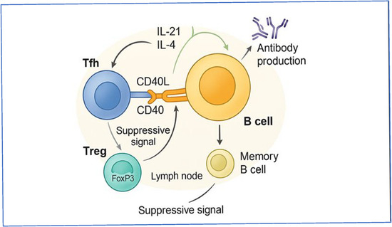
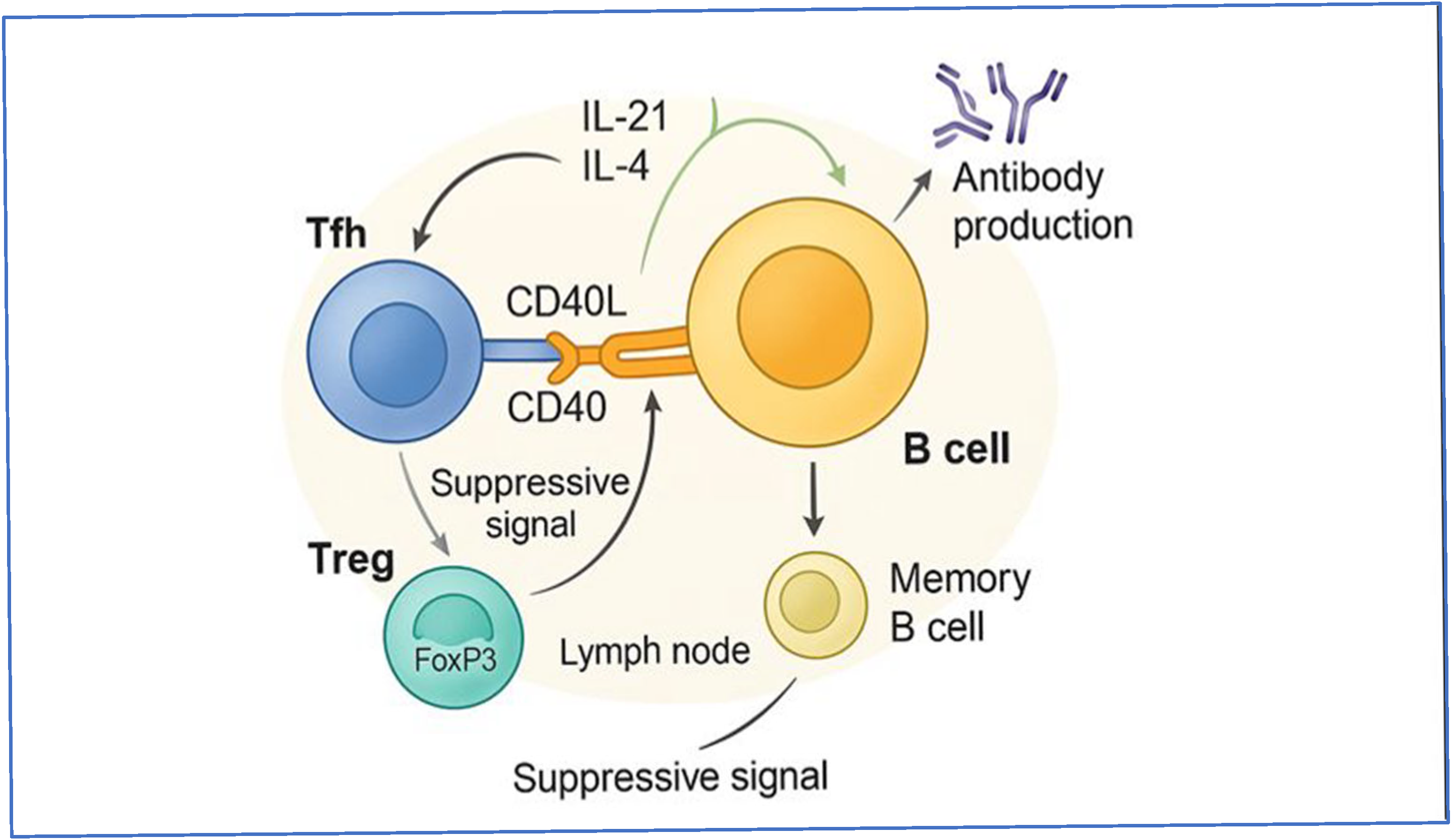
Regulatory cytokines generated during T-DC interactions affect early immune polarization, while cytokine signaling inside the T-B synapse works in a more limited and repetitive way, directly connecting T-cell assistance to B-cell selection results through germinal center cycling. Additionally, the participation of follicular regulatory T (Tfr) cells at the T-B interface adds a level of regulation that is mostly missing from T–DC interactions. Tfr cells moderate the intensity and duration of Tfh-mediated assistance, avoiding excessive or autoreactive B-cell responses and preserving immunological tolerance [39,40]. T-B cell synapses are therefore a crucial gatekeeper where immune responses are honed, varied, and stabilized, even if T–DC contacts are essential for triggering adaptive immunity. The significance of T-B crosstalk resides in its capacity to amalgamate antigen specificity, prolonged cellular interaction, and localized cytokine signaling to produce robust, enduring, and self-tolerant humoral immune responses [36,38,40] (Figure 1).
Figure 1.
T-B cell interactions in humoral immune responses: This schematic depicts the coordinated interactions between T cells and B cells that form the basis of effective humoral immune responses within secondary lymphoid tissues. Antigen-specific B cells present processed antigen to CD4+ T follicular helper (Tfh) cells, establishing a direct T-B cell synapse that facilitates the delivery of contact-dependent signals via CD40–CD40L interactions. Tfh cells subsequently deliver directed cytokine assistance to B cells, predominantly through the secretion of interleukin-21 (IL-21) and interleukin-4 (IL-4), which function to enhance B cell activation, proliferation, differentiation, and antibody synthesis. Activated B cells that receive sufficient T-cell assistance may differentiate into memory B cells or antibody-producing cells, thereby supporting sustained humoral immunity. The figure also shows how regulatory T cells (Tregs) that express FoxP3 can change the way Tfh-mediated stimulation works by sending signals that stop it from being too strong and stop B-cells from responding in a way that is out of control or self-reactive. This balanced mix of signals that stimulate and control the T-B interface makes sure that humoral immunity works well while keeping immunological tolerance. (This figure was created by the authors by using schematic illustration tools).
7. T Follicular Helper (Tfh) Cells and Their Role in B Cell Activation
Follicular helper T (Tfh) cells represent a subpopulation of CD4 Th cells that infiltrate germinal centers and are crucial for the maturation of B cells into memory and long-lived plasma cells [41]. Many transcription factors and B cell lymphoma 6 (Bcl-6) help to differentiate Tfh cells in a complex, multi-stage process [42].
It is evident that the production of Tfh cells from naive T CD4+ cell precursors is a continuous process that entails interaction with certain APCs (antigen-presenting cells), including DCs in T cell zones of lymphoid tissues and activated B cells at the border of T cell zones and follicles. Tfh cells mostly serve to control the clonal selection of germinal center B cells and generate antibody signals, class switching, and somatic mutations in the B-cell [43]. Tfh cells express CD40L, a member of the tumor necrosis factor (TNF) family, therefore facilitating the GC reaction. This member interacts with CD40 on GC B cells, which activates the NF-κB pathway and causes B cell proliferation. The intravital imaging revealed that GC B cells receive assistance from Tfh cells through brief but extensive cell–cell contacts, also known as T-B entanglement. These interactions arise often in the LZ and get longer during positive selection, suggesting that B cells continuously absorb the degree of help they get from Tfh cells. Furthermore, included in Tfh cell support is the cytokine IL-21, which is necessary for the preservation and proliferation of GC B cells as well as for the formation of long-lived plasma cells [25,41].
8. T-B Cell Interactions and Their Function in Vaccine and Infection Responses
Immunity against several bacterial and viral infections is reliant on both the strength of the adaptive response and the variety and quality of antibodies produced by germinal center (GC) selection. In this scenario, support from Tfh (CD40L and cytokines like IL-21) is very important for keeping GC B cells alive, selecting them over and over again, and making high-affinity memory B cells and long-lived plasma cells. Conversely, excessive or inadequately controlled Tfh activity may enhance low-stringency germinal center output and elevate the risk of autoreactivity, emphasizing the significance of balanced regulation within follicles. Recent research in vaccine contexts indicates that coordinated follicular T-cell programs (encompassing Tfh and follicular regulatory circuits) are associated with optimal affinity maturation and sustained humoral immunity. This supports the notion that vaccine design and supplementation can be regarded, in part, as strategies to influence the GC microenvironmental “support versus restriction” balance to enhance antibody quality and longevity [44,45].
9. Alterations in T-B Cell Interactions in Autoimmune Diseases and Cancer Immunotherapy
Dysregulated T-B interaction networks cause autoimmune diseases by letting autoreactive B-cell clones become activated and chosen in ways that are not right. There is more and more evidence that autoimmune inflammation is linked to changes in the number and quality of follicular T-cell subsets. These changes, especially imbalanced Tfh activity and poor follicular regulation, change the stringency of GC, encourage pathogenic class switching, and keep autoantibody production going. These mechanistic insights are progressively shaping therapeutic strategies designed to reestablish immune tolerance through the rebalancing of the follicular niche, such as by modulating pathways that affect Tfh/Tfr differentiation and function, or by attenuating GC help signals in the presence of autoantibody-mediated disease [46].
In cancer, the principles of T-B collaboration are increasingly acknowledged within the tumor microenvironment via tertiary lymphoid structures (TLS)—structured lymph node–like aggregates comprising B-cell follicles, T cells, antigen presentation, and GC-like characteristics. TLS can function as localized centers of antigen-driven T-B cell collaboration, promoting B-cell antigen presentation and enabling coordinated antitumor T-cell responses. When it comes to clinical applications, many studies show that TLS and B cells are not just hanging around; they are actually linked to better antitumor immunity and, in some tumor settings, better responses to immune checkpoint blockade. These discoveries establish T-B circuitry and TLS biology as viable frameworks for biomarker creation and for strategic combinations that enhance effective anticancer immunity while mitigating ineffective or tolerable follicular responses [47,48].
10. Role of Regulatory T Cells (Tregs) in Modulating B Cell Responses and Mechanism Preventing Autoimmunity
The human immune system can identify and respond to foreign proteins, but it is uncommon for it to do so with its own self. When the immune system mistakenly targets its own protein, it can lead to autoimmune disorders. Programmed cell death (apoptosis) starts in the embryonic stage, and the immune system keeps it going by getting rid of self-reacting lymphocytes that arise inadvertently. Nevertheless, because the approach that is employed to differentiate between self and non-self is not reliable, it is not possible to foresee the deletion of all lymphocytes that are self-reactive. Some self-reacting cells keep escaping death under physiological selection mechanisms, which could affect the direction of future development. Similarly, it remains a complex task to clarify why a considerable proportion of individuals do not manifest autoimmune diseases despite the presence of autoreactive lymphocytes [49].
The phenomena of biological self–non-self-discrimination is governed by a specific subset of lymphocytes called regulatory T cells (Tregs), which are essential regulators of inflammatory responses and play a crucial role in immunological tolerance and homeostasis. Regulatory T cells (Treg), traditionally recognized in humans and other species as CD4+/Foxp3+ lymphocytes, constitute merely 5–10% of total CD4+ T cells, however, are crucial for maintaining immune system equilibrium throughout life [33,50].
Treg cells are instrumental in the regulation of T cell-mediated diseases, as they are primarily directed at T cells. Recent studies have also revealed, though, that Treg cells affect many other cells including DCs, macrophages, mast cells, B cells, and osteoclasts [51]. Treg cells can quiet B cell responses and B cell-mediated antibody production [33,49]. More recently added to the fraction of GC-residing Tregs constitutively expressing Foxp3 are T follicular regulating (Tfr) cells. Although specific settings have also revealed Tfr cell differentiation from peripherally induced Treg precursors, it is thought that Tfr cells are essentially formed from thymic-derived Treg precursors. The transcriptional programs related to Tfh cells (including Bcl-6 and Tfh cell markers CXCR5 and PD-1) are upregulated by Tregs that differentiate into the Tfr cell phenotype to varying degrees. Consequently, they occupy an intermediate position between Tfh cells and Tregs in respect to transcription. Tfr cells have been suggested to modulate a variety of aspects of the GC reaction, such as the formation of GCs, the specificity of antigens, the emergence of autoreactive or allergy-inducing B cell clones, and the transition to different isotypes [52].
Autoantibody production and autoimmunity are the result of self-reactive B lymphocytes. Negative selection is the primary mechanism by which self-reactive B cells are eradicated in the bone marrow. However, a portion of self-reactive B cells can evade negative selection in the bone marrow and migrate to the periphery. Other mechanisms, such as immune modulation, anergy, and deletion, in the periphery, maintain the control of these self-reactive B cells [53].
The thymus is instrumental in the establishment and maintenance of the complex equilibrium between immune reactions against invading microorganisms and immune tolerance toward oneself, which is essential for the prevention of autoimmune diseases. The elimination of self-reactive T cells is a critical mechanism for the prevention of autoimmunity, which is a component of the induction of central immune tolerance in the thymus. The emergence of autoimmune disorders may arise from the impairment of thymic T cell selection processes. The selection processes ensuring self-tolerance in the thymus’s dynamic milieu depend on T cell migration and interactions with thymic stromal cells. In this context, thymic epithelial cells are particularly significant, as they present self-antigens and induce the negative selection of autoreactive T cells. In addition, the synergistic functions of thymic fibroblasts, B cells, and dendritic cells in the development of regulatory T cells, antigen presentation, and selection are essential for the tightly regulated immune response [53].
11. Regulation of Germinal Center Response
The regulation of germinal center reactions is essential to produce high-affinity antibodies and for maintaining self-tolerance by preventing the emergence of autoreactive B cell clones. Effective GC responses call for the cooperation of several cell types. The stromal cell network regulates the dynamics of the germinal center by controlling antigen delivery and cell trafficking. Targeted T-B cell interactions allow Tfh cells to assist germinal center (GC) B cells, and Foxp3+ Tfr cells are crucial for regulating GC activity. While the ratio of T follicular regulatory cells to T follicular helper cells is an important factor in germinal center response control, other cell types play an equally important role in shaping the milieu of the germinal center and warding off autoimmunity [29].
12. Conclusions
This overview helps us understand the adaptive immune system, what it is made of, and how B and T cells work together in complex ways. T cells are the main cells that control cell-mediated immunity since they are in charge of finding and killing cells that are infected with viruses. The humoral response is brought about by B cells, which develop in the bone marrow and other lymphoid tissues. An efficient immune response depends on how T and B cells work together.
Autoimmune diseases are kept under check by the suppressive action of regulatory cells, known as Tregs, whereas memory B cell formation and antibody class switching is facilitated by follicular helper T cells. Germinal centers are important sites where memory B cells and long-lived plasma cells are generated. GCs also prevent the emergence of autoreactive B cells. Another type of T cell, known as follicular helper T (Tfh) cells, are required for B cell activation. Additionally, they help in the development of memory B cells and antibody class switching. Since dysregulated T-B cell interactions are associated with autoimmune illnesses, immunodeficiencies, and inflammatory disorders, maintaining immunological homeostasis and preventing autoimmune disorders depend on the balance between Tfh and Tfr cells.
Author Contributions
Conceptualization, M.S.I., A.F.A., M.A.B., H.H.Q., A.T., N.I. and K.H.A.; writing—original draft preparation, M.S.I., A.F.A., M.A.B., H.H.Q., A.T., O.M.F., S.M., N.I. and M.P.; writing—review and editing, M.S.I., A.F.A., M.A.B., H.H.Q., A.T., O.M.F., S.M., N.I., M.P. and K.H.A.; visualization, M.S.I., A.F.A., M.A.B., H.H.Q., A.T., O.M.F., S.M. and M.P.; funding acquisition, A.F.A., M.A.B. and H.H.Q. All authors have read and agreed to the published version of the manuscript.
Funding
This research received no external funding.
Informed Consent Statement
Not applicable.
Data Availability Statement
No new data were created or analyzed in this study.
Conflicts of Interest
The authors declare no conflicts of interest.
References
- DeMaio, A.; Mehrotra, S.; Sambamurti, K.; Husain, S. The role of the adaptive immune system and T cell dysfunction in neurodegenerative diseases. J. Neuroinflamm. 2022, 19, 251. [Google Scholar] [CrossRef] [Scilit] [PubMed]
- Keselowsky, B.G.; Acharya, A.; Lewis, J.S. Innate and Adaptive Immunity: The Immune Response to Foreign Materials; Academic Press: Cambridge, MA, USA, 2020. [Google Scholar]
- Marshall, J.S.; Warrington, R.; Watson, W.; Kim, H.L. An introduction to immunology and immunopathology. Allergy Asthma Clin. Immunol. 2018, 14, 49. [Google Scholar] [CrossRef] [Scilit] [PubMed]
- Chi, H.; Pepper, M.; Thomas, P.G. Principles and therapeutic applications of adaptive immunity. Cell 2024, 187, 2052–2078. [Google Scholar] [CrossRef] [Scilit] [PubMed]
- Kaiko, G.E.; Horvat, J.C.; Beagley, K.W.; Hansbro, P.M. Immunological decision-making: How does the immune system decide to mount a helper T-cell response? Immunology 2008, 123, 326–338. [Google Scholar] [CrossRef] [Scilit]
- Lu, L.L.; Suscovich, T.J.; Fortune, S.M.; Alter, G. Beyond binding: Antibody effector functions in infectious diseases. Nat. Rev. Immunol. 2017, 18, 46–61. [Google Scholar] [CrossRef] [Scilit]
- Germain, R.N. T-cell development and the CD4–CD8 lineage decision. Nat. Rev. Immunol. 2002, 2, 309–322. [Google Scholar] [CrossRef] [Scilit]
- Sun, L.; Su, Y.; Jiao, A.; Wang, X.; Zhang, B. T cells in health and disease. Signal Transduct. Target. Ther. 2023, 8, 235. [Google Scholar] [CrossRef] [Scilit]
- Taniuchi, I. CD4 Helper and CD8 Cytotoxic T Cell Differentiation. Annu. Rev. Immunol. 2018, 36, 579–601. [Google Scholar] [CrossRef] [Scilit]
- Bonilla, F.A.; Oettgen, H.C. Adaptive immunity. J. Allergy Clin. Immunol. 2010, 125, S33–S44. [Google Scholar] [CrossRef] [Scilit]
- Kurd, N.; Robey, E.A. T-cell selection in the thymus: A spatial and temporal perspective. Immunol. Rev. 2016, 271, 114–126. [Google Scholar] [CrossRef] [Scilit]
- Takada, K.; Takahama, Y. Positive-selection-inducing self-peptides displayed by cortical thymic epithelial cells. In Advances in Immunology; Academic Press Inc.: Cambridge, MA, USA, 2015; pp. 87–110. [Google Scholar]
- Xiong, Y.; Bosselut, R. CD4–CD8 differentiation in the thymus: Connecting circuits and building memories. Curr. Opin. Immunol. 2012, 24, 139–145. [Google Scholar] [CrossRef] [Scilit] [PubMed]
- Takaba, H.; Takayanagi, H. The Mechanisms of T Cell Selection in the Thymus. Trends Immunol. 2017, 38, 805–816. [Google Scholar] [CrossRef] [Scilit] [PubMed]
- Klein, L.; Kyewski, B.; Allen, P.M.; Hogquist, K.A. Positive and negative selection of the T cell repertoire: What thymocytes see (and don’t see). Nat. Rev. Immunol. 2014, 14, 377–391. [Google Scholar] [CrossRef] [Scilit] [PubMed]
- Goronzy, J.J.; Weyand, C.M. Successful and Maladaptive T Cell Aging. Immunity 2017, 46, 364–378. [Google Scholar] [CrossRef] [Scilit]
- Barnaba, V. T Cell Memory in Infection, Cancer, and Autoimmunity. Front. Immunol. 2022, 12, 811968. [Google Scholar] [CrossRef] [Scilit]
- Gebhardt, T.; Wakim, L.M.; Eidsmo, L.; Reading, P.C.; Heath, W.R.; Carbone, F.R. Memory T cells in nonlymphoid tissue that provide enhanced local immunity during infection with herpes simplex virus. Nat. Immunol. 2009, 10, 524–530. [Google Scholar] [CrossRef] [Scilit]
- Lebien, T.W.; Tedder, F.T. B lymphocytes: How they develop and function. Blood 2008, 112, 1570–1580. [Google Scholar] [CrossRef] [Scilit]
- Barrio, L.; Román-García, S.; Díaz-Mora, E.; Risco, A.; Jiménez-Saiz, R.; Carrasco, Y.R.; Cuenda, A. B Cell Development and T-Dependent Antibody Response Are Regulated by p38γ and p38δ. Front. Cell Dev. Biol. 2020, 8, 189. [Google Scholar] [CrossRef] [Scilit]
- Rukundo, T.B. Cross-reactivity in Adaptive Immunity: An Overview. Newport Int. J. Bio-Log. Appl. Sci. 2024, 5, 39–43. Available online: https://nijournals.org/cross-reactivity-in-adaptive-immunity-an-overview/ (accessed on 5 October 2025). [CrossRef] [Scilit]
- Hjálmsdóttir, Á.; Hasler, F.; Waeckerle-Men, Y.; Duda, A.; López-Deber, M.P.; Pihlgren, M.; Vukicevic, M.; Kündig, T.M.; Johansen, P. T cell independent antibody responses with class switch and memory using peptides anchored on liposomes. npj Vaccines 2024, 9, 115. [Google Scholar] [CrossRef] [Scilit]
- Roco, J.A.; Mesin, L.; Binder, S.C.; Nefzger, C.; Gonzalez-Figueroa, P.; Canete, P.F.; Ellyard, J.; Shen, Q.; Robert, P.A.; Cappello, J.; et al. Class-Switch Recombination Occurs Infrequently in Germinal Centers. Immunity 2019, 51, 337–350.e7. [Google Scholar] [CrossRef] [Scilit] [PubMed]
- De Silva, N.S.; Klein, U. Dynamics of B cells in germinal centres. Nat. Rev. Immunol. 2015, 15, 137–148. [Google Scholar] [CrossRef] [Scilit] [PubMed]
- Victora, G.D.; Nussenzweig, M.C. Annual Review of Immunology. Germinal Cent. 2025, 10, 20. [Google Scholar] [CrossRef] [Scilit]
- Cooper, L.; Good-Jacobson, K.L. B Cells Hit a Class Ceiling in the Germinal Center. Immunity 2019, 51, 206–208. [Google Scholar] [CrossRef] [Scilit]
- Tsai, D.-Y.; Hung, K.-H.; Chang, C.-W.; Lin, K.-I. Regulatory mechanisms of B cell responses and the implication in B cell-related diseases. J. Biomed. Sci. 2019, 26, 64. [Google Scholar] [CrossRef] [Scilit]
- Palm, A.-K.E.; Henry, C. Remembrance of Things Past: Long-Term B Cell Memory After Infection and Vaccination. Front. Immunol. 2019, 10, 1787. [Google Scholar] [CrossRef] [Scilit]
- Stebegg, M.; Kumar, S.D.; Silva-Cayetano, A.; Fonseca, V.R.; Linterman, M.A.; Graca, L. Regulation of the Germinal Center Response. Front. Immunol. 2018, 9, 2469. [Google Scholar] [CrossRef] [Scilit]
- Kometani, K.; Kurosaki, T. Differentiation and maintenance of long-lived plasma cells. Curr. Opin. Immunol. 2015, 33, 64–69. [Google Scholar] [CrossRef] [Scilit]
- Puri, K.D.; Di Paolo, J.A.; Gold, M.R. B-Cell Receptor Signaling Inhibitors for Treatment of Autoimmune Inflammatory Diseases and B-Cell Malignancies. Int. Rev. Immunol. 2013, 32, 397–427. [Google Scholar] [CrossRef] [Scilit]
- Peng, S.L. Signaling in B cells via Toll-like receptors. Curr. Opin. Immunol. 2005, 17, 230–236. [Google Scholar] [CrossRef] [Scilit]
- Wang, P.; Zheng, S.G. Review Article Regulatory T cells and B cells: Implication on autoimmune diseases. Int. J. Clin. Exp. Pathol. 2013, 6, 2668. [Google Scholar] [PubMed] [PubMed Central]
- Steinman, R.M.; Banchereau, J. Taking dendritic cells into medicine. Nature 2007, 449, 419–426. [Google Scholar] [CrossRef] [Scilit]
- Qi, H.; Cannons, J.L.; Klauschen, F.; Schwartzberg, P.L.; Germain, R.N. SAP-controlled T-B cell interactions underlie germinal centre formation. Nature 2008, 455, 764–769. [Google Scholar] [CrossRef] [Scilit] [PubMed]
- Crotty, S. T Follicular Helper Cell Biology: A Decade of Discovery and Diseases. Immunity 2019, 50, 1132–1148. [Google Scholar] [CrossRef] [Scilit]
- Vinuesa, C.G.; Linterman, M.A.; Yu, D.; Maclennan, I.C.M. Follicular Helper T Cells. Annu. Rev. Immunol. 2016, 34, 335–368. [Google Scholar] [CrossRef] [Scilit] [PubMed]
- Victora, G.D.; Nussenzweig, M.C. Germinal centers. Annu. Rev. Immunol. 2012, 30, 429–457. [Google Scholar] [CrossRef] [Scilit]
- Sage, P.T.; Sharpe, A.H. T follicular regulatory cells in the regulation of B cell responses. Trends Immunol. 2015, 36, 410–418. [Google Scholar] [CrossRef] [Scilit]
- Lu, Y.; Craft, J. T Follicular Regulatory Cells: Choreographers of Productive Germinal Center Responses. Front. Immunol. 2021, 12, 679909. [Google Scholar] [CrossRef] [Scilit]
- Weinstein, J.S.; A Bertino, S.; Hernandez, S.G.; Poholek, A.C.; Teplitzky, T.B.; Nowyhed, H.N.; Craft, J. B Cells in T Follicular Helper Cell Development and Function: Separable Roles in Delivery of ICOS Ligand and Antigen. J. Immunol. 2014, 192, 3166–3179. [Google Scholar] [CrossRef] [Scilit]
- Crotty, S. T Follicular Helper Cell Differentiation, Function, and Roles in Disease. Immunity 2014, 41, 529–542. [Google Scholar] [CrossRef] [Scilit]
- Eivazi, S.; Bagheri, S.; Hashemzadeh, M.S.; Ghalavand, M.; Qamsari, E.S.; Dorostkar, R.; Yasemi, M. Development of T follicular helper cells and their role in disease and immune system. Biomed. Pharmacother. 2016, 84, 1668–1678. [Google Scholar] [CrossRef] [Scilit] [PubMed]
- Ritzau-Jost, J.; Hutloff, A. T cell/b cell interactions in the establishment of protective immunity. Vaccines 2021, 9, 1074. [Google Scholar] [CrossRef] [Scilit] [PubMed]
- Cavazzoni, C.B.; Hanson, B.L.; Podestà, M.A.; Bechu, E.D.; Clement, R.L.; Zhang, H.; Daccache, J.; Reyes-Robles, T.; Hett, E.C.; Vora, K.A.; et al. Follicular T cells optimize the germinal center response to SARS-CoV-2 protein vaccination in mice. Cell Rep. 2022, 38, 110399. [Google Scholar] [CrossRef] [Scilit] [PubMed]
- Qi, J.; Liu, C.; Bai, Z.; Li, X.; Yao, G. T follicular helper cells and T follicular regulatory cells in autoimmune diseases. Front. Immunol. 2023, 14, 1178792. [Google Scholar] [CrossRef] [Scilit]
- Flippot, R.; Teixeira, M.; Rey-Cardenas, M.; Carril-Ajuria, L.; Rainho, L.; Naoun, N.; Jouniaux, J.-M.; Boselli, L.; Naigeon, M.; Danlos, F.-X.; et al. B cells and the coordination of immune checkpoint inhibitor response in patients with solid tumors. J. Immunother. Cancer 2024, 12, e008636. [Google Scholar] [CrossRef] [Scilit]
- Hegoburu, A.; Amer, M.; Frizelle, F.; Purcell, R. B cells and tertiary lymphoid structures in cancer therapy response. BJC Rep. 2025, 3, 40. [Google Scholar] [CrossRef] [Scilit]
- Goswami, T.K.; Singh, M.; Dhawan, M.; Mitra, S.; Bin Emran, T.; Rabaan, A.A.; Al Mutair, A.; Al Alawi, Z.; Alhumaid, S.; Dhama, K. Regulatory T cells (Tregs) and their therapeutic potential against autoimmune disorders—Advances and challenges. Hum. Vaccines Immunother. 2022, 18, 2035117. [Google Scholar] [CrossRef] [Scilit]
- Ou, Q.; Power, R.; Griffin, M.D. Revisiting regulatory T cells as modulators of innate immune response and inflammatory diseases. Front. Immunol. 2023, 14, 1287465. [Google Scholar] [CrossRef] [Scilit]
- Zhao, D.-M.; Thornton, A.M.; DiPaolo, R.J.; Shevach, E.M. Activated CD4+CD25+ T cells selectively kill B lymphocytes. Blood 2006, 107, 3925–3932. [Google Scholar] [CrossRef] [Scilit]
- Yanaba, K.; Bouaziz, J.-D.; Matsushita, T.; Magro, C.M.; St. Clair, E.W.; Tedder, T.F. B-lymphocyte contributions to human autoimmune disease. Immunol. Rev. 2008, 223, 284–299. [Google Scholar] [CrossRef] [Scilit]
- Shirafkan, F.; Hensel, L.; Rattay, K. Immune tolerance and the prevention of autoimmune diseases essentially depend on thymic tissue homeostasis. Front. Immunol. 2024, 15, 1339714. [Google Scholar] [CrossRef] [Scilit]
Disclaimer/Publisher’s Note: The statements, opinions and data contained in all publications are solely those of the individual author(s) and contributor(s) and not of MDPI and/or the editor(s). MDPI and/or the editor(s) disclaim responsibility for any injury to people or property resulting from any ideas, methods, instructions or products referred to in the content. |
© 2025 by the authors. Licensee MDPI, Basel, Switzerland. This article is an open access article distributed under the terms and conditions of the Creative Commons Attribution (CC BY) license (https://creativecommons.org/licenses/by/4.0/).