


Abstract
Background: Differentiating atypical endometrial hyperplasia, also known as endometrial intraepithelial neoplasia (EAH/EIN) from endometrial hyperplasia without atypia is crucial due to the higher risk of progression to endometrioid adenocarcinoma, associated with atypical lesions. Immunohistochemical markers such as PAX2 and PTEN have emerged as potential adjuncts to improve diagnostic accuracy in morphologically challenging cases. Objective: To evaluate the diagnostic value of PAX2 and PTEN expression in distinguishing atypical from endometrial hyperplasia without atypia in the Bulgarian population. Materials and Methods: A total of 96 endometrial hyperplasia cases (48 typical, 48 atypical) were included. Histopathological evaluation was performed on hematoxylin and eosin–stained sections, with two experienced pathologists confirming diagnoses according to the WHO criteria. Immunohistochemical analysis of PTEN and PAX2 was conducted on formalin-fixed, paraffin-embedded tissue sections. Results: PTEN expression loss was observed in 6% (3/48) of hyperplasia without atypia cases, compared with 81.3% (38/48) of EAH/EIN cases. For PAX2, strong nuclear staining was retained in 60% (29/48) of endometrial hyperplasia without atypia cases, with no complete loss of expression. In contrast, 64.6% (31/48) of EAH/EIN cases showed complete loss of PAX2 expression, while only 35.4% (17/48) preserved nuclear immunoreactivity. Together, these results highlight clear and consistent differences in PTEN and PAX2 expression between hyperplasia without atypia and EAH/EIN and may aid pathologists in distinguishing these two entities in routine diagnostic practice. Conclusions: Expression loss of PAX2 and PTEN is significantly associated with EAH/EIN. Immunohistochemical evaluation of these markers provides valuable adjunctive information for the diagnosis of morphologically ambiguous cases and may enhance diagnostic reproducibility and accuracy. Incorporating PAX2 and PTEN into routine assessment may guide appropriate clinical management and risk stratification of patients with endometrial hyperplasia.
1. Introduction
Endometrial hyperplasia (EH) is a benign proliferative disorder of the endometrium driven by unopposed estrogenic stimulation. It commonly arises in settings such as obesity, chronic anovulation, and exogenous hormone exposure [1,2]. Its clinical relevance lies in its role as a precursor to endometrioid adenocarcinoma, the most frequent subtype of endometrial carcinoma [3]. The 1994 WHO classification, based on architectural complexity and cytologic atypia, suffered from poor reproducibility. The 2014 WHO revision simplified this framework into a two-tier system: hyperplasia without atypia, which carries minimal malignant potential, and atypical hyperplasia/endometrial intraepithelial neoplasia (EAH/EIN), which has a strong association with concurrent carcinoma or progression to malignancy [4].
Management is primarily guided by the presence of atypia. Hyperplasia without atypia generally regresses with progestin therapy or correction of hormonal imbalance. Oral progestins and the levonorgestrel-releasing intrauterine system (LNG-IUS) are first-line options, with the latter demonstrating superior regression rates [5,6]. Hysterectomy is seldom required and is reserved for persistent, recurrent, or medically refractory disease [7]. In contrast, EAH/EIN shows a markedly elevated risk of coexistent or future carcinoma; up to one-third of patients harbor carcinoma at diagnosis, making hysterectomy the preferred treatment in women who have completed childbearing [3]. Fertility-sparing therapy with high-dose progestins or LNG-IUS may be offered to select younger patients but requires rigorous surveillance with repeat sampling due to high rates of persistence and progression [8,9].
PAX2, a transcription factor involved in Müllerian development, has emerged as a useful immunohistochemical marker in evaluating endometrial proliferative lesions. PAX2 loss is commonly observed in EAH/EIN but typically preserved in hyperplasia without atypia and normal proliferative endometrium [10,11], reflecting underlying clonal genetic alterations rather than hormonally mediated changes [12]. In morphologically equivocal cases, PAX2 immunostaining supports diagnostic consistency, with studies showing strong associations between partial or complete loss of expression and EAH/EIN or endometrioid carcinoma, whereas diffuse nuclear positivity favors benign hyperplasia [13,14]. Although not definitive on its own, PAX2 enhances diagnostic accuracy when integrated with morphology [15].
PTEN, a tumor suppressor gene at 10q23, is also frequently altered in endometrial neoplasia. Immunohistochemical PTEN loss is characteristic of EAH/EIN and early endometrioid carcinoma, whereas intact expression is typical of hyperplasia without atypia and benign endometrium [16]. Because focal PTEN loss may be seen in non-neoplastic glands, interpretation requires morphologic correlation, and PTEN is most informative when used as part of a marker panel [12,17]. Combined evaluation of PAX2 and PTEN has been shown to improve reproducibility and diagnostic discrimination between EAH/EIN and hyperplasia without atypia, particularly in challenging cases [13,14,15].
Overall, PAX2 and PTEN serve as valuable adjunctive markers in distinguishing EAH/EIN from hyperplasia without atypia. Loss of either marker correlates with premalignant lesions, while retained expression supports a benign diagnosis. Given the morphologic overlap among endometrial proliferations, this study aims to assess the utility of PAX2 and PTEN expression patterns in differentiating EAH/EIN from endometrial hyperplasia without atypia.
2. Materials and Methods
We performed a retrospective review of 3360 curettage specimens with clinical data for abnormal uterine bleeding from the archive of University Hospital “St. George,” 4004 Plovdiv, Bulgaria, covering a 10-year period (2016–August 2025).
Among these, 64 cases had a morphological diagnosis of endometrial hyperplasia (EH) and 53 cases were diagnosed as EH/EIN. The remaining morphological findings for the above clinical indication included proliferative endometrium, endometrial polyp, dysfunctional endometrium, and endometrial carcinoma. Following the application of the exclusion criteria during data processing, 49 EH cases remained (in 7 cases, trimming of the microscopic area was observed, and in another 9, cold ischemia was recorded), and there were 48 EH/EIN cases (in 5 cases, trimming of the microscopic area was observed). The cases were divided into two working groups of 48 endometrial curettage specimens diagnosed with EH and 48 endometrial curettage specimens diagnosed with EH/EIN.
The inclusion criteria required that all participants be women aged 18 years or older and that the analyzed samples consist of endometrial curettage specimens with a confirmed histological diagnosis of endometrial hyperplasia (EH) or EH/EIN. A group of normal endometrium was also included as a control. Only samples fixed in 10% buffered formalin and containing sufficient tissue for immunohistochemical evaluation of PTEN and PAX2 were accepted. Additionally, the tissue morphology had to be well preserved to allow for reliable interpretation of the immunohistochemical results. Clinical data had to confirm the presence of abnormal uterine bleeding, and patient consent for scientific use of the material (ethical approval) was required. This survey is part of a project named “Diagnostic and Prognostic Reliability of Immunohistochemical Biomarkers in Patients with Leiomyoma, Precursor Endometrial Lesions and Endometrial Carcinoma”, appearing in the informed consent document.
The exclusion criteria eliminated cases in which no morphologically identifiable precursor endometrial lesions were present, as well as samples with poor fixation or insufficient tissue for the evaluation of all four immunohistochemical markers. Patients were excluded if they had invasive endometrial carcinoma or had undergone chemotherapy, radiotherapy, or prolonged hormonal therapy immediately prior to biopsy, as these treatments might alter biomarker expression. Technical limitations also served as exclusion factors, including cases in which the immunohistochemical protocol could not be applied due to missing controls, non-functional antibodies, excessive background staining, or severe tissue degeneration or distortion that made marker assessment impossible.
For the immunohistochemical analysis, the following reagents were used:
- Primary monoclonal antibodies, summarized in Table 1;
- Secondary antibodies and visualization agents.
Table 1.
Manufacturer of primary antibodies.
Immunohistochemical staining was performed using a universal mouse/rabbit polymer-based detection system (EnVision FLEX, High pH, Dako).
The immunohistochemical procedure included the following steps:
- (a)
- Deparaffinization
Sections of 4 μm thickness were prepared from archival paraffin blocks and mounted on adhesive slides. The slides were incubated in a thermostat at 58 °C for 2 h. Deparaffinization was performed using a programmed protocol on an Dako Autostainer (Dako, Glostrup, Denmark).
- (b)
- Heat-induced antigen retrieval
Performed according to a preset program on the automated Dako Autostainer.
- (c)
- Blocking of endogenous peroxidase
Carried out using a preset program on the automated Dako Autostainer.
- (d)
- Incubation with the primary antibody
Performed automatically according to the manufacturer’s preset protocol on the Dako Autostainer.
- (e)
- Visualization of the reaction
Achieved with DAB chromogen solution, applied using a preset program on the automated Dako Autostainer.
- (f)
- Counterstaining
Slides were counterstained with hematoxylin for 5 min using a preset program on the Dako Autostainer.
In the present study, a dichotomized, summary-based classification system with an intermediate category, adapted from Mutter et al. [12], was applied for the assessment of PTEN, due to its diagnostic clarity and reproducibility. PTEN is considered positive when the glandular epithelium demonstrates nuclear and/or cytoplasmic staining of an intensity comparable to the adjacent stroma or normal endometrium. Negative expression is defined as complete loss of staining in the epithelial cells of the glands, with stromal cells serving as an internal positive control. This method is preferred in routine diagnostic practice due to its clarity, objectivity, and high reproducibility. Evaluation of PTEN expression was performed based on cytoplasmic and nuclear staining in the glandular epithelium, using stromal cells as an internal control:
- Positive expression: Diffuse, uniform cytoplasmic and/or nuclear staining of an intensity comparable to the stroma.
- Loss in isolated glands: Focal absence of expression in a limited number of glands with retained staining in the surrounding epithelium.
- Complete loss: Absence of staining in more than 90% of glandular structures.
This PTEN-specific classification ensures consistency with established literature standards while maintaining high diagnostic informativeness and reproducibility.
PAX2 expression was semiquantitatively assessed using the H-score method, calculated as the sum of the percentage of positive cells multiplied by staining intensity (0–3+). Resulting values range from 0 to 300, with higher scores indicating stronger expression. The method was adapted according to Allison and Monte [15]. In normal endometrium, PAX2 is expressed uniformly and homogeneously in the nuclei of the glandular epithelial cells. These glands provide a reference for normal expression and can be used as an internal positive control when evaluating immunohistochemical staining results.
3. Results
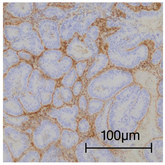
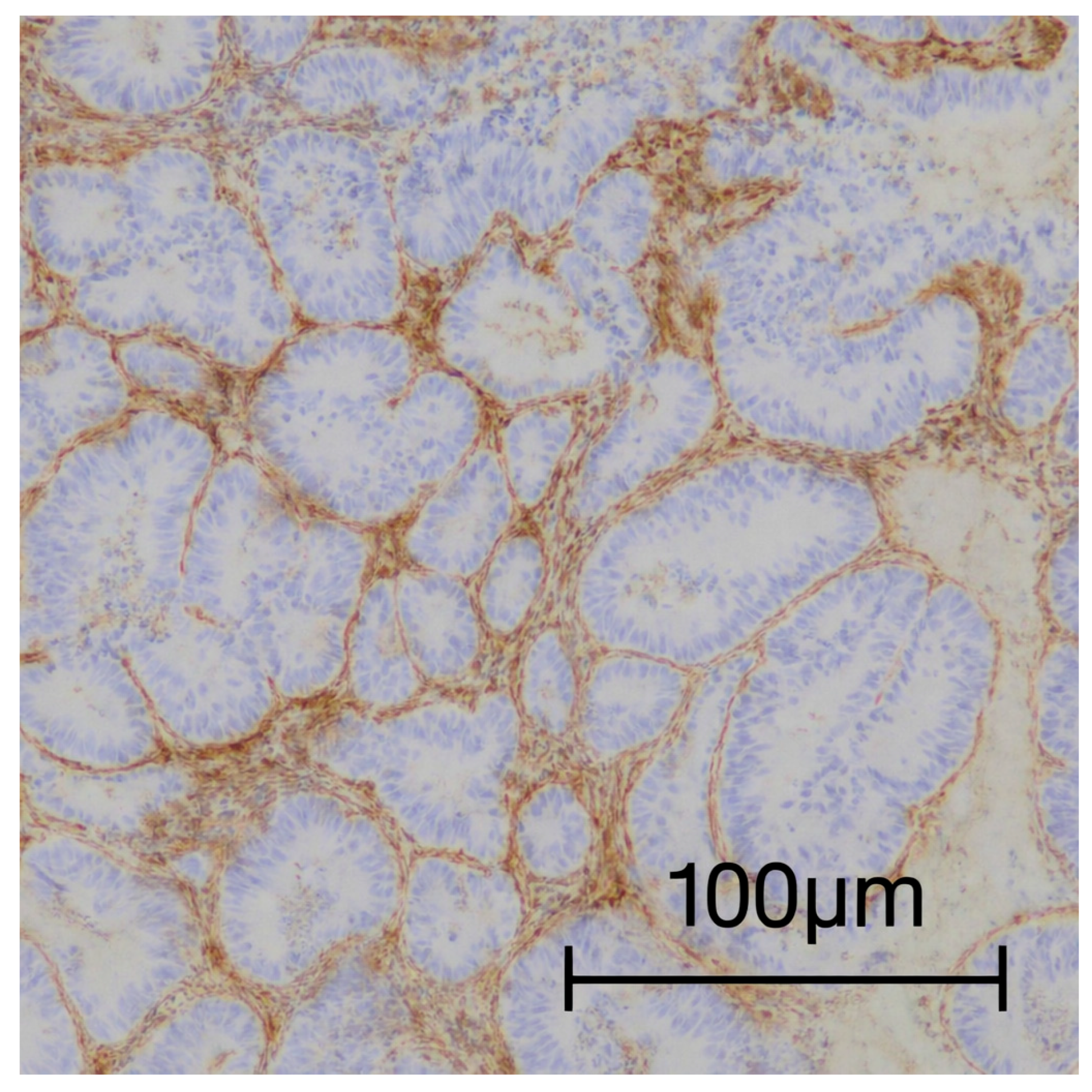
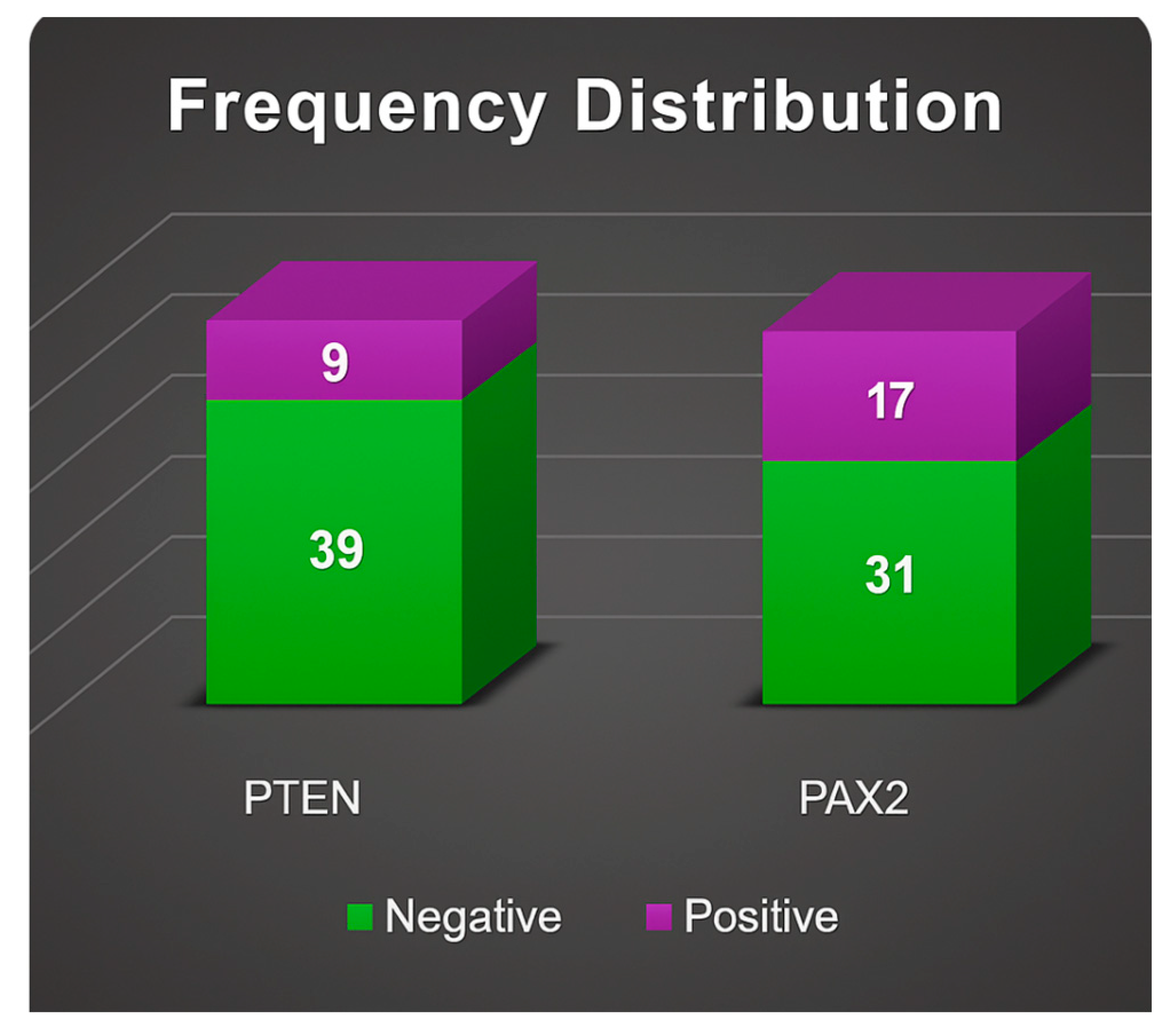
PTEN expression loss was identified in 6% (n = 3/48) of endometrial hyperplasia without atypia cases, whereas 81.3% (n = 38/48) of EAH/EIN cases showed loss of PTEN expression (Figure 1). In the immunohistochemical evaluation of PAX2 expression in cases of endometrial hyperplasia (EH), strong nuclear staining was identified in 29 cases (60%), whereas 19 cases (40%) exhibited weak nuclear immunoreactivity. Notably, no specimens demonstrated complete loss of PAX2 expression.
Figure 1.
General view of areas with EAH/EIN, showing loss of PTEN expression, with the positive internal control of the stromal cells.
These observations indicate that, in hyperplasia without atypia, nuclear PAX2 expression is largely retained across the epithelial component, consistent with its established function as a transcription factor regulating cellular differentiation and the preservation of endometrial glandular architecture. Published data further corroborate that sustained nuclear PAX2 expression is characteristic of normal and benign endometrial tissue, including EH, whereas diminished or absent expression is more commonly associated with atypical hyperplasia and malignant endometrial lesions.
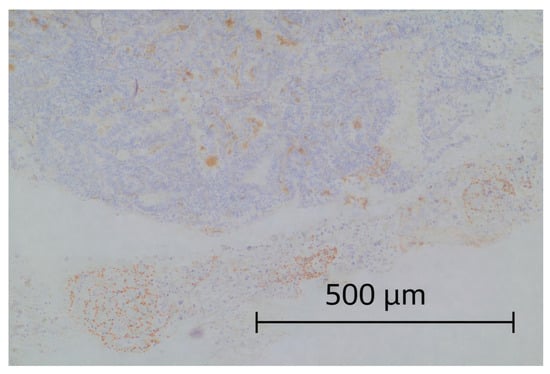
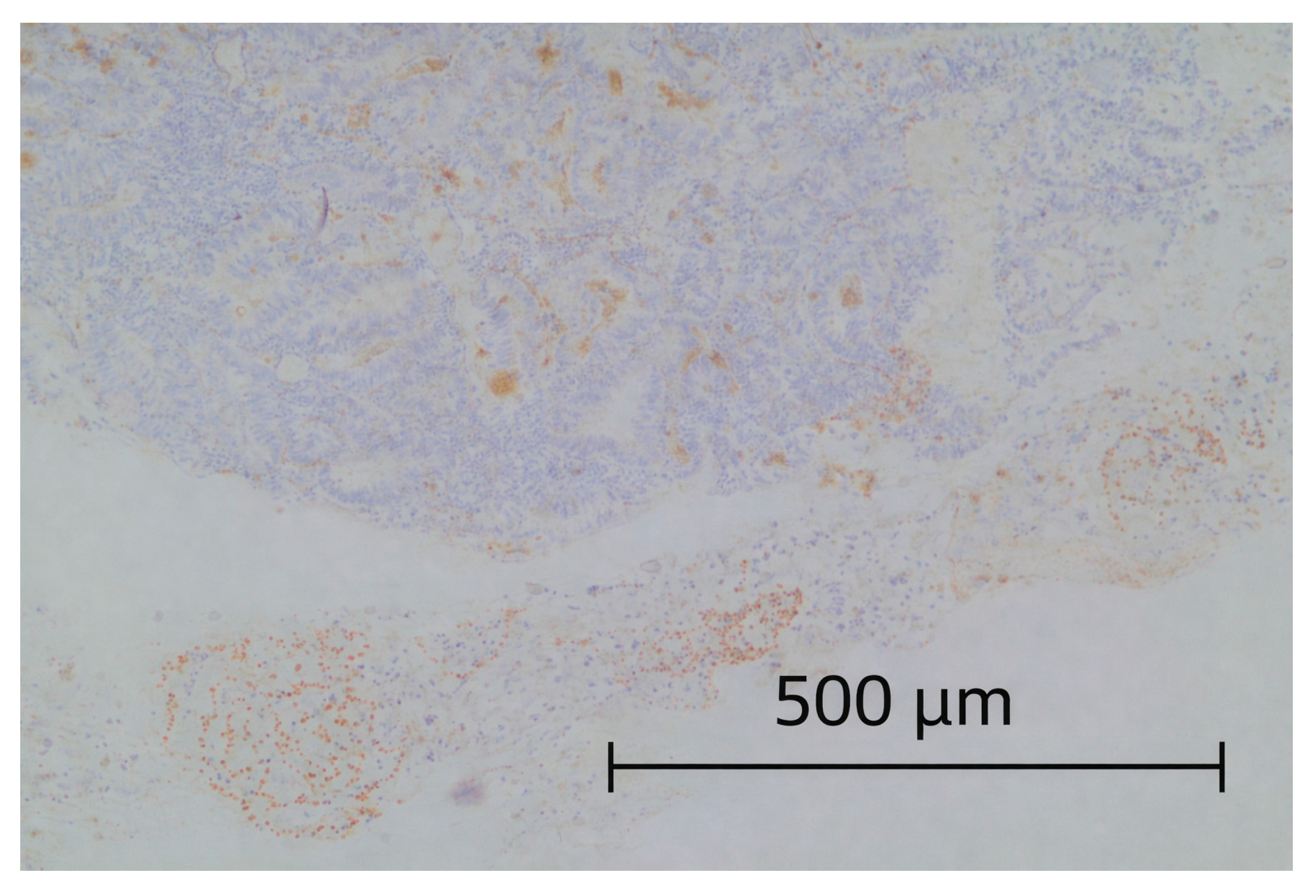
In EAH/EIN cases, PAX2 immunohistochemical analysis demonstrated a substantial loss of expression. A negative staining pattern was observed in 31 out of 48 examined cases (64.6%), while preserved immunoreactivity was detected in only 17 cases (35.4%) (Figure 2). These findings highlight the frequent and early suppression of PAX2 in precancerous endometrial lesions, with loss of expression considered indicative of gene inactivation and a potential marker of malignant transformation.
Figure 2.
General view of areas with EAH/EIN, showing loss of PAX2 expression, with a positive internal control at the bottom of the image consisting of normal glandular structures.
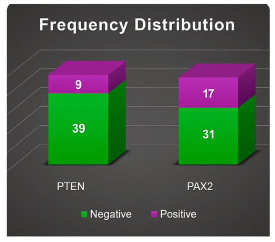
Welch’s t-test revealed a statistically significant difference in PTEN expression between hyperplasia without atypia and EAH/EIN groups (p < 0.001), as well as a statistically significant difference in PAX2 expression between the same groups (p < 0.001) (Figure 3). Concurrent loss of expression of both markers was observed in a substantial proportion of cases, suggesting a potential cumulative contribution to malignant transformation. These findings support the utility of PTEN and PAX2 immunohistochemical assessment as an adjunct tool in the histopathological evaluation of EAH/EIN.
Figure 3.
Frequency distribution of PTEN and PAX2 expression in EAH/EIN.
4. Discussion
Accurate differentiation between endometrial hyperplasia without atypia and EAH/EIN remains challenging, particularly in cases with borderline architectural or cytologic features. Because management decisions depend heavily on the presence of atypia, reliable ancillary tools are needed to support histopathologic assessment.
In this study, PAX2 and PTEN immunohistochemistry demonstrated significant value as adjunct markers in distinguishing EAH/EIN from endometrial hyperplasia without atypia. Consistent with previous studies in the literature, loss of PAX2 expression strongly correlated with EAH/EIN, whereas its retention supported a diagnosis of endometrial hyperplasia without atypia. PTEN loss showed a similar association with EAH/EIN, reflecting underlying clonal genetic alterations. Although focal PTEN loss can occur in benign endometrium, diffuse or multifocal absence in the setting of architectural complexity was highly suggestive of premalignant change.
Importantly, combined evaluation of PAX2 and PTEN improved diagnostic accuracy compared with either marker alone. Concurrent loss of both markers was frequently seen in EAH/EIN, while preservation of at least one favored endometrial hyperplasia without atypia. This complementary pattern reduces diagnostic ambiguity and may help minimize both overtreatment and undertreatment, particularly in patients undergoing fertility-sparing management.
5. Conclusions
Our study provides compelling evidence that the loss of PTEN and PAX2 expression is strongly correlated with EAH/EIN, whereas these markers are predominantly preserved in endometrial hyperplasia without atypia. The immunohistochemical evaluation of these molecular markers offers an objective adjunct to conventional histopathology, which can sometimes be limited by interobserver variability, especially in cases with subtle or equivocal cytologic atypia. PTEN, a tumor suppressor gene frequently altered in endometrial neoplasia, and PAX2, a transcription factor essential for Müllerian tract development, both exhibit distinct patterns of expression that reflect the underlying molecular pathogenesis of premalignant lesions. Their concurrent assessment allows for more precise differentiation between hormonally induced proliferative changes and clonal, neoplastic processes.
The clinical implications of these findings are substantial. Accurate distinction between endometrial hyperplasia without atypia and EAH/EIN is critical, as EAH/EIN carries a markedly higher risk of progression to endometrioid carcinoma. Immunohistochemical loss of PTEN and PAX2 may therefore serve as a predictive biomarker for malignant potential, enabling pathologists and clinicians to identify patients who may benefit from more aggressive surveillance or definitive therapeutic interventions. Incorporating these markers into routine diagnostic workflows has the potential to enhance reproducibility and reduce diagnostic ambiguity, particularly in morphologically challenging specimens where architectural and cytologic criteria alone may be insufficient.
Furthermore, the integration of PTEN and PAX2 assessment may contribute to individualized risk stratification. Patients exhibiting loss of either or both markers could be prioritized for close follow-up, hormonal therapy, or surgical management, while those retaining normal expression could be considered for conservative management strategies. In addition, the combined evaluation of these markers may facilitate the identification of early, pre-neoplastic lesions, thereby enabling timely intervention and potentially reducing the incidence of progression to invasive carcinoma.
In summary, our findings underscore the diagnostic and prognostic utility of PTEN and PAX2 immunohistochemistry in the evaluation of EH. By providing a molecular correlation to histopathologic assessment, these markers can improve diagnostic accuracy, inform clinical decision-making, and, ultimately, enhance patient care in the management of premalignant endometrial lesions. Future studies integrating additional molecular markers and longitudinal clinical outcomes may further refine risk prediction and expand the utility of immunohistochemical panels in endometrial pathology.
Author Contributions
Conceptualization, N.P. and E.U.; methodology, A.M.-K.; software, M.P.; validation, M.K.-I., A.M.-K., and N.P.; formal analysis, M.K.-I.; investigation, A.M.-K.; resources, E.U.; data curation, M.K.-I.; writing—original draft preparation, A.M.-K. and N.P.; writing—review and editing, A.M.; visualization, M.P.; supervision, E.U.; project administration, N.P. and A.M.; funding acquisition, E.U. All authors have read and agreed to the published version of the manuscript.
Funding
Contract No. BG-RRP-2.004-0007-C01 “Program for strategic research and innovation for the development of MU-Plovdiv (SRIPD-MUP)”, Pillar 2: Creation of a network of research higher schools of the Program for Accelerating Economic Recovery and Transformation through Science and Innovation from the “Innovative Bulgaria” component of the National Plan for Recovery and Sustainability, funded by the European Union-NextGenerationEU.
Institutional Review Board Statement
The study was conducted in accordance with the Declaration of Helsinki and approved by the Ethics Committee of the Medical University of Plovdiv (Protocol 3/28.10.2024; approval date 28 October 2024).
Informed Consent Statement
Informed consent was obtained from all subjects involved in the study.
Data Availability Statement
The original contributions presented in this study are included in the article. Further inquiries can be directed to the corresponding author.
Conflicts of Interest
The authors declare no conflicts of interest.
References
- Chou, A.J.; Bing, R.S.; Ding, D.C. Endometrial Atypical Hyperplasia and Risk of Endometrial Cancer. Diagnostics 2024, 14, 2471. [Google Scholar] [CrossRef] [Scilit] [PubMed]
- Léon-Castillo, A. Update in the molecular classification of endometrial carcinoma. Int. J. Gynecol. Cancer 2023, 33, 333–342. [Google Scholar] [CrossRef] [Scilit] [PubMed]
- Trimble, C.L.; Kauderer, J.; Zaino, R.; Silverberg, S.; Lim, P.C.; Burke, J.J.; Alberts, D.; Curtin, J. Concurrent endometrial carcinoma in women with a biopsy diagnosis of atypical endometrial hyperplasia: A Gynecologic Oncology Group study. Cancer 2006, 106, 812–819. [Google Scholar] [CrossRef] [Scilit] [PubMed]
- WHO Classification of Tumours Editorial Board. WHO Classification of Tumours of Female Reproductive Organs, 4th ed.; IARC (International Agency for Research on Cancer): Lyon, France, 2014. [Google Scholar]
- Gallos, I.D.; Shehmar, M.; Thangaratinam, S.; Papapostolou, T.K.; Coomarasamy, A.; Gupta, J.K. Oral progestogens vs levonorgestrel-releasing intrauterine system for endometrial hyperplasia: A systematic review and meta-analysis. Am. J. Obstet. Gynecol. 2010, 203, 547.e1–547.e10. [Google Scholar] [CrossRef] [Scilit] [PubMed]
- Abu Hashim, H. Levonorgestrel-releasing intrauterine system versus oral progestins for non-atypical endometrial hyperplasia: A systematic review and meta-analysis. Am. J. Obstet. Gynecol. 2015, 213, 469–478. [Google Scholar] [CrossRef] [Scilit] [PubMed]
- Royal College of Obstetricians and Gynaecologists (RCOG). Endometrial Hyperplasia, Green-Top Guideline No. 67; RCOG: London, UK, 2016. [Google Scholar]
- Orbo, A.; Vereide, A.B.; Arnes, M.; Pettersen, I.; Straume, B. Levonorgestrel-impregnated intrauterine device as treatment for endometrial hyperplasia: A national multicentre randomized trial. BJOG 2014, 121, 477–486. [Google Scholar] [CrossRef] [Scilit] [PubMed]
- Gunderson, C.C.; Fader, A.N.; Carson, K.A.; Bristow, R.E. Oncologic and reproductive outcomes with progestin therapy in women with endometrial hyperplasia and grade 1 adenocarcinoma: A systematic review. Gynecol. Oncol. 2012, 125, 477–482. [Google Scholar] [CrossRef] [Scilit] [PubMed]
- Monte, N.M.; Webster, K.A.; Neuberg, D.; Dressler, G.R.; Mutter, G.L. Joint loss of PAX2 and PTEN expression in endometrial precancers and cancer. Cancer Res. 2010, 70, 6225–6232. [Google Scholar] [CrossRef] [Scilit] [PubMed]
- Aguilar, M.; Chen, H.; Rivera-Colon, G.; Niu, S.; Carrick, K.; Gwin, K.; Cuevas, I.C.; Sahoo, S.S.; Li, H.D.; Zhang, S.; et al. Reliable identification of endometrial precancers through combined PAX2, β-catenin, and PTEN immunohistochemistry. Am. J. Surg. Pathol. 2022, 46, 404–414. [Google Scholar] [CrossRef] [Scilit] [PubMed]
- Mutter, G.L.; Lin, M.C.; Fitzgerald, J.T.; Kum, J.B.; Baak, J.P.; Lees, J.A.; Weng, L.P.; Eng, C. Altered PTEN expression as a diagnostic marker for the earliest endometrial precancers. J. Natl. Cancer Inst. 2000, 92, 924–930. [Google Scholar] [CrossRef] [Scilit] [PubMed]
- Quick, C.M.; Laury, A.R.; Monte, N.M.; Mutter, G.L. Utility of PAX2 immunohistochemistry in the diagnosis of endometrial intraepithelial neoplasia. Am. J. Surg. Pathol. 2012, 36, 1676–1683. [Google Scholar]
- Niu, S.; Molberg, K.; Castrillon, D.H.; Lucas, E.; Chen, H. Biomarkers in the diagnosis of endometrial precancers: Molecular characteristics, candidate immunohistochemical markers, and promising results of three-marker panel: Current status and future directions. Cancers 2024, 16, 1159. [Google Scholar] [CrossRef] [Scilit] [PubMed]
- Allison, K.H.; Upson, K.; Reed, S.D.; Jordan, C.D.; Newton, K.M.; Doherty, J.; Swisher, E.M.; Garcia, R.L. PAX2 loss by immunohistochemistry occurs early and often in endometrial hyperplasia. Int. J. Gynecol. Pathol. 2012, 31, 151–159. [Google Scholar] [CrossRef] [Scilit] [PubMed]
- Giannella, L.; Grelloni, C.; Bernardi, M.; Cicoli, C.; Lavezzo, F.; Sartini, G.; Natalini, L.; Bordini, M.; Petrini, M.; Petrucci, J.; et al. Atypical Endometrial Hyperplasia and Concurrent Cancer: A Comprehensive Overview on a Challenging Clinical Condition. Cancers 2024, 16, 914. [Google Scholar] [CrossRef] [Scilit] [PubMed]
- Ramzy, N.I.; Ibrahiam, W.S.; Ali, H.H.; Akle, M.M.A.; Khalifa, S.E. Possible role of PTEN expression in discriminating benign endometrial hyperplasia from atypical hyperplasia/endometrial intraepithelial neoplasia in a series of Egyptian patients. Curr. Women’s Health Rev. 2022, 18, 145–150. [Google Scholar]

Disclaimer/Publisher’s Note: The statements, opinions and data contained in all publications are solely those of the individual author(s) and contributor(s) and not of MDPI and/or the editor(s). MDPI and/or the editor(s) disclaim responsibility for any injury to people or property resulting from any ideas, methods, instructions or products referred to in the content. |
© 2026 by the authors. Licensee MDPI, Basel, Switzerland. This article is an open access article distributed under the terms and conditions of the Creative Commons Attribution (CC BY) license.