


Abstract
Objectives: This study evaluated the impact of combining icariin with a bovine xenograft on the enhancement of early socket healing in Wistar rats. Methods: Male Wistar rats underwent incisive extraction and were randomized into three groups (n = 8/group): (1) control, (2) xenograft, and (3) icariin–xenograft. On days 7 and 14, the animals were decapitated and their mandibles were examined. Histological analysis was conducted to assess the collagen matrix and the expression of BMP-2 and HIF-1α. Results: The icariin–xenograft group exhibited superior outcomes compared to the xenograft-alone and control groups. Histological analysis showed an earlier arrangement of connective tissue and an improved collagen matrix outcome in the icariin-treated sockets. Immunohistochemistry revealed elevated BMP-2 and HIF-1α expression in the icariin–xenograft group, indicating enhanced osteogenic and angiogenic signaling. Conclusions: Icariin-enhanced xenografts speed up the repair of early extraction sockets by enhancing the development of the collagen matrix and increasing the activity of pathways that promote osteogenesis and angiogenesis in low-oxygen conditions. This bioactive grafting technology appears to be a cost-effective method for preserving sockets and performing regenerative therapy in dentistry.
1. Introduction
Tooth extraction initiates a cascade of inflammatory and repair processes, which, when untreated, may cause considerable alveolar ridge resorption, which can alter the appearance of the alveolar ridge and make implantation difficult. Alveolar ridge preservation (ARP) methods utilizing xenogeneic biomaterials, specifically deproteinized bovine bone mineral (DBBM) and collagen matrices, facilitate maintenance of ridge volume by providing osteoconductive scaffolding for new bone formation [1,2]. Xenografts slow resorption and promote new bone growth, but the rate of critical bone formation remains unstable, especially in the early stages of recovery. This is why bioactive compounds are added to improve regenerative outcomes [3,4].
Early socket repair relies on the synchronized interactions between hypoxia-induced angiogenesis and osteogenesis. When oxygen levels are low after extraction, hypoxia-inducible factor-1α (HIF-1α) increases the expression of vascular endothelial growth factor (VEGF), making blood vessels more permeable and promoting the formation of new blood vessels, which are necessary for the development of granulation tissue. This effect is an angiogenic reaction that precedes the migration of osteoprogenitors, as well as the deposition of type I collagen, which in turn triggers bone-specific molecular cascades, such as activation of BMP-2, that promote osteoblast growth for mineralization of the matrix [5]. The healing of the socket at the early stage is of critical importance in the sense that it dictates the quality and quantity of alveolar bone that can be used in the future to place implants. There are early molecular events, including collagen fiber development, the role of BMP-2 in osteogenic signaling, and hypoxia responses mediated by HIF-1, which participate in the establishment of a stable ECM, initial bone remodeling, and angiogenic signaling. All these processes play a crucial role in maintaining the architecture of alveolar ridges, which subsequently results in the determination of primary implant stability and survival of implants in the long term.
Icariin (ICA) isolated from Epimedium species has demonstrated encouraging osteogenic activity. Icariin triggers the proliferation and differentiation of osteoblasts by promoting MAPK and BMP/Smad/Runx2 signaling, upregulating BMP-2, and enhancing collagen production [6,7,8]. In vivo research also demonstrates the ability of icariin to provide bone regeneration at a faster rate by activating BMP-2/Smad/Runx2 signaling in fractured animals. Moreover, icariin facilitates the osteogenesis–angiogenesis interaction, implying that it could improve mineralized tissue regeneration and initial vascularization [9,10]. Icariin aids not only in bone repair but also in soft tissue wound healing. Icariin increases keratinocyte proliferation and migration through the activation of the AKT/ERK pathway in cutaneous models, including diabetic wounds, and also induces angiogenesis and extracellular matrix organization [11]. Although oral and cutaneous environments have not been compared, these data suggest that icariin enhances early tissue repair mechanisms in both collagen maturation and vascularization.
Recombinant human BMP-2 has also been attempted in xenograft-based bone substitutes, which have proven to be effective in stimulating bone formation but with some issues of high cost and adverse effects related to the dose [12,13]. Thus, integrating xenografts with less expensive small-molecule enhancers, including icariin can be a more effective and less dangerous solution. Bovine xenografts have been shown to integrate and form bone in critical-size defects and extraction sockets in Wistar rats in rodent craniofacial models. Nevertheless, there is still variability in the early bone maturation [2,4,14]. This is specifically applicable to the study of the molecular markers related to the response to osteogenesis and hypoxia, including the BMP-2 and HIF-1α.
The commonly used recombinant growth factors that are used to promote osteogenesis, including rhBMP-2, are currently used in regenerative work but have been linked to adverse effects, high cost, and high dosing, which is supraphysiological. Conversely, the naturally occurring flavonoid icariin regulates several osteogenic and angiogenic signaling and BMP-2 and HIF-1 routes, without direct growth factor presentation. Even though previous studies support the use of icariin as an osteogenic and angiogenic agent and xenografts as an osteoconductive scaffold, none of them have assessed the combined effect of icariin and xenografts in Wistar rat extraction sockets and simultaneously examined collagen fiber organization, the expression of BMP-2, and the expression of HIF-1alfa throughout the early healing process. Also, the optimal dose of icariin and modes of delivery in oral bone regeneration are not clearly established. The study of these markers will help to determine whether icariin-containing xenografts are capable of balancing angiogenesis, the formation of collagen matrix, and osteoinduction in the process of alveolar healing. The research question of the study is whether the use of icariin with a bone xenograft will increase early socket repair in Wistar rats in terms of collagen fiber maturation and immunohistochemical levels of BMP-2 and HIF-1 compared with xenografts alone and untreated controls.
2. Materials and Methods
2.1. Study Design
This research was an in vivo experimental study that tested the effect of icariin-loaded xenografts on post-extraction wound healing in Wistar rats, and it was a randomized and controlled, as well as a post-test-only controlled study. A total of 48 male Wistar rats (10–12 weeks old; 200–250 g) were purchased from a certified laboratory breeding facility. The rats were kept in standard cages with clean bedding and were subjected to a 12 h light/dark cycle in order to stabilize the circadian rhythm. The temperature of the room was controlled to 29 ± 2 °C to keep the animals comfortable and reduce stress. The rats were given free access to water and a normal pellet diet, which meant that they did not lack sufficient food and water during the experiment. Checks were performed daily to ascertain environmental conditions in all groups [15,16].
2.2. Materials
The materials utilized in this study were of analytical and laboratory-grade quality. Icariin (ICA) was purchased as a crystalline substance from Sigma (St. Louis, MO, USA). The xenograft material consisted of deproteinized bovine bone mineral granules (0.25–1.0 mm) Osstem Bone Graft (Osstem Implant Co., Ltd., Seoul, Republic of Korea). These granules were used as the bone substitute material. A combination of ketamine and xylazine was given through an intramuscular injection to put the Wistar rats to sleep and relieve pain during surgery. After the tissue samples were taken, they were fixed in 10% neutral-buffered formalin (lab-grade) to maintain their shape for later histological investigation [16].
2.3. Icariin Preparation
We dissolved icariin in DMSO and then diluted it into normal saline to obtain a 10 µM concentration. The icariin concentration (10 µM) used was determined by previous studies, which revealed its ability to regulate the osteogenic and angiogenic cues without being cytotoxic. This is in a range that may be commonly biologically active to icariin. DBBM granules were soaked in an icariin solution for 30 min before use, as per the current osteogenesis-enhancement procedures [7,8].
2.4. Group-Based Treatment
- Control group (C): The socket was left to heal naturally.
- Xenograft group (X): DBBM was placed into the socket.
- Icariin + xenograft group (IX): The socket was filled with a mixture of icariin and xenograft.
Outcome endpoints were set at 7 and 14 days, representing early healing phases associated with HIF-1α activity, angiogenesis, and early osteogenesis. Hemostasis was achieved with gentle pressure. No membranes were placed to evaluate the biomaterial effect without barrier influence [4,16]. Digital images were captured at ×400 magnification from 3 standardized regions of interest within the extraction socket. For collagen fiber assessment, color thresholding was applied to isolate positively stained areas, the score of which was expressed as a percentage of the total area. BMP-2 and HIF-1α immunoreactivity were quantified as the positively stained area relative to the total field. All measurements were performed twice by a histologist observer.
2.5. Histological Processing and Staining
Specimens were embedded in paraffin and sectioned (4–5 µm). Staining used hematoxylin and eosin (H&E) and Masson’s trichrome for collagen fiber maturation.
2.6. Immunohistochemistry
HIF-1α and BMP-2 expression were evaluated by IHC. Antigen retrieval was then performed after deparaffinization and rehydration using a buffer (pH 6.0) in a microwave. The use of 3% hydrogen peroxide after 10 min to prevent endogenous peroxidase activity was followed by 5% bovine serum albumin (BSA) to eliminate the occurrence of nonspecific binding. HIF-1α and BMP-2 expression were analyzed using a light microscope at 400×.
2.7. Statistical
Data were presented as mean and standard deviation (Table 1, Table 2 and Table 3). The Shapiro–Wilk test was implemented to evaluate normality, and the one-way ANOVA followed by the Tukey post hoc test was used to compare groups. The Mann–Whitney test was used for non-parametric data. The threshold for significance was p < 0.05. SPSS V27 software was used to analyze all data.
Table 1.
Collagen fiber score (mean ± SD) on days 7 and 14 in each experimental group. Data is reported as mean ± SD. Distinct superscript letters (ᵃ,ᵇ) show statistically significant differences (p < 0.05).
Table 2.
HIF-1α expression (mean ± SD) on days 7 and 14 in each experimental group. Different superscript letters (a–c) show statistically significant differences (p < 0.05).
Table 3.
BMP-2 expression (mean ± SD) on days 7 and 14 in each experimental group. Different superscript letters (a–c) show statistically significant differences (p < 0.05).
3. Results
All of the Wistar rats did well with the experimental procedures and showed no signs of systemic infection, too much inflammation, or bad tissue reactions during the recovery period. All of the experimental groups had normal healing in the surgical sockets. Wound closure usually occurred between days 7 and 14 after extraction, and there were no reports of wound dehiscence or necrosis. Animals that received both icariin and bone xenograft (the IX group) healed their epithelial tissue faster and had less inflammatory exudate than the control and single-treatment groups (Figure 1, Figure 2, Figure 3 and Figure 4).
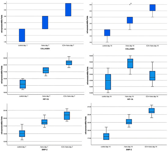
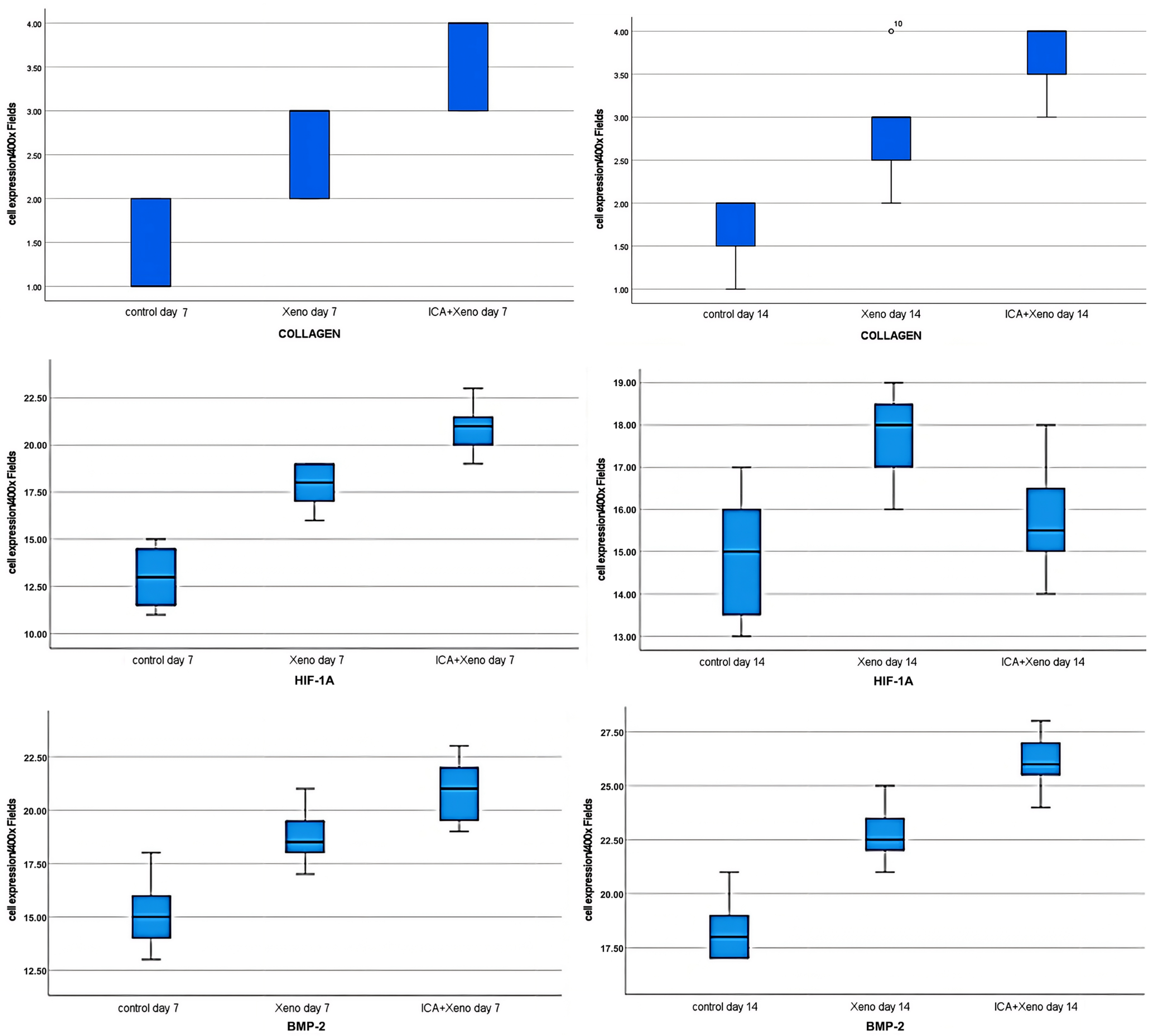
Figure 1.
Diagram of the results for the collagen fibers (top of boxplot), HIF1-α (in the middle of boxplot) and BMP-2 (at the bottom of boxplot) on days 7 and 14.
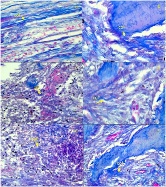
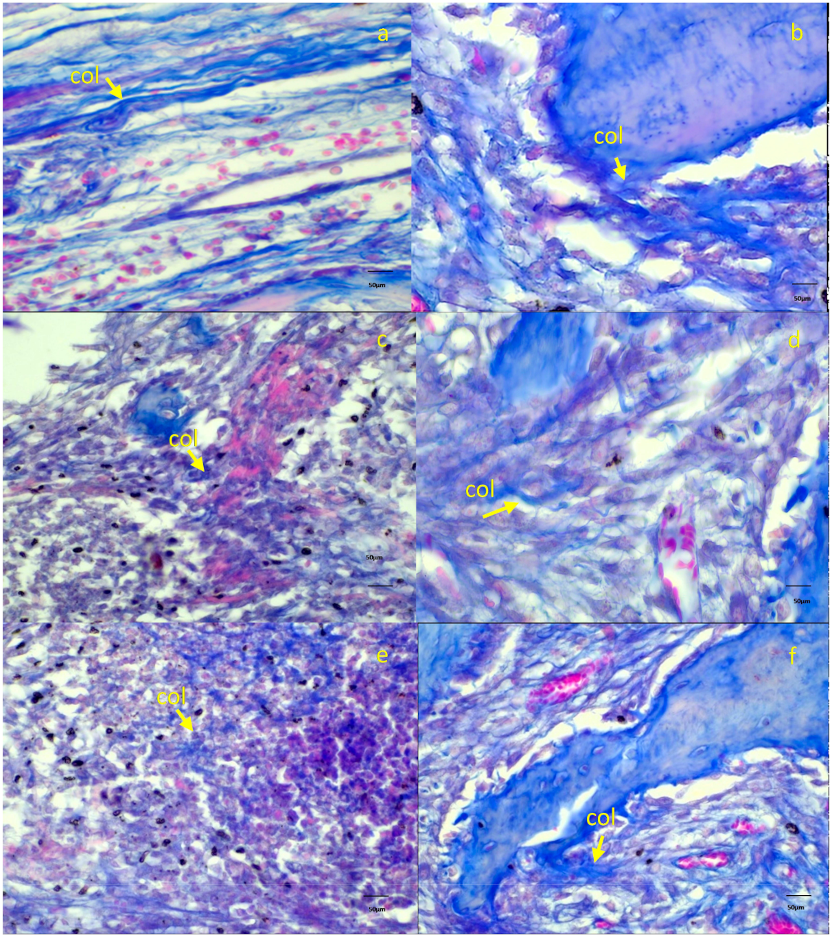
Figure 2.
Collagen fibers were evident in the socket region on days 7 and 14 (col arrow). Control group ((a) day 7, (b) day 14), xenograft ((c) day 7, (d) day 14), icariin combined with xenograft ((e) day 7, (f) day 14).
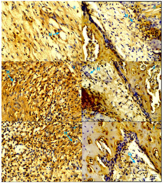
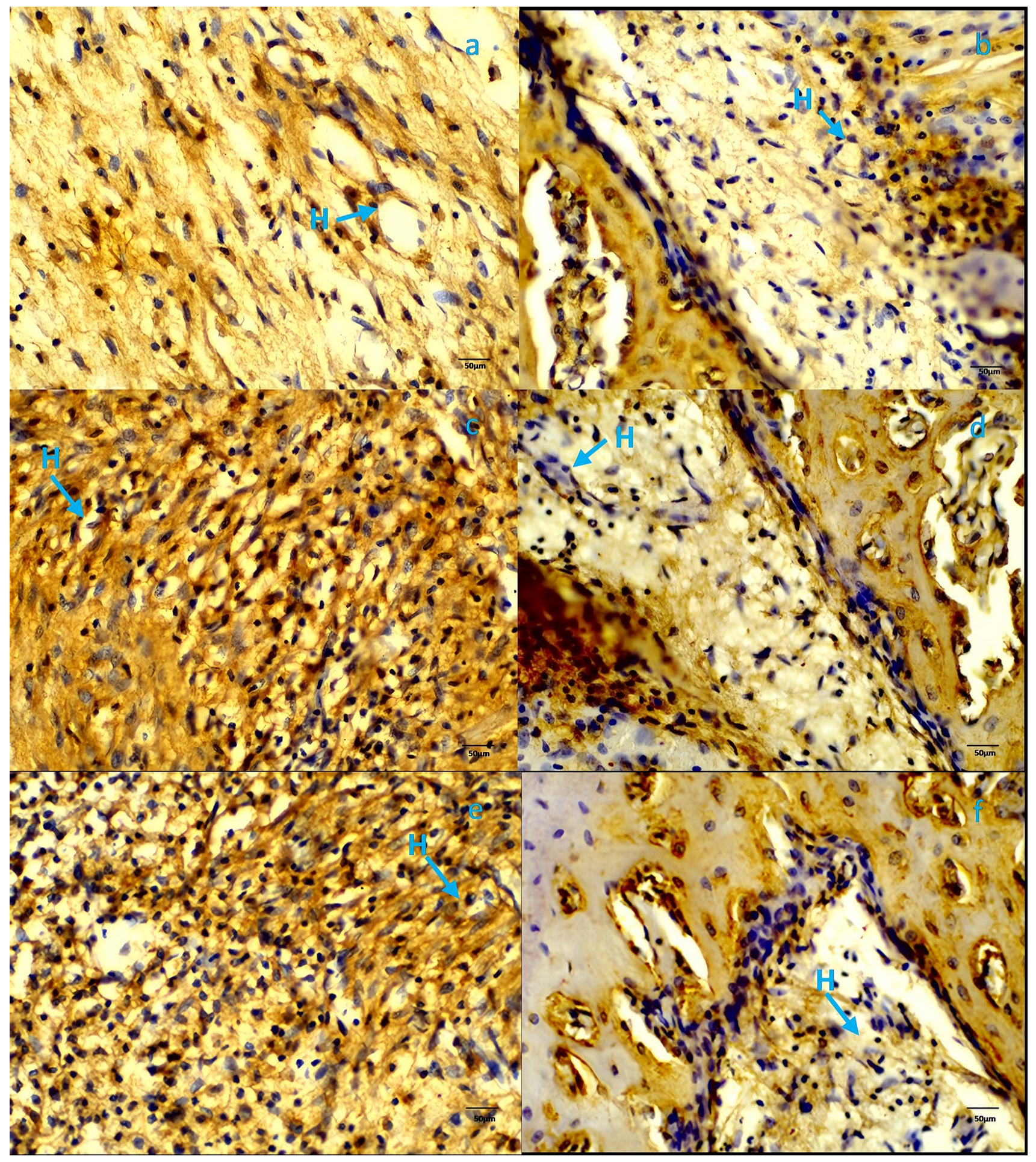
Figure 3.
HIF-1α expression was evident in the socket region on days 7 and 14 (H arrow). Control group ((a) day 7, (b) day 14), xenograft ((c) day 7, (d) day 14), icariin combined with xenograft ((e) day 7, (f) day 14).
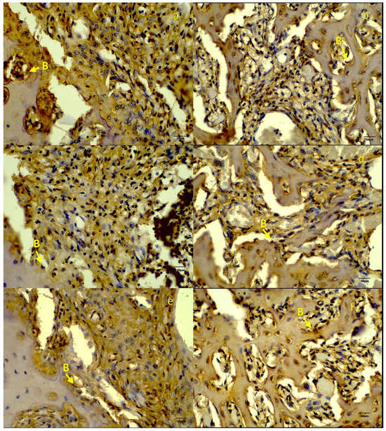
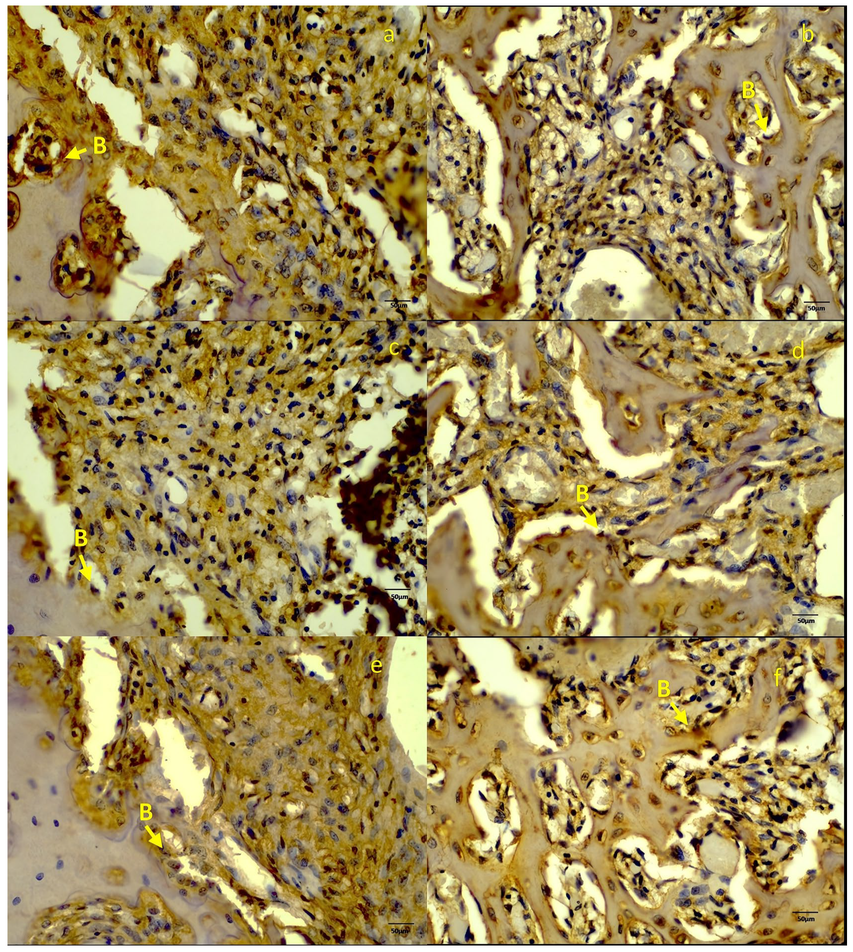
Figure 4.
BMP-2 expression was evident in the socket region on days 7 and 14 (B arrow). Control group ((a) day 7, (b) day 14), xenograft ((c) day 7, (d) day 14), icariin combined with xenograft ((e) day 7, (f) day 14).
3.1. Histological Assessment of Bone Healing
3.1.1. Control Group
The control sockets showed typical early healing signs at 7 days after extraction, such as a large number of inflammatory cells, fibroblast growth, and collagen. By day 14, granulation tissue filled up most of the socket area, and the wall of the socket was starting to develop woven bone. The alveolar bone was still not fully mature because there were still marrow gaps and fibrous tissue remnants.
3.1.2. Xenograft Group
On day 7, graft particles were found in sockets filled with the deproteinized bovine bone mineral. These particles were surrounded by inflammatory cells, immature osteoid tissue, and a collagen matrix. By day 14, the xenograft particles were embedded in a thick collagen matrix with active osteoblasts on the surface, indicating that the process of osteoconduction was underway. The xenograft group showed a significant amount of new bone area and more bone graft contact than the control group, but less than that of the IX combination group.
3.1.3. Icariin + Xenograft Combination Group (IX)
On day 7, there was minimal inflammatory infiltration accompanied by significant vascularization surrounding the graft particles and a dense collagen matrix. By day 14, newly formed woven bone was observed bridging the xenograft particles, indicating increased osteoconductive and osteoinductive activity. Histomorphometric analysis demonstrated that the IX group had the most significant collagen matrix, HIF-1α, and BMP-2 (p < 0.001) compared to all other groups, confirming improved outcomes of the combination of icariin and the xenograft.
HIF-1α expression showed a time-dependent pattern in the icariin + xenograft group. At day 7, higher HIF-1α expression was observed, consistent with an early hypoxic response, whereas by day 14, expression levels decreased across all groups, indicating resolution of hypoxia-associated signaling.
4. Discussion
The current study shows that, at extraction wounds in Wistar rats, ICA addition to a bone graft exerts a significant but less pronounced impact on the early wound healing dynamics, having a strong influence on the collagen fibre maturation dynamics, the upregulation of BMP-2 expression, and the enhancement of the HIF-1α signalling at day 7 and further improvement of these parameters at day 14 (Table 1, Table 2 and Table 3). We found that ICA has a dual osteogenic/angiogenic promoting effect, which accelerates the maturation of the extracellular matrix and increases osteo-angiogenic coupling by BMP-2 and HIF-1α. By day 7, the healing process in bone/graft environments usually includes granulation tissue, collagen production driven by fibroblasts, early vascular ingrowth, and the start of osteoid development. More numerous collagen fiber bundles were observed in our ICA + graft group than in the xenograft group. This was accompanied by considerably increased BMP-2 immuno-expression and higher HIF-1α levels. Collagen deposition: Early collagen fibre formation creates the provisional matrix upon which osteoprogenitors and blood vessels can anchor. Enhanced collagen at day 7 suggests ICA fosters a more robust scaffold early, potentially shortening the latency to woven bone formation. Literature shows that in alveolar extraction sockets, by 7–14 days collagen/fibroblast activity dominates before the onset of substantial mineralized bone [17]. BMP-2 upregulation: BMP-2 is a pivotal osteogenic growth factor, driving mesenchymal stem cell (MSC) commitment to the osteoblast lineage, activating Smad signalling and inducing downstream Runx2, Osx, COL1, and ALP. ICA has been shown to enhance BMP-2–mediated osteoblastic differentiation [8,18]. In our in vivo graft model, the early increase in BMP-2 suggests ICA accelerates the osteogenic switch within the granulation/collagen matrix at day 7. Although increased BMP-2 expression suggests activation of osteogenic-related signaling pathways, it does not provide direct functional evidence of osteoinduction. Therefore, these findings should be interpreted as indicative of molecular modulation rather than definitive proof of osteoinductive capacity. HIF-1α signalling and angiogenesis coupling: HIF-1α is a master regulator of hypoxia-driven angiogenesis and also importantly couples angiogenesis to osteogenesis [19]. Elevated HIF-1α at day 7 indicates that the ICA + graft environment triggers or sustains a hypoxia/angiogenic response earlier than graft alone. HIF-1α should be interpreted as an upstream hypoxia-responsive regulator and an indirect indicator of angiogenic signaling. Its increased expression reflects activation of pro-angiogenic pathways, such as VEGF signaling, rather than direct evidence of new blood vessel formation. This would facilitate microvascular ingrowth, enhance nutrient and oxygen supply for osteoprogenitors, and support ECM maturation. The co-existence of elevated HIF-1α and BMP-2 at day 7 aligns with the concept of osteo-angiogenic coupling.
Mechanistic enhanced effect: The improved collagen matrix provides a friendly architecture, and ICA initiates BMP-2 (osteogenesis) and HIF-1α (angiogenesis/hypoxia response) earlier, thereby catalysing the cascade, which would otherwise be slow. Supporting evidence: Zhang et al. 2019 demonstrated in vivo that a BioCaP scaffold with ICA + BMP-2 demonstrated superior bone formation as compared to BMP-2 alone [20]. Although their model could not explicitly analyse HIF-1α or collagen fibres during early time-points, their principle can be directly applied. In such a way, our data at day 7 indicate that ICA stimulates faster matrix deposition signalling activation, which preconditions increased bone regeneration. By day 14, in standard bone graft healing, one expects a transition from immature collagen to more organised fibre bundles, the beginning of woven bone deposition, increased vascular network stability, and declining hypoxic signalling as perfusion improves. In our study, the ICA + graft group showed more advanced collagen fibre organisation (denser, thicker fibres, better orientation), continued high BMP-2 expression and a maintained, albeit somewhat reduced, HIF-1α level compared to day 7. Collagen maturation and scaffold conversion: The progression from loose collagen to organised, thicker bundles by day 14 indicates accelerated maturation of the ECM. This hardened scaffold is more amenable to mineral deposition and supports osteoblast attachment, matrix mineralisation, and the formation of woven bone. The earlier and stronger collagen impetus in the ICA group likely compresses the time to functional bone matrix. Lasting expression of BMP-2: The continuous presence of BMP-2 at day 14 denotes that continued osteogenic signaling occurs after the initial phase, allowing the transition of the progenitor proliferation to matricular formation and mineralization. BMP-2 is very important in the initiation of the process as well as the preservation of osteoblast activity and collagen type I expression, which is consistent with the observation that our histology indicates the enhancement of collagen fibers. HIF-1 2 dynamics: Significantly, HIF-1 2, unlike the ICA group, attains steady levels at day 14 but can fall against controls, thus showing increased vascular perfusion and reduced hypoxia. This indicates that ICA initiates not only an early hypoxic/angiogenic response but also increases angiogenesis-related responses, facilitating a favorable outcome in the resolution of the hypoxic stimulus. Reviews on HIF-1α in bone regeneration emphasize that timely upregulation followed by resolution is ideal; chronic hypoxia or excessive HIF-1α may hinder bone formation [16,21]. Angio-osteogenic coupling realised: The overlapping timeline of initial HIF-1α/angiogenic activation, sustained BMP-2 and robust collagen scaffold formation points to efficient coupling of angiogenesis and osteogenesis. ICA appears to expedite this coupling so that by day 14, the environment is far more mature than the graft alone, which may still be in a slower matrix/vascular phase.
Bone graft milieu, and ICA effect: It should be pointed out that bone grafts provide an osteoconductive skeleton, but in many cases, they depend on host biology to provide osteogenesis and angiogenesis. ICA seems to provide an osteoinductive adjunct which enhances the host response in the graft microenvironment, supporting early collagen matrix deposition, active BMP-2 induction and earlier angiogenic support through HIF-1. This is in accord with the recent bone-tissue engineering approaches that portray the use of integrated osteogenic and angiogenic signals [22]. The ICA-enhanced graft scenario: At day 7, collagen levels are higher than expected for this time point, BMP-2 is already elevated, and HIF-1α is strongly present, suggesting accelerated initiation. At day 14, the collagen matrix is more mature/organised, BMP-2 is still elevated, and osteogenesis is ongoing; HIF-1α is still above baseline but trending downward, indicating an increased angiogenesis-related response and lessening hypoxia. This accelerated timeline may translate into earlier onset of woven bone, quicker graft integration, and potentially shorter healing periods. Despite the promising experimental findings, several potential inconveniences and risks should be considered before clinical translation.
5. Limitations and Future Directions
- –
- Specific type of graft and scaffold structure: The type of bone graft (xenograft, allograft, synthetic) and its microstructure (porosity, bioactivity) can have an impact on its healing speed and its behavior during the interaction with ICA. Further studies are required to examine various forms of grafts.
- –
- Results in a longer period of time: Day 7 and day 14 represent the results of early effects, but one should determine whether the early boost results in improved bone volume, mechanical strength, and graft integration at later stages.
- –
- The current research does not involve a direct description of the loading efficiency and release kinetics of icariin by the xenograft. Thus, the local concentration of icariin which was kept in the extraction socket during the time interval is not known precisely. This means that the biological effects that were observed must be regarded as the result of the icariin–xenograft system, but not as a well-regulated dose–response relationship. Further research that includes icariin-only and dose–response groups should be performed in the future to officially check the possible synergistic effects.
- –
- Although icariin is a naturally derived flavonoid, its clinical safety profile, particularly for topical application in oral tissues, requires careful consideration. From a regulatory perspective, the use of icariin as a bioactive adjunct in dental applications would require comprehensive toxicological evaluation, standardized formulation, and approval by relevant regulatory authorities before clinical translation.
- –
- Mechanistic pathway interrogation: Our findings support the current literature regarding BMP-2 and HIF-1α; however, further mechanistic studies (e.g., activation of Smad1/5/9, the cAMP/PKA/CREB axis, Wnt/β-catenin, and the expression of angiogenic factors such as VEGF and PDGF) are required to clarify the modulation of the osteo-angiogenic niche by ICA in vivo.
- –
- This study’s dosing schedule and application method were made for localized delivery in a rat extraction socket, so they cannot be used directly in human clinical practice.
- –
- Further research involving long-term micro-CT would provide us with figures on bone volume, trabecular architecture and mineral density. Biomechanical tests would assist us in determining whether the initial biological gains observed in this study can result in regenerated tissue functioning effectively.
6. Conclusions
In summary, our findings demonstrate that the integration of ICA with bone graft markedly improves early wound healing in a rat model, as evidenced by accelerated collagen matrix deposition, increased BMP-2 signaling, and a strong initial HIF-1α response at day 7, with a continued improved outcome of these parameters by day 14. This indicates that ICA serves as a significant complement to graft therapy, facilitating accelerated osteo-angiogenic coupling and scaffold formation. The early initiation of healing processes may enhance the integration of grafts and reduce healing timeframes in clinical settings and inform future translational and clinical investigations.
Author Contributions
Conceptualization, C.K. and I.K.D.; manuscript methodology, C.K.; manuscript data curation, C.K. and I.K.D.; manuscript writing—original draft preparation, C.K.; manuscript writing—review and editing, C.K. and I.K.D. All authors have read and agreed to the published version of the manuscript.
Funding
This research received no external funding.
Institutional Review Board Statement
All procedures adhered to institutional animal care and ARRIVE guidelines and received approval from the Institutional Animal Ethics Committee of the Faculty of Dentistry, Airlangga University (Ethic No: 1132/HRECC.FODM/XI/2025, Approval Date: 7 November 2025).
Informed Consent Statement
Not applicable.
Data Availability Statement
The original contributions presented in this study are included in the article. Further inquiries can be directed to the corresponding author(s).
Conflicts of Interest
The authors declare no conflicts of interest.
References
- Han, H.S.; Lee, J.T.; Oh, S.; Cho, Y.D.; Kim, S. Effectiveness of a collagen matrix seal and xenograft in alveolar ridge preservation: An experimental study in dogs. Sci. Rep. 2024, 14, 163. [Google Scholar] [CrossRef] [Scilit]
- Sukpaita, T.; Chirachanchai, S.; Chanamuangkon, T.; Pimkhaokham, A.; Ampornaramveth, R.S. Alveolar ridge preservation in rat tooth extraction model by chitosan-derived epigenetic modulation scaffold. J. Prosthodont. Res. 2024, 68, 299–309. [Google Scholar] [CrossRef] [Scilit] [PubMed]
- Durge, K.J.; Baliga, V.S.; Sridhar, S.B.; Dhadse, P.V.; Ragit, G.C. Extraction socket grafting using recombinant human bone morphogenetic protein-2-clinical implications and histological observations. BMC Res. Notes 2021, 14, 61. [Google Scholar] [CrossRef] [Scilit] [PubMed] [PubMed Central]
- Valencia-Llano, C.H.; López-Tenorio, D.; Saavedra, M.; Zapata, P.A.; Grande-Tovar, C.D. Comparison of Two Bovine Commercial Xenografts in the Regeneration of Critical Cranial Defects. Molecules 2022, 27, 5745. [Google Scholar] [CrossRef] [Scilit] [PubMed] [PubMed Central]
- Honnegowda, T.M.; Kumar, P.; Udupa, E.G.P.; Kumar, S.; Kumar, U.; Rao, P. Role of angiogenesis and angiogenic factors in acute and chronic wound healing. Plast. Aesthet. Res. 2015, 2, 243–249. [Google Scholar] [CrossRef] [Scilit]
- Wu, Y.; Xia, L.; Zhou, Y.; Ma, W.; Zhang, N.; Chang, J.; Lin, K.; Xu, Y.; Jiang, X. Evaluation of osteogenesis and angiogenesis of icariin loaded on micro/nano hybrid structured hydroxyapatite granules as a local drug delivery system for femoral defect repair. J. Mater. Chem. B 2015, 3, 4871–4883. [Google Scholar] [CrossRef] [Scilit] [PubMed]
- Xu, B.; Wang, X.; Wu, C.; Zhu, L.; Chen, O.; Wang, X. Flavonoid compound icariin enhances BMP-2 induced differentiation and signalling by targeting to connective tissue growth factor (CTGF) in SAMP6 osteoblasts. PLoS ONE 2018, 13, e0200367. [Google Scholar] [CrossRef] [Scilit] [PubMed] [PubMed Central]
- Chen, M.; Cui, Y.; Li, H.; Luan, J.; Zhou, X.; Han, J. Icariin Promotes the Osteogenic Action of BMP2 by Activating the cAMP Signaling Pathway. Molecules 2019, 24, 3875. [Google Scholar] [CrossRef] [Scilit] [PubMed] [PubMed Central]
- Zhang, X.; Chen, Y.; Zhang, C.; Zhang, X.; Xia, T.; Han, J.; Song, S.; Xu, C.; Chen, F. Effects of icariin on the fracture healing in young and old rats and its mechanism. Pharm. Biol. 2021, 59, 1245–1255. [Google Scholar] [CrossRef] [Scilit] [PubMed] [PubMed Central]
- Zheng, S.; Hu, G.Y.; Li, J.H.; Zheng, J.; Li, Y.K. Icariin accelerates bone regeneration by inducing osteogenesis-angiogenesis coupling in rats with type 1 diabetes mellitus. World J. Diabetes 2024, 15, 769–782. [Google Scholar] [CrossRef] [Scilit] [PubMed] [PubMed Central]
- Singh, W.R.; Sharma, A.; Devi, H.S.; Bhatia, A.; Patel, M.R.; Kumar, D. Icariin improves cutaneous wound healing in streptozotocin-induced diabetic rats. J. Tissue Viability 2022, 31, 197–206. [Google Scholar] [CrossRef] [Scilit] [PubMed]
- Coomes, A.M.; Mealey, B.L.; Huynh-Ba, G.; Barboza-Arguello, C.; Moore, W.S.; Cochran, D.L. Buccal bone formation after flapless extraction: A randomized, controlled clinical trial comparing recombinant human bone morphogenetic protein 2/absorbable collagen carrier and collagen sponge alone. J. Periodontol. 2014, 85, 525–535. [Google Scholar] [CrossRef] [Scilit] [PubMed]
- Uribe, F.; Vásquez, B.; Alister, J.P.; Olate, S. Comparison of rhBMP-2 in Combination with Different Biomaterials for Regeneration in Rat Calvaria Critical-Size Defects. Biomed. Res. Int. 2022, 2022, 6281641. [Google Scholar] [CrossRef] [Scilit] [PubMed] [PubMed Central]
- Hassumi, J.S.; Mulinari-Santos, G.; Fabris, A.L.D.S.; Jacob, R.G.M.; Gonçalves, A.; Rossi, A.C.; Freire, A.R.; Faverani, L.P.; Okamoto, R. Alveolar bone healing in rats: Micro-CT, immunohistochemical and molecular analysis. J. Appl. Oral Sci. 2018, 26, e20170326. [Google Scholar] [CrossRef] [Scilit] [PubMed] [PubMed Central]
- Garcia, V.G.; Dall Agnol, G.S.; Campista, C.C.C.; Bury, L.L.; Ervolino, E.; Longo, M.; Mulinari-Santos, G.; Levin, L.; Theodoro, L.H. Evaluation of Two Anorganic Bovine Xenogenous Grafts in Bone Healing of Critical Defect in Rats Calvaria. Braz. Dent. J. 2024, 35, e246119. [Google Scholar] [PubMed] [PubMed Central]
- Khoswanto, C.; Dewi, I.K. The role of rhBMP-2 in mandibular bone regeneration following tooth extraction through HIF-1α and VEGF-A expression: An Immunohistochemical study. J. Oral Biol. Craniofacial Res. 2025, 15, 359–364. [Google Scholar] [CrossRef] [Scilit] [PubMed] [PubMed Central]
- Francisconi, C.F.; Colavite, P.M.; Fonseca, A.C.; Azevedo, M.C.S.; Tabanez, A.P.; Melchiades, J.L.; Vieira, A.E.; Repeke, C.E.P.; Claudino, M.; Garlet, G.P. Microtomographic, histomorphometric, and molecular features show a normal alveolar bone healing process in iNOS-deficient mice along a compensatory upregulation of eNOS and nNOS isoforms. J. Appl. Oral Sci. 2023, 31, e20220436. [Google Scholar] [CrossRef] [Scilit] [PubMed] [PubMed Central]
- Xie, L.; Liu, N.; Xiao, Y.; Liu, Y.; Yan, C.; Wang, G.; Jing, X. In Vitro and In Vivo Osteogenesis Induced by Icariin and Bone Morphogenetic Protein-2: A Dynamic Observation. Front. Pharmacol. 2020, 11, 1058. [Google Scholar] [CrossRef] [Scilit] [PubMed] [PubMed Central]
- Chen, W.; Wu, P.; Yu, F.; Luo, G.; Qing, L.; Tang, J. HIF-1α Regulates Bone Homeostasis and Angiogenesis, Participating in the Occurrence of Bone Metabolic Diseases. Cells 2022, 11, 3552. [Google Scholar] [CrossRef] [Scilit] [PubMed] [PubMed Central]
- Zhang, X.; Lin, X.; Liu, T.; Deng, L.; Huang, Y.; Liu, Y. Osteogenic Enhancement Between Icariin and Bone Morphogenetic Protein 2: A Potential Osteogenic Compound for Bone Tissue Engineering. Front. Pharmacol. 2019, 10, 201. [Google Scholar] [CrossRef] [Scilit] [PubMed] [PubMed Central]
- Hannah, S.S.; McFadden, S.; McNeilly, A.; McClean, C. “Take My Bone Away?” Hypoxia and bone: A narrative review. J. Cell. Physiol. 2021, 236, 721–740. [Google Scholar] [CrossRef] [Scilit] [PubMed]
- He, Y.; Liang, L.; Luo, C.; Zhang, Z.Y.; Huang, J. Strategies for in situ tissue engineering of vascularized bone regeneration (Review). Biomed. Rep. 2023, 18, 42. [Google Scholar] [CrossRef] [Scilit] [PubMed] [PubMed Central]

Disclaimer/Publisher’s Note: The statements, opinions and data contained in all publications are solely those of the individual author(s) and contributor(s) and not of MDPI and/or the editor(s). MDPI and/or the editor(s) disclaim responsibility for any injury to people or property resulting from any ideas, methods, instructions or products referred to in the content. |
© 2026 by the authors. Licensee MDPI, Basel, Switzerland. This article is an open access article distributed under the terms and conditions of the Creative Commons Attribution (CC BY) license.