


Abstract
A low-cost phantom for ultrasound diagnostics is proposed for training applications involving small parts imaging. The phantom is constructed from plexiglass and includes openings that enable evaluation of the spatial resolution, measurement accuracy, and grayscale representation. Different filler materials are used to study contrast and shadowing effects in B-mode imaging. Experimental studies using a linear transducer show good agreement between measured and actual dimensions, with acceptable deviations. Clear grayscale differences are observed depending on acoustic impedance. The proposed phantom is inexpensive, easy to fabricate, and suitable for educational and training purposes in ultrasound diagnostics.
1. Introduction
In recent years, medical imaging diagnostics has developed at a very rapid pace and has become a leading technique for providing quick and accurate diagnoses in modern medicine. Among the various imaging modalities, ultrasound diagnostics occupies a particularly important position due to its wide accessibility, relatively low cost, and non-invasive nature. Unlike other imaging techniques—such as radiology (including computed tomography), magnetic resonance imaging, and nuclear medicine—ultrasound diagnostics is considered safe and does not involve ionizing radiation. Its application in obstetrics is unparalleled, and it is also widely used in cardiology, where Doppler-based techniques provide additional functional information about blood flow.
The increasing use of ultrasound systems in clinical practice raises the need for improved training and continuous education of medical personnel and engineering specialists working with such equipment. In many cases, it is highly desirable for this training to be performed under controlled and simulated conditions without involving real patients. This approach allows repeated practice, controlled experimentation, and safe familiarization with the system’s capabilities and limitations [1,2,3,4]. The need for simulated training environments has led to the development and widespread use of phantoms in medical imaging. Phantoms are specially designed objects that mimic certain physical and acoustic properties of biological tissues and are used for calibration, testing, and training purposes [1,2,3]. In ultrasound diagnostics, phantoms are particularly valuable because they allow users to study image formation, evaluate system performance, and practice measurement techniques in a controlled and reproducible manner [1,2].
A key requirement for ultrasound phantoms is that the materials used should approximate the acoustic properties of real tissues, such as speed of sound, density, and acoustic impedance. The average speed of sound in soft biological tissues is typically assumed to be approximately 1540 m/s, while acoustic impedance values vary slightly depending on the specific tissue type. For example, water has an acoustic impedance of approximately 1.48 MRayl, while soft tissues are around 1.6 MRayl [1]. Matching these parameters is essential for achieving realistic image representation and meaningful training results. In addition to material properties, the geometric design of the phantom plays an important role. Phantoms often include structures such as inclusions, cavities, or channels that allow the study of object detectability, measurement accuracy, and grayscale representation. These features are particularly important when evaluating B-mode ultrasound imaging, which is the primary operating mode of most ultrasound systems [2].
2. Materials and Methods
2.1. Design Requirements for the Phantom
The development of the proposed phantom is based on several key requirements. First, the materials used should approximate, as closely as possible, the acoustic properties of biological tissues, particularly in terms of speed of sound and acoustic impedance. Second, the phantom should enable the visualization of objects with different geometries and sizes in B-mode ultrasound imaging. Third, it should allow for the investigation of grayscale variations and shadowing effects. Finally, the phantom should be inexpensive, easy to fabricate, and suitable for training purposes.
A primary objective is the observation and measurement of openings with different diameters, as well as the evaluation of grayscale levels produced by materials with varying acoustic properties. The phantom is specifically designed for studies involving small parts using a linear ultrasound transducer.
2.2. Acoustic Properties of Biological Tissues
The selection of suitable materials for the phantom requires consideration of the acoustic properties of biological tissues. Table 1 presents the main acoustic parameters [5,6]—speed of sound, density, and acoustic impedance—for several typical tissue types used in ultrasound diagnostics.
Table 1.
Acoustic parameters of the main tissue structures.
As shown in Table 1, the average speed of sound in soft tissues is approximately 1540 m/s, while the acoustic impedance is around 1.6 MRayl [1]. These values are used as a reference for selecting appropriate materials for the phantom.
2.3. Phantom Design and Geometry
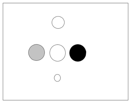
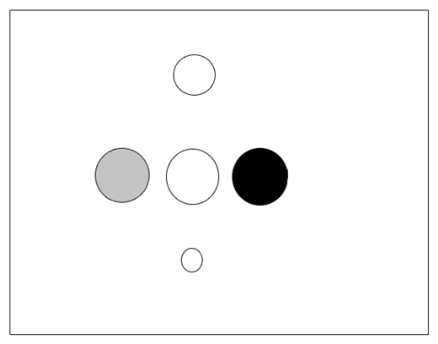
The proposed phantom is designed to include two main groups of objects for study:
- Vertically positioned openings with diameters of 10 mm, 8 mm, and 6 mm;
- Horizontally positioned openings with a diameter of 10 mm, intended for the placement of different filler materials.
The purpose of the vertical openings is to allow evaluation of spatial resolution and measurement accuracy at different depths. The horizontal openings are used to investigate grayscale representation and shadowing effects.
The overall dimensions of the phantom are 30 × 50 × 50 mm, with a maximum depth of 50 mm. The general design of the phantom is illustrated in Figure 1.
Figure 1.
Design of the proposed phantom showing the arrangement of the vertical openings (open circles; 10, 8 and 6 mm) and the horizontal openings intended for interchangeable filler materials (shaded circles; 10 mm).
The variation in size and depth of the openings enables analysis of the performance of the ultrasound transducer, particularly in terms of resolution and penetration depth.
2.4. Materials Selection and Fabrication
The base material selected for the phantom is a commercially available cast plexiglass (PMMA) sheet, obtained as generic engineering stock from a local supplier (Varna, Bulgaria), due to its mechanical stability, ease of fabrication, and suitable acoustic properties. In addition, several materials are used as fillers in order to produce different grayscale levels in ultrasound images.
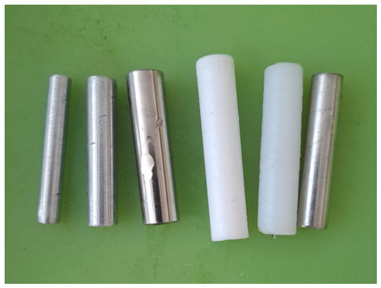
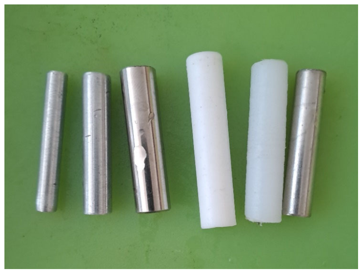
Table 2 presents the acoustic parameters [5,6,7] of the selected materials used for the fillers, along with their comparison to water. All filler elements were cut from standard engineering-grade rod of 10 mm diameter, obtained as generic material from the same local supplier (Varna, Bulgaria) as the PMMA sheet; no proprietary products were used.
Table 2.
Acoustic parameters of materials used for phantom fillers.
As shown in Table 2, materials such as Teflon and Nylon have acoustic properties relatively close to plexiglass, while Aluminum and Steel exhibit significantly higher acoustic impedance. This difference is expected to produce noticeable variations in grayscale and shadowing effects in the ultrasound images [8,9].
The different filler elements used in the phantom are illustrated in Figure 2.
Figure 2.
General view of the filler materials used for generating different grayscale levels.
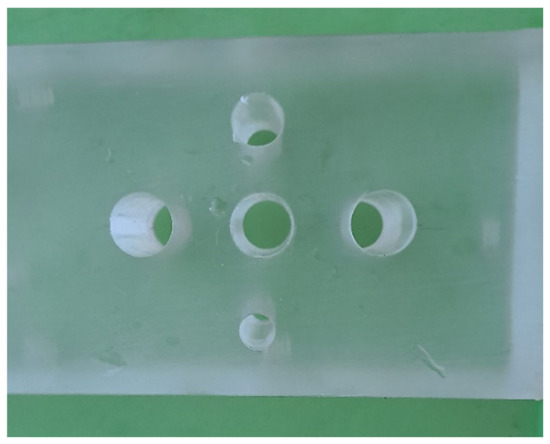
The phantom is fabricated from plexiglass using standard mechanical processing techniques. Openings of predefined diameters are drilled both vertically and horizontally according to the design shown in Figure 1. The horizontal openings are designed to accommodate interchangeable filler materials.
The fabrication process is intentionally kept simple in order to ensure low production cost and ease of replication. This makes the phantom suitable for educational and laboratory environments where access to complex manufacturing technologies may be limited.
2.5. Experimental Setup
The experimental studies are conducted using a digital ultrasound system (Mindray DP-10 with 75L38EB linear transducer; Shenzhen Mindray Bio-Medical Electronics Co., Ltd., Shenzhen, China) equipped with a linear transducer. The phantom is placed in a water environment during imaging in order to ensure proper acoustic coupling between the transducer and the phantom surface.
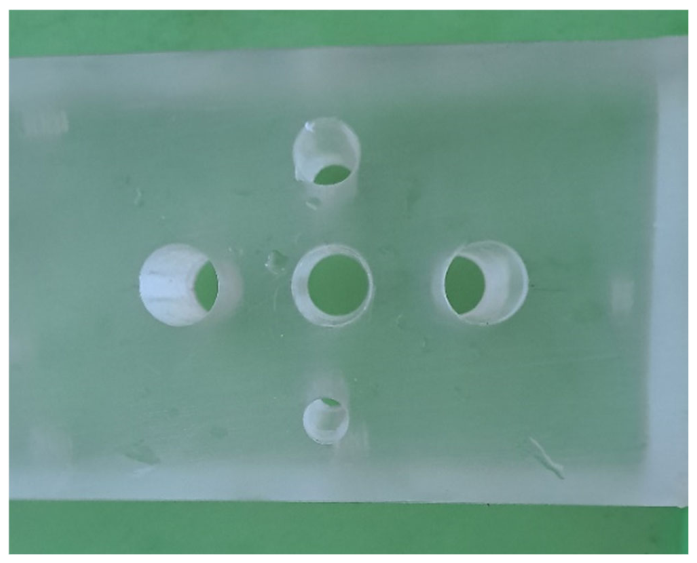
An example of the fabricated phantom is shown in Figure 3.
Figure 3.
Photograph of the fabricated phantom used in the experimental studies.
Ultrasound imaging is performed in B-mode, which is the standard mode for structural visualization in medical ultrasound systems [2].
2.6. Measurement Procedure
The evaluation of the phantom is performed through two main types of measurements:
- Measurement of the diameters of the vertical openings using the built-in software of the ultrasound system;
- Qualitative assessment of grayscale variations and shadowing effects for different filler materials.
The measurements of the openings are carried out by placing digital markers directly on the ultrasound images using the system software. The measured values are later compared with the actual dimensions of the openings in order to evaluate the accuracy of the imaging system.
For the grayscale analysis, different filler materials are inserted into the horizontal openings of the phantom. The resulting ultrasound images are visually assessed in terms of contrast and shadowing effects, depending on the acoustic properties of the materials used.
3. Results
3.1. Visualization of Vertical Openings
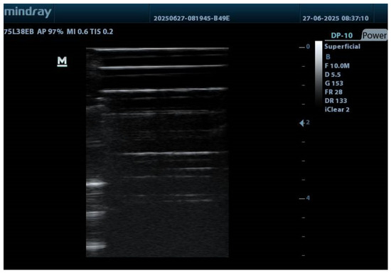
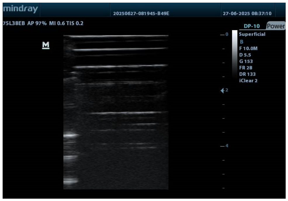
Ultrasound imaging of the developed phantom demonstrates clear visualization of the vertically positioned openings at different depths. The obtained B-mode image allows for observation of the shape and position of the openings, as well as their behavior with increasing depth.
An example of the ultrasound image of the vertical openings is shown in Figure 4.
Figure 4.
Ultrasound image of vertically positioned openings at different depths.
The results indicate good visibility of the openings, with minimal distortion of their shape. No significant shadowing effects are observed behind the openings, which facilitates accurate measurement and analysis.
3.2. Measurement Accuracy of Openings
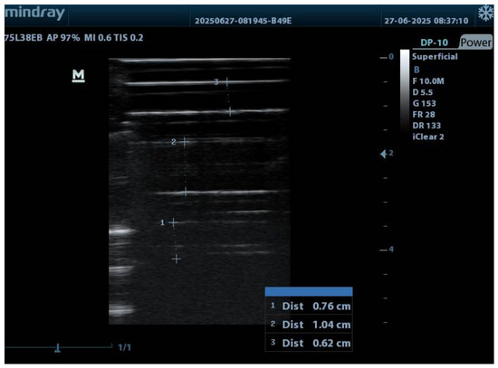
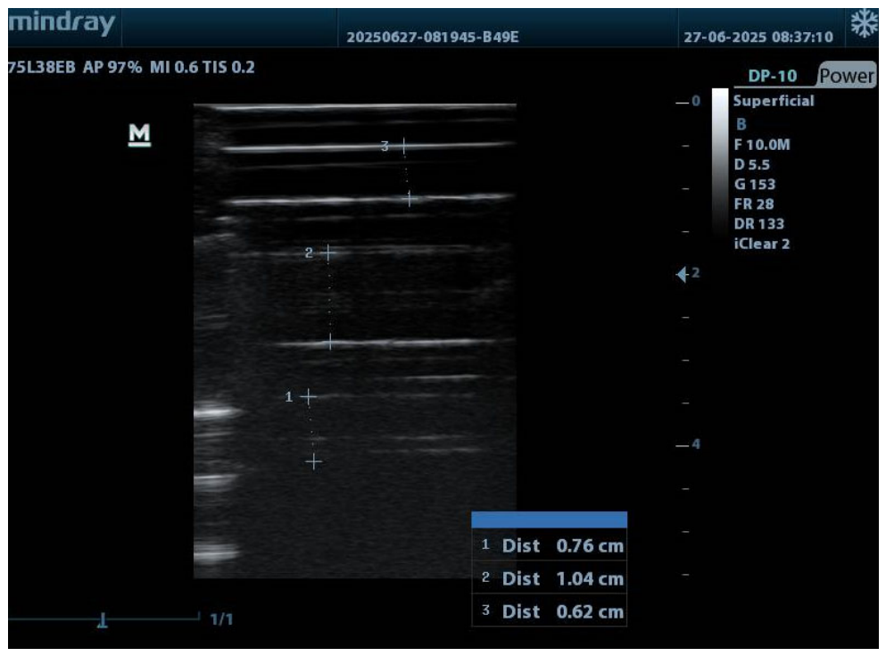
The diameters of the vertical openings are measured using the built-in software of the ultrasound system. The measurement process is illustrated in Figure 5.
Figure 5.
Measurement of the diameters of vertical openings using the ultrasound system software.
The obtained measurement results are summarized in Table 3, where the measured values are compared with the actual dimensions of the openings.
Table 3.
Results of the measurements of vertical openings.
The results show good agreement between the actual and measured values, with deviations within a few percent. These discrepancies can be attributed to the subjective placement of measurement markers and the resolution limitations of the ultrasound system.
3.3. Grayscale Imaging Results
Additional studies are conducted using different filler materials inserted into the horizontal openings of the phantom in order to evaluate grayscale representation and shadowing effects.
For materials such as Teflon and Nylon, the differences in grayscale levels are relatively small. This is due to the similarity of their acoustic properties to those of plexiglass and water, resulting in limited contrast in the ultrasound images.
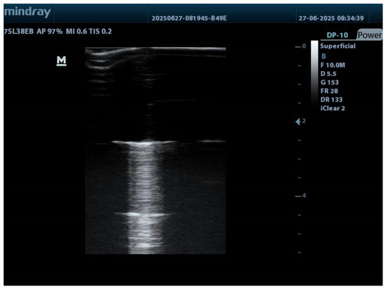
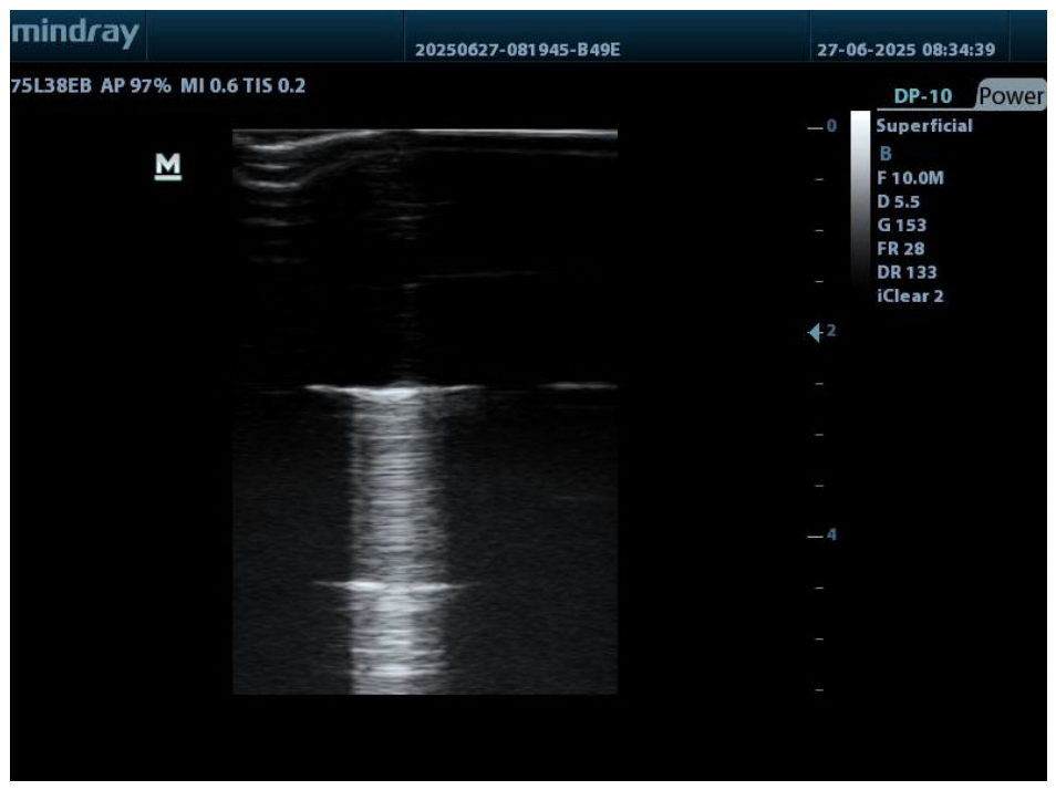
In contrast, materials such as Aluminum and Steel exhibit significantly higher acoustic impedance, which leads to pronounced grayscale differences and strong shadowing effects. An example of such behavior is shown in Figure 6, where a steel filler is used.
Figure 6.
Ultrasound image of a steel filler showing strong grayscale contrast and shadowing effect.
Two main observations can be made from Figure 6. First, the grayscale contrast between the filler and the surrounding material is clearly distinguishable. Second, a strong shadowing effect is observed behind the filler, which is caused by the large difference in acoustic impedance between the steel and the surrounding medium.
These results confirm that the phantom can be effectively used to demonstrate the influence of material properties on ultrasound image formation, particularly in terms of grayscale representation and shadowing.
4. Discussion
4.1. Accuracy and Measurement Limitations
The results obtained from the measurement of the vertical openings demonstrate good agreement between the actual and measured dimensions. The observed deviations are within a few percent, which is acceptable for practical training applications. These discrepancies are primarily attributed to the subjective placement of measurement markers on the ultrasound images, as well as the inherent resolution limitations of the ultrasound system.
In addition, factors such as image quality, operator experience, and system settings may influence the measurement accuracy. Despite these limitations, the results indicate that the proposed phantom provides sufficiently reliable conditions for evaluating measurement functions in ultrasound systems and for training purposes.
4.2. Influence of Acoustic Impedance on Imaging
The experimental results clearly demonstrate the influence of acoustic impedance on grayscale representation and shadowing effects in ultrasound imaging. Materials with acoustic properties similar to those of plexiglass and water, such as Teflon and Nylon, produce minimal contrast in the ultrasound images. This makes them less suitable for demonstrating grayscale variations.
In contrast, materials with significantly higher acoustic impedance, such as Aluminum and Steel, result in pronounced grayscale differences and strong shadowing effects. This behavior is consistent with the fundamental principles of ultrasound imaging, where large impedance mismatches lead to increased reflection of the acoustic wave and reduced transmission into deeper regions [8,9].
The observed shadowing effect behind high-impedance materials further confirms the expected interaction between the ultrasound wave and the medium. These findings highlight the importance of material selection when designing phantoms for specific training and experimental purposes.
4.3. Practical Applicability and Comparison with Existing Solutions
One of the primary objectives of the proposed phantom is its application in the training and self-training of students and medical personnel. The results show that the phantom provides clear visualization of objects with different sizes and enables practical measurement exercises using real ultrasound equipment. The ability to observe grayscale variations and shadowing effects further enhances its educational value, allowing users to better understand the relationship between material properties and image formation.
Compared to commercially available ultrasound phantoms [5,8,9], which often include complex tissue-mimicking materials and anatomically realistic structures, the proposed design is significantly simpler. However, this simplicity offers important advantages—due to its simple design and low cost, the phantom can be easily reproduced and used in laboratory settings, making it particularly suitable for educational institutions and training environments [2,3]. While advanced phantoms may provide higher realism, they are often expensive and less accessible for routine training and experimental use. In contrast, the proposed phantom provides a practical alternative for basic training and system evaluation, especially in educational contexts where simplicity and affordability are key factors [5,8,9].
4.4. Limitations of the Proposed Design
Despite its advantages, the proposed phantom has several limitations that should be considered. The geometric structures included in the phantom are relatively simple and do not fully replicate the complexity of real anatomical tissues. Additionally, the acoustic properties of the materials used, although selected to approximate biological tissues, do not perfectly match those of real human tissue.
The depth of the phantom is also limited, which restricts its use for studying deeper imaging scenarios or for applications involving convex transducers. Furthermore, the evaluation of grayscale differences is primarily qualitative, and no quantitative analysis of image intensity is performed.
These limitations suggest that while the phantom is suitable for basic training and experimental studies, further development would be required for more advanced applications.
5. Conclusions
In this study, a simple and cost-effective phantom for ultrasound medical diagnostics has been developed and experimentally evaluated. The proposed design is based on the use of plexiglass as a base material and incorporates vertical and horizontal openings that allow the investigation of spatial resolution, measurement accuracy, and grayscale representation.
The experimental results demonstrate that the phantom provides clear visualization of openings with different diameters, enabling reliable measurement using the built-in software of the ultrasound system. The measured values show good agreement with the actual dimensions, with deviations within an acceptable range for training applications.
Furthermore, the use of different filler materials allows effective demonstration of grayscale variations and shadowing effects, highlighting the influence of acoustic impedance on ultrasound image formation. Materials with higher acoustic impedance produce stronger contrast and pronounced shadowing, while materials with similar properties to the surrounding medium result in limited distinguishability.
Overall, the developed phantom fulfills its intended purpose as an inexpensive, easy-to-fabricate tool suitable for training and educational applications in the field of ultrasound diagnostics. It provides a practical solution for students and medical personnel to gain experience in image interpretation and measurement techniques.
Future developments of the proposed phantom may focus on increasing its complexity and expanding its range of applications. One possible direction is the design of phantoms suitable for use with convex transducers, as well as increasing the depth of the phantom to allow studies involving deeper imaging scenarios.
Additional improvements may include the incorporation of objects with more complex geometries, such as elliptical or spherical structures, which would enable more comprehensive evaluation of the ultrasound system’s capabilities. The development of phantoms that mimic specific anatomical structures and tissues could further enhance their usefulness in medical training.
Another promising direction is the use of 3D printing technologies for phantom fabrication. This approach would allow greater flexibility in design and more precise control over the acoustic properties of the materials used. Furthermore, future work may include the development of phantoms suitable for advanced ultrasound techniques, such as 3D imaging and Doppler-based studies.
Author Contributions
Conceptualization, E.B. and V.T.-S.; methodology, E.B.; validation, E.B. and A.M.; formal analysis, E.B., A.M. and V.T.-S.; investigation, E.B.; writing—original draft preparation, E.B., A.M. and V.T.-S.; writing—review and editing, E.B. and A.M.; visualization, E.B. All authors have read and agreed to the published version of the manuscript.
Funding
This research was funded by the Technical University of Varna.
Institutional Review Board Statement
Not applicable.
Informed Consent Statement
Not applicable.
Data Availability Statement
Data is contained within the article.
Acknowledgments
During the preparation of this manuscript, the authors used OpenAI′s ChatGPT4 for the purposes of text refinement, grammar correction, and improving the clarity of scientific writing, particularly to assist with English-language editing as the corresponding author is not a native English speaker. The authors have thoroughly reviewed and edited all AI-generated content and take full responsibility for the final text presented in this publication.
Conflicts of Interest
The authors declare no conflicts of interest.
Abbreviations
The following abbreviations are used in this manuscript:
| B-mode | Brightness mode (ultrasound imaging mode) |
| PMMA | Polymethyl methacrylate (plexiglass) |
| MRayl | Mega Rayl (unit of acoustic impedance) |
| ABS | Acrylonitrile butadiene styrene |
References
- Duck, F.A. Table of Density, Speed of Sound and Acoustic Impedance in Human Tissues. In Physical Properties of Tissue: A Comprehensive Reference Book; Academic Press: London, UK, 1990. [Google Scholar]
- Pierce, H. TU-C-202-01: Ultrasound Phantoms: B-Mode, Doppler Mode and Others. Med. Phys. 2010, 37, 3388. [Google Scholar] [CrossRef] [Scilit]
- CIRS. Near Field Ultrasound Phantom, Model 050 (Data Sheet 050 DS 053013). Available online: https://cspmedical.com/content/110-1045_050_near_field_ultrasound_phantom.pdf (accessed on 8 September 2026).
- Kenwright, D.; Lavarack, N.; Anderson, T.; Moran, C.M.; Hoskins, P.R. Wall-less Flow Phantom for High-Frequency Ultrasound Applications. Ultrasound Med. Biol. 2014, 41, 890–897. [Google Scholar] [CrossRef] [Scilit]
- Sun Nuclear (CIRS). Zerdine Ultrasound Phantoms—Multi-Purpose, Multi-Tissue Ultrasound Phantom, Model 040GSE. Available online: https://www.sunnuclear.com/products/zerdine-ultrasound-phantom (accessed on 8 September 2026).
- Velocity & Acoustic Impedance Table. Available online: https://content.ndtsupply.com (accessed on 30 April 2026).
- Harmsen, S.S. Acoustic Impedance Matching Using 3D Printing Lenses. Bachelor’s Thesis, University of Twente, Enschede, The Netherlands, 2018. [Google Scholar]
- Cardinal Health. Multipurpose Tissue/Cyst Ultrasound Phantom; Cardinal Health, Radiation Management Services: Cleveland, OH, USA, 2003; pp. 1–8. [Google Scholar]
- Multi-Purpose Phantom—N-365. Available online: https://www.kyotokagaku.com (accessed on 30 April 2026).

Disclaimer/Publisher’s Note: The statements, opinions and data contained in all publications are solely those of the individual author(s) and contributor(s) and not of MDPI and/or the editor(s). MDPI and/or the editor(s) disclaim responsibility for any injury to people or property resulting from any ideas, methods, instructions or products referred to in the content. |
© 2026 by the authors. Licensee MDPI, Basel, Switzerland. This article is an open access article distributed under the terms and conditions of the Creative Commons Attribution (CC BY) license.