


Abstract
A commonly employed imaging modality that relies on ionizing radiation is Computed Tomography (CT). While lowering the radiation dose is beneficial for patient health, it can result in reduced image quality. Therefore, improving low-dose CT (LDCT) reconstruction is a significant area of research. The LoDoPaB-CT benchmark evaluates LDCT reconstruction methods, where many top methods use UNet-type architectures. We explore a two-stage approach for LDCT reconstruction: the first stage employs traditional filtered backprojection (FBP), while the second stage performs CT image enhancement. Our training strategy involves pretraining a neural network to denoise natural grayscale images, which are corrupted by Gaussian noise, followed by fine-tuning the network for CT image enhancement using LDCT and normal-dose CT (NDCT) pairs. Experiments on various small subsets of the LoDoPaB-CT dataset demonstrate the effectiveness of our method, showing that less task-specific data are required for training.
1. Introduction
To provide non-invasive visualization of internal structures, Computed Tomography (CT) is essential in modern medical imaging. Its widespread use is partly due to its versatility, serving various diagnostic purposes [1,2]. However, minimizing ionizing radiation while maintaining image quality remains a critical challenge, particularly for low-dose CT (LDCT) reconstruction [3]. Recent research focuses on LDCT reconstruction, leveraging advanced algorithms and deep learning techniques [4,5,6,7,8,9,10].
Historically, iterative variational regularization methods, like total variation [11], total generalized variation [12], and Hessian Schatten norms [13], dominated CT image reconstruction. Recently, deep learning, especially convolutional neural networks (CNNs), has revolutionized medical image reconstruction, effectively reducing artifacts and noise [14]. Approaches range from fully learned methods, which use sinograms or raw projection data, to iterative and multi-stage methods integrating traditional techniques with neural networks. For instance, ISTA UNet [15], the Learned Primal-Dual algorithm [16], and ItNet [17] are notable for their success in LDCT reconstruction, particularly in the LoDoPaB-CT challenge [18].
Building on recent trends of pretraining models on large, diverse datasets from various domains, such as those used in Natural Language Processing (e.g., [19]) and Computer Vision (e.g., [20]), we introduce a two-stage approach for LDCT reconstruction. The first stage involves traditional filtered backprojection (FBP), while the second stage focuses on CT image enhancement. Two-stage methods using UNets or other neural networks are prevalent in CT image processing [9,10,15,21,22,23]. In the present work, we use the Gaussian denoiser DRUNet [24], which has been pretrained on several natural image datasets, including the Berkeley segmentation, DIV2K, Flick2K, and the Waterloo Exploration dataset. For the subsequent task of LDCT image enhancement, fine-tuning is performed using CT-specific images. We emphasize that our distinctive training strategy involves pretraining on non-CT images with different noise characteristics than LDCT images.
Furthermore, our method demonstrates robustness through experiments on various small subsets of the LoDoPaB-CT dataset, highlighting the advantage of our pretraining strategy over training the network from scratch. These experiments also show that our approach requires less task-specific data to achieve similar results.
2. Problem Formulation
CT images are reconstructed from X-Ray projection data measured at various angles. The forward model defines how the desired image relates to the collected projection data. Let y be the projection data, the true CT image, and R the Radon transform. With these definitions, the forward model can be simplified as follows:
where represents measurement errors, such as quantum noise, electronic noise, scatter, and artifacts from patient motion. For a detailed model description, we refer to [21]. CT image reconstruction aims to estimate x from the projection data y, utilizing the system matrix R.
This study addresses the issue of reconstructing LDCT images with a reduced radiation dose. The primary challenge is the increased noise level in the projection data resulting from the lower radiation dose. Additionally, the inversion of R is ill-posed, which further amplifies the noise.
2.1. The Two-Stage Approach
The methodology employed for the reconstruction of CT images comprises two stages. In the initial stage, reconstruction is performed utilizing the FBP, which is a conventional analytical method, but the noise is amplified in the measurements. In the subsequent stage, a neural network, designated as , is employed to enhance the LDCT image. Here, the low-dose FBP-reconstructed image is mapped to an enhanced version. Mathematically, the two-stage method can be expressed in the following way:
where denotes a neural network with learnable parameters , denotes the filtered backprojection, and is the final reconstruction result.
To enhance the performance of for CT image enhancement, we adopt a fine-tuning strategy. Initially, is pretrained on a large dataset to denoise natural grayscale images corrupted by Gaussian noise. Subsequently, we fine-tune for the specific task of LDCT image enhancement using paired data consisting of LDCT and normal-dose CT (NDCT) images. Fine-tuning adapts to the characteristics of LDCT image enhancement, aiming to improve the quality and fidelity of the FBP-reconstructed images.
2.2. DRUNet: Pretraining for Robust Image Restoration
The DRUNet [24] model is trained for various image restoration tasks, particularly for denoising natural grayscale images corrupted by Gaussian noise. It combines the effectiveness of UNets in image translation tasks, combined with the enhanced modeling capacity of ResNets through stacked residual blocks. The DRUNet model was trained on a dataset comprising 8794 natural images from the Berkeley Segmentation, DIV2K, Flick2K, and Waterloo Exploration datasets. The optimization process utilizes the ADAM algorithm with the objective of minimizing the mean absolute error (MAE) loss between the processed image and the ground truth. This pretrained network is then fine-tuned for the specific purpose of improving CT images using paired LDCT and NDCT images.
3. Implementation Details
Following the methodologies outlined in [10], we adopted a composite loss function to train our neural network architecture. Initially, we employed the MAE loss function, denoted as , which focuses on ensuring the accuracy of pixel intensity and is known for its robustness in handling outliers [25]. The MAE loss for a single sample is defined as
where N denotes the amount of pixels in the image, refers to the i-th pixel of the neural network output, and represents the i-th pixel from the ground truth (NDCT image).
To further enhance reconstruction quality, we integrated Structural Similarity (SSIM) [26] in our loss function. The SSIM metric evaluates the structural similarity of images by analyzing the statistical attributes of local regions, rather than focusing on the precision of individual pixels.
This composite loss function L is formulated as
where is calculated according to the method described in [26], adjusts the weight of the term, is the regularization hyperparameter, and represents the squared norm of the parameters of the model.
For optimization, we utilized the ADAM algorithm [27] with default parameters and . The initial learning rate was set to . In order to enhance the stability of our network, we employed two data augmentation techniques: rotational augmentation and Gaussian noise augmentation.
4. The LoDoPaB-CT Dataset
The LoDoPaB-CT dataset [18] consists of 46,573 pairs of chest CT images obtained from patients and their respective simulated low-dose measurements. The original data were obtained from the LIDC/IDRI dataset [28] and the ground truth images were cropped to pixels. This dataset serves as a benchmark for evaluating LDCT reconstruction algorithms, offering 35,820 training, 3522 validation, 3553 test, and 3678 challenge images.
5. Evaluation Metrics
We adopted the evaluation metrics established in the LoDoPaB-CT challenge [18], which include the peak signal-to-noise ratio (PSNR) and structural similarity index (SSIM). Additionally, we utilize their respective fixed-range (FR) versions of the PSNR and SSIM algorithms, namely, PSNR-FR and SSIM-FR. These metrics standardize the range of values rather than use the full dynamic range of the ground truth image. The PSNR measures the relationship between the highest achievable signal strength and the level of noise distortion [29]. On the other hand, SSIM, introduced by Wang et al. [26], evaluates the structural similarity of two images, as elaborated in the section on implementation details. Higher values for both the PSNR and SSIM indicate better reconstruction quality in our evaluations.
6. Experimental Results
To explore the impact of fine-tuning a pretrained network on our results, we evaluated the performance of the DRUNet in several scenarios. We compared the effectiveness of pretraining the DRUNet on the Gaussian denoising of natural grayscale images against the random initialization of the model weights. In both cases, the models are fine-tuned on various subsets of the LoDoPaB-CT dataset for comparison. We utilized ‘drunet_gray.pth’ as the pretrained model weights, which is available for download from a GitHub repository (https://github.com/cszn/DPIR/tree/master/model_zoo (accessed on 18 June 2024)).
In the LoDoPaB-CT dataset, we randomly selected 0.2%, 1%, and 5% of the training and validation datasets. Specifically, this selection resulted in 71 training and 7 validation images for the 0.2% case, 358 training and 35 validation images for the 1% case, and 1791 training and 176 validation images for the 5% case. When testing the trained networks, we selected 300 random images from the test set of the LoDoPaB-CT dataset.
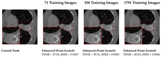
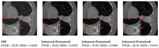
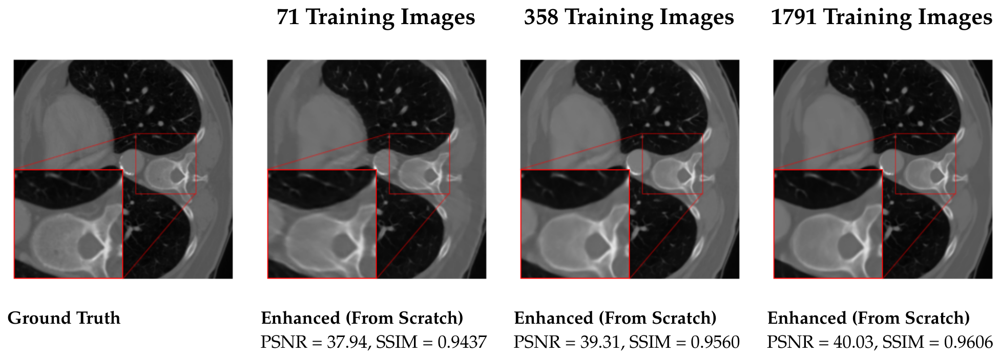
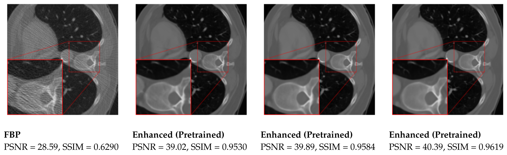
Table 1 outlines the results across various scenarios, while Figure 1 provides visual representations of an individual test samples for each scenario. A direct comparison reveals that fine-tuning the pretrained network consistently yields better metrics than training from scratch across all three cases. Additionally, the effectiveness of pretraining becomes more pronounced as the number of images for fine-tuning decreases. Notably, training the network from scratch with 358 images yields similar results to fine-tuning the pretrained network with only 71 images. Similarly, training from scratch with 1791 images shows comparable results to fine-tuning with 358 images. This demonstrates that less task-specific data are needed to achieve similar outcomes.
Table 1.
Performance comparison of the DRUNet trained from scratch and fine-tuned using the pretrained weights for Gaussian noise removal. The models are evaluated in terms of PSNR and SSIM across three subsets of the LoDoPaB-CT dataset.
Figure 1.
Reconstruction results for a test sample from the test set of the LoDoPaB-CT dataset. The initial column presents the ground truth, which is followed by the corresponding FBP, which represents the initial stage of our methodology. In the second stage, the DRUNet is employed to enhance the FBP result. Subsequent columns illustrate different amounts of training images (71, 358, and 1791). Each column compares results achieved by training the DRUNet from scratch versus fine-tuning the pretrained DRUNet. PSNR and SSIM metrics quantify the quality improvements, demonstrating better performance when leveraging the pretrained DRUNet.
7. Conclusions
In this study, we presented a two-stage method for reconstructing LDCT images by integrating classical FBP with deep learning techniques, aimed at enhancing image quality. Our training strategy involves the initial pretraining of a neural network on natural grayscale images to remove Gaussian noise, followed by fine-tuning specifically for LDCT image enhancement. This strategy diverges from conventional methods that typically rely on pretraining with solely domain-specific CT data.
Experiments on subsets of the LoDoPaB-CT dataset validated our approach, revealing improvements in both PSNR and SSIM metrics compared to training the network from scratch. Our findings illustrate the benefit of pretraining strategies, highlighting the potential for practical applications in medical imaging where minimizing data requirements is crucial.
For more details, the preprint of our paper can be found on arXiv [30]. Additional results using diverse architectures and datasets will be presented in future work.
Author Contributions
Conceptualization, T.S., T.M., M.S. and A.W.; methodology, T.S., T.M., M.S. and A.W.; software, T.S.; validation, T.S.; formal analysis, T.S.; investigation, T.S.; data curation, T.S.; writing—original draft preparation, T.S.; writing—review and editing, T.S., T.M., M.S. and A.W.; supervision, T.M., M.S. and A.W.; project administration, T.S.; funding acquisition, T.M. and A.W. All authors have read and agreed to the published version of the manuscript.
Funding
T.S., T.M., and A.W. appreciate the funding from the Hessian Ministry of Higher Education, Research, Science and the Arts through the “Programm zum Aufbau eines akademischen Mittelbaus an hessischen Hochschulen” and financial support from the EUt+/h-da (https://h-da.de/hochschule/eut (accessed on 18 June 2024)). M.S. appreciates the support of the DIBCO project financed by the research program “Informations- und Kommunikationstechnik” of the Bavarian State Ministry of Economic Affairs, Regional Development and Energy (DIK-2105-0044/DIK0264). A.W. appreciates the funding provided by the Deutsche Forschungsgemeinschaft (DFG) under project number 514177753.
Institutional Review Board Statement
Not applicable.
Informed Consent Statement
Not applicable.
Data Availability Statement
The LoDoPaB-CT dataset is publicly available from Zenodo at https://zenodo.org/records/3384092 (accessed on 18 June 2024). The pretrained DRUNet weights were obtained from the CSZN DPIR model zoo (https://github.com/cszn/DPIR/tree/master/model_zoo, accessed on 18 June 2024).
Conflicts of Interest
The authors declare no conflicts of interest.
References
- Lell, M.M.; Wildberger, J.E.; Alkadhi, H.; Damilakis, J.; Kachelriess, M. Evolution in Computed Tomography: The Battle for Speed and Dose. Investig. Radiol. 2015, 50, 629–644. [Google Scholar] [CrossRef] [Scilit]
- Kalender, W.A. Computed Tomography: Fundamentals, System Technology, Image Quality, Applications; John Wiley & Sons: Hoboken, NJ, USA, 2011. [Google Scholar]
- Yang, Q.; Yan, P.; Zhang, Y.; Yu, H.; Shi, Y.; Mou, X.; Kalra, M.K.; Zhang, Y.; Sun, L.; Wang, G. Low-Dose CT Image Denoising Using a Generative Adversarial Network With Wasserstein Distance and Perceptual Loss. IEEE Trans. Med. Imaging 2018, 37, 1348–1357. [Google Scholar] [CrossRef] [Scilit] [PubMed]
- Lu, S.; Yang, B.; Xiao, Y.; Liu, S.; Liu, M.; Yin, L.; Zheng, W. Iterative reconstruction of low-dose CT based on differential sparse. Biomed. Signal Process. Control 2023, 79, 104204. [Google Scholar] [CrossRef] [Scilit]
- Hu, Y.; Zheng, Z.; Yu, H.; Wang, J.; Yang, X.; Shi, H. Ultra-low-dose CT reconstructed with the artificial intelligence iterative reconstruction algorithm (AIIR) in 18F-FDG total-body PET/CT examination: A preliminary study. EJNMMI Phys. 2023, 10, 1. [Google Scholar] [CrossRef] [Scilit] [PubMed]
- Kulathilake, S.; Abdullah, N.; Sabri, A.; Lai, K.W. A review on Deep Learning approaches for low-dose Computed Tomography restoration. Complex Intell. Syst. 2021, 9, 2713–2745. [Google Scholar] [CrossRef] [Scilit]
- Li, H.; Yang, X.; Yang, S.; Wang, D.; Jeon, G. Transformer With Double Enhancement for Low-Dose CT Denoising. IEEE J. Biomed. Health Inform. 2023, 27, 4660–4671. [Google Scholar] [CrossRef] [Scilit] [PubMed]
- Wang, D.; Fan, F.; Wu, Z.; Liu, R.; Wang, F.; Yu, H. CTformer: Convolution-free Token2Token dilated vision transformer for low-dose CT denoising. Phys. Med. Biol. 2023, 68, 065012. [Google Scholar] [CrossRef] [Scilit]
- Mazandarani, F.N.; Babyn, P.; Alirezaie, J. UNeXt: A Low-Dose CT denoising UNet model with the modified ConvNeXt block. In Proceedings of the ICASSP 2023—2023 IEEE International Conference on Acoustics, Speech and Signal Processing (ICASSP), Rhodes, Greece, 16–20 April 2023; pp. 1–5. [Google Scholar]
- Xiong, L.; Li, N.; Qiu, W.; Zhang, Y. Re-UNet: A Novel Multi-scale Reverse U-shaped Network Architecture for Low-dose CT Image Reconstruction. SSRN 2023, 4426158. Available online: http://dx.doi.org/10.2139/ssrn.4426158 (accessed on 14 June 2026).
- Liu, Y.; Ma, J.; Fan, Y.; Liang, Z. Adaptive-weighted total variation minimization for sparse data toward low-dose x-ray computed tomography image reconstruction. Phys. Med. Biol. 2012, 57, 7923–7956. [Google Scholar] [CrossRef] [Scilit] [PubMed]
- Niu, S.; Gao, Y.; Bian, Z.; Huang, J.; Chen, W.; Yu, G.; Liang, Z.; Ma, J. Sparse-view x-ray CT reconstruction via total generalized variation regularization. Phys. Med. Biol. 2014, 59, 2997–3016. [Google Scholar] [CrossRef] [Scilit] [PubMed]
- Liu, L.; Li, X.; Xiang, K.; Wang, J.; Tan, S. Low-Dose CBCT Reconstruction Using Hessian Schatten Penalties. IEEE Trans. Med. Imaging 2017, 36, 2588–2599. [Google Scholar] [CrossRef] [Scilit] [PubMed]
- Litjens, G.; Kooi, T.; Bejnordi, B.E.; Setio, A.A.A.; Ciompi, F.; Ghafoorian, M.; Van Der Laak, J.A.W.M.; Van Ginneken, B.; Sánchez, C.I. A survey on deep learning in medical image analysis. Med. Image Anal. 2017, 42, 60–88. [Google Scholar] [CrossRef] [Scilit] [PubMed]
- Liu, T.; Chaman, A.; Belius, D.; Dokmanic, I. Interpreting U-Nets via Task-Driven Multiscale Dictionary Learning. arXiv 2020, arXiv:2011.12815. [Google Scholar]
- Adler, J.; Öktem, O. Learned Primal-Dual Reconstruction. IEEE Trans. Med. Imaging 2018, 37, 1322–1332. [Google Scholar] [CrossRef] [Scilit] [PubMed]
- Genzel, M.; Gühring, I.; Macdonald, J.; März, M. Near-Exact Recovery for Tomographic Inverse Problems via Deep Learning. In Proceedings of the 39th International Conference on Machine Learning, Baltimore, MD, USA, 17–23 July 2022; pp. 7368–7381. [Google Scholar]
- Leuschner, J.; Schmidt, M.; Ganguly, P.S.; Andriiashen, V.; Coban, S.B.; Denker, A.; Bauer, D.F.; Hadjifaradji, A.; Batenburg, K.J.; Maass, P.; et al. Quantitative Comparison of Deep Learning-Based Image Reconstruction Methods for Low-Dose and Sparse-Angle CT Applications. J. Imaging 2021, 7, 44. [Google Scholar] [CrossRef] [Scilit] [PubMed]
- Radford, A.; Narasimhan, K.; Salimans, T.; Sutskever, I. Improving Language Understanding by Generative Pre-Training. OpenAI. 2018. Available online: https://cdn.openai.com/research-covers/language-unsupervised/language_understanding_paper.pdf (accessed on 18 March 2026).
- Liu, Z.; Hu, H.; Lin, Y.; Yao, Z.; Xie, Z.; Wei, Y.; Ning, J.; Cao, Y.; Zhang, Z.; Dong, L.; et al. Swin transformer v2: Scaling up capacity and resolution. In Proceedings of the IEEE/CVF Conference on Computer Vision and Pattern Recognition, New Orleans, LA, USA, 19–20 June 2022; pp. 12009–12019. [Google Scholar]
- Leuschner, J.; Schmidt, M.; Baguer, D.O.; Maass, P. LoDoPaB-CT, a benchmark dataset for low-dose computed tomography reconstruction. Sci. Data 2021, 8, 109. [Google Scholar] [CrossRef] [Scilit] [PubMed]
- Pelt, D.M.; Batenburg, K.J.; Sethian, J.A. Improving tomographic reconstruction from limited data using mixed-scale dense convolutional neural networks. J. Imaging 2018, 4, 128. [Google Scholar] [CrossRef] [Scilit]
- Liu, T.; Chaman, A.; Belius, D.; Dokmanic, I. Learning Multiscale Convolutional Dictionaries for Image Reconstruction. IEEE Trans. Comput. Imaging 2022, 8, 425–437. [Google Scholar] [CrossRef] [Scilit]
- Zhang, K.; Li, Y.; Zuo, W.; Zhang, L.; Van Gool, L.; Timofte, R. Plug-and-Play Image Restoration with Deep Denoiser Prior. IEEE Trans. Pattern Anal. Mach. Intell. 2022, 44, 6360–6376. [Google Scholar] [CrossRef] [PubMed]
- Bishop, C. Pattern Recognition and Machine Learning; Springer: Berlin/Heidelberg, Germany, 2011. [Google Scholar]
- Wang, Z.; Bovik, A.C.; Sheikh, H.R.; Simoncelli, E.P. Image quality assessment: From error visibility to structural similarity. IEEE Trans. Image Process. 2004, 13, 600–612. [Google Scholar] [CrossRef] [Scilit] [PubMed]
- Kingma, D.P.; Ba, J. Adam: A method for stochastic optimization. arXiv 2014, arXiv:1412.6980. [Google Scholar]
- Armato, S.G., III; McLennan, G.; Bidaut, L.; McNitt-Gray, M.F.; Meyer, C.R.; Reeves, A.P.; Zhao, B.; Aberle, D.R.; Henschke, C.I.; Hoffman, E.A.; et al. The lung image database consortium (LIDC) and image database resource initiative (IDRI): A completed reference database of lung nodules on CT scans. Med. Phys. 2011, 38, 915–931. [Google Scholar] [CrossRef] [Scilit] [PubMed]
- Sara, U.; Akter, M.; Uddin, M.S. Image Quality Assessment through FSIM, SSIM, MSE and PSNR—A Comparative Study. J. Comput. Commun. 2019, 7, 8–18. [Google Scholar] [CrossRef] [Scilit]
- Selig, T.; März, T.; Storath, M.; Weinmann, A. Low-dose ct image reconstruction by fine-tuning a unet pretrained for gaussian denoising for the downstream task of image enhancement. arXiv 2024, arXiv:2403.03551. [Google Scholar]

Disclaimer/Publisher’s Note: The statements, opinions and data contained in all publications are solely those of the individual author(s) and contributor(s) and not of MDPI and/or the editor(s). MDPI and/or the editor(s) disclaim responsibility for any injury to people or property resulting from any ideas, methods, instructions or products referred to in the content. |
© 2026 by the authors. Licensee MDPI, Basel, Switzerland. This article is an open access article distributed under the terms and conditions of the Creative Commons Attribution (CC BY) license.