


Abstract
The choice of treatment for skin cancer depends on several factors: histology, location, size and depth of the cancer, available technologies, cost-effectiveness, general health, and patient preferences. Both superficial and electron beam radiotherapy have proven effective for certain types of skin cancer, with a cure rate greater than 90%, with the final option for a given patient being individualized. Providing superior esthetics in certain anatomical locations places superficial radiotherapy as the primary option for treating superficial tumors, as cure rates are similar to most surgical options. The current existence of brachytherapy equipment with specialized small-size applicators, extremely stable, easy to use, with outpatient application, and reduced treatment time, offers additional benefits compared to surgery.
1. Introduction
The worldwide incidence of non-melanoma skin cancer (NMSC) is continuously increasing with approximately 27 million cases between 1992 and 2021 [1], with a slightly increasing growth rate of approximately 1.2 million cases per year. EU member states rank second in the world in terms of incidence, mortality and prevalence of NMSC [1,2].
It is well known that cancer can be treated with surgery, radiotherapy and/or chemotherapy. The selected treatment should provide the highest cure rates with the lowest possible rate of side effects. Existing statistics show that surgery is still recognized as the most effective treatment, which can reach a cure rate of 99%. Traditional radiotherapy is usually used as monotherapy and is also an effective method in cases of medical contraindications to surgery, while chemotherapy is generally applicable only in limited cases.
Superficial radiotherapy (SRT) is a type of external beam radiotherapy that delivers low-energy X-rays to the skin surface. It has a history of use spanning approximately 100 years, rapidly implemented after the discovery of X-rays for the treatment of skin cancers and benign conditions. The development of Mohs surgery in the 1930s–1980s, which achieved a cure rate of nearly 100%, led to a long period of reduced demand for SRT. Technological advances in radiotherapy and brachytherapy equipment, in terms of treatment rate, are currently causing a renaissance of SRT as a non-surgical option. This recognition is also due to the development of 3D printing for irregular anatomical shapes and the availability of applicators specific to superficial brachytherapy [3].
SRT is a highly effective and cosmetically acceptable alternative, commonly used to treat superficial or shallow skin cancers, such as basal cell carcinoma and squamous cell carcinoma. SRT has been shown to currently provide a cure rate of 90–99% for skin cancer [4]. SRT should be the first choice for treating appropriate NMSC tumor types in patients with skin tumors up to 5 mm deep, as cure rates are already comparable to most surgical options and it provides superior esthetics in certain anatomical locations.
2. Methodology
2.1. Situation Analysis
In contrast to the registration of melanoma in Europe, non-melanoma skin cancer remains largely under-registered, which is reflected in its surveillance [5]. However, existing data on the incidence of NMSC in Eastern Europe show a slightly upward trend, with approximately 1.36 million new cases (age-standardized rate of 250.4 per 100,000) and a cumulative risk of approx. 26% [3].
The upward dynamics of NMSC are unfortunately also present in the Republic of Moldova, where it is a significant public health problem, ranking among the top five most frequently diagnosed malignant tumors. The incidence of NMSC, together with melanoma, has registered a rapid increase, especially in people over 50 years of age.
Data from the National Bureau of Statistics show an alarming increase of approximately 57% in the prevalence of patients registered annually with cancer (from 26.6 people per 10,000 inhabitants in 2015 to 42 people per 10,000 inhabitants in 2025). In the case of oncological patients with superficial localizations, we note an increase in the number of patients to 1113 (in 2024) compared to 1061 (in 2023). In December 2025, the development of a new program for 2026–2030 was initiated to improve early detection, diagnosis and treatment of cancer [6]. This new program will also incorporate the recommendations of the Integrated Cancer Control Capacity and Needs Assessment Mission (im-PACT Review Mission) organized in November 2025 in collaboration with the IAEA, WHO and IARC. The analysis of the structure of cancer incidence allows the formulation and support of the following arguments in the development and implementation of superficial brachytherapy treatment.
2.2. Evaluation and Argumentation
Brachytherapy is an effective and precise method for treating skin cancer, especially for superficially located tumors, such as basal cell carcinoma or squamous cell carcinoma. The choice of treatment becomes more complex among older people due to frailty, limited life expectancy and comorbidities. Anatomical location becomes an important factor, as NMSC lesions frequently occur on the ears, eyes and nose, where treatment can have significant cosmetic consequences. Thus, brachytherapy is an alternative when surgery could cause significant cosmetic or functional problems. Superficial brachytherapy delivers targeted radiation to the tumor with a less invasive approach, minimizing damage to surrounding healthy tissues and providing excellent cosmetic results. A comparison of cosmetic and recurrence rates of different treatments for early-stage skin cancers found that brachytherapy had the highest “good” cosmetic rate of 97.6%, followed by MMS (96.0%), CE (81%), and EBRT (74.6%) [4,5].
The economic burden of skin cancer treatment is substantial and continues to increase with increasing incidence. The average cost per patient increased from US$882 to US$1105 (p = 0.04). Hence, instead of difficult surgery or longer external radiotherapy sessions, NMSC brachytherapy requires shorter sessions, potentially reducing costs related to medical staff, equipment, and postoperative care. Frequently, brachytherapy equipment is not used (for gynecology) for the entire working day, usually until lunch; after that, it can be used to treat other sites.
2.3. Measures Taken
In September 2025, with the support of WHO, a set of applicators for superficial brachytherapy was purchased for the Oncology Institute of Moldova. This new set of applicators allows targeted irradiation of certain superficial locations. The purchased set is compatible with the treatment system of the already existing brachytherapy unit BRAVOS (Version 1.1) and with the Brachy Vision planning system (delivered by the IAEA in 2025). The applicator set contains:
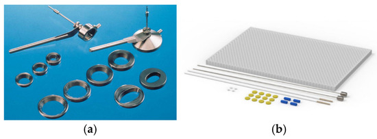
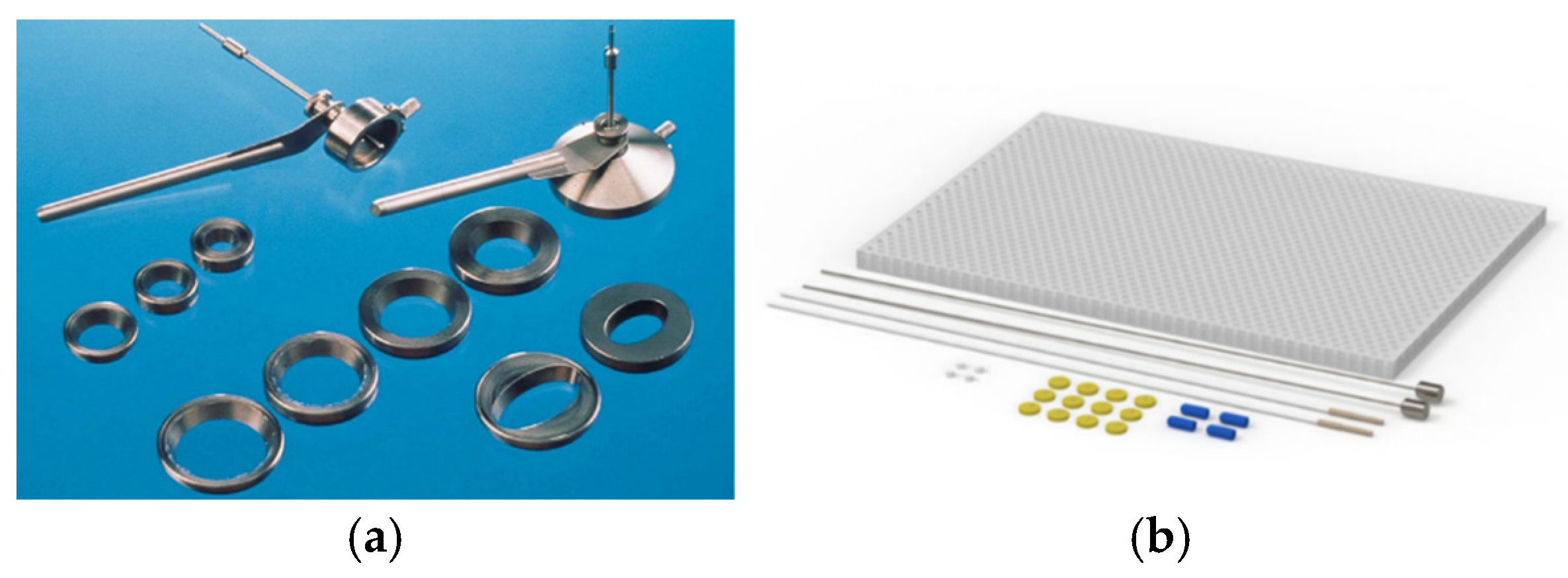
- A total of 10 inserts of different shapes and sizes. The small applicator is compatible with inserts for nominal treatment area diameters of 10, 15, 20 and 25 mm and the large applicator is compatible with inserts of 30, 35, 40 and 45 mm. There are also two types of oval inserts for the large applicator, with dimensions of 45 mm × 25 mm and 30 mm × 20 mm (Figure 1a).
- A flap matrix with a catheter set, for 3D planning (Figure 1b).
Figure 1.
Applicators used in superficial brachytherapy: (a) Varian vertical applicator set; (b) catheter flap array.
Obviously, facilitating the introduction of the new brachytherapy treatment method also required staff training, by organizing courses (on-site and in another country) with theoretical and clinical lectures, practical work and exchange of experience specific to this treatment method.
The commissioning phase followed, and to ensure quality and patient safety, planned checks were carried out to ensure the implementation of the new treatment method.
In this work, the emphasis is placed on the verification and testing of the set of vertical applicators, namely for the large applicator with the 30 mm diameter insert, because clinically it has proven to be the most common size of superficial tumors.
Table 1 indicates the type and model of the device used in the verification procedures.
Table 1.
Dosimetric devices and equipment according to the verification procedures.
3. Results and Discussion
3.1. End-to-End Procedure
- In general, this procedure is performed to verify the entire treatment flow for an oncologic patient, which was carried out according to the following steps: Scanning the phantom (a phantom was reconditioned to be used for this procedure) using an CT-Simulator and sending the obtained images to the planning system.
- Defining the areas of interest, created based on the obtained CT images.
- Creating a treatment plan to verify the dose in the areas of interest.
- Delivery of the treatment plan from the phantom to the treatment console.
- Determining the coincidence between the dose in the treatment plan and the dose delivered by the brachytherapy unit following measurements with the electrometer and the ionization chamber.
3.2. Quality Control
Quality control (QC) in brachytherapy includes specific, routine tests to ensure the accuracy, safety and functionality of the treatment equipment, especially for the HDR (High Dose Rate) afterloader and the radioactive source. This procedure is done with the main goal of checking the reliability of the treatment planning system (TPS) and delivered dose and to ensure patient safety during the treatment.
Mechanical and dose checks are the main categories related to quality control. In the present case, the dosimetric aspect is of particular interest, since a new type of applicator is used. The key aspects of the process performed are mentioned below, following IAEA Technical Reports Series No. 492 protocol [7].
3.2.1. Absolute Dose Measurement
In accordance with the IAEA Technical Reports Series No. 398 protocol, the delivered treatment dose was verified using an ionization chamber and an electrometer within a phantom [8]. The large vertical applicator with the 30 mm insert was located in the center of the phantom, and the ionization chamber was positioned at a depth of 5 mm in a predefined hole. The environmental conditions in which the measurements were performed are: temperature (T) and atmospheric pressure (P). The correction factor for pressure and temperature (kT,P) was determined according to the formula:
where kT,P is the factor used to correct the response of an ionization chamber for the effect of the differences that may exist between the standard reference temperature (T0) and pressure (P0) specified by the standards laboratory and the temperature (T) and pressure (P) of the chamber in the user cavity environmental conditions. To convert the electron charge to Gray (Gy), the corrected dosimeter reading (MQ) was used, calculated as follows:
The absorbed dose in water (DW) is determinate by the formula from:
where the M is the raw reading of the ion chamber, NDw is the calibration factor of the ion chamber and kQ,Q0 is beam quality correction factor.
After the delivery of the sample plan, the planned dose and the measured mean dose were 1 Gy and 1.02 Gy, respectively, resulting in a difference of 2%.
3.2.2. Percent Dose at Depth
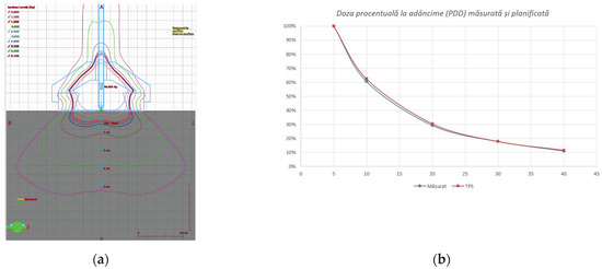
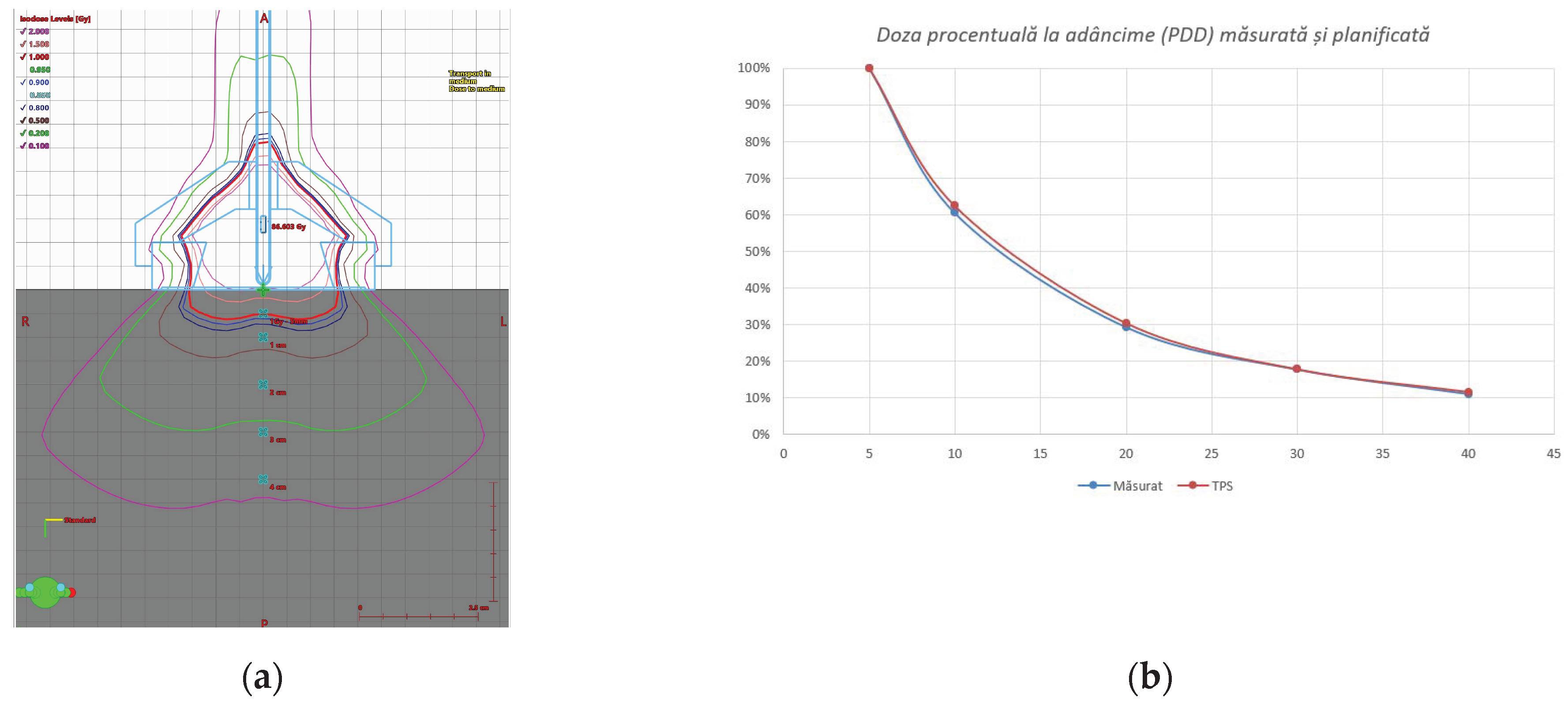
In radiotherapy, the percent dose at depth (PDD) curve represents the absorbed dose deposited by a beam of radiation in a medium, which varies as a function of depth along the beam axis. The dose values measured and calculated at different depths (Figure 2b) are reported relative to the maximum dose (Dmax), resulting in a percentage graph of the maximum dose. For PDD recording, a treatment plan was created, in which the initial source position was located 10 mm from the center of the first nominal timing position. The aforementioned applicator was used at depths of 5, 10, 20, 30 and 40 mm from the phantom surface.
Figure 2.
Dose lines: calculated dose distribution in the phantom (a); calculated percent dose at depth (b).
Figure 2a shows the dose distribution in the phantom, and the PDD calculated by the TPS and the measured one, respectively.
3.2.3. Relative Dosimetry Using Gafchromic Film Measurements
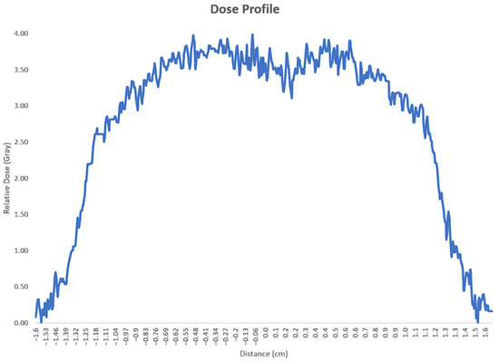
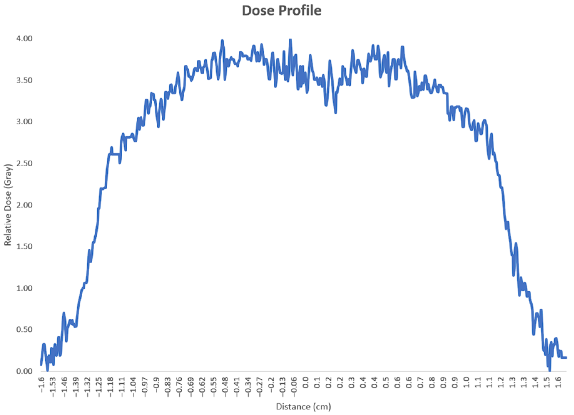
Another important aspect is the dose profile, which represents the radiation dose measured along two axes perpendicular to the central beam axis, usually transverse and in-plane, with a range extending beyond the edge of the radiation field. A Gafchromic film was used for the profile measurement, which was positioned at a depth of 4 mm in the center of the PMMA phantom. The film was exposed to a dose of 4 Gy, then scanned with an EPSON 12000XL scanner (Seiko Epson Corporation, Suwa, Japan), and the data were processed with ImageJ software (Version 1.54t). Figure 3 shows the scanned profile of irradiated film.
Figure 3.
Gafchromic irradiated film profile.
The dosimetric center of the Ir-192 source was strengthened by the fact that the profiles were analyzed at different angles of the applicator to the film.
As a test, a Gafchromic film was irradiated with the large applicator and the 30 mm insert. The difference between the width of the 90% dose level of the measured profile and that based on the Acuros calculation algorithm of the planning system was less than 2 mm. The steep measurement slope makes the measured profiles noisy.
4. Conclusions
The Institute of Oncology of Moldova, through the Laboratory of Radiation Oncology, currently has three treatment modalities: surgery, the linear accelerator equipped with electron energies and the recent brachytherapy unit equipped with superficial applicators.
The measurements performed demonstrate that the vertically oriented Varian surface applicators have precise modeled dosimetric characteristics, with the absolute dose value registering a difference of 2% and PDD curves with a difference of 2%. Therefore, they are admissible for clinical use for skin lesions with a diameter up to 40 mm and a depth of up to 5 mm.
Cutaneous brachytherapy is an effective treatment method with low implementation and maintenance costs, increasing the efficiency of the brachytherapy device by making a more efficient use of the Ir-192 source, while freeing up space on the linear accelerator for much more sophisticated treatments.
Author Contributions
Conceptualization, O.C.; methodology, O.C.; software, O.C.; validation, O.C.; formal analysis, O.C.; investigation, O.C.; resources, O.C. and A.B.; data curation, O.C.; writing—original draft preparation, O.C.; writing—review and editing, O.C. and A.B.; visualization, O.C. All authors have read and agreed to the published version of the manuscript.
Funding
This research received no external funding.
Institutional Review Board Statement
Not applicable.
Informed Consent Statement
Not applicable.
Data Availability Statement
The data presented in this study are available upon request from the corresponding author.
Conflicts of Interest
The authors declare no conflicts of interest.
References
- Ferlay, J.; Ervik, M.; Lam, F.; Laversanne, M.; Colombet, M.; Mery, L.; Piñeros, M.; Znaor, A.; Soerjomataram, I.; Bray, F. Global Cancer Observatory: Cancer Today. Lyon, France: International Agency for Research on Cancer. 2024. Available online: https://gco.iarc.who.int/today (accessed on 8 March 2026).
- Sendín-Martín, M.; Bueno-Molina, R.C.; Hernández-Rodríguez, J.-C.; Cayuela, L.; Cayuela, A.; Pereyra-Rodríguez, J.-J. Incidence and mortality of nonmelanoma skin cancer in Europe: Current trends and challenges. Clin. Transl. Oncol. 2026, 28, 302–319. [Google Scholar] [CrossRef] [Scilit] [PubMed] [PubMed Central]
- Bellis, R.; Rembielak, A.; Barnes, E.A.; Paudel, M.; Ravi, A. Additive manufacturing (3D printing) in superficial brachytherapy. J. Contemp. Brachyther. 2021, 13, 468–482. [Google Scholar] [CrossRef] [Scilit] [PubMed]
- Yu, L.; Oh, C.; Shea, C.R. The Treatment of Non-Melanoma Skin Cancer with Image-Guided Superficial Radiation Therapy: An Analysis of 2917 Invasive and In Situ Keratinocytic Carcinoma Lesions. Oncol. Ther. 2021, 9, 153–166. [Google Scholar] [CrossRef] [Scilit] [PubMed] [PubMed Central]
- Katalinic, A.; Hammas, K.; Taraszkiewicz, L.; Louwman, M.; Bartnicka, J.J.; Randi, G.; Bettio, M.; Stang, A.; Crocetti, E. A Narrative Review of European Registries for Skin Cancer: Where Are We and Where Should We Be? Cancer 2026, 18, 524. [Google Scholar] [CrossRef] [Scilit] [PubMed]
- Available online: https://particip.gov.md/ru/document/stages/anunt-de-initiere-a-elaborarii-programului-national-de-control-al-cancerului-pentru-anii-20262030/15643 (accessed on 8 March 2026).
- International Atomic Energy Agency. Dosimetry in Brachytherapy: An International Code of Practice for Secondary Standards Dosimetry Laboratories and Hospitals (Technical Reports Series No. 492). 2023. Available online: https://www-pub.iaea.org/MTCD/Publications/PDF/DOC-010-492_web.pdf (accessed on 30 March 2026).
- International Atomic Energy Agency. Absorbed Dose Determination in External Beam Radiotherapy An International Code of Practice for Dosimetry Based on Standards of Absorbed Dose to Water (Technical Reports Series No. 398, Rev. 1). 2024. Available online: https://www-pub.iaea.org/MTCD/Publications/PDF/p15048-DOC-010-398-Rev1_web.pdf (accessed on 30 March 2026).

Disclaimer/Publisher’s Note: The statements, opinions and data contained in all publications are solely those of the individual author(s) and contributor(s) and not of MDPI and/or the editor(s). MDPI and/or the editor(s) disclaim responsibility for any injury to people or property resulting from any ideas, methods, instructions or products referred to in the content. |
© 2026 by the authors. Licensee MDPI, Basel, Switzerland. This article is an open access article distributed under the terms and conditions of the Creative Commons Attribution (CC BY) license.