


Abstract
Bipolar forceps, powered by high-frequency alternating electrical current, are the main tools for electrocoagulation in surgical operations. Coagulation occurs through thermal heating; however, excessive temperatures can induce tissue damage. The study aims to measure the temperature increment between bipolar forceps’ tips on ex vivo biological tissues from the liver and white and grey matter of the calf brain. Three forceps, disposable and reusable, are tested using electrical power of 5, 10 and 15 W applied for 5 s, penetrating 1 mm into the tissue; the same procedure is followed for all the 135 tests performed. Infrared thermal measurements are collected during the transient, statistically analyzed in terms of the median and scatter of the peak temperatures, and compared among the forceps types. The outcomes allow evidence that disposable forceps are more performing than reusable ones for all tissues, e.g., limit excessive thermal heating and experience lower scatter, potentially reducing the risk of unintended injury to adjacent or peripheral tissues when applied in surgical operations.
1. Introduction and Rationale
While the use of thermal cautery dates as far back as 3000 B.C., when tools heated in the fire were used to reduce hemorrhage in accidental injuries, electrosurgery has been developed and applied in clinical practice since the 1920s to offer coagulation while aiming to avoid causing damage to the surrounding tissue. Amongst the most important contributions to electrosurgery, William Bovie, a physicist from Harvard, together with Harvey Williams Cushing, an American neurosurgeon and pioneer of neurosurgery, developed a monopolar tool that offered cutting and coagulation [1]. In 1940, Greenwood presented the use of a bipolar device in neurosurgery [2] which was commercialized later on in the 1960s by Malis [3,4].
The use of bipolar forceps in surgery has been developed through the years, and has been applied in different surgical fields [4,5,6,7]. Nowadays, forceps are tools commonly used during surgical procedures, to grasp and manipulate tissue, and coagulate blood vessels, ensuring hemostasis, while minimizing heat transfer and blood clots from sticking and charring. High radiofrequency surgery, or electrosurgery, refers to the passage of high-frequency electrical current from the forceps through the tissue, to achieve a specific surgical effect, such as cutting, coagulation or even vascular thermofusion [8]. In electrosurgery, the forceps are linked to a generator of alternating electrical current, which can be activated at different power levels and for different time intervals.
Electrosurgery is based on fast and selective tissue heating obtained through the passage of high-frequency alternating current (100 kHz–5 MHz) [9]. This frequency range allows for desired thermal effects without consequences on muscles or nerves [10]. Because this frequency falls in the range of AM radio waves, the energy used in electrosurgery is sometimes referred to as radiofrequency (RF) [11].
The current flows from the generator in a circuit created by the forceps’ tips manipulated by the surgeon. The current delivery causes movement of water molecules. The rapid movement of water molecules results in heat creation between the forceps’ tips and the tissue around them, that finally results in desiccation and coagulation of blood vessels [12].
Biological tissues react differently to temperature [13]: from 37 to 40 °C, no damage is caused to the tissue. Moving over 40 °C, initial damage to the tissue starts to be noticed, causing edema formation. The damage can be either recovered or proceeded to tissue devitalization, depending on the duration of the thermal source application. When the temperature reaches 60 °C, tissue cells start getting devitalized and denatured, and connective tissue shrinks. When the temperature reaches 100 °C, tissue fluid gets vaporized, and the result is either desiccation leading to tissue shrinkage or cutting, as the tissue undergoes mechanical tearing. If the temperature exceeds 150 °C, the tissue undergoes carbonization.
While the use of bipolar forceps is essential to coagulate or cut the tissue during surgery, it is important not to damage the surrounding tissue. Tissue damage can occur when using electrocoagulation devices. During neurosurgical procedures, cranial nerves or vital neural structures must be preserved, by avoiding excessive temperatures of thermal spread around the forceps’ tips. On the other side, overheating of the forceps’ tips can lead to unwanted effects, such as forceps’ tips adherence to tissue, electric sparks between the forceps’ tips, and charring, causing difficulties in removing carbonized tissue from the forceps’ tips. All these phenomena can cause the forceps to adhere to and even tear blood vessels and tissue with which they come in contact during the procedure [14].
Despite the advancement in the design of devices, attention still needs to be paid to any damage that can be caused by excessive heating and thermal injury to vital organ tissues, thermal spreading into adjacent tissue, inadvertent tip sticking to tissue and charring [15].
With the goal of minimizing tissue damage, several design innovations have been introduced, ranging from materials, coating, and irrigation systems. The efficacy of these designs has been analyzed in different studies [12,14,16,17,18,19].
The goal of this study is to assess the thermal profile induced by bipolar forceps during electrosurgical coagulation, to assess the thermal spread and the possible tissue damage. The study is carried out on ex vivo tissue samples of calf brain and liver, comparing the temperature evolution when different types of forceps are used. With respect to the existing literature, this study introduces an additional evaluation on different ex vivo tissue samples, to evaluate how the same bipolar forceps could impact different tissues, such as liver and brain, and compares reusable forceps with disposable ones. At the best of the authors’ knowledge, this is the first study comparing different tissues, to evaluate the thermal evolution in different settings.
2. Materials and Methods
2.1. Materials
2.1.1. The Infrared Camera
An infrared (IR) camera has been considered to be the appropriate device to measure the temperature increment produced by the use of forceps on ex vivo animal tissue, because it can measure the temperature remotely and it can measure the temperature peak at any point between the forceps’ tips and also in their surroundings without the need for physical contact, e.g., infrared thermography is a full-field non-contacting technique.
The IR camera used for the experiment is the FLIR-Cedip Titanium by FLIR Systems, (Täby, Sweden), endowed with an InSb focal plane array 320 × 256 working in the waveband 3–5 μm and having a Germanium lens with a focal length of 50 mm.
A thin sheet of polyethylene was placed in front of the lens to protect the lens from smoke or other debris that could be produced during the tests at high temperatures. The polyethylene is transparent to the infrared radiation, but it can absorb and reflect IR radiation, therefore lowering the intensity of the radiation reaching the sensor. This created the need for a calibration of the IR camera with the polyethylene sheet covering the lens, which was performed using a commercial black body (from HGH Ingenierie Systemes Infrarouges, Igny, France, model: RCN600). In order to obtain the calibration curve relating the temperature read by the camera with the effective temperature of the black body, the black body temperature was initially set to 30 °C, and it was increased up to 190 °C with steps of 10 °C. Afterwards, the temperature was brought back to 30 °C, always using steps of 10 °C.
The surface emissivity of the brain and liver under testing was not measured. However, the aim of the work was a comparison between the temperatures reached by different forceps, in the same conditions, rather than the measurement of the real temperature reached. Hence, the temperature values could be affected by a systematic underestimation.
2.1.2. The Forceps
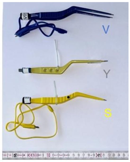
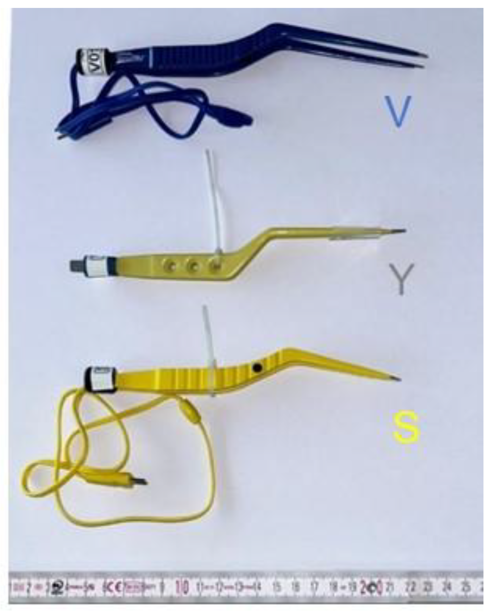
Three forceps were selected and tested (Figure 1), with 1 mm tip size. The forceps identified represent different options available on the market, and are indicated as
Figure 1.
The three different forceps used for the tests.
- V: VersaTru® from Integra LifeSciences—Princeton, NJ, USA (disposable)
- Y: Yasargil® from Aesculap B. Braun—Melsungen, Germany (multi-use)
- S: Spetzler-Malis® from Stryker—Kalamazoo, MI, USA (formerly Codman) (disposable)
Indeed, forceps can be of two kinds: reusable or multi-use, when manufactured to get clean, sterilized and used for multiple applications, or disposable, when manufactured for a single patient in a single surgery operation.
2.1.3. The Biological Tissues
As forceps are commonly used in neurosurgery and hepatobiliary surgery, brain and liver samples were selected from ex vivo calf samples.
Three fresh ex vivo tissues were analyzed:
- Calf liver
- Calf brain grey matter
- Calf brain white matter
These biological tissues were collected from a local butcher. The liver was cut into slices of about 10 mm thickness the same day of the tests; then, they were kept refrigerated at 4 °C before the beginning of the tests. The full brains were kept refrigerated at 4 °C and cut into slices of about 10–20 mm thickness a few minutes before the tests.
Using ex vivo materials guaranteed controllable test settings, including samples, forceps position, energy and duration. However, in all biological tissues, some degree of variability is to be expected. As all the animals may be different in terms of age, gender, fat content, and health state, a certain variation in terms of physiological parameters and physical properties may happen. As such, the testing phase was conducted on multiple liver and brain samples, with repeated measurements and different settings [20].
2.2. Methodss
2.2.1. The Experimental Setup
A forceps holder was designed using the software SolidWorks version 2024 SP5 by Dassault Systèmes and manufactured by the mechanical workshop of the Politecnico di Milano. It is composed of three support rods: two were positioned beneath the forceps to maintain a constant inclination, and one was placed above the forceps to ensure it remained stable throughout the tests. The metallic holder was connected to a micrometric linear positioning table to drive the forceps up and down with 0.01 mm resolution.
The idea was to arrange the forceps on the holder, fixing the angle between the forceps prongs and the horizontal direction at 45°. This angle simulates the actual orientation when the forceps are in use by the surgeon. In addition, it was decided to move down the forceps’ tips of 1 mm into the biological tissue after the first contact with its surface. This penetration depth (e.g., lesion depth) ensures that both tips of the forceps are properly embedded into the tissue, allowing for accurate and reliable measurements. Indeed, if only one tip is in contact with the tissue, only partial heating would occur, and the test would be invalid.
The distance between the forceps’ tips was set to 3 mm, as in the study [12], representing a standard distance used in surgical procedures to make the measurements easily repeatable and comparable with each other. To ensure accuracy, the distance between the tips of the forceps was measured using a calliper and the closure of the forceps was secured with a clamp.
The forceps were electrically powered with three power levels: (I) 5 W; (II) 10 W; (III) 15 W. The time interval during which the forceps were powered was 5 s; it was selected because it is typically higher than the duration of a surgical operation. The use of three different power settings, e.g., 5, 10 and 15 W, was necessary to provide a range for comparison and to reflect the actual operating conditions of the forceps during surgical procedures.
Though the forceps design varies according to the manufacturer, the generator used in the testing phase as well as other parameters like the penetration or the distance between the forceps’ tips, remained the same, thus allowing the measured differences to be attributed only to design, materials and features of the forceps.
Every measurement was repeated five times keeping fixed the same testing parameters. After each measurement the tips were cleaned with a physiological solution, to remove any debris or chars that can affect locally the current transmission in the following tests. A total of 135 measurements were performed over three test sessions.
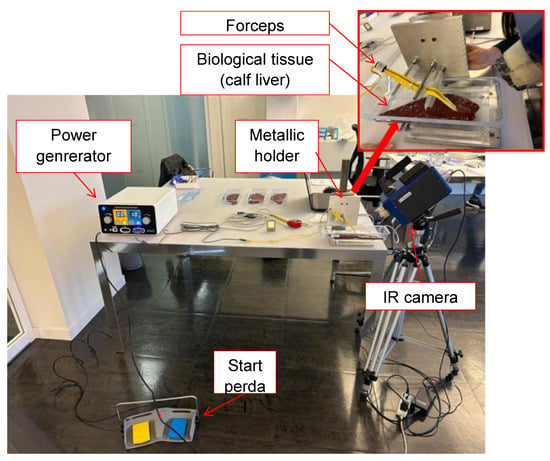
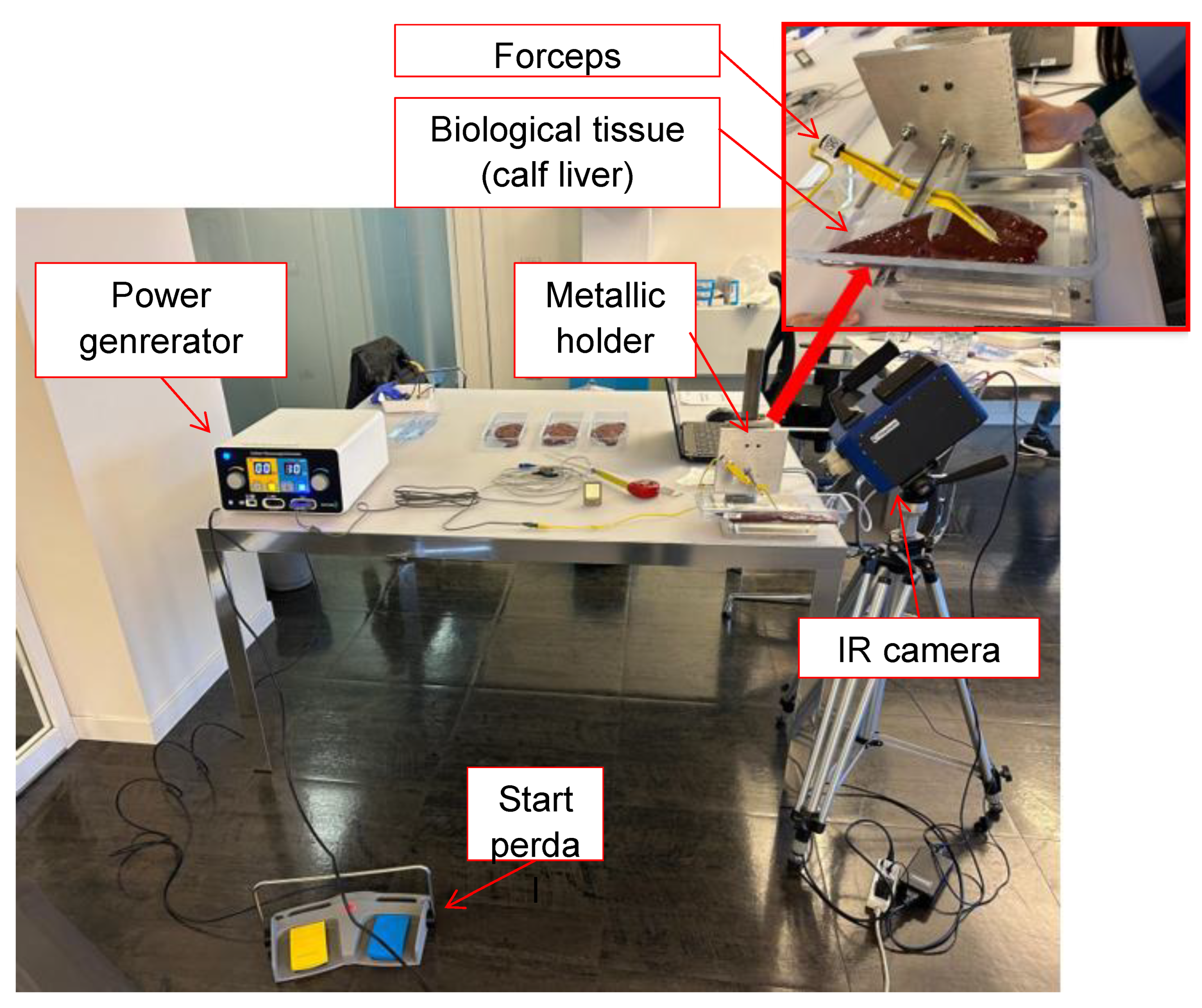
The experimental setup was composed of (see Figure 2)
Figure 2.
The setup of the instrumentation with a focus on the forceps and the holder.
- The power generator (Codman Electrosurgical Generator), with frequency 4 MHz
- The start pedal of generator
- The metallic holder for the forceps
- The IR camera
At the beginning of the tests, samples were exposed to the environment for a sufficient time to reach room temperature, allowing the initial surface temperature to be the same.
During the experiments, a crucial aspect was the continuous monitoring of the tissue degradation caused by exposure to air at room temperature. Indeed, the biological tissue dries out when exposed to air, leading to a change in its thermal conductivity. For this reason, additional measurements were carried out to monitor the degradation of both the liver and brain tissues. These extra tests were carried out with the V forceps, with the same setup as all the tests, and applying a power of 10 W. Both the liver and the brain were exposed to air at a room temperature of approximately 25 °C for the entire duration of the tests. The thermal measurements were collected at half-hour intervals.
The trends of the maximum temperature between the tips (after 5 s exposition at 10 W) revealed a decay and an increasing scatter as the exposure time increased. Indeed, the beginning of desiccation started affecting the thermal conductivity, and, consequently, the electrical flow. For the liver, a decay in the results was detected after two and a half hours. For the white matter and grey matter of the brain, the decay began faster than for the liver, starting after an hour and a half from the beginning of the manipulation phase.
These initial tests were essential in determining the time frame within which testing should be conducted, before replacing the tissue with a freshly cut one. Based on these results, the tests were carried out on all the selected biological tissues within one hour and a half since their exposure to air. After that time, the slice of biological material was replaced with a new one.
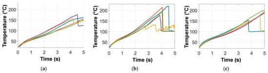
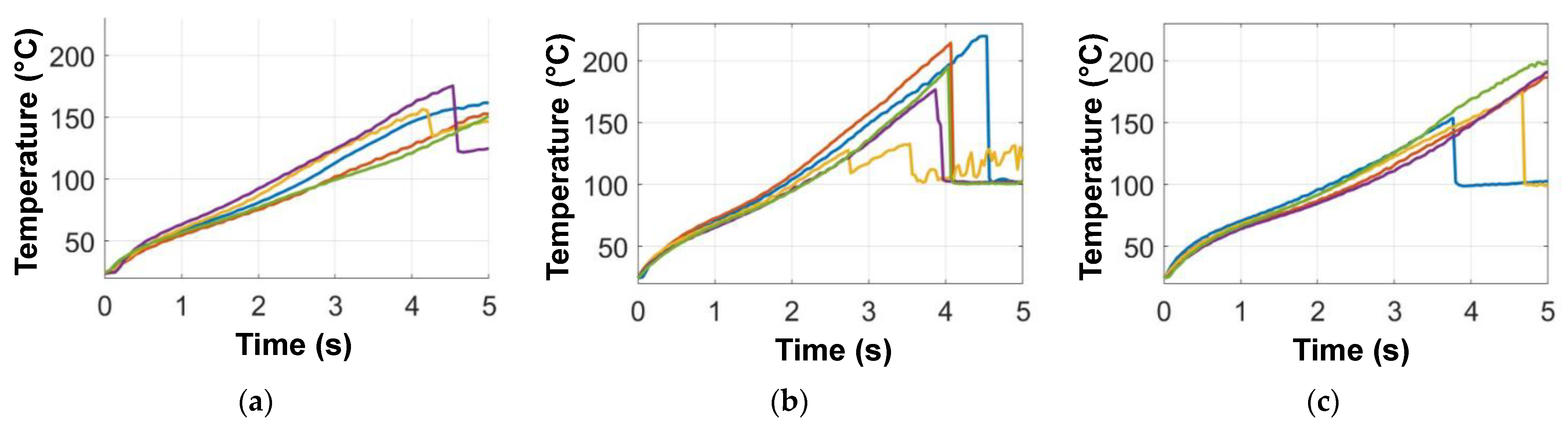
During the tests conducted at 15 W on the liver, some small explosions took place approximately 3 s after the current activation began, as shown in Figure 3. The figures show the trends of the maximum temperature in the heated region as a function of the time, over the 5 s of the test. The explosions occurred for all the three forceps. This happened only for two samples over 5 s for the V- and S forceps (Figure 3a,c), while the explosions occurred in all the samples for the reusable Y forceps (Figure 3b). Immediately after the explosion, the temperature suddenly dropped, invalidating the results. Such explosions were attributed to the sudden vaporization of water molecules within the cells caused by the high temperatures reached. This phenomenon was tackled considering only the data obtained before the explosions, thus in general limiting to the first 3 s the analysis of the data collected on the liver at 15 W.
Figure 3.
Graphs showing the temperature variation with time and the sudden temperature drops due to the small explosions during the tests on liver at 15 W, for the (a) V; (b) Y; (c) S forceps. Five tests per forceps are shown, over the 5 s test time.
2.2.2. Statistical Analysis
The first step, after recording the raw thermal data, consisted in correcting them by using the calibration curve to account for the polyethylene film in front of the lens.
The second step consisted in a statistical analysis of the results using the software Matlab version R2024a by MathWorks. The temperature data were used to create a time diagram for each measurement, grouping together the five measurements obtained for the same material, the same forceps and the same power. Then, these thermal data were handled by a Matlab script, comparing the three forceps in three charts:
- the BoxPlot. It represents the statistical distribution of the maximum temperature reached during the 5 tests (same biological material, same forceps, and same power), e.g., at the end of the test interval, that is in general 5 s or 3 s in case of the liver when the little explosions occurred. The BoxChart command allowed to show the following information: (1) the outline of the box, which extends from the 1st quartile to the 3rd quartile, representing Student’s t-distribution at 25% for the upper quartile and 75% for the lower quartile; (2) the line that separates the upper half from the lower half, that is the median, e.g., the 2nd quartile; (3) the whiskers, which extend from the upper and lower limits of the box to the maximum and minimum data values within a given limit (1.5 times the interquartile range). All values beyond these limits are considered outliers and are represented as circles in the graphs;
- a set of temperature–time trends for the five tests. It represents the trend of the five tests (for all collected trials), plotting the time on the x-axis and the maximum temperature between the forceps on the y-axis (some examples were shown in Figure 3);
- the average temperature trend among the 5 tests. The band of dispersion indicates the trend of the mean temperature with a 95% confidence interval, calculated using Student’s t-distribution.
The same procedure was followed for all tests except for those performed on the liver at 15 W power, due to explosions that caused an impulsive loss of energy, leading to a sudden temperature drop.
3. Results
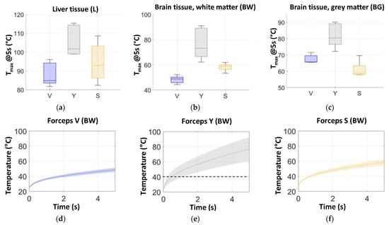
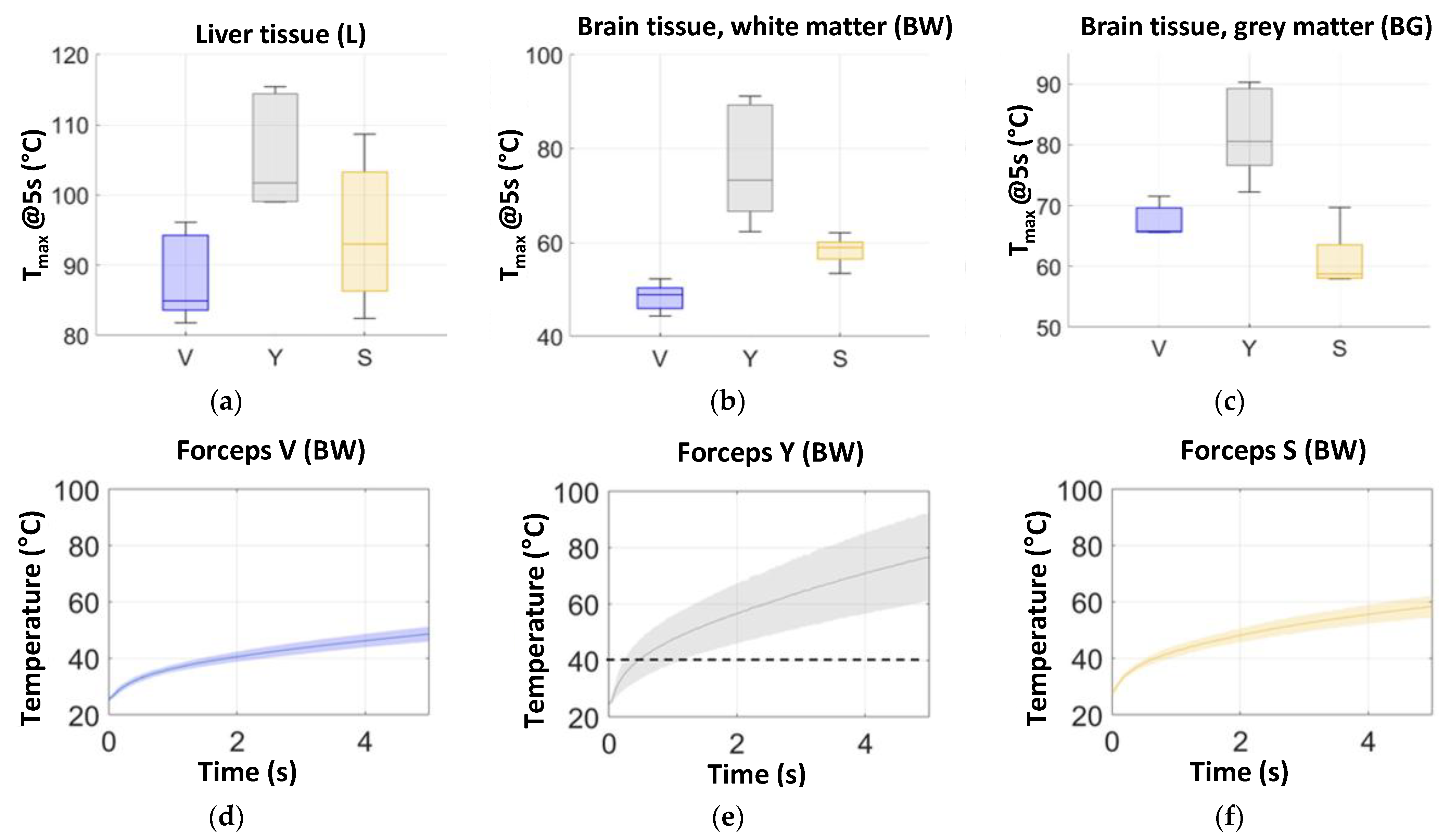
Figure 4 summarizes some of the most meaningful results collected during the tests at 10 W. Figure 4a–c show the BoxPlots of the three biological tissues, with the details of the tested forceps. For the sake of precision, Table 1 shows the numerical values of the median temperature, of the first and third quartiles at the end of the test on the liver and white and grey matter of the brain. These data correspond to the graphical representation of the BoxPlots of Figure 4a–c.
Figure 4.
Results of the experimental tests, powered at 10 W. Figures (a–c) in the upper part represent the BoxPlots of the maximum (final) temperature reached for: (a) the liver; (b) the white matter of the brain; (c) the grey matter of the brain. Figures (d–f) show the temperature vs. time dependence in terms of average curve and statistical scatter band on the white matter of the brain, measured for the forceps of type:(d) V; (e) Y and (f) S.
Table 1.
Summary of the median value reached between the forceps powered at 5 W, 10 W, 15 W. For each median value, the first quartile (Q1) and the third quartile (Q3) are reported. Temperatures are collected after 5 s, apart from the tests at 15 W on the liver, collected after 3 s.
Figure 4d–f display the average and the related statistical scatter band for the temperature–time curves on the white matter of the brain, measured for the forceps V, Y and S, respectively.
In some cases, the temperature variation from the beginning to the end of the test was considerably high, exceeding the range of the calibrated temperature windows available with the camera software. For this reason, the calibrated temperature window was chosen to cover the results at the end of the 5 s interval, rather than the beginning of the test. This means that, for instance, in the experiment of Figure 4e the temperature data below the dotted line at 40 °C may be less reliable than those above it. However, since the focus was on measuring the maximum temperature reached during the experiment, the limited reliability of the temperatures measured in the first 1–2 s of the tests was not considered to be particularly harmful.
In the case of the liver and the grey matter of the brain (Figure 4d,f), the range is more limited, and the selected calibrated temperature range gave reliable thermal results for the whole measurement.
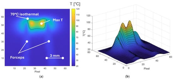
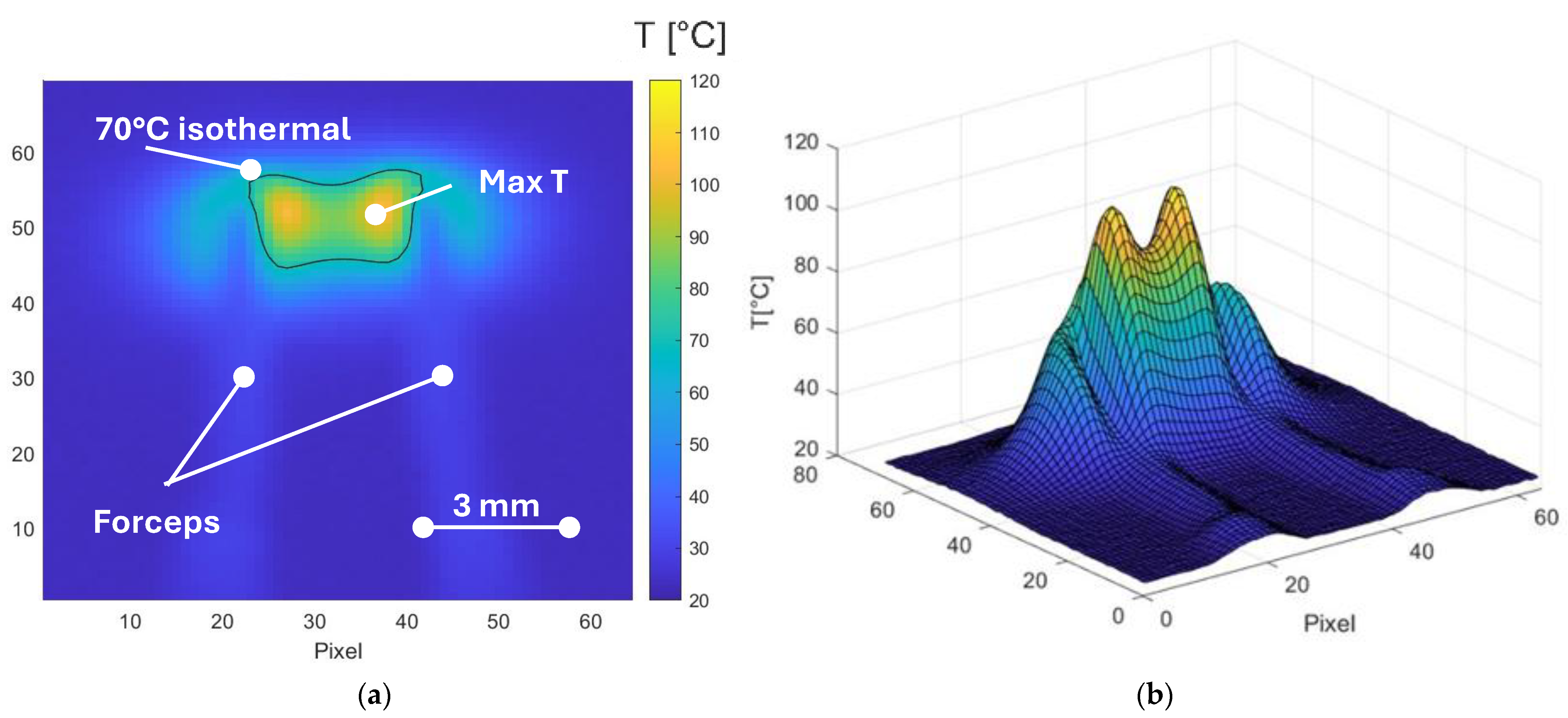
In addition to these outputs, a series of thermal maps were selected and plotted via Matlab in a 2D and 3D visualization. Useful plots consisted in (1) contour plot with isothermal lines, and (2) three-dimensional graphs of the temperature trends, both in correspondence of 5 s (or 3 s for the liver at 15 W). Figure 5 shows an example of these 2D and 3D images, related to the Y forceps used on liver at 10 W. The selected test was the average among the five tests. From these images it is possible to see how large the area is above a predetermined temperature threshold (e.g., 70 °C isothermal in Figure 5a) and where the maximum temperature is located.
Figure 5.
Example of thermal maps in 2D (a) and 3D (b) visualization. The test was performed at 10 W on the liver with Y forceps. The resolution is 0.19 mm/pixel.
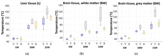
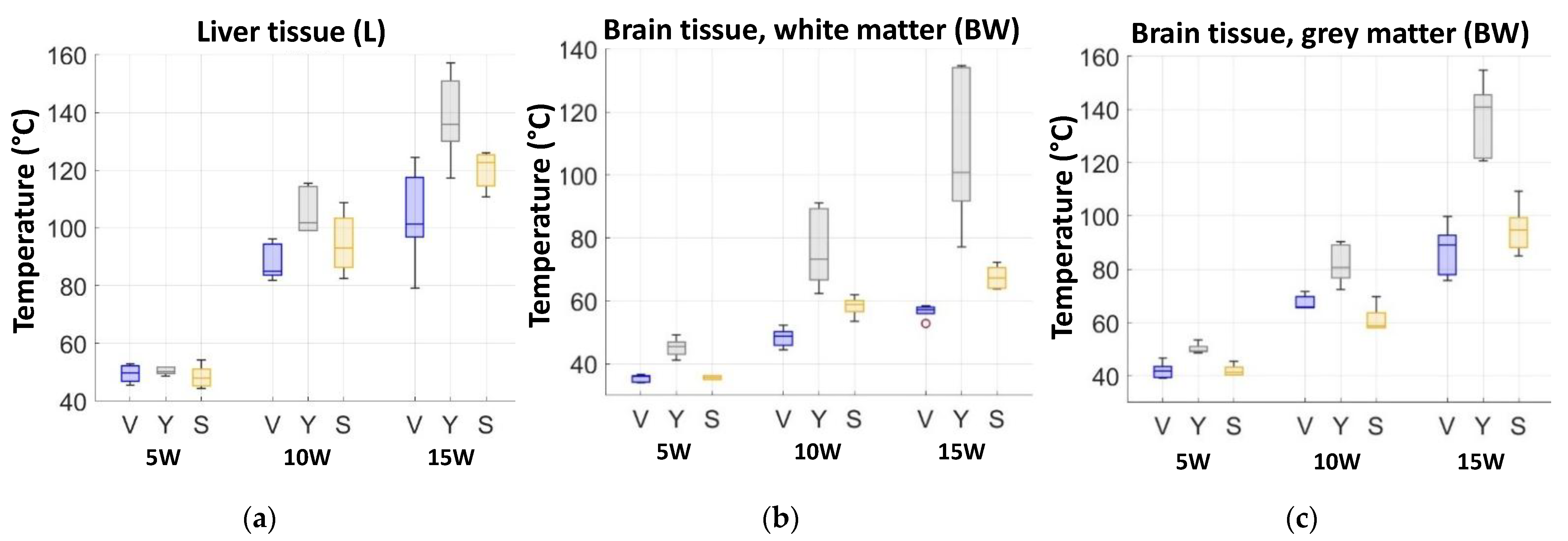
Figure 6 summarizes visually all the obtained results, grouped based on the analyzed biological tissues. The BoxPlot shows the median, the first and third quartiles of the maximum temperatures measured at the different power and for the three forceps.
Figure 6.
Histograms of the thermographic results for each biological tissue: (a) liver; (b) white matter of the brain; (c) grey matter of the brain. Temperatures are collected after 5 s, apart from the tests at 15 W on the liver that are collected after 3 s.
4. Discussion
4.1. Liver Tissue
Currently, liver resection can be considered the treatment of choice for liver tumours. Given the liver structure, which is very well vascularized, intraoperative hemorrhages still play an important role.
Bleeding is still considered a major concern for the hepatic surgeon, as it represents one of the major intraoperative surgical complications. Not only major bleedings need to be considered, but also, hemostasis of vascular structures under 1 mm of diameter is another important concern of the surgeon. In fact, the continuous bleeding of little vessels in parenchyma may represent a considerable part of intraoperative blood loss, and moreover it makes it hard for the surgeon to visualize the surgical field. Monopolar and bipolar forceps can be used to achieve coagulation, paying attention to thermal injury and tissue damage [5].
The results on hepatic tissue show that the three tested forceps induce similar temperatures only at the lowest power, e.g., 5 W (Figure 6a). Increasing the generator power to 10 and 15 W, the reusable forceps (Y-type) induces higher temperatures than the disposable forceps (V- and S-types). At 15 W, in particular, the temperatures are collected after 3 s due to the following local explosions, as shown in Figure 3. A certain similarity can be observed in the statistical variation between forceps V and Y. Both of these forceps exhibit significant variations in behaviour when increasing power levels. However, this variability may be attributed to the fact that these measurements were collected after 3 s due to the local explosions, that occurred for all the tests in the case of forceps Y. Despite this phenomenon, the temperatures reached by forceps Y remain higher than those recorded for forceps V and S. Indeed, the median temperature of the 15 W tests with the Y forceps is 135.87 °C, which is considered dangerous and excessive. Compared with the V- and S forceps, the Y forceps experiences temperatures 34.21% and 10.77% higher, respectively.
In addition, the comparison among forceps underlines that disposable forceps V and S allow better temperature control, not only generating lower temperatures but also experiencing more consistent behaviour (Figure 6), especially for power levels of 5 and 10 W. This consistency, represented by the limited scatter of the results given in detail in Table 1, is a key factor in clinical settings because it allows for repeatability. In other words, disposable forceps support the surgeon’s expectation of a given result when applying a defined energy level.
Together with the analysis of the maximum temperature, the thermal analysis also shows how the highest temperature is concentrated in the internal part of the forceps, between the two tips while a mild halo effect can be identified around the tips (Figure 5). This thermal pattern is consistent with surgical expectations, e.g., to contain thermal increase between the forceps’ tips and limit the thermal spread outside the concerned area. The targeted area of the liver tissue, e.g., that one between the tips, undergoes the strongest effect and could be subjected to unintended thermal injury during bipolar coagulation. It is interesting to notice as well how a correct use of the forceps can affect the results: if the two tips are inserted in the tissue with different depths, the thermal profile will differ.
The data obtained in this study echo those reported by Çavuşoĝlu et al. [17] on bovine liver. Among other forceps, that study compared Versatru and Spetzler Malis forceps, showing thermal damage lower for Versatru than Spetzler Malis. That study also proved that the single-use bipolar forceps induce lower heating and may reduce the risk of unintended injury to adjacent or peripheral tissues.
4.2. Brain Tissue
Coagulation with the use of bipolar forceps is common in neurosurgery. Given that neural tissues are susceptible to damage due to heat, it is extremely important to control the thermal spikes and the thermal spread into adjacent tissues.
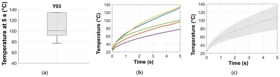
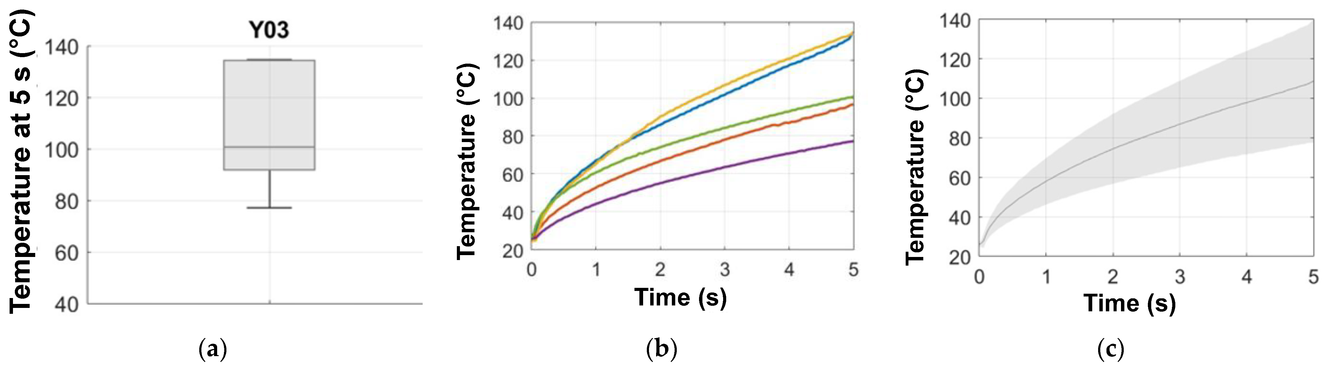
Among the three analyzed tissues, the white matter experiences the lowest heating (see Figure 6b). As for the liver, the white matter is subjected to the highest temperature increase when reusable forceps Y are used, as seen in results from Figure 4b,d–f and Figure 6b. In addition, these plots underline an important thermal variation range for the Y forceps, e.g., plot of Figure 4e compared to those of Figure 4d,f. The variability in the temperature range when applying the same power raises a question concerning the clinical impact and reliability. As an example, Table 1 and Figure 7 show that the white matter powered at 15 W experiences after 3 s a temperature increase of roughly 60 °C, while after 5 s the results obtained an increase to 80 °C as an average value, but with peaks up to 110 °C. This variability of the results suggests that a big error can be made in the prediction of the real temperature reached during the operation, with consequent possible tissue damage. It means that the surgeon’s expectation to deliver the same quantity of energy to the white matter is likely to fail when reusable forceps are used.
Figure 7.
Wide scatter of temperatures obtained on white matter using the Y forceps at 15 W: (a) BoxPlot of the maximum temperature (final, at 5 s); (b) thermal transients of the five tests; (c) average curve and statistical scatter band.
The grey matter experiences thermal heating comparable with the liver, reaching slightly lower maximum temperatures but higher than the white matter, e.g., Figure 6c. Comments similar to the previous tissues can be drawn about the performance of the disposable vs. the reusable forceps, which induces the highest temperature among the three tested forceps, reaching maximum temperatures at 15 W similar to the liver (e.g., the median values are 140.91 °C in the grey matter vs. 135.87 °C in the liver, despite this value being obtained after 3 s). On the other hand, the V- and S disposable forceps induced lower temperatures at 15 W, e.g., 89.10 and 94.74 °C, respectively, which are 58.15% and 48.73% lower than Y forceps.
4.3. Limitations
Despite the effort to complete the state-of-the-art literature by adding different specimens and associating a clinical perspective to the results, this study presents some limitations. First, the ex vivo specimens were maintained in a temperature-controlled condition until the testing phase. However, it was challenging to ensure that their temperature evolved uniformly without affecting the tissue features. In addition, once removed from a temperature-controlled fridge, the samples began to lose moisture, leading to a gradual degradation of their thermal properties, as noted in Section 2.2.1. That is the motivation to perform multiple tests with frequent sample replacement.
The temperatures recorded during these experiments do not accurately reflect in vivo conditions. Indeed, the initial temperatures are different between the in vivo and ex vivo tests. In addition, the ex vivo tests do not account for thermal variation due to perfusion that occurs in vivo. The blood flow happening in vivo may contribute in reducing the temperature, due to the heated blood circulation.
The irrigation phase, often part of surgical procedures, was not applied due to the difficulty of controlling quantity and timing. On the other hand, tests on ex vivo tissues allow angles of application, contact with the tissue and timings to be more controlled, resulting in higher repeatability than in a surgical setting, where the forceps application is operator-dependent.
It would be important to expand the results of the study by performing a histopathological analysis of the tissue, to evaluate the width and the extent of the possible damage to the tissue and refine the considerations for a clinical setting. Moreover, it would be interesting to calibrate the test condition by adapting the energy used with a stronger correlation to the specific surgery (neurosurgery, hepatobiliary surgery, urological surgery).
5. Conclusions
The study proposed and applied a testing procedure to compare the thermal performance of bipolar forceps on ex vivo calf liver and brain. Reusable and disposable forceps were compared in terms of surface temperature transients, applying 5 W, 10 W and 15 W with a generator and collecting thermal data for the test duration of 5 s. Temperature measurements were performed with an infrared camera in the tips region and were analyzed statistically, monitoring the maximum reached temperature. The thermal profiles suggest the following conclusions:
- All the analyzed tissues showed thermal trends increasing proportionally to the applied power. The thermal heating cannot be translated directly from one tissue to another due to their specific chemical and physical properties. Nevertheless, the thermal trends experience the common feature that the disposable forceps VersaTru and Spetzler-Malis induce temperatures lower than the reusable Yasargil, especially at 10 and 15 W power. In addition, these forceps also resulted in low repeatability and the highest scatter in the measurements, with temperature differences of up to 60 °C for similar tests; this could be problematic for the surgeon, because the huge scatter can induce overheating and tissue damage during the operation.
- The temperature field measurements showed that for all tissues the temperature peak happens between the forceps’ tips, next to the tips’ surface and spreads moderately outside.
- The hepatic tissue experienced the highest tissue heating. At 10 W, for instance, the median value of temperature for the Y forceps is 101.77 °C, instead the V- and S forceps are respectively 19.77% and 9.38% lower than the Y. The grey matter of the brain experiences intermediate heating, e.g., for the 10 W tests the higher value is reached by the Y forceps, the median value is 80.60 °C, that is 22.44% and 37.07% respectively higher than V- and S values. The heating in white matter is more limited even at 15 W, e.g., for the Y forceps the temperature reached is 100.87 °C and for V and S are respectively 57.28 °C and 67.40 °C, with maximum temperatures always below 100 °C for all the forceps.
- Among the three tested forceps, the disposable forceps Versatru demonstrated the lowest temperature scatter and lowest temperature peak in the brain tissue.
To summarize, the disposable forceps offer more stable and predictable performance, a high degree of repeatability and moderate thermal increase, when compared with the reusable one. Therefore, disposable forceps can be a safe and effective tool in liver and brain surgery. Randomized clinical studies would be advisable to clinically confirm the results of this study.
Author Contributions
Conceptualization, C.C., A.S., S.M., G.U.F., and C.V.; methodology, C.C., A.S., S.M., G.U.F. and C.V.; software, C.C., L.C. and A.S.; validation, C.C. and A.S.; formal analysis, C.C., L.C. and A.S.; investigation, C.C., A.S., S.M. and G.U.F.; resources, A.S., S.M., G.U.F. and C.V.; data curation, C.C. and A.S.; writing—original draft preparation, C.C., L.C., A.S. and C.V.; writing—review and editing, C.C., A.S., S.M., G.U.F. and C.V.; visualization, C.C., L.C., A.S., S.M., G.U.F. and C.V.; supervision, C.C., A.S. and C.V.; project administration, C.V.; funding acquisition, C.C., A.S. and C.V. All authors have read and agreed to the published version of the manuscript.
Funding
This research received no external funding.
Institutional Review Board Statement
Not applicable.
Informed Consent Statement
Not applicable.
Data Availability Statement
The raw data supporting the conclusions of this paper will be made available by the authors upon request.
Conflicts of Interest
Authors S.M., G.U.F. and C.V. were employed by the company Integra LifeSciences. The remaining authors declare that the research was conducted in the absence of any commercial or financial relationships that could be construed as a potential conflict of interest.
References
- Voorhees, J.R.; Cohen-Gadol, A.A.; Laws, E.R.; Spencer, D.D. Battling blood loss in neurosurgery: Harvey Cushing’s embrace of electrosurgery. J. Neurosurg. 2005, 102, 745–752. [Google Scholar] [CrossRef] [Scilit] [PubMed]
- Greenwood, J., Jr. Two point coagulation: A follow-up report of a new technic and instrument for electrocoagulation in neurosurgery. Arch. Phys. Ther. 1942, 23, 552–554. [Google Scholar]
- Malis, L.I. Bipolar Coagulation in Microsurgery. In Proceedings of the Micro-Vascular Surgery: Report of First Conference; Donaghy, R., Yasargil, M., Eds.; Mary Fletcher Hospital: Burlington, VT, USA, 1966; pp. 126–130. [Google Scholar]
- Bulsara, K.R.; Sukhla, S.; Nimjee, S.M. History of bipolar coagulation. Neurosurg. Rev. 2006, 29, 93–96. [Google Scholar] [CrossRef] [Scilit] [PubMed]
- Romano, F.; Garancini, M.; Uggeri, F.; Degrate, L.; Nespoli, L.; Gianotti, L.; Nespoli, A.; Uggeri, F. Bleeding in hepatic surgery: Sorting through methods to prevent it. HPB Surg. 2012, 2012, 1–12. [Google Scholar] [CrossRef] [Scilit] [PubMed]
- Malis, J.L. Technical contributions of Leonard I. Malis. Mt. Sinai J. Med. 1997, 64, 172–181. [Google Scholar] [PubMed]
- Yasargil, M.G. The advent of microsurgery. Mt. Sinai J. Med. 1997, 64, 164–165. [Google Scholar] [PubMed]
- Taheri, A.; Mansoori, P.; Sandoval, L.F.; Feldman, S.R.; Pearce, D.; Williford, P.M. Electrosurgery: Part I. Basics and principles. J. Am. Acad. Dermatol. 2014, 70, 591.e1–591.e14. [Google Scholar] [CrossRef] [Scilit] [PubMed]
- Cordero, I. Electrosurgical units: How They Work and How to Use Them Safely. Community Eye Heal. 2015, 28, 15–16. [Google Scholar]
- Gallagher, K.; Dhinsa, B.; Miles, J. Electrosurgery. Surgery 2011, 29, 70–72. [Google Scholar] [CrossRef] [Scilit]
- Vilos, G.A.; Rajakumar, C. Electrosurgical Generators and Monopolar and Bipolar Electrosurgery. J. Minim. Invasive Gynecol. 2013, 20, 279–287. [Google Scholar] [CrossRef] [Scilit] [PubMed]
- Elliott-Lewis, E.W.; Benzel, E.C. Thermal comparison of novel bipolar forceps in bovine liver. Neurosurgery 2010, 67, 160–164. [Google Scholar] [CrossRef] [Scilit] [PubMed]
- Berlien, H.-P.; Müller, G.J. Applied Laser Medicine. J. Biomed. Opt. 2004, 9, 844. [Google Scholar] [CrossRef] [Scilit]
- Elliott-Lewis, E.W.; Mason, A.M.; Barrow, D.L. Evaluation of a new bipolar coagulation forceps in a thermal damage assessment. Neurosurgery 2009, 65, 1182–1187. [Google Scholar] [CrossRef] [Scilit] [PubMed]
- El-Sayed, S.M.; Saridogan, E.; El-Sayed, M.M. Complications of electrosurgery: Mechanisms and prevention strategies. Facts Views Vis. ObGyn 2024, 16, 473–484. [Google Scholar] [CrossRef] [Scilit] [PubMed]
- Elliott-Lewis, E.W.; Jolette, J.; Ramos, J.; Benzel, E.C. Thermal damage assessment of novel bipolar forceps in a sheep model of spinal surgery. Neurosurgery 2010, 67, 166–171. [Google Scholar] [CrossRef] [Scilit] [PubMed]
- Çavuşoglu, N.; Güngör, A.; Aksu, M.E.; Emel, E.; Berker, B.B.; Dogruel, Y.; Türe, U. Comparative Assessment of Thermal Damage Induced by Bipolar Forceps in a Bovine Liver. Oper. Neurosurg. 2024, 28, 832–840. [Google Scholar] [CrossRef] [Scilit] [PubMed]
- Chen, R.; Than, K.; Wang, A.; Park, P.; Shih, A. Comparison of thermal coagulation profiles for bipolar forceps with different cooling mechanisms in a porcine model of spinal surgery. Surg. Neurol. Int. 2013, 4, 113. [Google Scholar] [CrossRef] [Scilit] [PubMed]
- Siracusano, S.; Marchioro, G.; Scutelnic, D.; Brunelli, M.; Talamini, R.; Porcaro, A.B.; Fiorini, P.; Muradore, R.; Daffara, C. Measuring thermal spread during bipolar cauterizing using an experimental pneumoperitoneum and thermal sensors. Front. Surg. 2023, 10, 1115570. [Google Scholar] [CrossRef] [Scilit] [PubMed]
- Karaki, W.; Akyildiz, A.; De, S.; Borca-Tasciuc, D.A. Energy dissipation in ex vivo porcine liver during electrosurgery. IEEE Trans. Biomed. Eng. 2017, 64, 1211–1217. [Google Scholar] [CrossRef] [Scilit] [PubMed]

Disclaimer/Publisher’s Note: The statements, opinions and data contained in all publications are solely those of the individual author(s) and contributor(s) and not of MDPI and/or the editor(s). MDPI and/or the editor(s) disclaim responsibility for any injury to people or property resulting from any ideas, methods, instructions or products referred to in the content. |
© 2026 by the authors. Licensee MDPI, Basel, Switzerland. This article is an open access article distributed under the terms and conditions of the Creative Commons Attribution (CC BY) license.