


Abstract
This study investigates the effects of heliomycin and its derivative LCTA-2614 on both hormone-dependent and hormone-independent breast cancer cell lines. Biological activity was assessed using the MTT assay, flow cytometry, and immunoblotting. Heliomycin exhibited potent antiproliferative activity across breast cancer cell lines of various molecular subtypes, with half-maximal inhibitory concentrations (IC50) of 0.65 μM in MCF7, 0.95 μM in MDA-MB-231, and 0.79 μM in HCC1954 cells. The water-soluble derivative LCTA-2614 showed comparable activity, with IC50 values of 0.86 μM in MCF7, 0.68 μM in MDA-MB-231, and 0.60 μM in HCC1954 cells. The compound LCTA-2614 demonstrated a more selective effect on tumor cells compared to heliomycin. Importantly, both compounds maintained their antiproliferative potency under hypoxic conditions, a known driver of chemoresistance. Additionally, compound LCTA-2614 induced apoptosis in hormone-dependent MCF7 cells through a p53-associated pathways. These findings highlight heliomycin as promising molecular scaffolds for the development of new chemotherapeutic agents. Their retained activity under hypoxia suggests particular potential for the treatment of solid tumors with extensive hypoxic regions.
1. Introduction
The search for and development of new antitumor agents remain among the key challenges of modern oncology and pharmacology [1,2,3,4,5]. Despite significant advances in targeted therapy and immunotherapy, the problems of drug resistance, systemic toxicity, and the limited efficacy of chemotherapeutic drugs remain highly relevant [6,7,8,9]. In this regard, natural compounds of microbial origin are of particular interest, as they have been historically used as a source of a several of clinically effective antitumor agents [10,11,12].
Antibiotics synthesized by actinomycetes of the genus Streptomyces have played an important role in the development of cancer chemotherapy [13,14,15,16,17]. Notable examples include anthracyclines (doxorubicin, daunorubicin), bleomycin, mitomycin C, actinomycin D and aureolic acid derivatives [16,18,19,20]. In this context, heliomycin [21,22], also known as resistomycin, represents a promising but, to date, insufficiently exploited secondary microbial metabolite with pronounced biological activity.
Heliomycin was discovered more than half a century ago; however, its clinical development has been limited by several factors, including poor water solubility and unfavorable pharmacokinetic properties. Nevertheless, in recent years, interest in heliomycin has increased significantly due to advances in the chemical modification of natural compounds and a deeper understanding of the molecular mechanisms of carcinogenesis [23,24,25,26,27]. Contemporary studies demonstrate that both heliomycin itself and its novel hydrophilic and functionalized derivatives exhibit pronounced antitumor activity and are capable of affecting key signaling pathways in tumor cells.
This study investigates the effects of heliomycin and its novel derivative LCTA-2614 on both hormone-dependent and hormone-independent breast cancer cell lines. In a separate series of experiments, the activity of the compounds under hypoxic conditions was analyzed.
2. Materials and Methods
2.1. Reagents
Reagents used for cell culture experiments, including Dulbecco’s Modified Eagle Medium (DMEM) and Roswell Park Memorial Institute (RPMI)-1640 medium, penicillin and streptomycin antibiotics, L-alanyl-L-glutamine, and vitamin supplements for RPMI-1640 medium, were purchased from PanEco (Moscow, Russia). Primary antibodies were obtained from Cell Signaling Technology (Danvers, MA, USA). Fetal bovine serum was purchased from HyClone (Logan, UT, USA); propidium iodide, RNAse A, sodium citrate, and Igepal CA-630 were purchased from Sigma-Aldrich (Saint Louis, MO, USA).
2.2. Synthesis of Heliomycin Derivatives
The scheme of modification of heliomycin, physicochemical and spectral characteristics of series semisynthetic derivatives, including 7-deoxy-7-(2-aminoethyl)amino-10-O-methylheliomycin (LCTA-2614), was described previously [27].
2.3. Cell Lines
Human HER2/neu-positive HCC1954, triple-negative MDA-MB-231, and ERα-positive MCF7 breast cancer cell lines were obtained from the American Type Culture Collection (Manassas, VA, USA) and stored in the biobank of the N.N. Blokhin National Medical Research Center of Oncology (Moscow, Russia) until use. Cell line identity was confirmed by short tandem repeat analysis (Gordiz, Moscow, Russia, 8 January 2025). MCF7 and MDA-MB-231 cells were cultured in DMEM (containing 4.5 g/L glucose), while HCC1954 cells were maintained in RPMI-1640 medium (PanEco, Russia). Both media contained 50 IU/mL penicillin, 50 μg/mL streptomycin, 2 mM L-alanyl-L-glutamine, and 10% fetal bovine serum. RPMI-1640 medium was additionally supplemented with a vitamin mixture (PanEco, Russia). Cells were incubated at 37 °C in a humidified atmosphere containing 5% CO2. Cell cultivation was performed using an NU-5840E incubator (NuAire, Plymouth, MN, USA). Cells in the logarithmic growth phase were used for experiments.
2.4. Evaluation of the Antiproliferative Activity and Selectivity of the Compounds
Antiproliferative activity of heliomycin and its derivative LCTA-2614 was evaluated using the MTT assay based on the reduction of 3-(4,5-dimethylthiazol-2-yl)-2,5-diphenyltetrazolium bromide by viable cells [28]. The assay was performed with modifications as described in [29,30]. Assessment of compound toxicity toward mammary epithelial cells was carried out using the MCF-10A non-cancerous cell line, as described in a previously published study [31]. IC50 values were calculated using non-linear regression analysis in GraphPad Prism 10 (GraphPad Software, Boston, MA, USA).
2.5. Cell Cycle Analysis by Flow Cytometry
Cell cycle distribution was analyzed by the DNA content measurement [32]. The MCF7 cells were cultivated in the DMEM, seeded in 60 mm dishes, and then treated with indicated concentrations of LCTA-2614 for 72 h. Cell sediments were washed with cold phosphate-buffered saline (PanEco, Russia) and incubated with a cell cycle lysis buffer containing 50 μg/mL of propidium iodide, 100 μg/mL of RNAse A, 0.1% sodium citrate, and 0.3% Igepal CA-630 for 30 min in the dark at +4 °C. Cell cycle data were acquired using a Cytoflex flow cytometer 26 (Beckman Coulter, Brea, CA, USA) in the PerCP-A channel. At least 10,000 events were acquired per sample. Data analysis was performed with CytExpert (Beckman Coulter, USA), Microsoft Excel (Microsoft, Redmond, WA, USA), and GraphPad Prism 10 software (USA).
2.6. Immunoblotting
The MCF7 cells were washed twice in cold phosphate-buffered saline and incubated for 10 min on ice in a lysis buffer containing 50 mM Tris-HCl at a pH of 7.4, 1% SDS, 1% Igepal CA-630, 0.25% sodium deoxycholate, 150 mM NaCl, 1 mM EDTA, 1 mM PMSF, 1 µg/mL each of aprotinin, leupeptin, and pepstatin, 1 mM Na-orthovanadate, and 1 mM NaF. Samples were sonicated 4 times for 10 s each at 30% output and centrifuged for 5 min at 15,000× g, as described in [33]. Proteins were separated by SDS-PAGE, transferred to nitrocellulose membranes, and probed with primary anti-p53 antibodies. GAPDH was used as a loading control. Detection was performed using HRP-conjugated secondary antibodies as described in [34] and chemiluminescence imaging (ImageQuant LAS 4000, GE Healthcare, Chicago, IL, USA).
2.7. Statistical Analysis
Each experiment was repeated three times with three technical replicates. Statistical analysis was performed using Microsoft Excel (USA). Results were expressed as the mean ± S.D. (standard deviation value) if not stated explicitly. A p-value of <0.05 was considered statistically significant.
3. Results
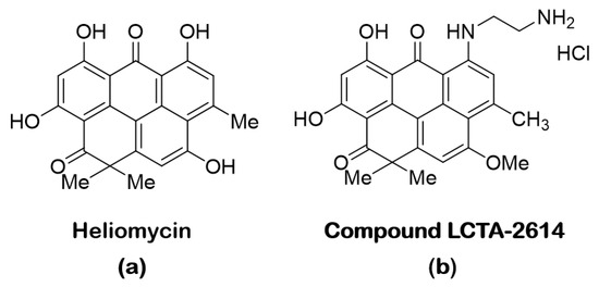
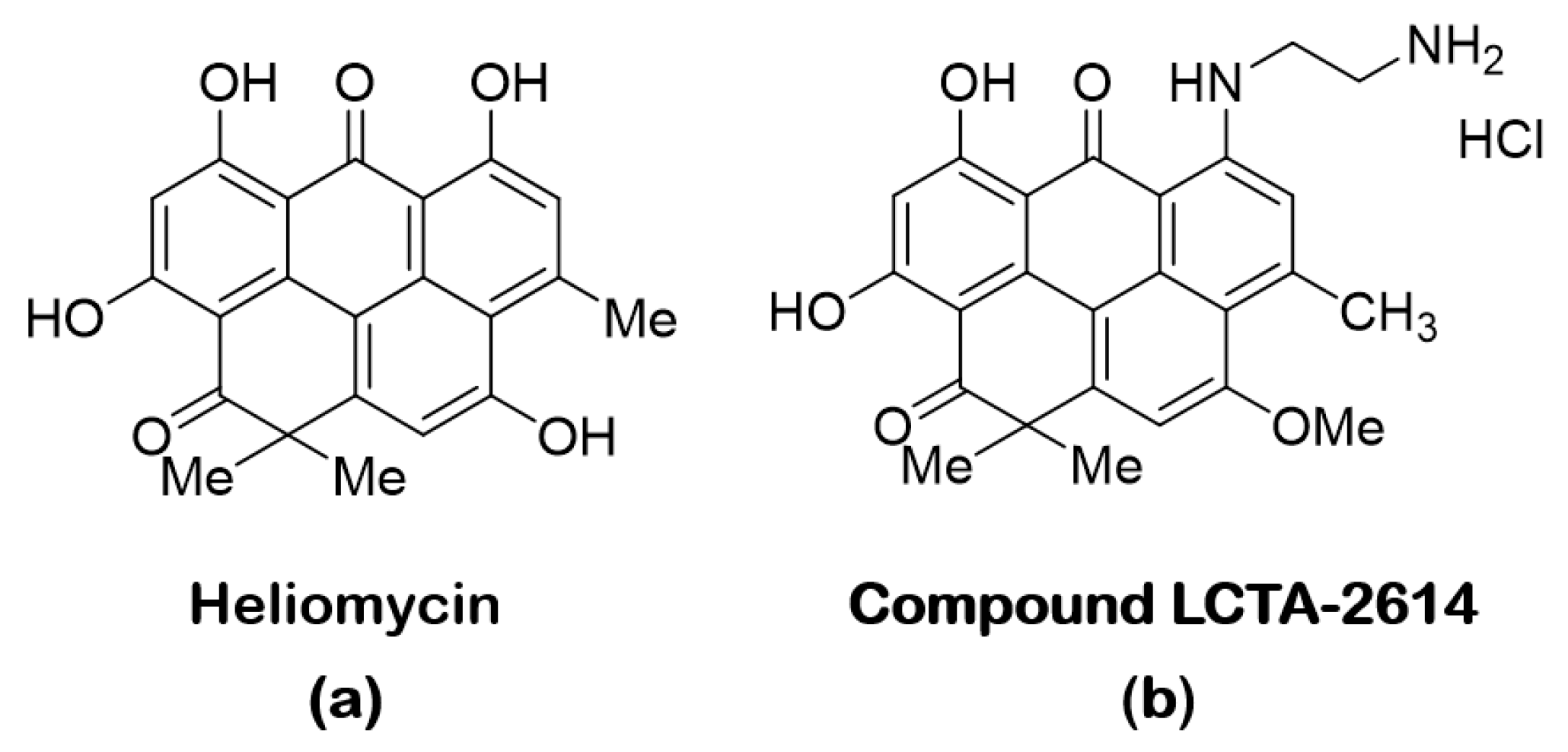
The heliomycin (Figure 1a) and its derivatives were synthesized and characterized as described early [27]. The heliomycin derivative LCTA-2614 (Figure 1b) demonstrated the highest antiproliferative activity in the series of the synthesized compounds, suppressing tumor cells at low, submicromolar concentrations, and was chosen as the most active for further studies on breast cancer cells.
Figure 1.
Structure of heliomycin (a) and its derivative LCTA-2614 (b). The synthesis of LCTA-2614 has been previously described in [27].
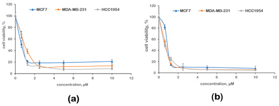
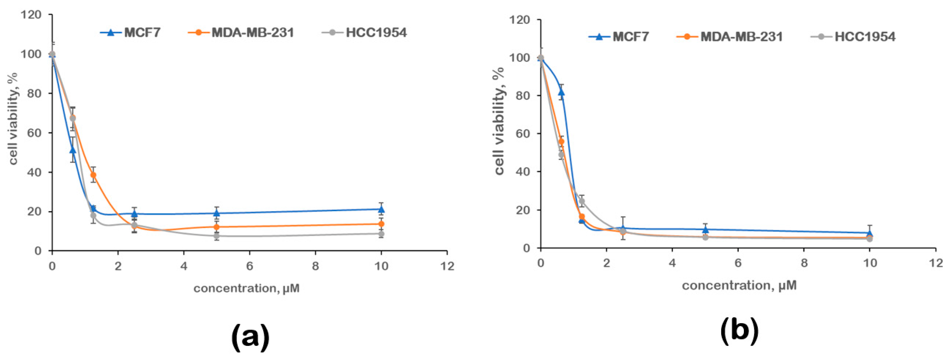
The antiproliferative activity of the compounds was evaluated using the MTT assay. Heliomycin inhibited cell growth in the submicromolar concentration range, showing IC50 values from 0.65 to 0.95 μM (Figure 2, Table 1). Luminal MCF7 breast cancer cells were more sensitive to its action. The activity of the novel heliomycin derivative LCTA-2614 was also high. For all breast cancer cell lines, its IC50 values did not exceed 1 μM. HER2-positive breast cancer cells were more sensitive to the action of the compound LCTA-2614.
Figure 2.
Antiproliferative activity of heliomycin (a) and its derivative LCTA-2614 (b) against ERα-positive MCF7, triple-negative MDA-MB-231, and HER2-positive HCC1954 breast cancer cells. The survival of cells was assessed after a 72 h incubation with compounds: the results of the MTT assay. The results present the average value ± the standard deviation of three independent tests.
Table 1.
Antiproliferative activity of the heliomycin and its derivative LCTA-2614 against ERα-positive MCF7, triple-negative MDA-MB-231, and HER2-positive HCC1954 breast cancer cells. The survival of cells was assessed after a 72 h incubation with compounds: the results of the MTT assay. The results present the mean value ± the standard deviation of three separate tests. IC50—half-maximal inhibitory concentration; hypoxia—1% O2.
The progression of malignant neoplasms is accompanied by tissue hypoxia. Hypoxic conditions determine changes in the phenotype of tumor cells, including the development of resistance. A number of chemotherapeutic agents lose their potency under reduced oxygen levels. Therefore, the evaluation of the antiproliferative activity of new compounds under hypoxic conditions is an important stage of preclinical development. Hypoxia was modeled using a dual-gas incubator with the ability to reduce the oxygen level to 1%. As under normoxic conditions, prior to the MTT assay, breast cancer cells were incubated with the compounds for 72 h. The data presented in Table 1 indicate that hypoxia did not reduce the activity of heliomycin or its derivative LCTA-2614. It is important to note that the compound LCTA-2614 showed very high activity against aggressive HER2+ breast cancer, including experiments in hypoxia.
Evaluation of compound toxicity at the preclinical stage may be performed using normal epithelial cell lines. The non-tumorigenic MCF-10A human mammary epithelial cell line serves as a valuable in vitro model for evaluating the toxicity of novel compounds, particularly in the context of breast cancer research and drug safety profiling [35,36]. Derived from normal breast tissue, MCF-10A cells retain key characteristics of differentiated mammary epithelium, including polarized growth, making them suitable for distinguishing compound-specific cytotoxicity from off-target effects observed in tumorigenic lines such as MCF7 or MDA-MB-231. MCF-10A cells were incubated with heliomycin and its derivative LCTA-2614 for 72 h, followed by the MTT assay. Table 2 presents a comparison of compound activity against epithelial cells and HER2+ HCC1954 cancer cells. The selectivity index was calculated as the ratio of the IC50 value in epithelial cells to the IC50 value in tumor cells. It was found that heliomycin is sufficiently toxic toward epithelial cells, with a selectivity index of approximately 1. This indicates that epithelial cells died at a rate comparable to that of tumor cells upon incubation with heliomycin. The cytotoxicity of the new heliomycin derivative was more pronounced in tumor cells than in epithelial cells. The selectivity index was 2.7, indicating the promise of further development of this heliomycin modification.
Table 2.
Evaluation of compound toxicity and selectivity. The results of the MTT assay performed after 72 h incubation with the compounds are presented.
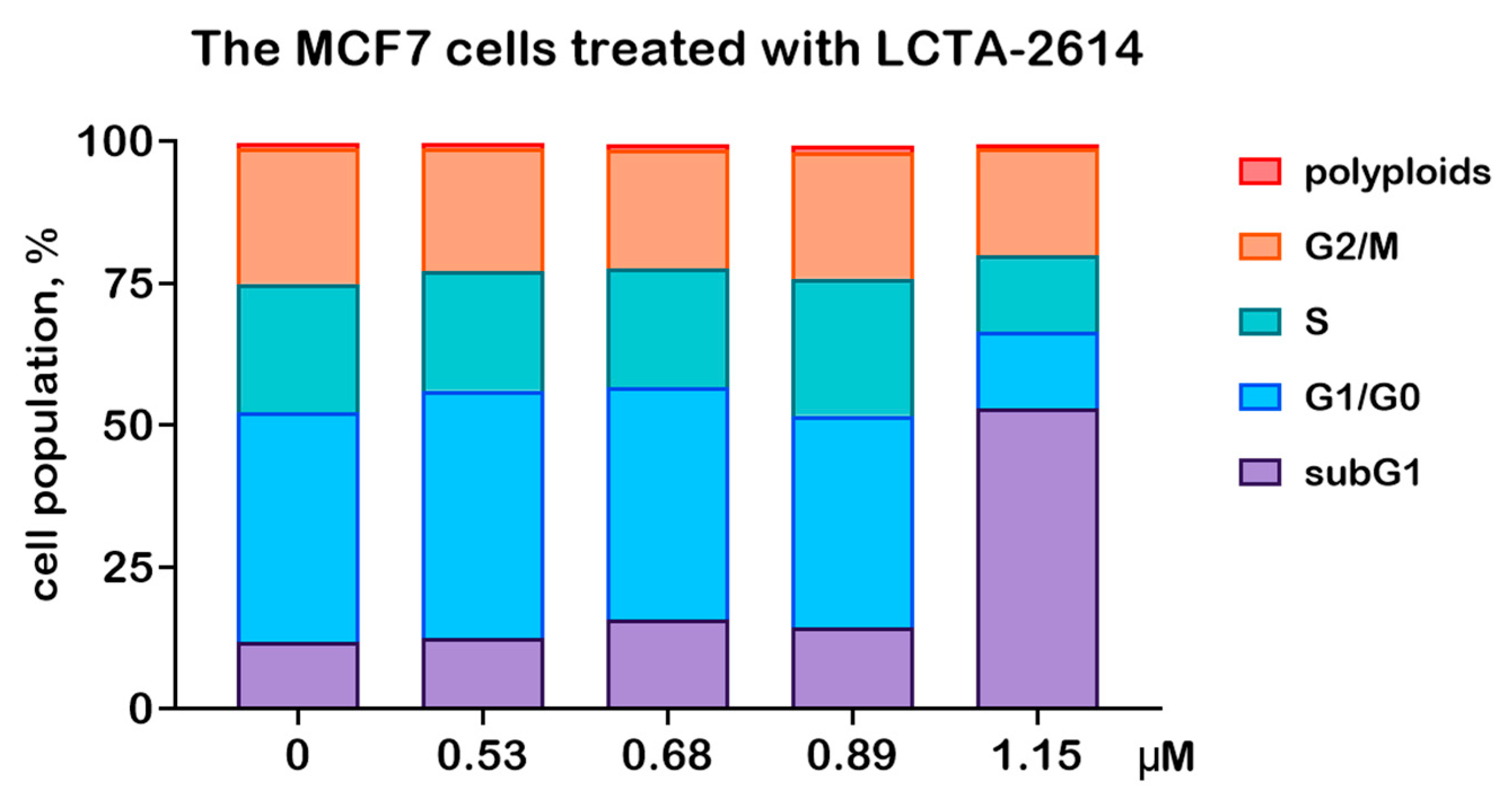
Further studies were aimed at elucidating the mechanisms of action of the novel heliomycin derivative LCTA-2614 in MCF7 cells possessing wild-type p53. The cell cycle of the MCF7 cells treated with compound LCTA-2614 for 72 h was analyzed by measuring the DNA content. Cells were stained with propidium iodide following permeabilization. This method quantifies the percentage of cells in the G1/G0 (interphase), S (DNA replication), and G2/M (mitotic division) phases, as well as polyploid and dead (subG1) cell populations. A 1.3-fold serial dilution series was used to generate concentrations ranging from 0.53 to 1.15 µM. The highest tested concentration was 1.15 µM of LCTA-2614; an equivalent volume of vehicle (0.01% dimethyl sulfoxide) served as the control.
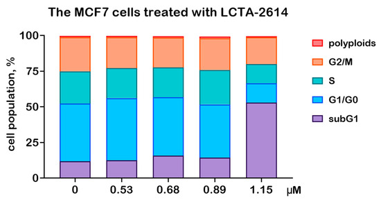
Treatment with LCTA-2614 at 1.15 µM resulted in a dramatic increase in the proportion of dead cells (Figure 3 and Table 3). Notably, a cytotoxic effect was absent at all lower concentrations and was observed exclusively at this highest dose. This all-or-nothing response is indicative of a sharp concentration threshold for the compound activity. Furthermore, no significant accumulation of cells was detected in any specific phase of the cell cycle (G1/G0, S, or G2/M) prior to cell death.
Figure 3.
Cell cycle distribution of the MCF7 cells treated with compound LCTA-2614 for 72 h.
Table 3.
The percentage of the MCF7 cells treated with LCTA-2614 for 72 h in various phases of the cell cycle.
Therefore, exposure of the MCF7 cells to compound LCTA-2614 at its minimal cytotoxic concentration of 1.15 µM induces significant and abrupt cell death without preceding cell cycle arrest.
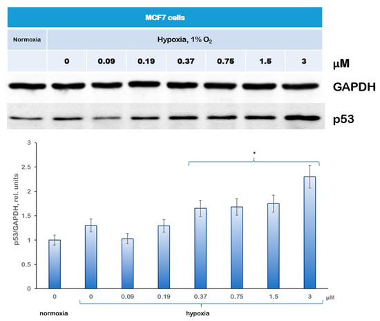
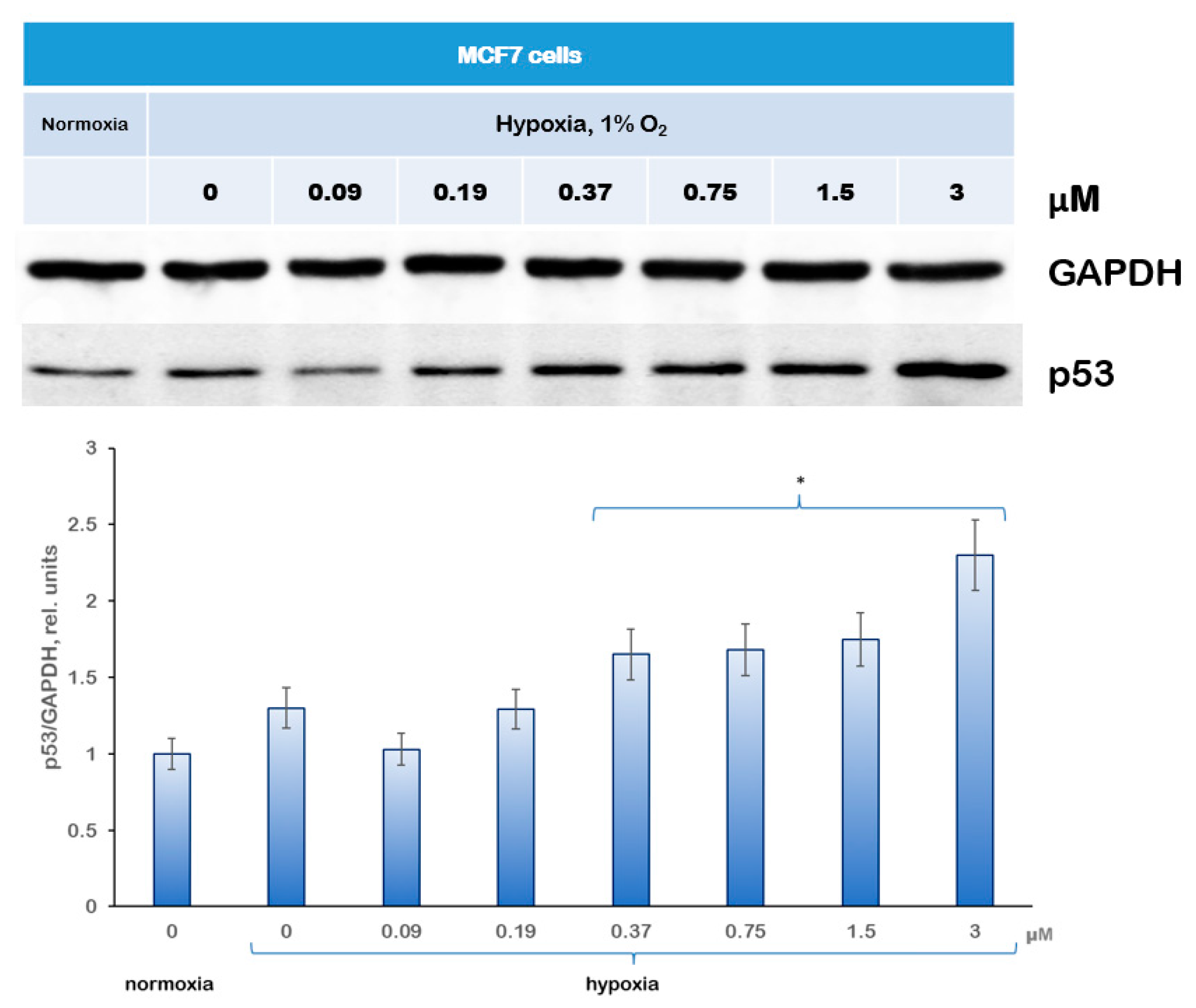
The expression of the tumor suppressor p53 was analyzed by immunoblotting. MCF7 cells were incubated under hypoxic conditions with the compound LCTA-2614 at concentrations ranging from 0.09 to 3.0 μM, after which samples were prepared for analysis. Figure 4 shows that treatment with compound LCTA-2614 led to an increase in p53 expression. Thus, the apoptosis induced by compound LCTA-2614 is partly mediated by p53-assosiated pathways.
Figure 4.
Immunoblotting of tumor suppressor p53 in MCF7 cells treated with compound LCTA-2614. GAPDH was used as loading control. The diagram presents the results of densitometry analysis performed in ImageJ, Version 1.54 (Bethesda, MD, USA) according to the protocol and recommendations described in references [37,38,39]. * p < 0.05 versus non-treated cells under hypoxia.
4. Discussion
Initial studies of heliomycin focused on its antibacterial and antiviral activities [21,40]. It was demonstrated that the compound effectively inhibited the growth of a number of Gram-positive bacteria, including Staphylococcus aureus and Bacillus subtilis [41]. The antibacterial mechanism of action was associated with disruption of energy metabolism and damage to cellular membranes. However, with the development of new classes of antibiotics, interest in heliomycin in this field gradually declined, and further studies were redirected toward its antitumor properties. In this direction, some progress has already been achieved [23,24,25,27].
The first evidence of the antitumor activity of heliomycin emerged from experimental in vitro and in vivo studies, where suppression of tumor growth of various origins was demonstrated. In particular, heliomycin exhibited cytotoxicity toward leukemia, sarcoma, and carcinoma cells. Subsequently, studies revealed that heliomycin possesses a broad spectrum of antiproliferative activity, including effects on breast, prostate, lung, and colon cancer cells. In many cases, the sensitivity of tumor cells exceeded that of normal cells, indicating potential selectivity of action. The low water solubility of heliomycin was one of the reasons that necessitated the targeted synthesis of new derivatives [27].
Previously, we synthesized a series of hydrophilic heliomycin derivatives in order to improve aqueous solubility and enhance the biological availability of the compounds. The physicochemical properties of the new compounds were characterized using spectroscopic methods. The ability of the derivatives to stabilize the DNA i-motif was evaluated using biophysical approaches, including circular dichroism spectroscopy and thermal denaturation experiments [27]. It was shown that a number of the synthesized compounds effectively stabilize i-motif structures while having minimal influence on the canonical DNA form, indicating their structural selectivity. Continuing research in this direction, we demonstrate here a high antiproliferative potential of heliomycin and its derivatives against several breast cancer cell lines. The IC50 values of compound LCTA-2614 did not exceed one μM, and its activity was comparable to that of the parent antibiotic. At the same time, the selectivity of LCTA-2614 was higher than that of heliomycin. An important observation is the high activity of LCTA-2614 under hypoxic conditions—retention of activity under such conditions highlights the promise of further development. The established ability of compound LCTA-2614 to induce apoptosis and to stimulate p53 accumulation places it among pro-apoptotic anti-breast cancer agents.
5. Conclusions
The activity of heliomycin and its new water-soluble derivative was characterized in breast cancer cells. The antiproliferative and pro-apoptotic potential of the water-soluble heliomycin derivative LCTA-2614 provides a basis for further development of new semisynthetic anticancer agents based on this antibiotic.
Author Contributions
Conceptualization, A.E.S. and A.M.S.; methodology, A.S.T. and A.M.S.; synthesis A.S.T.; validation, D.I.S., A.I.K., A.S.T., A.L.M. and A.M.S.; formal analysis, D.I.S., A.I.K. and A.M.S.; investigation, A.L.M., D.I.S., A.S.T. and A.I.K.; resources, A.E.S.; data curation, A.E.S. and A.M.S.; writing—original draft preparation, D.I.S. and A.M.S.; writing—review and editing, A.I.K., A.M.S. and A.E.S.; visualization, D.I.S. and A.I.K.; supervision, A.M.S. and A.E.S.; project administration, A.E.S.; funding acquisition, A.E.S. All authors have read and agreed to the published version of the manuscript.
Funding
This research was partly funded by the Russian Science Foundation, grant number 25-73-20069, https://rscf.ru (accessed on 15 January 2026).
Institutional Review Board Statement
Not applicable.
Informed Consent Statement
Not applicable.
Data Availability Statement
The data presented in this study are available on request from the corresponding authors.
Acknowledgments
The authors dedicate this article to the outstanding Soviet microbiologists Brazhnikova M.G. and Gauze G.F., who made significant contributions to the development of heliomycin and the analysis of its activity. The authors are grateful to Danila V. Sorokin and Anna E. Tischenko for their help in carrying out the analyses and thank the reviewers for their insightful and constructive comments, which greatly improved the manuscript.
Conflicts of Interest
The authors declare no conflicts of interest.
Abbreviations
| DMEM | Dulbecco’s Modified Eagle Medium |
| ERα | Estrogen Receptor α |
| HER2 | Human Epidermal Growth Factor Receptor 2 |
| IC50 | Half-maximal Inhibitory Concentration |
| RPMI | Roswell Park Memorial Institute |
References
- Hu, Z.; Bousbaa, H. Recent Advances in Anticancer Strategies. Cancers 2025, 17, 173. [Google Scholar] [CrossRef] [Scilit]
- Kifle, Z.D.; Tadele, M.; Alemu, E.; Gedamu, T.; Ayele, A.G. A recent development of new therapeutic agents and novel drug targets for cancer treatment. SAGE Open Med. 2021, 9, 1–12. [Google Scholar] [CrossRef] [Scilit]
- Zykova, S.S.; Gessel, T.; Galembikova, A.; Mozhaitsev, E.S.; Borisevich, S.S.; Igidov, N.; Egorova, E.S.; Mikheeva, E.; Khromova, N.; Kopnin, P.; et al. Hypoxia-Inducible Factor-1α, a Novel Molecular Target for a 2-Aminopyrrole Derivative: Biological and Molecular Modeling Study. Cancers 2025, 18, 115. [Google Scholar] [CrossRef] [Scilit] [PubMed]
- Krylova, L.V.; Otvagin, V.F.; Gribova, G.P.; Kuzmina, N.S.; Fedotova, E.A.; Zelepukin, I.V.; Nyuchev, A.V.; Kustov, A.V.; Morshnev, P.K.; Berezin, D.B.; et al. Developing Chlorin/Arylaminoquinazoline Conjugates with Nanomolar Activity for Targeted Photodynamic Therapy: Design, Synthesis, SAR, and Biological Evaluation. J. Med. Chem. 2025, 68, 1901–1923. [Google Scholar] [CrossRef] [Scilit]
- Svirshchevskaya, E.V.; Kostenko, V.V.; Boyko, A.A.; Shevtsov, M.; Kholodenko, R.V.; Grechikhina, M.V.; Gracheva, I.A.; Fedorov, A.Y.; Sapozhnikov, A.M. Core-Shell Chitosan Particles Targeting Membrane-Bound Heat Shock Protein 70 for Cancer Therapy. Nanomaterials 2024, 14, 1873. [Google Scholar] [CrossRef] [Scilit] [PubMed]
- Tamatam, R.; Mohammed, A. Small molecule anticancer drugs approved during 2021–2022: Synthesis and clinical applications. Eur. J. Med. Chem. 2024, 272, 116441. [Google Scholar] [CrossRef] [Scilit] [PubMed]
- Mikhaevich, E.I.; Sorokin, D.V.; Scherbakov, A.M. Honokiol inhibits the growth of hormone-resistant breast cancer cells: Its promising effect in combination with metformin. Res. Pharm. Sci. 2023, 18, 580–591. [Google Scholar] [CrossRef] [Scilit]
- Scherbakov, A.M.; Basharina, A.A.; Sorokin, D.V.; Mikhaevich, E.I.; Mizaeva, I.E.; Mikhaylova, A.L.; Bogush, T.A.; Krasil’nikov, M.A. Targeting hormone-resistant breast cancer cells with docetaxel: A look inside the resistance. Cancer Drug Resist. 2023, 6, 103–115. [Google Scholar] [CrossRef] [Scilit] [PubMed]
- Scherbakov, A.M.; Krasil’nikov, M.A.; Kushlinskii, N.E. Molecular mechanisms of hormone resistance of breast cancer. Bull. Exp. Biol. Med. 2013, 155, 384–395. [Google Scholar] [CrossRef] [Scilit]
- Newman, D.J.; Cragg, G.M. Microbial antitumor drugs: Natural products of microbial origin as anticancer agents. Curr. Opin. Investig. Drugs 2009, 10, 1280–1296. [Google Scholar]
- Giurini, E.F.; Godla, A.; Gupta, K.H. Redefining bioactive small molecules from microbial metabolites as revolutionary anticancer agents. Cancer Gene Ther. 2024, 31, 187–206. [Google Scholar] [CrossRef] [Scilit]
- Hsieh, C.Y.; Liou, Y.F.; Shih, Y.T.; Tikhomirov, A.S.; Shchekotikhin, A.E.; Chueh, P.J. Olivomycin A Targets Epithelial-Mesenchymal Transition, Apoptosis, and Mitochondrial Quality Control in Renal Cancer Cells. Antioxidants 2025, 14, 1348. [Google Scholar] [CrossRef] [Scilit]
- Rui, S.; Fengrui, G.; Yining, Z.; Hong, S.; Xuewen, Y.; Changping, W.; Chunjia, Y. Biological activity of secondary metabolites of actinomycetes and their potential sources as antineoplastic drugs: A review. Front. Microbiol. 2025, 16, 1550516. [Google Scholar] [CrossRef] [Scilit]
- Olano, C.; Méndez, C.; Salas, J.A. Antitumor Compounds from Marine Actinomycetes. Mar. Drugs 2009, 7, 210–248. [Google Scholar] [CrossRef] [Scilit]
- Karthik, Y.; Arthikala, M.K.; Nanjareddy, K.; Kalyani, M.I. Mangrove soil-derived Streptomyces viridiostaticus as a source of dual therapeutic bioactives: Antimicrobial proteins and Tryprostatin B alkaloid with anticancer potency. Mol. Biol. Rep. 2026, 53, 259. [Google Scholar] [CrossRef] [Scilit] [PubMed]
- Al-Shaibani, M.M.; Zin, N.M.; Remali, J.; Sidik, N.M.; Al-Mekhlafi, N.A.; Mariappan, V.; Sukri, A. Anticancer compounds from Streptomyces: Insights from metagenomics and mechanistic perspective. Folia Microbiol. 2025, 70, 1159–1172. [Google Scholar] [CrossRef] [Scilit] [PubMed]
- Law, J.W.; Law, L.N.; Letchumanan, V.; Tan, L.T.; Wong, S.H.; Chan, K.G.; Ab Mutalib, N.S.; Lee, L.H. Anticancer Drug Discovery from Microbial Sources: The Unique Mangrove Streptomycetes. Molecules 2020, 25, 5365. [Google Scholar] [CrossRef] [Scilit]
- Sritharan, S.; Sivalingam, N. A comprehensive review on time-tested anticancer drug doxorubicin. Life Sci. 2021, 278, 119527. [Google Scholar] [CrossRef] [Scilit] [PubMed]
- Maddock, C.L.; D’Angio, G.J.; Farber, S.; Handler, A.H. Biological Studies of Actinomycin D. Ann. N. Y. Acad. Sci. 1960, 89, 386–398. [Google Scholar] [CrossRef] [Scilit] [PubMed]
- Watson, R.A.; De La Peña, H.; Tsakok, M.T.; Joseph, J.; Stoneham, S.; Shamash, J.; Joffe, J.; Mazhar, D.; Traill, Z.; Ho, L.-P.; et al. Development of a best-practice clinical guideline for the use of bleomycin in the treatment of germ cell tumours in the UK. Br. J. Cancer 2018, 119, 1044–1051. [Google Scholar] [CrossRef] [Scilit] [PubMed]
- Brazhnikova, M.G.; Uspenskaia, T.A.; Sokolova, L.B.; Preobrazhenskaia, T.P.; Gauze, G.F.; Ukholina, R.S.; Shorin, V.A.; Rossolimo, O.K.; Vertogradova, T.P. New anti-viral antibiotic heliomycin. Antibiotiki 1958, 3, 29–34. [Google Scholar] [PubMed]
- Brockmann, H.; Schmidt-Kastner, G. Resistomycin, ein neues Antibioticum aus Actinomyceten. Naturwissenschaften 1951, 38, 479–480. [Google Scholar] [CrossRef] [Scilit]
- Islam, A.; Chang, Y.C.; Chen, X.C.; Weng, C.W.; Chen, C.Y.; Wang, C.W.; Chen, M.K.; Tikhomirov, A.S.; Shchekotikhin, A.E.; Chueh, P.J. Water-soluble 4-(dimethylaminomethyl)heliomycin exerts greater antitumor effects than parental heliomycin by targeting the tNOX-SIRT1 axis and apoptosis in oral cancer cells. eLife 2024, 12, RP87873. [Google Scholar] [CrossRef] [Scilit] [PubMed]
- Lin, M.H.; Islam, A.; Liu, Y.H.; Weng, C.W.; Zhan, J.H.; Liang, R.H.; Tikhomirov, A.S.; Shchekotikhin, A.E.; Chueh, P.J. Antibiotic heliomycin and its water-soluble 4-aminomethylated derivative provoke cell death in T24 bladder cancer cells by targeting sirtuin 1 (SIRT1). Am. J. Cancer Res. 2022, 12, 1042–1055. [Google Scholar]
- Alazzouni, A.S.; Mahmoud, A.A.; Omran, M.M.; Essawy, E.A.; Abdalla, M.S.; Abdelfattah, M.S. Inhibitory and ameliorative effect of heliomycin derived from actinomycete on induced hepatocellular carcinoma in rats. Naunyn-Schmiedeberg’s Arch. Pharmacol. 2021, 394, 1091–1102. [Google Scholar] [CrossRef] [Scilit]
- Nadysev, G.Y.; Tikhomirov, A.S.; Lin, M.H.; Yang, Y.T.; Dezhenkova, L.G.; Chen, H.Y.; Kaluzhny, D.N.; Schols, D.; Shtil, A.A.; Shchekotikhin, A.E.; et al. Aminomethylation of heliomycin: Preparation and anticancer characterization of the first series of semi-synthetic derivatives. Eur. J. Med. Chem. 2018, 143, 1553–1562. [Google Scholar] [CrossRef] [Scilit]
- Tikhomirov, A.S.; Abdelhamid, M.A.S.; Nadysev, G.Y.; Zatonsky, G.V.; Bykov, E.E.; Chueh, P.J.; Waller, Z.A.E.; Shchekotikhin, A.E. Water-Soluble Heliomycin Derivatives to Target i-Motif DNA. J. Nat. Prod. 2021, 84, 1617–1625. [Google Scholar] [CrossRef] [Scilit] [PubMed]
- Iselt, M.; Holtei, W.; Hilgard, P. The tetrazolium dye assay for rapid in vitro assessment of cytotoxicity. Arzneimittelforschung 1989, 39, 747–749. [Google Scholar]
- Scherbakov, A.M.; Vorontsova, S.K.; Khamidullina, A.I.; Mrdjanovic, J.; Andreeva, O.E.; Bogdanov, F.B.; Salnikova, D.I.; Jurisic, V.; Zavarzin, I.V.; Shirinian, V.Z. Novel pentacyclic derivatives and benzylidenes of the progesterone series cause anti-estrogenic and antiproliferative effects and induce apoptosis in breast cancer cells. Investig. New Drugs 2023, 41, 142–152. [Google Scholar] [CrossRef] [Scilit]
- Ilovaisky, A.I.; Scherbakov, A.M.; Chernoburova, E.I.; Shchetinina, M.A.; Merkulova, V.M.; Bogdanov, F.B.; Sorokin, D.V.; Salnikova, D.I.; Bozhenko, E.I.; Zavarzin, I.V.; et al. Secosteroid diacylhydrazines as novel effective agents against hormone-dependent breast cancer cells. J. Steroid Biochem. Mol. Biol. 2024, 244, 106597. [Google Scholar] [CrossRef] [Scilit]
- Monzote, L.; Scherbakov, A.M.; Scull, R.; Satyal, P.; Cos, P.; Shchekotikhin, A.E.; Gille, L.; Setzer, W.N. Essential Oil from Melaleuca leucadendra: Antimicrobial, Antikinetoplastid, Antiproliferative and Cytotoxic Assessment. Molecules 2020, 25, 5514. [Google Scholar] [CrossRef] [Scilit]
- Scherbakov, A.M.; Kuznetsov, Y.V.; Yastrebova, M.A.; Khamidullina, A.I.; Sorokin, D.V.; Tserfas, M.O.; Levina, I.S. Antiproliferative effects of 13α/β-steroids on triple-negative MDA-MB-231 breast cancer cells: Unraveling intracellular signaling without ERα. Braz. J. Pharm. Sci. 2023, 59, e22540. [Google Scholar] [CrossRef] [Scilit]
- Scherbakov, A.M.; Stefanova, L.B.; Sorokin, D.V.; Semina, S.E.; Berstein, L.M.; Krasil’nikov, M.A. Snail/beta-catenin signaling protects breast cancer cells from hypoxia attack. Exp. Cell Res. 2013, 319, 3150–3159. [Google Scholar] [CrossRef] [Scilit]
- Mruk, D.D.; Cheng, C.Y. Enhanced chemiluminescence (ECL) for routine immunoblotting: An inexpensive alternative to commercially available kits. Spermatogenesis 2011, 1, 121–122. [Google Scholar] [CrossRef] [Scilit]
- Engel, A.; Frenzel, F.; Niemann, B.; Braeuning, A.; Lampen, A.; Buhrke, T. The use of 3D cultures of MCF-10A and MCF-12A cells by high content screening for effect-based analysis of non-genotoxic carcinogens. Toxicol. Vitr. 2019, 59, 55–63. [Google Scholar] [CrossRef] [Scilit] [PubMed]
- Kuznetsov, Y.V.; Tserfas, M.O.; Scherbakov, A.M.; Andreeva, O.E.; Salnikova, D.I.; Bozhenko, E.I.; Zavarzin, I.V.; Levina, I.S. Synthesis of 13β- and 13α-epimers of 3-hydroxy-17-hydroxymethylestra-1,3,5(10)-triene and considerations on their hormonal and antiproliferative potency. Steroids 2024, 212, 109527. [Google Scholar] [CrossRef] [Scilit]
- Schneider, C.A.; Rasband, W.S.; Eliceiri, K.W. NIH Image to ImageJ: 25 years of image analysis. Nat. Methods 2012, 9, 671–675. [Google Scholar] [CrossRef] [Scilit]
- Gassmann, M.; Grenacher, B.; Rohde, B.; Vogel, J. Quantifying Western blots: Pitfalls of densitometry. Electrophoresis 2009, 30, 1845–1855. [Google Scholar] [CrossRef] [Scilit]
- Alonso Villela, S.M.; Kraïem, H.; Bouhaouala-Zahar, B.; Bideaux, C.; Aceves Lara, C.A.; Fillaudeau, L. A protocol for recombinant protein quantification by densitometry. Microbiologyopen 2020, 9, 1175–1182. [Google Scholar] [CrossRef] [Scilit]
- Adinarayana, G.; Venkateshan, M.R.; Bapiraju, V.V.; Sujatha, P.; Premkumar, J.; Ellaiah, P.; Zeeck, A. Cytotoxic compounds from the marine actinobacterium. Bioorganicheskaia Khimiia 2006, 32, 328–334. [Google Scholar] [CrossRef] [Scilit] [PubMed]
- Bershteĭn, E.M.; Vasil’eva, N.G.; Poliak, M.S.; Grigor’eva, V.M.; Astanina, L.N. Nutrient medium for determining the biological activity of heliomycin. Antibiotiki 1984, 29, 766–770. [Google Scholar] [PubMed]

Disclaimer/Publisher’s Note: The statements, opinions and data contained in all publications are solely those of the individual author(s) and contributor(s) and not of MDPI and/or the editor(s). MDPI and/or the editor(s) disclaim responsibility for any injury to people or property resulting from any ideas, methods, instructions or products referred to in the content. |
© 2026 by the authors. Licensee MDPI, Basel, Switzerland. This article is an open access article distributed under the terms and conditions of the Creative Commons Attribution (CC BY) license.