


Abstract
Pectin was extracted from Citrus sinensis fruit peel, an agro-waste material, by an acid-assisted method, and the percentage of pectin extracted was 26.8%. The extracted pectin was then modified by ionic cross-linking of Cu2+ and Zn2+ ions with the pectin to form metal–pectin complexes by a controlled coordination method. The structure of the modified pectin complexes was studied by FT-IR, XRD, SEM, and EDX techniques. The coordination of metal ions with the carboxylate functional group of the pectin backbone was confirmed by the shift in the characteristic peaks in the FT-IR spectra. The amorphous nature of the pectin backbone remained unchanged after modification, as evidenced by the XRD patterns. The morphology of the pectin changes from flakes to aggregated form, as evidenced by the SEM images. The presence of metal ions in the complexes was confirmed by the EDX technique. Swelling and pH stability tests have shown reduced swelling potential and an increase in the structural stability of the metal–pectin complexes, especially at a neutral pH, which were attributed to the formation of a cross-linked ionic network. In addition, the metal-modified pectin material has shown increased antimicrobial potential compared to the native material. This is a testament to the role of transition metals in enhancing the bio-functional potential of materials. In conclusion, the present work provides an efficient and sustainable strategy for the valorization of citrus waste materials for various potential applications.
1. Introduction
Pectin is a structurally complex, anionic heteropolysaccharide composed predominantly of α-1,4-linked D-galacturonic acid residues that are variably methyl-esterified and partially acetylated, a heterogeneity that governs its physicochemical behaviour [1]. Citrus by-products, and orange peel in particular, form one of the largest and most accessible agro-industrial waste streams and represent a rich, renewable source of this polysaccharide [2]. Acid-assisted extraction followed by alcohol precipitation is a well-established and efficient route for isolating pectin from orange peel, yielding a polysaccharide structurally comparable to commercial citrus pectin and rich in galacturonic acid [3,4]. The yield and physicochemical characteristics of the extracted pectin—including methoxy content and degree of esterification—are strongly influenced by extraction parameters such as acid type, pH, temperature and time, and can be further improved through optimization approaches such as response surface methodology [5,6,7]. The degree of esterification, in turn, is directly correlated with the morphological ordering, thermal behaviour and ion-binding capacity of pectin-based systems [1].
Because the free carboxylate groups of the galacturonan backbone act as effective metal-binding sites, pectin and related pectic materials have been extensively explored as low-cost, biodegradable adsorbents and ligands for metal ions, including for the removal and recovery of heavy-metal ions from aqueous media [8]. Spectroscopic and solution-equilibrium studies have shown that divalent metal ions coordinate with the non-esterified carboxylate groups of pectin and polygalacturonic acid, producing characteristic shifts in the carboxylate stretching bands and stable coordination complexes in aqueous solution [9,10]. This binding is selective and strongly dependent on the degree of esterification and chain conformation, with transition-metal ions generally showing greater affinity than alkaline–earth metals such as calcium [11,12]. Building on this coordination chemistry, biologically essential metals such as copper and zinc have more recently been incorporated into pectin to generate bioactive, rather than purely adsorptive, metal–pectin materials [13,14], since copper and zinc complexes are well known for their pronounced antimicrobial activity, arising from membrane disruption, the generation of reactive oxygen species and interference with essential microbial biomolecules [15,16]. Consistent with this, metal–polysaccharide systems such as silver–polygalacturonate, copper-enriched composites and cross-linked pectin films have all been shown to acquire enhanced antibacterial functionality upon metal incorporation [17,18,19,20].
Despite this considerable interest in pectin extraction, metal–polygalacturonate coordination chemistry and the antimicrobial potential of transition-metal complexes, comparative studies that extract pectin directly from orange peel waste and systematically evaluate the resulting Cu2+– and Zn2+–pectin ionic complexes—in terms of their structure, physicochemical stability and antibacterial performance—remain limited. The present study therefore aims to extract pectin from orange peel waste by an acid-assisted method, prepare Cu2+– and Zn2+–pectin ionic complexes through controlled meta–-carboxylate coordination, and characterize the resulting materials using FT-IR, XRD, SEM and EDX to establish the mode and structural consequences of metal binding, alongside their swelling behaviour, pH stability and antimicrobial activity relative to native pectin. The novelty of this work lies in the direct, integrated valorisation of citrus peel waste into structurally characterized, antimicrobially active Cu2+– and Zn2+–pectin materials, providing a sustainable route from agricultural by-product to a functional biomaterial with potential application as an antimicrobial coating or packaging component.
2. Materials and Methods
2.1. Materials and Reagents
Fresh peels of Citrus sinensis (sweet orange) were obtained from a local fruit market for pectin extraction. Copper sulfate pentahydrate (CuSO4·5H2O) was obtained from Simson Pharma Ltd., Mumbai, India. Zinc chloride (SQ grade) and citric acid monohydrate were obtained from Qualigens (Thermo Fisher Scientific India Pvt. Ltd., Mumbai, India). Analytical-grade glacial acetic acid was obtained from Merck Life Science Pvt. Ltd., Mumbai, India. Ethanol (95%, AR grade) was obtained from Changshu Hongshen Fine Chemical Co., Ltd., Changshu, China. Double-distilled water was obtained from Hi-Media Laboratories Pvt. Ltd., Thane, India. All reagents were used without further purification. All synthesis chemicals and reagents purchase from Chemifine Formulations Pvt. Ltd, Nellore, India.
2.2. Preparation of Orange Peel Powder
Fresh orange peels were thoroughly washed with tap water, followed by distilled water to remove surface contaminants. The cleaned peels were cut into small pieces and dried in a hot-air oven at 55 ± 2 °C for 24–36 h until constant weight was achieved. The dried material was subjected to mechanical grinding, followed by sieving to obtain fine powder. The powder obtained from the peel of the orange was stored in airtight containers and maintained at room temperature, for future use, as described above, with modifications in the drying method [5].
2.3. Isolation and Purification of Pectin
Pectin was extracted using the modified method for the acid extraction of citrus pectin, as described in [2,7]. In summary, 10 g of dried powder was mixed with 250 mL of distilled water, and the pH was adjusted to 1.8–2.0 using 0.1 M citric acid. The mixture was then subjected to heat treatment at 80–85 °C using constant magnetic stirring at 600 rpm for 60 min. The mixture was then subjected to hot filtration using muslin cloth followed by Whatman No. 1 filter paper, then cooled to room temperature. Pectin precipitate was obtained by adding two volumes of 95% ethanol with constant stirring. The mixture was left to settle for 12 h. The precipitated pectin was centrifuged at 5000 rpm for 10 min, and then washed a few times with ethanol to ensure that all sugars and impurities were removed from solution. The purified pectin was then dried at 45–50 °C to constant weight, after which it was ground into a fine powder. The percentage of pectin extraction was found to be 26.8%, within the expected range for citrus pectin, thus verifying the efficiency of the chosen protocol.
2.4. Synthesis of Metal–Pectin Ionic Complexes
Metal–pectin ionic complexes were synthesized by controlling the coordination of metal ions according to the principle of the ionotropic gelation method, with some modifications to the conditions of the reaction [14,21]. In the preparation of the polymer solution, 1.0 g of extracted pectin was dispersed in 100 mL of distilled water by magnetic stirring at 600 rpm for 60 min to produce a homogeneous viscous solution. The pH was adjusted to 4.5–5.0 using sodium hydroxide or acetic acid to ensure sufficient ionization of the carboxyl groups of pectin without precipitation of metal hydroxides. A 0.1 M copper (II) solution was prepared by dissolving 0.50 g CuSO4·5H2O in 20 mL of distilled water, while a 0.1 M zinc (II) solution was prepared by dissolving 0.273 g ZnCl2 in 20 mL of distilled water. The metal solutions were added to the pectin solution in a dropwise manner while stirring at 50–55 °C for 20–30 min. The reaction mixture was then stirred for 90 min to ensure the equal coordination of metal ions with pectin’s carboxylate groups. The complexation reaction was accompanied by an increase in viscosity, and for copper, it was accompanied by a colour change. The reaction mixtures were left to ripen at room temperature for 16–20 h to allow for the formation of a three-dimensional ionic structure. This coordination reaction is consistent with previously reported interaction between divalent metal ions and polygalacturonic acid through metal–carboxylate binding models [10,14].
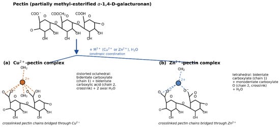
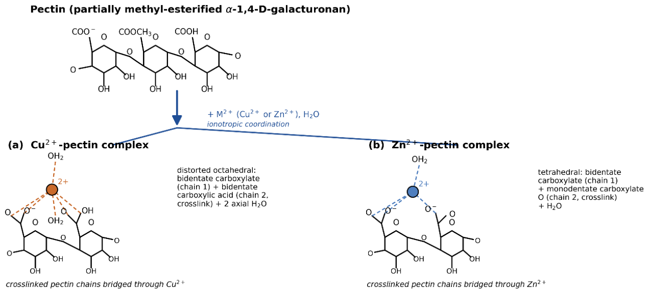
The obtained complexes were filtered, washed extensively with distilled water to remove loosely bound metal ions, and then dried at 45–50 °C to a constant weight. The samples, once dried, were pulverized and kept under suitable conditions for later physicochemical and antimicrobial evaluation. Schematic representation of the coordination of pectin carboxylate/carboxylic-acid groups with Cu2+ and Zn2+ ions is represented in Scheme 1.
Scheme 1.
Schematic representation of the coordination of pectin carboxylate/carboxylic-acid groups with Cu2+ and Zn2+ ions. Cu2+ adopts a distorted octahedral geometry through bidentate carboxylate/carboxylic-acid chelation from cross-linked chains and axial coordinated water, while Zn2+ adopts a tetrahedral geometry through bidentate carboxylate chelation together with a monodentate carboxylate oxygen and a coordinated water molecule.
2.5. Physicochemical Characterization
The extracted pectin and metal complexes of copper–pectin and zinc–pectin were characterized using various physicochemical techniques. These techniques were employed to evaluate the structure, composition, and morphology of the compounds. The techniques included Fourier Transform Infrared Spectroscopy (FTIR), X-Ray Diffraction (XRD) studies, Scanning Electron Microscopy (SEM), and Energy-Dispersive Spectroscopy (EDX). The FTIR spectra were obtained in the range of 4000–400 cm−1 by the KBr pellet method. In the FTIR analysis, the emphasis was placed on the identification of functional groups and changes in the characteristic peaks in the functional groups, especially those of the carboxylate groups (-COO−). Changes in the peaks were monitored to prove the interaction of divalent metal ions with the pectin backbone, as observed in the formation of pectinate complexes [9,22]. The X-ray diffraction (XRD) analysis was conducted to observe the effect of changes in the amorphous and crystalline properties of the pectin by incorporating metal ions into the structure. In the XRD analysis, CuKα radiation of wavelength 1.5406 Å was utilized. For the examination of surface morphology of samples, Scanning Electron Microscopy (SEM) was used. The results obtained in this analysis were used for a comparative evaluation of native pectin and its metal complexes. It was possible to visualize changes in surface roughness, aggregation, and compact structure formation after complexation of pectin with metals. Energy-Dispersive X-ray (EDX) analysis was conducted in combination with SEM for elemental analysis of samples. The appearance of specific peaks for metals (Cu and Zn) in the EDX spectra of corresponding complexes confirmed the complexation of metal ions in pectin structures, whereas in the EDX spectrum of native pectin, only carbon and oxygen peaks were identified as predominant elements. The results obtained in this analysis were found to be in agreement with previously conducted investigations on metal complexes of pectin [9,12,23]. All analysis techniques were conducted in an ambient environment using standardized methods for analysis of polymer–metal complexes based on pectin structures [18,24].
2.6. Swelling Study
The swelling properties of native pectin and its metal complexes were investigated by using a gravimetric method based on previously used methods for pectin-based matrices and hydrogels [25,26]. In this regard, dried samples of native pectin and its metal complexes (approximately 100 mg each) were precisely weighed and individually placed in 10 mL of distilled water at 25 ± 2 °C. At predetermined time points of 30, 60, 120, 180, and 240 min, each sample was removed from the water, gently blotted using filter paper to remove any excess water on its surface, and then weighed. The percentage of swelling was calculated using the following formula:
where Ws is the weight of the swollen sample and Wd is the initial dry weight of each sample. All experiments were conducted in triplicate, and results were expressed as mean values [27].
2.7. pH Stability Study
The pH stability of native pectin and its metal complexes was studied using solutions of different pH values according to a general methodology used for pH-sensitive pectin-based systems [28,29]. Dry samples of pectin, Cu–pectin, and Zn–pectin (about 100 mg each) were added individually into 10 mL of buffer solutions of pH 2.0, 4.0, 7.0, and 9.0, and kept at 25 ± 2 °C for 24 h in a shaking environment. After 24 h, the samples were removed from each buffer solution, washed carefully with distilled water to remove salts, and dried in an oven at 40 °C until a constant weight was reached. The percentage stability was calculated by using the following formula:
where Wi is the initial weight of the sample and Wf is the final weight of the sample after treatment. All experiments were done in triplicate, and results were calculated as mean values [29].
2.8. Evaluation of Antimicrobial Activity
Five bacterial strains—three Gram-negative (Escherichia coli, Pseudomonas aeruginosa, and Serratia marcescens) and two Gram-positive (Bacillus subtilis and Staphylococcus aureus)—were chosen in order to assess the antibacterial activity of pectin–metal ion complexes using the broth dilution technique [25]. Bacterial culture in Luria Broth was prepared using a prelature of bacteria. To obtain the desired concentration of chemicals to examine microbial strains, DMSO was utilized as a medium. Before adding previously cultivated bacterial species, the sugar tube containing Luria Broth as a solution was autoclaved to sterilize the test chemicals at a particular concentration. The tubes carrying a particular concentration of test substances were filled with various bacterial species and then incubated for 24 h at the ideal temperature. The experiment was repeated until the tube only showed a slight turbidity. The minimum inhibitory concentration (MIC) for each drug was the lowest concentration that, after a 24 h subculture, did not exhibit any bacterial growth [26,30].
3. Results and Discussion
3.1. FTIR Spectral Analysis
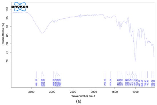
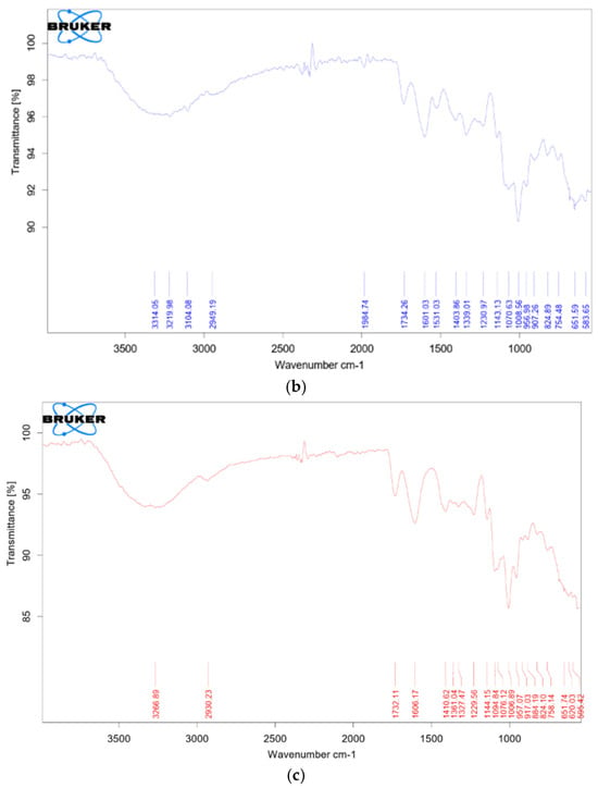
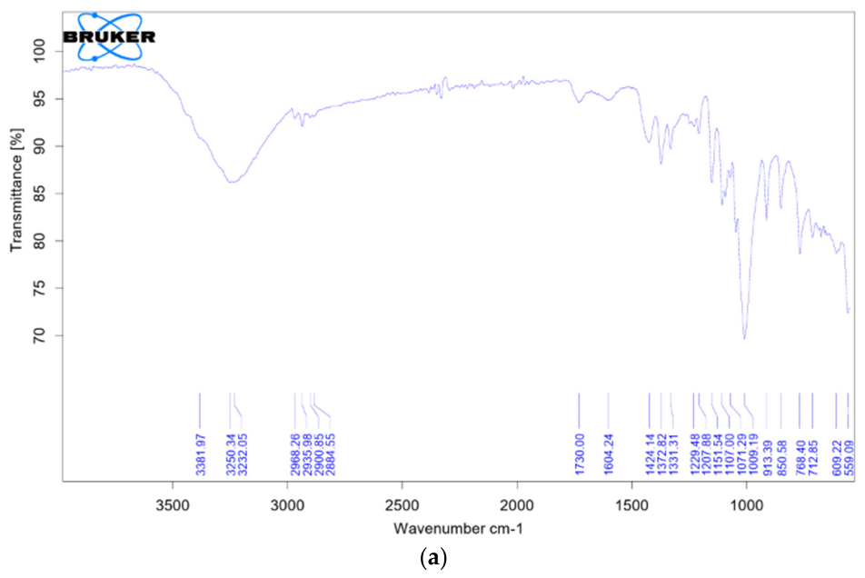
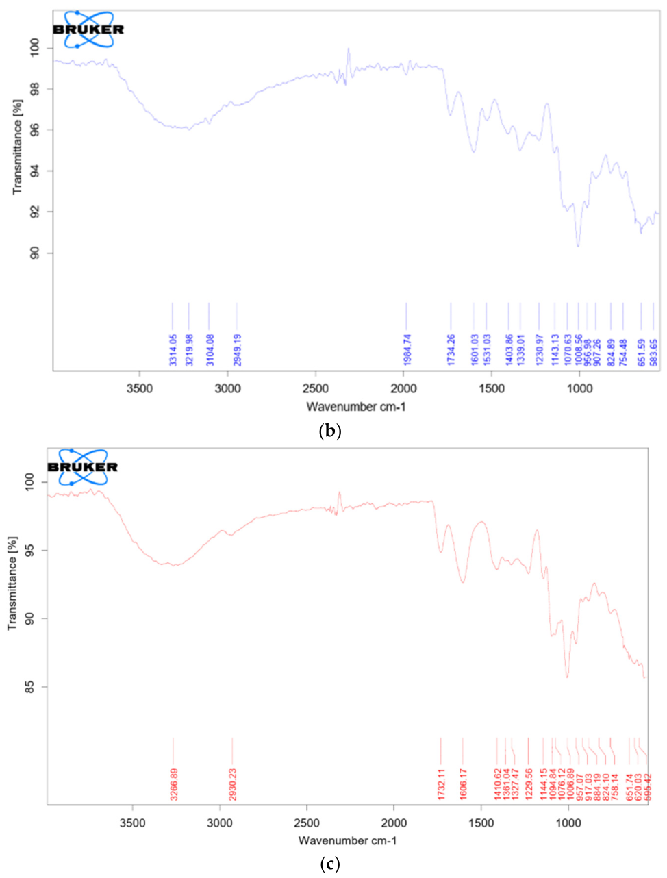
The FTIR spectrum (Figure 1) of pure pectin displayed a broad O–H stretching band at ~3368 cm−1 and a C–H stretching vibration at ~2930 cm−1. A comparatively weak ester carbonyl band was observed at 1738 cm−1, while asymmetric and symmetric stretching vibrations of carboxylate (COO−) groups appeared at 1604 and 1422 cm−1, respectively, consistent with reported spectra of citrus pectin and sodium pectate systems [9,23]. The comparatively low intensity of this ester C=O band, relative to the COO− bands, suggests that the extracted pectin has a relatively low degree of methyl-esterification; this is consistent with the acidic (pH 1.8–2.0) and thermal (80–85 °C) conditions used during extraction, which are known to promote partial acid-catalyzed de-esterification of the methyl ester groups on the galacturonan backbone, in addition to any low intrinsic esterification of the parent orange-peel pectin. Following coordination with Cu2+, the asymmetric and symmetric COO− bands shifted to 1601 and 1431 cm−1, respectively, resulting in a decrease in Δν (from 182 to 170 cm−1). Such variations in carboxylate stretching frequencies are indicative of metal–carboxylate coordination and reflect replacement of protonated carboxyl groups by Cu2+ ions within the polygalacturonate framework [17,23]. Comparable spectral behaviour has been documented for pectin–metal systems, where coordination predominantly involves ionized carboxylate sites without disruption of the glycosidic backbone [22]. In the Zn–pectin complex, the COO− asymmetric and symmetric bands appeared at 1606 and 1410 cm−1, respectively, with a corresponding change in Δν from native pectin. The effect of zinc coordination has been shown to mainly affect the carboxylate region while maintaining other structural aspects of the polymer [9], which is in agreement with the current observations. The maintenance of typical glycosidic bands in the 1200–1000 cm−1 region further supports that metal binding does not disrupt the underlying polysaccharide structure. In summary, the systematic changes in the COO− stretching vibrations and corresponding Δν values offer clear spectroscopic support for the existence of ionic coordination between Cu2+/Zn2+ metal ions and pectin carboxylate groups, as has been previously reported for metal–polygalacturonate complexes [23].
Figure 1.
FTIR spectra of (a) native pectin, (b) Cu–pectin, and (c) Zn–pectin complexes showing characteristic functional group vibrations.
3.2. X-Ray Diffraction Analysis
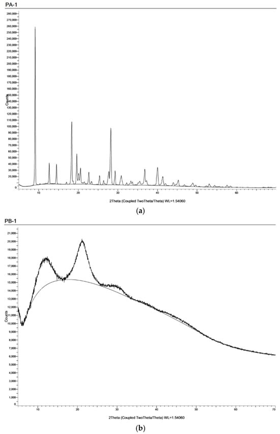
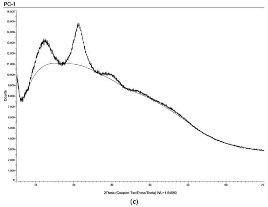
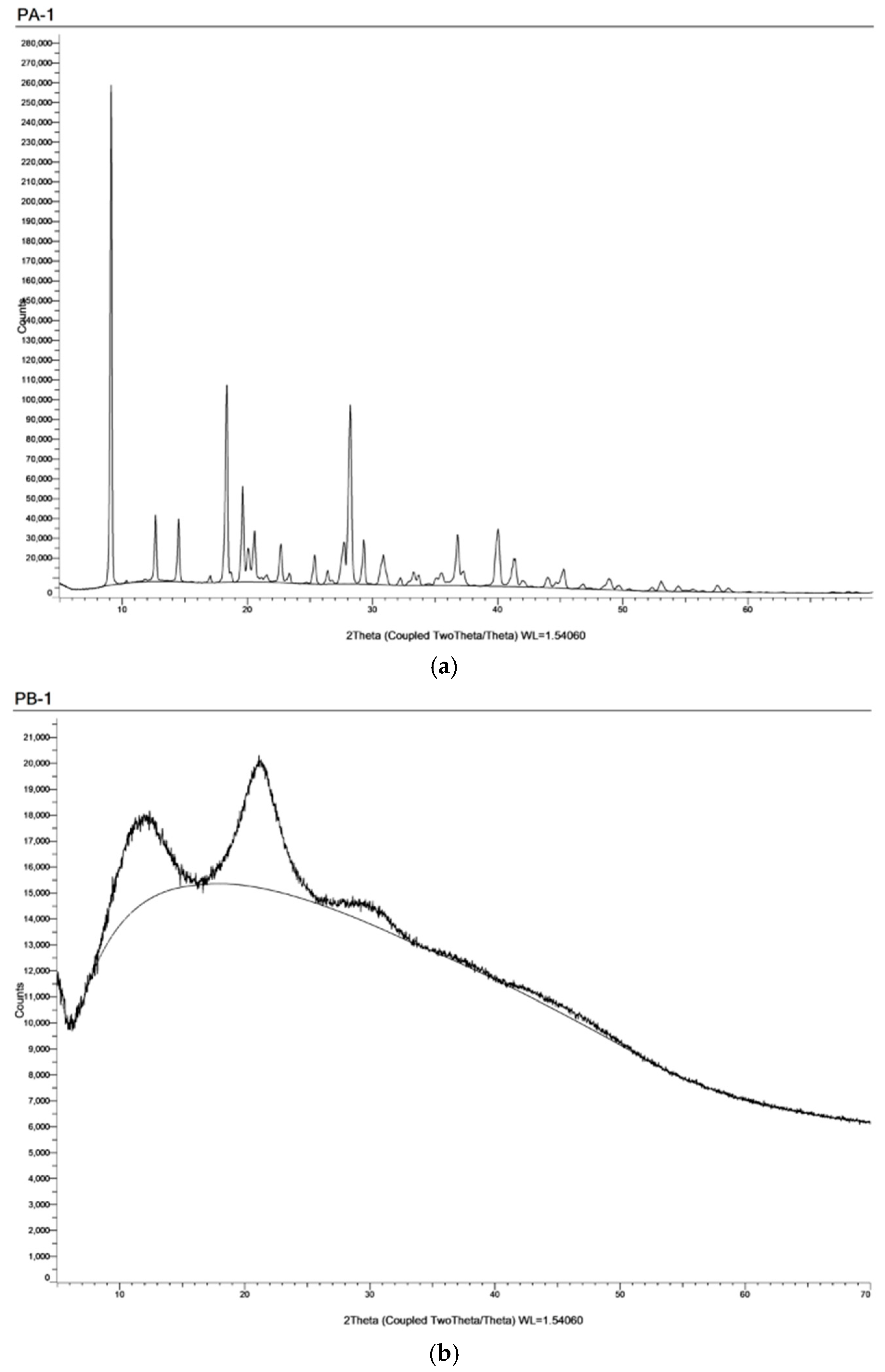
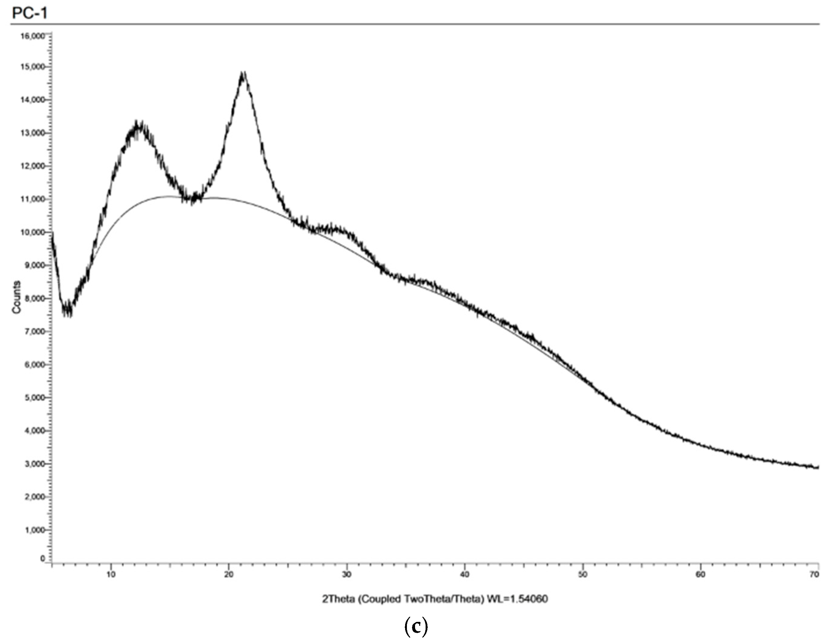
The XRD pattern of pure pectin (Figure 2) showed a broad diffraction halo around 2θ ≈ 18–22°, indicating its largely amorphous nature. A weak reflection at around 2θ ≈ 9–10° indicated a very small extent of short-range ordering due to hydrogen bonding between chains. Such diffraction patterns have been reported for pectate and polygalacturonate systems [23]. After the coordination reaction with Cu2+ and Zn2+ ions, the diffraction patterns showed a broad amorphous halo with a decrease in the intensity of the low-angle reflection and a slight broadening of the peaks. These observations suggest a partial loss of localized order due to metal–carboxylate interactions. Notably, no new sharp reflections of crystalline metal oxides or metal salts were detected, thus proving that the incorporation of the metal ions was achieved via ionic coordination rather than the formation of crystalline structures. Similar findings of the retention of amorphous characteristics after complexation with metal ions have also been reported for the case of polygalacturonate–metal complexes [17] and pectin–metal complexes [22]. From these findings, it is clear that the formation of homogeneous pectin–metal ionic complexes were achieved.
Figure 2.
XRD patterns of (a) native pectin, (b) Cu–pectin, and (c) Zn–pectin complexes showing amorphous diffraction profiles.
3.3. Scanning Electron Microscopy (SEM) Analysis
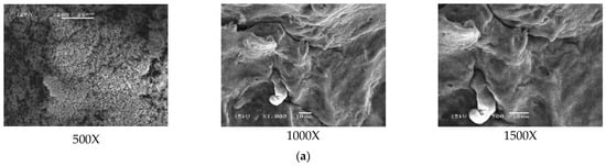
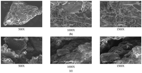
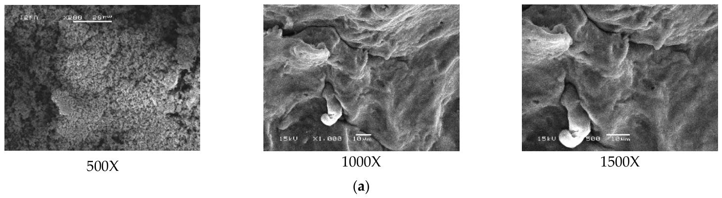
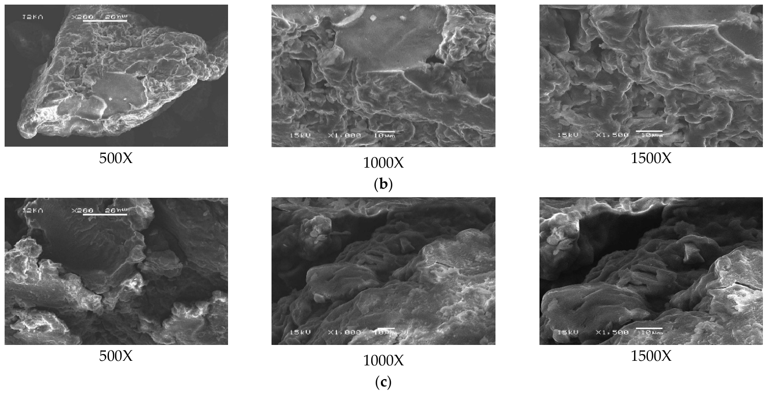
The SEM images (Figure 3) also showed distinct differences in the morphology of the native pectin and its complexes. The morphology of the native pectin was found to be irregular flakes with a smooth surface, irrespective of the magnification. At higher magnifications, loosely packed sheet-like structures could also be observed, as expected for dried amorphous structures [23]. The morphology of the copper–pectin complex was characterized by the presence of a clumped, compact region with a rough surface, indicating the fusion of particles. The transformation from dispersed flakes to clumped structures can be attributed to the increased intermolecular interactions, likely due to the ionic cross-linking between the copper ions and the carboxylate groups along the polymer chain. A similar observation of the compaction of the structure due to the formation of the complex has been reported for the complexes formed between the polygalacturonate structure and metal ions [17]. The structure of the zinc–pectin complex was found to be more packed and clumped compared to the native pectin structure. The structure was non-crystalline, but it showed increased aggregation and surface roughness. Such a morphological change triggered by coordination is in line with the observations made in pectin–metal complexes [22]. From the SEM analysis, it is apparent that the addition of divalent metal ions triggers a more compact and aggregated microstructure in the pectin matrix, indicating the successful formation of a coordinated ionic network.
Figure 3.
SEM micrographs of (a) native pectin, (b) Cu–pectin complex, and (c) Zn–pectin complex at different magnifications.
3.4. Energy-Dispersive X-Ray (EDX) Analysis
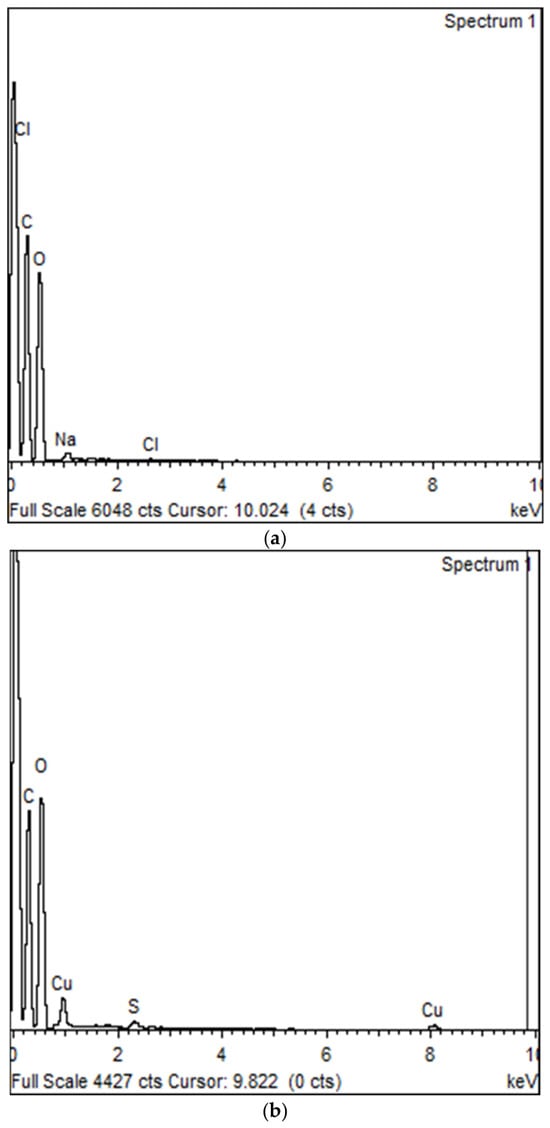
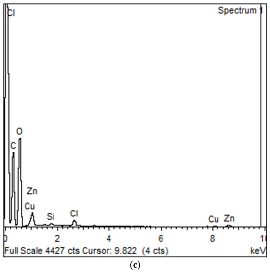
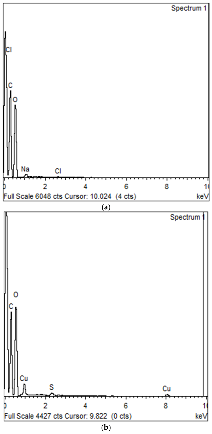
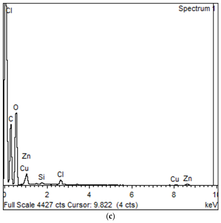
An Energy-Dispersive X-ray (EDX) (Figure 4) analysis of the pure pectin, copper–pectin, and zinc–pectin samples was performed to analyze their composition. The composition (Table 1) of the pure pectin sample was found to consist of carbon (44.41 wt%) and oxygen (54.66%), which indicates the polysaccharide nature of the pectin. In addition, there were traces of sodium and chlorine. No metal (Cu, Zn) peaks were observed in the spectrum, thus confirming that the native pectin was metal ion-free before complex formation. These minor Na and Cl signals are most likely residual mineral electrolytes intrinsic to the orange-peel biomass (citrus tissue naturally contains Na+, K+ and Cl−) that were carried through the acid extraction and ethanol-precipitation steps, rather than being introduced by the extraction reagents (citric acid and ethanol) used, which do not themselves contain Na or Cl; trace residues from the wash/rinse water cannot, however, be entirely excluded. The composition of the copper–pectin sample was found to consist of carbon (42.25 wt%), oxygen (50.79%), and copper (6.50%), thus confirming the successful formation of the metal–polymer complex. However, the metal content was found to be at moderate levels, thus indicating that the copper ions were only bound to the accessible sites of the macromolecule. This phenomenon can be explained by the coordination of copper ions with the carboxyl groups of the galacturonic acid units of the pectin molecule [9,12]. Similarly, the spectrum of zinc–pectin showed the presence of carbon (39.28 wt%) and oxygen (52.84 wt%), and a peak corresponding to zinc (5.07 wt%). This showed that zinc was present in the complex. However, the amount of zinc was not very high, indicating partial complexation of pectin, which might be related to the structural diversity and extent of esterification of pectin. In addition, similar results were reported for pectin–metal complexes [12,23]. It is also noted that a significant Cl content (1.37 wt%) was detected in the Zn–pectin complex and a smaller S content (0.47 wt%) in the Cu–pectin complex, corresponding to the Cl− and SO42− counter-ions of the ZnCl2 and CuSO4·5H2O precursor salts, respectively. This indicates that some loosely associated counter-ions/salt were retained despite washing, and that more extensive washing of the complexes would be advisable in future work and for reporting scientific communications to unambiguously distinguish coordinated metal from residual, non-coordinated salt. In conclusion, the presence of peaks corresponding to copper and zinc in Cu–pectin and Zn–pectin indicates successful complex formation, though not in high amounts. Similar results have been reported for complexes of metals and pectin, indicating the coordination of metals with carboxyl groups of pectin [12].
Figure 4.
Energy-dispersive X-ray (EDX) spectra of (a) native pectin, (b) copper–pectin complex, and (c) zinc–pectin complex.
Table 1.
Elemental composition (wt% and at%) of pure pectin, copper–pectin, and zinc–pectin obtained from EDX analysis.
3.5. Swelling Study
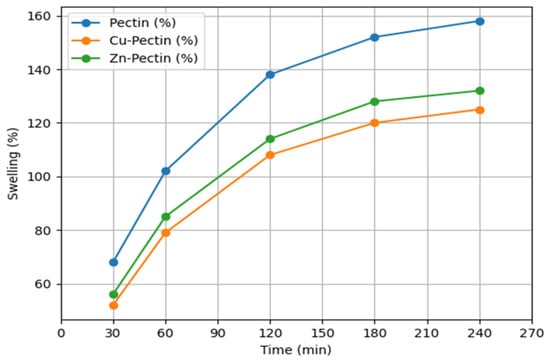
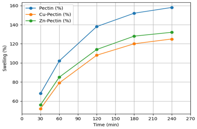
The swelling ability of native pectin, Cu–pectin, and Zn–pectin complexes is presented in Table 2 and Figure 5. Native pectin had the highest swelling ability throughout the experiment. The swelling ability of native pectin began at 68% after 30 min and reached 158% after 240 min. Conversely, Cu–pectin and Zn–pectin complexes recorded low swelling ability, i.e., 125% and 132%, respectively, after 240 min. The swelling ability of the complexes, i.e., pectin > Zn–pectin > Cu–pectin, indicates that the coordination of metals reduces the water-uptake ability of the polymer matrix [27]. This is possibly due to the hydrophilic hydroxyl and carboxyl groups in native pectin, which allow for the easier diffusion of water into the structure. After coordination with Cu2+ and Zn2+, it was observed that swelling of the material decreased, possibly forming a more compact structure. The coordination of metal ions and carboxyl groups of pectin restricts chain mobility and hence reduces the free volume [28]. The slightly lower swelling of Cu–pectin compared to Zn–pectin possibly indicates that copper ions formed a denser structure within the pectin structure.
Table 2.
Swelling percentage of pectin, Cu–pectin, and Zn–pectin complexes at different time intervals.
Figure 5.
Time-dependent swelling profiles of native pectin and its metal complexes (Cu–pectin and Zn–pectin).
3.6. pH Stability Study
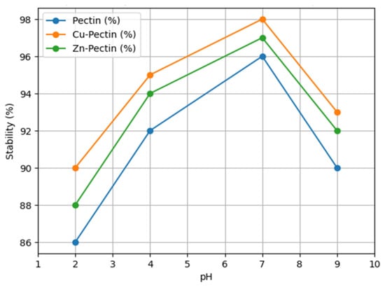
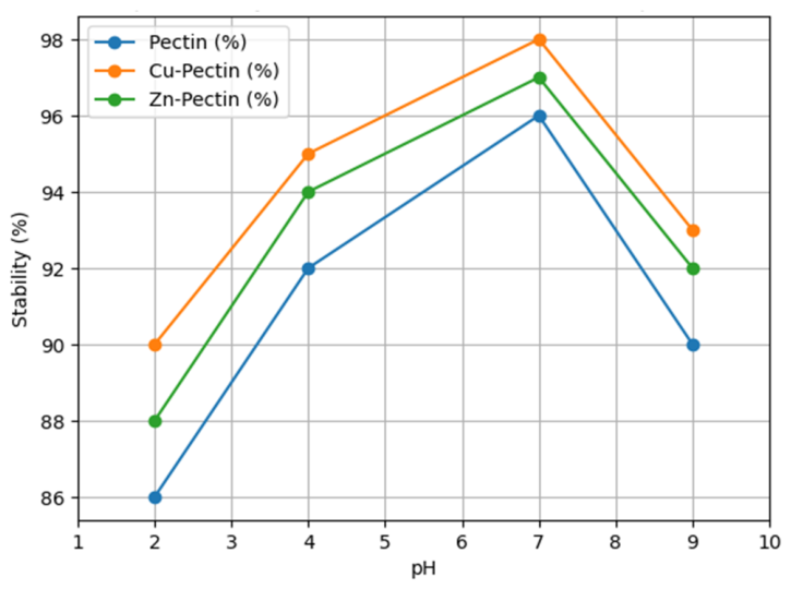
The pH stability study for native pectin and its metal complexes is depicted in Table 3 and Figure 6. Native pectin samples showed 86%, 92%, 96%, and 90% of their initial masses after being kept in solutions with pH 2, 4, 7, and 9, respectively. On the other hand, Cu–pectin samples showed 90%, 95%, 98%, and 93%, and Zn–pectin samples showed 88%, 94%, 97%, and 92% of their initial masses after being kept in solutions of pH 2, 4, 7, and 9, respectively. All samples showed maximum stability in pH 7 solutions. Lower stabilities of the samples were observed in acidic and alkaline solutions. This is in accordance with the pH-stability characteristics of pectin, in which pH stability is associated with the pH-dependent ionization of carboxyl groups and their effect on the overall integrity and stability of the polymer [29]. The increased stability of the complexes in relation to the native pectin might be related to coordination effects between the divalent metal ions and the carboxylate groups of the polymer backbone, leading to a more compact and rigid network and consequently increasing resistance to structural change in different pH media. Among all samples studied, the Cu–pectin complex was more stable, followed by Zn–pectin and then native pectin, confirming the effect of coordination on the physicochemical stability of pectin complexes [28].
Table 3.
pH stability (%) of pectin, Cu–pectin, and Zn–pectin complexes at different pH values.
Figure 6.
Stability (%) of pectin and metal–pectin complexes as a function of pH.
3.7. Antimicrobial Activity Assessment
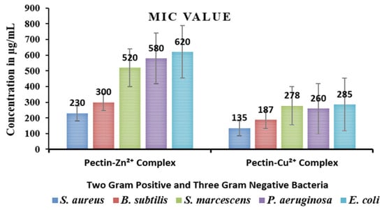
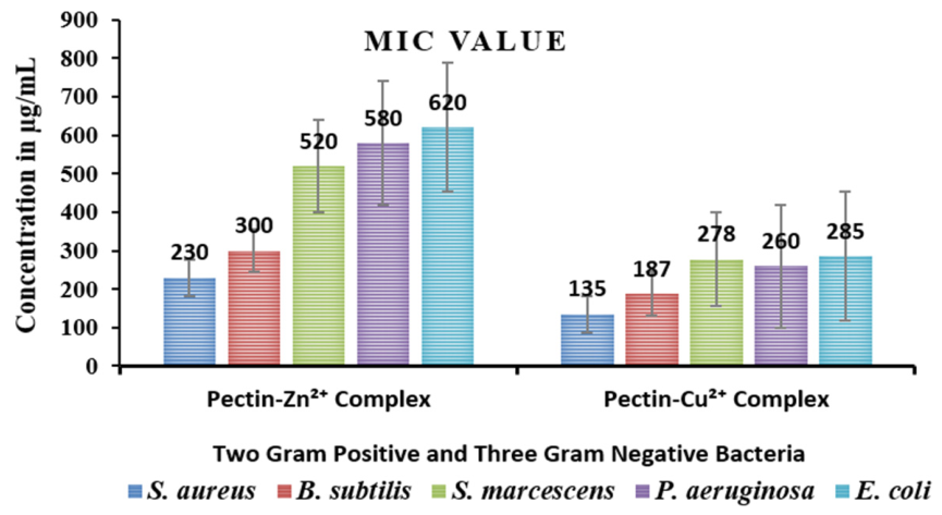
Using the minimum inhibitory concentration (MIC) method, the antibacterial activity of the pectin-based metal complexes was assessed against three Gram-negative bacteria (S. marcescens, P. aeruginosa, and E. coli) and two Gram-positive bacteria (S. aureus and B. subtilis) that is shown in Figure 7 The findings showed that both complexes showed significant antibacterial activity, but the zinc and copper derivatives’ efficacy differed noticeably. Gram-positive bacteria (S. aureus: 230 µg/mL; B. subtilis: 300 µg/mL) were more susceptible to the pectin–Zn2+ complex’s action than Gram-negative pathogens (S. marcescens: 520 µg/mL; P. aeruginosa: 580 µg/mL; E. coli: 620 µg/mL). The pectin–Cu2+ complex, on the other hand, showed considerably lower MIC values, ranging from 135 to 285 µg/mL, indicating increased antibacterial efficacy. Gram-negative bacteria (S. marcescens: 278 µg/mL; P. aeruginosa: 260 µg/mL; E. coli: 285 µg/mL) had somewhat higher MIC values than S. aureus (135 µg/mL) and B. subtilis (187 µg/mL).
Figure 7.
Antibacterial activity of pectin–Zn+2 and pectin–Cu+2 complexes.
In spite of the structural variations in their cell envelopes, the observed trend shows that both complexes are more efficient against Gram-positive bacteria than Gram-negative bacteria. Gram-negative bacteria have an extra lipopolysaccharide outer membrane that functions as a permeability barrier, preventing the complexes from penetrating. The inherent characteristics of copper ions can account for the greater antibacterial activity of the pectin–Cu2+ complex compared to the pectin–Zn2+ complex. Cu2+ is redox-active and can produce reactive oxygen species (ROS), which damage cellular components, cause oxidative stress, and interfere with essential biological activities. Moreover, strong interactions between copper ions and proteins and nucleic acids can block enzymes and cause cell death. Zn2+ ions, on the other hand, have relatively limited activity since they are redox-inactive and mainly work via inhibiting enzymes and destabilizing membranes. The carboxylate (–COO−) groups of pectin play a crucial role in coordinating the metal ions, enhancing the stability of the complexes and facilitating the controlled release of metal ions at the microbial surface. Generally, our results suggest that pectin’s antimicrobial ability is greatly increased by metal coordination, with the copper complex exhibiting encouraging activity and promise for future development as an antibacterial agent.
4. Conclusions
In this investigation, pectin was successfully isolated from orange peel waste and employed to form copper–pectin and zinc–pectin ionic complexes via coordination with divalent metal ions. The pectin extraction process was found to be effective in producing pectin with characteristics similar to those reported for pectin isolated from citrus fruits. Physicochemical characterization of the complexes confirmed the formation of metal–pectin complexes. The coordination of Cu2+ and Zn2+ with the carboxylate groups of pectin was confirmed by FTIR, whereas the retention of the amorphous structure of pectin was confirmed by XRD. The SEM analysis revealed changes in the surface morphology of the complexes, indicating a more compact form of pectin, which resulted from the ionic cross-linking of pectin. The presence of metal ions was confirmed by EDX analysis. The results of the swelling study showed a decrease in the water uptake capacity of pectin after coordination with metals, indicating a denser network. The pH stability of the complexes was also examined, which showed the increased stability of the complexes compared with the native pectin, especially at a neutral pH, with the Cu–pectin complex having the maximum stability. Most importantly, the antimicrobial study showed an increase in the antibacterial properties of the complexes compared with the native pectin, which confirmed that the addition of transition metals increases the bioactivity of the material. In conclusion, this research has shown a possible and efficient method for utilizing agricultural waste materials for the production of even more useful functional biomaterials. The metal complexes synthesized in this research have great potential for use as antimicrobial coatings, medicine, and even “green” packaging materials.
Author Contributions
Conceptualization, D.P. and M.M.; methodology, D.P. and M.M.; software, D.P. and M.M.; validation, D.P. and M.M.; formal analysis, D.P., M.M. and A.J.; investigation, D.P., M.M. and A.J.; resources, D.P., M.M. and A.J.; data curation, D.P., M.M. and A.J.; writing—original draft preparation, D.P. and M.M.; writing—review and editing, A.J. and M.V.; visualization, A.J. and M.V.; supervision, A.J.; project administration, A.J. All authors have read and agreed to the published version of the manuscript.
Funding
This research received no external funding.
Institutional Review Board Statement
Not applicable.
Informed Consent Statement
Not applicable.
Data Availability Statement
The original contributions presented in this study are included in the article. Further iquiries can be directed to the corresponding author.
Acknowledgments
The authors are thankful to the Department of Chemistry, Parul Institute of Applied Sciences, Faculty of Applied Sciences, Parul University, Vadodara and Department of Chemistry, Krishna School of Science (KSS), Kiran & Pallavi Patel Global University, Vadodara for providing the necessary laboratory facilities, instrumentation support and academic environment required to carry out this research. The institutional support and technical assistance received during the experimental and analytical phases of this work are gratefully acknowledged.
Conflicts of Interest
The authors declare no conflicts of interest.
References
- Renard, C.M.; Jarvis, M.C. Acetylation and methylation of homogalacturonans 2: Effect on ion-binding properties and conformations. Carbohydr. Polym. 1999, 39, 209–216. [Google Scholar] [CrossRef] [Scilit]
- Figueira, O.; Pereira, V.; Castilho, P.C. A two-step approach to orange peel waste valorization: Consecutive extraction of pectin and hesperidin. Foods 2023, 12, 3834. [Google Scholar] [CrossRef] [Scilit] [PubMed]
- Tiwari, A.K.; Saha, S.N.; Yadav, V.P.; Upadhyay, U.K.; Katiyar, D.; Mishra, T. Extraction and characterization of pectin from orange peels. Int. J. Biotechnol. Biochem. 2017, 13, 39–47. [Google Scholar]
- Alam, Y.B.; Tankoiko, S.; Matlubah, U.S.; Agustina, E.; Hidayati, I.; Andiarna, F. Isolation and Characterization of Pectin from Orange Peel (Citrus sp.). In Proceedings of the International Conference on Sustainable Health Promotion, Harstad, Norway, 10–12 June 2023; Volume 3, pp. 245–252. [Google Scholar]
- Akhter, M.J.; Sarkar, S.; Sharmin, T.; Mondal, S.C. Extraction of pectin from powdered citrus peels using various acids: An analysis contrasting orange with lime. Appl. Food Res. 2024, 4, 100614. [Google Scholar] [CrossRef] [Scilit]
- Fakayode, O.A.; Abobi, K.E. Optimization of oil and pectin extraction from orange (Citrus sinensis) peels: A response surface approach. J. Anal. Sci. Technol. 2018, 9, 1–6. [Google Scholar] [CrossRef] [Scilit]
- Seggiani, M.; Puccini, M.; Pierini, M.; Giovando, S.; Forneris, C. Effect of different extraction and precipitation methods on yield and quality of pectin. Int. J. Food Sci. Technol. 2009, 44, 574–580. [Google Scholar] [CrossRef] [Scilit]
- Wang, R.; Liang, R.; Dai, T.; Chen, J.; Shuai, X.; Liu, C. Pectin-based adsorbents for heavy metal ions: A review. Trends Food Sci. Technol. 2019, 91, 319–329. [Google Scholar] [CrossRef] [Scilit]
- Assifaoui, A.; Loupiac, C.; Chambin, O.; Cayot, P. Structure of calcium and zinc pectinate films investigated by FTIR spectroscopy. Carbohydr. Res. 2010, 345, 929–933. [Google Scholar] [CrossRef] [Scilit] [PubMed]
- Davarski, K.A.; Manolov, S.N.; Petrova, I.N.; Mavrov, V.D. Complex equilibria in the system M2+-polygalacturonic acid (pectin)-H2O (M=Co, Ni, Cu, Zn, Cd and Pb). J. Coord. Chem. 1994, 33, 75–81. [Google Scholar] [CrossRef] [Scilit]
- Dronnet, V.M.; Renard, C.M.; Axelos, M.A.; Thibault, J.F. Characterisation and selectivity of divalent metal ions binding by citrus and sugar-beet pectins. Carbohydr. Polym. 1996, 30, 253–263. [Google Scholar] [CrossRef] [Scilit]
- Khotimchenko, M.Y.; Kolenchenko, E.A.; Khotimchenko, Y.S. Zinc-binding activity of different pectin compounds in aqueous solutions. J. Colloid Interface Sci. 2008, 323, 216–222. [Google Scholar] [CrossRef] [Scilit] [PubMed]
- Debon, S.J.; Tester, R.F. In vitro binding of calcium, iron and zinc by non-starch polysaccharides. Food Chem. 2001, 73, 401–410. [Google Scholar] [CrossRef] [Scilit]
- Schlemmer, U. Studies of the binding of copper, zinc and calcium to pectin, alginate, carrageenan and gum guar in HCO−3 CO2 buffer. Food Chem. 1989, 32, 223–234. [Google Scholar] [CrossRef] [Scilit]
- Ngece, K.; Khwaza, V.; Paca, A.M.; Aderibigbe, B.A. The antimicrobial efficacy of copper complexes: A review. Antibiotics 2025, 14, 516. [Google Scholar] [CrossRef] [Scilit] [PubMed]
- Damena, T.; Alem, M.B.; Zeleke, D.; Desalegn, T.; Eswaramoorthy, R.; Demissie, T.B. Novel zinc (II) and copper (II) complexes of 2-((2-hydroxyethyl) amino) quinoline-3-carbaldehyde for antibacterial and antioxidant activities: A combined experimental, DFT, and docking studies. ACS Omega 2022, 7, 26336–26352. [Google Scholar] [CrossRef] [Scilit] [PubMed]
- Nurmatova, M.; Sabirova, K.; Rashidova, S. Synthesis and Properties of Polymer Metal Complexes of Polygalacturonic Acid With d-Metal Ions. Int. J. Polym. Sci. 2025, 2025, 4216357. [Google Scholar] [CrossRef] [Scilit]
- Nemtarev, A.V.; Kuznetsova, E.V.; Yergeshov, A.A.; Eflova, D.S.; Ishkaeva, R.A.; Valiullina, I.R.; Mironov, V.F.; Salakhieva, D.V.; Abdullin, T.I. Synthesis and Comparative Study of the Structure and Antibacterial Activity of Polygalacturonate Complexes with Ionic and Nanoparticulate Silver. Polymers 2025, 17, 2798. [Google Scholar] [CrossRef] [Scilit] [PubMed]
- Kudzin, M.H.; Boguń, M.; Mrozińska, Z.; Kaczmarek, A. Physical properties, chemical analysis, and evaluation of antimicrobial response of new polylactide/alginate/copper composite materials. Mar. Drugs 2020, 18, 660. [Google Scholar] [CrossRef] [Scilit] [PubMed]
- Piacenza, E.; Presentato, A.; Alduina, R.; Scurria, A.; Pagliaro, M.; Albanese, L.; Meneguzzo, F.; Ciriminna, R.; Chillura Martino, D.F. Cross-linked natural IntegroPectin films from Citrus biowaste with intrinsic antimicrobial activity. Cellulose 2022, 29, 5779–5802. [Google Scholar] [CrossRef] [Scilit]
- Gawkowska, D.; Cybulska, J.; Zdunek, A. Structure-related gelling of pectins and linking with other natural compounds: A review. Polymers 2018, 10, 762. [Google Scholar] [CrossRef] [Scilit] [PubMed]
- Ma, X.; Jing, J.; Yu, J.; Wang, J.; Zhu, H.; Hu, Z. Synthesis and characterization of a novel apple pectin–Fe (III) complex. ACS Omega 2021, 6, 1391–1399. [Google Scholar] [CrossRef] [Scilit] [PubMed]
- Minzanova, S.T.; Mironov, V.F.; Vyshtakalyuk, A.B.; Tsepaeva, O.V.; Mironova, L.G.; Mindubaev, A.Z.; Nizameev, I.R.; Kholin, K.V.; Milyukov, V.A. Complexation of pectin with macro-and microelements. Antianemic activity of Na, Fe and Na, Ca, Fe complexes. Carbohydr. Polym. 2015, 134, 524–533. [Google Scholar] [CrossRef] [Scilit] [PubMed]
- Mendes, J.P.; Esperança, J.M.; Medeiros, M.J.; Pawlicka, A.; Silva, M.M. Structural, morphological, ionic conductivity, and thermal properties of pectin-based polymer electrolytes. Mol. Cryst. Liq. Cryst. 2017, 643, 266–273. [Google Scholar] [CrossRef] [Scilit]
- Vora, M.; Gosai, D.P.; Dhobi, T.K.; Patel, N.J.; Thakor, P.; Thakkar, A.B.; Rojmala, J.V.; Waghela, B.N. Investigation of heterocycle-fused 2, 5-dihydro-1H-benzo[b,1,4]diazepine conjugate as potential anti-cancer agents: Design, synthesis and in silico docking activity. Synth. Commun. 2026, 13, 1–7. [Google Scholar] [CrossRef] [Scilit]
- Lunagariya, M.V.; Thakor, K.P.; Varma, R.R.; Waghela, B.N.; Pathak, C.; Patel, M.N. Synthesis, characterization and biological application of 5-quinoline 1, 3, 5-trisubstituted pyrazole based platinum (ii) complexes. MedChemComm 2018, 9, 282–298, Correction in MedChemCommun 2018, 9, 383–384. https://doi.org/10.1039/c8md90005a. [Google Scholar] [PubMed]
- Sriamornsak, P.; Thirawong, N.; Weerapol, Y.; Nunthanid, J.; Sungthongjeen, S. Swelling and erosion of pectin matrix tablets and their impact on drug release behavior. Eur. J. Pharm. Biopharm. 2007, 67, 211–219. [Google Scholar] [CrossRef] [Scilit] [PubMed]
- Han, S.S.; Ji, S.M.; Park, M.J.; Suneetha, M.; Uthappa, U.T. Pectin based hydrogels for drug delivery applications: A mini review. Gels 2022, 8, 834. [Google Scholar] [CrossRef] [Scilit] [PubMed]
- Kapoor, D.U.; Garg, R.; Gaur, M.; Pareek, A.; Prajapati, B.G.; Castro, G.R.; Suttiruengwong, S.; Sriamornsak, P. Pectin hydrogels for controlled drug release: Recent developments and future prospects. Saudi Pharm. J. 2024, 32, 102002. [Google Scholar] [CrossRef] [Scilit] [PubMed]
- Lunagariya, M.V.; Thakor, K.P.; Waghela, B.N.; Vaidya, F.U.; Pathak, C.; Patel, M.N. Design, synthesis, MTT assay, DNA interaction studies of platinum (II) complexes. J. Biomol. Struct. Dyn. 2018, 36, 14–31. [Google Scholar] [CrossRef] [Scilit] [PubMed]

Disclaimer/Publisher’s Note: The statements, opinions and data contained in all publications are solely those of the individual author(s) and contributor(s) and not of MDPI and/or the editor(s). MDPI and/or the editor(s) disclaim responsibility for any injury to people or property resulting from any ideas, methods, instructions or products referred to in the content. |
© 2026 by the authors. Licensee MDPI, Basel, Switzerland. This article is an open access article distributed under the terms and conditions of the Creative Commons Attribution (CC BY) license.