


Abstract
Human dihydrofolate reductase (hDHFR) is a key enzyme in folate metabolism and an established therapeutic target in cancer and other diseases. Methotrexate (MTX), a clinically used hDHFR inhibitor, is effective but associated with toxicity and resistance, highlighting the need for alternative inhibitors. Here, we developed and validated a structure-based pharmacophore model derived from the hDHFR–MTX crystal complex and applied it to DrugBank for drug repurposing. Pharmacophore screening, Lipinski filtering, molecular docking, GNINA CNN scoring, and protein–ligand interaction analysis were integrated to prioritize potential hDHFR-targeting drugs. Ertugliflozin emerged as a promising computational candidate, warranting further molecular dynamics and experimental validation.
1. Introduction
Human dihydrofolate reductase (hDHFR) is a key enzyme in folate metabolism that catalyzes the reduction in dihydrofolate to tetrahydrofolate, which is required for purine and thymidylate biosynthesis and consequently for DNA synthesis and cellular proliferation [1]. Its central role in cellular metabolism has established hDHFR as an important therapeutic target, particularly in cancer and inflammatory diseases [2]. Methotrexate (MTX) is one of the most widely used hDHFR inhibitors and has demonstrated considerable clinical success in the treatment of several cancers and immune-mediated inflammatory diseases [3]. However, its therapeutic application remains limited by adverse effects, suboptimal pharmacokinetic properties, and the emergence of resistance mechanisms, including hDHFR overexpression and mutations [4,5,6,7]. These limitations support the search for alternative hDHFR inhibitors with improved pharmacological profiles.
Drug repurposing provides an efficient strategy for identifying new therapeutic applications for existing drugs, potentially reducing the time, cost, and risks associated with conventional drug development [8,9,10]. Computer-aided drug design (CADD), particularly pharmacophore-based virtual screening, can facilitate this process by identifying compounds capable of reproducing the key spatial and chemical features required for target recognition [11,12].
In this study, we developed and validated a structure-based pharmacophore model derived from the hDHFR–MTX crystal complex and applied it to the DrugBank database to identify existing drugs with potential hDHFR inhibitory activity. Pharmacophore screening was combined with Lipinski filtering, molecular docking, GNINA CNN scoring, and protein–ligand interaction analysis to prioritize candidates for further investigation. This integrated workflow provides a computational strategy for identifying potential hDHFR drug-repurposing candidates and prioritizing compounds for subsequent molecular dynamics simulations and experimental validation.
2. Materials and Methods
2.1. Pharmacophore Modeling
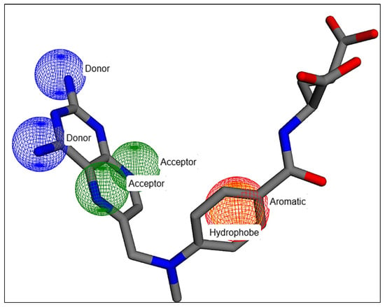
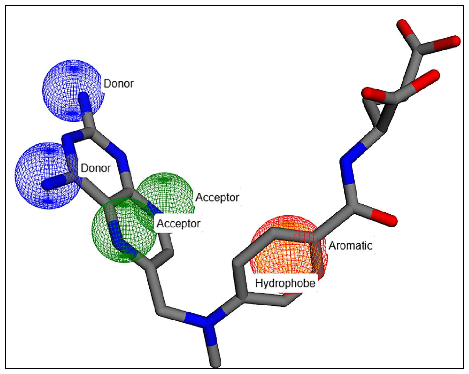
A structure-based pharmacophore model was generated from the crystal structure of the hDHFR–MTX complex (PDB ID: 1U72) [13] using Pharmit [14]. The final model comprised six essential features, including two hydrogen bond acceptors, two hydrogen bond donors, one aromatic feature, and one hydrophobic region (Figure 1). These features represent the key interactions required for ligand recognition within the hDHFR active site.
Figure 1.
Pharmacophore model.
2.2. Pharmacophore Validation
The pharmacophore model was evaluated using an external validation dataset of 410 compounds, comprising 38 experimentally confirmed active compounds and 372 inactive compounds, independently compiled in our previous hDHFR QSAR study [15]. Model performance was assessed using ROC-AUC, Average Precision (AP), Enrichment Factor (EF), and Güner–Henry (GH) metrics [16,17,18,19].
2.3. DrugBank Virtual Screening
The validated pharmacophore model was applied to the DrugBank database (version 5.1.21, released in June 2026), comprising 11,904 approved, investigational, and experimental drugs. DrugBank is a comprehensive resource containing drug and drug-related information [20]. Compounds satisfying all pharmacophore constraints were subsequently filtered according to Lipinski’s Rule of Five [21]. Duplicate structures were removed before molecular docking.
2.4. Molecular Docking
Molecular docking was performed using Smina (15 October 2019 release), developed by David R. Koes and collaborators at the University of Pittsburgh, Pittsburgh, Pennsylvania, USA. Smina is based on AutoDock Vina (version 1.1.2) [22,23]. The hDHFR structure from PDB entry 1U72 was prepared by removing the co-crystallized MTX and water molecules. Ligands were converted to three-dimensional conformations before docking. The search space was centered on the MTX-binding site at x = 30.62, y = 16.88, and z = −1.62 Å, with dimensions of 30 × 30 × 30 Å. Exhaustiveness was set to 10, with a maximum of 10 poses generated per ligand and a random seed of 42. The highest-ranked pose according to the predicted Smina affinity was retained for subsequent analysis. The docking protocol was validated by redocking independently prepared MTX into the prepared receptor using the same parameters. The best-ranked redocked pose was compared with the crystallographic MTX conformation using heavy-atom root-mean-square deviation (RMSD).
2.5. GNINA Scoring and PLIP Analysis
Four prioritized complexes, comprising MTX (CID 126941), CID 446744, CID 46937152, and ertugliflozin (CID 44814423), were additionally evaluated using GNINA v1.0.1 [24]. GNINA docking was performed using the same receptor and search-box coordinates, with exhaustiveness set to 40, up to 10 docking modes, and a random seed of 42. The resulting poses were evaluated using GNINA binding affinity, CNN pose score, and CNN affinity. The highest-ranked pose was retained for structural analysis.
Protein–ligand interactions were characterized using the Protein–Ligand Interaction Profiler (PLIP) [25,26], including hydrogen bonds, hydrophobic contacts, salt bridges, π-stacking, and halogen bonds. Interactions involving the NADPH cofactor were excluded from comparative interpretation because they were consistently observed across the complexes. Selected poses were visualized using BIOVIA Discovery Studio Visualizer v25.1.0.24284.
3. Results
3.1. Pharmacophore Validation
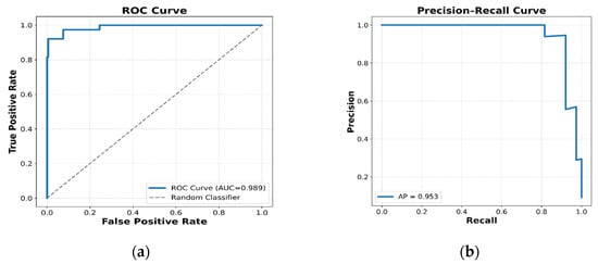
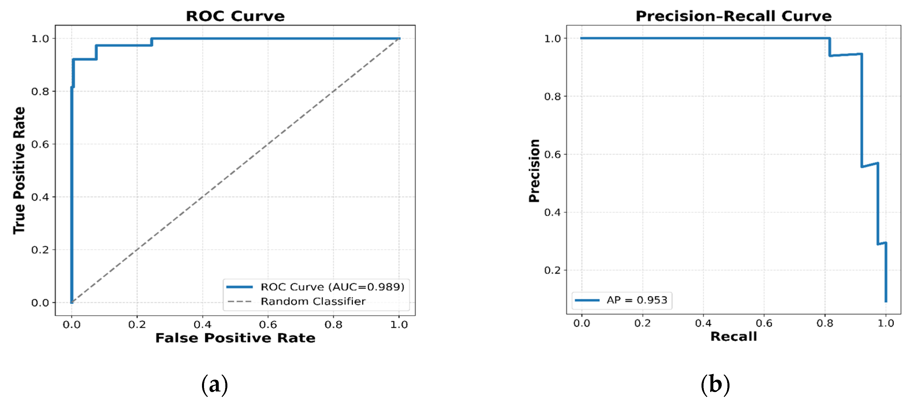
The final structure-based pharmacophore model consisted of six features: two hydrogen-bond acceptors, two hydrogen-bond donors, one aromatic feature, and one hydrophobic feature (Figure 1). Validation against the external dataset of 410 compounds demonstrated excellent discrimination between active and inactive compounds, with a ROC-AUC of 0.989 and an AP of 0.95 (Figure 2). The model also showed strong early enrichment at the top 5% of the ranked database, with an EF of 10.79 and a GH score of 0.882. These results demonstrate a high capacity to prioritize experimentally active hDHFR ligands and support the application of the model for virtual screening.
Figure 2.
Validation of the pharmacophore model: (a) Receiver Operating Characteristic (ROC) curve showing the model’s discrimination between active and inactive compounds (AUC = 0.989). The dashed line represents a random classifier. (b) Corresponding Precision–Recall curve (Average Precision, AP = 0.95).
3.2. DrugBank Virtual Screening
Screening of the 11,904 DrugBank compounds against the validated pharmacophore identified 212 compounds satisfying all pharmacophore constraints. Lipinski filtering reduced this set to 35 drug-like compounds, and removal of duplicate structures resulted in 15 unique compounds for molecular docking.
3.3. Molecular Docking
The Smina docking protocol was first validated by redocking the crystallographic MTX ligand. The best-ranked redocked pose showed a predicted affinity of −11.1 kcal/mol and a heavy-atom RMSD of 1.122 Å relative to the crystallographic conformation. The RMSD below 2.0 Å indicates successful reproduction of the experimental binding orientation and supports the use of the docking protocol for the screened compounds.
The 15 pharmacophore-selected compounds exhibited predicted Smina affinities ranging from −7.0 to −11.3 kcal/mol (Table 1). CID 446744 showed the strongest predicted affinity (−11.3 kcal/mol), followed by ertugliflozin (CID 44814423; −11.1 kcal/mol) and CID 46937152 (−9.9 kcal/mol). Overall, nine of the 15 compounds (60%) exhibited predicted affinities ≤ −9.0 kcal/mol.
Table 1.
Smina docking results for MTX and all 15 hDHFR candidates.
Importantly, experimentally characterized DHFR inhibitor scaffolds were recovered among the highest-ranked compounds. CID 446744, a 2,4-diaminopteridine derivative, has previously been reported as a DHFR inhibitor [27,28], while CID 46937152 has been structurally characterized as a potent and selective DHFR inhibitor in complex with mouse DHFR [29]. These compounds therefore provided useful retrospective evidence that the screening workflow could recover chemically relevant DHFR ligands.
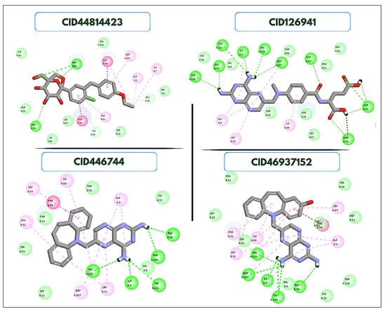
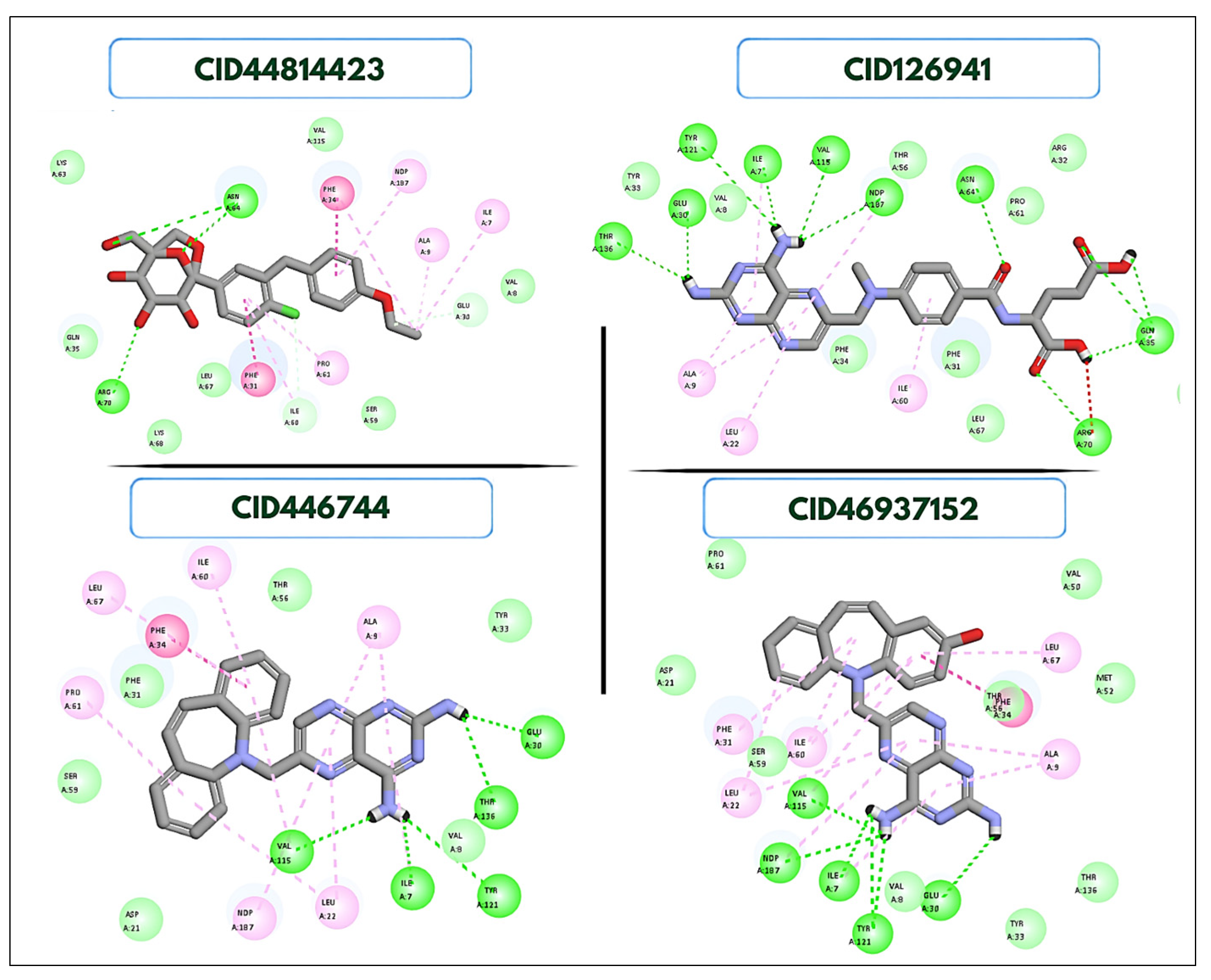
Ertugliflozin (CID 44814423) exhibited a predicted affinity of −11.1 kcal/mol, comparable to the redocked MTX reference. The docking poses of the three highest-ranked compounds are shown in Figure 3.
Figure 3.
Predicted binding modes of the prioritized compounds in the hDHFR MTX-binding site, with protein–ligand interactions visualized using BIOVIA Discovery Studio Visualizer.
3.4. GNINA CNN Scoring and PLIP Analysis
GNINA evaluation provided complementary assessment of the four prioritized complexes (Table 2). MTX (CID 126941) showed the highest CNN pose score (0.9710), followed by CID 446744 (0.7122), ertugliflozin (0.6202), and CID 46937152 (0.5499). The corresponding GNINA binding affinities were −11.23, −11.30, −11.58, and −8.45 kcal/mol, respectively. Ertugliflozin therefore showed the most favorable GNINA binding affinity among the four compounds, although its CNN pose score was lower than those of MTX and CID 446744.
Table 2.
GNINA docking and scoring results for MTX and three prioritized hDHFR candidates.
PLIP analysis revealed that MTX (CID 126941) formed an extensive interaction network involving hydrogen bonds, salt bridges, and hydrophobic contacts with residues including Ile7, Gln35, Glu30, Arg70, and Thr136 (Table 3). CID 446744 retained interactions with Ile7, Glu30, and Thr136, together with multiple hydrophobic contacts. CID 46937152 formed hydrogen bonds with Ile7 and Thr136 and a salt bridge with Glu30, as well as π-stacking with Phe34.
Table 3.
PLIP-derived protein–ligand interactions of MTX and prioritized DrugBank compounds.
Ertugliflozin (CID 44814423) displayed a distinct interaction pattern, including hydrogen bonds with Asn64 and Arg70, hydrophobic contacts with Ile7, Phe31, Phe34, and Ile60, π-stacking with Phe34, and a halogen bond involving Ser59. These interactions provided a structural rationale for its predicted binding within the hDHFR active site.
4. Discussion
The results demonstrate that the validated structure-based pharmacophore model efficiently prioritized compounds capable of reproducing the key interaction features of the hDHFR–MTX complex. Its high ROC-AUC and AP values, together with strong EF and GH scores, indicate good discrimination and early enrichment of active compounds. The recovery of previously characterized DHFR inhibitor scaffolds among the highly ranked DrugBank compounds provides an additional retrospective indication that the screening workflow captured chemically relevant hDHFR-binding chemotypes.
The docking results further prioritized several compounds with predicted affinities comparable to or stronger than the redocked MTX reference. CID 446744 and ertugliflozin (CID 44814423) showed Smina affinities of −11.3 and −11.1 kcal/mol, respectively. The recovery of CID 446744 and CID 46937152 is consistent with previous experimental evidence for their DHFR inhibitory activity [26,27,28], supporting the ability of the pharmacophore-based workflow to identify known target-relevant compounds.
Ertugliflozin (CID 44814423) was particularly noteworthy because it combined a favorable Smina docking affinity with the most favorable GNINA binding affinity among the four complexes analyzed. PLIP further revealed several interactions within the hDHFR active site, including contacts involving Phe34 and Ile7 that are also represented in the interaction network of other prioritized compounds. However, the distinct interaction profile of ertugliflozin (CID 44814423) should not be interpreted as evidence of experimentally confirmed hDHFR inhibition. Docking scores and CNN-based predictions are computational estimates and cannot establish binding affinity or biological activity.
An additional limitation is that the present analysis represents a static structural assessment. Protein flexibility, solvent effects, ligand protonation states, and the energetic stability of the predicted complexes were not fully addressed. Molecular dynamics simulations and subsequent biochemical inhibition assays will therefore be necessary to evaluate the stability and biological relevance of the predicted ertugliflozin–hDHFR complex. Nevertheless, the combination of pharmacophore validation, retrospective recovery of known DHFR inhibitors, docking, GNINA scoring, and interaction analysis provides a rational basis for prioritizing compounds for experimental investigation.
Author Contributions
Conceptualization, I.M. and N.E.A.; methodology, I.M.; software, I.M.; validation, I.M. and N.E.A.; formal analysis, I.M.; investigation, I.M.; data curation, I.M.; writing—original draft preparation, I.M.; writing—review and editing, I.M. and N.E.A.; visualization, I.M.; supervision, N.E.A.; project administration, N.E.A. All authors have read and agreed to the published version of the manuscript.
Funding
This research received no external funding.
Institutional Review Board Statement
Not applicable.
Informed Consent Statement
Not applicable.
Data Availability Statement
The data supporting the findings of this study are available from the corresponding authors upon reasonable request.
Acknowledgments
During the preparation of this manuscript, the authors used OpenAI ChatGPT (GPT-5.5) for language editing, text refinement, and improvement of scientific writing. The authors reviewed and edited the output and took full responsibility for the content of this publication.
Conflicts of Interest
The authors declare no conflicts of interest.
Abbreviations
The following abbreviations are used in this manuscript:
| AP | Average Precision |
| CADD | Computer-Aided Drug Design |
| CID | Compound Identifier (PubChem Compound ID) |
| CNN | Convolutional Neural Network |
| DHFR | Dihydrofolate Reductase |
| EF | Enrichment Factor |
| GH | Güner–Henry Score |
| hDHFR | Human Dihydrofolate Reductase |
| HBA | Hydrogen Bond Acceptor |
| HBD | Hydrogen Bond Donor |
| MD | Molecular Dynamics |
| MTX | Methotrexate |
| NADPH | Nicotinamide Adenine Dinucleotide Phosphate |
| PDB | Protein Data Bank |
| PLIP | Protein–Ligand Interaction Profiler |
| QSAR | Quantitative Structure–Activity Relationship |
| RMSD | Root Mean Square Deviation |
| ROC | Receiver Operating Characteristic |
| ROC-AUC | Area Under the Receiver Operating Characteristic Curve |
| RO5 | Rule of Five |
| VS | Virtual Screening |
References
- Abali, E.E.; Skacel, N.E.; Celikkaya, H.; Hsieh, Y. Regulation of Human Dihydrofolate Reductase Activity and Expression. Vitam. Horm. 2008, 79, 267–292. [Google Scholar] [CrossRef] [Scilit] [PubMed]
- Hsieh, Y.-C.; Tedeschi, P.; AdeBisi Lawal, R.; Banerjee, D.; Scotto, K.; Kerrigan, J.E.; Lee, K.-C.; Johnson-Farley, N.; Bertino, J.R.; Abali, E.E. Enhanced Degradation of Dihydrofolate Reductase through Inhibition of NAD Kinase by Nicotinamide Analogs. Mol. Pharmacol. 2013, 83, 339–353. [Google Scholar] [CrossRef] [Scilit] [PubMed]
- Tripathy, S.; Dassarma, B.; Jha, B.; Azad, M.A.; Saha, I.; Gupta, P.; Pramanik, D.D.; Chosdol, K.; Pramanik, A. Advances in Methotrexate Nano-Formulations to Enhance Therapeutic Efficacy against Drug-Resistant Breast Cancer. Discov. Nano 2026, 21, 96. [Google Scholar] [CrossRef] [Scilit] [PubMed]
- Rana, S.; Dranchak, P.; Dahlin, J.L.; Lamy, L.; Li, W.; Oliphant, E.; Shrimp, J.H.; Rajacharya, G.H.; Tharakan, R.; Holland, D.O.; et al. Methotrexate-Based PROTACs as DHFR-Specific Chemical Probes. Cell Chem. Biol. 2024, 31, 221–233.e14. [Google Scholar] [CrossRef] [Scilit] [PubMed]
- Neradil, J.; Pavlasovsa, G.; Sramek, M.; Kyr, M.; Veselska, R.; Sterba, J. DHFR-Mediated Effects of Methotrexate in Medulloblastoma and Osteosarcoma Cells: The Same Outcome of Treatment with Different Doses in Sensitive Cell Lines. Oncol. Rep. 2015, 33, 2169–2175. [Google Scholar] [CrossRef] [Scilit] [PubMed]
- Menon, A.; Murugesan, P.; Tamilarasan, P.P.; Palanisamy, S.A.; Parthasarathy, A.K. Beyond Folate Antagonism: The Unique Pharmacological Blueprint of Methotrexate. Int. J. Basic Clin. Pharmacol. 2026, 15, 195–200. [Google Scholar] [CrossRef] [Scilit]
- Göker, E.; Waltham, M.; Kheradpour, A.; Trippett, T.; Mazumdar, M.; Elisseyeff, Y.; Schnieders, B.; Steinherz, P.; Tan, C.; Berman, E. Amplification of the Dihydrofolate Reductase Gene Is a Mechanism of Acquired Resistance to Methotrexate in Patients with Acute Lymphoblastic Leukemia and Is Correlated with p53 Gene Mutations. Blood 1995, 86, 677–684. [Google Scholar] [CrossRef] [Scilit]
- Akodad, S.; Niu, X.; Secades, B.; Stevens, H. Impact of Drug Repurposing between 1985 and 2024 on Pharmaceutical Innovation. Commun. Med. 2026, 6, 84. [Google Scholar] [CrossRef] [Scilit] [PubMed]
- Recino, A.; Rayner, M.L.D.; Rohn, J.L.; Della Pasqua, O.; Carter, B.; Cupani, A.; Dahlmann-Noor, A.; Foltynie, T.; Furman, J.; Hobbiger, S.; et al. Therapeutic Innovation in Drug Repurposing: Challenges and Opportunities. Drug Discov. Today 2025, 30, 104390. [Google Scholar] [CrossRef] [Scilit] [PubMed]
- Kulkarni, V.S.; Alagarsamy, V.; Solomon, V.R.; Jose, P.A.; Murugesan, S. Drug Repurposing: An Effective Tool in Modern Drug Discovery. Russ. J. Bioorganic Chem. 2023, 49, 157–166. [Google Scholar] [CrossRef] [Scilit] [PubMed]
- Giordano, D.; Biancaniello, C.; Argenio, M.A.; Facchiano, A. Drug Design by Pharmacophore and Virtual Screening Approach. Pharmaceuticals 2022, 15, 646. [Google Scholar] [CrossRef] [Scilit] [PubMed]
- Zheng, M. Ligand-Based Virtual Screening. In Artificial Intelligence for Drug Design; Li, H., Zheng, M., Zhu, F., Bai, F., Eds.; Springer: Singapore, 2026; pp. 745–759. [Google Scholar] [CrossRef] [Scilit]
- Cody, V.; Luft, J.R.; Pangborn, W. Understanding the Role of Leu22 Variants in Methotrexate Resistance: Comparison of Wild-Type and Leu22Arg Variant Mouse and Human Dihydrofolate Reductase Ternary Crystal Complexes with Methotrexate and NADPH. Acta Crystallogr. Sect. D Biol. Crystallogr. 2005, 61, 147–155. [Google Scholar] [CrossRef] [Scilit] [PubMed]
- Koes, D.R. The Pharmit Backend: A Computer Systems Approach to Enabling Interactive Online Drug Discovery. IBM J. Res. Dev. 2018, 62, 3:1–3:6. [Google Scholar] [CrossRef] [Scilit] [PubMed]
- Maattallaoui, I.; Sakho, M.; Maatallaoui, A.; Catalán, E.B.; Aouad, N.E. Development of QSAR Models and Web Applications for Predicting hDHFR Inhibitor Bioactivity Using Machine Learning. Molecules 2025, 30, 4618. [Google Scholar] [CrossRef] [Scilit] [PubMed]
- Fawcett, T. An Introduction to ROC Analysis. Pattern Recognit. Lett. 2006, 27, 861–874. [Google Scholar] [CrossRef] [Scilit]
- Davis, J.; Goadrich, M. The Relationship between Precision-Recall and ROC Curves. In Proceedings of the 23rd International Conference on Machine Learning; ACM Press: New York, NY, USA, 2006; pp. 233–240. [Google Scholar] [CrossRef] [Scilit]
- Lätti, S.; Niinivehmas, S.; Pentikäinen, O.T. ROCKER: Open Source, Easy-to-Use Tool for AUC and Enrichment Calculations and ROC Visualization. J. Cheminformatics 2016, 8, 45. [Google Scholar] [CrossRef] [Scilit] [PubMed]
- Junaidin, J.; Lestari, D.; Kurniawan, M.F.; Khairul Ikram, N.K. Ligand-Based Pharmacophore Modeling, Molecular Docking, and Molecular Dynamic Studies of HMG-CoA Reductase Inhibitors. Inform. Med. Unlocked 2022, 32, 101063. [Google Scholar] [CrossRef] [Scilit]
- Wishart, D.S.; Knox, C.; Guo, A.C.; Shrivastava, S.; Hassanali, M.; Stothard, P.; Chang, Z.; Woolsey, J. DrugBank: A Comprehensive Resource for in Silico Drug Discovery and Exploration. Nucleic Acids Res. 2006, 34, D668–D672. [Google Scholar] [CrossRef] [Scilit] [PubMed]
- Lipinski, C.A.; Lombardo, F.; Dominy, B.W.; Feeney, P.J. Experimental and Computational Approaches to Estimate Solubility and Permeability in Drug Discovery and Development Settings. Adv. Drug Deliv. Rev. 2001, 46, 3–26. [Google Scholar] [CrossRef] [Scilit] [PubMed]
- Koes, D.R.; Baumgartner, M.P.; Camacho, C.J. Lessons Learned in Empirical Scoring with Smina from the CSAR 2011 Benchmarking Exercise. J. Chem. Inf. Model. 2013, 53, 1893–1904. [Google Scholar] [CrossRef] [Scilit] [PubMed]
- Trott, O.; Olson, A.J. AutoDock Vina: Improving the Speed and Accuracy of Docking with a New Scoring Function, Efficient Optimization, and Multithreading. J. Comput. Chem. 2010, 31, 455–461. [Google Scholar] [CrossRef] [Scilit] [PubMed]
- McNutt, A.T.; Francoeur, P.; Aggarwal, R.; Masuda, T.; Meli, R.; Ragoza, M.; Sunseri, J.; Koes, D.R. GNINA 1.0: Molecular Docking with Deep Learning. J. Cheminformatics 2021, 13, 43. [Google Scholar] [CrossRef] [Scilit] [PubMed]
- Salentin, S.; Schreiber, S.; Haupt, V.J.; Adasme, M.F.; Schroeder, M. PLIP: Fully Automated Protein–Ligand Interaction Profiler. Nucleic Acids Res. 2015, 43, W443–W447. [Google Scholar] [CrossRef] [Scilit] [PubMed]
- Schake, P.; Bolz, S.N.; Linnemann, K.; Schroeder, M. PLIP 2025: Introducing Protein–Protein Interactions to the Protein–Ligand Interaction Profiler. Nucleic Acids Res. 2025, 53, W463–W465. [Google Scholar] [CrossRef] [Scilit] [PubMed]
- Rosowsky, A.; Cody, V.; Galitsky, N.; Fu, H.; Papoulis, A.T.; Queener, S.F. Structure-Based Design of Selective Inhibitors of Dihydrofolate Reductase: Synthesis and Antiparasitic Activity of 2,4-Diaminopteridine Analogues with a Bridged Diarylamine Side Chain. J. Med. Chem. 1999, 42, 4853–4860. [Google Scholar] [CrossRef] [Scilit] [PubMed]
- Rosowsky, A.; Fu, H.; Chan, D.C.M.; Queener, S.F. Synthesis of 2,4-Diamino-6-[2′-O-(ω-Carboxyalkyl)Oxydibenz[b,f]azepin-5-yl]methylpteridines as Potent and Selective Inhibitors of Pneumocystis carinii, Toxoplasma gondii, and Mycobacterium avium Dihydrofolate Reductase. J. Med. Chem. 2004, 47, 1475–1486. [Google Scholar] [CrossRef] [Scilit] [PubMed]
- Cody, V.; Pace, J.; Rosowsky, A. Structural Analysis of a Holoenzyme Complex of Mouse Dihydrofolate Reductase with NADPH and a Ternary Complex with the Potent and Selective Inhibitor 2,4-Diamino-6-(2′-Hydroxydibenz[b,f]azepin-5-yl)methylpteridine. Acta Crystallogr. Sect. D Biol. Crystallogr. 2008, 64, 977–984. [Google Scholar] [CrossRef] [Scilit] [PubMed]

Disclaimer/Publisher’s Note: The statements, opinions and data contained in all publications are solely those of the individual author(s) and contributor(s) and not of MDPI and/or the editor(s). MDPI and/or the editor(s) disclaim responsibility for any injury to people or property resulting from any ideas, methods, instructions or products referred to in the content. |
© 2026 by the authors. Licensee MDPI, Basel, Switzerland. This article is an open access article distributed under the terms and conditions of the Creative Commons Attribution (CC BY) license.