


Abstract
Species of the genus Pyrus are used as food (their fruits) and in traditional medicine for various ailments (their leaves). Previous studies have reported polyphenols, hydroquinone glycosides, triterpenes, and sterols in pear leaf extracts. This study comparatively characterized the micromorphological features and total polyphenol and flavone contents of Pyrus communis L. leaves collected from Popești-Leordeni and Răcoasa, Romania, and assessed the preliminary toxicity of the corresponding extracts. Phytotoxicity was evaluated using the Triticum aestivum assay over a concentration range of 3.33–0.03%, and lethality toward invertebrates was assessed at concentrations below 1% using the Artemia franciscana Kellogg, 1906 bioassay.
1. Introduction
Pyrus communis L. (European pear), Rosaceae, is widely cultivated for its edible fruits, whereas its leaves are a less extensively investigated plant material with potential phytochemical value. Pear leaves contain several classes of specialized metabolites, including phenolic acids, flavonoids, flavan-3-ols, procyanidins, hydroquinone derivatives such as arbutin, and triterpenoids [1,2,3]. Comparative analyses of different anatomical parts of pear have shown that leaves may contain substantially higher concentrations of phenolic compounds than the pulp, peel, and seeds and may also exhibit considerable antioxidant activity [2]. These findings support further investigation of pear leaves as a potential source of biologically active constituents.
The phenolic composition of pear leaves is not constant. Differences have been reported among cultivars and rootstocks and according to leaf developmental stage, mechanical injury, and growing conditions [1,3]. In particular, environmental variables such as radiation exposure, temperature, and nutrient availability may affect the biosynthesis and accumulation of phenolic compounds involved in plant protection and adaptation [1]. Pear leaves grown under different radiation environments have shown marked differences in hydroxycinnamic acids and flavonoids, while natural field conditions were associated with higher concentrations of several phenolic classes [1]. Consequently, comparisons between plant materials collected from different geographical areas may provide useful information about variation in their morphological and phytochemical characteristics. However, the effects of environmental conditions must be interpreted together with other potential sources of variation, including genotype, rootstock, leaf age, and cultivation practices.
Leaf anatomical and surface characteristics provide complementary information about plant identity and structural variability. Microscopic investigations of pear leaves have shown differences in traits such as lamina and palisade thickness, palisade-to-spongy tissue ratio, and cuticle development [4]. Optical microscopy and scanning electron microscopy can therefore contribute to the characterization and comparison of leaf samples. In addition, the potential use of phenolic-rich plant extracts requires preliminary evaluation of their biological safety. Plant-based assays such as the Triticum aestivum test offer simple and cost-effective methods for detecting concentration-dependent effects on germination, root growth, and, when cytogenetic endpoints are included, meristematic cell division [5]. Artemia franciscana nauplii provide a complementary invertebrate model in which mortality can be used as an endpoint for preliminary toxicity screening [6].
In this context, the present study aimed to compare P. communis leaves collected from two geographical areas of Romania, Răcoasa in Vrancea County and Popești-Leordeni in the peri-urban Bucharest–Ilfov area. The samples were characterized macroscopically and microscopically using optical microscopy and scanning electron microscopy, and their total phenolic and total flavonoid contents were determined using spectrophotometric methods. The potential phytotoxicity of the corresponding extracts was investigated using the Triticum assay, while lethality toward an invertebrate model was assessed using A. franciscana nauplii. By integrating morphological, phytochemical, and preliminary toxicological observations, this work seeks to characterize variability in pear leaves originating from two Romanian regions and to provide initial information regarding their potential as sources of bioactive compounds.
2. Materials and Methods
2.1. Materials
The plant material consisted of pear (Pyrus communis L.) leaves collected during August and September from private household gardens in two regions of Romania. In each region, leaves were collected from 10 trees and pooled to obtain one composite regional sample. The PB sample was collected in Popești-Leordeni, Ilfov County (44.3693194° N, 26.1438812° E), from trees approximately 10 years old. The PV sample was collected in Răcoasa, Vrancea County (45.9909924° N, 26.8809485° E), from trees approximately 20 years old. The two regional composite samples were processed and analyzed separately. Soil properties and local meteorological variables were not measured directly at individual trees; information from official regional sources [7,8] was used only to describe the general environmental context. A voucher specimen (no. 26-27/2016) was deposited in the Department of Pharmaceutical Botany and Cellular Biology. Species identity was confirmed macroscopically and microscopically using optical microscopy and scanning electron microscopy (SEM).
Reagents: NaOH/iodine green/alum carmine/Na2CO3 (sodium carbonate)/AlCl3/CH3COONa (sodium acetate)/methanol/Folin–Ciocâlteu reagent/quercetin/gallic acid.
2.2. Methods
Surface preparations were cleared with 5% NaOH, and transverse sections were double-stained with iodine green and alum carmine and examined under a Nikon Labophot-2 microscope (Nikon Corporation, Tokyo, Japan; 10× eyepiece, 4×–40× objectives) [9]. The results were compared with data from the specialized literature [10,11].
2.2.1. Scanning Electron Microscopy (SEM) Analysis
The surface characteristics of leaves from both samples were analyzed using scanning electron microscopy (SEM). Analysis was performed on a high-resolution dual-beam electron microscope (SEM/FIB), model Quanta 3D FEG (FEI Company, Hillsboro, OR, USA), operating at an accelerating voltage of 5 kV in low-vacuum mode (~50 Pa pressure), equipped with a low-vacuum secondary electron detector (LVSED). The samples were minimally prepared by mounting on a double-adhesive carbon tape, without coating [12].
2.2.2. Total Polyphenol Content
The total polyphenol content was determined using the Folin–Ciocâlteu spectrophotometric method. The powdered plant material (<250 μm) was extracted with 50% methanol at a plant-to-solvent ratio of 5 g/100 mLThe extraction was carried out in an ultrasonic bath (Elmasonic S15H, Elma Schmidbauer GmbH, Singen, Germany) for 30 min at 45 °C. The extract solution was centrifuged for 10 min at 6000 rpm (C2 centrifuge, Centurion Scientific Ltd., Chichester, West Sussex, UK). A 50-fold dilution was also made from the supernatant for determination. Then, 1 mL was transferred to a 10 mL volumetric flask. 5 mL of Folin–Ciocâlteu reagent diluted in water (1:10) was added to the solution, and the flask was filled to the mark with 7.5% Na2CO3.
The control sample was prepared under the same conditions, but distilled water was used instead of the Folin–Ciocâlteu reagent. The prepared samples were kept in the dark for 1 h. Absorbance was measured at 765 nm using a HALO DB-20 UV-VIS spectrophotometer (Dynamica GmbH, Salzburg-Mayrwies, Austria). The reference solution was prepared from gallic acid in 70% methanol and used to construct a 7-point calibration curve (r = 0.9996).
2.2.3. Flavonoid Assay
The total flavonoid content was determined by a spectrophotometric method based on chelation with AlCl3 in sodium acetate (Romanian Pharmacopoeia, 10th edition, Cynarae folium monograph). The plant material was extracted in the same manner as for polyphenols. For determination, 0.5 mL of the extract solution from the supernatant was placed in a 10 mL reaction flask, 2 mL of 10% sodium acetate and 1 mL of 0.25% AlCl3 were added, and the volume was made up to the mark with 50% methanol. Absorbance was measured at 430 nm against a 10-point calibration curve of quercetin (r = 0.9992) [13,14].
2.2.4. Artemia Lethality Test
The lethality of the extracts was investigated using the Artemia franciscana Kellogg test (Ocean Star International, Inc., Snowville, UT, USA), as described previously [15]. Extract solutions were tested at concentrations of 1%, 0.5%, 0.25%, 0.125%, and 0.0675%, and artificial seawater was used as the negative control. Expanded experimental details are provided in Supplementary Methods S1.
2.2.5. Triticum Test (Phytotoxicity Test)
The toxicity of the pear leaf extracts was investigated using the Constantinescu phytobiological method (Triticum aestivum assay), as described previously [16,17]. The hydroalcoholic extract solutions were evaporated to dryness, and the residues were redissolved in distilled water. Five concentrations were prepared: 3.3%, 2.5%, 1.66%, 0.33%, and 0.033%. Distilled water was used as the negative control. Expanded experimental details are provided in Supplementary Methods S2.
2.3. Statistical Analysis
All statistical modeling and data visualizations were performed in R (version 4.5.3) using the “brms” [18,19], “nlme” [20,21], “emmeans” [22], “drc” [23], and “ggplot2” [24] packages.
Due to the lack of normality and heteroscedasticity (residual variance being narrower at higher concentrations), root length was analyzed with a Bayesian distributional regression model. The model was built with the “brms” R package (through a “cmdstanr” [25] interface) under a Student’s t-distribution family. The latter includes a degrees-of-freedom parameter (ν) that controls tail thickness, providing more reliable results compared to the Gaussian family. Both the location parameter (μ) and the log-scale residual scale parameter (σ) were modeled simultaneously as functions of the predictors. Root length was modeled as a function of day (main effect), extract, concentration (treated as a factor to be able to detect discrete level effects), and the extract × concentration interaction. We accounted for residual heteroscedasticity by allowing the residual scale parameter (σ) to change with concentration levels. Posterior sampling was done using Hamiltonian Monte Carlo (HMC) with No-U-Turn Sampling (NUTS) across 4 chains. Each chain ran for 2000 iterations, including 1000 warmup draws, resulting in 4000 post-warmup draws. Model convergence was confirmed with potential scale reduction factors (R^ = 1.00) and sufficient effective sample sizes (Bulk-ESS > 1000, Tail-ESS > 1000). Post hoc comparisons of each concentration with the control (similar to Dunnett’s test) were made using emmeans from the posterior distribution. Differences were deemed statistically credible if the 95% credible intervals did not include zero.
To ensure that the conclusions were reliable regardless of the modeling approach used, a classical Generalized Least Squares (GLS) regression model was performed as a sensitivity analysis with the “nlme” package. Heteroscedasticity was addressed by using a variance function (varIdent (form = ~ 1|Conc)) to provide separate residual variance estimates for each concentration level. Pairwise Dunnett-adjusted post hoc tests (p < 0.05) were used to compare these results with those from the primary Bayesian model.
In addition to testing categorical contrasts, median inhibitory concentration (IC50) values were estimated by treating concentration as a continuous variable. Four-parameter log-logistic models (LL.4) were built for the two extracts using the drc package, with separate models for each day and pooled across all three days, as the latter option would increase statistical power. To prevent larger values from Day 3 from having too strong an influence over the pooled model, root lengths were normalized as a percentage of the daily control mean. Because delta-method calculations can yield biologically impossible negative lower bounds, 95% confidence intervals were estimated using a non-parametric bootstrapping approach. For each fitted model, 1999 bootstrap replicates were generated by resampling with replacement; for the pooled analysis, resampling was stratified by Day to maintain the original day-level balance of the experimental design. The dose–response model was refit to each replicate and the IC50 re-estimated; replicates where the model failed to converge were excluded. Point estimates were taken as the IC50 from the model fitted to the original data, and 95% confidence intervals were calculated using the bias-corrected and accelerated (BCa) method or the percentile method where BCa could not be computed, using the “boot” package [26,27] in R.
3. Results and Discussion
Macroscopic analysis of the pear leaves collected from the two geographical regions of Romania confirmed, based on the description in the Flora Republicii Populare Romîne, that the species is Pyrus communis L., syn. Pyrus sativa DC., syn. Pyrus communis var. sativa DC.
3.1. Micromorphological and Anatomical Analysis
3.1.1. Light Microscopy (LM)
Although collected from two geographical regions characterized in the specialized literature by different pedoclimatic conditions [7,8], these did not result in significant morpho-anatomical differences, as expected. Thus, both the PV and PB samples showed a characteristic leaf structure: heterogeneously asymmetric, with bistratified palisade tissue. In the midrib, the collateral vascular bundle was surrounded by a sclerenchymatous pericycle. These anatomical characteristics are consistent with previous studies [28].
3.1.2. Scanning Electron Microscopy (SEM)
Scanning electron microscopy (SEM) analysis revealed the characteristics of the adaxial and abaxial epidermis of the pear leaves collected from the two geographical regions. Stomata were observed only on the abaxial surface of the leaf (Supplementary Figure S1). The wax deposits on the abaxial surface show the same epicuticular pattern (striations) for both analyzed samples, and the shape is similar to that reported in the study by Fernández V. et al. (2008) [29]. The adaxial surface of the PB samples appears to present a higher density of wax crystals compared to PV. This may be explained by the plant’s need to adapt to a more polluted environment in the peri-urban area and to stress factors such as dehydration. No tectorial hairs were observed on either of the examined leaf surfaces [30].
3.2. Determination of Active Principles (Polyphenols, Flavones)
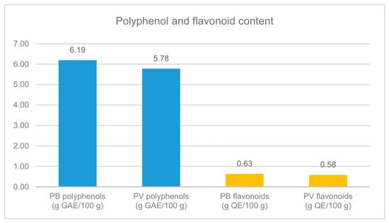
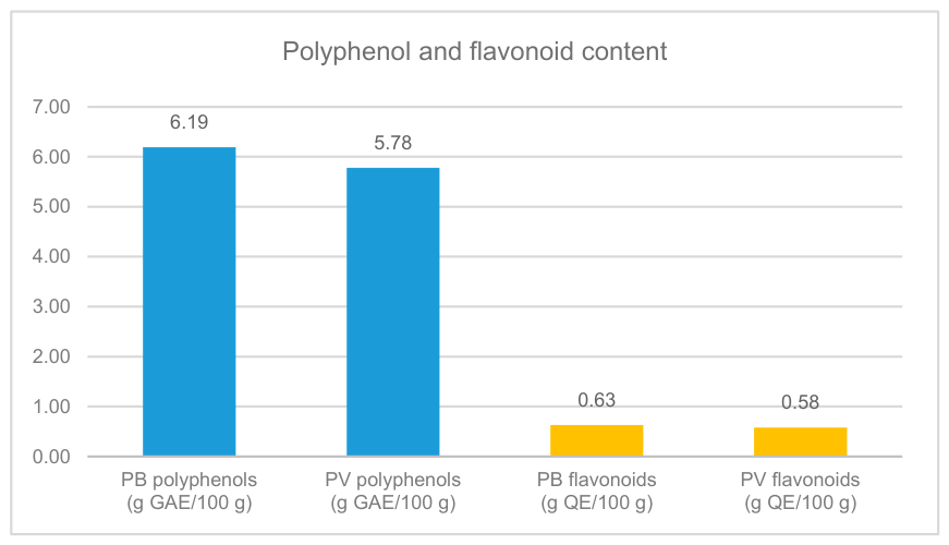
The polyphenol and flavone content was found to be higher in extracts from plant products collected in the Bucharest metropolitan area (Popești-Leordeni, Ilfov). The results are presented in Table 1 and Figure 1.
Table 1.
Total polyphenol and flavone contents of Pyrus communis L. leaf samples collected from two geographical regions of Romania.
Figure 1.
Polyphenol and flavone content of plant products collected from the two geographical regions.
It is well known that secondary metabolites, including polyphenols and flavones, are produced by plants in response to abiotic and biotic stress factors [31,32,33]. The plant materials were collected during the same period of the year and at the same vegetative stage; however, the sampled trees differed in approximate age (about 10 years in Popești-Leordeni and about 20 years in Răcoasa), and soil and local meteorological variables were not measured directly. Therefore, the observed quantitative differences should be interpreted as comparative regional observations and cannot be attributed exclusively to environmental conditions.
3.3. Determination of Phytotoxicity of Plant Products
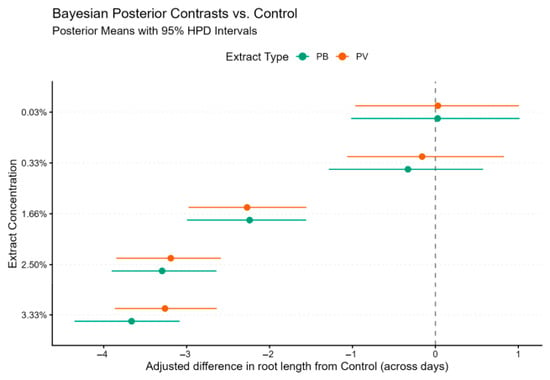
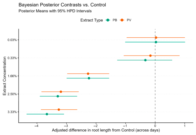
Root growth in the Triticum assay exhibited pronounced concentration-dependent inhibition for both extracts (PB and PV). Relative to Day 1, root length increased on Day 2 (βDay 2 = 0.43, 95% HPD interval: [0.30, 0.57]) and Day 3 (βDay 3 = 0.64, 95% HPD interval: [0.51, 0.79]). Bayesian posterior contrasts for root growth relative to the negative control are shown in Figure 2.
Figure 2.
Forest plot illustrating the Bayesian posterior contrasts for Triticum root growth compared with the negative control at different extract concentrations. Overlapping intervals between PB and PV across all concentrations indicate no statistically credible difference between the two extracts.
Dunnett-style posterior contrasts comparing each concentration with the negative control indicated that the two lowest concentrations, 0.03% and 0.33%, produced no credible inhibition. Credible inhibition was observed beginning at 1.66% for both extracts, whereas severe phytotoxicity occurred at the higher concentrations, with the greatest mean reductions observed at 3.33%. No credible effect of extract origin was identified (βPV = 0.02, 95% HPD interval: [−0.83, 0.90]), and no credible extract × concentration interaction was detected. The frequentist GLS sensitivity analysis was consistent with the Bayesian results and identified the same concentration threshold, with statistically significant inhibition beginning at 1.66% for both PB and PV (Dunnett-adjusted p < 0.001). Thus, the conclusions were robust to the statistical modeling approach.
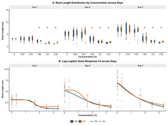
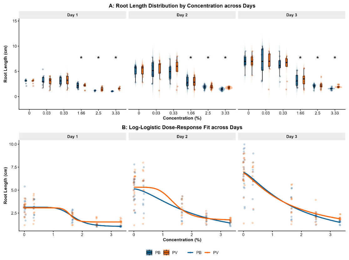
The global four-parameter log-logistic models fitted to the pooled, control-normalized data estimated similar IC50 values for the two extracts (Table 2). Although the IC50 for PV was slightly lower than for PB and its confidence interval was narrower, the intervals overlapped substantially. IC50 estimates for each Day (Table 3) were characterized by wide, overlapping confidence intervals, particularly for PB. These findings are consistent with those from the primary Bayesian model, which also detected no statistically credible difference between the two extracts. The observed root-length distributions and fitted dose–response curves across the three measurement days are shown in Figure 3.
Table 2.
Global log-logistic IC50 estimates (pooled across days).
Table 3.
Day-specific log-logistic IC50 estimates.
Figure 3.
Effect of plant extract concentration on Triticum root growth across the 3 days of measurement. (A) Violin plots showing the variation in root lengths by extract, concentration, and measurement day. Violins indicate kernel density distributions, boxplots indicate medians and interquartile ranges, and points represent individual observations. Asterisks (*) mark groups exhibiting credible inhibition relative to the control based on Bayesian 95% HPD intervals. (B) Four-parameter log-logistic dose–response curves fitted across the three measurement days.
3.4. Determination of Lethality of Plant Products
The Artemia test showed no mortality after 24 h for either pear leaf extract under the tested concentrations and experimental conditions. Further assay details are provided in Supplementary Methods S1.
4. Conclusions
The comparative analysis of Pyrus communis L. leaves collected from the two Romanian regions revealed no major morpho-anatomical differences between the samples. The PB sample from Ilfov County exhibited higher total polyphenol and flavone contents than the PV sample from Vrancea County. These differences may be associated with the environmental conditions specific to the two regions; however, a direct causal relationship cannot be established based on the available data.
In the Triticum assay, both extracts inhibited root elongation at concentrations of 1.66% and above. No statistically credible difference between PB and PV was detected. Neither extract caused mortality in Artemia franciscana under the tested concentrations and experimental conditions.
Future research should focus on the identification and individual quantification of phenolic and flavonoid compounds, as well as on the evaluation of the biological activities and safety of the extracts. Such investigations may clarify the potential use of pear leaves as a source of bioactive compounds.
The main limitations of this study were the inability to identify the cultivar due to the absence of fruits, the difference in the approximate ages of the sampled trees between regions, the use of trees grown in private household gardens rather than a controlled orchard, and the absence of direct soil and local meteorological measurements at the sampling sites. Consequently, the available regional environmental information should be interpreted only as contextual, and no direct causal relationship between environmental conditions and the observed phytochemical differences can be established.
Supplementary Materials
The following supporting information can be downloaded at: https://www.mdpi.com/article/10.3390/chemproc2026022116/s1, Figure S1: SEM micrographs of the abaxial surface of Pyrus communis L. leaves collected from Ilfov and Vrancea counties; Figure S2: Cytogenetic observations in root-tip cells of Triticum aestivum exposed to pear leaf extracts; Figure S3: Sampling locations of Pyrus communis L. leaf material in Romania; Supplementary Method S1: Artemia franciscana lethality assay; Supplementary Method S2: Triticum aestivum phytotoxicity assay.
Author Contributions
Conceptualization, A.-I.A., C.-S.S. and R.A.; methodology, A.-I.A. and R.A.; validation, A.-I.A., C.-S.S., and R.A.; formal analysis, A.-I.A. and R.A.; investigation, A.-I.A. and C.-S.S.; resources, A.-I.A., R.A., C.-S.S., M.D., and M.-V.H.; data curation, C.-S.S., A.-I.A., and R.A.; writing—original draft preparation, A.-I.A.; writing—review and editing, A.-I.A., C.-S.S., R.A., M.D., and M.-V.H.; visualization, A.-I.A., C.-S.S., and R.A.; supervision, A.-I.A., R.A., and C.-S.S.; project administration, A.-I.A. and R.A.; All authors have read and agreed to the published version of the manuscript.
Funding
This research received no external funding.
Institutional Review Board Statement
Not applicable.
Informed Consent Statement
Not applicable.
Data Availability Statement
The data supporting the findings of this study are included in the article. Additional raw data are available from the corresponding author upon reasonable request.
Acknowledgments
The authors would like to thank Cornel Munteanu for assistance with SEM image acquisition. The support provided by the Romanian Academy, the European Union through the European Regional Development Fund (ERDF), and the Romanian Government for the acquisition of research infrastructure under Project INFRANANOCHEM, No. 19/01.03.2009, is gratefully acknowledged. During the preparation of this manuscript, C.S.S. used [OpenAI Codex 5.6 Sol and a locally hosted Qwen 3.8 27B model] for the purposes of finding and formatting references, language editing, terminology checking, and finding duplicates. The authors have reviewed and edited the outputs and take full responsibility for the content of this publication.
Conflicts of Interest
The authors declare no conflicts of interest.
References
- Andreotti, C.; Costa, G.; Treutter, D. Composition of phenolic compounds in pear leaves as affected by genetics, ontogenesis and the environment. Sci. Hortic. 2006, 109, 130–137. [Google Scholar] [CrossRef] [Scilit]
- Kolniak-Ostek, J. Chemical composition and antioxidant capacity of different anatomical parts of pear (Pyrus communis L.). Food Chem. 2016, 203, 491–497. [Google Scholar] [CrossRef] [Scilit] [PubMed]
- Żbikowska, B.; Kotowska, M.; Gamian, A.; Augustyniak, D.; Korzekwa, K.; Patek, K.; Matuła, K.; Sroka, Z. Antimicrobial and Antiradical Activity of Extracts from Leaves of Various Cultivars of Pyrus communis and Pyrus pyrifolia. Biomolecules 2025, 15, 821. [Google Scholar] [CrossRef] [Scilit] [PubMed]
- Chen, B.; Wang, C.; Tian, Y.; Chu, Q.; Hu, C. Anatomical characteristics of young stems and mature leaves of dwarf pear. Sci. Hortic. 2015, 186, 172–179. [Google Scholar] [CrossRef] [Scilit]
- Jităreanu, A.; Caba, I.-C.; Trifan, A.; Pădureanu, S.; Agoraei, L. Triticum aestivum Assay—A Useful Tool for Environmental Monitoring and Toxicity Assessment. Not. Bot. Horti Agrobot. Cluj.-Nap. 2019, 47, 1005–1018. [Google Scholar] [CrossRef] [Scilit]
- Manfra, L.; Savorelli, F.; Pisapia, M.; Magaletti, E.; Cicero, A.M. Long-term Lethal Toxicity Test with the Crustacean Artemia franciscana. J. Vis. Exp. 2012, 62, e3790. [Google Scholar] [CrossRef] [Scilit] [PubMed]
- Dinu, M.; Ancuceanu, R.V.; Anghel, A.I.; Rebegea, O.C.; Hovanet, M.V.; Creţu, O.D.; Olaru, O.T. Pharmaceutical Botany: Theoretical and Practical Bases, Cytology, Histology, Organography, 3rd ed.; Universitară: Bucharest, Romania, 2013. [Google Scholar]
- Săvulescu, T. (Ed.) Flora Republicii Populare Romîne; Editura Academiei Republicii Populare Romîne: București, Romania, 1956; Volume IV, p. 203. [Google Scholar]
- World Flora Online. Pyrus communis L. Available online: https://identify.plantnet.org/en-au/canada/species/Pyrus%20communis%20L./data (accessed on 10 August 2026).
- Dinu, M.; Ancuceanu, R.; Radulescu, V.; Visan, D.-C.; Munteanu, C.; Arama, C.-C. A comparative morphological and chemical study of the seeds from Trigonella foenum graecum (L.) and T. caerulea (L.) ser. species. Farmacia 2013, 61, 1069–1081. [Google Scholar]
- Ancuceanu, R.; Anghel, A.I.; Hovanet, M.; Dinu, M.; Olaru, O.; Dune, A.; Ciolea, M.; Stoicescu, C.; Popescu, C. Variation of iron contents, polyphenols and flavonoids in Petroselinum crispum (Mill.) Fuss (Apiaceae). Farmacia 2018, 66, 275–281. [Google Scholar]
- Ancuceanu, R.; Hovanet, M.; Anghel, A.I.; Dinu, M.; Dune, A.; Ciolea, M.; Olaru, O.; Popescu, C. Variation of polyphenols and iron concentration in Mentha × piperita L. by development stage and soil type. Farmacia 2017, 65, 748–754. [Google Scholar]
- Anghel, A.I.; Ancuceanu, R.; Popescu, M.L.; Gard, C.E.; Dinu, M.; Hovanet, M.V.; Nencu, I. Preliminary research regarding the cytotoxicity and antioxidant activity of Arbutus unedo L. leaves. Farmacia 2021, 69, 260–266. [Google Scholar] [CrossRef] [Scilit]
- Dinu, M.; Anghel, A.I.; Olaru, O.; Seremet, O.; Calalb, T.; Cojocaru-Toma, M.; Negreș, S.; Hovanet, M.; Zbarcea, C.; Ancuceanu, R. Toxicity investigation of an extract of Amaranthus retroflexus L. (Amaranthaceae) leaves. Farmacia 2017, 65, 289–294. [Google Scholar]
- Ancuceanu, R.; Zbarcea, C.E.; Cojocaru-Toma, M.; Calalb, T.; Anghel, A.I.; Negres, S.; Dinu, M.; Hovanet, M.V. Non-clinical toxicity and phytotoxicity of two herbal extracts. Farmacia 2016, 64, 763–769. [Google Scholar]
- Bürkner, P.-C. Brms: An R Package for Bayesian Multilevel Models Using Stan. J. Stat. Softw. 2017, 80, 1–28. [Google Scholar] [CrossRef] [Scilit]
- Bürkner, P.-C. Bayesian Item Response Modeling in R with brms and Stan. J. Stat. Softw. 2021, 100, 1–54. [Google Scholar] [CrossRef] [Scilit]
- Pinheiro, J.; Bates, D.; R Core Team. Nlme: Linear and Nonlinear Mixed Effects Models. R package. 2025. Available online: https://CRAN.R-project.org/package=nlme (accessed on 29 October 2026).
- Pinheiro, J.C.; Bates, D.M. Mixed-Effects Models in S and S-PLUS; Springer: New York, NY, USA, 2000. [Google Scholar] [CrossRef] [Scilit]
- Lenth, R.V.; Piaskowski, J. Emmeans: Estimated Marginal Means, aka Least-Squares Means. R package, 2026. Available online: https://CRAN.R-project.org/package=emmeans (accessed on 29 October 2026).
- Ritz, C.; Baty, F.; Streibig, J.C.; Gerhard, D. Dose-Response Analysis Using R. PLoS ONE 2015, 10, e0146021. [Google Scholar] [CrossRef] [Scilit] [PubMed]
- Wickham, H. Ggplot2: Elegant Graphics for Data Analysis; Springer: New York, NY, USA, 2016. [Google Scholar]
- Gabry, J.; Češnovar, R.; Johnson, A.; Bronder, S. Cmdstanr: R Interface to “CmdStan”. R package. 2025. Available online: https://mc-stan.org/cmdstanr/ (accessed on 29 October 2026).
- Canty, A.; Ripley, B. Boot: Bootstrap Functions. R package. 2025. Available online: https://CRAN.R-project.org/package=boot (accessed on 29 October 2026).
- Davison, A.C.; Hinkley, D.V. Bootstrap Methods and Their Applications; Cambridge University Press: Cambridge, UK, 1997. [Google Scholar]
- Direcția Pentru Agricultură Județeană Vrancea; Ministerul Agriculturii și Dezvoltării Rurale. Informații Privind Condițiile Agroclimatice ale Județului Vrancea. Available online: http://www.dajvrancea.ro/4-informatii-privind-conditiile-agroclimatice-ale-judetului-vrancea/ (accessed on 12 August 2026).
- Popești-Leordeni, O. Strategia de Dezvoltarea Durabilă a Orașului Popești-Leordeni, jud. Ilfov, 2023–2030; Primăria Orașului Popești-Leordeni: Popești-Leordeni, Romania, 2026; pp. 43–45. Available online: https://xn--primria-popeti-leordeni-7tc270e.ro/files/primarie/programe-strategii/strategia_dezvoltare_2023-2030_final.pdf (accessed on 23 August 2026).
- Kārkliņa, K.; Lācis, G.; Lāce, B. Differences in Leaf Morphological Parameters of Pear (Pyrus communis L.) Based on Their Susceptibility to European Pear Rust Caused by Gymnosporangium sabinae (Dicks.) Oerst. Plants 2021, 10, 1024. [Google Scholar] [CrossRef] [Scilit] [PubMed]
- Fernández, V.; Eichert, T.; Río, V.; López-Casado, G.; Heredia-Guerrero, J.; Abadía, A.; Heredia, A.; Abadía, J. Leaf structural changes associated with iron deficiency chlorosis in field-grown pear and peach: Physiological implications. Plant Soil 2008, 311, 161–172. [Google Scholar] [CrossRef] [Scilit]
- Fernández, V.; Guzmán-Delgado, P.; Graça, J.; Santos, S.; Gil, L. Cuticle Structure in Relation to Chemical Composition: Re-assessing the Prevailing Model. Front. Plant Sci. 2016, 7, 427. [Google Scholar] [CrossRef] [Scilit] [PubMed]
- Sharma, A.; Shahzad, B.; Rehman, A.; Bhardwaj, R.; Landi, M.; Zheng, B. Response of Phenylpropanoid Pathway and the Role of Polyphenols in Plants under Abiotic Stress. Molecules 2019, 24, 2452. [Google Scholar] [CrossRef] [Scilit] [PubMed]
- Yeshi, K.; Crayn, D.; Ritmejerytė, E.; Wangchuk, P. Plant Secondary Metabolites Produced in Response to Abiotic Stresses Has Potential Application in Pharmaceutical Product Development. Molecules 2022, 27, 313. [Google Scholar] [CrossRef] [Scilit] [PubMed]
- Patil, J.R.; Mhatre, K.J.; Yadav, K.; Yadav, L.S.; Srivastava, S.; Nikalje, G.C. Flavonoids in plant-environment interactions and stress responses. Discov. Plants 2024, 1, 68. [Google Scholar] [CrossRef] [Scilit]

Disclaimer/Publisher’s Note: The statements, opinions and data contained in all publications are solely those of the individual author(s) and contributor(s) and not of MDPI and/or the editor(s). MDPI and/or the editor(s) disclaim responsibility for any injury to people or property resulting from any ideas, methods, instructions or products referred to in the content. |
© 2026 by the authors. Licensee MDPI, Basel, Switzerland. This article is an open access article distributed under the terms and conditions of the Creative Commons Attribution (CC BY) license.