


Abstract
Background and objectives: Diabetic microangiopathy is a frequent complication of diabetes mellitus, and its early identification remains challenging, particularly in resource-limited settings. This study aimed to develop a simple clinical scoring system based on cutaneous manifestations to estimate the risk of microangiopathy. Methods: An observational cross-sectional pilot study was conducted in 30 adults with diabetes mellitus attending a specialized diabetic foot unit. Lower-limb cutaneous manifestations (xerosis, absence of hair, hyperpigmentation, nail-plate thickening, skin atrophy and hypertrophy) were assessed during physical examination. Cutaneous findings significantly associated with clinically established microangiopathy were incorporated into a preliminary clinical screening index. Its discriminative ability was evaluated using receiver operating characteristic (ROC) analysis. As a secondary exploratory analysis, the relationship between the index and transcutaneous oxygen pressure (TcPO2) was investigated. Results: Five cutaneous manifestations were included in the final clinical screening index. The index demonstrated excellent discriminative ability for identifying clinically established microangiopathy (AUC = 0.95; 95% CI: 0.88–1.00). A cut-off value of ≥4 points yielded a sensitivity of 76.2% and a specificity of 100%. Higher index scores showed a trend towards lower TcPO2 values and a greater frequency of reduced tissue oxygenation, although these exploratory associations were not statistically significant. Conclusions: The proposed score shows promising discriminative performance. Although physiological associations were not statistically significant, observed trends support its potential clinical relevance. Further validation in larger cohorts is required.
1. Introduction
Microcirculatory disorders occur in the context of numerous diseases, particularly vascular pathologies, as well as metabolic conditions such as diabetes mellitus (DM) [1]. Chronic hyperglycemia promotes both direct and indirect cellular damage through the formation of advanced glycation end products (AGEs), which impair the function of keratinocytes and fibroblasts and disrupt protein synthesis, proliferation, and migration. Moreover, they can interfere with vasodilation mechanisms by reducing the bioavailability of nitric oxide (NO) [2,3]. The interaction of AGEs with their specific receptors induces the release of pro-inflammatory cytokines and reactive oxygen species, which leads to oxidative stress. In addition, AGEs interact with type I collagen and epidermal growth factor, which results in the suppression of cutaneous regeneration processes [4].
A key histopathological feature of diabetic microangiopathy is the thickening of the capillary basement membrane, which compromises the diffusion of adequate amounts of nutrients and oxygen into the skin [1]. Among the most common cutaneous manifestations associated with DM are diabetic dermopathy, scleroderma-like changes, and xerosis. It is estimated that between 30% and 70.3% of individuals with DM present with some form of cutaneous alteration, which may even precede the clinical diagnosis of DM [5,6].
Diabetic dermopathy is the most common dermatological manifestation of DM and occurs in up to 50% of individuals with this condition [7]. The number of lesions increases with the duration of DM, patient age, and HbA1c levels [8,9]. It presents as rounded, dull, erythematous papules that progressively develop into brown, well-circumscribed atrophic macules. Eventually, the lesions resolve and leave behind concave areas with hyperpigmentation. They typically appear bilaterally and most often in the pretibial region, and the lesions are completely asymptomatic [9,10]. Diagnosis is primarily clinical [10]. Diabetic dermopathy is especially prevalent among patients with microangiopathy and may precede the onset of neuropathy and retinopathy. Nephropathy and cardiovascular disease are also frequently observed in individuals with diabetic dermopathy, as Shemer et al. [8] reported. An alternative hypothesis proposes that subcutaneous nerve degeneration is the primary pathogenic mechanism underlying diabetic dermopathy, but its pathogenesis is not completely understood [10,11]. Scleroderma-like skin changes occur in approximately 10% to 50% of individuals with DM. Risk factors for developing this cutaneous condition include type 1 DM, the presence of nephropathy and retinopathy [12], and longer disease duration. These skin changes typically manifest bilaterally in the acral regions, with the skin appearing thickened, indurated, and waxy or sclerotic. These changes are not accompanied by pain [13].
Xerosis is characterized by impairment of the lipid barrier in the stratum corneum that leads to disruption of the cutaneous barrier and a reduction in natural moisturizing factors [14]. It occurs in up to 44% of patients with DM [15], and the affected skin may exhibit scaling, cracking, or a rough texture. These changes are most frequently localized to the feet. Furthermore, this cutaneous manifestation can give rise to further complications such as fissures, secondary infections, and diabetic foot ulcers (DFU).
In patients with DM, xerosis often occurs as a consequence of long-term sustained hyperglycemia in which AGEs are deposited on collagen [5,16]. It may also be linked to microvascular complications in the context of diabetic polyneuropathy, where impaired sweating due to autonomic nerve dysfunction contributes to skin dryness [17]. Treatment is centered on the application of emollient creams containing active ingredients such as urea, which help restore the integrity of the skin barrier [18,19]. Yellowish discoloration and asymptomatic thickening of the nail plate are commonly observed in older patients with type 2 DM. These benign alterations typically affect the soles of the feet or the nail plate of the hallux. The accumulation of various substances, including AGEs, may contribute to the morphological changes, but the exact pathogenesis remains a subject of debate [12].
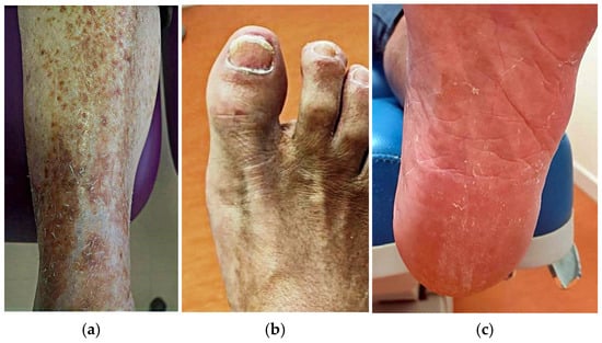
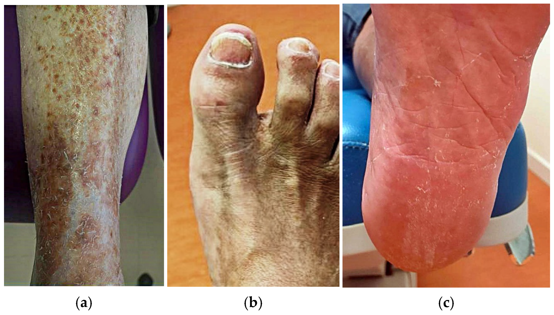
Other common manifestations of diabetic microangiopathy include hair loss and skin atrophy or thinning in the lower extremities. Chronic reduction in oxygen and nutrient supply secondary to vascular impairment induced by hyperglycemia may damage the hair follicles, leading to hair abnormalities such as thinning, fragility, or loss. Studies have shown that up to 26.6% of patients with type 2 DM exhibit an absence of hair on the lower limbs [20,21]. Cutaneous atrophy is characterized by loose, wrinkled, and shiny skin that is often accompanied by telangiectasias and hypopigmentation and is similarly associated with decreased blood flow to the skin [22]. The treatment of these manifestations is primarily based on glycemic control to prevent the progression of microvascular complications [9,23]. The main cutaneous manifestations considered in this study are illustrated in Figure 1.
Figure 1.
Cutaneous manifestations assessed in the study. The figure illustrates the main lower-limb cutaneous findings considered in the proposed clinical screening index, including hair loss, hyperpigmentation (a), nail-plate thickening (b), xerosis and skin atrophy (c).
Several cutaneous manifestations have individually been associated with diabetic microvascular disease, including xerosis, hair loss, pigmentary changes, nail abnormalities, and skin atrophy. However, these findings are usually reported as isolated clinical observations, and to our knowledge they have not previously been integrated into a simple composite clinical screening index capable of identifying patients with clinically established diabetic microangiopathy. Although previous studies have explored individual cutaneous manifestations [8,24], skin-derived biomarkers [25], and quantitative measures of skin microvascular dysfunction in relation to diabetic complications [26], we found limited evidence for a clinical composite index integrating multiple readily observable cutaneous manifestations to indicate clinically established diabetic microvascular involvement. Given the wide range of cutaneous manifestations associated with impaired microcirculation in patients with DM, it is essential to establish an early referral protocol to facilitate the identification of individuals who have higher risk of developing microvascular complications. Developing such an index could facilitate bedside assessment using routine physical examination and may help identify patients requiring further vascular evaluation, particularly in settings where specialized diagnostic tests are not readily available.
Therefore, this pilot study aimed to develop a simple bedside clinical screening index based on lower-limb cutaneous manifestations that may raise clinical suspicion of diabetic microvascular involvement and help prioritize patients for further diagnostic evaluation. A secondary objective was to explore the relationship between this index and tissue oxygenation measured by transcutaneous oxygen pressure (TcPO2).
2. Materials and Methods
2.1. Study Design
A pilot observational cross-sectional study was conducted to investigate the association between lower-limb cutaneous manifestations and clinically established diabetic microangiopathy and to develop a preliminary clinical screening index. Adult patients with type 1 or type 2 diabetes mellitus attending a specialized diabetic foot unit were consecutively recruited between October 2023 and December 2024. All patients meeting the predefined eligibility criteria during the recruitment period were considered for inclusion, regardless of their vascular status or the presence of diabetic microvascular complications.
Inclusion criteria were age ≥18 years, a diagnosis of type 1 or type 2 diabetes mellitus, and the ability to provide written informed consent. Patients with major lower-limb amputation, defined as an amputation at or proximal to the ankle joint, were excluded because complete assessment of the predefined cutaneous variables was not possible. No additional exclusion criteria based on peripheral arterial disease or other vascular conditions were applied, as vascular assessment formed part of the study protocol and was unknown at the time of enrollment. This approach was intended to minimize selection bias and reflect routine clinical practice.
The study was approved by the local Research Ethics Committee (approval number 22/722-E) and conducted in accordance with the Declaration of Helsinki [27]. All participants provided written informed consent before enrollment.
The present study represents a secondary analysis of a previously characterized cohort of individuals with diabetes mellitus [28]. The previous study investigated changes in lower-limb microcirculation following a 15 min walking intervention, whereas the present analysis addresses a distinct research question. The clinical scoring system and its preliminary discriminatory assessment presented in the current study have not been previously reported.
2.2. Clinical Assessment
All participants underwent a standardized clinical examination of both lower limbs performed by the same investigator. To minimize observer bias, the dermatological assessment was systematically completed before obtaining the participants’ medical history or reviewing information regarding diabetes-related complications. The presence or absence of predefined cutaneous manifestations—including xerosis, absence of hair, hyperpigmentation, nail-plate thickening, skin atrophy, and edema—was recorded using standardized clinical criteria. Severity, extent, or anatomical distribution of individual lesions was not graded.
Following completion of the dermatological examination, participants were evaluated for clinically established diabetic microangiopathy according to the predefined study criteria. Microangiopathy was defined as the presence of diabetic peripheral neuropathy together with diabetic retinopathy and/or diabetic nephropathy. This definition was adopted to increase diagnostic specificity by identifying patients with clinically established multisystem microvascular involvement rather than isolated complications. Diabetic peripheral neuropathy is a multifactorial complication in which microvascular dysfunction plays a major pathogenic role through chronic impairment of the vasa nervorum, contributing to nerve ischemia and progressive neural damage [29,30].
Peripheral neuropathy was assessed according to the recommendations of the International Working Group on the Diabetic Foot (IWGDF) and the American Diabetes Association (ADA) [31,32]. Protective sensation was evaluated using a 10 g Semmes–Weinstein monofilament and vibration perception using a 128 Hz tuning fork, which constituted the principal criteria for diagnosing diabetic peripheral neuropathy. Superficial thermal sensation was additionally assessed using a RollTemp® (Somedic SenseLab AB, Sösdala, Sweden) device as part of the neurological examination. The study focused on diabetic patients, and peripheral neuropathy was evaluated within the clinical context of DM. No specific investigations were performed to identify alternative etiologies of neuropathy. Information regarding diabetic retinopathy and diabetic nephropathy was obtained from the participants’ medical records and corroborated during the clinical interview.
All participants underwent a standardized vascular assessment comprising pedal pulse examination, ankle–brachial index (ABI), toe–brachial index (TBI), and TcPO2. While ABI and TBI were used to characterize lower-limb macrovascular status, TcPO2 was analyzed as an exploratory physiological measure of tissue perfusion to examine its relationship with the proposed clinical screening index.
2.3. Sample-Size Calculation
Given the exploratory nature of this pilot study, a formal sample size calculation was initially performed to estimate the minimum number of subjects required for detecting differences between groups. The calculation was conducted using Granmo® software (version 7.12; Institut Municipal d’Investigació Mèdica, Barcelona, Spain). As this was an exploratory pilot study comparing patients with and without clinically established microangiopathy, the calculation assumed a prevalence of cutaneous manifestations of 30% in patients without microangiopathy and 80% in those with microangiopathy, with 80% statistical power and a two-sided significance level of 10%. Under these assumptions, a minimum sample of 28 participants was required.
However, given the exploratory aim of deriving a preliminary clinical index, a final sample of 30 patients was included. Therefore, the results of this study should be interpreted as hypothesis-generating and require confirmation in larger cohorts.
2.4. Data Collection
Two investigators participated in the clinical assessment. The cutaneous manifestations included in the clinical index were systematically recorded by the primary investigator (LPA), while the second investigator participated in the overall clinical assessment (ATG). Circulatory-status parameters were analyzed for the study, including the presence or absence of distal pulses, ABI, TBI, TcPO2, and skin temperature. Vascular screening was performed using the PeriFlux 6000 Combined System® (PeriMed AB, Järfälla, Sweden), and skin temperature was measured with a contactless infrared thermometer. Prior to examination, patients were asked to remain barefoot and at rest for 10 min. All data were collected at a specialized diabetic foot unit.
2.5. Data Analysis
Data analysis was performed using SPSS (version 27.0; IBM Corp., Armonk, NY, USA). Categorical variables were summarized as absolute frequencies and percentages, whereas continuous variables were expressed as mean, standard deviation (SD), and 95% confidence intervals (95% CI), where appropriate.
Associations between lower-limb cutaneous manifestations and clinically established diabetic microangiopathy were initially explored using contingency tables. Owing to the small sample size and sparse cell counts, Fisher’s exact test was used to assess statistical significance. Odds ratios (ORs) with 95% confidence intervals were calculated whenever estimation was statistically appropriate.
Because of the limited sample size and the exploratory nature of this pilot study, regression-based weighting was intentionally not performed to avoid model overfitting. Instead, variables significantly associated with clinically established microangiopathy were first identified through bivariate analysis. A pragmatic weighting strategy was subsequently applied, whereby variables showing larger and more consistent effect sizes (OR >10 or complete separation) were assigned two points, whereas variables demonstrating moderate but statistically significant associations were assigned one point. The purpose of this approach was not to derive an optimized prediction model but to construct a simple preliminary clinical screening index suitable for future validation. The discriminative ability of the proposed clinical screening index for identifying clinically established diabetic microangiopathy was evaluated using receiver operating characteristic (ROC) curve analysis. The area under the curve (AUC) with its corresponding 95% confidence interval was calculated, and the optimal cut-off value was selected based on the best balance between sensitivity and specificity.
As a secondary exploratory analysis, the relationship between the clinical screening index and TcPO2 was assessed using Spearman’s rank correlation coefficient. Differences in TcPO2 values between predefined risk groups were analyzed using the Mann–Whitney U test.
All statistical tests were two-tailed, and a p-value < 0.05 was considered statistically significant.
3. Results
The study included 30 patients, including 19 males (63.3%). The mean age was 70.1 years (SD 6.6), and the mean BMI was 27.6 (SD 3.07). There were 28 patients (93.3%) who had type 2 DM with a mean disease duration of 19.7 years (SD 12.3), and 14 (46.7%) subjects were receiving combined treatment with insulin and oral antidiabetics. The average glucose level was 129.4 mg/dL (SD 33.7), and the average HbA1c was 7.01% (SD 1.1) (53 mmol/mol; SD ± 9.7).
There were 12 (40.0%) subjects who had a history of DFU, and 5 (16.7%) had undergone amputation. Other clinical characteristics of the sample are shown in Table 1. Neurological and vascular parameters of the sample can be found in Table 2. Table 3 shows data on the cutaneous manifestations of vascular impairment. Of the 30 included participants, 21 (70.0%) fulfilled the predefined criteria for clinically established diabetic microangiopathy, whereas 9 (30.0%) did not.
Table 1.
Clinical characteristics.
Table 2.
Vascular and neurological screening.
Table 3.
Cutaneous manifestations of vascular disorder.
Bivariate analysis showed significant associations between several cutaneous manifestations and the presence of microangiopathy. Xerosis demonstrated a strong association, although estimates were imprecise due to sparse data (OR = 48.0; 95% CI: 4.30–535.26; p < 0.001). Similarly, absence of hair on the lower limbs was significantly associated with microangiopathy (OR = 14.88; 95% CI: 2.20–100.66; p = 0.004). Nail-plate thickening also showed a statistically significant association, although with a lower magnitude of effect (OR = 7.0; 95% CI: 1.14–42.97; p = 0.046).
Hyperpigmentation and skin atrophy were significantly associated with microangiopathy (p = 0.001 and p = 0.029, respectively); however, OR could not be reliably estimated due to sparse data and the presence of zero-frequency cells, suggesting a strong but statistically unstable association. No significant association was found between edema and microangiopathy (p = 0.626), and this variable was therefore excluded from subsequent analyses.
A preliminary clinical screening index was developed incorporating the five cutaneous manifestations significantly associated with microangiopathy. Based on the observed effect sizes and the consistency of the associations identified in the bivariate analyses, a pragmatic weighting strategy was adopted. Xerosis and absence of hair showed the strongest associations with clinically established microangiopathy and were therefore assigned two points each. Nail-plate thickening, hyperpigmentation and skin atrophy demonstrated significant but comparatively smaller or less stable associations and were assigned one point each. The total index ranged from 0 to 7 points, with higher scores indicating a greater likelihood of clinically established microangiopathy.
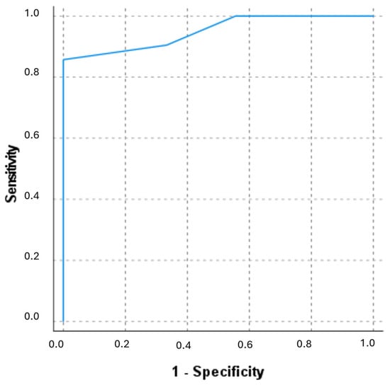
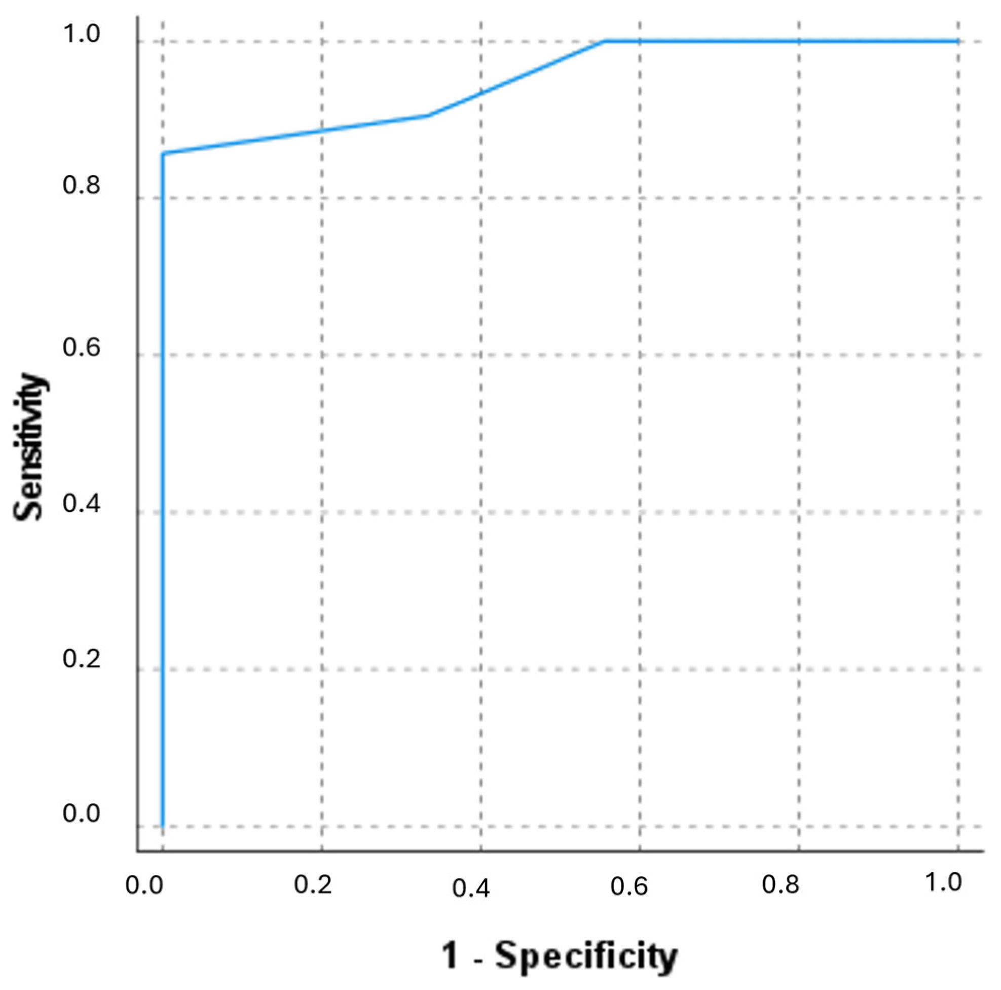
The ROC curve analysis demonstrated excellent discriminative ability of the clinical index for identifying patients with microangiopathy (AUC = 0.950; 95% CI: 0.878–1.000) (Figure 2). The optimal cut-off value was established at ≥4 points, which provided a sensitivity of 76.2% and a specificity of 100%, allowing accurate classification of high-risk patients.
Figure 2.
Receiver operating characteristic (ROC) curve of the proposed bedside clinical screening index for discriminating between patients with and without clinically established diabetic microvascular involvement.
Based on the predefined cut-off (≥4 points), 16 participants (53.3%) were classified as high risk and 14 (46.7%) as low risk. Exploratory analyses showed a consistent trend towards lower TcPO2 values in patients classified as high risk according to the proposed index, although statistical significance was not reached (Spearman’s rho = −0.207, p = 0.272).
TcPO2 values were lower in the high-risk group compared with the low-risk group; however, this difference did not reach statistical significance (Mann–Whitney U test, U = 76.5, Z = −1.477, p = 0.140). Despite the lack of statistical significance, the observed trend suggests a potential association between higher clinical risk scores and reduced tissue perfusion.
4. Discussion
Microangiopathy is the main contributing factor to the development of diabetic neuropathy. Impaired vasodilation of small vessels causes endothelial dysfunction of the vasa nervorum, which leads to hypoxia in both myelinated and unmyelinated nerve fibers in patients with DM. Neuropathy and peripheral arterial disease are the two complications that have the most influence on the development of DFU. For this reason, early detection of microvascular complications is essential for the prevention of these lesions.
Cutaneous manifestations of diabetic microangiopathy are extremely valuable for healthcare professionals as they can provide information even before more severe complications arise. In the present study, the most frequent dermopathies were xerosis and hair loss on the lower limbs, followed by hyperpigmentation, skin hypertrophy, and nail thickening, as detailed in Table 3. These prevalence rates align with published data [13,22,33]. Moreover, our findings are consistent with previous observations reporting associations between diabetic skin manifestations and microvascular complications. Shemer et al. [8] demonstrated that diabetic dermopathy was significantly associated with retinopathy, nephropathy, and neuropathy, and that the prevalence of skin lesions increased as the number of microvascular complications accumulated. Building upon these observations, the present study explored whether multiple lower-limb cutaneous manifestations could be integrated into a pragmatic clinical screening index. For the development of this clinical tool, only the manifestations with statistically significant associations were included, with tissue hypertrophy being excluded as it was a less relevant sign. The weighting strategy was intentionally simplified to improve clinical applicability and reduce overfitting given the small sample size.
A recent study by Özer et al. [34] further supports the potential value of clinically assessed cutaneous findings as markers of diabetic complications. Using the Overall Dry Skin (ODS) score, the authors demonstrated associations between the severity of xerosis and diabetic retinopathy, nephropathy, and peripheral polyneuropathy. However, the ODS score was specifically designed to quantify the severity of xerosis, whereas the index proposed in the present study integrates multiple clinically observable cutaneous manifestations that may reflect different aspects of diabetic microvascular involvement.
Therefore, this study proposes a novel clinical screening index based on easily identifiable cutaneous manifestations associated with diabetic microangiopathy. The main finding is that the index demonstrated strong discriminative ability in this exploratory cohort, suggesting its potential utility as a simple bedside tool for risk stratification in patients with DM The observed high diagnostic performance (AUC = 0.95; 95% CI: 0.88–1.00) indicates that combinations of clinical skin findings may capture relevant patterns associated with microvascular disease. This supports the concept that cutaneous alterations reflect underlying chronic tissue hypoperfusion and microcirculatory dysfunction in DM.
Notably, although neither the correlation analysis nor the group comparison reached statistical significance, a consistent directional trend was observed across analyses. Higher clinical risk scores were associated with lower TcPO2 values and were more frequently observed in patients classified within the high-risk group. This pattern, while not statistically confirmed in this sample, aligns with the expected physiological relationship between increasing microangiopathic burden and reduced tissue perfusion. Although these findings did not reach statistical significance, the observed trends may be consistent with the biological rationale underlying the proposed screening index. However, these observations require confirmation in larger cohorts.
These results are consistent with previous studies that have highlighted the value of cutaneous findings as indirect markers of vascular damage. Demiseren et al. [35] demonstrated that skin diseases in which microangiopathy plays a pathogenic role are significantly associated with diabetic nephropathy, neuropathy, and retinopathy. Furthermore, they may indicate the presence of microvascular complications associated with DM. In the present study, we found similar prevalence rates for nephropathy and retinopathy, while the prevalence of neuropathy was higher, possibly due to all data being collected in a specialized diabetic foot unit, where patients with more severe conditions are more frequently encountered. Similarly, it has been observed that patients with poorer glycemic control are at greater risk of developing cutaneous alterations [36,37]. Consistently, we found that the patients included in this study had HbA1c levels above 7%, which may predispose them to the development of dermopathies associated with poor metabolic control. However, in our study, HbA1c was not significantly associated with either clinically established microangiopathy or the proposed cutaneous score in this exploratory cohort. This finding should be interpreted cautiously given the limited sample size and warrants further evaluation in larger studies.
Some of the limitations of this study include the small sample size and the potential subjectivity in the assessment of certain physical signs. Additionally, clinical examination and classification were not performed under blinded conditions. Future validation studies should include independent blinded assessment of cutaneous manifestations to minimize observer bias. Future research with larger samples and long-term follow-up could help to validate the index as a predictive tool for complications associated with diabetes mellitus, such as DFU. The proposed clinical index demonstrated a high discriminative ability in this exploratory cohort; however, the absence of significant correlations with TcPO2 and the limited sample size suggest that these findings should be interpreted as preliminary. Such an approach may be particularly valuable in settings where access to complementary investigations is limited. Another important limitation of this pilot study is that participants were recruited from a specialized diabetic foot unit, resulting in a population with a high prevalence of advanced diabetic complications. Consequently, the proposed clinical screening index was developed in a high-risk cohort and requires external validation in broader diabetic populations before routine clinical use.
Because the score was derived and evaluated within the same small pilot cohort, its apparent discriminative performance may be optimistic. No internal resampling or external validation was performed, and the proposed weighting strategy was intentionally pragmatic rather than regression-based to avoid unstable estimates in a limited sample. Accordingly, the reported performance should be interpreted as preliminary and requires confirmation in larger independent cohorts. Furthermore, calibration analyses were beyond the scope of the present pilot study. Interobserver reliability was not evaluated because all cutaneous examinations were performed by a single trained examiner. Future external validation studies should evaluate both discrimination and calibration in larger independent populations and assess the reproducibility of the proposed index by examining agreement between independent observers.
One of the strengths of the study is its methodological structure, through which independent researchers collected and analyzed the data, lending credibility to the work. The study was overseen by two independent researchers, who ensured the accuracy of the research. Furthermore, all participants underwent a standardized clinical and vascular assessment performed within a specialized diabetic foot unit.
The proposed index is intended as a simple bedside clinical screening aid based on easily recognizable cutaneous manifestations that may increase clinical suspicion of diabetic microvascular involvement and support decisions regarding further vascular, neurological, ophthalmological, or nephrological evaluation. From a clinical perspective, this index has the advantage of relying on simple and non-invasive variables that can be assessed without specialized equipment, making it potentially useful in primary care or resource-limited settings.
Future studies with larger, independent cohorts are required to externally validate the score, refine its weighting system, and assess its predictive value for clinically relevant outcomes such as diabetic foot ulceration or limb ischemia progression.
5. Conclusions
The proposed bedside clinical screening index showed promising discriminative performance in this pilot study and may represent a simple, rapid, and cost-effective tool based on readily observable cutaneous manifestations. Rather than serving as a diagnostic instrument, the index may help raise clinical suspicion of diabetic microvascular involvement and support the prioritization of patients for further vascular, neurological, ophthalmological, and nephrological assessment, particularly in resource-limited settings. Given the exploratory nature of this study, the proposed index should be considered preliminary and requires external validation in larger, more diverse populations before routine clinical implementation.
Author Contributions
Conceptualization: L.P.-A., A.T.-G. and F.J.Á.-A.; Methodology: L.P.-A., A.T.-G. and Y.G.-Á.; Formal analysis: L.P.-A., A.T.-G., Y.G.-Á. and S.T.-R.; Investigation: L.P.-A., A.T.-G. and J.L.L.-M.; Data curation: L.P.-A., A.T.-G. and S.T.-R.; Writing—original draft preparation: L.P.-A. and A.T.-G.; Writing—review and editing: A.T.-G., S.T.-R., F.J.Á.-A., L.P.-A. and J.L.L.-M.; Supervision: A.T.-G. and J.L.L.-M.; Project administration: J.L.L.-M. All authors have read and agreed to the published version of the manuscript.
Funding
This research received no external funding.
Institutional Review Board Statement
The study was conducted in accordance with the Declaration of Helsinki, and approved by the Ethics Committee of Hospital Clínico San Carlos (protocol code number 22/722-E with an approval date of 2 January 2023) for studies involving humans.
Informed Consent Statement
Informed consent was obtained from all subjects involved in the study. Written informed consent has been obtained from the patient(s) to publish this paper.
Data Availability Statement
The data that support the findings of this study are available from the corresponding author upon reasonable request.
Conflicts of Interest
The authors declare no conflicts of interest.
Abbreviations
The following abbreviations are used in this manuscript:
| PAD | peripheral arterial disease |
| DM | Diabetes mellitus |
| NO | nitric oxide |
| TcPO2 | transcutaneous oxygen pressure |
| AGEs | advanced glycation end products |
| ROC | receiver operating characteristic |
| AUC | area under the curve |
References
- Lutze, S.; Westphal, T.; Jünger, M.; Arnold, A. Microcirculation disorders of the skin. J. Dtsch. Dermatol. Ges. 2024, 22, 236–264. [Google Scholar] [CrossRef] [Scilit]
- Blakytny, R.; Jude, E.B. Altered Molecular Mechanisms of Diabetic Foot Ulcers. Int. J. Low. Extrem. Wounds 2009, 8, 95–104. [Google Scholar] [CrossRef] [Scilit]
- Obrosova, I.G. Increased Sorbitol Pathway Activity Generates Oxidative Stress in Tissue Sites for Diabetic Complications. Antioxid. Redox Signal. 2005, 7, 1543–1552. [Google Scholar] [CrossRef] [Scilit]
- Stern, D.; Yan, S.D.; Yan, S.F.; Schmidt, A.M. Receptor for advanced glycation endproducts: A multiligand receptor magnifying cell stress in diverse pathologic settings. Adv. Drug Deliv. Rev. 2002, 54, 1615–1625. [Google Scholar] [CrossRef] [Scilit]
- Lima, A.L.; Illing, T.; Schliemann, S.; Elsner, P. Cutaneous Manifestations of Diabetes Mellitus: A Review. Am. J. Clin. Dermatol. 2017, 18, 541–553. [Google Scholar] [CrossRef] [Scilit]
- Salari, N.; Hosseinian-Far, A.; Hosseinian-Far, M.; Kavoussi, H.; Jalali, R.; Vaisi-Raygani, A.; Rasoulpoor, S.; Rasoulpoor, S.; Mohammadi, M.; Shabani, S. Evaluation of skin lesions in diabetic patients: A systematic review and meta-analysis. J. Diabetes Metab. Disord. 2020, 19, 1909–1916. [Google Scholar] [CrossRef] [Scilit]
- Bustan, R.S.; Wasim, D.; Yderstræde, K.B.; Bygum, A. Specific skin signs as a cutaneous marker of diabetes mellitus and the prediabetic state—A systematic review. Dan. Med. J. 2017, 64, A5316. [Google Scholar]
- Shemer, A.; Bergman, R.; Linn, S.; Kantor, Y.; Friedman-Birnbaum, R. Diabetic dermopathy and internal complications in diabetes mellitus. Int. J. Dermatol. 1998, 37, 113–115. [Google Scholar] [CrossRef] [Scilit]
- Morgan, A.J.; Schwartz, R.A. Diabetic dermopathy: A subtle sign with grave implications. J. Am. Acad. Dermatol. 2008, 58, 447–451. [Google Scholar] [CrossRef] [Scilit]
- Naik, P.P.; Farrukh, S.N. Clinical Significance of Diabetic Dermatopathy. Diabetes Metab. Syndr. Obes. 2020, 13, 4823–4827. [Google Scholar] [CrossRef] [Scilit]
- de Oliveira Abate, M.C.M.; Aroucha, P.M.T.; da Nóbrega, D.V.M.; Rocha, I.P.M.; Soares, S.D.; Reis, A.A.; Paliares, I.C.; de Giuffrida, F.M.A.; Dib, S.A.; Reis, A.F.; et al. Cutaneous manifestations of diabetes mellitus: A narrative review. Einstein 2025, 23, eRW1193. [Google Scholar] [CrossRef] [Scilit]
- Edwards, E.; Yosipovitch, G. Skin Manifestations of Diabetes Mellitus. In Endotext; Feingold, K.R., Ahmed, S.F., Anawalt, B., Blackman, M.R., Boyce, A., Chrousos, G., Corpas, E., de Herder, W.W., Dhatariya, K., Dungan, K., et al., Eds.; MDText.com, Inc.: South Dartmouth, MA, USA, 2000. Available online: http://www.ncbi.nlm.nih.gov/books/NBK481900/ (accessed on 21 April 2025).
- Yosipovitch, G.; Hodak, E.; Vardi, P.; Shraga, I.; Karp, M.; Sprecher, E.; David, M. The prevalence of cutaneous manifestations in IDDM patients and their association with diabetes risk factors and microvascular complications. Diabetes Care 1998, 21, 506–509. [Google Scholar] [CrossRef] [Scilit]
- Yosipovitch, G.; Misery, L.; Proksch, E.; Metz, M.; Ständer, S.; Schmelz, M. Skin Barrier Damage and Itch: Review of Mechanisms, Topical Management and Future Directions. Acta Derm. Venereol. 2019, 99, 1201–1209. [Google Scholar] [CrossRef] [Scilit]
- Goyal, A.; Raina, S.; Kaushal, S.S.; Mahajan, V.; Sharma, N.L. Pattern of Cutaneous Manifestations in Diabetes Mellitus. Indian J. Dermatol. 2010, 55, 39–41. [Google Scholar] [CrossRef] [Scilit]
- Stingeni, L.; Tramontana, M.; Cordera, L.; Castello, M.; Parodi, A. Xerosis in Patients with Type 2 Diabetes: An Italian Multicentre Study. Acta Derm. Venereol. 2021, 101, 263. [Google Scholar] [CrossRef] [Scilit]
- Sun, P.-C.; Chen, C.-S.; Kuo, C.-D.; Lin, H.-D.; Chan, R.-C.; Kao, M.-J.; Wei, S.-H. Impaired microvascular flow motion in subclinical diabetic feet with sudomotor dysfunction. Microvasc. Res. 2012, 83, 243–248. [Google Scholar] [CrossRef] [Scilit]
- Tejeda Ramírez, S.; Tardáguila-García, A.; López-Moral, M.; Sanz-Corbalán, I.; García-Madrid, M.; Lázaro-Martínez, J.L. Randomized Double-blind Cost-effectiveness Comparison of Two 10% Urea Creams in Patients with Diabetic Foot Syndrome. Adv. Skin Wound Care 2024, 37, 1–7. [Google Scholar] [CrossRef] [Scilit]
- Celleno, L. Topical urea in skincare: A review. Dermatol. Ther. 2018, 31, e12690. [Google Scholar] [CrossRef] [Scilit]
- Miranda, J.J.; Taype-Rondan, A.; Tapia, J.C.; Gastanadui-Gonzalez, M.G.; Roman-Carpio, R. Hair follicle characteristics as early marker of Type 2 Diabetes. Med. Hypotheses 2016, 95, 39–44. [Google Scholar] [CrossRef] [Scilit]
- Chatterjee, N.; Chattopadhyay, C.; Sengupta, N.; Das, C.; Sarma, N.; Pal, S.K. An observational study of cutaneous manifestations in diabetes mellitus in a tertiary care Hospital of Eastern India. Indian J. Endocrinol. Metab. 2014, 18, 217. [Google Scholar] [CrossRef] [Scilit]
- Lause, M.; Kamboj, A.; Fernandez Faith, E. Dermatologic manifestations of endocrine disorders. Transl. Pediatr. 2017, 6, 300–312. [Google Scholar] [CrossRef] [Scilit]
- Perez, M.I.; Kohn, S.R. Cutaneous manifestations of diabetes mellitus. J. Am. Acad. Dermatol. 1994, 30, 519–531. [Google Scholar] [CrossRef] [Scilit]
- Mirhoseini, M.; Saleh, N.; Momeni, A.; Deris, F.; Asadi-Samani, M. A study on the association of diabetic dermopathy with nephropathy and retinopathy in patients with type 2 diabetes mellitus. J. Nephropathol. 2016, 5, 139–143. [Google Scholar] [CrossRef] [Scilit]
- Gerrits, E.G.; Lutgers, H.L.; Kleefstra, N.; Graaff, R.; Groenier, K.H.; Smit, A.J.; Gans, R.O.; Bilo, H.J. Skin autofluorescence: A tool to identify type 2 diabetic patients at risk for developing microvascular complications. Diabetes Care 2008, 31, 517–521. [Google Scholar] [CrossRef] [Scilit]
- Tehrani, S.; Bergen, K.; Azizi, L.; Jörneskog, G. Skin microvascular reactivity correlates to clinical microangiopathy in type 1 diabetes: A pilot study. Diabetes Vasc. Dis. Res. 2020, 17, 1479164120928303. [Google Scholar] [CrossRef] [Scilit]
- World Medical Association. World Medical Association Declaration of Helsinki: Ethical Principles for Medical Research Involving Human Subjects. JAMA 2013, 310, 2191–2194. [Google Scholar] [CrossRef] [Scilit] [PubMed]
- Palacios-Abril, L.; Tardáguila-García, A.; Álvaro-Afonso, F.J.; Tejeda-Ramírez, S.; López-Moral, M.; Lázaro-Martínez, J.L. Fifteen-minute walk improves microcirculation in people with diabetes mellitus. Microvasc. Res. 2026, 164, 104897. [Google Scholar] [CrossRef] [Scilit]
- Faselis, C.; Katsimardou, A.; Imprialos, K.; Deligkaris, P.; Kallistratos, M.; Dimitriadis, K. Microvascular Complications of Type 2 Diabetes Mellitus. Curr. Vasc. Pharmacol. 2020, 18, 117–124. [Google Scholar] [CrossRef] [Scilit]
- Hicks, C.W.; Selvin, E. Epidemiology of Peripheral Neuropathy and Lower Extremity Disease in Diabetes. Curr. Diabetes Rep. 2019, 19, 86. [Google Scholar] [CrossRef] [Scilit]
- Schaper, N.C.; van Netten, J.J.; Apelqvist, J.; Bus, S.A.; Fitridge, R.; Game, F.; Monteiro-Soares, M.; Senneville, E. Practical guidelines on the prevention and management of diabetes-related foot disease (IWGDF 2023 update). Diabetes Metab. Res. Rev. 2024, 40, e3657. [Google Scholar] [CrossRef] [Scilit]
- American Diabetes Association Professional Practice Committee for Diabetes. 12. Retinopathy, Neuropathy, and Foot Care: Standards of Care in Diabetes—2026. Diabetes Care 2025, 49, S261–S276. [Google Scholar] [CrossRef] [Scilit]
- Kamel, M.I.; Elhenawy, Y.I.; Saudi, W.M. Relation between cutaneous and extracutaneous complications in pediatric patients with type 1 diabetes. Dermato-Endocrinology 2018, 10, e1467717. [Google Scholar] [CrossRef] [Scilit]
- Özer, Ö.; Menteşoğlu, D.; Yorulmaz, G. Is dry skin a clue to diabetic retinopathy? Clinical insights from the overall dry skin score. Front. Endocrinol. 2026, 17, 1871805. [Google Scholar] [CrossRef] [Scilit]
- Demirseren, D.D.; Emre, S.; Akoglu, G.; Arpacı, D.; Arman, A.; Metin, A.; Cakır, B. Relationship between skin diseases and extracutaneous complications of diabetes mellitus: Clinical analysis of 750 patients. Am. J. Clin. Dermatol. 2014, 15, 65–70. [Google Scholar] [CrossRef] [Scilit]
- Khuraiya, S.; Lal, N.; Naseerudin; Jain, V.; Kachhawa, D. A cross sectional study of cutaneous manifestations in 300 patients of diabetes mellitus. Int. J. Adv. Med. 2019, 6, 150–154. [Google Scholar] [CrossRef] [Scilit][Green Version]
- Zakharov, P.; Talary, M.S.; Kolm, I.; Caduff, A. Full-field optical coherence tomography for the rapid estimation of epidermal thickness: Study of patients with diabetes mellitus type 1. Physiol. Meas. 2010, 31, 193–205. [Google Scholar] [CrossRef] [Scilit]

Disclaimer/Publisher’s Note: The statements, opinions and data contained in all publications are solely those of the individual author(s) and contributor(s) and not of MDPI and/or the editor(s). MDPI and/or the editor(s) disclaim responsibility for any injury to people or property resulting from any ideas, methods, instructions or products referred to in the content. |
© 2026 by the authors. Licensee MDPI, Basel, Switzerland. This article is an open access article distributed under the terms and conditions of the Creative Commons Attribution (CC BY) license.