


Abstract
Objectives: This study aimed to assess the effects of voluntary exercise on type 1 diabetes mellitus (T1D) development and splenic immunological profiles in non-obese diabetic (NOD) mice, a spontaneous model of human T1D. Methods: Prediabetic female NOD mice were randomly assigned to sedentary or exercise groups, with mice in the exercise group given 10-week wheel access and sedentary mice receiving none. Late-time mice were monitored to diabetes onset or 24 weeks of age; early-time mice were analyzed immediately post-intervention. Blood glucose, food intake, water consumption, and body mass were monitored weekly. At the endpoints, splenocyte counts, T and B cell subsets, and mitogen-stimulated cytokine production were analyzed using flow cytometry. Results: Mice in the exercise group ran an average of 20.76 ± 0.22 km/day. By the late-time endpoint, 75% of mice in the exercise group remained non-diabetic versus 35% of sedentary mice (p = 0.006). Mice in the exercise group demonstrated lower blood glucose (p = 0.015), visceral fat mass (p = 0.035), and water intake (p < 0.001) but higher food intake (p = 0.001), with no difference in body mass (p = 0.389) compared to sedentary mice. No differences were observed in splenocyte counts or Th, Tc, Treg, or B cell populations at either time point (p ≥ 0.185). Early-time point cytokines also did not differ between groups (p ≥ 0.08). Conclusions: Voluntary exercise reduces T1D incidence and mitigates hyperglycemia in NOD mice, suggesting a protective effect against disease progression. Despite the benefits, physical activity did not alter splenic Tcell subsets or inflammatory cytokines, demonstrating systemic immunomodulation may not be the primary driver of benefit. Our results indicate that voluntary exercise protects against T1D through tissue-specific or metabolic mechanisms, which warrant further mechanistic investigation.
1. Introduction
Type 1 diabetes mellites (T1D) is a chronic autoimmune disease marked by the selective destruction of pancreatic β-cells by autoreactive T lymphocytes, ultimately leading to absolute insulin deficiency and persistent hyperglycemia. Beyond glycemic control, T1D has systemic consequences including impaired skeletal muscle regeneration, altered energy metabolism, and increased cardiovascular risk [1,2,3]. The global incidence of T1D has been increasing steadily with environmental and genetic factors, both implicated in T1D pathogenesis [4,5]. Despite advancements in understanding the etiology of T1D, effective strategies to prevent or delay disease progression remain limited. Recent studies have begun to explore how modifiable lifestyle factors, such as physical activity, may exert immunomodulatory effects capable of influencing the course of autoimmune diabetes.
Regular physical activity is widely recognized as a cornerstone of diabetes management due to its beneficial effects on glucose uptake, insulin sensitivity, body composition, and inflammation [6,7,8,9]. Emerging research highlights the beneficial role of exercise in modulating immune responses in individuals with T1D. Physical activity has been shown to reduce systemic inflammation, improve glycemic control, and preserve β-cell function, particularly when initiated early in the disease course [6,10]. Several studies have demonstrated exercise-induced shifts in cytokine profiles toward an anti-inflammatory state, along with mobilization of innate and adaptive immune cells [11,12,13]. Notably, a single bout of vigorous exercise have been shown to mobilize highly differentiated CD8+ T cells and natural killer cells, although these responses appear blunted in individuals with T1D compared to healthy controls [12]. This attenuated mobilization may reflect altered immune cell trafficking or underlying metabolic disturbances unique to T1D. Furthermore, muscle-residing regulatory T cells (Tregs), which are essential for tissue repair and immune regulation, appear responsive to exercise-induced signals and may play a protective role in modulating autoimmunity [13]. Despite these advances, the direct effect of exercise on T cell subpopulations, particularly islet-reactive and regulatory T cells, remains poorly understood. Clarifying how different exercise modalities influence T cell phenotypes, activation status, and migratory patterns is essential for understanding the immunomodulatory potential of physical activity in T1D and for developing targeted exercise-based interventions aimed at altering disease trajectory.
The non-obese diabetic (NOD) mouse model has been instrumental in unraveling the immunopathology of T1D, as it closely mimics human disease progression, including the gradual autoimmune infiltration of pancreatic islets [14,15]. If physical activity lowers hyperglycemia by preserving pancreatic β-cell function through the reduction in inflammatory cytokines or attenuation of T lymphocyte-mediated attack, this would represent a favorable therapeutic mechanism. However, the extent to which exercise exerts immunomodulatory effects in experimental T1D mice is not clear. Also, the extent to which physical activity affects immunological cytokines regardless of glycemic levels has not been thoroughly investigated. Thus, to better elucidate the immunomodulatory properties of physical activity, this study was designed to test the hypotheses that voluntary exercise will (1) delay the T1D progression, (2) modulate splenic T cell differentiation and inflammatory cytokines, and (3) lower glycemia compare to the sedentary controls.
2. Materials and Methods
2.1. Animals and Ethical Approvals
All experimental protocols and animal care procedures were approved by the Institutional Animal Care and Use Committee at St. Cloud State University (IACUC protocol #5-146) and complied with the NIH guidelines regarding the use of animals. We selected female NOD/ShiLtJ mice for this study because they exhibit a high and predictable T1D incidence (60–85%) between 12 and 24 weeks of age [16,17], providing early-onset, reproducible, and statistically robust disease progression essential for controlled investigations of autoimmune diabetes. Female NOD/ShiLtJ mice (n = 64) were obtained from Jackson Laboratories (Bar Harbor, ME, USA), bred on-site and used at 5–9 weeks of age. Mice were housed in groups of four to five and had access to rodent chow (AIN-93G, Research Diets, Inc., New Brunswick, NJ, USA) and water ad libitum. The housing room was maintained on a 14:10 h light:dark cycle with controlled temperature and humidity. Weekly water consumption and food intake were measured, and body mass was determined on a weekly basis starting at the first week of exercise and continuing throughout the entire experimental period until the endpoint.
2.2. Study 1 (Late-Time NOD)
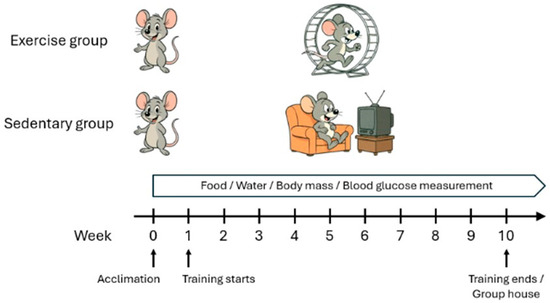
Forty prediabetic female NOD mice (age 6.2 ± 0.3 weeks) were randomly assigned to sedentary (LSED; n = 17) or exercise (LEXE; n = 20) groups. Three sedentary mice died unexpectedly before scheduled hyperglycemia assessment and confirmation of diabetes onset (≥250 mg/dL on two consecutive readings). As they did not meet predefined inclusion criteria, these mice were excluded from outcome analyses. The final sample sizes used for statistical comparisons reflect only animals that survived to diabetes onset assessments, ensuring transparency and reproducibility of data analysis. Mice underwent 10 weeks of voluntary wheel running or remained sedentary individually, then were group-housed and monitored until 24 weeks of age or diabetes onset, whichever occurred first (Figure 1). At the endpoint, mice were euthanized via CO2 asphyxiation and visceral fat mass was recorded. Spleens were collected for analysis of spleen mass, total splenocyte count, cell viability, major immune cell populations (T and B cells), and T cell subsets (Th, Tc, and Treg).
Figure 1.
Exercise group mice were individually housed with running wheels, while sedentary mice were individually housed without wheels. After a one-week acclimation to single housing, mice underwent 10 weeks of voluntary running or sedentary conditions. Following training, exercised and sedentary mice were group-housed in standard cages until the experimental endpoints.
2.3. Study 2 (Early-Time NOD)
A subset of 24 prediabetic female NOD mice (age 5.9 ± 0.3 weeks) was analyzed early to assess exercise effects before diabetes onset. Mice were randomly assigned to sedentary (ESED; n = 12) or exercise (EEXE; n = 11) groups. One mouse in the exercise group developed hyperglycemia (≥250 mg/dL on two consecutive measurements) during the training period. Because the study targeted prediabetic NOD mice, animals meeting diabetes criteria were prospectively excluded. This mouse was removed from subsequent analyses, and statistical comparisons were performed using only normoglycemic mice. The exercise group underwent 10 weeks of voluntary wheel running, while sedentary mice were individually housed without wheels. This cohort aimed to determine whether exercise modulates immune function during the prediabetic stage, and all mice in both groups were confirmed to be in prediabetic condition based on blood glucose measurements taken at the study endpoint. After 10 weeks, mice were euthanized for ex vivo analyses identical to Study 1, with additional assessment of cytokine profiles (IL-2, IL-4, IL-6, IL-17A, IFN-γ, TNF-α, IL-10) in mitogen-stimulated T cells.
2.4. Voluntary Wheel Running
Exercise group mice were individually housed in wheel-running cages (Model 80820, Lafayette Instrument Co., Lafayette, IN, USA) equipped with 12.7 cm diameter wheels, 5.72 cm width, and 38-rod running surfaces. Revolutions were recorded via an optical sensor using Activity Wheel Monitor Software (Model 86065, Lafayette Instrument Co.). Wheels were calibrated weekly with 1 g resistance brakes (Model 86070-B1, Lafayette Instrument Co.) to ensure consistent resistance across mice [18,19]. Sedentary mice were individually housed in standard cages without wheels. All mice underwent a one-week acclimation to individual housing before the 10-week voluntary running period to mitigate stress-related confounders. After 10 weeks, all mice were group-housed in regular cages until the experimental endpoint (Figure 1).
2.5. Blood Glucose Determination
Blood glucose levels were measured weekly from 12 to 24 weeks of age in exercise group and sedentary mice, as described previously [16,20]. Baseline levels were taken before the 10-week training period, and monitoring continued until diabetes onset or 24 weeks of age. Mice were restrained in a restrainer (Braintree Sci., Braintree, MA, USA), and blood was collected from the lateral tail vein using a 26G × 5/8” needle. A 0.6 µL sample was analyzed with an Accu-Check Aviva glucose meter (Roche Diagnostics, Indianapolis, IN, USA). Diabetes was defined as two consecutive glucose readings ≥ 250 mg/dL, with the first reading considered diagnostic.
2.6. Relationship Between Blood Glucose and Running Distance
Correlation analysis was performed to determine whether voluntary running distance was associated with glycemic outcomes in exercise group NOD mice at the endpoint. To further evaluate exercise exposure in relation to T1D disease progression, we additionally analyzed the relationship between individual average running distance and blood glucose at diabetes onset (Study 1).
2.7. Ex Vivo Immunological Analysis and Single Cell Suspension Preparation
Mice were continuously monitored until they became diabetic or reached 24 weeks of age, whichever occurred first in Study 1 or until completion of 10-week wheel running in Study 2. At the endpoints, mice were euthanized and spleens were aseptically harvested and weighed. Spleen mass was used to calculate the spleen index (spleen mass/body mass) as an indicator of relative spleen size.
Single cell suspensions of isolated spleens were done as described by Cetkovic-Cvrlje et al. [16,21]. In brief, a spleen was forced through a 70 mm nylon mesh strainer (BD Falcon, San Jose, CA, USA); the resulting suspension was then treated with ACK Lysis Buffer (NH4Cl 8.29 g/L, KHCO3 1.0 g/L, EDTA Na2 2H2O 0.0375 g/L; Lonza BioWhittaker, Rockville, MD, USA) to remove erythrocytes and washed three times using phosphate-buffered saline (PBS, pH 7.5). Total splenocyte counts were determined using a hemocytometer, and cell viability was assessed by trypan blue exclusion.
2.8. Immunophenotyping
Immunophenotyping was performed as previously described [22,23]. Briefly, after counting, splenic single cell suspensions were aliquoted into labeled tubes containing 1 mL of FACS buffer (1× PBS, 0.1% sodium azide, 1% fetal calf serum) and centrifuged at 1200 rpm for 5 min at 4 °C. Cell pellets were resuspended in FACS buffer and stained with fluorochrome-conjugated monoclonal antibodies (1:100 dilution) to label surface markers for immune cell identification. The antibodies utilized were: peridinin chlorophyll-a protein (PerCP)-conjugated anti-CD4 (clone RM4-5), fluorescein isothiocyanate (FITC)-conjugated anti-CD8 (clone 53-6.7), allophycocyanin (APC)-conjugated anti-CD25 (clone 3C7), phycoerythrin (PE)-conjugated anti-CD3 (clone 145-2C11), and APC-conjugated anti-CD45R/B220 (clone RA3-6B2) (all from BD Biosciences). Obtained on the gated lymphocyte population, specific CD markers were used to identify T (CD3+), Th (CD4+), Tc (CD8+), Treg (CD4+CD25+), and B cells (CD45R/B220+), respectively. Samples were vortexed, protected from light, and incubated for 45 min in the dark. After washing three times with FACS buffer, cells were resuspended in 500 µL of FACS buffer, kept on ice, and analyzed using an BD Accuri™ C6 Plus flow cytometer (BD Biosciences, Franklin Lakes, NJ, USA). Data were analyzed with BD Accuri™ C6 Plus software and exported to Excel for further statistical and graphical analysis. Specific regions were marked, followed by setting the gates and quadrants during data analysis. A minimum of 10,000 cells in each sample were assessed.
2.9. Splenic T Cell Stimulation and Cytokine Analysis
Splenic T cell stimulation and cytokine analysis were performed as described previously [24,25]. Splenocytes were plated at 4 × 10⁶ cells per well (500 μL) in duplicate wells of a 24-well plate containing complete RPMI-1640 medium supplemented with penicillin, streptomycin, and 10% FBS. Cultures were incubated for 48 h at 37 °C in 5% CO2, and stimulated with Concanavalin A (ConA, 3 mg/mL; Sigma-Aldrich, St. Louis, MO, USA) to induce T cell activation. After incubation, plates were centrifuged at 1200 rpm for 10 min at 4 °C, and supernatants were collected and stored at −80 °C for cytokine analysis. Cytokine concentrations were quantified using the Cytometric Bead Array Mouse Th1/Th2/Th17 Cytokine Kit (BD Biosciences), measuring IL-2, IL-4, IL-6, IL-10, IL-17A, IFN-γ, and TNF-α. For each sample, 50 μL of supernatant was incubated with cytokine-specific capture beads and PE-conjugated detection reagents for 2 h at 20 °C, protected from light. Serial 1:2–1:256 dilutions of standard cytokines were used to generate standard curves for quantification. Samples were acquired on an Accuri C6 Plus flow cytometer and analyzed with FCAP Array v3.0 software (BD Biosciences).
2.10. Statistical Analyses
To analyze the effects of intervention (sedentary vs. exercise) and time (weeks 1–10) on water consumption, food intake, body mass, and glycemia level, repeated measures of Two-Way ANOVAs were performed. When main effects or interactions were significant (p < 0.05), Holm–Sidak post hoc tests were performed to determine which combinations of conditions were different from one another. Diabetes incidence was assessed using survival analysis. Visceral fat mass, spleen index, cell counts, cell viability, immunophenotyping, and cytokine profile were analyzed using a two-tailed unpaired Student’s t-test. Statistical analyses were carried out using GraphPad Prism version 10.6 (Boston, MA, USA). For all tests, a significant threshold of α = 0.05 was applied to determine statistically significant differences between groups. Results were reported as means ± standard error mean (Means ± SEM).
3. Results
3.1. Physical Activity and Visceral Fat Mass
Within 10 weeks of voluntary exercise training in Study 1, exercise group mice ran 20.76 ± 0.22 km/day. Visceral fat mass was greater in sedentary controls compared to exercise group mice (262.8 ± 23.9 vs. 188.0 ± 24.0 mg, p = 0.035), indicating voluntary exercise reduces visceral adiposity linked to inflammation and insulin resistance in female mice.
3.2. T1D Incidence
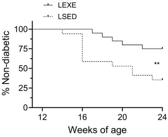
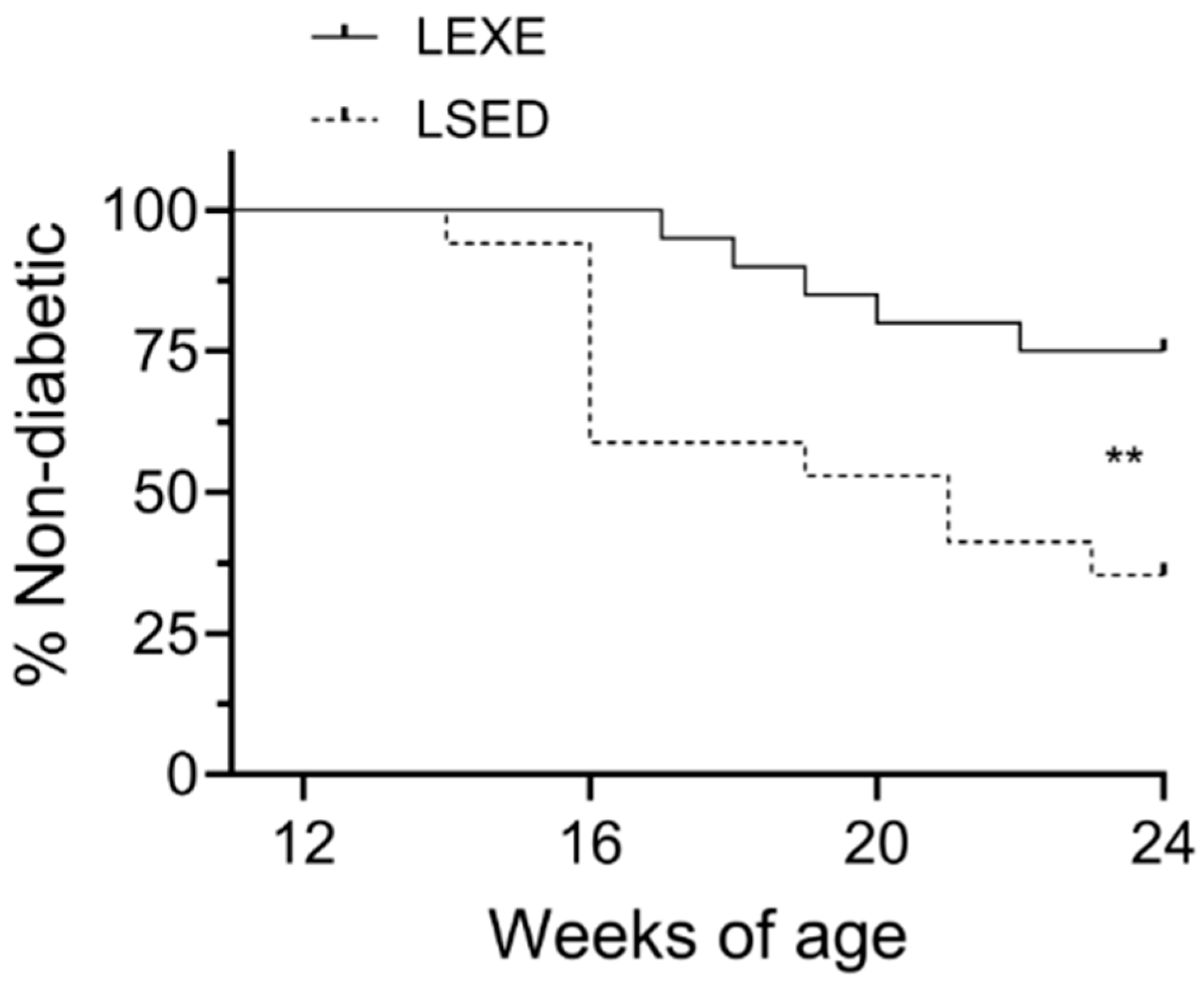
In Study 1, voluntary exercise was associated with a delayed onset and reduced incidence of T1D in female NOD mice compared to sedentary controls (p = 0.006; Figure 2). Overall, at the end of the experimental period (24 weeks of age), 15 of 20 (75%) exercised mice remained non-diabetic, whereas only 6 of 17 (35.3%) sedentary mice remained non-diabetic. Diabetes onset was also delayed in the exercise group, with the first case appearing at 17 weeks of age compared to 14 weeks in sedentary mice. By 19 weeks of age, 50% of sedentary mice had developed diabetes, while the exercise group mice only reached 25% incidence by 24 weeks, demonstrating that voluntary physical activity confers a significant protective effect against T1D onset in NOD mice. Consistent with these findings, survival analysis yielded a hazard ratio of 0.386 for the exercise group relative to sedentary controls, indicating a 38.6% reduced instantaneous risk of developing diabetes over time in exercising mice.
Figure 2.
Voluntary exercise reduced diabetes incidence and delayed onset in female NOD mice. By 24 weeks of age, 75% of exercised mice remained non-diabetic versus 35.3% of sedentary controls (p = 0.006). Diabetes onset occurred later in exercised mice (17 weeks) than sedentary mice (14 weeks). By 19 weeks of age, 50% of sedentary mice were diabetic, while exercised mice reached only 25% incidence by 24 weeks, indicating a protective effect of physical activity. LEXE: Late-time exercise group mice (n = 20); LSED: late-time sedentary mice (n = 17). Diabetes incidence was assessed using survival analysis. ** p < 0.01.
3.3. Glycemic Profiles
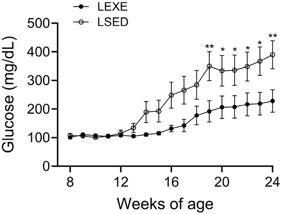
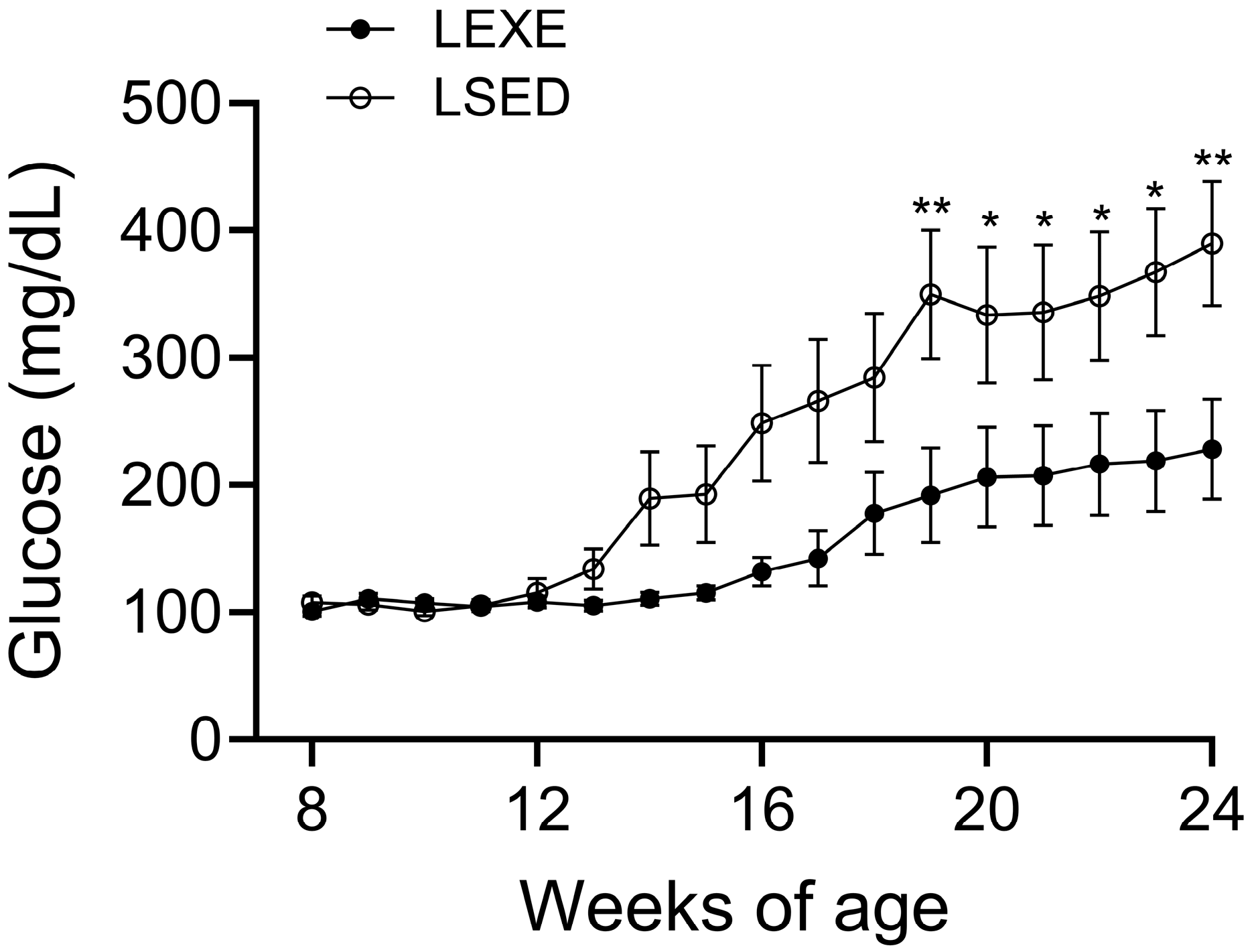
Throughout the observation period in Study 1, the exercised group demonstrated lower average blood glucose levels compared to sedentary controls (p = 0.015; Figure 3). Weekly monitoring from 8 to 24 weeks of age revealed that voluntary exercise effectively attenuated the rise in blood glucose over time. At 12 weeks of age, both groups had comparable baseline glucose levels (108.0 ± 4.6 mg/dL in exercise vs. 115.2 ± 11.3 mg/dL in sedentary mice; p > 0.999). However, by 19 weeks of age, exercised mice maintained lower glucose levels (191.7 ± 37.3 mg/dL) compared to sedentary mice (349.8 ± 50.3 mg/dL; p = 0.009), and this difference became more pronounced by 24 weeks (228.3 ± 39.3 mg/dL vs. 389.9 ± 48.9 mg/dL, respectively; p = 0.006). These results illustrate that voluntary physical activity helps maintain better glycemic control and delays the progression of hyperglycemia in NOD mice.
Figure 3.
Voluntary exercise reduced blood glucose levels in NOD mice throughout Study 1. Exercised mice showed lower average glucose than sedentary controls (p = 0.015). Although baseline levels at 12 weeks were similar (p > 0.999), exercised mice maintained lower glucose starting from 19 weeks and thereafter (p ≤ 0.019), indicating delayed hyperglycemia progression. LEXE: late-time exercise group mice (n = 20); LSED: late-time sedentary mice (n = 17). Results were analyzed using a repeated measure of Two-Way ANOVA (intervention × time). * p < 0.05, ** p < 0.01.
3.4. Correlation Between Glycemic Profiles and Running Distances
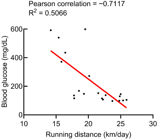
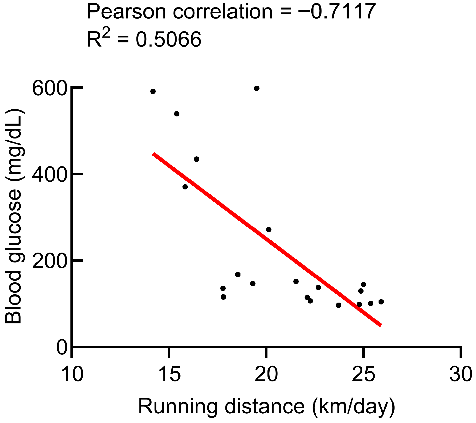
In Study 1, linear regression of endpoint non-fasting blood glucose values against daily running distance revealed a significant negative association in late-time exercise group NOD mice (Pearson’s r = −0.7117; R2 = 0.5066, p < 0.001), indicating that greater voluntary running activity was associated with lower contemporaneous glucose concentrations at disease onset. Furthermore, analysis of individual average running distance versus blood glucose at diabetes onset demonstrated a strong predictive dose–response relationship between cumulative exercise exposure and glycemia at onset (Figure 4).
Figure 4.
Scatter plot showing relationships between glucose level and daily running distance in late-time exercise group NOD mice (Study 1). Linear regression revealed a significant negative association (Pearson’s r = −0.7117; R2 = 0.5066, p < 0.001). Individual average running distance and blood glucose at diabetes onset analysis revealed a strong predictive relationship between running activity and glycemia at onset. Each plot indicates one mouse. LEXE: Late-time exercise group mice (n = 20).
3.5. Weekly Food Intake, Water Consumption, and Body Mass
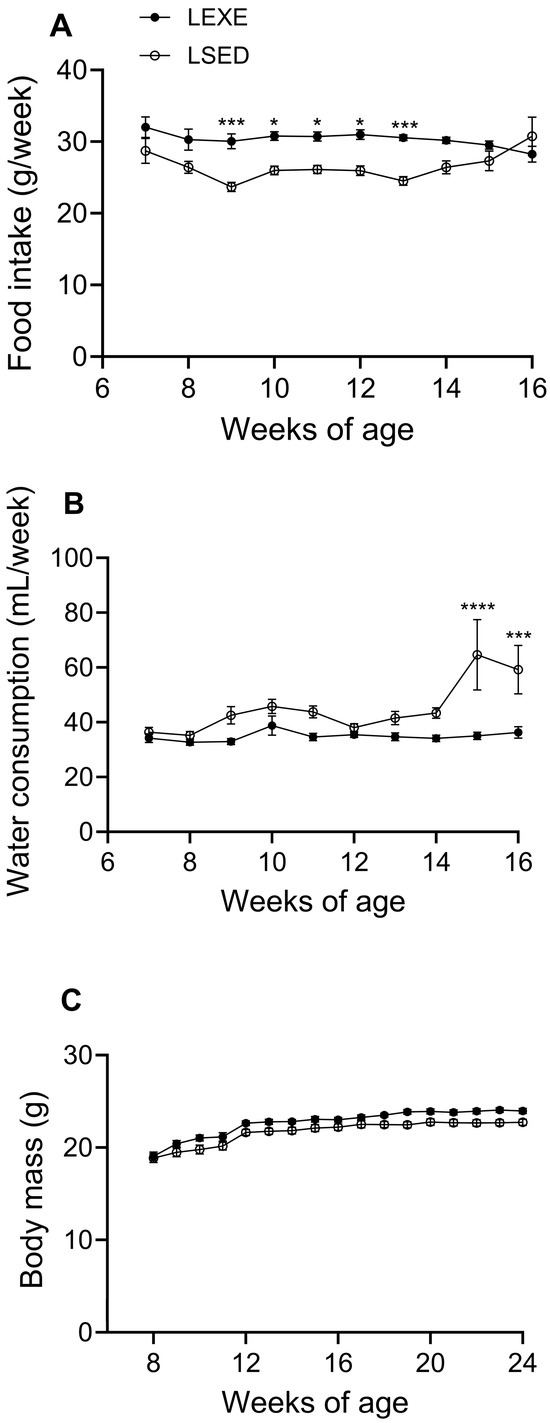
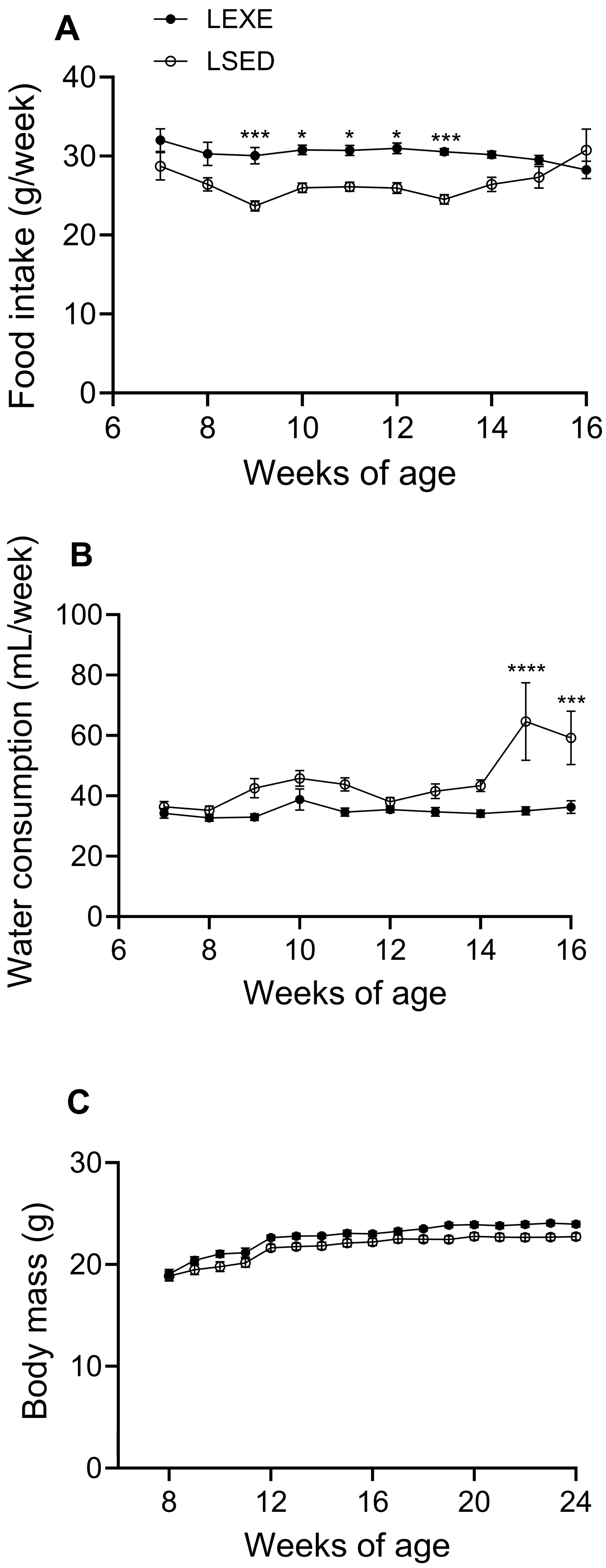
In Study 1, food intake was higher in exercise group mice compared to sedentary controls (30.3 ± 0.4 vs. 26.6 ± 0.6 g/week, p = 0.001; Figure 5A), indicating increased energy demands associated with physical activity. Water consumption was lower in exercise group mice compared to sedentary controls (36.3 ± 1.3 vs. 59.2 ± 8.9 mL/week, p < 0.001; Figure 5B), suggesting that physical activity may delay the onset of T1D during the prediabetic stage, as polydipsia is a key early indicator of disease progression. All mice gained weight over the course of the study in a time-dependent manner (Figure 5C). However, no difference in body mass was observed between exercise and sedentary groups at all time points (23.9 ± 0.3 vs. 22.7 ± 0.3 g, p = 0.389), suggesting that voluntary exercise did not attenuate weight gain.
Figure 5.
Exercise group mice consumed more food than sedentary controls (p = 0.0014; (A)), reflecting increased energy demands. Water intake was lower in exercise group mice (p < 0.001; (B)), consistent with delayed prediabetic polydipsia. All mice gained weight over time (C), but body mass did not differ between groups at multiple time points, including the endpoint (p = 0.389). LEXE: Late-time exercise group mice (n = 20); LSED: late-time sedentary mice (n = 17). Results were analyzed using a repeated measure of Two-Way ANOVA (intervention × time). * p < 0.05, *** p < 0.001, **** p < 0.0001.
3.6. Spleen Mass, Spleen Index, Total Count of Splenocytes, and Cell Viability
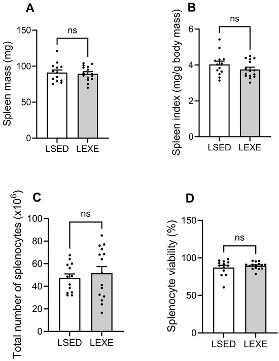
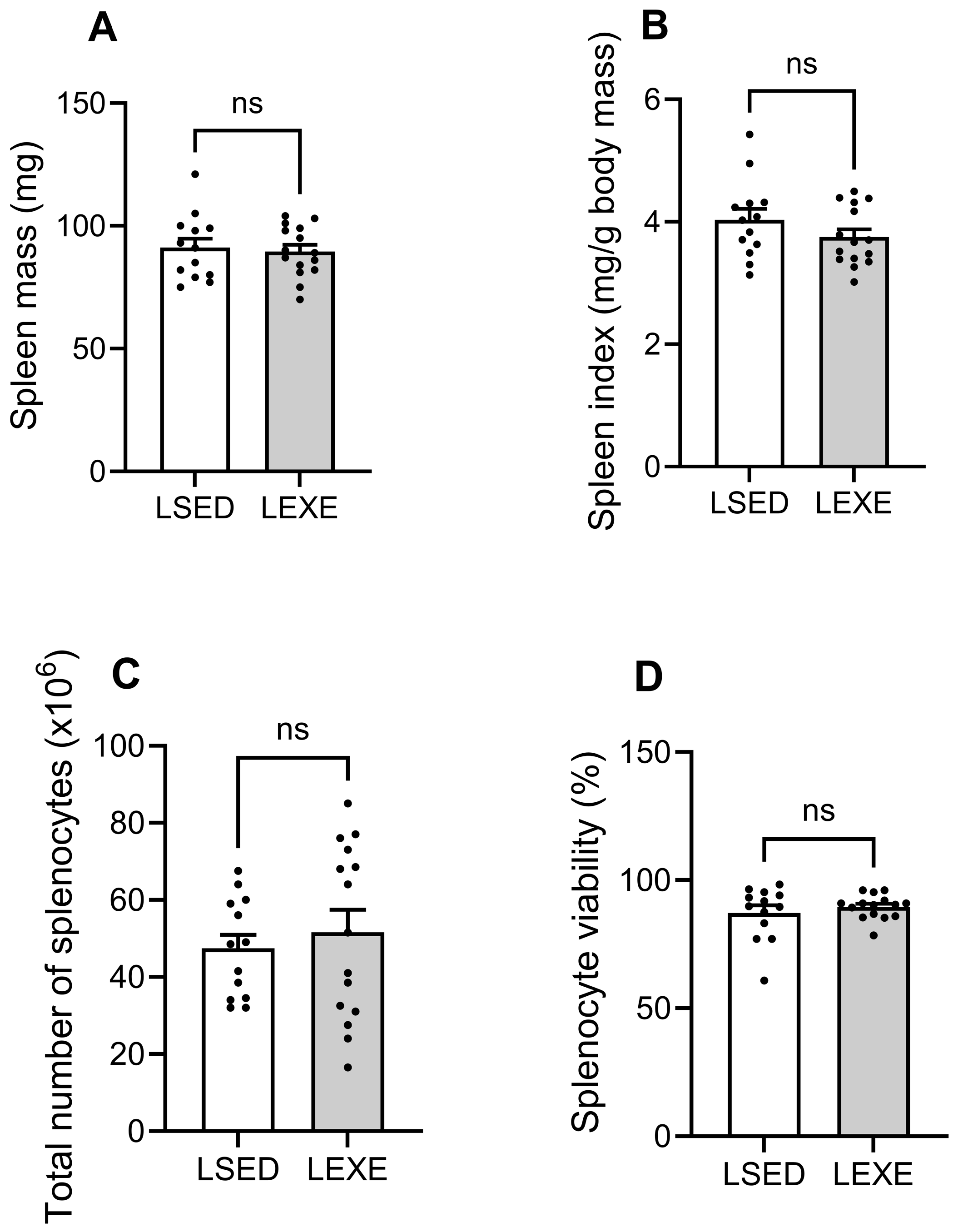
Ex vivo analysis of immunological parameters in late-time mice (Study 1) revealed no differences between exercise and sedentary NOD mice. Spleens were dissected and weighed, showing comparable spleen mass between the two groups (91.2 ± 3.7 mg vs. 89.6 ± 2.7 mg; p = 0.735; Figure 6A). Similarly, the spleen index (spleen mass/body mass) did not differ within groups (4.0 ± 0.2 vs. 3.8 ± 0.1 mg/g; p = 0.210; Figure 6B). Single cell suspensions prepared from spleens were used to assess total splenocyte counts and cell viability. No differences were observed in total splenocyte numbers (47.4 ± 3.6 vs. 51.6 ± 5.8 × 10⁶ cells; p = 0.547; Figure 6C) or in cell viability (87.2 ± 2.9% vs. 89.6 ± 1.2%; p = 0.460; Figure 6D).
Figure 6.
Ex vivo immunological parameters in exercise vs. sedentary late-time NOD mice (Study 1). Spleen mass (p = 0.735) and spleen index (p = 0.210) were comparable between groups (A,B). Total splenocyte counts (p = 0.547) and cell viability (p = 0.460) also showed no differences (C,D), indicating that voluntary exercise did not alter these systemic immune measures. LSED: Late-time sedentary mice (n = 13); LEXE: late-time exercise group mice (n = 15). Results were analyzed using a two-tailed unpaired Student’s t-test; ns, no significance.
Likewise, in early-time mice (Study 2), no differences were observed between exercise and sedentary NOD mice in any spleen parameters. Spleen mass (77.0 ± 1.8 vs. 74.8 ± 1.7 mg; p = 0.092) and spleen index (3.3 ± 0.1 vs. 3.3 ± 0.1 mg/g; p = 0.662) were comparable between groups (Supplemental Figure S1A,B). Similarly, total splenocyte counts (44.9 ± 4.2 vs. 43.9 ± 4.6 ×10⁶ cells; p = 0.885) and cell viability (90.4 ± 1.5 vs. 89.6 ± 1.3%; p = 0.696) showed no significance (Supplemental Figure S1C,D). These results suggest that voluntary exercise did not alter spleen mass or overall splenocyte yield and viability in NOD mice regardless of disease stage.
3.7. Immunophenotyping of Splenocytes
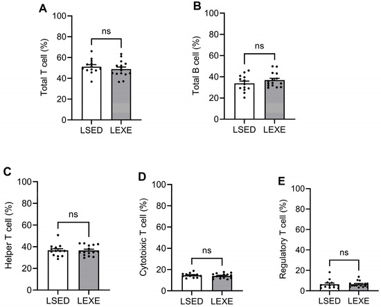
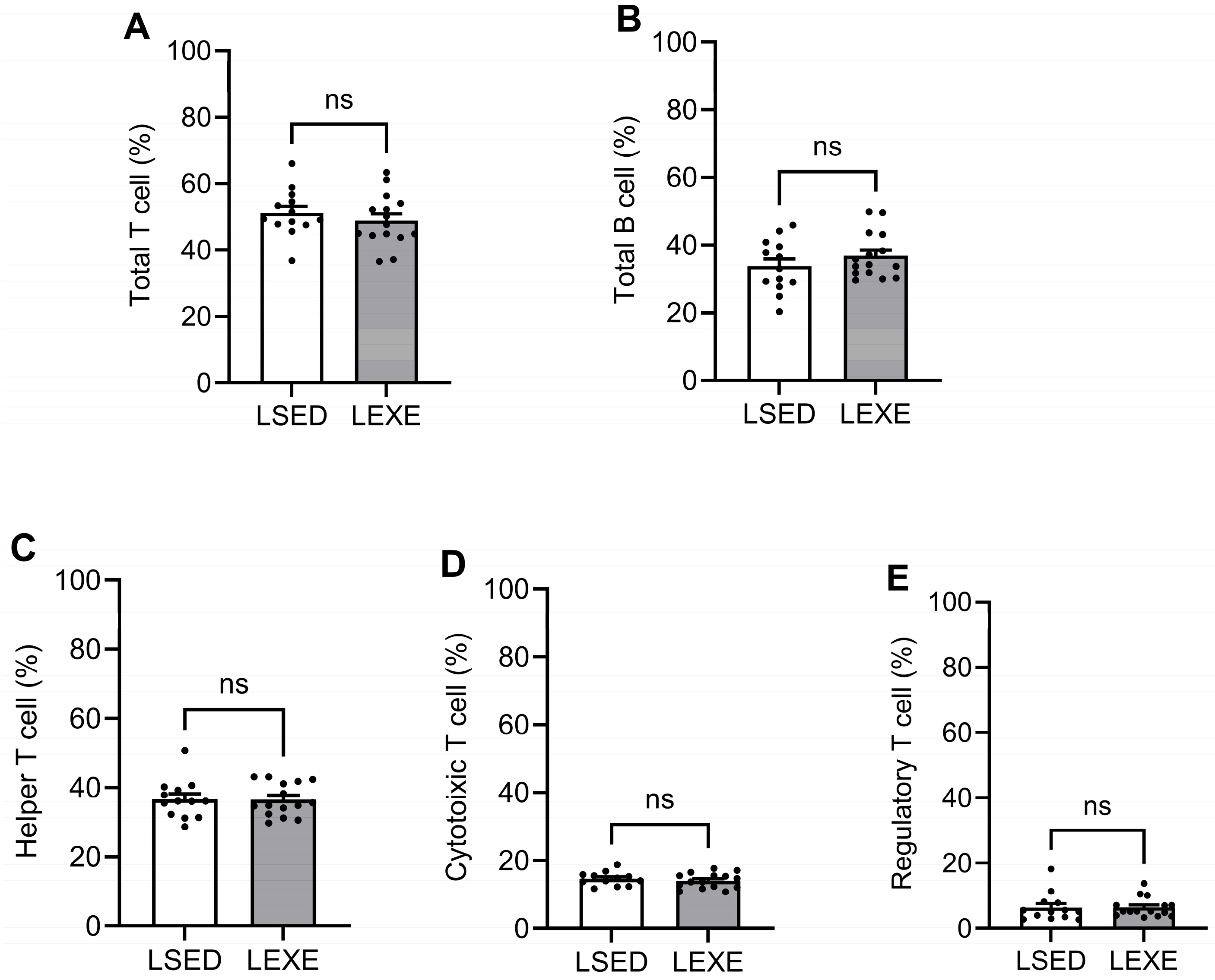
Representative flow cytometry dot plots of splenocytes from exercise and sedentary late-time NOD mice demonstrated major immune cell subpopulations (Supplemental Figure S2). Despite the significance observed in glycemic levels and diabetes incidence in late-time mice (Study 1), immunophenotyping of splenocytes revealed no differences in the major immune cell populations between exercise and sedentary NOD mice. The percentages of total T cells (48.9 ± 2.1 vs. 51.2 ± 2.0%; p = 0.423), B cells (36.8 ± 1.7 vs. 33.8 ± 2.1%; p = 0.278), helper T cells (Th; 36.5 ± 1.2 vs. 36.6 ± 1.5%; p = 0.967), cytotoxic T cells (Tc; 14.0 ± 0.6 vs. 14.6 ± 0.6%; p = 0.484), and regulatory T cells (Treg; 6.4 ± 0.8 vs. 6.4 ± 1.2%; p = 0.978) were similar between exercise and sedentary groups (Figure 7A–E). Additionally, no differences were observed in absolute lymphocyte numbers in each subpopulation (Table 1) or in the Treg to Tc ratio (Table 1) between exercise and sedentary NOD mice. This data reveals that voluntary exercise did not alter the overall composition of major splenic immune cell subsets in experimental T1D mice.
Figure 7.
Immunophenotyping of splenocytes from exercise and sedentary late-time NOD mice revealed no differences in major immune cell subpopulations (Study 1). (A) Percentages of total T cells (p = 0.423), (C) helper T cells (p = 0.185), (D) cytotoxic T cells (p = 0.821), (E) regulatory T cells (p = 0.608), and (B) total B cells (p = 0.278) were comparable between groups. These findings indicate that voluntary exercise did not alter the overall composition of major splenic immune cell subsets in experimental T1D mice. LSED: Late-time sedentary mice (n = 13); LEXE: late-time exercise group mice (n = 15). Results were analyzed using a two-tailed unpaired Student’s t-test; ns, no significance.
Table 1.
Comparison of absolute numbers of splenic lymphocyte subsets and ratios of regulatory T cells vs. cytotoxic T cells (Treg/Tc) in late-time NOD (Study 1).
Similarly, in early-time mice (Study 2), no differences were observed between the exercise and sedentary group NOD mice in the distribution of major splenic immune cell populations. The percentages of total T cells (48.1 ± 2.2 vs. 51.1 ± 2.9%; p = 0.410), helper T cells (Th; 37.2 ± 1.9 vs. 36.5 ± 2.1%; p = 0.806), cytotoxic T cells (Tc; 13.9 ± 0.5 vs. 14.8 ± 0.6%; p = 0.307), regulatory T cells (Treg; 4.7 ± 0.3 vs. 5.0 ± 0.4%; p = 0.727), and B cells (35.0 ± 1.9 vs. 34.1 ± 1.9%; p = 0.729) were similar between groups (Supplemental Figure S3A–E). These findings illustrate that voluntary exercise did not affect the composition of major splenic immune cell subsets in prediabetic NOD mice.
3.8. Cytokine Profiles
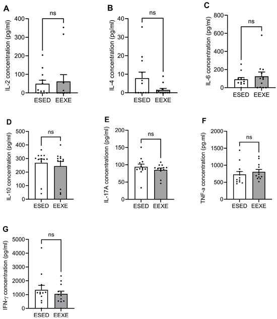
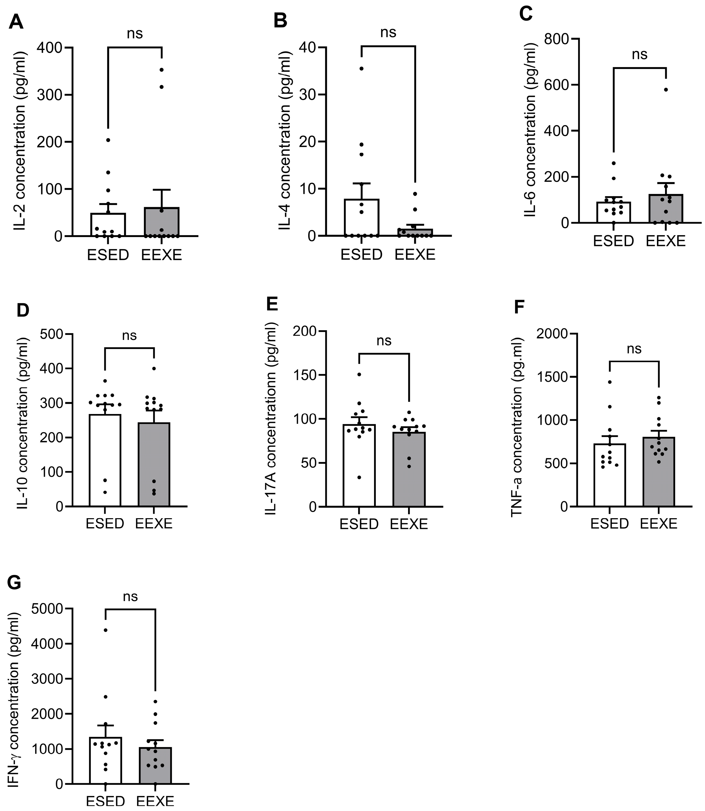
In early-time mice (Study 2), cytokine analysis of ConA-stimulated splenocyte cultures revealed no differences in cytokine production between exercise and sedentary groups. Concentrations of IL-2 (61.4 ± 37.2 vs. 49.3 ± 19.1 pg/mL; p = 0.776), IL-4 (vs. 1.6 ± 0.8 vs. 7.9 ± 3.2 pg/mL; p = 0.080), IL-6 (125.2 ± 46.8 vs. 92.0 ± 20.4 pg/mL; p = 0.525), IL-10 (243.9 ± 34.6 vs. 267.5 ± 28.9 pg/mL; p = 0.606), IL-17A (85.5 ± 5.1 vs. 94.1 ± 7.9 pg/mL; p = 0.372), TNF-α (807.0 ± 69.9 vs. 729.2 ± 87.4 pg/mL; p = 0.494), and IFN-γ (1054.3 ± 198.6 vs. 1343.0 ± 330.5 pg/mL; p = 0.464) were comparable between exercise and sedentary group mice (Figure 8A–G). These outcomes indicate that voluntary exercise did not alter Th1, Th2, or Th17 cytokine profiles in early-stage prediabetic experimental NOD mice. However, given the relatively large variance observed in several cytokines, the study may have been underpowered to detect small-to-moderate effect sizes, and thus subtle immunomodulatory differences cannot be fully excluded.
Figure 8.
Cytokine production by ConA-stimulated splenocytes in early-time NOD showed no differences between exercise and sedentary group mice (Study 2). Levels of (A) IL-2 (p = 0.776), (B) IL-4 (p = 0.080), (C) IL-6 (p = 0.525), (D) IL-10 (p = 0.606), (E) IL-17A (p = 0.372), (F) TNF-α (p = 0.494), and (G) IFN-γ (p = 0.464) were comparable between groups. These results indicate that voluntary exercise did not alter Th1, Th2, or Th17 cytokine responses in early-stage experimental T1D. ESED: Early-time sedentary mice (n = 12); EEXE: early-time exercise group mice (n = 11). Results were analyzed using a two-tailed unpaired Student’s t-test; ns, no significance.
4. Discussion
Our results support our hypothesis that voluntary exercise (1) delays the onset of hyperglycemia resulting from pancreatic β-cell loss and (2) slows the progression of T1D compared to sedentary controls. Consistent with this prediction, voluntary exercise delayed diabetes onset and reduced disease incidence in NOD mice, while also lowering glycemia levels throughout the study period. Exercised mice maintained lower average blood glucose levels and showed a slower rise in glycemia compared to sedentary mice, indicating preserved β-cell function and delayed autoimmune destruction. These findings demonstrate that physical activity alone serves as an independent protective factor against T1D progression in this model. Furthermore, our findings align with both clinical and preclinical evidence showing that structured aerobic or combined resistance–aerobic exercise enhances glycemic regulation and metabolic stability in individuals with T1D [26,27,28,29].
A primary objective and innovation of our study was to evaluate whether voluntary exercise modulates splenic immunomodulatory capacity in experimental T1D. Contrary to reports describing exercise-driven shifts in inflammatory cytokines and immune markers [30,31], we detected no differences in splenic T cell subsets or cytokine profiles between exercise and sedentary group NOD mice, indicating that in this context exercise exerts limited systemic immune modulation. This aligns with studies showing that while exercise can elevate adaptive stress response mediators such as VEGF and HIF-1α, downstream cytokine and immune progenitor responses in autoimmune diabetes are subtle and variable [28,32]. Moreover, the presence of mild diabetic myopathy may limit skeletal muscle adaptation to exercise and contribute to the heterogeneity of immune and metabolic responses [33]. Together, these results support emerging evidence that the benefits of physical activity in T1D arise predominantly from tissue-specific or metabolic adaptations rather than broad changes in immune cell composition, which highlights the importance of further mechanistic exploration into how physical activity influences autoimmunity and splenic immune cell regulation.
We evaluated whether physical activity influences splenic immunoregulation in T1D by assessing splenic T cell-associated cytokine expression. Voluntary exercise did not alter splenic levels of pro- or anti-inflammatory cytokines, including IL-10, IL-4, IL-17A, TNF-α, IL-6, IFN-γ, and IL-2, nor did it modify major CD4+ or CD8+ T cell subsets, indicating minimal immunomodulatory impact. These data align with previous studies [27,34], suggesting that physical activity exerts inconsistent or subtle immune effects in models of diabetes. In rodent studies, exercise has been shown to partially restore impaired natural killer T cell activity and reverse zinc transporter suppression in the spleen and thymus of streptozotocin-induced diabetic animals [35], whereas other reports describe significant oxidative stress associated with acute exercise that is mitigated by melatonin [36]. Additionally, experimental work in islet-transplanted animals demonstrates that glucose homeostasis and insulin regulation during exercise are largely preserved irrespective of graft site, suggesting that systemic metabolic benefits may be more prominent than immune modulation [37,38]. Together, these observations support the conclusion that, in the setting of ongoing β-cell autoimmunity, the capacity of voluntary exercise to broadly remodel splenic T cell populations or cytokine signaling is limited, and exercise-derived benefits may occur through metabolic, oxidative, or tissue-specific pathways rather than through robust modification of splenic immune profiles.
Previous studies have suggested that physical activity, particularly structured moderate-intensity training, may promote a shift in the immune profile toward a more anti-inflammatory state [29,39]. Codella et al. reported no significant differences in the cytokines examined despite a trend toward reduced proinflammatory markers such as IL-6 and CCL4 with treadmill training [39]. Similarly, Oharomari et al. observed no changes in serum IL-6 or TNF-α levels, even though exercise reduced insulitis in NOD mice [29]. Additionally, the stress-buffering effects of exercise may help normalize glutamine metabolism and reduce sympathetic activation of Th1-dominant pathways, theoretically allowing a shift toward Th2 cytokines and increased antioxidant defenses via glutathione synthesis [40]. However, our data using a voluntary wheel-running paradigm suggests that, in the absence of externally imposed exercise intensity or duration, this form of physical activity is insufficient to provoke measurable changes in splenic T cell cytokine production during the progression of T1D.
Our results of no difference in IL-6 levels between exercise and sedentary mice may be explained by the context-dependent nature of IL-6 expression and action during physical activity. IL-6 is known to be acutely upregulated in skeletal muscle in response to contraction, where it functions locally to promote glucose uptake and fatty acid oxidation through activation of pathways such as AMP-activated protein kinase (AMPK) and enhanced glucose transporter protein type-4 (GLUT4) expression [41,42,43]. However, this transient increase in IL-6 during muscle contraction may not be captured at resting time points, particularly in chronic exercise studies where systemic IL-6 levels can return to baseline rapidly after cessation of activity [44]. Additionally, the metabolic effects of IL-6 may depend on interaction with other exercise-induced factors, which may be absent or diminished at rest. Thus, our observed lack of difference in IL-6 levels may reflect the timing of sample collection relative to exercise sessions, rather than an absence of IL-6-mediated metabolic adaptation.
During exercise, skeletal muscle contractions acutely stimulate the production of both pro- and anti-inflammatory cytokines to support metabolic demands and maintain immune balance [45,46]. However, many of these cytokine changes, particularly the anti-inflammatory effects mediated by IL-10 and IL-4, and the controlled expression of TNF-α, are tightly regulated and may return to baseline shortly after exercise ends [47,48]. Thus, if tissue or serum samples were collected at rest, rather than immediately post-exercise, these dynamic fluctuations may not be detected. Furthermore, the anti-inflammatory cytokine response to exercise, including the suppression of TNF-α, is thought to be partially mediated by IL-6 and its downstream signaling, which also relies on acute muscle contraction [49]. Our results may therefore reflect a limitation in sampling timing and highlight the importance of capturing cytokine expression at or near the time of muscle activity to fully assess exercise-induced immunomodulation.
Our study is limited by its focus on splenic immune composition, which does not necessarily reflect functional activation status, antigen specificity, trafficking behavior, or tissue residency. We did not directly assess immune cell composition, cytokine profiles, or activation states within pancreatic islets or pancreatic lymph nodes—key sites of autoimmune initiation and insulitis—nor did we evaluate insulitis severity, as the primary objective was to examine systemic and splenic immune responses to voluntary exercise rather than pancreatic histopathology. Therefore, the absence of detectable splenic alterations does not exclude localized immune modulation at disease-relevant tissues. Future studies should incorporate insulitis grading and tissue-specific immune profiling to determine whether exercise alters pancreatic immune infiltration. In addition, antigenic triggers derived from the pancreas, including misfolded insulin, are known to induce the autoimmune cascades in NOD mice [50,51], and exercise may attenuate this response by lessening pancreatic beta cell stress [29,52]. Exercise may also regulate the activation status of specific T cell and B cell subpopulations and thereby modulate their corresponding cytokine production [53,54]. Moreover, assessment of β-cell mass preservation, glucose-stimulated insulin secretion, β-cell stress markers, and peripheral insulin sensitivity will be necessary to clarify whether exercise-mediated protection reflects intrinsic β-cell preservation, improved metabolic resilience, altered systemic insulin dynamics, or tissue-specific immune regulation.
In conclusion, voluntary exercise reduces T1D incidence and mitigates hyperglycemia in experimental diabetic mice, suggesting a protective effect against disease progression. However, physical activity did not alter splenic CD4+ or CD8+ T cell subsets or associated cytokines, indicating that its benefits occur independently of systemic immunomodulation. These results suggest that voluntary exercise protects against T1D through tissue-specific or metabolic effects rather than broad immune remodeling. Future studies should determine whether exercise-mediated protection involves pancreatic immune modulation, enhanced β-cell resilience, or systemic metabolic adaptations contributing to reduced type 1 diabetes incidence.
Supplementary Materials
The following supporting information can be downloaded at https://www.mdpi.com/article/10.3390/diabetology7040064/s1: Figure S1: Ex vivo immunological parameters in exercise vs. sedentary group mice in early-time NOD (Study 2); Figure S2: Representative flow cytometry plots of splenocytes from exercise and sedentary group late-time NOD mice in major immune cell subpopulations (Study 1). Figure S3: Immunophenotyping of splenocytes from exercise and sedentary group mice in early-time NOD (Study 2).
Author Contributions
Conceptualization, M.C.-C. and G.L.-C.; methodology, M.C.-C., H.A., M.A.N. and G.L.-C.; software, G.L.-C. and S.S.K.C.; validation, M.C.-C., S.S.K.C. and G.L.-C.; formal analysis, S.S.K.C. and G.L.-C.; investigation, H.A. and M.A.N.; resources, M.C.-C. and G.L.-C.; data curation, H.A. and M.A.N.; writing—original draft preparation, G.L.-C.; writing—review and editing, M.C.-C., S.S.K.C. and G.L.-C.; visualization, S.S.K.C. and G.L.-C.; supervision, M.C.-C. and G.L.-C.; project administration, M.C.-C. and G.L.-C.; funding acquisition, M.C.-C. and G.L.-C. All authors have read and agreed to the published version of the manuscript.
Funding
This research was funded by the Early Career Grant (G.L.-C.), Faculty Improvement Grant Research Grant (G.L.-C.), and Student Mentor Collaboration Grant (M.C.-C., H.A., M.A.N., and G.L.-C.) from St. Cloud State University; NASA Connecticut Space Grant Consortium Faculty Research Grant—PTE Federal Award No. 80NSSC25M7127 (G.L.-C.); Greenberg Junior Faculty Research Grant (G.L.-C.), and College of Arts and Sciences Dean’s Faculty Research Funds (G.L.-C.) from the University of Hartford.
Institutional Review Board Statement
The animal study protocol was approved by the Institutional Animal Care and Use Committee of St. Cloud State University (IACUC#5-146, 18 September 2023).
Informed Consent Statement
Not applicable.
Data Availability Statement
The original contributions presented in this study are included in the article. Further inquiries can be directed to the corresponding author.
Acknowledgments
We are thankful for all the funds and grants that made this study possible. We gratefully acknowledge the valuable contributions and support of Barb Kjellberg, Mary Dowst, Kyle Reason, and Brian Lorenz. We also thank Mariama Suwareh, Efaith Borbor, Fathaa Mahamud, Christabel Ofori, Lauren Wilfong, Taylor Larsen, and Alex Math for their assistance with data collection and project support.
Conflicts of Interest
The authors declare no conflicts of interest.
Abbreviations
The following abbreviations are used in this manuscript:
| CCL4 | Chemokine (C-C motif) ligands 4 |
| EEXE | Early-time exercise |
| ESED | Early-time sedentary |
| HIF-1α | Hypoxia-inducible factor 1-alpha |
| IFN-γ | Interferon gamma |
| LEXE | Late-time exercise |
| LSED | Late-time sedentary |
| NOD | Non-Obese Diabetic |
| T1D | Type 1 Diabetes Mellitus |
| TNF-α | Tumor necrosis factor alpha |
| VEGF | Vascular endothelial growth factor |
References
- Dial, A.G.; Grafham, G.K.; Monaco, C.M.F.; Voth, J.; Brandt, L.; Tarnopolsky, M.A.; Hawke, T.J. Alterations in skeletal muscle repair in young adults with type 1 diabetes mellitus. Am. J. Physiol. Cell Physiol. 2021, 321, C876–C883. [Google Scholar] [CrossRef] [Scilit]
- Hebert, S.L.; Nair, K.S. Protein and energy metabolism in type 1 diabetes. Clin. Nutr. 2010, 29, 13–17. [Google Scholar] [CrossRef] [Scilit] [PubMed]
- Sinclair, A.J.; Dunning, T.; Dhatariya, K.; an International Group of Experts. Clinical guidelines for type 1 diabetes mellitus with an emphasis on older adults: An Executive Summary. Diabet. Med. 2020, 37, 53–70. [Google Scholar] [CrossRef] [Scilit]
- Norris, J.M.; Johnson, R.K.; Stene, L.C. Type 1 diabetes-early life origins and changing epidemiology. Lancet Diabetes Endocrinol. 2020, 8, 226–238. [Google Scholar] [CrossRef] [Scilit]
- Xia, Y.; Xie, Z.; Huang, G.; Zhou, Z. Incidence and trend of type 1 diabetes and the underlying environmental determinants. Diabetes Metab. Res. Rev. 2019, 35, e3075. [Google Scholar] [CrossRef] [Scilit]
- Kanaley, J.A.; Colberg, S.R.; Corcoran, M.H.; Malin, S.K.; Rodriguez, N.R.; Crespo, C.J.; Kirwan, J.P.; Zierath, J.R. Exercise/Physical Activity in Individuals with Type 2 Diabetes: A Consensus Statement from the American College of Sports Medicine. Med. Sci. Sports Exerc. 2022, 54, 353–368. [Google Scholar] [CrossRef] [Scilit] [PubMed]
- Kaza, M.; Tsentidis, C.; Vlachopapadopoulou, E.; Karanasios, S.; Sakou, I.-I.; Mastorakos, G.; Karavanaki, K. The Role of Exercise on Cardiometabolic Profile and Body Composition in Youth with Type 1 Diabetes. Children 2022, 9, 1840. [Google Scholar] [CrossRef] [Scilit]
- Kirwan, J.P.; Sacks, J.; Nieuwoudt, S. The essential role of exercise in the management of type 2 diabetes. Cleve Clin. J. Med. 2017, 84, S15–S21. [Google Scholar] [CrossRef] [Scilit] [PubMed]
- Rangel, M.A.; Calejo, R.; Lopes, V.; Campos, R.A.; Leite, A.L. Body composition in a pediatric population with type-1 diabetes—The importance of planned physical exercise. Ann. Endocrinol. 2025, 86, 101670. [Google Scholar] [CrossRef] [Scilit]
- Tsalamandris, S.; Antonopoulos, A.S.; Oikonomou, E.; Papamikroulis, G.A.; Vogiatzi, G.; Papaioannou, S.; Deftereos, S.; Tousoulis, D. The Role of Inflammation in Diabetes: Current Concepts and Future Perspectives. Eur. Cardiol. 2019, 14, 50–59. [Google Scholar] [CrossRef] [Scilit]
- Becker, M.W.; Peters, L.D.; Myint, T.; Smurlick, D.; Powell, A.; Brusko, T.M.; Phelps, E.A. Immune engineered extracellular vesicles to modulate T cell activation in the context of type 1 diabetes. Sci. Adv. 2023, 9, eadg1082. [Google Scholar] [CrossRef] [Scilit]
- Curran, M.; Campbell, J.; Drayson, M.; Andrews, R.; Narendran, P. Type 1 diabetes impairs the mobilisation of highly-differentiated CD8+T cells during a single bout of acute exercise. Exerc. Immunol. Rev. 2019, 25, 64–82. [Google Scholar]
- Blank, M.; Israeli, D.; Shoenfeld, Y. Exercise, autoimmune diseases and T-regulatory cells. J. Autoimmun. 2024, 149, 103317. [Google Scholar] [CrossRef] [Scilit]
- Pearson, J.A.; Wong, F.S.; Wen, L. The importance of the Non Obese Diabetic (NOD) mouse model in autoimmune diabetes. J. Autoimmun. 2016, 66, 76–88. [Google Scholar] [CrossRef] [Scilit]
- Yoshida, K.; Kikutani, H. Genetic and immunological basis of autoimmune diabetes in the NOD mouse. Rev. Immunogenet. 2000, 2, 140–146. [Google Scholar] [PubMed]
- Cetkovic-Cvrlje, M.; Olson, M.; Schindler, B.; Gong, H.K. Exposure to DDT metabolite p,p′-DDE increases autoimmune type 1 diabetes incidence in NOD mouse model. J. Immunotoxicol. 2016, 13, 108–118. [Google Scholar] [CrossRef] [Scilit] [PubMed]
- Serreze, D.V.; Dwyer, J.R.; Racine, J.J. Advancing Animal Models of Human Type 1 Diabetes. Cold Spring Harb. Perspect. Med. 2024, 14, a041587. [Google Scholar] [CrossRef] [Scilit]
- Cabelka, C.A.; Baumann, C.W.; Collins, B.C.; Nash, N.; Le, G.; Lindsay, A.; Spangenburg, E.E.; Lowe, D.A. Effects of ovarian hormones and estrogen receptor alpha on physical activity and skeletal muscle fatigue in female mice. Exp. Gerontol. 2019, 115, 155–164. [Google Scholar] [CrossRef] [Scilit] [PubMed]
- Call, J.A.; McKeehen, J.N.; Novotny, S.A.; Lowe, D.A. Progressive resistance voluntary wheel running in the mdx mouse. Muscle Nerve 2010, 42, 871–880. [Google Scholar] [CrossRef] [Scilit]
- Cetkovic-Cvrlje, M.; Thinamany, S.; Bruner, K.A. Bisphenol A (BPA) aggravates multiple low-dose streptozotocin-induced Type 1 diabetes in C57BL/6 mice. J. Immunotoxicol. 2017, 14, 160–168. [Google Scholar] [CrossRef] [Scilit]
- Cetkovic-Cvrlje, M.; Gerling, I.C.; Muir, A.; Atkinson, M.A.; Elliott, J.F.; Leiter, E.H. Retardation or acceleration of diabetes in NOD/Lt mice mediated by intrathymic administration of candidate β-cell antigens. Diabetes 1997, 46, 1975–1982. [Google Scholar] [CrossRef] [Scilit] [PubMed]
- Cetkovic-Cvrlje, M.; Roers, B.A.; Schonhoff, D.; Waurzyniak, B.; Liu, X.P.; Uckun, F.M. Treatment of post-bone marrow transplant acute graft-versus-host disease with a rationally designed JAK3 inhibitor. Leuk. Lymphoma 2002, 43, 1447–1453. [Google Scholar] [CrossRef] [Scilit] [PubMed]
- Cetkovic-Cvrlje, M.; Uckun, F.M. Dual targeting of Bruton’s tyrosine kinase and Janus kinase 3 with rationally designed inhibitors prevents graft-versus-host disease (GVHD) in a murine allogeneic bone marrow transplantation model. Br. J. Haematol. 2004, 126, 821–827. [Google Scholar] [CrossRef] [Scilit]
- Cetkovic-Cvrlje, M.; Roers, B.A.; Waurzyniak, B.; Liu, X.P.; Uckun, F.M. Targeting Janus kinase 3 to attenuate the severity of acute graft-versus-host disease across the major histocompatibility barrier in mice. Blood 2001, 98, 1607–1613. [Google Scholar] [CrossRef] [Scilit]
- Cetkovic-Cvrlje, M.; Uckun, F.M. Effect of targeted disruption of signal transducer and activator of transcription (Stat)4 and Stat6 genes on the autoimmune diabetes development induced by multiple low doses of streptozotocin. Clin. Immunol. 2005, 114, 299–306. [Google Scholar] [CrossRef] [Scilit]
- Adamo, M.; Codella, R.; Casiraghi, F.; Ferrulli, A.; Macri, C.; Bazzigaluppi, E.; Terruzzi, I.; Inverardi, L.; Ricordi, C.; Luzi, L. Active Subjects with Autoimmune Type 1 Diabetes Have Better Metabolic Profiles Than Sedentary Controls. Cell Transplant. 2017, 26, 23–32. [Google Scholar] [CrossRef] [Scilit]
- Farinha, J.B.; Ramis, T.R.; Vieira, A.F.; Macedo, R.C.O.; Rodrigues-Krause, J.; Boeno, F.P.; Schroeder, H.T.; Muller, C.H.; Boff, W.; Krause, M.; et al. Glycemic, inflammatory and oxidative stress responses to different high-intensity training protocols in type 1 diabetes: A randomized clinical trial. J. Diabetes Complicat. 2018, 32, 1124–1132. [Google Scholar] [CrossRef] [Scilit] [PubMed]
- Nazari, M.; Shabani, R.; Hassanzadeh-Rad, A.; Esfandiari, M.A.; Dalili, S. Effect of concurrent resistance-aerobic training on inflammatory factors and growth hormones in children with type 1 diabetes: A randomized controlled clinical trial. Trials 2023, 24, 519. [Google Scholar] [CrossRef] [Scilit]
- Oharomari, L.K.; de Moraes, C.; Navarro, A.M. Exercise Training but not Curcumin Supplementation Decreases Immune Cell Infiltration in the Pancreatic Islets of a Genetically Susceptible Model of Type 1 Diabetes. Sports Med. Open 2017, 3, 15. [Google Scholar] [CrossRef] [Scilit]
- Galassetti, P.R.; Iwanaga, K.; Crisostomo, M.; Zaldivar, F.P.; Larson, J.; Pescatello, A. Inflammatory cytokine, growth factor and counterregulatory responses to exercise in children with type 1 diabetes and healthy controls. Pediatr. Diabetes 2006, 7, 16–24. [Google Scholar] [CrossRef] [Scilit]
- Zebrowska, A.; Hall, B.; Maszczyk, A.; Banas, R.; Urban, J. Brain-derived neurotrophic factor, insulin like growth factor-1 and inflammatory cytokine responses to continuous and intermittent exercise in patients with type 1 diabetes. Diabetes Res. Clin. Pract. 2018, 144, 126–136. [Google Scholar] [CrossRef] [Scilit]
- Hall, B.; Zebrowska, A.; Sikora, M.; Siatkowski, S.; Robins, A. The Effect of High-Intensity Interval Exercise on Short-Term Glycaemic Control, Serum Level of Key Mediator in Hypoxia and Pro-Inflammatory Cytokines in Patients with Type 1 Diabetes-An Exploratory Case Study. Nutrients 2023, 15, 3749. [Google Scholar] [CrossRef] [Scilit] [PubMed]
- Minnock, D.; Annibalini, G.; Valli, G.; Saltarelli, R.; Krause, M.; Barbieri, E.; De Vito, G. Altered muscle mitochondrial, inflammatory and trophic markers, and reduced exercise training adaptations in type 1 diabetes. J. Physiol. 2022, 600, 1405–1418. [Google Scholar] [CrossRef] [Scilit]
- Bock, P.M.; Monteiro, R.B.; Maraschin, C.K.; Alegretti, A.P.; Farias, M.G.; Spagnol, F.; Lopez, P.; Santos, L.P.; Helal, L.; Moraes, R.S.; et al. Circulating endothelial progenitor cells and inflammatory markers in type 1 diabetes after an acute session of aerobic exercise. Arch. Endocrinol. Metab. 2024, 68, e230499. [Google Scholar] [CrossRef] [Scilit] [PubMed]
- Takeuchi, Y.; Nishikawa, H. Roles of regulatory T cells in cancer immunity. Int. Immunol. 2016, 28, 401–409. [Google Scholar] [CrossRef] [Scilit]
- Kodama, S.; Kuhtreiber, W.; Fujimura, S.; Dale, E.A.; Faustman, D.L. Islet regeneration during the reversal of autoimmune diabetes in NOD mice. Science 2003, 302, 1223–1227. [Google Scholar] [CrossRef] [Scilit]
- Itoh, T.; Nishinakamura, H.; Kumano, K.; Takahashi, H.; Kodama, S. The Spleen Is an Ideal Site for Inducing Transplanted Islet Graft Expansion in Mice. PLoS ONE 2017, 12, e0170899. [Google Scholar] [CrossRef] [Scilit]
- Ugurlu, I.; Baltaci, S.B.; Unal, O.; Mogulkoc, R.; Ucaryilmaz, H.; Baltaci, A.K. Chronic Running Exercise Regulates Cytotoxic Cell Functions and Zinc Transporter SLC39A10/ZIP10 Levels in Diabetic Rats. Biol. Trace Elem. Res. 2022, 200, 699–705. [Google Scholar] [CrossRef] [Scilit] [PubMed]
- Codella, R.; Lanzoni, G.; Zoso, A.; Caumo, A.; Montesano, A.; Terruzzi, I.M.; Ricordi, C.; Luzi, L.; Inverardi, L. Moderate Intensity Training Impact on the Inflammatory Status and Glycemic Profiles in NOD Mice. J. Diabetes Res. 2015, 2015, 737586. [Google Scholar] [CrossRef] [Scilit]
- da Silva Krause, M.; de Bittencourt, P.I.H., Jr. Type 1 diabetes: Can exercise impair the autoimmune event? The L-arginine/glutamine coupling hypothesis. Cell Biochem. Funct. 2008, 26, 406–433. [Google Scholar] [CrossRef] [Scilit]
- Chan, M.H.; McGee, S.L.; Watt, M.J.; Hargreaves, M.; Febbraio, M.A. Altering dietary nutrient intake that reduces glycogen content leads to phosphorylation of nuclear p38 MAP kinase in human skeletal muscle: Association with IL-6 gene transcription during contraction. FASEB J. 2004, 18, 1785–1787. [Google Scholar] [CrossRef] [Scilit]
- O’Neill, H.M.; Maarbjerg, S.J.; Crane, J.D.; Jeppesen, J.; Jorgensen, S.B.; Schertzer, J.D.; Shyroka, O.; Kiens, B.; van Denderen, B.J.; Tarnopolsky, M.A.; et al. AMP-activated protein kinase (AMPK) beta1beta2 muscle null mice reveal an essential role for AMPK in maintaining mitochondrial content and glucose uptake during exercise. Proc. Natl. Acad. Sci. USA 2011, 108, 16092–16097. [Google Scholar] [CrossRef] [Scilit]
- Wolsk, E.; Mygind, H.; Grondahl, T.S.; Pedersen, B.K.; van Hall, G. IL-6 selectively stimulates fat metabolism in human skeletal muscle. Am. J. Physiol. Endocrinol. Metab. 2010, 299, E832–E840. [Google Scholar] [CrossRef] [Scilit] [PubMed]
- Fischer, C.P. Interleukin-6 in acute exercise and training: What is the biological relevance? Exerc. Immunol. Rev. 2006, 12, 6–33. [Google Scholar]
- Argiles, J.M.; Campos, N.; Lopez-Pedrosa, J.M.; Rueda, R.; Rodriguez-Manas, L. Skeletal Muscle Regulates Metabolism via Interorgan Crosstalk: Roles in Health and Disease. J. Am. Med. Dir. Assoc. 2016, 17, 789–796. [Google Scholar] [CrossRef] [Scilit]
- Docherty, S.; Harley, R.; McAuley, J.J.; Crowe, L.A.N.; Pedret, C.; Kirwan, P.D.; Siebert, S.; Millar, N.L. The effect of exercise on cytokines: Implications for musculoskeletal health: A narrative review. BMC Sports Sci. Med. Rehabil. 2022, 14, 5. [Google Scholar] [CrossRef] [Scilit]
- Moldoveanu, A.I.; Shephard, R.J.; Shek, P.N. The cytokine response to physical activity and training. Sports Med. 2001, 31, 115–144. [Google Scholar] [CrossRef] [Scilit] [PubMed]
- Petersen, A.M.; Pedersen, B.K. The anti-inflammatory effect of exercise. J. Appl. Physiol. 2005, 98, 1154–1162. [Google Scholar] [CrossRef] [Scilit]
- Nara, H.; Watanabe, R. Anti-Inflammatory Effect of Muscle-Derived Interleukin-6 and Its Involvement in Lipid Metabolism. Int. J. Mol. Sci. 2021, 22, 9889. [Google Scholar] [CrossRef] [Scilit] [PubMed]
- Bicer, M.; Akil, M.; Baltaci, A.K.; Mogulkoc, R.; Sivrikaya, A.; Gunay, M.; Akkus, H. Protective effect of melatonin on lipid peroxidation in various tissues of diabetic rats subjected to an acute swimming exercise. Bratisl. Lek. Listy 2012, 113, 698–701. [Google Scholar] [CrossRef] [Scilit]
- Omer, A.; Duvivier-Kali, V.F.; Aschenbach, W.; Tchipashvili, V.; Goodyear, L.J.; Weir, G.C. Exercise induces hypoglycemia in rats with islet transplantation. Diabetes 2004, 53, 360–365. [Google Scholar] [CrossRef] [Scilit]
- Yardley, J.E.; Rees, J.L.; Funk, D.R.; Toghi-Eshghi, S.R.; Boule, N.G.; Senior, P.A. Effects of Moderate Cycling Exercise on Blood Glucose Regulation Following Successful Clinical Islet Transplantation. J. Clin. Endocrinol. Metab. 2019, 104, 493–502. [Google Scholar] [CrossRef] [Scilit] [PubMed]
- Belotto, M.F.; Magdalon, J.; Rodrigues, H.G.; Vinolo, M.A.; Curi, R.; Pithon-Curi, T.C.; Hatanaka, E. Moderate exercise improves leucocyte function and decreases inflammation in diabetes. Clin. Exp. Immunol. 2010, 162, 237–243. [Google Scholar] [CrossRef] [Scilit] [PubMed]
- Quickfall, M.; Cocks, M.; Long, H.M.; Di Rosa, F.; Andrews, R.; Narendran, P.; Hesketh, K.; Wadley, A.J. EXTOD-Immune: A randomised controlled trial to investigate whether a remotely monitored, home-based exercise intervention can reduce disease activity in people with type 1 diabetes. BMJ Open Sport Exerc. Med. 2024, 10, e002144. [Google Scholar] [CrossRef] [Scilit] [PubMed]

Disclaimer/Publisher’s Note: The statements, opinions and data contained in all publications are solely those of the individual author(s) and contributor(s) and not of MDPI and/or the editor(s). MDPI and/or the editor(s) disclaim responsibility for any injury to people or property resulting from any ideas, methods, instructions or products referred to in the content. |
© 2026 by the authors. Licensee MDPI, Basel, Switzerland. This article is an open access article distributed under the terms and conditions of the Creative Commons Attribution (CC BY) license.