


Abstract
Metal nanoparticles such as silver nanoparticles (AgNPs) have attracted considerable scientific interest due to their diverse properties, some of which are antimicrobial, anticancer, and wound healing. Plant-mediated green synthesis offers an alternative to conventional physical and chemical methods for the preparation of nanostructures. It uses phytochemicals such as flavonoids, phenols, and alkaloids as natural reducing and capping agents. However, stand-alone green AgNPs face significant limitations, including batch-to-batch variability, colloidal instability, and uncontrolled ion release. This review provides a comparative evaluation of plant-mediated green AgNPs and advanced biohybrid nanostructures—including polymer, lipid, and hydrogel systems. Preparation/synthesis conditions, particle size, zeta potential, and biological applications are summarized. The comparison shows that biohybrid systems significantly improve colloidal stability, enable sustained and tunable drug release, and improve biocompatibility compared to single nanoparticles. In addition, biohybrid systems offer greater formulation flexibility (films, hydrogels, liposomes, nanocomposites) for targeted drug delivery, antimicrobial therapy, and environmental remediation. These findings highlight the potential of green biohybrid AgNPs as next-generation platforms for applications in various fields of life. Along with this, the need for standardized synthesis protocols and comprehensive safety assessments before their clinical application is also highlighted.
1. Introduction
Over the past decade, nanomaterials have gained widespread attention across diverse fields due to their unique physical, chemical, and electronic properties [1]. Owing to their small size, these materials readily interact with biological systems, cellular structures, and biomolecules. Their capacity to cross physiological boundaries—such as the blood–brain and placental barriers—highlights their growing significance in targeted drug delivery formulations [2].
Due to their high surface-area-to-volume ratio and tunable morphology, metallic NPs exhibit enhanced physicochemical properties relative to bulk materials. Consequently, noble metal nanoparticles (NPs)—particularly silver and gold—are widely employed in catalysis, biosensing, antimicrobial treatments, photothermal therapy, and drug development [3].
Among various nanomaterials, silver NPs, defined within the 1–100 nm scale, play a crucial role across fields such as medicine, agriculture, environmental remediation, and energy. Silver NPs stand out for their cost-effective synthesis and diverse biological activities, including antibacterial, anticancer, and wound-healing properties [4,5].
Despite their broad applicability, silver nanoparticles (AgNPs) pose potential risks that are closely associated with conventional synthesis techniques [6]. Various physical (lithography, irradiation, laser ablation, condensation) and chemical methods (chemical reduction, the sol-gel method, microemulsion techniques, photochemical methods, electrochemical synthesis, and microwave-assisted synthesis) are employed in the production of AgNPs [7]. Although physical methods may offer high purity, they are limited by high energy consumption and potentially low product yields [8].
Chemical synthesis of NPs uses aqueous or organic media, along with metal precursors, reducing agents, and capping agents to control particle size and inhibit agglomeration. Compared with physical routes, chemical approaches are typically favored for their higher yields [9].
Silver nanoparticle synthesis is broadly classified into top-down and bottom-up approaches [10]. The top-down route mechanically reduces bulk silver using physical processes such as laser ablation and ball milling. Although it offers reliable control over size distribution, it requires high energy input and often introduces surface imperfections [11,12].
In contrast, the bottom-up approach constructs NPs from atomic precursors. Chemical reduction is widely used for rapid, large-scale production; however, standard synthetic reducing agents (e.g, NaBH4, DMF) produce hazardous waste. Consequently, biogenic bottom-up strategies leveraging plant extracts and microorganisms present a sustainable, non-toxic, and economically viable route [11,12].
Paul Anastas, widely recognized as the father of green chemistry, first introduced the concept of “green synthesis.” The core idea is the development of chemical products without the use of potentially hazardous substances, representing a safer approach to chemical processes [13]. Key green chemistry practices in nanoparticle production include the use of safe solvents, the application of eco-friendly substances for stabilization and reduction, and the development of energy-efficient reaction methods [14]. Green synthesis of silver nanoparticles is an environmentally friendly approach for producing nanosized silver particles using natural biological resources, such as plant extracts, bacteria, fungi, or other biomaterials. These biological materials act as reducing or stabilizing agents, reducing the need for hazardous chemicals during nanoparticle production [15].
Green synthesis of silver biohybrid systems is also an environmentally friendly approach in which silver nanoparticles are combined with biological components, such as microorganisms, proteins, enzymes, polymers, or other biomolecules, to form hybrid nanostructures. The biological component not only participates in the synthesis or stabilization of the silver nanoparticles but also becomes part of the resulting hybrid system [16].
However, while green-synthesized AgNPs avoid toxic waste, unmodified metal NPs still encounter formulation challenges, such as particle aggregation and uncontrolled release. To address these limitations, recent research has focused on green biohybrids (such as polymer-and lipid-based nanocomposites, hydrogels, and co-delivery carriers). This review provides a concise comparative analysis of conventional plant-mediated metallic nanoparticles and green biohybrids, highlighting how hybrid platforms enhance stability and drug-delivery performance. Therefore, this review does not aim to provide another general overview of silver nanoparticle (AgNP) synthesis using plant extracts; instead, it focuses on the transition from standalone “green” AgNPs to advanced biohybrid systems and compares them. A key emphasis is placed on overcoming the limitations of standard “green” AgNPs using polymer and lipid matrices or hydrogels. The progress in green synthesis is the mitigation of colloidal instability, aggregation, uncontrolled release, and variability in biological activity. Through summary tables covering synthesis parameters, physicochemical characteristics, stability, release kinetics, and biomedical applications, the review illustrates the evolution of “green” AgNPs into more stable and functionally controlled biohybrid platforms.
2. Methods
To develop the concept and framework of this review article, relevant studies on the green synthesis of AgNPs were evaluated using scientific databases, including Scopus, Google Scholar, ResearchGate, PubMed, and ScienceDirect. The literature search focused on terms associated with the green and plant-mediated synthesis of AgNPs. The primary keywords included “silver nanoparticles,” “green synthesis,” “plant extracts,” “phytochemicals,” “nanobiotechnology,” “eco-friendly synthesis,” “biosynthesis,” “sustainable nanotechnology,” “green biohybrid AgNPs,” “plant-mediated green AgNPs,” and “advanced biohybrid nanostructures.” The selection of articles included studies providing significant information on the synthesis of silver nanoparticles (AgNPs) using plant extracts, as well as their physicochemical characteristics, stability, biological activity, or silver-containing biohybrid systems. Priority was given to original studies presenting precise quantitative data or biological findings. Exclusion criteria included a lack of sufficient methodological or experimental information. Studies containing specific data on synthesis conditions, physicochemical characteristics, and/or biological efficacy were selected for the preparation of the comparative tables. In total, 149 relevant publications were selected and critically analyzed. All the analyzed information covers the period from 2010 to 2026, with most sources dating from the last five years. The findings from these studies were used to establish a comparative assessment of plant-mediated green AgNPs and advanced biohybrid nanoplatforms.
3. Need for Green Synthesis
“Green synthesis” represents an alternative for obtaining a variety of biocompatible nanoparticles by replacing hazardous chemicals with natural bioresources [17].
Greenly synthesized metal nanoparticles offer various advantages, including reduced toxicity, minimal environmental risk, high yields, energy efficiency, and cost-effectiveness due to the easy availability of plant materials. In addition to being necessary for the realization of “green” synthesis, phytochemicals can also exhibit therapeutic properties. For example, in addition to acting as reducing and capping agents, polyphenols exhibit intrinsic therapeutic properties, offering antioxidant protection along with particle stabilization [3,18].
A diverse range of biological entities—including microorganisms, plants, and their isolated biomolecules—serve as primary sources for nanoparticle synthesis. Extracts derived from various plant organs (such as leaves, roots, seeds, flowers, and bark) alongside microalgae, bacteria, and fungi are extensively utilized to produce nanoparticles with controlled particle size and distinct morphologies [18].
Some disadvantages of conventional green plant synthesis are longer processing times, limited control over particle size, morphology, and crystallinity, and sensitivity to temperature, light, and pH during prolonged storage [9].
These inherent restrictions of pristine green NPs further emphasize the need for green biohybrid systems that can stabilize the metallic core and ensure consistent performance.
4. Plant-Derived AgNPs: Representative Examples and Biological Efficacy
Table 1 summarizes representative literature examples of plant-mediated green synthesis of AgNPs, highlighting the dual role of botanical extracts as reducing and capping agents alongside their physical characteristics and biological activities.
Table 1.
Physical characteristics and biological activities of green-synthesized AgNPs.
Particle sizes reported in literature vary widely, spanning from ~11–12 nm [24,27] to over 130 nm [25], with morphology varying from spherical [21,22,23,28] to flower-like [20], cubic [19], and triangular/hexagonal nanoplates depending on the botanical source and reaction conditions. Zeta potential values consistently fall within the moderately to highly negative range (typically −15 to −45 mV), e.g., [20] (−45 mV) [29], (−21.6 mV) [30], (−15.41 mV). Exceptionally high stability is observed in AgNPs synthesized using Kedrostis nana, with mean values of −81.08, −88.5, and −83.5 mV for nanoparticles derived from leaves, stems, and tubers, respectively [40], highlighting the significant variability in colloidal stability depending on the plant part used. This negative surface charge highlights the capping role of extract polyphenols and flavonoids, providing electrostatic colloidal stabilization [43].
When we talk about bioactivity, plant-mediated AgNPs demonstrate potent antibacterial activity against both Gram-positive and Gram-negative pathogens (e.g., E. coli, S. aureus, P. aeruginosa). For example, AgNPs from Conocarpus lancifolius showed notable activity against S. pneumoniae and S. aureus, along with antifungal and antiproliferative effects on MDA-MB-231 breast cancer cells [21]. Similarly, C. arabica-mediated AgNPs (20–30 nm) demonstrated antibacterial potency against E. coli and S. aureus comparable to the standard antibiotic ampicillin [23]. In another study, Eulophia herbacea- derived AgNPs showed activity against S. aureus, B. subtilis, E. coli, and P. aeruginosa, together with synergistic efficacy alongside streptomycin and chloramphenicol [27]. Similarly, Elaeagnus angustifolia-mediated AgNPs demonstrated antibacterial potency against E. coli (MIC = 113.24 μg/mL) and K. pneumoniae, alongside notable anticancer activity against PC-3 cells (IC50 = 58.77 μg/mL) [41]. Withania coagulans-mediated AgNPs were effective against Klebsiella pneumoniae and Salmonella typhi, with inhibition zones exceeding 28 mm at low concentrations [36].
Panax ginseng-mediated AgNPs (10–30 nm) were effective against a broader panel including B. anthracis, V. parahaemolyticus, S. enterica, S. aureus, E. coli, and B. cereus [31]. Achillea millefolium-derived AgNPs (14–20 nm) exhibited shape- and solvent-dependent broad-spectrum antibacterial activity together with enhanced antioxidant properties [32].
Certain botanicals, including Origanum vulgare [19] and Alpinia galanga [26], additionally confer antifungal or antioxidant effects. Several studies report notable cytotoxicity; for instance, Borago officinalis-mediated AgNPs against A549 and HeLa cell lines, and Taraxacum officinale-mediated AgNPs [33], which achieved up to 95% growth inhibition in HepG2 cells at 200 μg/mL, far exceeding the performance of unformulated extracts.
Syzygium cumini-mediated AgNPs, for instance, displayed antioxidant activity (DPPH IC50 = 40.40 μg/mL) approaching that of the vitamin C standard (36.18 μg/mL), alongside antibacterial and anti-inflammatory effects [34]. Several studies report notable cytotoxicity: Ananas comosus-derived AgNPs reduced viability across glioblastoma (U118), colorectal adenocarcinoma (Caco-2), and ovarian sarcoma (SKOV-3) cell lines [35]; Kedrostis nana tuber-derived AgNPs exhibited cytotoxicity toward HeLa cells (IC50 = 33.49%), though selectivity was limited by comparable toxicity toward non-cancerous HEK293 cells [40]. Buddleja asiatica-derived AgNPs showed concentration-dependent cytotoxicity in RD cells (IC50 estimated at 6.25–12.5 mg/mL), far exceeding the performance of unformulated extracts [42].
This article focuses on comparing the synthesis, characterization, and potential applications of two types of silver-based materials obtained via green synthesis. Due to the diversity of experimental designs, starting materials, and production methods, such a comparison is quite difficult. Drawing a definitive conclusion regarding the most optimal conditions, the best plant material, and the most therapeutically active nanostructures is nearly impossible. Nevertheless, if one attempts to compare and draw conclusions regarding, for instance, the antimicrobial efficacy of the described green synthesis methods for silver nanoparticles, certain trends emerge. Key reaction parameters such as pH, temperature, precursor concentration, the extract-to-silver ratio, and reaction duration influence Ag+ reduction, nucleation, particle growth, and aggregation. This influence, in turn, affects particle size, morphology, and colloidal stability. Smaller, well-dispersed AgNPs are often associated with stronger antimicrobial effects; however, this relationship is not an absolute rule and should not be viewed in isolation from surface chemistry and the composition of the phytochemical coating (corona). The botanical source is another factor determining AgNP properties. Extracts containing varying ratios of bioactive compounds exhibit differing reducing and stabilizing (capping) capabilities. Ultimately, the plant material influences both the formation of the nanoparticles and their surface properties. The manifestation of any therapeutic effect or biological activity results from the interplay of the particles’ physicochemical characteristics, the phytochemicals associated with their surfaces, the release of Ag+ ions, and the experimental conditions. Therefore, a precise quantitative comparison between green-synthesized silver nanostructures cannot be made; instead, the available scientific literature can be summarized in terms of probable trends and influences. All these findings illustrate that both the botanical source and resulting nanoparticle morphology strongly influence the spectrum and potency of biological activity. This variability underscores why standardized characterization protocols remain essential for translational success.
5. Mechanism of Green Synthesis Using Plant Extracts
5.1. Phytochemicals as Reducing and Capping Agents
Green synthesis has emerged as a versatile strategy for producing NPs with controlled sizes, shapes, and structural characteristics. A wide array of natural materials—including plant extracts, fruits, vegetables, and biological derivatives—contain active biomolecules capable of reducing metal cations into metallic NPs [44]. While conventional nanoparticle fabrication typically requires a distinct reducing agent, a stabilizing agent, and a solvent, green approaches utilize natural biomass—such as microorganisms and plant extracts—which acts dual-actively as both a reducing and capping matrix (Figure 1) [45].
Figure 1.
Plant-mediated synthesis of green and hybrid AgNPs (created in BioRender, Georgieva, P. (2026) https://BioRender.com/q1hdyyc) [46,47].
The surface modification of green-synthesized AgNPs involves coordinated contributions from multiple phytochemical classes. Hydroxyl groups from polyphenols and flavonoids drive Ag+ reduction. Carbonyl groups coordinate directly with the metal surface; proteins contribute capping via amide linkages. Polysaccharides provide steric stabilization through their C–O–C backbone, collectively preventing agglomeration via coordination bonding, electrostatic interaction, and hydrogen bonding [48]. Terpenoids can perform both roles simultaneously. They adsorb onto the nascent surface while donating electrons for reduction, with their oxygen-containing groups converting from C–O to –C–O configurations during this process [49]. Alkaloids, a diverse group of naturally occurring compounds, have also been used in the green synthesis of NPs. Their nitrogen-containing heterocyclic structures allow them to function as both reducing and capping agents. Almadiy and Nenaah (2018) synthesized green AgNPs using two steroidal alkaloids, α-chaconine and α-solanine, extracted from potatoes (Solanum tuberosum L.) [50]. The resulting AgNPs had a particle-size range of 40–80 nm and demonstrated high biosafety. Saponins, particularly triterpenoid saponins such as platycodin D from Platycodi Radix, can likewise act as effective reducing agents in AgNP synthesis, with hydroxyl, aromatic C=C, and C–O functional groups directly implicated in the reduction process. Notably, this reaction proceeds without the introduction of any additional chemical agents, representing an entirely green and sustainable synthesis route [51]. Figure 2 illustrates this multi-component mechanism.
Figure 2.
Schematic representation of the phytochemical-mediated surface modification mechanism of AgNPs (created in BioRender, Georgieva, P. (2026), https://BioRender.com/0mnmp8u).
The green synthesis methods could be classified according to both the biological source and the physical/chemical technique used to facilitate nanoparticle formation. Consequently, the primary sources enabling green synthesis give their names to the main synthesis methods: plant-mediated synthesis, microbial synthesis, fungi-mediated synthesis, algal synthesis, and biomolecule-mediated synthesis [52]. In this review, we will focus primarily on plant-mediated green synthesis. This does not imply that other methods for producing green silver nanoparticles do not exist; on the contrary, examples include hydrothermal synthesis, ultrasound/sonochemical synthesis, microwave-assisted synthesis, photochemical synthesis, and laser ablation in green/biological media-though these fall under the category of green-assisted physical or physicochemical methods [53]. Since the primary objective here is not to compare synthesis methods, we will instead present the fundamental mechanisms of plant-based production, characterization, potential medical applications, and other related aspects.
5.2. Reaction Pathways and Proposed Mechanisms
Bioreduction of silver ions to zero-valent silver occurs through electron transfer from plant phytoconstituents such as phenols, alkaloids, terpenoids, and glycosides. In aqueous solution, AgNO3 dissociates into Ag+ and NO3− ions, where the electron-donating functional groups –OH and –COOH are deprotonated to negatively charged compounds (O- and COO-). These functional groups interact with Ag+ cations, donating electrons to facilitate their reduction to Ag0. This transition is usually characterized by a color change from yellowish green to brown (Figure 2).
Despite these advantages, the main limitation of this green route is its slower reaction rate than chemical synthesis [46,47]. Furthermore, these bioactive molecules act as critical stabilizers that inhibit uncontrolled particle growth and aggregation. This ensures long-term colloidal stability. By coating nanoparticle surfaces, these biocompatible agents determine size, morphology, and physicochemical properties. The coating, in some cases, minimizes cellular toxicity and nonspecific interactions. Therefore, effective coating optimizes AgNPs for targeted drug delivery and antimicrobial therapies [54,55]. An analysis of the literature reveals a lack of consistent characterization or, indeed, a complete absence of information regarding the organic material involved in green synthesis processes. The biologically active substances forming a stable shell on the nanoparticle surface cannot be regarded merely as impurities. Typically, their quantity and composition are not reported, making it difficult to distinguish the biological contributions of the metallic silver core and released Ag+ ions from those of the phytochemical shell. Consequently, future research should include a quantitative assessment of both surface-bound phytochemicals and residual, unbound organic components.
5.3. Factors Affecting Green Synthesis
Modifying NPs dimensions and morphology directly improves their overall efficiency. These characteristics are regulated by adjusting parameters including pH, temperature, exposure time, and the concentration of both the precursor salt and botanical extract [56] (Figure 3).
Figure 3.
Factors influencing the plant-mediated synthesis of silver nanoparticles (created in BioRender, Georgieva, P. (2026), https://BioRender.com/ud1rw0k) [56,57,58,59,60,61,62].
5.3.1. pH
pH determines the synthesis rate of AgNPs, their surface charge, and spherical morphology. Acidic conditions can inactivate the reducing phytochemical moieties altogether. This results in partial reduction, SPR band broadening, and particle agglomeration via weak interactions between Ag0 nuclei and phenolic -OH groups. Strongly alkaline conditions (pH 10) favor the formation of smaller, more monodisperse nanoparticles, evidenced by a hypsochromic SPR shift [63]. This pH-dependence has also been directly confirmed in other plant-mediated AgNP systems: Dubey et al. reported that AgNPs exhibited lower zeta potential values under acidic conditions, whereas higher zeta potentials were observed as pH increased toward alkaline conditions. This reflects more efficient polyphenol-mediated capping and stabilization at elevated pH [64]. It should be noted, however, that excessively alkaline conditions can also destabilize phenolic compounds themselves, promoting their auto-oxidation and degradation. This may reduce the pool of active reducing agents available for nanoparticle synthesis [65].
The main conclusion about the influence of pH in the synthesis of silver nanoparticles is that alkaline conditions accelerate the reduction of Ag+ and produce smaller, negatively charged NPs. Acidic conditions slow down the reduction kinetics and favor the synthesis of larger particles [56]. This size modulation directly impacts biological performance, as smaller NPs demonstrate superior membrane permeability and antimicrobial activity [58].
5.3.2. Temperature
Reaction temperature plays a pivotal role in dictating the rate of production, overall yield, size, and morphological traits of metallic NPs [56]. Green synthesis of AgNPs is conventionally performed at ambient temperature to preserve the plant compounds from thermal degradation; though this often prolongs reaction time [59]. Although the processes proceed slowly at room temperature, small, predominantly spherical particles are obtained. Increasing the temperature of the “green” synthesis accelerates the reaction rate and results in larger particles. However, this can also trigger certain unwanted reactions. The formation of larger particles is attributed to rapid nucleation followed by coalescence; consequently, the particles may exhibit irregular shapes and undergo aggregation. Another observed effect is a change in the color of the resulting powder at elevated temperatures [53]. At higher temperatures (80–100 °C), the proteins present in the extracts may undergo denaturation, leading to nanoparticle aggregation and increased particle size [66]. Additionally, enzymatic activity may decrease, potentially reducing the stability of the resulting particles [53].
5.3.3. Concentration of Plant Extract
Studies show that another important parameter is the concentration of the plant extract used in the synthesis of silver nanoparticles. This concentration influences the rate of the process, as well as the size and shape of the particles [56]. As the concentration of the plant extract increases, the yield of AgNPs increases to an optimal ratio. After this point, excess phytochemicals lead to secondary reduction of existing nuclei, causing particle growth and agglomeration [60].
5.3.4. Effect of Stirring Time
Another important parameter for the size of nanoparticles and the maintenance of colloidal stability is the duration of stirring. It has been found that prolonged stirring after complete addition of the precursor causes particle aggregation, leading to a color transition (yellow-dark yellow-violet-gray) and eventual colloidal disintegration [61].
5.3.5. Effect of AgNO3 Concentration
Additionally, precursor concentration directly dictates optical properties and particle formation. To prevent premature precipitation, AgNO3 must be added dropwise into the reducing agent rather than in reverse [61]. Literature data indicate that higher AgNO3 concentrations increase the size and production rate of AgNPs. Hu et al. (2025) found that increasing the AgNO3 concentration leads not only to an increase in particle size but also to aggregation, due to rapid nucleation and particle growth [67].
5.3.6. Storage Conditions
Finally, long-term stability is highly sensitive to storage conditions. Ambient light exposure destabilizes AgNPs within 30 days, with heavy precipitation by day 100, whereas dark storage substantially reduces agglomeration; optimal preservation is achieved by storing AgNPs in the dark at 5 °C for up to 6 months [62].
6. Characterization of Green-Synthesized Silver Nanoparticles
Spectroscopic, structural, and charge-analysis techniques are commonly used to characterize green-synthesized AgNPs to establish their physicochemical profiles. Complete characterization is essential to confirm successful bioreduction, structural integrity, surface chemistry, and stability of the nanoparticles (Figure 4).
Figure 4.
Overview of characterization methods for plant-mediated silver nanoparticles (created in BioRender, Georgieva, P. (2026), https://BioRender.com/ndsx8d2) [68,69,70,71,72,73,74,75,76,77,78,79].
6.1. Spectroscopic Techniques
6.1.1. UV-Vis Spectroscopy: Surface Plasmon Resonance (SPR) Analysis
UV-Vis spectroscopy is a convenient method to monitor the bioreduction kinetics and colloidal stability of AgNPs. These NPs are characterized by strong absorption in the visible region, typically peaking between 400 and 500 nm. The reason is that they are driven by surface plasmon resonance (SPR), caused by the collective oscillation of free conduction electrons in response to incident light [68]. Furthermore, this characteristic is highly sensitive to particle dimensions, dielectric medium, and local chemical environment [69]. AgNPs with non-spherical or irregular geometries can display multiple plasmonic resonance modes, with the number and characteristics of these resonances depending on the nanoparticle’s composition [80,81].
6.1.2. Fourier-Transform Infrared Spectroscopy: Identification of Functional Groups Involved
Fourier transform infrared spectroscopy (FTIR) is a widely used method for identifying molecular functional groups by converting an interferogram into an infrared spectrum using the Fourier transform algorithm [70]. In green nanotechnology, FTIR is essential for identifying the surface functional groups and metabolites responsible for reducing, capping, and stabilizing NPs [71].
6.2. Structural and Morphological Analysis
6.2.1. X-Ray Diffraction: Confirmation of Crystalline Structure
X-ray diffraction (XRD) provides structural characterization down to atomic resolution. This is possible based on Bragg’s law and the elastic scattering of hard sub-nanometer X-rays by atomic electron clouds. Because X-ray scattering depends on the atomic number of the elements, XRD offers high chemical sensitivity for phase identification. In addition, crystallinity can be determined, and the size of nanomaterials can be estimated [69,72].
6.2.2. SEM/TEM: Morphology and Size Distribution
Scanning electron microscopy (SEM) is a high-resolution surface imaging technique used to evaluate nanoparticle size distribution, surface morphology, topography, and degree of aggregation [73,74]. Coupled with Energy Dispersive X-ray Spectroscopy (EDX), SEM also enables quantitative chemical composition analysis. However, SEM cannot reveal internal particle structures [69].
Transmission electron microscopy (TEM) is commonly used to complement SEM. In TEM, an electron beam is passed through thin samples, achieving subnanometer resolution (approximately 0.2 nm). This allows the visualization of internal structures and fine interactions between nanostructures and biological tissues without markers [75,76]. However, TEM requires meticulous, time-consuming sample preparation, incurs high operational costs, and provides static rather than dynamic cellular insights [75].
6.2.3. EDX: Elemental Composition
Energy-dispersive X-ray spectroscopy (EDX) measures element-specific X-ray emission spectra to evaluate the elemental composition and qualitative purity of synthesized metallic NPs [71].
6.3. Stability and Surface Chemistry
Zeta Potential, Dynamic Light Scattering, and Surface Charge Analysis
Dynamic light scattering (DLS) and UV-Vis spectroscopy are simple, rapid, and uncalibrated techniques widely used for evaluating colloidal NPs [77]. DLS determines the hydrodynamic radius and particle size distribution. This is done by measuring the variations in the scattering of laser light over time. The only problem is that polydisperse samples can result in screening of the signal of smaller particles from larger ones [71,77]. UV-Vis spectroscopy complements DLS by assessing size, shape, aggregation, and concentration changes through light intensity absorption [77]. While DLS is commonly used to determine AgNPs’ hydrodynamic particle size and size distribution, zeta potential measurements indicate their colloidal stability [82].
Zeta potential is an extremely important property because it measures the surface charge. Its value can predict the electrostatic repulsion and physical stability of nanoparticle suspensions. Precisely, zeta potential directly influences bio-nano interactions and pharmacokinetic behavior [69,78]. Absolute values exceeding +30 mV or −30 mV signify high colloidal stability that prevents aggregation over time. Values between −10mV and +10 mV denote near-neutral, unstable particles [78,79]. Dubey et al. reported that AgNPs exhibited lower zeta potential values under acidic conditions, whereas higher zeta potentials were observed as the pH increased toward alkaline conditions [64].
In contrast to conventional AgNPs prepared with simple small-molecule reducing agents like citrate, phytogenic AgNPs possess a complex, multi-component phytochemical corona (rich in polyphenols, proteins, and carbohydrates). This distinction is evidenced by characteristic Uv-Vis absorption features at 260–280 nm, which are exclusive to phytochemically capped nanoparticles and confirm the integration of bio-organic capping agents onto the metallic core [83].
This distinction is further illustrated by chitosan-capped AgNPs, where increasing the molecular weight of the chitosan shell from 20 to 800 kDa increased the hydrodynamic diameter more than tenfold (115 nm to 2430 nm), reflecting the formation of a progressively thicker, hydrated biopolymer coating not captured by TEM, which measures only the desiccated electron-dense core [84].
The FTIR characterization of phytogenic biohybrids presents unique analytical challenges compared to conventional nanoparticles. Due to the presence of a complex botanical matrix, the resulting spectra feature significant peak overlap from various functional groups associated with reducing, capping, and stabilizing biomolecules [85].
Overall, these results indicate that although standard analytical techniques (UV-Vis, DLS/TEM, FTIR) apply to both conventional and phytoengineered biohybrid AgNPs, their interpretation differs substantially because of the multi-component phytochemical or polymeric coating. These observations underscore the need for complementary, comparative characterization strategies specific to biohybrid systems.
6.4. Techniques for Investigating the Phytochemical Thermal Layer
While the techniques described above confirm the presence of a biomolecular corona, they provide limited direct information on the specific type and quantity of organic material retained on green-synthesized AgNPs. This organic layer is formed during the reduction process: biologically active substances rich in hydroxyl and carbonyl groups, such as phenols and flavonoids, reduce Ag+ to Ag0 while undergoing oxidation. This generates biomolecules that adsorb onto the nanoparticle surface via coordination bonds, hydrogen bonds, and electrostatic interactions. A protective coating layer is thus formed, which is not completely removed during standard washing [86]. Although existing methods allow for the detection of the phytochemical corona, they provide limited insight into its specific organic composition and quantity [87]. Several complementary methods address this gap.
Thermogravimetric analysis (TGA) resolves sequential weight-loss stages corresponding to adsorbed moisture, the phytochemical capping layer, and thermally resistant aromatic residues; for example, Viburnum opulus-derived AgNPs exhibited a 19% mass loss (100–350 °C) attributed to phenolic, flavonoid, and carbohydrate desorption, and a further 16% loss above 350 °C (resistant aromatic residues) [88].
Comparing total phenolic and flavonoid content between the crude extract and the resulting nanoparticles, via the Folin-Ciocalteu and aluminium chloride assays, offers a cost-effective means of estimating biomolecule consumption during synthesis. This was demonstrated by a marked decrease in phenolic content following AgNP formation using both acorn [89] and hemp [90] leaf extracts, with GC-MS/UHPLC-MS further identifying specific constituents potentially contributing to stabilization.
The composition and quantity of organic components vary depending on the plant source. For instance, AgNPs derived from Withania coagulans exhibit a total mass loss of 53.73% [36], whereas those derived from Matricaria chamomilla show a total loss of 36.63% [91]. Silver nanoparticles synthesized using Cynoglossum creticum display a higher total organic content (65.5%), distributed across three distinct stages: 4.24% (at temperatures < 133 °C), 32.9% (degradation of plant biomolecules, 133–400 °C), and 28.4% (at temperatures > 400 °C). It was found that only 34.46% of the initial mass remains as pure silver at 1000 °C [92]. In a profile characterized by two more clearly defined stages, silver nanoparticles obtained using Anchusa officinalis show a total organic content of 14% [93]. Green synthesis involving a composite of crude Agave americana extract and silver nanoparticles results in a mass loss of as much as 99.1%. The likely reason is the high organic fraction of the unpurified sample, rather than that of the isolated nanoparticle core [94]. An analysis of these organic content values (ranging from 14% to nearly 99% by mass), determined via TGA, implies that “purity” cannot be taken for granted and must be measured.
X-ray photoelectron spectroscopy (XPS) resolves the elemental composition and chemical bonding environment within the outermost few nanometers of the nanoparticle. It could confirm the presence of carbon- and oxygen-containing surface species originating from phytochemical capping agents [95]. Surface-enhanced Raman spectroscopy (SERS) exploits the intrinsic plasmonic activity of silver itself to amplify Raman signals from adsorbed molecules. This enables direct molecular fingerprinting of the capping layer with enhancement factors reaching 103–106. That’s why the method has additionally been used to compare how different plant extracts influence nanoparticle morphology and resulting SERS activity [96].
Combining TGA and colorimetric assays for quantitative estimation, XPS and SERS for molecular/elemental identification represents a robust current strategy for characterizing the biologically active compounds associated with green-synthesized AgNPs.
7. Applications of Plant-Mediated Silver Nanoparticles
Green-synthesized AgNPs could be exploited in various fields of medicine, pharmacy, and the environment (Figure 5).
Figure 5.
Multidisciplinary applications of biogenic silver nanoparticles (created in BioRender, Georgieva, P. (2026), https://BioRender.com/pk2cwvv).
7.1. Antimicrobial and Antiviral Applications
Green-synthesized AgNPs have emerged as potent platforms for antimicrobial and antiviral therapy, with bioactivity strongly modulated by particle size, shape, and surface functionalization [97,98]. It has been found that smaller particles generally show increased bactericidal efficacy. On the other hand, particle morphology further determines their efficacy. The strongest bactericidal activity is observed in triangular-shaped particles, followed by pentagonal/hexagonal/cubic/nanorod-shaped ones. The least active are spherical ones. The reason for this is the highly reactive facets of triangular AgNPs, which promote membrane disruption [99]. Efficacy is also strain-dependent: spherical AgNPs are notably active against E. coli and Bacillus species, whereas triangular and cubic nanostructures show greater potency against Staphylococcus spp. [43].
The antimicrobial action occurs through a multi-targeted mechanism. It first begins with direct particle-membrane contact, combined with the release of Ag+ ions. This leads to the generation of reactive oxygen species (ROS), disrupting membrane permeability and inhibiting protein and nucleic acid synthesis. In turn, this mechanism significantly limits the development of microbial resistance [100,101]. AgNPs also exert antiviral effects, including extracellular inhibition via binding to viral surface proteins and suppression of intracellular replication, as demonstrated for small AgNPs (1–10 nm) against HIV-1 and Tacaribe virus at non-toxic concentrations [97,100].
7.2. Antioxidant and Anti-Inflammatory Properties
Phyto-mediated AgNPs also act as effective agents against oxidative stress and inflammation. For example, Viburnum opulus-capped AgNPs (~25 nm) suppress pro-inflammatory IL-6 in UVB-exposed keratinocytes and reduce paw edema in rat models. Another study showed that Astragalus tribuloides-mediated AgNPs display stronger antioxidant and anti-inflammatory activity than the native plant extract alone [102].
7.3. Environmental Applications
AgNPs serve as effective catalysts for dye and pollutant degradation under ambient conditions because of their chemical stability and photocatalytic efficiency [103]. Representative examples include Acorus calamus-derived AgNPs, which degrade organic dyes while showing in vitro anticancer activity and in vivo safety in rat models [104]. S. acuminata-derived AgNPs exhibit strong catalytic performance in NaBH4-mediated degradation of multiple industrial dyes [105].
7.4. Biomedical and Therapeutic Potentials
Green AgNPs show broad therapeutic relevance spanning wound healing, anticancer therapy, biosensing, and drug delivery. In wound models, Catharanthus roseus-derived AgNPs combined broad-spectrum antimicrobial activity with measurable healing effects in albino rats [106]. Another study reported that Ehretia cymosa-derived AgNPs formulated into topical creams outperformed ciprofloxacin against S. aureus and E. coli [107]. Anticancer effects are largely size-dependent, driven by ROS overproduction and mitochondrial disruption [108]. Punica granatum-derived AgNPs induced apoptosis via DNA fragmentation in HeLa cells [109]. Beyond therapy, AgNPs’ localized surface plasmon resonance enables sensitive optical biosensing [110,111]. On the other hand, surface-functionalized formulations (e.g., folic acid-conjugated chitosan-AgNP systems for co-delivery of Imatinib) illustrate their growing role as targeted drug-delivery platforms [112].
7.5. Agricultural and Industrial Uses
Pest Control, Packaging, and Coating
Green AgNPs offer eco-friendly alternatives to synthetic pesticides, with extracts such as Feronia elephantum, Heliotropium indicum, and Mimosa pudica showing potent larvicidal and antiparasitic activity against mosquito and tick vectors [113,114]. In food packaging, AgNPs incorporated into starch or Punica granatum-based nanocomposite films enhance mechanical performance [115]. It turns out that industrial scale-up requires rigorous assessment of nanoparticle migration and long-term food safety [116].
8. Influence of the Characteristics of Green-Synthesized Nanostructures on Their Application and Properties
Although directly comparing green-synthesized silver nanoparticles presents a significant challenge, Kalantari and Turner conclude that differences in particle size, stability, and biological activity stem primarily from varying biochemical reaction conditions rather than independent parameters [87]. The clearest correlation concerns particle size: individual silver nanoparticles (AgNPs) smaller than ~30 nm (e.g., from Eulophia herbacea (11.70 nm) [27] and Panax ginseng (10–30 nm) [31]) exhibit activity against the broadest spectrum of pathogens. Larger particles (>60 nm) demonstrate a narrower spectrum of action. This inverse relationship between size and activity is confirmed by a large-scale study of AgNP antibacterial activity data (covering 45 plant sources and 36 bacterial species). The analysis identifies particle size as the “most significant parameter” predicting activity (R2 > 0.94) [117].
The findings regarding the influence of plant extract concentration on the properties of the resulting nanostructures are also summarized in Table 1. Although some studies indicate a proportional relationship between plant extract concentration and silver nanoparticle formation, this correlation is only partially evident from the data in Table 1. Specifically, standalone AgNPs synthesized using a 1:4 extract-to-AgNO3 ratio consistently yielded small particles (10–27 nm) across four distinct plant species (Eulophia herbacea [27], Elaeagnus angustifolia [41], Buddleja asiatica [42], Achillea millefolium [32]). In contrast, more dilute ratios (1:10–1:20) more frequently resulted in a significant yield of larger particles (63–132.6 nm; Nephelium lappaceum [25], Rheum palmatum [29], Convolvulus arvensis [37]), consistent with the mechanism described by Dube et al. [86] There are, however, exceptions to this general trend (e.g., Alpinia galanga, 1:9 ratio yielding 20.82 nm particles [26]) due to additional factors such as microwave-assisted heating. These results demonstrate that the interplay of all parameters influences the quantitative and qualitative characteristics of the resulting silver nanoparticles. As for the properties of the smaller particles, smaller, more uniform particles typically exhibit enhanced activity due to a higher surface-area-to-volume ratio and improved membrane interaction [86]. However, this relationship is not universal; the influence of synthesis temperature on particle size varies across studies [57,118]. Other research has found that surface charge, rather than size, is a better indicator of sustained antimicrobial activity across a wide pH range. Particles with a more acidic character and a positive charge (which are also larger) have been shown to exhibit higher efficacy against E. coli compared to smaller particles synthesized in an alkaline environment [58]. This demonstrates that the application scope of silver nanostructures obtained via green synthesis and their antimicrobial activity in particular are determined by a multitude of factors both before and after synthesis, rather than solely by particle size.
9. Challenges and Limitations
9.1. Standardization Issues—Variation in Plant Composition, Extract Preparation, and Reaction Parameters
The biggest problem in green synthesis of nanoparticles is batch-to-batch inconsistency, limited interlaboratory validation, and scalability issues. One of the reasons is the difference in plant growth conditions (temperature, light, soil chemistry). This in turn leads to fluctuations in phytochemical composition. The different concentrations of phytochemicals combined with sensitive reaction parameters (pH, temperature, extract ratios) and non-standardized extraction methods leave green synthesis largely confined to laboratories for scientific purposes. Therefore, green-synthesized nanoparticles would hardly reach clinical trials and implementation in the medical and pharmaceutical industries. The hope is mainly in environmental applications [119].
9.2. Scalability and Reproducibility
Another major challenge in large-scale green synthesis is that metal NPs easily oxidize in air under mild conditions due to their high surface-to-volume ratio. Additionally, differences between plant extracts cause substantial variation in particle size and shape. Thus, making it difficult to maintain consistent batch quality at scale [120].
9.3. Toxicological Concerns and Biosafety
Comprehensive biosafety evaluation-encompassing biocompatibility, cellular interactions, and long-term physicochemical stability is crucial before clinical translation [121]. During green synthesis, non-target plant compounds may unexpectedly attach to the nanoparticle surface, altering their biological fate and risk profile. This raises concerns, including toxicity from interactions with cellular molecules (proteins, lipids, nucleic acids), potential cardiovascular and pulmonary dysfunction, low drug-loading capacity, and unexpected burst drug release. To mitigate these toxicological concerns, assessing the long-term fate, bioavailability, and excretion pathways is critical for regulatory approval [122].
Furthermore, the toxicity of AgNPs varies substantially based on particle size, exposure duration, and surface coatings. Small-sized AgNPs cause cytotoxicity by increasing ROS, depleting glutathione, and causing lipid peroxidation. This oxidative stress leads to genotoxicity, DNA damage, apoptosis, and necrosis [123,124]. Beyond biomedical applications, AgNPs are used in wastewater treatment for multi-pollutant and heavy-metal removal; their environmental releases pose severe ecotoxicological risks to marine and freshwater organisms [125].
10. Phyto-Engineered AgNP Biohybrid Systems
Although green-synthesized AgNPs offer clear sustainability benefits, translating pristine NPs into clinical use is constrained by swift agglomeration, rapid and largely uncontrolled Ag+ ion release, and reduced stability under physiological conditions [126].
Integrating AgNPs into polymer matrices directly addresses these limitations: hybrid polymer–metal nanoparticles offer notable advantages over conventional metal nanoparticles with respect to stability, chemical resistance, antibacterial activity, and drug-delivery properties [127]. Indeed, plant-mediated hybrid polymer-AgNPs synthesized using a stimuli-responsive microgel matrix demonstrate enhanced and temperature-dependent antibacterial activity compared to AgNPs alone, owing to controlled release kinetics and polymer-mediated interactions [127]. By combining the antimicrobial potency of silver with the tunable physicochemical properties of polymers, such composites achieve improved colloidal stability, sustained release profiles, enhanced mechanical strength, and, in some cases, targeted delivery capabilities [126]. This complementary relationship forms the basis for the comparative analysis presented in this review. Table 2 presents further details on synthesis conditions, characterization, and therapeutic action of green-synthesized biohybrids based on available literature.
Table 2.
Physical characteristics and biological activities of green-synthesized biohybrids.
Recent studies highlight the therapeutic and multifunctional scope of such plant-mediated hybrid constructs. The literature summarizes a broad spectrum of cytotoxicity outcomes in Table 2, where IC50 values vary markedly with the plant extract used, the capping strategy employed, and the cell line tested. While potent systems such as HeLa-targeted AgNPs from Kedrostis nana tubers (33.49% at 200 µg/mL)-demonstrate high potency in the µg/mL range, less cytotoxic systems require mg/mL levels [40]. Such discrepancies underline the urgent need to standardize toxicological endpoints for rigorous meta-analysis.
Beyond the core reaction conditions, biohybrid fabrication techniques require additional methodological considerations that standalone green synthesis does not involve, such as the conjugation chemistry and purification methods used to attach and isolate the coating material. Post-synthesis approaches range from simple mechanical mixing [128] to alkaline-pH chitosan coating [129] and reverse micellization [135]. On the other hand, in-situ techniques favor growing nanoparticles directly within structured polymer networks [127]. Encapsulation-based strategies are the most complex, typically combining emulsion, dialysis, or crosslinking steps with dedicated purification [63,131,132,133]. This range of approaches confirms that biohybrid fabrication constitutes a distinct methodological layer beyond conventional green synthesis, rather than a simple extension of it.
10.1. Multifunctional and Biomimetic Systems
Biomimetic composites incorporating turmeric-generated Ag/AgCl NPs (28.4 ± 8.9 nm; ξ = −28.03 mV, shifting to 38.9 ± 11.9 nm and ξ = +14.17 mV upon chitosan capping), chlorophyll, and chitosan have shown promising, non-hemolytic cytotoxicity against cancer cell lines (HT-29 and HepG2), combined with strong radical scavenging and antimicrobial action against Enterococcus faecalis [128].
Similarly, chitosan capping of Plantago major and Trifolium repens-mediated spherical AgNPs significantly improved colloidal stability and reduced particle sizes (54 nm and 45 nm, compared to 88 nm and 79 nm for uncapped AgNPs). This functionalization enhanced colloidal stability and boosted antioxidant (up to ≈85% H2O2 scavenging), antidiabetic (α-amylase inhibition up to 43.5%), and broad-spectrum antimicrobial activity [129].
Beyond polysaccharide coatings, alternative polymeric and lipid networks provide key functional benefits. Biohybrid AgNPs (81.96 ± 22.95 nm; 150.94 ± 50.06 nm upon alginate capping) synthesized using Vaccinium Arctostaphylos fruit extract were coated with alginate and conjugated with docetaxel, providing controlled drug release over 24 h. This formulation sensitized LNCaP prostate cancer cells to X-irradiation by inducing apoptotic pathways and inhibiting cellular proliferation [131].
10.2. Topical Delivery and Skin Protection
For topical and dermatological applications, encapsulating Vitis vinifera (grape pomace) extract-mediated AgNPs within phospholipid and propylene glycol liposomes yielded highly stable nanovesicles (98 ± 6 nm to 100 ± 5 nm, spherical shape, and high negative surface charge (ZP = −49 mV to −50 mV). These lipid formulations exhibited strong antibacterial activity against Staphylococcus aureus and Pseudomonas aeruginosa while protecting skin cells from oxidative stress, highlighting their potential for topical delivery [132].
Furthermore, ultrasmall green Ag (≈10 nm, spherically shaped) synthesized from Papaver somniferum pods was incorporated into polyvinyl alcohol hydrogels, providing broad-spectrum, sustained microbial inhibition (24 h) and promoting in vivo skin repair with high biocompatibility over a 14-day evaluation period [133].
10.3. Polymeric Matrices for Controlled Release
Encapsulation of Eugenia jambolana-mediated AgNPs into biodegradable PLGA matrices produced ultra-fine, spherical polymer-silver nanocomposites (PLGA-AgNCs). The resulting nanocomposites exhibited surfactant-dependent characteristics: PLGA-AgNCs showed a size of 293.9 ± 4.3 nm with a zeta potential of −11.44 ± 0.57 mV, whereas PLGA-AgNCs presented a larger size of 357.9 ± 3.8 nm and a higher negative surface charge of −22 ± 0.22 mV [63]. The prepared AgNCs ensured colloidal stability and prevented agglomeration. These biohybrid systems followed a tri-phasic growth pattern with Fickian diffusion-controlled release and demonstrated superior DPPH radical scavenging activity alongside enhanced antibacterial potency against Escherichia coli and Pseudomonas aeruginosa compared to standalone AgNPs and standard Norfloxacin [63].
Pronounced antibiotic synergism was also demonstrated for Salvia officinalis-mediated AgNPs (17.61 nm, zeta potential-16.2 mV, spherical shape), which potentiated colistin against multi-drug resistant pathogens (A. baumannii strain (85.57%), Enterobacter cloacae (53.63%), E. coli (35.76%), Klebsiella pneumoniae (35.19%), Salmonella typhimurium (33.06%), and Pseudomonas aeruginosa (13.75%) [134].
Furthermore, PEGMA-functionalized Caralluma tuberculata-mediated AgNPs 13.07 nm, ζ = −7.8 mV, uncapped) and 28.1 nm, ζ = −18.6 mV, functionalized), showed 30-day aqueous stability, superior catalytic reduction of 4-nitrophenol, and antioxidant, antiproliferative, antidiabetic, antileishmanial, and anti-aggregation properties [135].
Not all hybrid systems, however, translate into uniformly favorable safety profiles. A stimuli-responsive AgNPs hybrid synthesized from Calotropis gigentea extract (Ag core ~80 nm; hydrodynamic diameter ~700–1150 nm) showed a temperature-dependent MIC against S. typhimurium (700 μg/mL at 20 °C vs. 250 μg/mL at 37 °C), attributed to tighter nanoparticle packing at physiological temperature. However, its IC50 on HCT116 cells (125 μg/mL) was considerably lower than for bare AgNPs (1200 μg/mL), indicating greater mammalian toxicity despite improved antibacterial selectivity—highlighting that polymer encapsulation does not uniformly enhance biocompatibility [127].
Environmental applications are also notable: RGO-AgNP hybrids co-reduced via Brassica nigra seed extract combined antimicrobial activity against E. coli, V. parahaemolyticus, S. enterica, P. aeruginosa, and B. cereus with photocatalytic degradation of the dye DB-14 [130].
10.4. Comparison Between Green-Synthesized AgNPs and Biohybrid Systems
Green-synthesized AgNPs and silver biohybrid systems are similar in that both types of carriers incorporate silver into biologically derived media. However, they differ in their structural organization, synthesis strategies, and functional properties [136]. Green-synthesized AgNPs consist of a metallic silver core surrounded by a naturally derived capping layer of plant or microbial metabolites. They are typically produced by mixing silver precursors with plant extracts, polysaccharides, or microbial filtrates. Their main advantages are: high biocompatibility, low cost, and easy synthesis at room temperature [137]. Their colloidal stability mainly depends on the phytochemical corona formed during synthesis, which helps to limit the aggregation of the nanoparticles. In addition, organic functional groups originating from polyphenols, flavonoids, proteins, and other metabolites present in the biological extract can be retained on their surface [138]. The advantage of green synthesis of such particles is the high scalability, as well as the low-cost renewable resources of plant materials or agricultural wastes, which are used as reducing and capping agents [9].
In contrast, silver biohybrids are formed by combining or linking silver nanoparticles with biological macromolecules, cells or structural scaffolds [139]. Their synthesis involves the integration of inorganic silver into functional biological or organic frameworks. This provides dual functionality, allowing the antimicrobial, catalytic or physicochemical properties of silver to be combined with the catalytic, sensing, targeting or structural characteristics of the biological component [140]. Biohybrid systems can also provide improved stability and extended shelf life, as the nanoparticles can be immobilized in a biological host or hybrid matrix. The aim is to reduce aggregation and provide targeted interactions. Their surface characteristics are largely determined by the embedded biological component, which may include protein, cellular, polysaccharide, or other biomolecular signatures [141]. However, compared to conventional green AgNPs, their production can be more labor-intensive and expensive, especially when maintenance and processing of complex biological components are required.
When considering stability, silver nanoparticles exhibit moderate stability due to their propensity to aggregate in ionic environments, whereas Biohybrid Systems exhibit high stability owing to their protective polymer/lipid shield. The release kinetics show rapid, uncontrolled, and burst Ag release from conventional AgNPs. In comparison, Biohybrids show Sustained and tunable release profiles. When discussing biocompatibility and toxicity, silver nanoparticles show good compatibility but are limited by concentration-dependent cytotoxicity. Biohybrids, on the other hand, are preferred because the matrix shields non-target host tissues. Another question is which is the more functional carrier. AgNPs are primarily limited to direct NP-mediated action, whereas biohybrids are highly compact and functional, enabling multi-drug delivery, bioimaging, or catalysis. AgNPs can be in the form of colloids or liquid suspensions, while biohybrid systems can be incorporated into hydrogels, films, liposomes, nanocomposites, and other innovative drug delivery systems [141,142].
In summary, green-synthesized AgNPs offer ease of preparation, accessibility, and scalability, while silver biohybrid systems provide greater control, multifunctionality, and opportunities for improved stability and targeted applications (Figure 6). These differences make biohybrid nanocarriers particularly promising for advanced biomedical (For example, a docetaxel-conjugated alginate-AgNP system for prostate cancer [131]), environmental [130], catalytic [135], and drug-delivery applications (for example, a PLGA-encapsulated system using Eugenia jambolana [63] and many others [143,144]. This multifunctional versatility distinguishes biohybrid platforms from standalone AgNPs, which remain largely confined to direct antimicrobial or ion-mediated action. For greater clarity regarding the potential applications of both types of plant-mediated silver nanoparticles and biohybrid systems, Table 1 and Table 2 could be analyzed.
Figure 6.
Green AgNPs: From advantages and limitations to biohybrid solutions (created in BioRender. Georgieva, P (2026), https://BioRender.com/3ai4eye) [9,119,120,122,126].
In green synthesis, the resulting phytochemical corona, dominated by hydroxyl(-OH), carboxyl (-COOH), and amide (-NH-C=O) groups, confers a net anionic surface charge and hydrophilic character on the nanoparticle. Hydrophilic surfaces generally exhibit superior dispersion in aqueous media, whereas hydrophobic surfaces tend to promote aggregation and plasma protein adsorption [145]. This combination of anionic charge and hydrophilicity thus promotes electrostatic repulsion between particles (enhancing colloidal stability). This enables dispersion in aqueous biological fluids, in contrast to nanoparticles capped with hydrophobic synthetic surfactants. Together, these properties underlie the improved biocompatibility of green-synthesized AgNPs when interacting with cellular membranes and physiological environments.
Direct comparisons between plant-mediated and chemically synthesized AgNPs consistently support the biocompatibility advantage of phytochemical capping. Even among purely chemical reducing agents, the choice of agent matters: glucose-reduced AgNPs demonstrated a more favorable selectivity index (SI = 2.44) and lower cytotoxicity toward healthy peripheral blood mononuclear cells compared to polyvinylpyrrolidone (PVP)-reduced AgNPs [146], confirming that biological or carbohydrate-like agents inherently improve the safety profile of AgNPs relative to synthetic polymers.
This advantage becomes more pronounced when plant extracts are directly compared against chemical synthesis. A combined in vitro (HEK-293) and in vivo (Drosophila) study found that turmeric-extract-derived AgNPs displayed superior biocompatibility and wound-healing capacity, with the least cytotoxicity among all tested formulations, whereas the chemically synthesized counterpart proved cytotoxic [147]. This underscores the role of the specific phytochemical capping agent in determining biological outcome. Similarly, Azadirachta indica-mediated AgNPs demonstrated superior antimicrobial efficacy over their chemically synthesized counterparts [148]. The produced particles are larger and more variably sized (19–43 nm vs. 8–13 nm) without requiring any additional capping agent, since the plant extract inherently provided both reducing and capping functions [148].
The conclusion is that the biological origin of the reducing/capping agent (rather than synthesis speed or size uniformity alone) is a key determinant of the resulting nanoparticle’s biocompatibility and functional performance relative to conventional chemical synthesis.
11. Possibility for Large-Scale Production of Green-Synthesized Silver Nanosystems
Significant progress has been made in the large-scale production of silver nanoparticles and biohybrids via green synthesis, marking a transition from laboratory experiments to sustainable industrial chemistry. Although “green” chemistry successfully replaces toxic reducing agents (such as sodium borohydride) with plant extracts, microorganisms, or agricultural biowaste, scaling up these processes presents several challenges. One such challenge is the variability of bioactive components in plant sources. Differences in the composition of plant extracts, microorganisms, or other biological sources can lead to changes in nanoparticle size, surface properties, and chemical composition. These variations may, in turn, influence how the nanoparticles behave in the body, including their distribution, pharmacokinetics, and interactions with cells [149]. Furthermore, as previously discussed, numerous parameters influence the green synthesis process, such as pH regulation and the use of predominantly alkaline conditions to achieve smaller particle sizes. Silver nanoparticle synthesis also depends on temperature; it is typically carried out at room temperature because the plant extracts contain biologically active components. However, some researchers also experiment with higher temperatures, reaching up to 80 degrees, which influence the final product. Finally, post-synthesis processing is crucial; it must be carried out in a way that does not damage the resulting coating. Isolating the synthesized AgNPs or biohybrids from raw biomass requires specialized large-scale centrifuges and adequate laboratory space. The use of “Quality by Design” approaches and artificial intelligence could help optimize synthesis conditions, identify key process parameters, and predict nanoparticle behavior in vivo [149]. At the same time, standardized toxicity assessment methods and more consistent regulatory requirements are needed to ensure the safety, quality, and reliability of these materials. Collectively, these advances could help make green-synthesized nanoparticles more suitable for practical, clinical, and commercial applications.
12. Conclusions and Future Perspectives
Although green synthesis routes provide a sustainable and non-toxic alternative to conventional AgNP production, raw nanoparticles are often hampered by poor batch variability, limited colloidal stability, and uncontrolled Ag+ release. Ultimately, it became clear that the green synthesis of AgNPs focuses primarily on the process of nanoparticle synthesis. The green synthesis of silver biohybrid systems focuses on producing silver nanoparticles and combining them with biological components to create a functional hybrid material. Therefore, green-synthesized AgNPs can be considered a component or starting material for silver biohybrid systems [6,16]. Integrating green AgNPs into biohybrid systems (e.g., chitosan and alginate matrices, lipid carriers, and hydrogels) overcomes these shortcomings, offering improved stability, tunable release, and multifunctional formulation versatility for drug delivery, wound healing, and environmental applications. Consequently, biohybrid AgNPs hold high promise for biomedical transition, provided that future efforts establish universal manufacturing standards and extensive in vivo safety data.
Author Contributions
Conceptualization, Y.G. and P.G.; methodology, Y.G. and P.P.; investigation, P.G., P.K. and Y.G.; resources, P.K., Y.G. and P.G.; writing—original draft preparation, P.G., P.P. and Y.G.; writing—review and editing, P.K., Y.G. and P.P.; visualization, P.G.; formal analysis, P.K. and P.P.; project administration, P.K. and Y.G.; software, P.G. and Y.G.; supervision, Y.G. and P.K. All authors have read and agreed to the published version of the manuscript.
Funding
This research received no external funding.
Institutional Review Board Statement
Not applicable.
Informed Consent Statement
Not applicable.
Data Availability Statement
No new data were created or analyzed in this study. Data sharing is not applicable to this article.
Acknowledgments
We would like to express our gratitude to the program “Research, Innovation and Digitalisation for Smart Transformation” 2021–2027, funded by the European Union, and Project BG16RFPR002-1.014-0007, Center for Competence “PERIMED-2” for supporting this article.
Conflicts of Interest
The authors declare no conflicts of interest.
Abbreviations
The following abbreviations are used in this manuscript:
| Ag | Silver |
| NPs | Nanoparticles |
| AgNPs | Silver nanoparticles |
| SPR | Surface plasmon resonance |
| RT | Room temperature |
| PEGMA | Poly(ethylene glycol) Monomethacrylate |
| UV-Vis | Ultraviolet–visible spectroscopy |
| TEM | Transmission electron microscopy |
| SEM | Scanning electron microscopy |
| EDX | Energy-Dispersive X-ray Spectroscopy |
| XRD | X-ray diffraction |
| DLS | Dynamic light scattering |
| FTIR | Fourier Transform Infrared spectroscopy |
| HRTEM | High-Resolution Transmission Electron Microscopy |
| DPPH | 2,2-diphenyl-1-picrylhydrazyl method |
| ROS | Reactive oxygen species |
| TGA | Thermogravimetric analysis |
| XPS | X-ray photoelectron spectroscopy |
| SERS | Surface-enhanced Raman spectroscopy |
References
- Agbaje, L.; Elegbede, J.A.; Akinola, P.O.; Ajayi, V.A. Biomedical Applications of Green Synthesized-Metallic Nanoparticles: A Review. Pan Afr. J. Life Sci. 2019, 3, 157–182. [Google Scholar] [CrossRef] [Scilit]
- Singh, H.; Desimone, M.F.; Pandya, S.; Jasani, S.; George, N.; Adnan, M.; Aldarhami, A.; Bazaid, A.S.; Alderhami, S.A. Revisiting the Green Synthesis of Nanoparticles: Uncovering Influences of Plant Extracts as Reducing Agents for Enhanced Synthesis Efficiency and Its Biomedical Applications. Int. J. Nanomed. 2023, 18, 4727–4750. [Google Scholar] [CrossRef] [Scilit]
- Yadi, M.; Mostafavi, E.; Saleh, B.; Davaran, S.; Aliyeva, I.; Khalilov, R.; Nikzamir, M.; Nikzamir, N.; Akbarzadeh, A.; Panahi, Y.; et al. Current developments in green synthesis of metallic nanoparticles using plant extracts: A review. Artif. Cells Nanomed. Biotechnol. 2018, 46, 336–343. [Google Scholar] [CrossRef] [Scilit]
- Almatroudi, A. Silver nanoparticles: Synthesis, characterisation and biomedical applications. Open Life Sci. 2020, 15, 819–839. [Google Scholar] [CrossRef] [Scilit]
- Kurul, F.; Doruk, B.; Topkaya, S.N. Principles of green chemistry: Building a sustainable future. Discov. Chem. 2025, 2, 68. [Google Scholar] [CrossRef] [Scilit]
- Eker, F.; Akdaşçi, E.; Duman, H.; Bechelany, M.; Karav, S. Green Synthesis of Silver Nanoparticles Using Plant Extracts: A Comprehensive Review of Physicochemical Properties and Multifunctional Applications. Int. J. Mol. Sci. 2025, 26, 6222. [Google Scholar] [CrossRef] [Scilit]
- Ibrahim, N.H.; Taha, G.M.; Hagaggi, N.S.A.; Moghazy, M.A. Green synthesis of silver nanoparticles and its environmental sensor ability to some heavy metals. BMC Chem. 2024, 18, 7. [Google Scholar] [CrossRef] [Scilit]
- Nguyen, N.P.U.; Dang, N.T.; Doan, L.; Nguyen, T.T.H. Synthesis of Silver Nanoparticles: From Conventional to ‘Modern’ Methods—A Review. Processes 2023, 11, 2617. [Google Scholar] [CrossRef] [Scilit]
- Arshad, F.; Naikoo, G.A.; Hassan, I.U.; Chava, S.R.; El-Tanani, M.; Aljabali, A.A.; Tambuwala, M.M. Bioinspired and Green Synthesis of Silver Nanoparticles for Medical Applications: A Green Perspective. Appl. Biochem. Biotechnol. 2024, 196, 3636–3669. [Google Scholar] [CrossRef] [Scilit]
- Dager, A.; Baliyan, A.; Kurosu, S.; Maekawa, T.; Tachibana, M. Ultrafast synthesis of carbon quantum dots from fenugreek seeds using microwave plasma enhanced decomposition: Application of C-QDs to grow fluorescent protein crystals. Sci. Rep. 2020, 10, 12333. [Google Scholar] [CrossRef] [Scilit]
- Rafique, M.; Sadaf, I.; Rafique, M.S.; Tahir, M.B. A review on green synthesis of silver nanoparticles and their applications. Artif. Cells Nanomed. Biotechnol. 2017, 45, 1272–1291. [Google Scholar] [CrossRef] [Scilit]
- Mustapha, T.; Misni, N.; Ithnin, N.R.; Daskum, A.M.; Unyah, N.Z. A Review on Plants and Microorganisms Mediated Synthesis of Silver Nanoparticles, Role of Plants Metabolites and Applications. Int. J. Environ. Res. Public Health 2022, 19, 674. [Google Scholar] [CrossRef] [Scilit]
- Girotra, V.; Kaushik, P.; Vaya, D. Exploring sustainable synthesis paths: A comprehensive review of environmentally friendly methods for fabricating nanomaterials through green chemistry approaches. Turk. J. Chem. 2024, 48, 703–725. [Google Scholar] [CrossRef] [Scilit]
- Madani, M.; Hosny, S.; Alshangiti, D.M.; Nady, N.; Alkhursani, S.A.; Alkhaldi, H.; Al-Gahtany, S.A.; Ghobashy, M.M.; Gaber, G.A. Green synthesis of nanoparticles for varied applications: Green renewable resources and energy-efficient synthetic routes. Nanotechnol. Rev. 2022, 11, 731–759. [Google Scholar] [CrossRef] [Scilit]
- Asif, M.; Yasmin, R.; Asif, R.; Ambreen, A.; Mustafa, M.; Umbreen, S. Green Synthesis of Silver Nanoparticles (AgNPs), Structural Characterization, and their Antibacterial Potential. Dose-Response 2022, 20, 15593258221088709. [Google Scholar] [CrossRef] [Scilit]
- Kafesa, A.; Baehaki, F. Green Synthesis of Silver Nanoparticles. In Nanotechnology and Nanomaterials; Pham, V.P., Ed.; IntechOpen: London, UK, 2026; Volume 15, ISBN 978-1-80631-454-6. [Google Scholar]
- Santhosh, P.B.; Genova, J.; Chamati, H. Green Synthesis of Gold Nanoparticles: An Eco-Friendly Approach. Chemistry 2022, 4, 345–369. [Google Scholar] [CrossRef] [Scilit]
- Alharbi, N.S.; Alsubhi, N.S.; Felimban, A.I. Green synthesis of silver nanoparticles using medicinal plants: Characterization and application. J. Radiat. Res. Appl. Sci. 2022, 15, 109–124. [Google Scholar] [CrossRef] [Scilit]
- Shaik, M.; Khan, M.; Kuniyil, M.; Al-Warthan, A.; Alkhathlan, H.; Siddiqui, M.; Shaik, J.; Ahamed, A.; Mahmood, A.; Khan, M.; et al. Plant-Extract-Assisted Green Synthesis of Silver Nanoparticles Using Origanum vulgare L. Extract and Their Microbicidal Activities. Sustainability 2018, 10, 913. [Google Scholar] [CrossRef] [Scilit]
- Ajitha, B.; Reddy, Y.A.K.; Jeon, H.-J.; Ahn, C.W. Synthesis of silver nanoparticles in an eco-friendly way using Phyllanthus amarus leaf extract: Antimicrobial and catalytic activity. Adv. Powder Technol. 2018, 29, 86–93. [Google Scholar] [CrossRef] [Scilit]
- Oves, M.; Ahmar Rauf, M.; Aslam, M.; Qari, H.A.; Sonbol, H.; Ahmad, I.; Sarwar Zaman, G.; Saeed, M. Green synthesis of silver nanoparticles by Conocarpus Lancifolius plant extract and their antimicrobial and anticancer activities. Saudi J. Biol. Sci. 2022, 29, 460–471. [Google Scholar] [CrossRef] [Scilit]
- Singh, H.; Du, J.; Yi, T.-H. Green and rapid synthesis of silver nanoparticles using Borago officinalis leaf extract: Anticancer and antibacterial activities. Artif. Cells Nanomed. Biotechnol. 2017, 45, 1310–1316. [Google Scholar] [CrossRef] [Scilit]
- Dhand, V.; Soumya, L.; Bharadwaj, S.; Chakra, S.; Bhatt, D.; Sreedhar, B. Green synthesis of silver nanoparticles using Coffea arabica seed extract and its antibacterial activity. Mater. Sci. Eng. C 2016, 58, 36–43. [Google Scholar] [CrossRef] [Scilit]
- Kumar, B.; Smita, K.; Cumbal, L.; Debut, A. Green synthesis of silver nanoparticles using Andean blackberry fruit extract. Saudi J. Biol. Sci. 2017, 24, 45–50. [Google Scholar] [CrossRef] [Scilit]
- Kumar, B.; Smita, K.; Cumbal, L.; Angulo, Y. Fabrication of silver nanoplates using Nephelium lappaceum (Rambutan) peel: A sustainable approach. J. Mol. Liq. 2015, 211, 476–480. [Google Scholar] [CrossRef] [Scilit]
- Joseph, S.; Mathew, B. Microwave Assisted Biosynthesis of Silver Nanoparticles Using the Rhizome Extract of Alpinia galanga and Evaluation of Their Catalytic and Antimicrobial Activities. J. Nanoparticles 2014, 2014, 967802. [Google Scholar] [CrossRef] [Scilit]
- Pawar, J.S.; Patil, R.H. Green synthesis of silver nanoparticles using Eulophia herbacea (Lindl.) tuber extract and evaluation of its biological and catalytic activity. SN Appl. Sci. 2020, 2, 52. [Google Scholar] [CrossRef] [Scilit]
- Soshnikova, V.; Kim, Y.J.; Singh, P.; Huo, Y.; Markus, J.; Ahn, S.; Castro-Aceituno, V.; Kang, J.; Chokkalingam, M.; Mathiyalagan, R.; et al. Cardamom fruits as a green resource for facile synthesis of gold and silver nanoparticles and their biological applications. Artif. Cells Nanomed. Biotechnol. 2018, 46, 108–117. [Google Scholar] [CrossRef] [Scilit]
- Arokiyaraj, S.; Vincent, S.; Saravanan, M.; Lee, Y.; Oh, Y.K.; Kim, K.H. Green synthesis of silver nanoparticles using Rheum palmatum root extract and their antibacterial activity against Staphylococcus aureus and Pseudomonas aeruginosa. Artif. Cells Nanomed. Biotechnol. 2017, 45, 372–379. [Google Scholar] [CrossRef] [Scilit]
- Sumitha, S.; Vasanthi, S.; Shalini, S.; Chinni, S.V.; Gopinath, S.C.B.; Anbu, P.; Bahari, M.B.; Harish, R.; Kathiresan, S.; Ravichandran, V. Phyto-Mediated Photo Catalysed Green Synthesis of Silver Nanoparticles Using Durio Zibethinus Seed Extract: Antimicrobial and Cytotoxic Activity and Photocatalytic Applications. Molecules 2018, 23, 3311. [Google Scholar] [CrossRef] [Scilit]
- Singh, P.; Kim, Y.J.; Wang, C.; Mathiyalagan, R.; Yang, D.C. The development of a green approach for the biosynthesis of silver and gold nanoparticles by using Panax ginseng root extract, and their biological applications. Artif. Cells Nanomed. Biotechnol. 2015, 1150–1157. [Google Scholar] [CrossRef] [Scilit]
- Yousaf, H.; Mehmood, A.; Ahmad, K.S.; Raffi, M. Green synthesis of silver nanoparticles and their applications as an alternative antibacterial and antioxidant agents. Mater. Sci. Eng. C 2020, 112, 110901. [Google Scholar] [CrossRef] [Scilit]
- Saratale, R.G.; Benelli, G.; Kumar, G.; Kim, D.S.; Saratale, G.D. Bio-fabrication of silver nanoparticles using the leaf extract of an ancient herbal medicine, dandelion (Taraxacum officinale), evaluation of their antioxidant, anticancer potential, and antimicrobial activity against phytopathogens. Environ. Sci. Pollut. Res. 2018, 25, 10392–10406. [Google Scholar] [CrossRef] [Scilit]
- Chakravarty, A.; Ahmad, I.; Singh, P.; Ud Din Sheikh, M.; Aalam, G.; Sagadevan, S.; Ikram, S. Green synthesis of silver nanoparticles using fruits extracts of Syzygium cumini and their bioactivity. Chem. Phys. Lett. 2022, 795, 139493. [Google Scholar] [CrossRef] [Scilit]
- Baran, A.; Keskin, C.; Baran, M.F.; Huseynova, I.; Khalilov, R.; Eftekhari, A.; Irtegun-Kandemir, S.; Kavak, D.E. Ecofriendly Synthesis of Silver Nanoparticles Using Ananas comosus Fruit Peels: Anticancer and Antimicrobial Activities. Bioinorg. Chem. Appl. 2021, 2021, 2058149. [Google Scholar] [CrossRef] [Scilit]
- Khan, A.; Younis, T.; Anas, M.; Ali, M.; Shinwari, Z.K.; Khalil, A.T.; Munawar, K.S.; Mohamed, H.E.A.; Hkiri, K.; Maaza, M.; et al. Withania coagulans-mediated green synthesis of silver nanoparticles: Characterization and assessment of their phytochemical, antioxidant, toxicity, and antimicrobial activities. BMC Plant Biol. 2025, 25, 574. [Google Scholar] [CrossRef] [Scilit]
- Al-Audah, S.A.; Alghamdi, A.I.; Alsanie, S.I.; Alabdalla, N.M.; Alawdah, A.; Alenezi, N.; AlShammari, A.; Taha, I.; Albarrag, A.; Aldakeel, S.; et al. Green Synthesis of Silver Nanoparticles with Antibacterial, Anti-Inflammatory, and Antioxidant Activity Using Convolvulus arvensis. Int. J. Mol. Sci. 2026, 27, 1210. [Google Scholar] [CrossRef] [Scilit]
- Pavić, V.; Kalinić, L.; Užarević, Z.; Kovač-Andrić, E.; Ćorić, I.; Jakovljević Kovač, M.; Džemaili, E.; Mihajlović, L.; Gvozdić, V. Green Synthesis of Silver Nanoparticles Using Ligusticum mutellina (L.) Crantz. Molecules 2026, 31, 1279. [Google Scholar] [CrossRef] [Scilit]
- Lemeitaron, N.P.; Shigwenya, M.E.; Munuhe, L.N.; Makhanu, S.D.; Kinyanjui, K.P. Green synthesis of silver nanoparticles via aqueous extracts of Prunus africana and their antimicrobial activities. S. Afr. J. Chem. Eng. 2026, 57, 100883. [Google Scholar] [CrossRef] [Scilit]
- Doolabh, K.; Naidoo, Y.; Nundkumar, N.; Pillay, K. Green synthesis of silver nanoparticles from Kedrostis nana (Lam.) Cogn. extracts and its biological activities. Front. Chem. 2026, 14, 1894101. [Google Scholar] [CrossRef] [Scilit]
- Baran, A.; Ghorbanzadeh, V.; Doğan, Y.; Ahmadian, E.; Zulfugarova, P.; Mohamed, A.J. Green synthesis of silver nanoparticles from Elaeagnus angustifolia extract: Characterization and evaluation of antibacterial and cytotoxic properties. Discov. Nano 2026, 21, 2. [Google Scholar] [CrossRef] [Scilit]
- Fatima, M.; Amjad, M.S.; Mehmood, A.; Khan, S.; Binish, Z.; Arshad, H.; Qureshi, H.; Anwar, T.; El-Beltagi, H.S.; Alsudays, I.M.; et al. Green synthesis of silver nanoparticles from Buddleja asiatica L. and their multifunctional applications for industrial and bio-based products. Sci. Rep. 2026, 16, 23620. [Google Scholar] [CrossRef] [Scilit]
- Restrepo, C.V.; Villa, C.C. Synthesis of silver nanoparticles, influence of capping agents, and dependence on size and shape: A review. Environ. Nanotechnol. Monit. Manag. 2021, 15, 100428. [Google Scholar] [CrossRef] [Scilit]
- Tatarchuk, T.; Shyichuk, A.; Sojka, Z.; Gryboś, J.; Naushad, M.; Kotsyubynsky, V.; Kowalska, M.; Kwiatkowska-Marks, S.; Danyliuk, N. Green synthesis, structure, cations distribution and bonding characteristics of superparamagnetic cobalt-zinc ferrites nanoparticles for Pb(II) adsorption and magnetic hyperthermia applications. J. Mol. Liq. 2021, 328, 115375. [Google Scholar] [CrossRef] [Scilit]
- Khandel, P.; Yadaw, R.K.; Soni, D.K.; Kanwar, L.; Shahi, S.K. Biogenesis of metal nanoparticles and their pharmacological applications: Present status and application prospects. J. Nanostructure Chem. 2018, 8, 217–254. [Google Scholar] [CrossRef] [Scilit]
- Vidyasagar; Patel, R.R.; Singh, S.K.; Singh, M. Green synthesis of silver nanoparticles: Methods, biological applications, delivery and toxicity. Mater. Adv. 2023, 4, 1831–1849. [Google Scholar] [CrossRef] [Scilit]
- Villagrán, Z.; Anaya-Esparza, L.M.; Velázquez-Carriles, C.A.; Silva-Jara, J.M.; Ruvalcaba-Gómez, J.M.; Aurora-Vigo, E.F.; Rodríguez-Lafitte, E.; Rodríguez-Barajas, N.; Balderas-León, I.; Martínez-Esquivias, F. Plant-Based Extracts as Reducing, Capping, and Stabilizing Agents for the Green Synthesis of Inorganic Nanoparticles. Resources 2024, 13, 70. [Google Scholar] [CrossRef] [Scilit]
- Alrofaidi, M.A.; Naouar, A.; Ali, N.A.A.; Alghamdi, A.; Essghaier, B.; Abdelhak, J. Green synthesis of silver nanoparticles using Achillea arabica and their antioxidant, anti-inflammatory, antidiabetic, and antimicrobial activities. Discov. Nano 2026, 21, 183. [Google Scholar] [CrossRef] [Scilit]
- Parlinska-Wojtan, M.; Kus-Liskiewicz, M.; Depciuch, J.; Sadik, O. Green synthesis and antibacterial effects of aqueous colloidal solutions of silver nanoparticles using camomile terpenoids as a combined reducing and capping agent. Bioprocess Biosyst. Eng. 2016, 39, 1213–1223. [Google Scholar] [CrossRef] [Scilit]
- Almadiy, A.A.; Nenaah, G.E. Ecofriendly Synthesis of Silver Nanoparticles Using Potato Steroidal Alkaloids and Their Activity Against Phytopathogenic Fungi. Braz. Arch. Biol. Technol. 2018, 61, e18180013. [Google Scholar] [CrossRef] [Scilit]
- Choi, Y.; Kang, S.; Cha, S.-H.; Kim, H.-S.; Song, K.; Lee, Y.J.; Kim, K.; Kim, Y.S.; Cho, S.; Park, Y. Platycodon saponins from Platycodi Radix (Platycodon grandiflorum) for the Green Synthesis of Gold and Silver Nanoparticles. Nanoscale Res. Lett. 2018, 13, 23. [Google Scholar] [CrossRef] [Scilit]
- Khan, S.; Singh, T.; Sharma, P.; Vishnoi, B.; Sharma, G.; Singh, D.; Roy, S. Plant-Mediated Green Synthesis of Nanomaterials for Sustainable Energy Applications. Sustain. Process. Connect 2026, 2, 2026.0024. [Google Scholar] [CrossRef] [Scilit]
- Ahmed, R.; Manik, K.H.; Islam, M.S.; Rhine, A.; Mim, J.J.; Hossain, N. Green synthesis methods for nanoparticles: Principles, biological routes, and physicochemical approaches toward sustainable nanotechnology. Next Mater. 2026, 11, 101929. [Google Scholar] [CrossRef] [Scilit]
- Javed, R.; Zia, M.; Naz, S.; Aisida, S.O.; Ain, N.U.; Ao, Q. Role of capping agents in the application of nanoparticles in biomedicine and environmental remediation: Recent trends and future prospects. J. Nanobiotechnology 2020, 18, 172. [Google Scholar] [CrossRef] [Scilit]
- Javed, R.; Sajjad, A.; Naz, S.; Sajjad, H.; Ao, Q. Significance of Capping Agents of Colloidal Nanoparticles from the Perspective of Drug and Gene Delivery, Bioimaging, and Biosensing: An Insight. Int. J. Mol. Sci. 2022, 23, 10521. [Google Scholar] [CrossRef] [Scilit]
- Azad, A.; Zafar, H.; Raza, F.; Sulaiman, M. Factors Influencing the Green Synthesis of Metallic Nanoparticles Using Plant Extracts: A Comprehensive Review. Pharm. Front. 2023, 05, e117–e131. [Google Scholar] [CrossRef] [Scilit]
- Fahim, M.; Shahzaib, A.; Nishat, N.; Jahan, A.; Bhat, T.A.; Inam, A. Green synthesis of silver nanoparticles: A comprehensive review of methods, influencing factors, and applications. JCIS Open 2024, 16, 100125. [Google Scholar] [CrossRef] [Scilit]
- Miranda, A.; Akpobolokemi, T.; Chung, E.; Ren, G.; Raimi-Abraham, B.T. pH Alteration in Plant-Mediated Green Synthesis and Its Resultant Impact on Antimicrobial Properties of Silver Nanoparticles (AgNPs). Antibiotics 2022, 11, 1592. [Google Scholar] [CrossRef] [Scilit]
- Ahmed, R.H.; Mustafa, D.E. Green synthesis of silver nanoparticles mediated by traditionally used medicinal plants in Sudan. Int. Nano Lett. 2020, 10, 1–14. [Google Scholar] [CrossRef] [Scilit]
- Vanlalveni, C.; Lallianrawna, S.; Biswas, A.; Selvaraj, M.; Changmai, B.; Rokhum, S.L. Green synthesis of silver nanoparticles using plant extracts and their antimicrobial activities: A review of recent literature. RSC Adv. 2021, 11, 2804–2837. [Google Scholar] [CrossRef] [Scilit]
- Badi’ah, H.I.; Seedeh, F.; Supriyanto, G.; Zaidan, A.H. Synthesis of Silver Nanoparticles and the Development in Analysis Method. IOP Conf. Ser. Earth Environ. Sci. 2019, 217, 012005. [Google Scholar] [CrossRef] [Scilit]
- Velgosova, O.; Čižmárová, E.; Málek, J.; Kavuličova, J. Effect of storage conditions on long-term stability of Ag nanoparticles formed via green synthesis. Int. J. Miner. Metall. Mater. 2017, 24, 1177–1182. [Google Scholar] [CrossRef] [Scilit]
- Parmar, A.; Kaur, G.; Kapil, S.; Sharma, V.; Sachar, S.; Sandhir, R.; Sharma, S. Green chemistry mediated synthesis of PLGA-Silver nanocomposites for antibacterial synergy: Introspection of formulation parameters on structural and bactericidal aspects. React. Funct. Polym. 2019, 141, 68–81. [Google Scholar] [CrossRef] [Scilit]
- Dubey, S.P.; Lahtinen, M.; Sillanpää, M. Tansy fruit mediated greener synthesis of silver and gold nanoparticles. Process Biochem. 2010, 45, 1065–1071. [Google Scholar] [CrossRef] [Scilit]
- Vuolo, M.M.; Lima, V.S.; Maróstica, M.R., Jr. Phenolic Compounds. In Bioactive Compounds; Elsevier: Amsterdam, The Netherlands, 2019; pp. 33–50. ISBN 978-0-12-814774-0. [Google Scholar]
- Hosny, S.; Gaber, G.A.; Ragab, M.S.; Ragheb, M.A.; Anter, M.; Mohamed, L.Z. A Comprehensive Review of Silver Nanoparticles (AgNPs): Synthesis Strategies, Toxicity Concerns, Biomedical Applications, AI-Driven Advancements, Challenges, and Future Perspectives. Arab. J. Sci. Eng. 2026, 51, 13667–13714. [Google Scholar] [CrossRef] [Scilit]
- Hu, M.; Liu, Q.; Yang, D.; Wang, Q.; Chen, X.; Chang, C.; Zheng, G.; Meng, Y. The factors influencing the synthesis of silver nanoparticles using natural polysaccharides. New J. Chem. 2025, 49, 10597–10607. [Google Scholar] [CrossRef] [Scilit]
- Bamal, D.; Singh, A.; Chaudhary, G.; Kumar, M.; Singh, M.; Rani, N.; Mundlia, P.; Sehrawat, A.R. Silver Nanoparticles Biosynthesis, Characterization, Antimicrobial Activities, Applications, Cytotoxicity and Safety Issues: An Updated Review. Nanomaterials 2021, 11, 2086. [Google Scholar] [CrossRef] [Scilit]
- Patil, R.B.; Chougale, A.D. Analytical methods for the identification and characterization of silver nanoparticles: A brief review. Mater. Today Proc. 2021, 47, 5520–5532. [Google Scholar] [CrossRef] [Scilit]
- Khan, S.A.; Khan, S.B.; Khan, L.U.; Farooq, A.; Akhtar, K.; Asiri, A.M. Fourier Transform Infrared Spectroscopy: Fundamentals and Application in Functional Groups and Nanomaterials Characterization. In Handbook of Materials Characterization; Sharma, S.K., Ed.; Springer International Publishing: Cham, Switzerland, 2018; pp. 317–344. ISBN 978-3-319-92954-5. [Google Scholar]
- Gour, A.; Jain, N.K. Advances in green synthesis of nanoparticles. Artif. Cells Nanomed. Biotechnol. 2019, 47, 844–851. [Google Scholar] [CrossRef] [Scilit]
- Giannini, C.; Ladisa, M.; Altamura, D.; Siliqi, D.; Sibillano, T.; De Caro, L. X-ray Diffraction: A Powerful Technique for the Multiple-Length-Scale Structural Analysis of Nanomaterials. Crystals 2016, 6, 87. [Google Scholar] [CrossRef] [Scilit]
- Panja, A.; Mishra, A.K.; Dash, M.; Pandey, N.K.; Singh, S.K.; Kumar, B. Silver Nanoparticles–A Review. Eurasian J. Med. Oncol. 2021, 5, 95. [Google Scholar] [CrossRef] [Scilit]
- Akhtar, K.; Khan, S.A.; Khan, S.B.; Asiri, A.M. Scanning Electron Microscopy: Principle and Applications in Nanomaterials Characterization. In Handbook of Materials Characterization; Sharma, S.K., Ed.; Springer International Publishing: Cham, Switzerland, 2018; pp. 113–145. ISBN 978-3-319-92954-5. [Google Scholar]
- Malatesta, M. Transmission Electron Microscopy as a Powerful Tool to Investigate the Interaction of Nanoparticles with Subcellular Structures. Int. J. Mol. Sci. 2021, 22, 12789. [Google Scholar] [CrossRef] [Scilit]
- Rauwel, P.; Küünal, S.; Ferdov, S.; Rauwel, E. A Review on the Green Synthesis of Silver Nanoparticles and Their Morphologies Studied via TEM. Adv. Mater. Sci. Eng. 2015, 2015, 682749. [Google Scholar] [CrossRef] [Scilit]
- Tomaszewska, E.; Soliwoda, K.; Kadziola, K.; Tkacz-Szczesna, B.; Celichowski, G.; Cichomski, M.; Szmaja, W.; Grobelny, J. Detection Limits of DLS and UV-Vis Spectroscopy in Characterization of Polydisperse Nanoparticles Colloids. J. Nanomater. 2013, 2013, 313081. [Google Scholar] [CrossRef] [Scilit]
- Kamble, S.; Agrawal, S.; Cherumukkil, S.; Sharma, V.; Jasra, R.V.; Munshi, P. Revisiting Zeta Potential, the Key Feature of Interfacial Phenomena, with Applications and Recent Advancements. ChemistrySelect 2022, 7, e202103084. [Google Scholar] [CrossRef] [Scilit]
- Manosalva, N.; Tortella, G.; Cristina Diez, M.; Schalchli, H.; Seabra, A.B.; Durán, N.; Rubilar, O. Green synthesis of silver nanoparticles: Effect of synthesis reaction parameters on antimicrobial activity. World J. Microbiol. Biotechnol. 2019, 35, 88. [Google Scholar] [CrossRef] [Scilit]
- Salem, J.K.; El-Nahhal, I.M.; Najri, B.A.; Hammad, T.M. Utilization of surface Plasmon resonance band of silver nanoparticles for determination of critical micelle concentration of cationic surfactants. Chem. Phys. Lett. 2016, 664, 154–158. [Google Scholar] [CrossRef] [Scilit]
- Shrivas, K.; Sahu, S.; Patra, G.K.; Jaiswal, N.K.; Shankar, R. Localized surface plasmon resonance of silver nanoparticles for sensitive colorimetric detection of chromium in surface water, industrial waste water and vegetable samples. Anal. Methods 2016, 8, 2088–2096. [Google Scholar] [CrossRef] [Scilit]
- Khane, Y.; Fenniche, F.; Zoulikha, H. Green/Biological Synthesis of Silver Nanoparticles. In Nanotechnology and Nanomaterials; Pham, V.P., Ed.; IntechOpen: London, UK, 2026; Volume 15, ISBN 978-1-80631-454-6. [Google Scholar]
- Abraham, A.N.; Sharma, T.K.; Bansal, V.; Shukla, R. Phytochemicals as Dynamic Surface Ligands to Control Nanoparticle–Protein Interactions. ACS Omega 2018, 3, 2220–2229. [Google Scholar] [CrossRef] [Scilit]
- Kulikouskaya, V.; Hileuskaya, K.; Kraskouski, A.; Kozerozhets, I.; Stepanova, E.; Kuzminski, I.; You, L.; Agabekov, V. Chitosan-capped silver nanoparticles: A comprehensive study of polymer molecular weight effect on the reaction kinetic, physicochemical properties, and synergetic antibacterial potential. SPE Polym. 2022, 3, 77–90. [Google Scholar] [CrossRef] [Scilit]
- Pasieczna-Patkowska, S.; Cichy, M.; Flieger, J. Application of Fourier Transform Infrared (FTIR) Spectroscopy in Characterization of Green Synthesized Nanoparticles. Molecules 2025, 30, 684. [Google Scholar] [CrossRef] [Scilit]
- Dube, E. From Plant Metabolites to Functional Nanomaterials: Advances in Phytochemical-Mediated Silver Nanoparticle Synthesis and Applications. Micro 2026, 6, 40. [Google Scholar] [CrossRef] [Scilit]
- Kalantari, H.; Turner, R.J. Biogenic nanoparticles: Overview of challenges in production, characterization and use. Front. Chem. 2026, 14, 1930144. [Google Scholar] [CrossRef] [Scilit]
- David, L.; Moldovan, B. Green Synthesis of Biogenic Silver Nanoparticles for Efficient Catalytic Removal of Harmful Organic Dyes. Nanomaterials 2020, 10, 202. [Google Scholar] [CrossRef] [Scilit]
- Kurt, M.; Güngör, S.; Akkuş, G.U.; Evcin, A.; Korcan, S.E. Phytochemical Profiling and Green Synthesis of Silver Nanoparticles from Quercus robur Acorn: Characterization and Biological Evaluation. Molecules 2026, 31, 1653. [Google Scholar] [CrossRef] [Scilit]
- Csakvari, A.C.; Moisa, C.; Radu, D.G.; Olariu, L.M.; Lupitu, A.I.; Panda, A.O.; Pop, G.; Chambre, D.; Socoliuc, V.; Copolovici, L.; et al. Green Synthesis, Characterization, and Antibacterial Properties of Silver Nanoparticles Obtained by Using Diverse Varieties of Cannabis sativa Leaf Extracts. Molecules 2021, 26, 4041. [Google Scholar] [CrossRef] [Scilit]
- Alshehri, A.A.; Malik, M.A. Phytomediated Photo-Induced Green Synthesis of Silver Nanoparticles Using Matricaria chamomilla L. and Its Catalytic Activity against Rhodamine B. Biomolecules 2020, 10, 1604. [Google Scholar] [CrossRef] [Scilit]
- Boudagha, S.E.I.; Sobhi, C.; Bendif, H.; Uyanikgil, E.Ö.Ç.; Abdennouri, A.; Ökeer, M.; Bensouici, C.; Boudiaf, M.; Zouaoui, A.; Rudayni, H.A.; et al. Green Synthesis of Silver Nanoparticles Using Cynoglossum creticum Leaf Extract: Eco-Friendly Approach for Antibacterial, Antioxidant, and Sensing Applications. Appl. Biochem. Biotechnol. 2025, 197, 7981–8007. [Google Scholar] [CrossRef] [Scilit]
- Keskin, C.; Aslan, S.; Baran, M.F.; Baran, A.; Eftekhari, A.; Adıcan, M.T.; Ahmadian, E.; Arslan, S.; Mohamed, A.J. Green Synthesis and Characterization of Silver Nanoparticles Using Anchusa Officinalis: Antimicrobial and Cytotoxic Potential. Int. J. Nanomed. 2025, 20, 4481–4502. [Google Scholar] [CrossRef] [Scilit]
- Tiwari, A.P.; Tiwari, R. Agave americana leaf extract-mediated green synthesis of silver nanoparticles: Physicochemical characterization, antibacterial activity, antioxidant capacity, and cytotoxicity toward SiHa cells. Health Nanotechnol. 2026, 2, 14. [Google Scholar] [CrossRef] [Scilit]
- Lina, J.M.; Mostafa, M.S.; Yeasmin, S.; Tasnim, N.; Dipto, A.R.; Das, H.; Amin, M.N.; Khan, N.M.-M.U.; Habib, A.; Rahman, M. Green-synthesized silver nanoparticles for improved heat stress resilience and germination in potato seeds. RSC Adv. 2025, 15, 39975–39987. [Google Scholar] [CrossRef] [Scilit]
- Mogilipuri, J.; Bhaskar, S.P.; Soma, V.R.; Mohan, S. Phytogenic silver nanoparticles synthesized from Dendrophthoe falcata and Ocimum tenuiflorum: SERS and ultrafast nonlinear optical studies. RSC Adv. 2026, 16, 17337–17350. [Google Scholar] [CrossRef] [Scilit]
- Luceri, A.; Francese, R.; Lembo, D.; Ferraris, M.; Balagna, C. Silver Nanoparticles: Review of Antiviral Properties, Mechanism of Action and Applications. Microorganisms 2023, 11, 629. [Google Scholar] [CrossRef] [Scilit]
- Jain, N.; Jain, P.; Rajput, D.; Patil, U.K. Green synthesized plant-based silver nanoparticles: Therapeutic prospective for anticancer and antiviral activity. Micro Nano Syst. Lett. 2021, 9, 5. [Google Scholar] [CrossRef] [Scilit]
- Shahzadi, S.; Fatima, S.; Ul Ain, Q.; Shafiq, Z.; Janjua, M.R.S.A. A review on green synthesis of silver nanoparticles (SNPs) using plant extracts: A multifaceted approach in photocatalysis, environmental remediation, and biomedicine. RSC Adv. 2025, 15, 3858–3903. [Google Scholar] [CrossRef] [Scilit]
- Jain, A.S.; Pawar, P.S.; Sarkar, A.; Junnuthula, V.; Dyawanapelly, S. Bionanofactories for Green Synthesis of Silver Nanoparticles: Toward Antimicrobial Applications. Int. J. Mol. Sci. 2021, 22, 11993. [Google Scholar] [CrossRef] [Scilit]
- Rautela, A.; Rani, J.; Debnath (Das), M. Green synthesis of silver nanoparticles from Tectona grandis seeds extract: Characterization and mechanism of antimicrobial action on different microorganisms. J. Anal. Sci. Technol. 2019, 10, 5. [Google Scholar] [CrossRef] [Scilit]
- Sharifi-Rad, M.; Pohl, P.; Epifano, F.; Álvarez-Suarez, J.M. Green Synthesis of Silver Nanoparticles Using Astragalus tribuloides Delile. Root Extract: Characterization, Antioxidant, Antibacterial, and Anti-Inflammatory Activities. Nanomaterials 2020, 10, 2383. [Google Scholar] [CrossRef] [Scilit]
- Jaast, S.; Grewal, A. Green synthesis of silver nanoparticles, characterization and evaluation of their photocatalytic dye degradation activity. Curr. Res. Green Sustain. Chem. 2021, 4, 100195. [Google Scholar] [CrossRef] [Scilit]
- Nakkala, J.R.; Mata, R.; Raja, K.; Khub Chandra, V.; Sadras, S.R. Green synthesized silver nanoparticles: Catalytic dye degradation, in vitro anticancer activity and in vivo toxicity in rats. Mater. Sci. Eng. C 2018, 91, 372–381. [Google Scholar] [CrossRef] [Scilit]
- Bogireddy, N.K.R.; Kiran Kumar, H.A.; Mandal, B.K. Biofabricated silver nanoparticles as green catalyst in the degradation of different textile dyes. J. Environ. Chem. Eng. 2016, 4, 56–64. [Google Scholar] [CrossRef] [Scilit]
- Al-Shmgani, H.S.A.; Mohammed, W.H.; Sulaiman, G.M.; Saadoon, A.H. Biosynthesis of silver nanoparticles from Catharanthus roseus leaf extract and assessing their antioxidant, antimicrobial, and wound-healing activities. Artif. Cells Nanomed. Biotechnol. 2017, 45, 1234–1240. [Google Scholar] [CrossRef] [Scilit]
- Adeleye, O.A.; Aremu, O.K.; Iqbal, H.; Adedokun, M.O.; Bamiro, O.A.; Okunye, O.L.; Femi-Oyewo, M.N.; Sodeinde, K.O.; Yahaya, Z.S.; Awolesi, A.O. Green Synthesis of Silver Nanoparticles Using Extracts of Ehretia cymosa and Evaluation of Its Antibacterial Activity in Cream and Ointment Drug Delivery Systems. J. Nanotechnol. 2023, 2023, 2808015. [Google Scholar] [CrossRef] [Scilit]
- Ratan, Z.A.; Haidere, M.F.; Nurunnabi, M.; Shahriar, S.M.; Ahammad, A.J.S.; Shim, Y.Y.; Reaney, M.J.T.; Cho, J.Y. Green Chemistry Synthesis of Silver Nanoparticles and Their Potential Anticancer Effects. Cancers 2020, 12, 855. [Google Scholar] [CrossRef] [Scilit]
- Sarkar, S.; Kotteeswaran, V. Green synthesis of silver nanoparticles from aqueous leaf extract of Pomegranate (Punica granatum) and their anticancer activity on human cervical cancer cells. Adv. Nat. Sci. Nanosci. Nanotechnol. 2018, 9, 025014. [Google Scholar] [CrossRef] [Scilit]
- Juma, M.W.; Birech, Z.; Mwenze, N.M.; Ondieki, A.M.; Maaza, M.; Mokhotjwa, S.D. Localized surface plasmon resonance sensing of Trenbolone acetate dopant using silver nanoparticles. Sci. Rep. 2024, 14, 5721. [Google Scholar] [CrossRef] [Scilit]
- Unser, S.; Bruzas, I.; He, J.; Sagle, L. Localized Surface Plasmon Resonance Biosensing: Current Challenges and Approaches. Sensors 2015, 15, 15684–15716. [Google Scholar] [CrossRef] [Scilit]
- Dubey, R.K.; Shukla, S.; Hussain, Z. Green Synthesis of Silver Nanoparticles; A Sustainable Approach with Diverse Applications. Chin. J. Appl. Physiol. 2023, 39, e20230007. [Google Scholar] [CrossRef] [Scilit]
- Govindarajan, M. Green Synthesized Silver Nanoparticles: A Potential New Insecticide for Mosquito Control. In Nanoparticles in the Fight Against Parasites; Mehlhorn, H., Ed.; Parasitology Research Monographs; Springer International Publishing: Cham, Switzerland, 2016; Volume 8, pp. 99–153. ISBN 978-3-319-25290-2. [Google Scholar]
- Marimuthu, S.; Rahuman, A.A.; Rajakumar, G.; Santhoshkumar, T.; Kirthi, A.V.; Jayaseelan, C.; Bagavan, A.; Zahir, A.A.; Elango, G.; Kamaraj, C. Evaluation of green synthesized silver nanoparticles against parasites. Parasitol. Res. 2011, 108, 1541–1549. [Google Scholar] [CrossRef] [Scilit]
- Mohseni, M.S.; Khalilzadeh, M.A.; Mohseni, M.; Hargalani, F.Z.; Getso, M.I.; Raissi, V.; Raiesi, O. Green synthesis of Ag nanoparticles from pomegranate seeds extract and synthesis of Ag-Starch nanocomposite and characterization of mechanical properties of the films. Biocatal. Agric. Biotechnol. 2020, 25, 101569. [Google Scholar] [CrossRef] [Scilit]
- Jafarzadeh, S.; Nooshkam, M.; Zargar, M.; Garavand, F.; Ghosh, S.; Hadidi, M.; Forough, M. Green synthesis of nanomaterials for smart biopolymer packaging: Challenges and outlooks. J. Nanostruct. Chem. 2024, 14, 113–136. [Google Scholar] [CrossRef] [Scilit]
- Dagne, W.; Benor, A.; Tizazu, G. Predicting antibacterial activity of silver nanoparticles using physicochemical descriptors. iScience 2026, 29, 117194. [Google Scholar] [CrossRef] [Scilit]
- Liu, H.; Zhang, H.; Wang, J.; Wei, J. Effect of temperature on the size of biosynthesized silver nanoparticle: Deep insight into microscopic kinetics analysis. Arab. J. Chem. 2020, 13, 1011–1019. [Google Scholar] [CrossRef] [Scilit]
- Abegunde, S.M.; Afolayan, B.O.; Ilesanmi, T.M. Ensuring sustainable plant-assisted nanoparticles synthesis through process standardization and reproducibility: Challenges and future directions–A review. Sustain. Chem. One World 2024, 3, 100014. [Google Scholar] [CrossRef] [Scilit]
- Ying, S.; Guan, Z.; Ofoegbu, P.C.; Clubb, P.; Rico, C.; He, F.; Hong, J. Green synthesis of nanoparticles: Current developments and limitations. Environ. Technol. Innov. 2022, 26, 102336. [Google Scholar] [CrossRef] [Scilit]
- Velidandi, A.; Dahariya, S.; Pabbathi, N.P.P.; Kalivarathan, D.; Baadhe, R.R. A Review on Synthesis, Applications, Toxicity, Risk Assessment and Limitations of Plant Extracts Synthesized Silver Nanoparticles. NanoWorld J. 2020, 6, 35–60. [Google Scholar] [CrossRef] [Scilit]
- Kotcherlakota, R.; Das, S.; Patra, C.R. Therapeutic applications of green-synthesized silver nanoparticles. In Green Synthesis, Characterization and Applications of Nanoparticles; Elsevier: Amsterdam, The Netherlands, 2019; pp. 389–428. ISBN 978-0-08-102579-6. [Google Scholar]
- Wei, J.; Ju, X.; Zou, X.; Xie, R.; Wang, W.; Liu, Y.; Chu, L. Drug Delivery: Multi-Stimuli-Responsive Microcapsules for Adjustable Controlled-Release (Adv. Funct. Mater. 22/2014). Adv. Funct. Mater. 2014, 24, 3290. [Google Scholar] [CrossRef] [Scilit][Green Version]
- Noga, M.; Milan, J.; Frydrych, A.; Jurowski, K. Toxicological Aspects, Safety Assessment, and Green Toxicology of Silver Nanoparticles (AgNPs)—Critical Review: State of the Art. Int. J. Mol. Sci. 2023, 24, 5133. [Google Scholar] [CrossRef] [Scilit]
- Abbas, R.; Luo, J.; Qi, X.; Naz, A.; Khan, I.A.; Liu, H.; Yu, S.; Wei, J. Silver Nanoparticles: Synthesis, Structure, Properties and Applications. Nanomaterials 2024, 14, 1425. [Google Scholar] [CrossRef] [Scilit]
- Madhusudanan, M.; Zhang, J.; Pandit, S.; Singh, P.; Jeong, G.; Khan, F.; Mijakovic, I. Green Synthesis of Silver Nanoparticles: A Review of Polymer and Antimicrobial Drug Combinations for Enhanced Antimicrobial Applications. Adv. NanoBiomed Res. 2025, 5, 2400194. [Google Scholar] [CrossRef] [Scilit]
- Verma, S.K.; Jha, E.; Kiran, K.J.; Bhat, S.; Suar, M.; Mohanty, P.S. Synthesis and characterization of novel polymer-hybrid silver nanoparticles and its biomedical study. Mater. Today Proc. 2016, 3, 1949–1957. [Google Scholar] [CrossRef] [Scilit]
- Barbinta-Patrascu, M.-E.; Gorshkova, Y.; Ungureanu, C.; Badea, N.; Bokuchava, G.; Lazea-Stoyanova, A.; Bacalum, M.; Zhigunov, A.; Petrovic, S. Characterization and Antitumoral Activity of Biohybrids Based on Turmeric and Silver/Silver Chloride Nanoparticles. Materials 2021, 14, 4726. [Google Scholar] [CrossRef] [Scilit]
- Khalid, S.; Sabir, S.M.; Shahida, S.; Khan, G.-L.; Siddique, S.; Sher, F.; Khan, M.I.; Luque, R. Exploring the multifaceted potential of chitosan-capped silver nanoaprticles: Antioxidant, antimicrobial and antidiabetic properties. Results Eng. 2026, 30, 110480. [Google Scholar] [CrossRef] [Scilit]
- Karthik, C.; Swathi, N.; Prabha, S.P.; Caroline, D.G. Green synthesized rGO-AgNP hybrid nanocomposite–An effective antibacterial adsorbent for photocatalytic removal of DB-14 dye from aqueous solution. J. Environ. Chem. Eng. 2020, 8, 103577. [Google Scholar] [CrossRef] [Scilit]
- Keshavarz, B.; Gharbavi, M.; Bagherpour, G.; Rezaeejam, H.; Johari, B. Green-Synthesized Silver Nanoparticles Coated with Alginate and Conjugated to Docetaxel Drug: Combination Therapy Under X-Irradiation on LNCaP Prostate Cancer Cells. J. Polym. Environ. 2025, 33, 3029–3049. [Google Scholar] [CrossRef] [Scilit]
- Castangia, I.; Marongiu, F.; Manca, M.L.; Pompei, R.; Angius, F.; Ardu, A.; Fadda, A.M.; Manconi, M.; Ennas, G. Combination of grape extract-silver nanoparticles and liposomes: A totally green approach. Eur. J. Pharm. Sci. 2017, 97, 62–69. [Google Scholar] [CrossRef] [Scilit]
- Shehzad, A.; Tariq, F.; Al Saidi, A.K.; Khan, K.A.; Khan, S.A.; Alshammari, F.H.; Ul-Islam, M. Green-synthesized silver nanoparticle-infused PVA hydrogels: A sustainable solution for skin repair. Results Chem. 2025, 17, 102584. [Google Scholar] [CrossRef] [Scilit]
- Yassin, M.T.; Mostafa, A.A.-F.; Al-Askar, A.A.; Al-Otibi, F.O. Synergistic Antibacterial Activity of Green Synthesized Silver Nanomaterials with Colistin Antibiotic against Multidrug-Resistant Bacterial Pathogens. Crystals 2022, 12, 1057. [Google Scholar] [CrossRef] [Scilit]
- Anwar, N.; Khan, A.; Shah, M.; Walsh, J.J.; Saleem, S.; Anwar, Z.; Aslam, S.; Irshad, M. Hybridization of green synthesized silver nanoparticles with poly(ethylene glycol) methacrylate and their biomedical applications. PeerJ 2022, 10, e12540. [Google Scholar] [CrossRef] [Scilit]
- Ibrahim, D.M.; Idris, H.M.; Mohammed, H.M. Comparison between Green and Chemical Synthesis of Silver Nanoparticles: Characterization and Antibacterial Activit. Al-Mukhtar J. Basic Sci. 2024, 22, 103–113. [Google Scholar] [CrossRef] [Scilit]
- Jiang, X.; Khan, S.; Dykes, A.; Stulz, E.; Zhang, X. Biogenic Synthesis of Silver Nanoparticles and Their Diverse Biomedical Applications. Molecules 2025, 30, 3104. [Google Scholar] [CrossRef] [Scilit]
- Reijnders, L. Is ‘Green’ Gold and Silver Nanoparticle Synthesis Environmentally Friendly? Nanomaterials 2025, 15, 1095. [Google Scholar] [CrossRef] [Scilit]
- Barbinta-Patrascu, M.E.; Badea, N.; Ungureanu, C.; Constantin, M.; Pirvu, C.; Rau, I. Silver-based biohybrids “green” synthesized from Chelidonium majus L. Opt. Mater. 2016, 56, 94–99. [Google Scholar] [CrossRef] [Scilit]
- Hasan, R.; Bhuia, M.S.; Chowdhury, R.; Khan, M.A.; Mazumder, M.; Yana, N.T.; Alencar, M.V.O.B.D.; Ansari, S.A.; Ansari, I.A.; Islam, M.T. Piperine exerts anti-inflammatory effects and antagonises the properties of celecoxib and ketoprofen: In vivo and molecular docking studies. Nat. Prod. Res. 2026, 40, 1051–1066. [Google Scholar] [CrossRef] [Scilit]
- Tabrizi, E.; Li, B. Silver integrated hybrids and nanocomposites for next-generation biomedicine: Beyond antimicrobial coatings toward smart sense–response–heal platforms. Mater. Today Bio 2025, 35, 102609. [Google Scholar] [CrossRef] [Scilit]
- Gudikandula, K.; Dasari, T.; Sundaram, R. Biological Agents for the Synthesis of Silver Nanoparticles and Their Applications. In Toxicity of Nanoparticles–Recent Advances and New Perspectives; Muzibur Rahman, M., Uddin, J., Mohamed Asiri, A., Rezaur Rahman, M., Eds.; IntechOpen: London, UK, 2024; ISBN 978-1-83768-452-6. [Google Scholar]
- Periasamy, S.; Jegadeesan, U.; Sundaramoorthi, K.; Rajeswari, T.; Tokala, V.N.B.; Bhattacharya, S.; Muthusamy, S.; Sankoh, M.; Nellore, M.K. Comparative Analysis of Synthesis and Characterization of Silver Nanoparticles Extracted Using Leaf, Flower, and Bark of Hibiscus rosasinensis and Examine Its Antimicrobicidal Activity. J. Nanomater. 2022, 2022, 8123854. [Google Scholar] [CrossRef] [Scilit]
- Barbinta-Patrascu, M.-E.; Nichita, C.; Enculescu, M.; Maraloiu, V.-A.; Bacalum, M.; Ungureanu, C.; Negrila, C.C.; Zgura, I. Bioactive Hybrids Containing Artificial Cell Membranes and Phyto-Gold–Silver Chloride Bio-Nanoparticles. Int. J. Mol. Sci. 2024, 25, 11929. [Google Scholar] [CrossRef] [Scilit]
- Ly, P.-D.; Ly, K.-N.; Phan, H.-L.; Nguyen, H.H.T.; Duong, V.-A.; Nguyen, H.V. Recent advances in surface decoration of nanoparticles in drug delivery. Front. Nanotechnol. 2024, 6, 1456939. [Google Scholar] [CrossRef] [Scilit]
- Aguirre-León, J.G.; Sulbarán-Rangel, B.; Pérez-Guerrero, E.E.; Topete-Camacho, A.; García-Iglesias, T.; Sánchez-Hernández, P.E.; Ramos-Solano, M.; Machado-Sulbaran, A.C. The Impact of the Reducing Agent on the Cytotoxicity and Selectivity Index of Silver Nanoparticles in Leukemia and Healthy Cells. Nanomaterials 2025, 15, 1858. [Google Scholar] [CrossRef] [Scilit]
- Desai, A.S.; Singh, A.; Edis, Z.; Haj Bloukh, S.; Shah, P.; Pandey, B.; Agrawal, N.; Bhagat, N. An In Vitro and In Vivo Study of the Efficacy and Toxicity of Plant-Extract-Derived Silver Nanoparticles. J. Funct. Biomater. 2022, 13, 54. [Google Scholar] [CrossRef] [Scilit]
- Khan, K.; Javed, S. Silver nanoparticles synthesized using leaf extract of Azadirachta indica exhibit enhanced antimicrobial efficacy than the chemically synthesized nanoparticles: A comparative study. Sci. Prog. 2021, 104, 00368504211012159. [Google Scholar] [CrossRef] [Scilit]
- Al Hinai, A.; Al-Maliki, R.M.; Meskher, H.; Bassim, S. Green nanoparticles in drug delivery: Translational challenges and AI solutions. Next Mater. 2026, 13, 103081. [Google Scholar] [CrossRef] [Scilit]
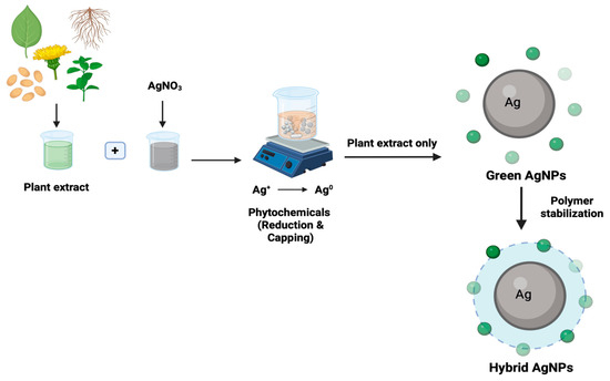
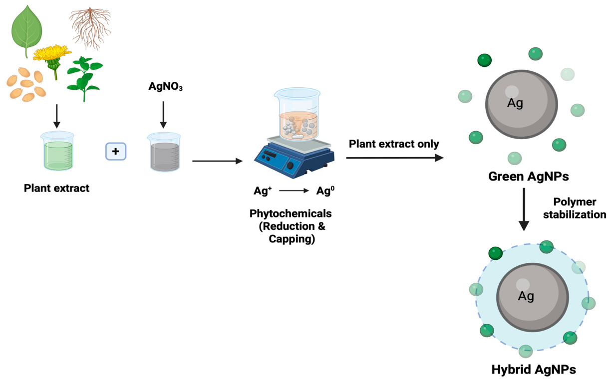
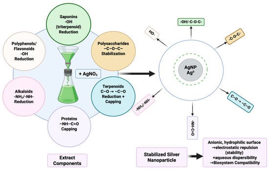
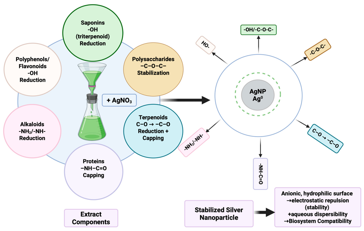
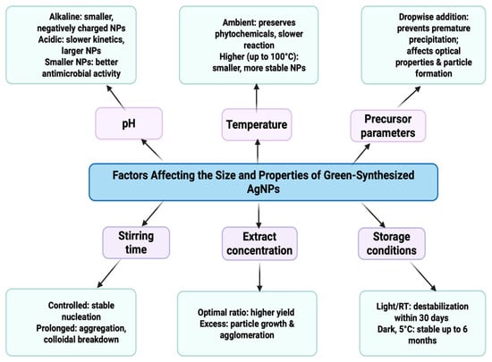
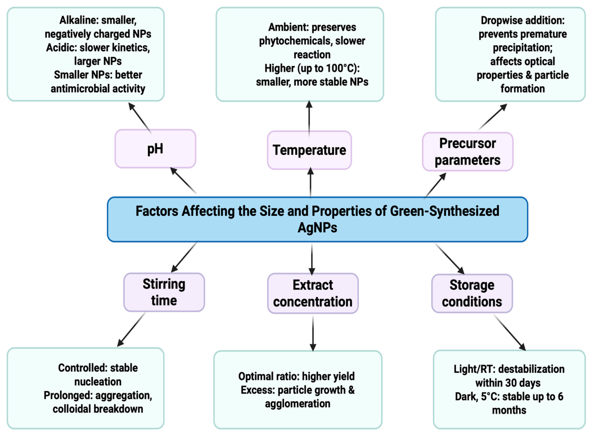
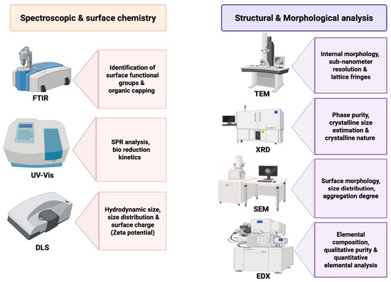
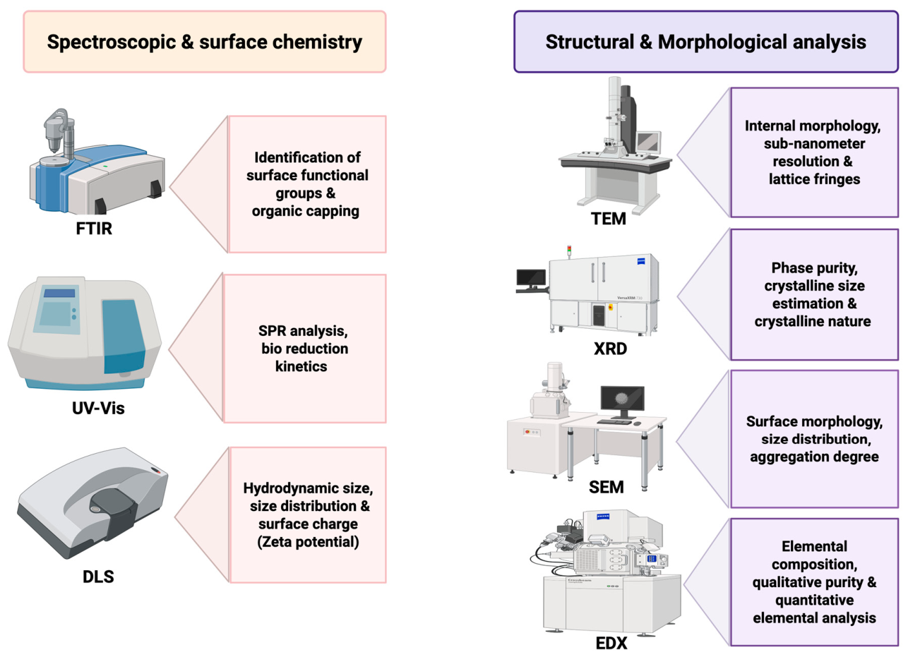
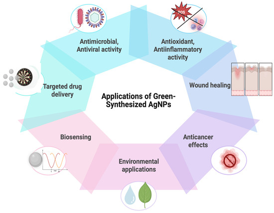
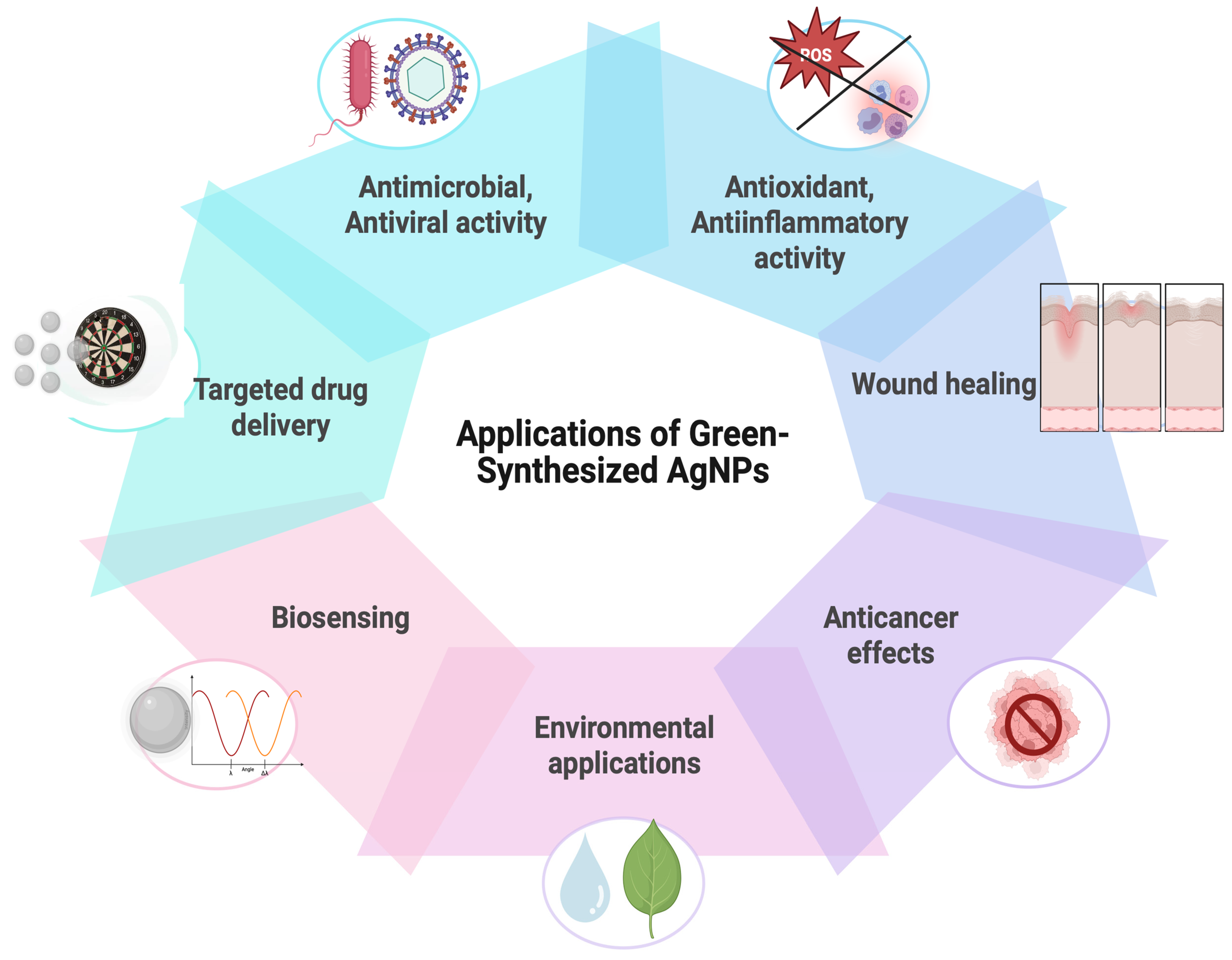
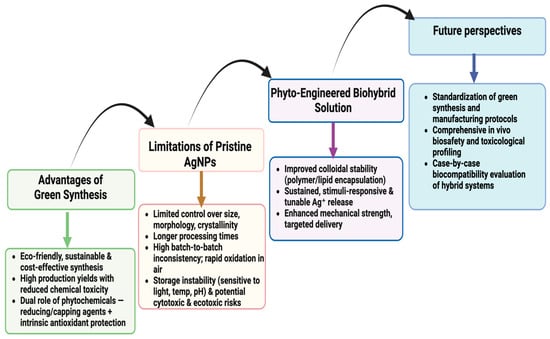
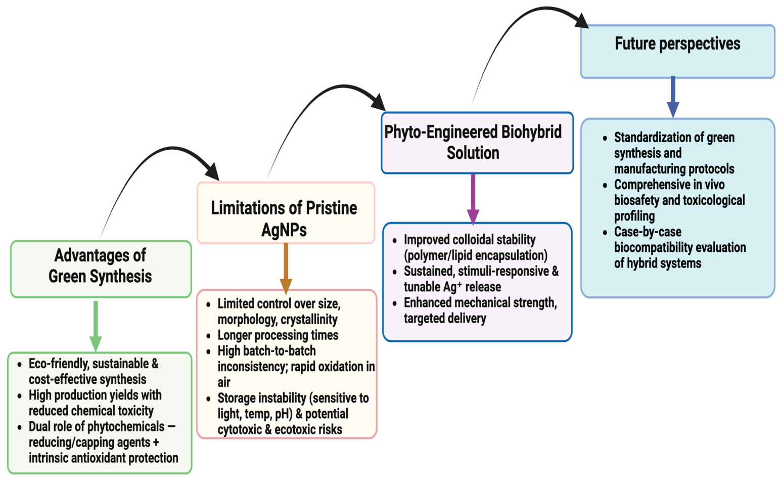

Disclaimer/Publisher’s Note: The statements, opinions and data contained in all publications are solely those of the individual author(s) and contributor(s) and not of MDPI and/or the editor(s). MDPI and/or the editor(s) disclaim responsibility for any injury to people or property resulting from any ideas, methods, instructions or products referred to in the content. |
© 2026 by the authors. Licensee MDPI, Basel, Switzerland. This article is an open access article distributed under the terms and conditions of the Creative Commons Attribution (CC BY) license.