


Abstract
Supercritical carbon dioxide extraction (SFE-CO2) offers a sustainable approach to recovering plant-derived bioactive compounds; however, its influence on the phytochemical composition and biological properties of Anabasis setifera remains underexplored. This study investigated the effects of extraction pressure (200, 250, and 325 bar) at 50 °C on extract yield, phytochemical profiles and in vitro biological activities. Extraction at 250 bar achieved the highest yield (5.82%) and the highest total quantified concentration of phenolic acids and flavonoids (2995.18 µg/g dry extract), with particularly high levels of gallic, coumaric, syringic, and caffeic acids. However, rutin and quercetin were preferentially recovered at 200 and 325 bar, respectively, demonstrating pressure-dependent phytochemical selectivity. Extracts obtained at 200 and 250 bar generally exhibited stronger antimicrobial activity than those obtained at 325 bar, with minimum inhibitory concentrations (MICs) as low as 7.80 µg/mL. Notably, the 250-bar extract demonstrated the strongest DPPH radical-scavenging activity (IC50 = 14.01 µg/mL), inhibited bacterial supernatant-induced hemolysis by up to 98.27%, and effectively suppressed heat-induced protein denaturation. All extracts exhibited comparable cytotoxicity against SK-OV-3 ovarian cancer cells (IC50 = 98.65–102.46 µg/mL). Collectively, these findings demonstrate that extraction pressure governs both phytochemical recovery and associated biological responses, highlighting SFE-CO2 as a promising strategy for obtaining chemically enriched A. setifera extracts.
1. Introduction
Natural products have long served as important sources of biologically active compounds and continue to contribute substantially to the discovery and development of therapeutic agents. In recent decades, increasing attention has been directed toward bioactive substances derived from plants and microorganisms because of their structural diversity and potential applications in managing various human and animal diseases [1]. The pharmacological properties of medicinal plants are largely associated with their complex phytochemical composition, which includes phenolic compounds, flavonoids, alkaloids, saponins, terpenoids, vitamins, and other secondary metabolites [2]. These constituents occur in different concentrations across plant tissues, including leaves, stems, roots, seeds, fruits, and grains, with their abundance and biological activities influenced by genetic, environmental, and extraction-related factors [3,4]. Accordingly, identifying suitable botanical sources and developing efficient extraction strategies are essential steps in obtaining bioactive compounds for further chemical characterization and biological evaluation.
Anabasis setifera, a perennial plant belonging to the family Amaranthaceae, is adapted to arid and environmentally challenging habitats. Members of the genus Anabasis have attracted scientific interest because of their diverse secondary metabolites and reported biological activities. Phytochemical investigations have identified several classes of potentially bioactive constituents, including flavonoids, phenolic compounds, terpenoids, alkaloids, and saponins, which may contribute to their pharmacological properties [5]. In addition, A. setifera has a history of traditional medicinal use, including reported applications for inflammatory and renal conditions, while experimental investigations have suggested anti-inflammatory activity [6,7]. These observations provide a scientific rationale for further exploring the phytochemical composition and biological potential of this species. Nevertheless, its pharmacological properties remain incompletely characterized, and the relationships among extraction conditions, recovered constituents, and biological activities require further investigation. Importantly, reported traditional uses and preliminary experimental findings do not establish clinical efficacy or safety.
The search for new bioactive compounds is particularly relevant in the context of major global health challenges, including cancer and antimicrobial resistance. Cancer remains a leading cause of morbidity and mortality worldwide, and its growing global burden continues to place considerable demands on healthcare systems [8]. Although advances in chemotherapy, immunotherapy, and molecularly targeted treatments have improved outcomes for several malignancies, therapeutic resistance, treatment-associated toxicity, and limited responses in certain patient populations remain substantial challenges [9,10,11]. Consequently, natural products continue to be investigated as sources of structurally diverse compounds that may provide new molecular scaffolds for anticancer drug discovery. Of particular interest are plant-derived metabolites capable of modulating cellular processes associated with cancer development and progression [12]. However, the identification of cytotoxic activity in preliminary experimental models represents an initial stage of investigation and must be followed by mechanistic, selectivity, and safety assessments before therapeutic relevance can be established.
Similarly, antimicrobial resistance (AMR) represents an escalating global public health concern that threatens the effectiveness of existing antimicrobial therapies [13]. The emergence and dissemination of drug-resistant microorganisms contribute to persistent infections, prolonged hospitalization, increased healthcare costs, and higher mortality rates [14,15,16]. The declining effectiveness of certain conventional antimicrobial agents underscores the importance of identifying compounds with potentially useful antimicrobial properties [17]. In this context, medicinal plants represent a valuable reservoir of chemically diverse metabolites that warrant systematic investigation. Evaluating the antimicrobial and anticancer activities of botanical extracts may therefore provide preliminary evidence of their biological potential and help identify promising candidates for subsequent compound isolation and mechanistic studies.
Despite the recognized phytochemical potential of A. setifera, the recovery and characterization of its bioactive constituents depend strongly on the extraction methodology employed [18]. Conventional extraction approaches commonly rely on organic solvents of differing polarities, including methanol, ethanol, and ethyl acetate, to recover phytochemicals from plant matrices [10]. Although these methods are widely established, they may involve substantial solvent consumption, extended processing periods, and additional solvent-removal procedures. Furthermore, solvent polarity and extraction conditions can influence the composition, recovery, and stability of the extracted constituents. These considerations have encouraged the development of alternative extraction technologies that combine chemical selectivity, process efficiency, and improved environmental sustainability.
Among these approaches, supercritical fluid extraction (SFE) has emerged as a promising technique for recovering valuable natural products from botanical resources [19]. Supercritical carbon dioxide (SC-CO2) is particularly attractive because of its relatively low critical temperature, chemical stability, low toxicity, and ease of separation from the final extract. Its favorable mass-transfer properties, including low viscosity and relatively high diffusivity, facilitate penetration into plant matrices and the recovery of soluble constituents [20]. Furthermore, the solvent density and extraction selectivity can be adjusted by modifying operating parameters, particularly pressure and temperature, allowing the extraction process to be tailored to compounds with different physicochemical characteristics [21].
An important consideration, however, is the relatively low polarity of SC-CO2, which generally favors the extraction of lipophilic constituents and may limit the recovery of highly polar compounds, including many phenolics and flavonoid glycosides [22]. When appropriate, the incorporation of a polar co-solvent, such as ethanol, can modify the solvating properties of the extraction medium and broaden the range of recoverable phytochemicals [23]. Among these variables, extraction pressure was selected as the primary experimental factor because, at constant temperature and solvent composition, pressure is the principal means of tuning SC-CO2 density and therefore its solvating power, while also influencing viscosity, diffusivity, and matrix penetration [21]. Temperature, static and dynamic extraction times, sample mass, and co-solvent use were deliberately held constant so that differences in yield and phytochemical profiles could be attributed to pressure rather than to confounding process variables. The objective was therefore not to rank all possible SFE parameters, but to establish whether pressure alone can drive selective recovery of A. setifera constituents under otherwise identical conditions. Under appropriately selected operating conditions, SFE can reduce the use of conventional organic solvents, facilitate solvent removal, and potentially limit the degradation of thermally sensitive compounds [24]. These characteristics have supported its growing application in pharmaceutical, food, cosmetic, and nutraceutical research, where efficient extraction and reproducible chemical characterization are increasingly important.
Against this background, investigating SFE as an alternative strategy for recovering bioactive constituents from A. setifera offers an opportunity to connect sustainable extraction technology with phytochemical research and preliminary biological evaluation. Although previous studies have examined the phytochemical composition and biological properties of A. setifera using conventional extraction approaches, the potential of SFE to provide extracts with distinct chemical and biological characteristics warrants further investigation. The present study therefore aims to apply supercritical fluid extraction to A. setifera and evaluate the biological properties of the resulting extracts, with particular emphasis on their potential antimicrobial and anticancer activities. By examining this underexplored extraction approach, the study seeks to contribute to the development of more sustainable methods for obtaining plant-derived bioactive materials and to provide a scientific foundation for their subsequent chemical and pharmacological investigation.
2. Materials and Methods
2.1. Plant Material Collection, Taxonomic Identification, and Sample Preparation
Fresh leaves of Anabasis setifera were collected from the Hail Region, Saudi Arabia. Botanical identification was performed based on morphological characteristics, at the Medical Diagnostic and Research Center (MDRC), University of Ha’il, Hail, Saudi Arabia, using established taxonomic keys for the flora of Saudi Arabia. The collected leaves were thoroughly washed with distilled water to remove surface impurities and subsequently dried at 50 °C for 72 h under controlled conditions. Following drying, the plant material was finely ground and passed through a 0.5 mm sieve to obtain a homogeneous powder with a particle size of ≤0.5 mm. The powdered material was subsequently dried at 105 °C until a constant mass was achieved to minimize variability associated with residual moisture. Extraction yields were calculated relative to the dry mass of the plant material. The prepared powder was then used for all subsequent supercritical carbon dioxide extraction (SFE-CO2) experiments.
2.2. Supercritical CO2 Extraction of Anabasis setifera and Determination of Extraction Yield
Bioactive constituents were recovered from the powdered leaves of A. setifera using supercritical carbon dioxide extraction (SFE-CO2) [18]. The extraction system consisted of a high-pressure extraction vessel, a CO2 delivery pump, a temperature-control unit, and an extract collection chamber. Carbon dioxide was selected as the extraction solvent because of its favorable physicochemical properties under supercritical conditions and its ability to be removed from the recovered extract through depressurization. Approximately 5 g of dried plant powder was introduced into the extraction vessel, which was subsequently sealed. Liquid CO2 was supplied to the system, and the extraction pressure and temperature were adjusted to establish supercritical conditions. No co-solvent or modifier was used; all extractions were performed with pure CO2 (100%) to ensure that pressure was the only variable affecting extraction efficiency and selectivity.
The extraction process consisted of an initial static phase of 15 min, followed by a dynamic extraction phase of 40 min. To investigate the influence of extraction pressure on extract recovery, three pressures (200, 250, and 325 bar) were evaluated in separate extraction runs. The extraction temperature was maintained at 50 °C, while the static and dynamic extraction durations remained constant at 15 and 40 min, respectively. This experimental design enabled assessment of the pressure-dependent variation in extraction yield under otherwise consistent operating conditions. Following extraction, the system was depressurized, and the recovered extracts were collected and weighed. Extraction yield was determined gravimetrically and expressed as a percentage of the initial dry weight of the plant material according to the following equation:
All extraction experiments were conducted under the specified conditions to facilitate comparison of extraction efficiency among the three pressure treatments.
2.3. High-Performance Liquid Chromatography (HPLC)-Photodiode Array Analysis of Phenolic Acids and Flavonoids
2.3.1. HPLC-PDA Instrumentation and Chromatographic Separation Conditions
The phenolic acid and flavonoid profiles of A. setifera extracts were investigated using HPLC coupled with photodiode array detection (PDA). Chromatographic analyses were performed using a Shimadzu Prominence HPLC system (Shimadzu Corporation, Kyoto, Japan) equipped with a Shimpack VP-ODS C18 reversed-phase analytical column (250 × 4.6 mm internal diameter, 5 µm particle size) and a corresponding guard column. Chromatographic separation was achieved using a binary mobile phase consisting of acetonitrile (solvent A) and an aqueous solution containing 1% (v/v) acetic acid (solvent B). The column temperature was maintained at 28 °C, the mobile-phase flow rate was set at 0.7 mL/min, and the injection volume was 20 µL. A gradient elution program was employed to achieve separation of the targeted phytochemical constituents. The proportion of solvent A was increased from 10% to 63% during the first 11 min, followed by an isocratic period at 63% solvent A for an additional 10 min. Subsequently, the proportion of solvent A was increased to 90% at 26 min. The mobile-phase composition was then returned to the initial conditions of 10% solvent A and 90% solvent B at 31 min, followed by a 5-min re-equilibration period before the subsequent injection. Chromatographic detection was performed at 272 nm using the PDA detector. The resulting chromatograms and spectral data were recorded and processed for qualitative identification and quantitative determination of the targeted phenolic acids and flavonoids [25].
2.3.2. Reference Standards and Chromatographic Identification of Phytochemical Constituents
Authentic reference standards of gallic acid, chlorogenic acid, catechin, methyl gallate, caffeic acid, syringic acid, rutin, ellagic acid, coumaric acid, vanillin, ferulic acid, naringenin, rosmarinic acid, daidzein, quercetin, cinnamic acid, kaempferol, and hesperetin were obtained from Sigma-Aldrich (St. Louis, MO, USA). Individual stock solutions were prepared in methanol, and working standard solutions were obtained by serial dilution. Identification of the target compounds was based on chromatographic retention times and PDA spectral characteristics relative to those of the corresponding authentic standards. Compound assignments were further examined through co-injection with reference standards. For positive identification, a retention-time deviation within ± 2% and a PDA spectral match factor of at least 0.99 were required. While these criteria provide reasonable confidence for the targeted phenolic acids and flavonoids, HPLC-PDA identification cannot fully exclude co-eluting isomers or structurally related compounds, and confirmation by mass spectrometry would further strengthen compound assignment.
Using these identification criteria, the prominent chromatographic peak observed at a retention time of approximately 17.16 min was assigned to quercetin based on its retention behavior, co-injection results, and PDA spectral agreement with the authentic reference standard.
2.3.3. External Standard Calibration, Quantification, and Analytical Method Performance
Quantitative determination of the identified phenolic acids and flavonoids was performed using the external standard calibration method. Calibration curves were generated individually for each target analyte using six concentration levels, with each calibration solution injected into triplicate. The calibration curves demonstrated satisfactory linearity over the evaluated concentration ranges, with coefficients of determination (R2) of at least 0.999. Analytical sensitivity was assessed by determining the limits of detection (LOD) and quantification (LOQ), corresponding to signal-to-noise ratios of 3:1 and 10:1, respectively. The LOD values ranged from 0.05 to 0.15 µg/mL, whereas the LOQ values ranged from 0.15 to 0.50 µg/mL. The precision of the chromatographic method was evaluated through intra-day and inter-day analyses using six replicate injections. The relative standard deviation (RSD) was below 2.0% for intra-day precision and below 3.0% for inter-day precision. The concentrations of the identified compounds were calculated from their respective external calibration curves and corrected for the applicable dilution factors. Quantitative results were expressed as micrograms of compound per gram of dry extract (µg/g dry extract). Each independent extract was analyzed in triplicate (technical/analytical replicates). These technical replicates were averaged before statistical analysis and were not treated as independent experimental replicates. The three independent SFE-CO2 extraction experiments performed for each pressure condition constituted the experimental replicates (n = 3) used for statistical comparisons.
2.4. Evaluation of Antimicrobial Activity of Anabasis setifera Extracts
2.4.1. Test Microorganisms and Preparation of Microbial Inocula
The antimicrobial potential of A. setifera extracts was evaluated against a panel of bacterial and fungal reference strains comprising Bacillus subtilis (ATCC 6633), methicillin-resistant Staphylococcus aureus (MRSA; ATCC 33591), Salmonella typhi (ATCC 6539), Proteus vulgaris (ATCC 13315), Candida albicans (ATCC 10221), and Aspergillus terreus (AUMC 15762). Standardized microbial suspensions were prepared from the respective cultures. Bacteria were grown on Mueller-Hinton agar (MHA) and yeast on Sabouraud dextrose agar (SDA) at 37 °C for 24 h. Aspergillus terreus was grown on SDA at 30 °C for 5–7 days to induce sporulation. Bacterial and yeast suspensions were adjusted to 0.5 McFarland standard (1.5 × 10⁸ CFU/mL) and further diluted in Mueller-Hinton broth (MHB) or Sabouraud dextrose broth (SDB) to obtain final inocula of 106 CFU/mL for the agar well diffusion assay and 5 × 10⁵ CFU/mL for broth microdilution. A conidial suspension of A. terreus was prepared in SDB containing 0.05% Tween 80 and adjusted to 104 spores/mL.
These preparations were used for the agar well diffusion assay, while the appropriate bacterial and yeast inocula were used for broth microdilution testing.
2.4.2. Screening of Antimicrobial Activity by the Agar Well Diffusion Assay
The preliminary antimicrobial activity of the extracts was determined using the agar well diffusion method, with minor modifications to the previously reported procedure [26]. Standardized microbial suspensions were distributed uniformly over the surfaces of the respective agar media. Wells (6 mm diameter) were prepared aseptically, and 50 µL of extract solution (prepared in DMSO at 10 mg/mL and filter-sterilized through 0.22 µm membranes) was introduced into each well. Gentamicin (10 µg/well) and amphotericin B (10 µg/well) served as positive controls for bacterial and fungal assays, respectively, while 50 µL of DMSO served as the vehicle control. Plates were incubated at 37 °C for 24 h for bacteria and C. albicans, and at 30 °C for 48–72 h for A. terreus. Inhibition zones were measured in millimeters. Following the prescribed diffusion and incubation periods, antimicrobial activity was assessed by measuring the diameters of the inhibition zones in millimeters. All experiments were performed in triplicate, and the resulting inhibition-zone measurements were recorded for comparisons among extracts obtained under different extraction pressures.
2.4.3. Determination of Minimum Inhibitory Concentration (MIC) by Broth Microdilution
The MIC of each extract was determined using the broth microdilution method in 96-well microplates. Twofold serial dilutions of the extracts were prepared in MHB or SDB in 96-well round-bottom microplates to cover the concentration range of 1000–0.49 µg/mL in a final volume of 200 µL. The extract solutions were combined with standardized bacterial or yeast inocula (final concentration ~5 × 10⁵ CFU/mL), and the final concentration of DMSO was maintained at ≤1% (v/v). Controls included growth controls (inoculum without extract), sterility controls (medium only), and vehicle controls (DMSO at ≤1%). Plates were incubated at 37 °C for 24 h (bacteria and C. albicans) or at 30 °C for 48–72 h (A. terreus). The MIC was defined as the lowest concentration of extract at which no visible microbial growth was observed. All MIC determinations were conducted in triplicate.
2.4.4. Determination of Minimum Bactericidal (MBC) and Minimum Fungicidal Concentrations (MFC)
The MBC and MFC values were determined by subculturing 10 µL from wells showing no visible growth onto MHA (bacteria) or SDA (fungi). Plates were incubated at 37 °C for 24 h (bacteria and C. albicans) or at 30 °C for 48–72 h (A. terreus). The MBC or MFC was defined as the lowest extract concentration at which no colony formation was observed. The bactericidal assessment was performed for the tested bacterial strains, whereas fungicidal assessment was performed for C. albicans. Aspergillus terreus was not included in MIC or MFC determination because no inhibition zone was observed during the initial agar well diffusion screening. All MBC and MFC determinations were performed in triplicate.
2.5. Assessment of Cytotoxic Activity Against SK-OV-3 Human Ovarian Cancer Cells Using the MTT Assay
The in vitro cytotoxic activity of A. setifera extracts was evaluated against the human ovarian cancer cell line SK-OV-3, obtained from Vacsera (Giza, Egypt), using the colorimetric MTT assay [27]. This assay measures cellular metabolic activity as an indicator of cell viability following exposure to the tested extracts. SK-OV-3 cells were seeded into 96-well culture plates at a density of 5 × 10⁴ cells per well and maintained in RPMI-1640 medium supplemented with 10% heat-inactivated fetal bovine serum (FBS), penicillin (100 U/mL), and streptomycin (100 µg/mL). The cells were incubated for 24 h at 37 °C in a humidified atmosphere containing 5% CO2 to allow cell attachment. Following the initial incubation, the culture medium was partially removed and replaced with fresh medium containing different concentrations of A. setifera extracts (31.25–1000 µg/mL). The treated cells were subsequently incubated for an additional 24 h under the same culture conditions. At the end of the treatment period, 10 µL of MTT solution (5 mg/mL) was added to each well, and the plates were incubated for an additional 4 h to allow metabolically active cells to reduce MTT to insoluble formazan crystals. Subsequently, the supernatant was carefully removed, and 150 µL of DMSO was added to each well to dissolve the resulting formazan crystals. Absorbance was measured at 490 nm using a microplate reader. Untreated cells maintained under identical experimental conditions served as negative control. The percentage of cytotoxicity was determined using the following equation:
where Atreated represents the absorbance of extract-treated cells and Acontrol represents the absorbance of untreated control cells. In addition to the colorimetric measurements, morphological changes in treated and untreated SK-OV-3 cells were examined and documented using phase-contrast light microscopy (Nikon Corporation, Tokyo, Japan). These observations provided complementary qualitative information regarding treatment-associated alterations in cellular morphology.
2.6. Evaluation of the Effects of Anabasis setifera Extracts on Bacterial Supernatant-Induced Erythrocyte Hemolysis
The effects of A. setifera extracts on the hemolytic activity of bacterial culture supernatants were investigated using a modified erythrocyte hemolysis assay [28]. The assay was designed to assess whether exposure of bacterial cultures to sub-inhibitory extract concentrations altered the capacity of their extracellular products to induce erythrocyte lysis. Bacterial cultures were standardized to an optical density of 0.4 at 595 nm and exposed to extract concentrations corresponding to 25%, 50%, and 75% of the previously determined MIC values. Untreated bacterial cultures were included as controls. Following treatment, the cultures were centrifuged at 20,000× g for 20–25 min, and the resulting supernatants were collected for evaluation of hemolytic activity. An aliquot of 500 µL of each bacterial culture supernatant was mixed with a freshly prepared 2% erythrocyte suspension in isotonic saline. The reaction mixtures were incubated at 36 °C for 2 h to allow erythrocyte lysis to occur. After incubation, the samples were centrifuged at 15,000× g for 10 min at 4 °C to separate intact erythrocytes from the supernatant. The absorbance of the released hemoglobin was subsequently measured at 545 nm. Erythrocytes treated with 0.1% sodium dodecyl sulfate (SDS) served as the positive control representing maximal hemolysis, whereas erythrocytes incubated in sterile medium without treatment served as the negative control representing baseline hemolysis. The percentage of hemolysis was calculated according to the following equation:
where Asample represents the absorbance of the tested sample, Anegative corresponds to the absorbance of the negative control, and Apositive represents the absorbance of the SDS-treated positive control. All measurements were performed in triplicate. Hemolytic activity in extract-treated groups was compared with that of untreated bacterial controls to evaluate treatment-associated differences in erythrocyte lysis.
2.7. Determination of Antioxidant Activity Using the DPPH Radical-Scavenging Assay
The in vitro free radical-scavenging activity of A. setifera extracts was evaluated using the 2,2-diphenyl-1-picrylhydrazyl (DPPH) assay, following a previously described method with modifications [29]. This assay is based on the reduction of the stable DPPH radical by antioxidant constituents, resulting in a decrease in absorbance that can be quantified spectrophotometrically. A fresh DPPH solution was prepared by dissolving 2.6 mg of DPPH into 100 mL of methanol. Serial concentrations of the plant extracts ranging from 1.95 to 1000 µg/mL were prepared, and the appropriate aliquot of each extract solution was mixed with 0.96 mL of the DPPH solution. The reaction mixtures were incubated at approximately 26 °C for 60 min in the dark to minimize light-induced changes during the reaction. Following incubation, absorbance was measured at 520 nm using a UV-Vis spectrophotometer (PerkinElmer GmbH, Rodgau, Germany). A control solution containing DPPH without plant extract was used to determine the initial radical absorbance, while the test samples contained DPPH and the corresponding extract concentrations. DPPH radical-scavenging activity was calculated using the following equation:
where Acontrol represents the absorbance of the DPPH control solution and Asample represents the absorbance of the reaction mixture containing the tested extract. The resulting percentage scavenging values were used to evaluate the concentration-dependent antioxidant activity of extracts obtained under different SFE conditions.
2.8. Evaluation of In Vitro Protein Denaturation Inhibition Using the Bovine Serum Albumin Assay
The ability of A. setifera extracts to inhibit heat-induced protein denaturation was evaluated using a modified bovine serum albumin (BSA) denaturation assay [30]. This method provides an in vitro measure of the capacity of tested compounds to reduce thermally induced protein structural alterations and is commonly employed as a preliminary screening approach for potential anti-inflammatory activity. Briefly, 50 µL of each extract solution at concentrations of 1.56, 3.12, 6.25, 12.5, 25, 50, 100, and 200 µg/mL was mixed with 450 µL of a 1% (w/v) aqueous BSA solution. The pH of each reaction mixture was adjusted to 6.3 using the minimum necessary volume of 1 N hydrochloric acid. The prepared reaction mixtures were initially maintained at room temperature for 20 min to allow interactions between the extract constituents and BSA. Subsequently, the samples were subjected to thermal denaturation by incubation in a water bath at 55 °C for 30 min. Following heat treatment, the reaction mixtures were cooled to room temperature, and their absorbance was measured at 670 nm using a BioSystems 310 Plus spectrophotometer (BioSystems S.A., Barcelona, Spain). Diclofenac sodium was employed as the reference anti-inflammatory agent, while DMSO served as the negative control. The percentage inhibition of heat-induced BSA denaturation was calculated according to the following equation:
where Acontrol represents the absorbance of the negative control and Asample represents the absorbance of the extract-treated reaction mixture. The concentration-dependent inhibition of BSA denaturation was evaluated to compare the protective effects of extracts obtained at the different extraction pressures.
2.9. Statistical Analysis and Determination of Half-Maximal Effect Concentrations
Statistical analyses were performed using Minitab software, version 18 (Minitab LLC, State College, PA, USA). All experiments were conducted using three independent experimental replicates (n = 3), corresponding to three separate SFE-CO2 extraction runs for each pressure condition, and results were expressed as the mean ± standard deviation (SD). For each independent extract, analytical and biological measurements were performed in triplicate (technical replicates); these technical replicates were averaged before statistical analysis and were not treated as independent experimental replicates. Thus, the reported standard deviations and statistical comparisons reflect independent extraction variability rather than only analytical precision. For single-response variables, including extraction yield, individual phytochemical concentrations, antimicrobial inhibition zones, MIC, MBC, MFC, and hemolysis measurements at each sub-MIC level, differences among extraction treatments were assessed using one-way analysis of variance (ANOVA), with extraction pressure (200, 250, and 325 bar) as the fixed factor. For concentration-dependent responses, including DPPH radical-scavenging activity, BSA denaturation inhibition, and SK-OV-3 cytotoxicity, two-way ANOVA was performed using extraction pressure and extract concentration as fixed factors, including their interaction. When statistically significant differences were identified, Tukey’s honestly significant difference (HSD) post hoc test was applied for multiple pairwise comparisons. The probability value of p < 0.05 was considered statistically significant. Half-maximal effect concentrations (IC50, as applicable to the respective assay) were estimated by nonlinear regression of the concentration-response data using a four-parameter logistic (4PL) model, with concentrations transformed to the log10 scale. The IC50 was defined as the extract concentration corresponding to 50% of the fitted maximal response.
3. Results and Discussion
3.1. Supercritical CO2 Extraction at 250 Bar Maximizes the Recovery of Anabasis setifera Constituents
The extraction efficiency of Anabasis setifera leaves was investigated using SFE-CO2 at three operating pressures (200, 250, and 325 bar), while maintaining a constant extraction temperature of 50 °C, a static extraction period of 15 min, and a dynamic extraction period of 40 min. As presented in Table 1, extraction pressure substantially influenced the recovery of plant-derived constituents, revealing a nonlinear relationship between pressure and extraction yield. Among the investigated conditions, the highest extraction yield was obtained at 250 bar, producing 0.291 g of extract, corresponding to 5.82% of the initial dry plant mass. In comparison, extraction at 200 bar yielded 0.159 g (3.18%), whereas increasing the pressure to 325 bar resulted in a lower yield of 0.112 g (2.24%). Accordingly, the extract mass recovered at 250 bar was approximately 1.83-fold and 2.60-fold greater than those obtained at 200 and 325 bar, respectively. These findings demonstrate that an intermediate extraction pressure was more favorable for recovering extractable constituents from A. setifera under the experimental conditions employed.
Table 1.
Effect of extraction pressure on the yield of Anabasis setifera leaf extract obtained by supercritical CO2 extraction (SFE-CO2).
The pressure-dependent yield observed here is best interpreted as a density-driven solubility and mass-transfer effect. Raising pressure from 200 to 250 bar increases SC-CO2 density and solvent strength, improving dissolution of extractable constituents. However, above an optimum, further densification at 325 bar can reduce diffusivity and matrix penetration and/or shift the solubility balance, so yield need not increase monotonically with pressure [21,31,32,33]. Because temperature, static/dynamic time, sample mass, and co-solvent use were held constant, the 250-bar maximum is attributed specifically to extraction pressure and its effect on CO2 density and solvating capacity, rather than to any of the other listed factors.
The observed pressure-dependent variation in extraction yield may be explained by changes in the physicochemical properties of supercritical CO2, particularly its density, solvent strength, diffusivity, and interactions with plant-derived constituents. Increasing extraction pressure generally enhances CO2 density, thereby modifying its solvating capacity and potentially improving the dissolution of specific phytochemicals. However, the relationship between extraction pressure and extraction efficiency is influenced by the chemical characteristics of the plant matrix and the solubility behavior of individual constituents. In addition to density and solvent strength, two mass-transfer parameters are particularly relevant: matrix swelling by SC-CO2 and the diffusion rate of solutes out of the plant matrix. Increasing pressure generally increases CO2 sorption into the plant matrix, which can promote swelling and plasticization of cell-wall polymers and improve solvent access to intracellular constituents. However, higher pressure also increases CO2 density and reduces diffusivity, and excessive swelling or matrix compaction can increase mass-transfer resistance. The nonlinear yield pattern observed here that maximum at 250 bar and lower at 325 bar, is therefore consistent with a balance between pressure-enhanced swelling/solvation and pressure-reduced diffusion/matrix penetration [21,31,32,33]. Direct measurement of the degree of swelling of A. setifera at different pressures was not performed in this study, and this is acknowledged as a limitation. Consequently, the reduced yield observed at 325 bar suggests that increasing pressure beyond an optimal range did not further enhance the overall extraction performance of A. setifera [31].
From a mechanistic perspective, the recovery results obtained in this study cannot be classified exclusively as either thermodynamic (partition/solubility-controlled) or kinetic (diffusion-controlled). Because extractions were performed at a fixed dynamic time of 40 min, the observed pressure-dependent yield and phytochemical selectivity reflect the net outcome of two opposing pressure effects. First, a thermodynamic effect, whereby increasing pressure enhances SC-CO2 density and solvating power, favoring the dissolution of target compounds; second, a kinetic effect, whereby increasing pressure reduces SC-CO2 diffusivity and may increase mass-transfer resistance within the plant matrix. The nonlinear yield pattern observed here that maximum at 250 bar and lower at 325 bar, is therefore consistent with a balance between these thermodynamic and kinetic influences rather than with either pure partition control or pure diffusion control [21,31,32,33]. A time-resolved extraction design would be required to separate these contributions, and this is acknowledged as a limitation of the present study.
The similarity between these findings and the present results highlights the importance of optimizing extraction pressure according to the physicochemical characteristics of the botanical material rather than assuming a direct relationship between increasing pressure and extraction yield. Importantly, the present findings establish 250 bar as the most favorable extraction pressure among the investigated conditions, providing the highest recovery of A. setifera constituents and demonstrating the importance of pressure optimization in improving the efficiency of SFE-CO2 extraction.
3.2. Extraction at 250 Bar Enhances the Overall Recovery of Phenolic Acids and Flavonoids While Modulating Individual Phytochemical Profiles
The phenolic acid and flavonoid compositions of A. setifera extracts obtained at different SFE-CO2 pressures were characterized using HPLC-PDA. The quantitative distribution of the identified compounds is documented (Table 2), while the corresponding chromatographic profiles are illustrated (Figure 1, Figure 2 and Figure 3). The results revealed distinct pressure-dependent differences in the concentrations and relative distributions of the detected phytochemical constituents, demonstrating that extraction pressure influenced both overall extract recovery and phytochemical selectivity. The extract obtained at 250 bar exhibited the highest total quantified concentration of phenolic acids and flavonoids, defined as the sum of the individual concentrations of all identified and quantified compounds, reaching 2995.18 µg/g dry extract, compared with 2103.61 µg/g at 200 bar and 2269.62 µg/g at 325 bar. Each individual compound concentration was determined using its respective external calibration curve and expressed as µg/g dry extract; the total was then calculated by summing these individual values. Thus, extraction at 250 bar increased the summed concentration of the identified compounds by approximately 42.4% and 32.0% relative to extraction at 200 and 325 bar, respectively. This enrichment was particularly evident for several phenolic acids and flavonoids, including gallic acid (912.24 µg/g), p-coumaric acid (356.38 µg/g), syringic acid (263.78 µg/g), caffeic acid (263.39 µg/g), kaempferol (255.69 µg/g), and chlorogenic acid (244.49 µg/g). These findings indicate that the intermediate extraction pressure favored the recovery of several major phytochemical constituents. Gallic acid was the predominant identified constituent in the 250- and 325-bar extracts, reaching concentrations of 912.24 and 461.49 µg/g dry extract, respectively. In contrast, rutin was the most abundant quantified compound in the extract obtained at 200 bar (633.22 µg/g), followed by gallic acid (323.83 µg/g) and p-coumaric acid (279.63 µg/g). This variation demonstrates that different extraction pressures promoted distinct phytochemical profiles, reflecting differences in the extraction behavior of individual compounds. Several constituents exhibited particularly notable pressure-dependent patterns. Rutin and ellagic acid reached their highest concentrations at 200 bar, with values of 633.22 and 98.59 µg/g, respectively. Rosmarinic acid (22.32 µg/g) was detected only in the 200-bar extract, whereas syringic acid was detected at 250 and 325 bar, with concentrations of 263.78 and 215.59 µg/g, respectively. Quercetin was most abundant in the extract obtained at 325 bar (234.38 µg/g), compared with 151.40 µg/g at 200 bar and 113.22 µg/g at 250 bar. Similarly, catechin reached its highest concentration at 325 bar (88.36 µg/g). In contrast, methyl gallate, daidzein, and hesperetin were not detected under any of the extraction conditions investigated. The chromatographic profiles further demonstrated the distinct chemical characteristics of the extracts. A prominent chromatographic peak at approximately 17.16 min was assigned to quercetin based on retention-time agreement, co-injection with the authentic reference standard, and PDA spectral matching, with a reported spectral match factor of at least 0.99. Variations in chromatographic peak intensities among the three extracts were consistent with their different phytochemical distributions and demonstrated the sensitivity of HPLC-PDA profiling for evaluating extraction-dependent compositional changes.
Table 2.
HPLC-PDA quantification of phenolic acids and flavonoids in A. setifera extracts obtained by SFE-CO2 at 200, 250, and 325 bar. Concentrations are expressed as µg/g dry extract. Area, peak area; Conc., concentration; n.d., not detected (below the limit of detection).
Figure 1.
HPLC-PDA chromatogram of the phenolic acids and flavonoids separated from the A. setifera extract obtained by SFE-CO2 at 200 bar.
Figure 2.
HPLC-PDA chromatogram of the phenolic acids and flavonoids separated from the A. setifera extract obtained by SFE-CO2 at 250 bar.
Figure 3.
HPLC-PDA chromatogram of the phenolic acids and flavonoids separated from the A. setifera extract obtained by SFE-CO2 at 325 bar.
The observed differences in phytochemical recovery are consistent with previous investigations demonstrating the influence of SFE operating conditions on the selective extraction of plant-derived metabolites [34]. It was reported that extraction of Moringa peregrina at 25 MPa provided greater recovery of selected bioactive constituents than extraction at 10 or 40 MPa [32]. Similarly, Almehayawi et al. [19] observed that certain phenolic compounds, including caffeic acid, methyl gallate, syringic acid, and ferulic acid, became detectable at higher extraction pressures in M. oleifera. Furthermore, da Silva et al. [33] reported favorable extraction yields and phenolic recovery from M. oleifera leaves at 20 MPa and 60 °C. These findings support the role of pressure-dependent solvent properties in determining the chemical composition of botanical extracts. Previous phytochemical investigations of Anabasis species also provide a relevant scientific context for the present findings. Abdelaziz et al. [35] reported substantial flavonoid and phenolic contents in the ethyl acetate extract of A. setifera, with concentrations of 5163 and 4264 µg/mL, respectively. The same extract demonstrated antioxidant activity (EC50 = 30.6 µg/mL) and cytotoxic activity against Hep-G2 and MCF-7 cancer cells, with IC50 values of 36.4 and 44 µg/mL, respectively. These observations highlight the phytochemical richness of A. setifera and the potential contribution of its secondary metabolites to biological activity. The biological significance of the identified compounds is supported by previous research describing the antioxidant, antimicrobial, and anti-inflammatory properties of several phenolic acids and flavonoids. Gallic acid, caffeic acid, and chlorogenic acid are recognized for their electron-donating and radical-scavenging properties, while p-coumaric acid and syringic acid have been associated with antioxidant and inflammation-related biological effects [33,36,37,38,39]. Similarly, rutin, quercetin, and kaempferol have attracted considerable attention because of their diverse pharmacological properties, including effects on oxidative signaling and cellular responses [38,39,40]. The enrichment of these compounds in the extracts provides a plausible phytochemical basis for the biological activities evaluated in the subsequent experiments. Notably, extraction at 250 bar produced the highest cumulative concentration of quantified phenolic acids and flavonoids, while individual compounds exhibited distinct pressure-dependent recovery patterns, demonstrating the potential of SFE-CO2 to modulate the phytochemical composition of A. setifera extracts. The pressure-dependent selectivity observed for individual constituents may also be influenced by differences in molecular weight and diffusion rates. Compounds with lower molecular weight generally diffuse more rapidly within the plant matrix and may be recovered more readily under conditions where diffusivity is reduced (e.g., at higher pressure), whereas larger or more polar glycosides such as rutin may require lower pressure, where SC-CO2 diffusivity is higher, to achieve effective mass transfer. In the present study, rutin (MW ≈ 610 g/mol) was preferentially recovered at 200 bar, whereas the smaller aglycone quercetin (MW ≈ 302 g/mol) was enriched at 325 bar, consistent with a possible diffusion-related effect. However, because all extractions were performed at a fixed dynamic time of 40 min, these observations cannot be attributed definitively to molecular-weight-dependent diffusion kinetics. A time-resolved extraction study (sampling at 5, 10, 20, 30, 40, and 50 min) would be required to separate thermodynamic (solubility) from kinetic (diffusion) contributions, and this is recommended for future work.
3.3. Extracts Obtained at 200 and 250 Bar Exhibit Stronger Antimicrobial Activity than Those Obtained at 325 Bar
The antimicrobial activity of A. setifera extracts obtained under different SFE-CO2 pressures was investigated against selected bacterial and fungal microorganisms using the agar well diffusion and broth microdilution methods. Inhibition-zone measurements are reported (Table 3 and Figure 4), while the corresponding MIC, MBC, and MFC values are presented in Table 4. The agar well diffusion assay demonstrated that extraction pressure influenced the antimicrobial performance of A. setifera extracts. Overall, extracts obtained at 200 and 250 bar produced larger inhibition zones than those obtained at 325 bar. The strongest bacterial inhibition was observed against Bacillus subtilis, for which the 200- and 250-bar extracts produced inhibition zones of 30 mm, compared with 25 mm for the 325-bar extract and 32 mm for the positive control. Similarly, both the 200- and 250-bar extracts produced 20-mm inhibition zones against methicillin-resistant Staphylococcus aureus (MRSA), whereas the 325-bar extract produced a smaller zone of 16 mm. Against Salmonella typhi, the 250-bar extract exhibited the strongest inhibitory activity, producing an inhibition zone of 27 mm, followed by 24 mm at 200 bar and 16 mm at 325 bar. In contrast, Proteus vulgaris showed comparatively lower susceptibility, with inhibition zones of 13, 14, and 11 mm for extracts obtained at 200, 250, and 325 bar, respectively. The antifungal assessment revealed substantial activity against Candida albicans, with inhibition zones of 28 mm for the 200- and 250-bar extracts, compared with 20 mm for the 325-bar extract. No measurable inhibition zone was detected against Aspergillus terreus under the investigated conditions, whereas the corresponding positive control produced a 20-mm inhibition zone. These findings indicate that the antimicrobial effects of A. setifera extracts were influenced by both the extraction conditions and the tested microorganism. The relatively large inhibition zones observed against B. subtilis and C. albicans suggest that these organisms were particularly responsive to the recovered phytochemical mixtures under the experimental conditions. The broth microdilution assay provided further evidence of the differences in antimicrobial activity among extraction pressures. Against B. subtilis, the lowest MIC was recorded for the 200-bar extract (7.80 µg/mL), followed by the 250-bar extract (15.62 µg/mL) and the 325-bar extract (31.25 µg/mL). Against MRSA, both the 200- and 250-bar extracts exhibited MIC values of 31.25 µg/mL, whereas the MIC increased to 125 µg/mL for the 325-bar extract. Similarly, extracts obtained at 200 and 250 bar showed MIC values of 15.62 µg/mL against Salmonella typhi, compared with 125 µg/mL for the extract obtained at 325 bar. Against P. vulgaris, the MIC values were 62.5 µg/mL for both lower-pressure extracts and 125 µg/mL for the 325-bar extract. The antifungal results followed a comparable pattern, with MIC values of 15.62 µg/mL for the 200- and 250-bar extracts against C. albicans, increasing to 62.5 µg/mL at 325 bar. The MBC and MFC results further demonstrated pressure-dependent variations in antimicrobial performance. For B. subtilis, MBC values increased from 15.62 µg/mL at 200 bar to 31.25 µg/mL at 250 bar and 125 µg/mL at 325 bar. In contrast, the 250-bar extract exhibited the lowest MBC against MRSA (31.25 µg/mL), compared with 62.5 µg/mL at 200 bar and 500 µg/mL at 325 bar. A similar trend was observed against Salmonella typhi, for which the lowest MBC was associated with the 250-bar extract (15.62 µg/mL), followed by 62.5 µg/mL at 200 bar and 500 µg/mL at 325 bar. Against C. albicans, MFC values were 31.25, 15.62, and 62.5 µg/mL for extracts obtained at 200, 250, and 325 bar, respectively. Collectively, these measurements indicate that the 200- and 250-bar extracts generally displayed more favorable inhibitory and bactericidal or fungicidal activities than the 325-bar preparation.
Table 3.
Antimicrobial activity of A. setifera extracts obtained by SFE-CO at 200, 250, and 325 bar, expressed as inhibition-zone diameter (mm) in the agar well diffusion assay.
Figure 4.
Antimicrobial activity of A. setifera extracts obtained by SFE-CO2 at different pressures, determined by the agar well diffusion assay. Test organisms were Candida albicans (CA), Aspergillus terreus (AT), Proteus vulgaris (PV), Salmonella typhi (ST), methicillin-resistant Staphylococcus aureus (SA), and Bacillus subtilis (BS). Wells: P, positive control; N, negative control (DMSO); 1, extract obtained at 250 bar; 2, extract obtained at 200 bar; 3, extract obtained at 325 bar. Positive controls were gentamicin (10 µg/well) for all bacterial strains (Bacillus subtilis, methicillin-resistant Staphylococcus aureus, Salmonella typhi and Proteus vulgaris) and amphotericin B (10 µg/well) for fungal strains (Candida albicans and Aspergillus terreus).
Table 4.
Minimum inhibitory concentrations (MIC) and minimum bactericidal or fungicidal concentrations (MBC/MFC) of A. setifera extracts obtained by SFE-CO2 at 200, 250, and 325 bar.
The observed antimicrobial activity may be associated with the diverse phytochemical constituents recovered during SFE-CO2 extraction. Phenolic acids and flavonoids have been investigated for their interactions with microbial membranes, proteins, and other cellular components. Compounds such as gallic acid, caffeic acid, syringic acid, and quercetin may contribute to antimicrobial responses through their respective physicochemical and biological properties. The pressure-dependent differences in the concentrations of these compounds provide a possible explanation for the variations observed among the extracts. The present findings are consistent with previous investigations demonstrating the antimicrobial potential of SFE-derived botanical extracts. It was reported enhanced antimicrobial activity against Helicobacter pylori for M. peregrina extracts obtained at 25 MPa compared with extracts obtained at 10 and 40 MPa [32]. Similarly, Almehayawi et al. [19] described increased antifungal activity of M. oleifera extracts obtained at 25 MPa against several Candida species, including C. glabrata, C. tropicalis, and C. albicans. These observations reinforce the importance of extraction-pressure selection in obtaining phytochemical mixtures with favorable antimicrobial properties. Taken together, the agar diffusion and broth microdilution findings demonstrate that A. setifera extracts obtained at 200 and 250 bar generally exhibited stronger antimicrobial activity than those recovered at 325 bar, indicating that extraction pressure plays an important role in determining the antimicrobial performance of the resulting phytochemical mixtures.
3.4. Extraction at 250 Bar Produces the Greatest Inhibition of Bacterial Supernatant-Induced Erythrocyte Hemolysis
The effects of A. setifera extracts on bacterial supernatant-induced erythrocyte hemolysis were evaluated at sub-inhibitory concentrations corresponding to 25%, 50%, and 75% of the previously determined MIC values. The extracts exhibited concentration-dependent inhibition of hemolytic activity, with substantial differences among extraction pressures and bacterial strains (Table 5 and Figure 5). Across the investigated microorganisms, increasing extract concentrations were generally associated with progressively greater hemolysis inhibition. Among the three extraction conditions, the 250-bar extract consistently produced the highest inhibition percentages at corresponding sub-MIC levels, followed by the 200-bar extract, whereas the 325-bar extract exhibited lower activity. For B. subtilis, the 250-bar extract produced hemolysis inhibition values of 90.21%, 92.82%, and 96.85% at 25%, 50%, and 75% of the MIC, respectively. The corresponding inhibition percentages for the 200-bar extract were 76.71%, 84.43%, and 91.40%. By comparison, the 325-bar extract produced 12.50%, 49.50%, and 80.35% inhibition at the same relative concentrations. A similar concentration-dependent pattern was observed against MRSA. The 250-bar extract increased hemolysis inhibition from 75.91% at 25% of the MIC to 94.71% at 75% of the MIC. The corresponding values for the 200-bar extract ranged from 64.19% to 93.05%, whereas those of the 325-bar extract increased from 7.82% to 67.13%. These findings indicate that extracts obtained at lower and intermediate extraction pressures were more effective in reducing hemolytic activity associated with the tested bacterial supernatants. The extracts also demonstrated considerable inhibition of hemolysis associated with Salmonella typhi and P. vulgaris. At 75% of the MIC, the 250-bar extract achieved inhibition values of 96.80% and 98.27%, respectively. These values exceeded those recorded for the corresponding 200-bar extracts (95.33% and 94.89%) and 325-bar extracts (92.07% and 76.87%). The highest inhibition observed in the study was 98.27%, recorded against P. vulgaris following treatment with the 250-bar extract at 75% of the MIC.
Table 5.
Inhibition (%) of bacterial supernatant-induced hemolysis by A. setifera extracts obtained by SFE-CO2 at 200, 250, and 325 bar, applied at 25%, 50%, and 75% of their respective MICs.
Figure 5.
Inhibition of bacterial supernatant-induced hemolysis by A. setifera extracts obtained by SFE-CO2 at 325 bar (series A), 200 bar (series B), and 250 bar (series C), applied at sub-inhibitory concentrations against P. vulgaris (PV), Salmonella typhi (ST), methicillin-resistant S. aureus (SA), and B. subtilis (BS). In each series, tube 1 is the negative control (erythrocytes without hemolytic challenge, showing no hemolysis); tubes 2, 3, and 4 contain the extract at 75%, 50%, and 25% of its MIC, respectively; and tube 5 is the positive control (complete hemolysis induced by SDS).
The pronounced activity of the 250-bar extract may be associated with its comparatively high concentrations of several phenolic acids and flavonoids, including gallic acid, caffeic acid, syringic acid, chlorogenic acid, and p-coumaric acid. These compounds have been associated with diverse biological properties and may influence the extracellular activities of bacteria through interactions with cellular, or secreted components. The correspondence between phytochemical enrichment and hemolysis inhibition provides a plausible explanation for the greater performance of the 250-bar extract. Previous studies have also described antihemolytic properties in other members of the genus Anabasis. Gheraissa et al. [41] reported antihemolytic activity in Anabasis oropediorum, with flavonoid constituents proposed as potential contributors to the observed effects. These findings provide additional support for the biological relevance of phytochemicals recovered from Anabasis species. An important observation was that the relative inhibition of bacterial supernatant-associated hemolysis did not consistently follow the same pattern as the MIC results. For example, the 200-bar extract exhibited the lowest MIC against B. subtilis, whereas the 250-bar extract produced the greatest hemolysis inhibition. This distinction suggests that the measured responses represent different aspects of extract-associated biological activity and may reflect interactions between phytochemical constituents and bacterial extracellular products. Interestingly, the 250-bar extract consistently exhibited the strongest inhibition of bacterial supernatant-induced hemolysis, reaching 98.27% against P. vulgaris, thereby revealing a prominent pressure-dependent biological response associated with the phytochemical composition of A. setifera extracts.
3.5. Extraction at 250 Bar Enhances the DPPH Radical-Scavenging Activity of Anabasis setifera Extracts
The antioxidant potential of A. setifera extracts obtained under different SFE-CO2 pressures was evaluated using the DPPH radical-scavenging assay over a concentration range of 1.95–1000 µg/mL. The results demonstrated concentration-dependent increases in radical-scavenging activity for all extracts, with distinct differences among the investigated extraction pressures (Table 6). Among the tested preparations, the 250-bar extract consistently exhibited the greatest DPPH radical-scavenging activity throughout the concentration range. At the lowest investigated concentration (1.95 µg/mL), this extract produced 28.5% inhibition, compared with 23.8% and 19.6% for the 200- and 325-bar extracts, respectively. Increasing the extract concentration to 31.25 µg/mL enhanced the radical-scavenging activity to 59.1% for the 250-bar extract, whereas the corresponding values for the 200- and 325-bar extracts were 55.5% and 52.6%. At 125 µg/mL, the 250-bar extract achieved 74.4% inhibition, compared with 69.2% for the 200-bar extract and 66.9% for the 325-bar extract. The concentration-dependent increase continued at higher concentrations, with the 250-bar extract reaching 87.1% inhibition at 500 µg/mL and 92.5% at 1000 µg/mL. In comparison, the 200- and 325-bar extracts produced 88.6% and 85.5% inhibition, respectively, at the highest investigated concentration. Under the same experimental conditions, the reference antioxidant ascorbic acid exhibited 98.1% DPPH inhibition at 1000 µg/mL. The calculated IC50 values further demonstrated the differences in radical-scavenging activity among the extracts. The 250-bar extract exhibited the lowest IC50 (14.01 ± 0.21 µg/mL), followed by the 200-bar extract (22.06 ± 0.21 µg/mL) and the 325-bar extract (28.09 ± 0.32 µg/mL). Ascorbic acid exhibited the lowest overall IC50 (2.74 ± 0.84 µg/mL), consistent with its strong radical-scavenging activity. These findings identify the 250-bar extract as the most active A. setifera preparation in the DPPH assay.
Table 6.
DPPH radical-scavenging activity (%) and IC50 values (µg/mL) of A. setifera extracts obtained by SFE-CO2 at 200, 250, and 325 bar, compared with ascorbic acid.
The greater antioxidant activity of the 250-bar extract corresponded with its higher cumulative concentration of HPLC-quantified phenolic acids and flavonoids. In particular, gallic acid, caffeic acid, chlorogenic acid, syringic acid, and p-coumaric acid contain structural features associated with radical-scavenging activity, including hydroxyl groups capable of participating in electron- or hydrogen-transfer reactions. The enrichment of these compounds in the 250-bar extract may therefore contribute to its greater capacity to reduce DPPH radicals. Previous studies have documented antioxidant activity in several Anabasis species, providing a broader context for the present findings. For instance, A. aretioides extract exhibited an EC50 of 52.91 µg/mL [42], while crude A. articulata extract showed an EC50 of 90 µg/mL [43]. Other investigations reported considerably lower EC50 values of 3.2–4.9 µg/mL for A. articulata leaf extracts [44]. The diversity of these reported values reflects differences in botanical composition, extraction procedures, and the resulting concentrations of radical-scavenging constituents. The present findings demonstrate a consistent relationship between the phytochemical enrichment observed at 250 bar and its superior DPPH radical-scavenging response. This association highlights the importance of optimizing extraction conditions not only to maximize the quantity of recovered material but also to obtain extracts enriched in constituents associated with antioxidant activity. Of particular significance, the 250-bar extract combined the highest cumulative concentration of quantified phenolic acids and flavonoids with the strongest DPPH radical-scavenging activity, demonstrating a favorable association between extraction-dependent phytochemical composition and antioxidant performance.
3.6. Extracts Obtained at 250 and 325 Bar Exhibit Strong Inhibition of Heat-Induced BSA Denaturation
The inhibitory effects of A. setifera extracts on heat-induced protein denaturation were evaluated using the BSA assay over a concentration range of 1.56–200 µg/mL, with diclofenac sodium serving as the reference compound. All extracts exhibited concentration-dependent inhibition of BSA denaturation, although the magnitude of the response varied considerably with extraction pressure (Table 7). Among the tested extracts, the 250-bar preparation consistently demonstrated the greatest percentage of inhibition across the investigated concentrations. At 1.56 µg/mL, the 250-bar extract produced 46.3% inhibition, closely matching the response of diclofenac sodium (45.8%) and exceeding those of the 325-bar (43.3%) and 200-bar (19.3%) extracts. At 6.25 µg/mL, the 250-bar extract achieved 65.3% inhibition, compared with 62.7% for diclofenac sodium, 61.7% for the 325-bar extract, and 38.2% for the 200-bar extract. The differences among extraction pressures remained evident at intermediate concentrations. At 25 µg/mL, the 250-bar extract produced 84.0% inhibition, followed by diclofenac sodium (82.5%), the 325-bar extract (79.5%), and the 200-bar extract (57.0%). At 50 µg/mL, the corresponding inhibition values were 89.8%, 88.9%, 85.7%, and 65.6%, respectively. These observations demonstrate that the 250-bar extract maintained greater inhibitory activity than the other plant extracts throughout the evaluated concentration range. At higher concentrations, diclofenac sodium exhibited the greatest inhibitory effect, reaching 94.3% and 97.6% at 100 and 200 µg/mL, respectively. The corresponding values for the 250-bar extract were 92.4% and 96.2%, whereas the 325-bar extract produced 90.7% and 94.1% inhibition. The 200-bar extract remained comparatively less effective, achieving 74.6% and 83.1% inhibition at the same concentrations. The calculated IC50 values further supported the observed differences in BSA denaturation inhibition. The 250-bar extract exhibited the lowest IC50 among the plant extracts (1.60 ± 0.25 µg/mL), followed by the 325-bar extract (2.21 ± 0.36 µg/mL). In contrast, the 200-bar extract showed a substantially higher IC50 (15.46 ± 0.25 µg/mL), indicating lower activity in this assay. Diclofenac sodium exhibited an IC50 of 1.91 ± 0.41 µg/mL. According to the reported statistical grouping, the IC50 values of the 250- and 325-bar extracts were not significantly different from that of diclofenac sodium, despite their numerical differences.
Table 7.
Inhibition (%) of heat-induced bovine serum albumin (BSA) denaturation and IC50 values (µg/mL) of A. setifera extracts obtained by SFE-CO2 at 200, 250, and 325 bar, compared with diclofenac sodium.
The inhibition of heat-induced BSA denaturation may be associated with interactions between the extracted phytochemical constituents and the protein structure. Phenolic acids and flavonoids contain functional groups capable of participating in hydrogen bonding and other noncovalent interactions with proteins, potentially influencing their conformational stability during thermal exposure. The strong inhibition observed for the 250-bar extract corresponded with its relatively high concentrations of gallic acid, caffeic acid, syringic acid, chlorogenic acid, and other phenolic compounds, suggesting a possible contribution of these constituents to the observed response [30]. The differences among extraction pressures further emphasize the importance of phytochemical composition in determining the functional properties of the recovered extracts. Although the 200-bar extract contained relatively high concentrations of rutin and selected other compounds, its BSA denaturation inhibition was substantially lower than that of the 250- and 325-bar extracts. This observation suggests that the overall response may reflect the combined properties of multiple constituents rather than the concentration of any single compound. Collectively, the findings demonstrate that the 250-bar extract exhibited the strongest inhibition of heat-induced BSA denaturation among the tested plant preparations, with activity approaching that of diclofenac sodium under the experimental conditions, supporting the biological relevance of pressure-dependent phytochemical recovery.
3.7. Anabasis setifera Extracts Exhibit Comparable Concentration-Dependent Cytotoxic Activity Against SK-OV-3 Ovarian Cancer Cells
The in vitro cytotoxic activity of A. setifera extracts obtained at different SFE-CO2 pressures was investigated against the human ovarian cancer cell line SK-OV-3 using the MTT assay. Concentration-dependent cytotoxic responses are summarized in Table 8, while representative morphological alterations are illustrated in Figure 6 and Figure 7. All extracts produced concentration-dependent reductions in MTT-associated cellular viability, although the magnitude of the response varied according to extract concentration and extraction pressure. At the lowest tested concentration (31.25 µg/mL), the extracts exhibited minimal cytotoxic activity, with values of 2.45%, 0%, and 1.01% for the 200-, 250-, and 325-bar extracts, respectively. At 62.5 µg/mL, the 325-bar extract produced the greatest cytotoxic response (9.89%), followed by the 200-bar extract (3.56%) and the 250-bar extract (0.05%). A pronounced increase in cytotoxicity was observed at 125 µg/mL, where the 250-bar extract exhibited the greatest response (75.44%), followed by the 325-bar extract (71.61%) and the 200-bar extract (69.22%). This concentration represented a marked transition in the cellular response, with substantially greater reductions in MTT-associated viability than those observed at lower extract concentrations. Further increases in extract concentration produced near-maximal cytotoxic responses across all extraction treatments. At 250 µg/mL, cytotoxicity reached 96.54%, 96.81%, and 96.76% for extracts obtained at 200, 250, and 325 bar, respectively. At 500 and 1000 µg/mL, all extracts-maintained cytotoxicity values of approximately 96–97%, indicating that the measured response approached a plateau within the investigated concentration range. The calculated IC50 values were 102.46 ± 1.13 µg/mL for the 200-bar extract, 98.65 ± 0.32 µg/mL for the 250-bar extract, and 100.19 ± 1.21 µg/mL for the 325-bar extract. Although the 250-bar extract exhibited the numerically lowest IC50, the differences among the three preparations were relatively small, indicating broadly comparable cytotoxic potency against SK-OV-3 cells. The MTT results were supported by morphological observations obtained using phase-contrast microscopy (Figure 7). At the lowest concentration (31.25 µg/mL), SK-OV-3 cells generally retained their characteristic adherent morphology following treatment with extracts obtained at all three pressures, consistent with the minimal cytotoxic responses observed in the MTT assay.
Table 8.
Cytotoxic activity (%) and IC50 values (µg/mL) of A. setifera extracts obtained by SFE-CO2 at 200, 250, and 325 bar against SK-OV-3 human ovarian cancer cells, determined by the MTT assay.
Figure 6.
Phase-contrast micrograph of untreated SK-OV-3 ovarian cancer cells (negative control) showing typical adherent epithelial morphology. The image was acquired at 200× total magnification; scale bars = 50 µm.
Figure 7.
Phase-contrast micrographs showing morphological changes in SK-OV-3 ovarian cancer cells exposed to increasing concentrations of A. setifera extracts obtained by SFE-CO2 at 200 bar (A), 250 bar (B), and 325 bar (C). Panels 1–6 correspond to 31.25, 62.5, 125, 250, 500, and 1000 µg/mL, respectively. All images were acquired at 200× total magnification; scale bars = 50 µm.
As extract concentrations increased, progressively more pronounced morphological alterations were observed, including cellular rounding, shrinkage, and detachment from the culture surface. These changes became particularly evident at concentrations of 250 µg/mL and above, corresponding to the concentration range associated with substantial reductions in MTT-associated cellular viability. The observed morphological alterations were consistent with the concentration-dependent responses obtained from the colorimetric assay. Untreated SK-OV-3 cells (Figure 6) exhibited their characteristic adherent morphology under the experimental culture conditions, providing a reference for comparison with extract-treated cultures. The differences between treated and untreated cells further supported the association between extract exposure and changes in cellular morphology. Previous investigations have demonstrated cytotoxic activity in extracts derived from various Anabasis species against several cancer cell models, including leukemia, lung, breast, and prostate cancer cells [45,46]. Similarly, SFE-derived Moringa peregrina extracts have been reported to exhibit cytotoxic activity, with an IC50 of approximately 91.94 ± 0.95 µg/mL [32,47]. These findings provide additional context for the biological activity observed in the present study. The cytotoxic responses may be associated with the diverse phytochemical constituents identified in the extracts, including gallic acid, caffeic acid, quercetin, rutin, and related phenolic compounds. Several of these constituents have been investigated for their effects on cellular proliferation, metabolic activity, oxidative signaling, and processes associated with cell survival. Their presence in the A. setifera extracts provides a plausible phytochemical basis for the concentration-dependent responses observed in SK-OV-3 cells. An interesting feature of the present findings is that extraction pressure had a pronounced influence on extraction yield, phytochemical composition, antioxidant activity, and BSA denaturation inhibition, whereas the differences in SK-OV-3 cytotoxic IC50 values were comparatively small. This suggests that the relationships between extraction conditions, phytochemical composition, and biological activity are dependent on the experimental endpoint. In particular, the enhanced recovery of several phenolic constituents at 250 bar was associated with only a modest numerical improvement in the measured cytotoxic response relative to the other extracts. Overall, A. setifera extracts obtained at all three extraction pressures exhibited concentration-dependent cytotoxic activity against SK-OV-3 ovarian cancer cells, with IC50 values ranging from 98.65 to 102.46 µg/mL, demonstrating broadly comparable biological responses despite substantial differences in extraction yield and phytochemical composition.
3.8. Limitations and Future Directions
Despite the promising findings, several limitations should be considered when interpreting the results of this study. First, the supercritical CO2 extraction experiments were conducted at a fixed temperature (50 °C) and extraction duration, with pressure as the only variable. In addition, matrix swelling and solute diffusivity were not directly measured; therefore, the mechanistic balance between swelling-enhanced solvation and diffusion-limited mass transfer at different pressures remains inferential and should be addressed in future studies using direct swelling and diffusivity measurements. Consequently, the individual and interactive effects of temperature, pressure, and extraction time on phytochemical recovery and biological activity could not be fully evaluated. In addition, because all extractions used a fixed dynamic time of 40 min, the migration kinetics of individual constituents were not measured. Therefore, the potential contribution of molecular-weight-dependent diffusion to the observed pressure-dependent selectivity remains inferential. Future studies should include time-course extraction experiments (e.g., sampling at 5, 10, 20, 30, 40, and 50 min) to determine whether the observed selectivity is thermodynamically (solubility) or kinetically (diffusion) controlled. Furthermore, because extraction pressure is known to directly influence the diffusion rate of targeted compounds by altering fluid density, viscosity, and matrix interactions [48,49], a time-resolved kinetic study would be required to separate thermodynamic (solubility) from kinetic (diffusion) contributions to the observed recovery patterns. Additionally, because the fixed-time design does not allow separation of thermodynamic (solubility) from kinetic (diffusion) contributions, and because pressure is known to directly influence the diffusion rate of targeted compounds [48,49], future time-resolved kinetic studies are required to definitively establish whether the observed recovery patterns are partition-controlled or diffusion-controlled. Second, compound identification was based on HPLC-PDA using retention-time matching (±2%), PDA spectral comparison (match factor ≥ 0.99), and co-injection with authentic standards. Although this approach is widely used for targeted phenolic profiling, it has inherent limitations: co-eluting isomers, structurally related compounds, and constituents lacking available authentic standards may remain unresolved or mis-assigned. LC-MS/MS or high-resolution mass spectrometry would provide confirmatory structural information (e.g., accurate mass and fragmentation patterns) and enable the detection of additional constituents not captured by PDA. However, LC-MS/MS data were not available for the present sample set; therefore, the identifications reported here should be regarded as targeted, standard-based assignments rather than definitive structural confirmations. Complementary analytical techniques, such as LC-MS/MS and GC-MS, should be incorporated in future studies to provide a more comprehensive characterization of the chemical constituents of A. setifera extracts. Third, the biological activities were evaluated exclusively using in-vitro assays, which do not fully reproduce the physiological complexity of living organisms or account for factors such as bioavailability, metabolism, and systemic toxicity. Furthermore, cytotoxicity was assessed using a single ovarian cancer cell line (SK-OV-3), without a nonmalignant cell comparator, preventing the determination of cancer-cell selectivity.
Future investigations should therefore employ multivariable optimization of SFE conditions, expand the analytical characterization of the recovered phytochemicals, and incorporate additional cancer and nonmalignant cell lines, such as WI-38, to establish selectivity indices. Mechanistic investigations and appropriately designed in vivo studies would further clarify the biological relevance, safety, and potential therapeutic applications of A. setifera extracts. Nevertheless, the present study provides a valuable foundation by demonstrating how extraction pressure influences phytochemical composition and multiple in vitro biological responses, supporting further investigation of SFE-CO2 as a sustainable approach for recovering bioactive constituents from A. setifera.
4. Conclusions
This study demonstrates the potential of SFE-CO2 as a sustainable and tunable approach for recovering bioactive constituents from Anabasis setifera. Extraction pressure emerged as a key determinant of extraction efficiency, phytochemical composition, and biological activity, with 250 bar providing the most favorable overall performance among the investigated conditions. At this pressure, the highest extraction yield (5.82%) and the highest total quantified concentration of phenolic acids and flavonoids (2995.18 µg/g dry extract; calculated as the sum of individual quantified compounds) were achieved, accompanied by enrichment of several prominent constituents, including gallic acid, coumaric acid, syringic acid, and caffeic acid. These compositional differences were associated with enhanced DPPH radical-scavenging activity, inhibition of heat-induced protein denaturation, and suppression of bacterial supernatant-induced hemolysis. Antimicrobial activity was also influenced by extraction pressure, with the 200- and 250-bar extracts generally outperforming the 325-bar preparation. In contrast, all three extracts exhibited broadly comparable concentration-dependent cytotoxicity against SK-OV-3 ovarian cancer cells, with IC50 values ranging from 98.65 to 102.46 µg/mL. The principal contribution of this study lies in demonstrating that optimization of SFE-CO2 pressure can influence not only extract yield but also the selective recovery of phytochemicals and their associated in vitro biological activities. Collectively, these findings establish a foundation for the analytical characterization and further biological investigation of A. setifera as a source of naturally derived bioactive constituents, while highlighting the potential of SFE-CO2 for sustainable botanical extraction.
Author Contributions
Conceptualization, H.Q.; formal analysis—HPLC analysis, antimicrobial and anti-inflammatory activities, A.S.B.; formal analysis—denaturation analysis, antioxidant and anticancer activities, H.Q.; investigation, A.S.B. and H.Q.; visualization and validation, A.S.B. and H.Q.; supervision, H.Q.; project administration, H.Q.; writing—original draft preparation, A.S.B. and H.Q.; writing—review and editing; A.S.B. and H.Q. All authors have read and agreed to the published version of the manuscript.
Funding
This research received no external funding.
Data Availability Statement
Data that supports the findings of this study are available within the article and from the corresponding author upon request.
Acknowledgments
Graphical abstract was created in BioRender. Qanash, H. (2026) https://BioRender.com/oegur3b (accessed on 16 November 2026).
Conflicts of Interest
The authors declare no conflicts of interest.
References
- Alharthi, F.; Althagafi, H.A.; Jafri, I.; Alqahtani, L.S.; Althagafi, H.; Oyouni, A.A.A.; Alghamdi, A.A.A.; Almotayri, A.M.; AlOmari, F.; Naguib, D.M. Molecular mechanism of anticancer activity of selected coastal marsh plants from Saudi Arabia. 3 Biotech 2026, 16, 120. [Google Scholar] [CrossRef] [Scilit] [PubMed]
- Abdelghany, T.M.; Bakri, Y.R.; Ganash, M.M.; Amin, M.; Qanash, B.H. Effect of Thevetia peruviana seeds extract for microbial pathogens and cancer control. Int. J. Pharmacol. 2021, 17, 643–655. [Google Scholar] [CrossRef] [Scilit]
- Qanash, H.; Yahya, R.; Bakri, M.M.; Bazaid, A.S.; Qanash, S.; Shater, A.F.; Abdelghany, T.M. Anticancer, antioxidant, antiviral and antimicrobial activities of Kei Apple (Dovyalis caffra) fruit. Sci. Rep. 2022, 12, 5914. [Google Scholar] [CrossRef] [Scilit] [PubMed]
- Alawlaqi, M.M.; Al-Rajhi, A.M.H.; Abdelghany, T.M.; Ganash, M.; Moawad, H. Evaluation of Biomedical Applications for Linseed Extract: Antimicrobial, Antioxidant, Anti-Diabetic, and Anti-Inflammatory Activities In Vitro. J. Funct. Biomater. 2023, 14, 300. [Google Scholar] [CrossRef] [Scilit] [PubMed]
- Kloub, S.M.; Banihani, S.A.; Atrooz, O.M.; Hananeh, W.M. The Effect of Alcoholic Extract of Anabasis syriaca Iljin on Biochemical and Histological Parameters in Rats. J. Toxicol. 2022, 2022, 6945745. [Google Scholar] [CrossRef] [Scilit] [PubMed]
- Nowrouzi, I.; Mohammadi, A.H.; Khaksar Manshad, A. Double-Chain Single-Head modification of extracted saponin from Anabasis Setifera plant and its effects on chemical enhanced oil recovery process by surfactant-alkali slug injection into carbonate oil reservoirs. J. Pet. Sci. Eng. 2021, 201, 108438. [Google Scholar] [CrossRef] [Scilit]
- Amtaghri, S.; Slaoui, M.; Eddouks, M. The Genus Anabasis: A Review on Pharmacological and Phytochemical Properties. Cardiovasc Hematol. Agents Med. Chem. 2025, 23, 11–28. [Google Scholar] [CrossRef] [Scilit] [PubMed]
- Morgan, E.; Arnold, M.; Gini, A.; Lorenzoni, V.; Cabasag, C.J.; Laversanne, M.; Vignat, J.; Ferlay, J.; Murphy, N.; Bray, F. Global burden of colorectal cancer in 2020 and 2040: Incidence and mortality estimates from GLOBOCAN. Gut 2023, 72, 338–344. [Google Scholar] [CrossRef] [Scilit] [PubMed]
- Al-Rajhi, A.M.H.; Ghany, T.M.A. In vitro repress of breast cancer by bio-product of edible Pleurotus ostreatus loaded with chitosan nanoparticles. Appl. Biol. Chem. 2023, 66, 33. [Google Scholar] [CrossRef] [Scilit]
- Shegebayev, Z.; Turgumbayeva, A.; Datkhayev, U.; Zhakipbekov, K.; Kalykova, A.; Kartbayeva, E.; Beyatli, A.; Tastambek, K.; Altynbayeva, G.; Dilbarkhanov, B.; et al. Pharmacological Properties of Four Plant Species of the Genus Anabasis, Amaranthaceae. Molecules 2023, 28, 4454. [Google Scholar] [CrossRef] [Scilit] [PubMed]
- El-Keblawy, A.; Gairola, S.; Bhatt, A. Maternal salinity environment affects salt tolerance during germination in Anabasis setifera: A facultative desert halophyte. J. Arid Land 2016, 8, 254–263. [Google Scholar] [CrossRef] [Scilit]
- Maphoso, T.M.; Ramali, D.P.; Mulaudzi, T.; Maharaj, V.; Driver, C.H.S.; Damane, B.P. Plant-Derived Modulators of Tumor Metabolism as Novel, Efficacious, and Low-Toxicity Therapeutic Agents for Cancer Treatment. Molecules 2026, 31, 1394. [Google Scholar] [CrossRef] [Scilit] [PubMed]
- Ahmed, S.K.; Hussein, S.; Qurbani, K.; Ibrahim, R.H.; Fareeq, A.; Mahmood, K.A.; Mohamed, M.G. Antimicrobial resistance: Impacts, challenges, and future prospects. J. Med. Surg. Public Health 2024, 2, 100081. [Google Scholar] [CrossRef] [Scilit]
- Chinemerem Nwobodo, D.; Ugwu, M.C.; Oliseloke Anie, C.; Al-Ouqaili, M.T.S.; Chinedu Ikem, J.; Victor Chigozie, U.; Saki, M. Antibiotic resistance: The challenges and some emerging strategies for tackling a global menace. J. Clin. Lab. Anal. 2022, 36, e24655. [Google Scholar] [CrossRef] [Scilit] [PubMed]
- Ntais, C.; Chatziprodromidou, I.P. Antimicrobial Resistance as a Global Public Health Challenge: Epidemiological Burden, Bioethical Dimensions and Emerging Therapeutic Strategies. Infect. Dis. Rep. 2026, 18, 70. [Google Scholar] [CrossRef] [Scilit] [PubMed]
- Salam, M.A.; Al-Amin, M.Y.; Salam, M.T.; Pawar, J.S.; Akhter, N.; Rabaan, A.A.; Alqumber, M.A.A. Antimicrobial Resistance: A Growing Serious Threat for Global Public Health. Healthcare 2023, 11, 1946. [Google Scholar] [CrossRef] [Scilit] [PubMed]
- SeyedAlinaghi, S.; Mehraeen, E.; Mirzapour, P.; Yarmohammadi, S.; Dehghani, S.; Zare, S.; Gholami, S.; Attarian, N.; Abiri, A.; Farahani Rad, F.; et al. A systematic review on natural products with antimicrobial potential against WHO’s priority pathogens. Eur. J. Med. Res. 2025, 30, 525. [Google Scholar] [CrossRef] [Scilit] [PubMed]
- Qanash, H.; Bazaid, A.S.; Binsaleh, N.K.; Alshammari, A.S.; Eltayeb, R. Therapeutic Potential of Plantago ovata Bioactive Extracts Obtained by Supercritical Fluid Extraction as Influenced by Temperature on Anti-Obesity, Anticancer, and Antimicrobial Activities. Plants 2025, 14, 1813. [Google Scholar] [CrossRef] [Scilit] [PubMed]
- Almehayawi, M.S.; Almuhayawi, M.S.; El-Fadl, S.R.A.; Nagshabandi, M.K.; Tarabulsi, M.K.; Selim, S.; Alruwaili, Y.S.; Mostafa, E.M.; Al Jaouni, S.K.; Abdelghany, T.M. Evaluating the anti-yeast, anti-diabetic, wound healing activities of Moringa oleifera extracted at different conditions of pressure via supercritical fluid extraction. BioResources 2024, 19, 5961–5977. [Google Scholar] [CrossRef] [Scilit]
- Bouakline, H.; Brahmi, M.; Ziani, I.; Idrissi Yahyaoui, M.; Angioni, A.; Atzei, A.; Corrias, F.; Gharsallaoui, A.; Asehraou, A.; Tahani, A.; et al. Optimization of CO2 Supercritical Extraction and Air-Drying Temperature Process on the Antimicrobial Properties of Pistacia lentiscus Leaves Essential Oil. Analytica 2026, 7, 54. [Google Scholar] [CrossRef] [Scilit]
- Bazaid, A.S.; Binsaleh, N.K.; Barnawi, H.; Alharbi, B.; Alsolami, A.; Selim, S.; Al Jaouni, S.K.; Saddiq, A.A.; Ganash, M.; Abdelghany, T.M.; et al. Unveiling the in vitro activity of extracted Euphorbia trigona via Supercritical Fluid Extraction against pathogenic yeasts, obesity, cancer, and its wound healing properties. Bioresour. Bioprocess. 2025, 12, 28. [Google Scholar] [CrossRef] [Scilit] [PubMed]
- Nagybákay, N.E.; Syrpas, M.; Vilimaitė, V.; Tamkutė, L.; Pukalskas, A.; Venskutonis, P.R.; Kitrytė, V. Optimized Supercritical CO2 Extraction Enhances the Recovery of Valuable Lipophilic Antioxidants and Other Constituents from Dual-Purpose Hop (Humulus lupulus L.) Variety Ella. Antioxidants 2021, 10, 918. [Google Scholar] [CrossRef] [Scilit] [PubMed]
- Lee, J.-E.; Jayakody, J.T.M.; Kim, J.-I.; Jeong, J.-W.; Choi, K.-M.; Kim, T.-S.; Seo, C.; Azimi, I.; Hyun, J.; Ryu, B. The Influence of Solvent Choice on the Extraction of Bioactive Compounds from Asteraceae: A Comparative Review. Foods 2024, 13, 3151. [Google Scholar] [CrossRef] [Scilit] [PubMed]
- Bazaid, A.S.; Alsalamah, S.A.; Alsaif, G.; Barnawi, H.; Aldarhami, A.; Qanash, H.; Alruhaili, M.H.; Almuhayawi, M.S.; Tarabulsi, M.K.; Jaouni, S.K.A.; et al. Extraction of Thymus vulgaris at different levels of pressure and their effect against food-born microorganisms with antioxidant and anti-diabetic activities. J. Food Meas. Charact. 2026, 20, 807–822. [Google Scholar] [CrossRef] [Scilit]
- Qanash, H.; Bazaid, A.S.; Aldarhami, A.; Alharbi, B.; Almashjary, M.N.; Hazzazi, M.S.; Felemban, H.R.; Abdelghany, T.M. Phytochemical Characterization and Efficacy of Artemisia judaica Extract Loaded Chitosan Nanoparticles as Inhibitors of Cancer Proliferation and Microbial Growth. Polymers 2023, 15, 391. [Google Scholar] [CrossRef] [Scilit] [PubMed]
- Abdel Ghany, T.M.; Ganash, M.; Alawlaqi, M.M.; Al-Rajhi, A.M.H. Antioxidant, Antitumor, Antimicrobial Activities Evaluation of Musa paradisiaca L. Pseudostem Exudate Cultivated in Saudi Arabia. BioNanoScience 2019, 9, 172–178. [Google Scholar] [CrossRef] [Scilit]
- Al-Rajhi, A.M.H.; Alsalamah, S.A.; Almotayri, A.M.; Almimoni, N.M.; Zeleei, R.I.; Al Abboud, M.A.; Shater, A.-R.M.; Mashraqi, A.; Sahagi, E.M.; Masmali, I.; et al. Comparative phytochemical characterization and in vitro biological activities of coffee (Coffea arabica) and loquat (Eriobotrya japonica) seed extracts. Sci. Rep. 2026, 16, 32073. [Google Scholar] [CrossRef] [Scilit]
- Alsalamah, S.A.; Alghonaim, M.I.; Ismail, K.S.; Mashraqi, A.; Abdelghany, T.M. Innovative strategy for extraction of green cardamom via super critical fluid extractor at different levels of pressure with its application against microorganisms in vitro and in silico. Bioresour. Bioprocess. 2025, 12, 146. [Google Scholar] [CrossRef] [Scilit] [PubMed]
- Qanash, H.; Al-Rajhi, A.M.H.; Bazaid, A.S.; Almarshadi, F.; Alesefir, W.; Aldarhami, A.; Hakami, W.; Duhduh, A.; Ahmed, A.O. Phytosterol-Rich Delonix regia Seed Oil Exhibits Antimicrobial, Antibiofilm, Radical-Scavenging and Pancreatic Lipase-Inhibitory Activities: Integrated GC-MS Profiling and Molecular Docking of Stigmasterol. Antioxidants 2026, 15, 1249. [Google Scholar] [CrossRef] [Scilit] [PubMed]
- Al-Rajhi, A.M.H.; Alsalamah, S.A.; Mashlawi, A.M.; Alawlaqi, M.M.; Abdelghany, T.M. Assessment of mustard oil and its ozonated form for their antimicrobial potency and their antioxidant and anti-inflammatory potential with molecular docking study. Appl. Biol. Chem. 2025, 68, 84. [Google Scholar] [CrossRef] [Scilit]
- Zhao, X.; Zeng, J.; Wang, Y. Optimization and Composition of Volatile Oil from Polygonatum odoratum (Mill Druce) using Supercritical Fluid Extraction. Trop. J. Pharm. Res. 2014, 13, 779. [Google Scholar] [CrossRef] [Scilit]
- Qanash, H.; El-Fadl, S.R.A.; Binsaleh, N.K.; Aljahdali, I.A.; Altayar, M.A.; Khalel, A.F.; Alsalamah, S.A.; Alghonaim, M.I.; Abdelghany, T.M. Ecofriendly extraction approach of Moringa peregrina biomass and their biological activities in vitro. Biomass Convers. Biorefinery 2024, 15, 12205–12215. [Google Scholar] [CrossRef] [Scilit]
- Silva, M.; Trancoso, J.; Tormen, L.; Bombardelli, M.; Corazza, M.; Bainy, E. Extraction of compounds from Moringa oleifera leaves using supercritical CO2 plus ethanol as a cosolvent. J. Food Process Eng. 2022, 45, e13979. [Google Scholar] [CrossRef] [Scilit]
- Ahmed, H.Y.; Kareem, S.M.; Atef, A.; Safwat, N.A.; Shehata, R.M.; Yosri, M.; Youssef, M.; Baakdah, M.M.; Sami, R.; Baty, R.S.; et al. Optimization of Supercritical Carbon Dioxide Extraction of Saussurea costus Oil and Its Antimicrobial, Antioxidant, and Anticancer Activities. Antioxidants 2022, 11, 1960. [Google Scholar] [CrossRef] [Scilit] [PubMed]
- Abdelaziz, A.M.; Abdel-Maksoud, M.A.; Fatima, S.; Almutairi, S.; Kiani, B.H.; Hashem, A.H. Anabasis setifera leaf extract from arid habitat: A treasure trove of bioactive phytochemicals with potent antimicrobial, anticancer, and antioxidant properties. PLoS ONE 2024, 19, e0310298. [Google Scholar] [CrossRef] [Scilit] [PubMed]
- Gül, F.; Başar, Y.; Altun, M.; Chi, G.F.; Behçet, L. Bio-Guided Isolation and Characterisation of Lanigeraflavone, a Novel Flavone From Kickxia lanigera (Desf.) Handel-Mazzetti: In Vitro and In Silico Antioxidant Proporties. Chem. Biodivers. 2026, 23, e03375. [Google Scholar] [CrossRef] [Scilit] [PubMed]
- Başar, Y.; Yenigün, S.; Demirtas, I.; Özen, T. In Vitro Xanthine Oxidase Inhibition, DNA Protection, Anti-Inflammatory, Antioxidant Activity, Phytochemical Analysis, and In Silico Studies of Punica granatum Peels and Flowers. Chem. Biodivers. 2025, 23, e02470. [Google Scholar] [CrossRef] [Scilit] [PubMed]
- Kulak, M.; Başar, Y.; Yenigün, S.; Gül, F.; Karadağ, M.; Demirtas, I.; Özen, T.; Tel, A.; Fru, C.; Yıldırım Kocaman, A.; et al. Essential Oil and Hydroethanolic Extract of Clinopodium serpyllifolium subsp. serpyllifolium: Enzyme Inhibition, Antiproliferative, Antimicrobial, Antibiofilm and In Silico Insights. Flavour Fragr. J. 2026, 41, 943–967. [Google Scholar] [CrossRef] [Scilit]
- Yenigun, S.; Ipek, Y.; Basar, Y.; Ozen, T.; Demirtas, I.; Behcet, L. Isolation, characterization, and pharmacological potential of hexapicenol from the endemic Nepeta baytopii as a new terpenoid: A combination analysis of in vitro, in silico, ADMET, and PASS. J. Indian Chem. Soc. 2025, 102, 102253. [Google Scholar] [CrossRef] [Scilit]
- Yenigun, S.; Basar, Y.; Demirtas, I.; Ozen, T.; Tel, A.Z. Exploration of chemical content and biological activities: A promising source on Capparis sicula subsp. herbacea grown in Türkiye with in vitro and in silico insights. Eur. Food Res. Technol. 2026, 252, 72. [Google Scholar] [CrossRef] [Scilit]
- Gheraissa, N.; Chemsa, A.E.; Cherrada, N.; Ebru, E.; Elsharkawy, E. Anabasis oropediorum Maire. as a health-promoting source: Phytochemical content, in vitro antioxidant, antidiabetic, antibacterial, and anti-inflammatory potential. J. Res. Pharm. 2023, 27, 1924–1935. [Google Scholar] [CrossRef] [Scilit]
- Senhaji, S.; Lamchouri, F.; Toufik, H. Phytochemical Content, Antibacterial and Antioxidant Potential of Endemic Plant Anabasis aretioïdes Coss. & Moq. (Chenopodiaceae). BioMed Res. Int. 2020, 2020, 6152932. [Google Scholar] [CrossRef] [Scilit] [PubMed]
- Al-Joufi, F.A.; Jan, M.; Zahoor, M.; Nazir, N.; Naz, S.; Talha, M.; Sadiq, A.; Nawaz, A.; Khan, F.A. Anabasis articulata (Forssk.) Moq: A Good Source of Phytochemicals with Antibacterial, Antioxidant, and Antidiabetic Potential. Molecules 2022, 27, 3526. [Google Scholar] [CrossRef] [Scilit] [PubMed]
- Benzineb, E.; Nadia, K.; Hamiani, A.; Bellahouel, S.; Hanane, Z.; Toumi, H. Phenolics Compounds and Biological Activity of Leaves of Anabasis Articulata, an Algerian Medicinal Plant. Int. J. Pharm. Res. Allied Sci. 2019, 8, 1–5. [Google Scholar] [CrossRef] [Scilit]
- Rudzińska, A.; Juchaniuk, P.; Oberda, J.; Wiśniewska, J.; Wojdan, W.; Szklener, K.; Mańdziuk, S. Phytochemicals in Cancer Treatment and Cancer Prevention-Review on Epidemiological Data and Clinical Trials. Nutrients 2023, 15, 1896. [Google Scholar] [CrossRef] [Scilit] [PubMed]
- Ismail, N.I.; Othman, I.; Abas, F.; Lajis, N.H.; Naidu, R. Mechanism of Apoptosis Induced by Curcumin in Colorectal Cancer. Int. J. Mol. Sci. 2019, 20, 2454. [Google Scholar] [CrossRef] [Scilit] [PubMed]
- Sehgal, R.; Sharma, A.K.; Singh, B.J.; Saini, R.V.; Saini, A.K.; Beniwal, V. Augmenting the antioxidant, anti-bacterial and anti-carcinogenic potential of Terminalia chebula and Terminalia bellirica after tannin acyl hydrolase mediated biotransformation. Biocatal. Agric. Biotechnol. 2024, 56, 103045. [Google Scholar] [CrossRef] [Scilit]
- Ito, T.; Aizawa, K.; Seta, J. Diffusion of linear aliphatic esters in polyethylene as a function of chain length of the esters, pressure and temperature. Colloid Polym. Sci. 1991, 269, 1224–1240. [Google Scholar] [CrossRef] [Scilit]
- Lou, X.; Janssen, H.-G.; Cramers, C.A. Parameters Affecting the Accelerated Solvent Extraction of Polymeric Samples. Anal. Chem. 1997, 69, 1598–1603. [Google Scholar] [CrossRef] [Scilit]
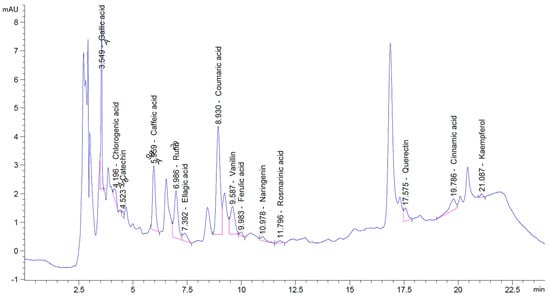
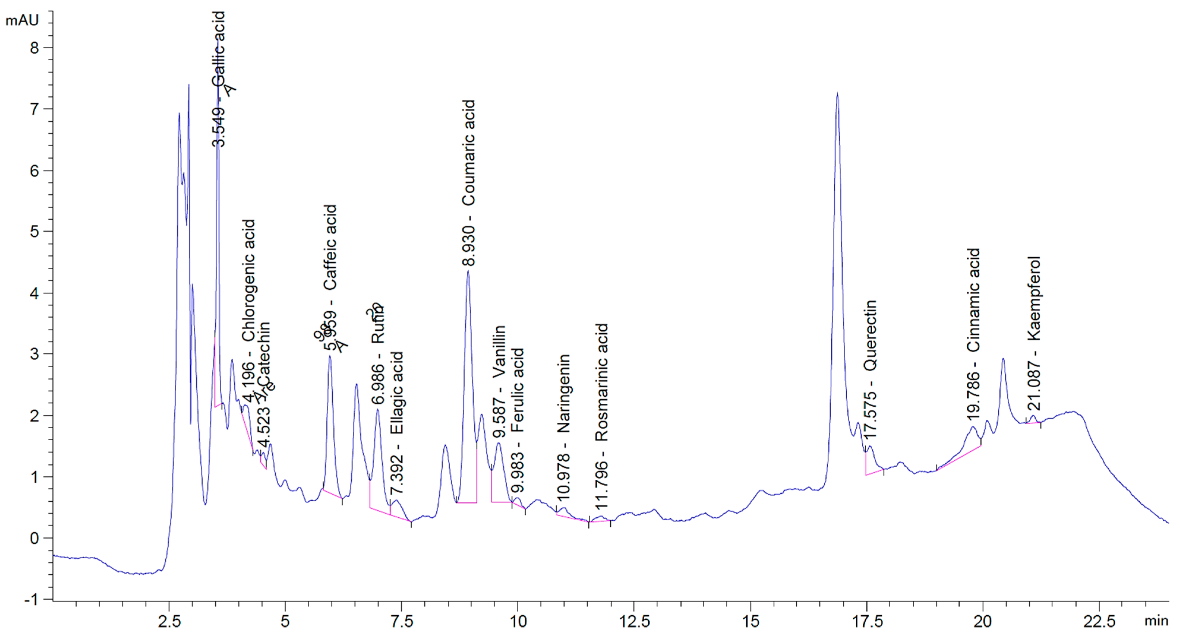
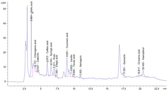
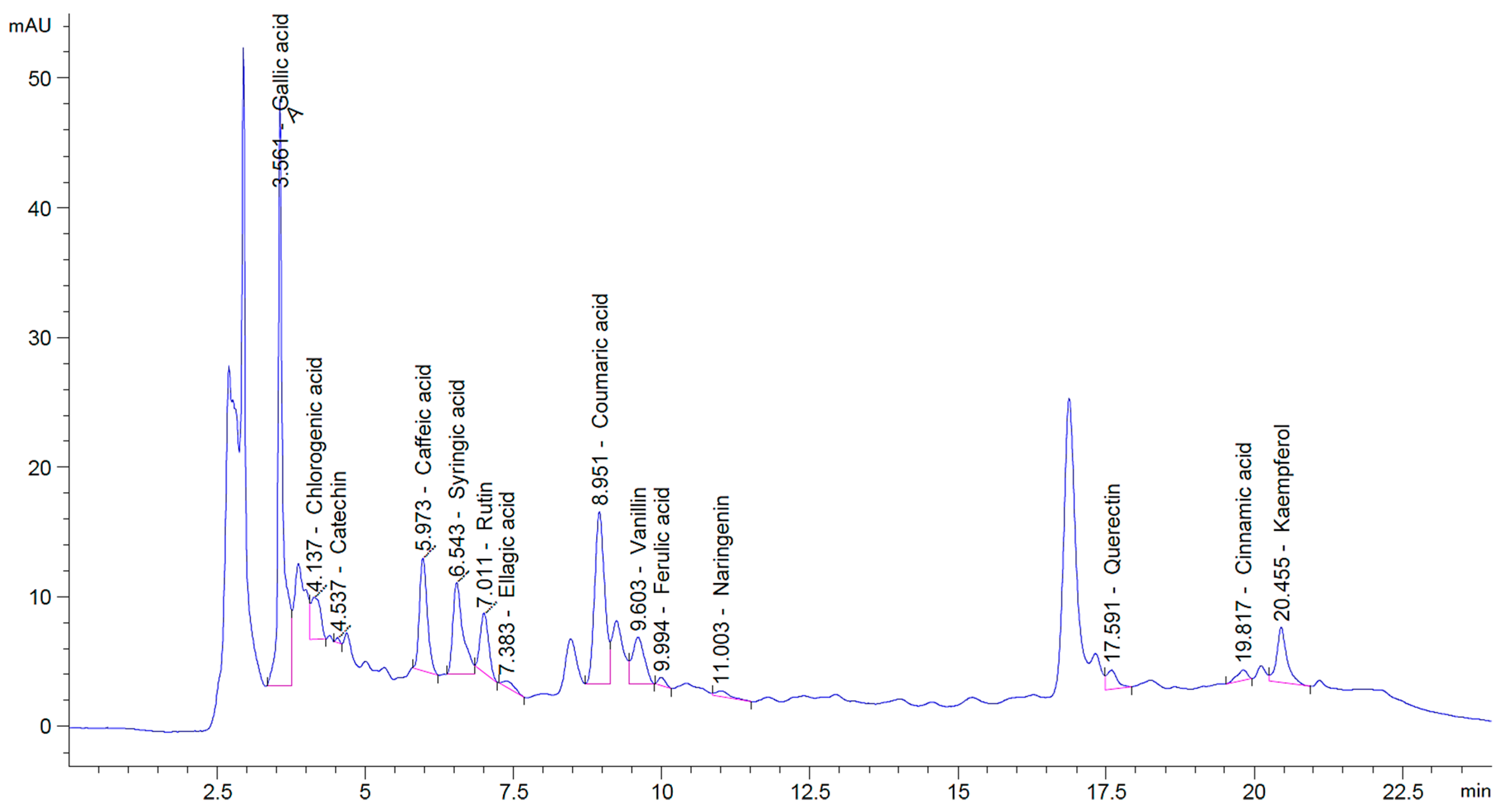
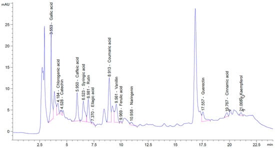
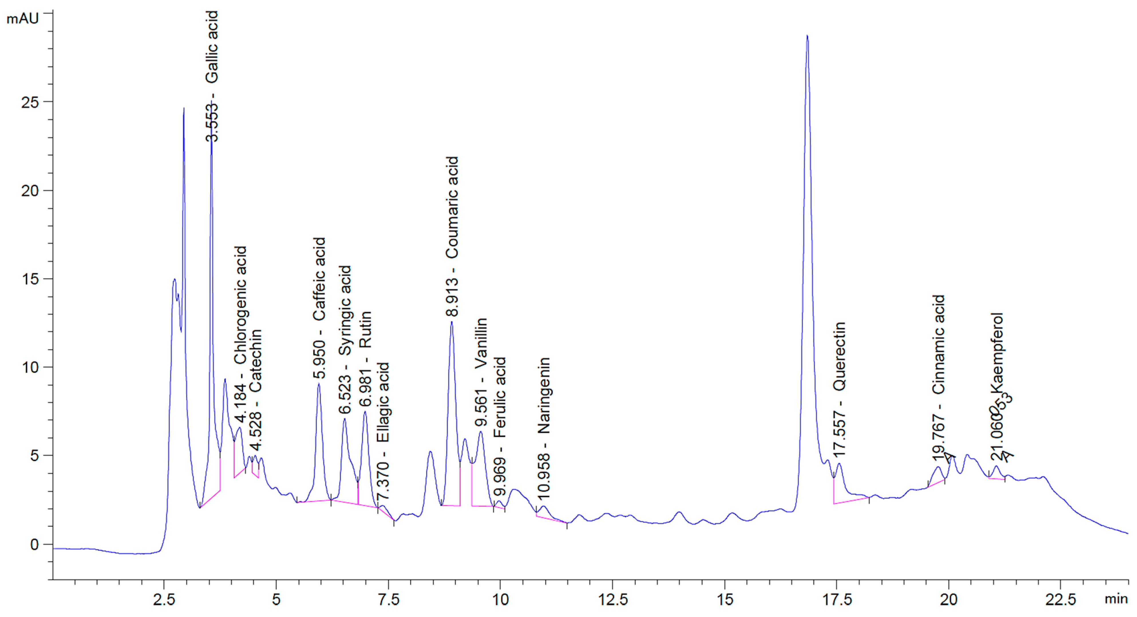
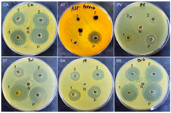
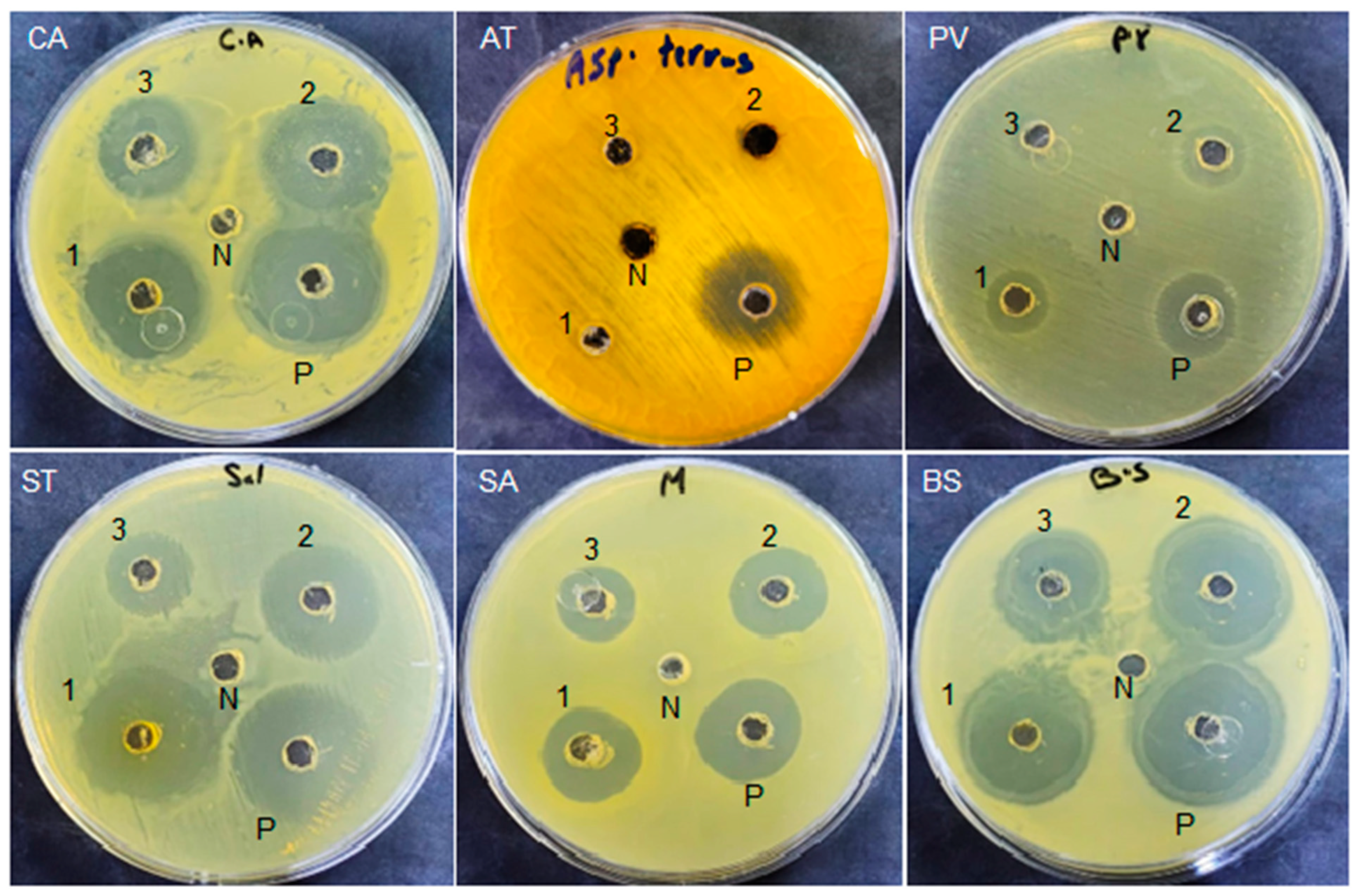
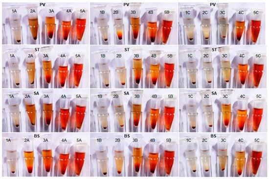
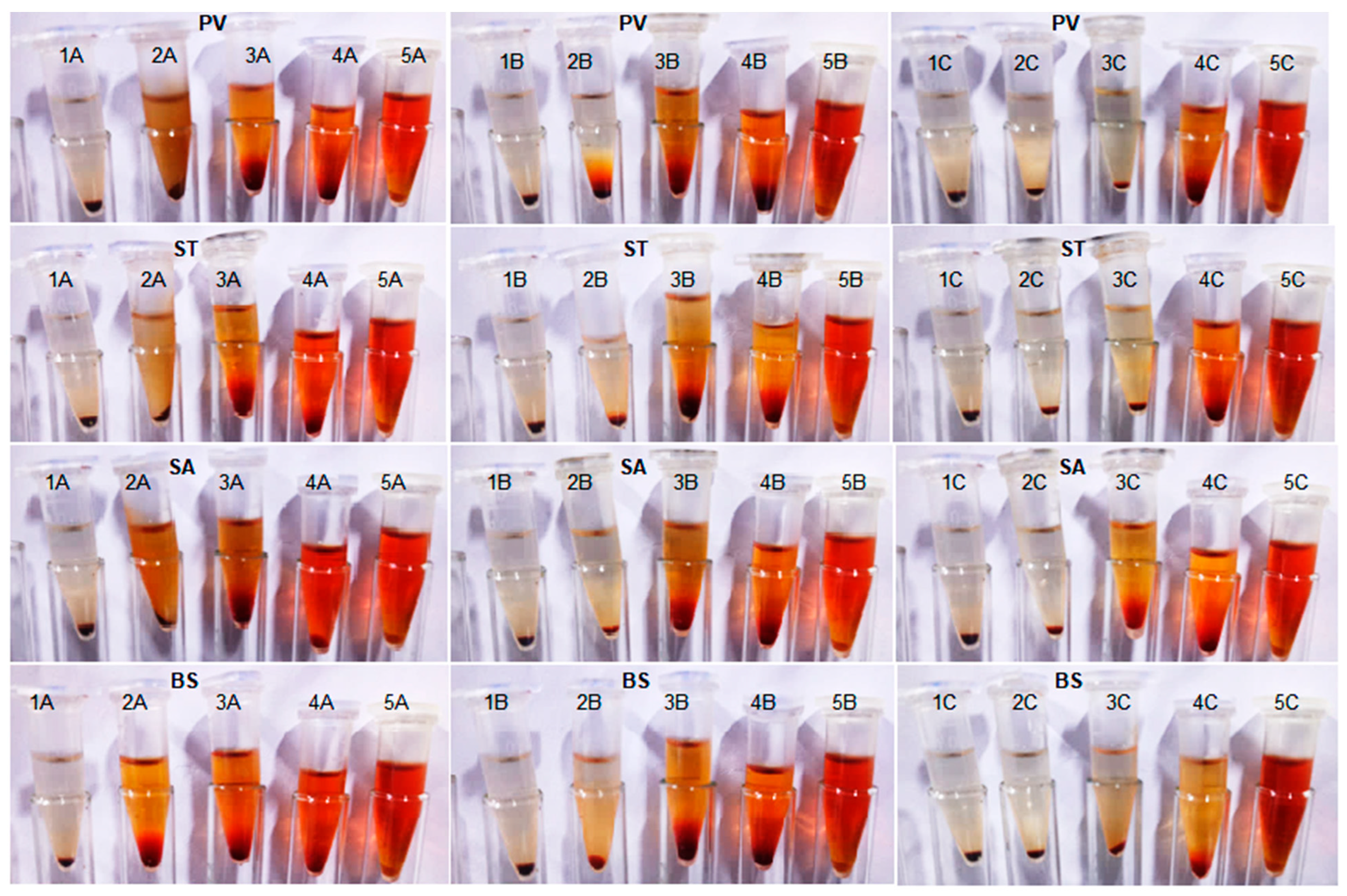
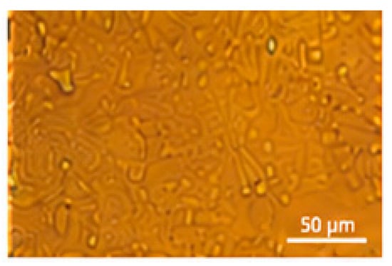
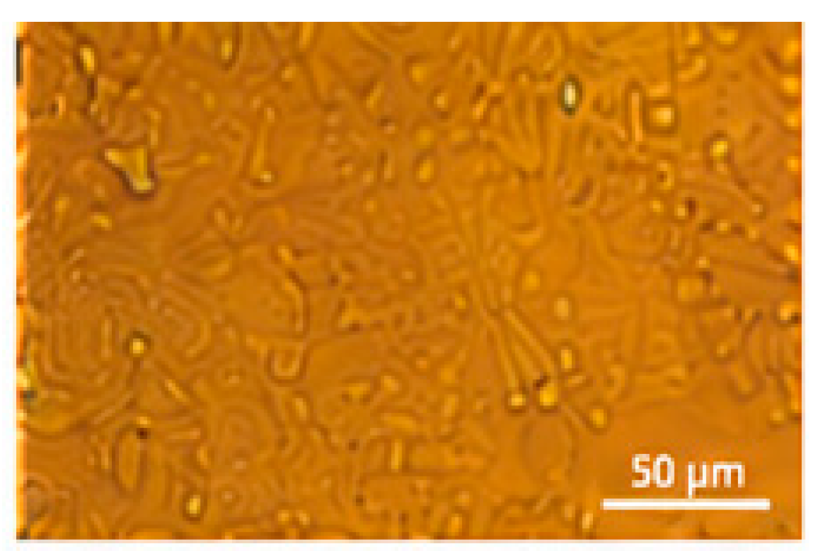
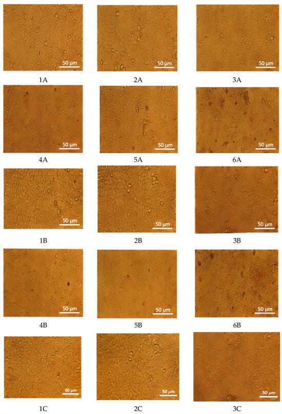
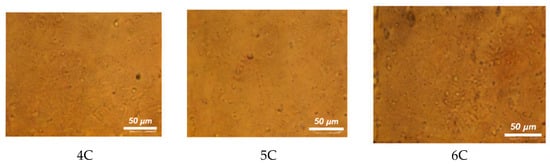
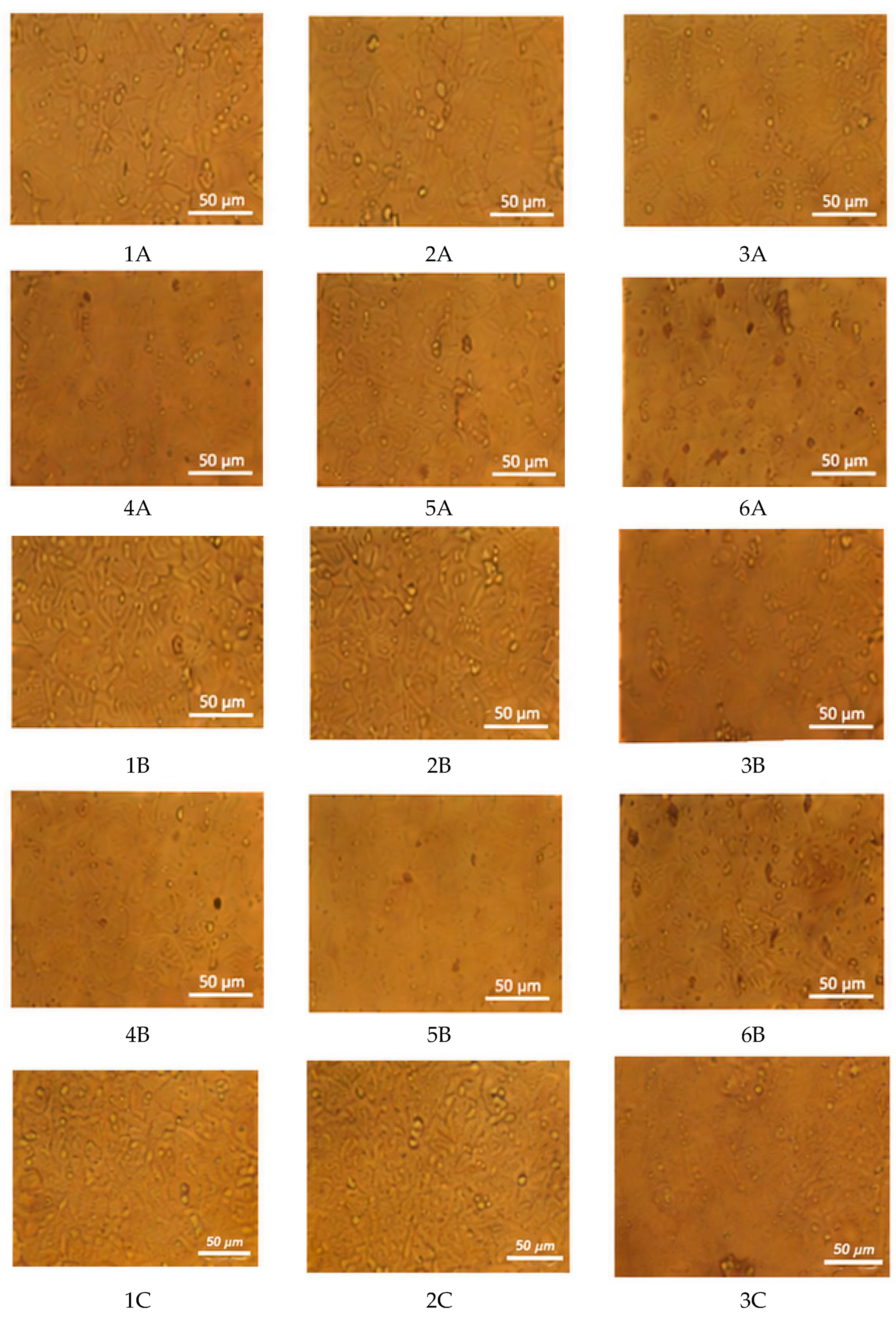
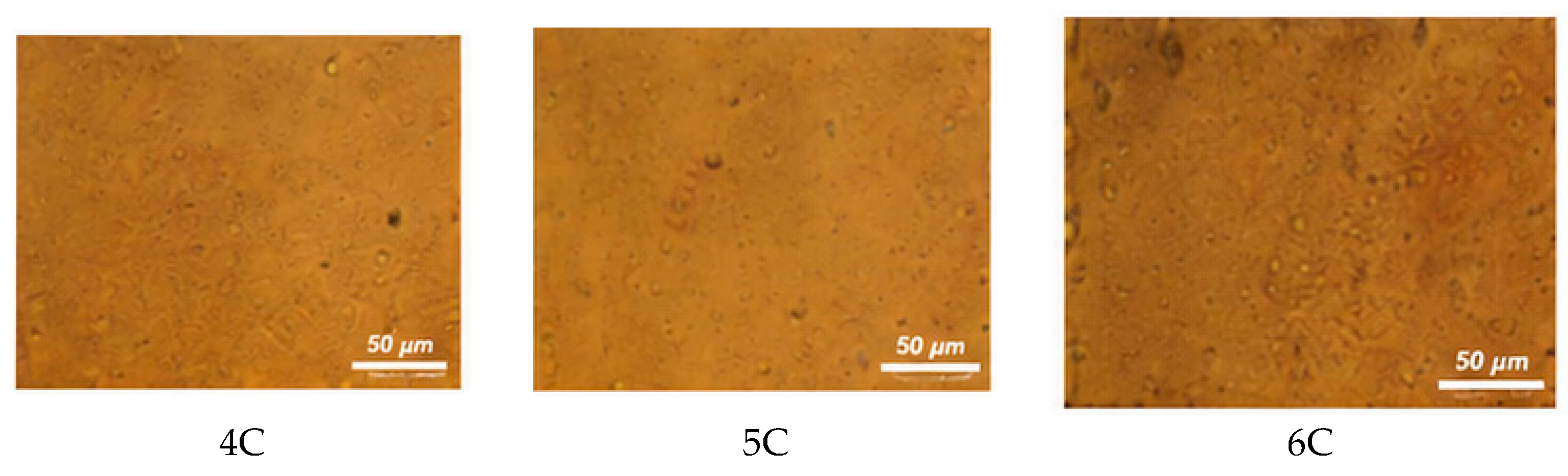

Disclaimer/Publisher’s Note: The statements, opinions and data contained in all publications are solely those of the individual author(s) and contributor(s) and not of MDPI and/or the editor(s). MDPI and/or the editor(s) disclaim responsibility for any injury to people or property resulting from any ideas, methods, instructions or products referred to in the content. |
© 2026 by the authors. Licensee MDPI, Basel, Switzerland. This article is an open access article distributed under the terms and conditions of the Creative Commons Attribution (CC BY) license.