


Abstract
Background/Objectives: The evolution of minimally invasive urologic surgery has progressively reduced incisional burden, yet successive platforms have largely preserved a cavity-based operative paradigm in which the peritoneal cavity serves as the dominant surgical workspace. The da Vinci SP robotic system (Intuitive Surgical, Sunnyvale, CA), through its single-cannula architecture and independently articulating instruments, enables surgery within confined anatomic spaces. We propose “regionalization” as a conceptual framework describing a shift from cavity-based toward compartment-based surgery, in which target organs are approached directly within their native anatomic regions. Methods: This narrative review examines contemporary single-port urologic surgery literature, including systematic reviews and meta-analyses, multi-institutional consortium data, propensity-matched comparative studies, and institutional series. The available evidence is considered in the context of a proposed compartment-based framework for SP urologic surgery. Results: Across the pelvic compartment, superior retroperitoneal compartment, and retroperitoneal reconstructive compartment, the SP platform expands the feasibility of direct transvesical, extraperitoneal, and retroperitoneal access while reducing reliance on the peritoneal cavity as the operative workspace. These approaches allow preservation of surrounding anatomic spaces, direct access to retroperitoneal organs, and greater flexibility in addressing anatomically distributed pathology from a single access site. Available comparative data generally demonstrate comparable oncologic and functional outcomes to multiport surgery, with potential perioperative advantages including shorter hospitalization and reduced analgesic requirements. Current evidence remains predominantly observational and concentrated at experienced high-volume centers. Conclusions: The SP platform enables urologic procedures traditionally organized around cavity-based access to be reorganized around the anatomic compartment of the target organ. This regionalization framework may offer meaningful perioperative advantages while preserving established surgical outcomes, but prospective multicenter studies, longer-term follow-up, and broader institutional experience are needed to define its clinical impact.
1. Introduction
The history of urologic surgery is a history of continuous access refinement. Open retropubic prostatectomy gave way to laparoscopy, which gave way to multiport robotic surgery, and with each transition the incisional burden was reduced while preserving surgical capability. In parallel, open retroperitoneal nephrectomy transitioned into a largely transperitoneal procedure with the rise of laparoscopy and multiport robotic surgery. Yet across these transitions, there was a constant theme: the abdominal or pelvic cavity was entered, a workspace was established within it, and the target organ was approached from within that shared cavity. The peritoneum was not just a route to the target organ; it was the operating theater.
This cavity-based approach has served urology well but carries inherent costs. Transperitoneal access mandates bowel mobilization which risks ileus and visceral injury, can require steep Trendelenburg positioning, and introduces the peritoneum as a middleman between the surgeon and the target [1]. Even the most refined multiport robotic techniques are centered around entering a shared cavity and establishing distributed port geometry to enable triangulated access. This limitation is especially evident for patients with extensive intra-abdominal adhesions from prior operations. Transperitoneal access for these patients is often technically hazardous or unsafe, effectively excluding them from minimally invasive options.
The da Vinci SP robotic surgical system (Intuitive Surgical, Sunnyvale, CA, USA), commercially released in 2018 following FDA approval, was designed with a different operating logic. Its defining feature is a single-port cannula capable of deploying a fully wristed three-dimensional camera and three independently articulating instruments, explicitly engineered for performance in narrow, confined, deep spaces [1,2]. The system eliminates the requirement for distributed port geometry because its instruments articulate independently from a single entry point. This difference, initially framed as a cosmetic or access advantage, carries a deeper surgical implication.
What the SP platform enables is the possibility of organizing surgery not around the cavity through which an organ is accessed, but around the anatomic compartment in which it resides. We propose that the SP robot is driving a paradigm shift from cavity-based surgery toward compartment-based surgery, what we define as the “regionalization” of urologic surgery. Rather than entering the abdomen, creating a workspace, and reaching the target from within a shared peritoneal environment, SP surgery increasingly allows direct access to a defined anatomic region: the superior retroperitoneum for kidney surgery, the extravesical or intravesical space for pelvic surgery, the retroperitoneal ureteral tract for reconstruction. Each organ is treated as residing in its own surgical territory rather than as an abdominal target. Of note, our institutional experience anecdotally demonstrates that even highly complex procedures such as retroperitoneal lymph node dissection (RPLND) can now be performed through these compartment-based access strategies.
This conceptual reframing is supported by a growing body of clinical evidence. Multi-institutional data from the SP Advanced Research Consortium (SPARC) and single-center series have documented the safety and efficacy of retroperitoneal, extraperitoneal, and transvesical SP approaches across a wide range of procedures [3,4,5]. In each case, the defining feature is not simply that a different access route was used but that the route directly targets the anatomic compartment relevant to the procedure, bypassing the peritoneal cavity entirely and operating within a regionally bounded surgical space. The objective of this review is to examine the evolving role of single-port robotic surgery across urologic procedures and to propose regionalization as a framework for understanding the shift from cavity-based to compartment-based surgical access.
2. Methodology
This article presents a narrative review of the contemporary literature on single-port robotic surgery in urology and uses the available evidence to develop a conceptual framework for regionalization. Published evidence relevant to single-port pelvic, renal, and reconstructive surgery was reviewed, including systematic reviews and meta-analyses, multi-institutional consortium studies, propensity-matched comparative analyses, and institutional case series. Literature was selected for its relevance to surgical access strategy, anatomic workspace, perioperative outcomes, functional and oncologic outcomes, and the technical capabilities of the SP platform that permit direct access to confined anatomic regions. Where available, recent comparative and higher-level evidence was emphasized. The literature was synthesized alongside contemporary institutional experience to examine how single-port technology is enabling a shift from cavity-based surgical access toward compartment-based approaches across urologic procedures. This review was narrative in nature and was not conducted as a systematic review or meta-analysis.
3. Conceptual Foundation: The Regionalization of Urologic Surgery
3.1. Defining Regionalization
The development of surgical technologies and techniques can require development of novel frameworks to conceptualize developing strategies. We propose conceptualizing SP robotic urologic surgeries via regionalization, which we define as the organization of a procedure around the anatomic compartment in which the target organ resides, rather than around the abdominal or pelvic cavity used as a passage to reach it. In the traditional paradigm, the surgeon enters the peritoneal cavity, navigates through it, and accesses the target organ as an abdominal structure. In the regionalized paradigm, the surgeon accesses the relevant anatomic region directly: the superior retroperitoneum for the kidney, the extravesical or intravesical space for the prostate, and retroperitoneal ureteral tract. The peritoneal cavity is no longer a necessary waypoint.
This distinction has practical consequences. Cavity-based surgery requires the entire peritoneal environment to be prepared and managed throughout the case, with bowel reflected, omentum repositioned, and visceral anatomy navigated at every step [1]. Compartment-based surgery confines the operative field to the relevant anatomic region, reducing the systemic footprint of the procedure. When the kidney is accessed retroperitoneally, the bowel is never encountered. When the prostate is accessed transvesically, the Retzius space is preserved intact. When ureteral reconstruction is performed via low anterior access (LAA), the entire upper urinary tract can be addressed without re-docking or repositioning [6].
3.2. Why the SP Platform Enables This Shift
Four features of the SP robotic system together enable compartment-based access in a way that multiport robotics cannot readily replicate.
The first is single-port access without triangulation dependence. Three independently articulating instruments and a flexible camera all operate through a single cannula, with each instrument articulating independently. The system does not require the distributed port geometry that multiport robotics demands. This frees the access site from the geometric constraints that make transperitoneal entry obligatory for most multiport surgeries [1,2].
The second is the freedom to operate in confined spaces. Because the SP instruments create their own operative geometry intracorporeally from a single access point, the system performs effectively in the tight anatomy of the retroperitoneum, the extravesical pelvis, and the bladder lumen. These are spaces where multiport robotics, with its requirement for external triangulation and significant working distance, is far less accommodating [3].
The third is the elimination of obligatory bowel mobilization. Direct compartment access means the bowel need not be displaced to reach retroperitoneal or pelvic organs. Retroperitoneal partial nephrectomy, extraperitoneal or transvesical prostatectomy, and retroperitoneal ureteral reconstruction are all performed without handling bowel, reducing the risk of postoperative ileus and related complications [4,6]. In avoiding the peritoneal cavity, there is a distinct advantage to SP surgery in patients who have had prior abdominal surgery or radiation because the surgeons can bypass a hostile abdomen filled with scar tissue and adhesions.
The fourth is dynamic relocation without re-docking. The SP robot can be repositioned to target different anatomic regions without changing the trocar position or re-docking the robot. This enables multi-region surgery from a single access site, a capability with particular relevance for ureteral reconstruction, nephroureterectomy, and combined pelvic procedures [1,6].
4. The SP Platform as an Enabler of Regional Access
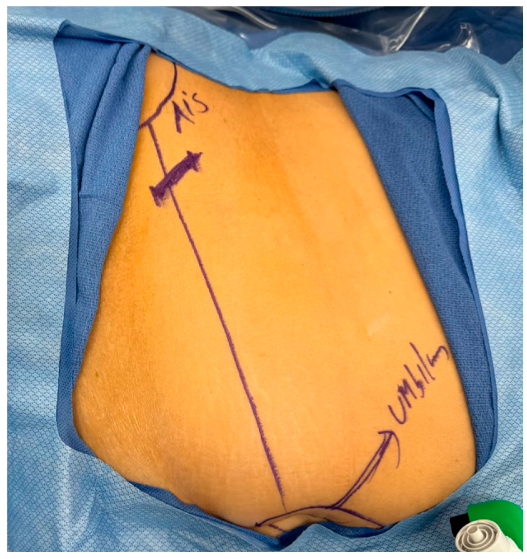
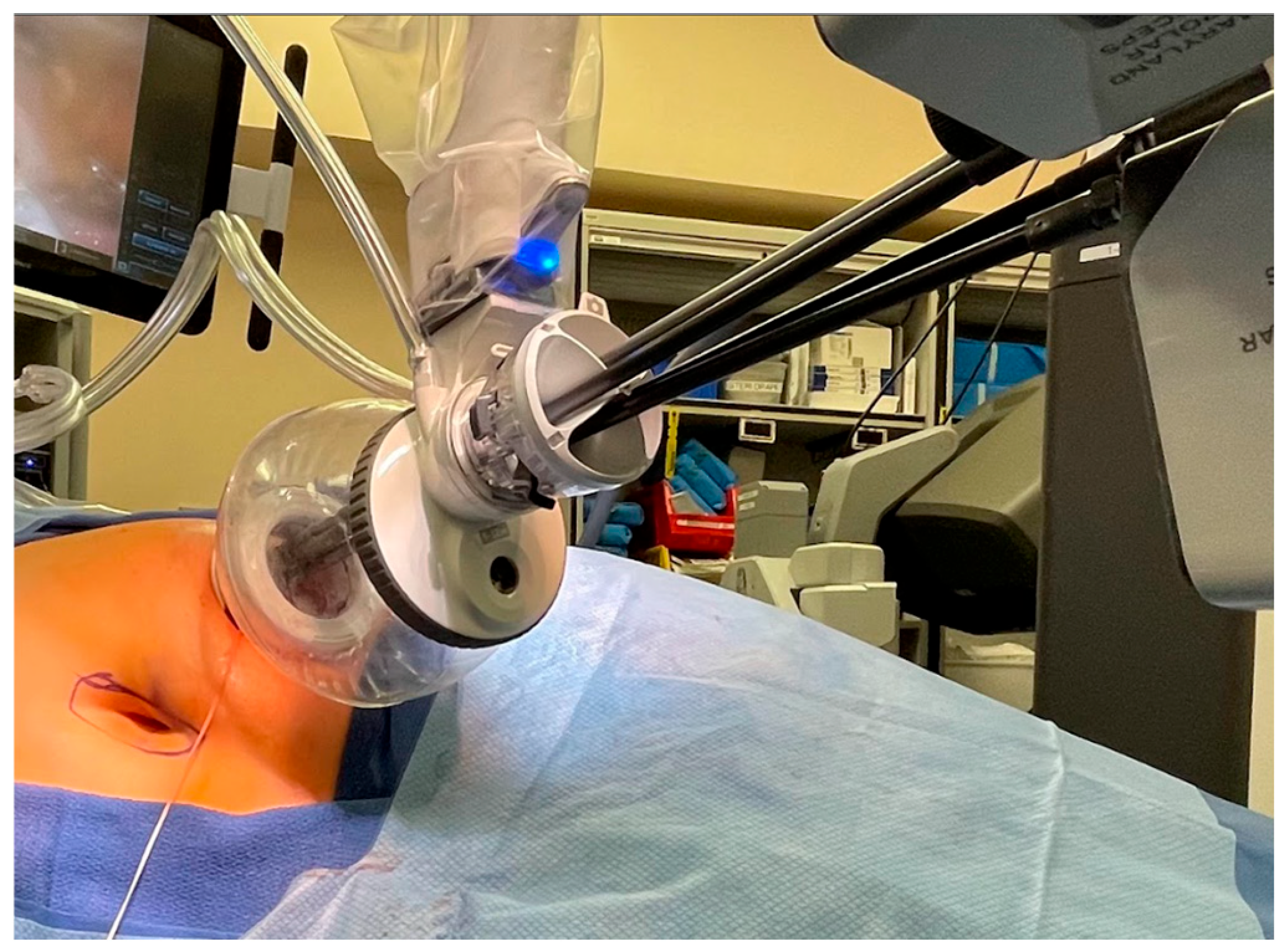
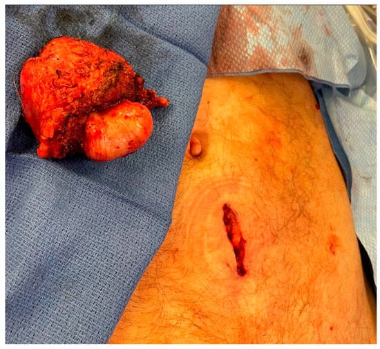
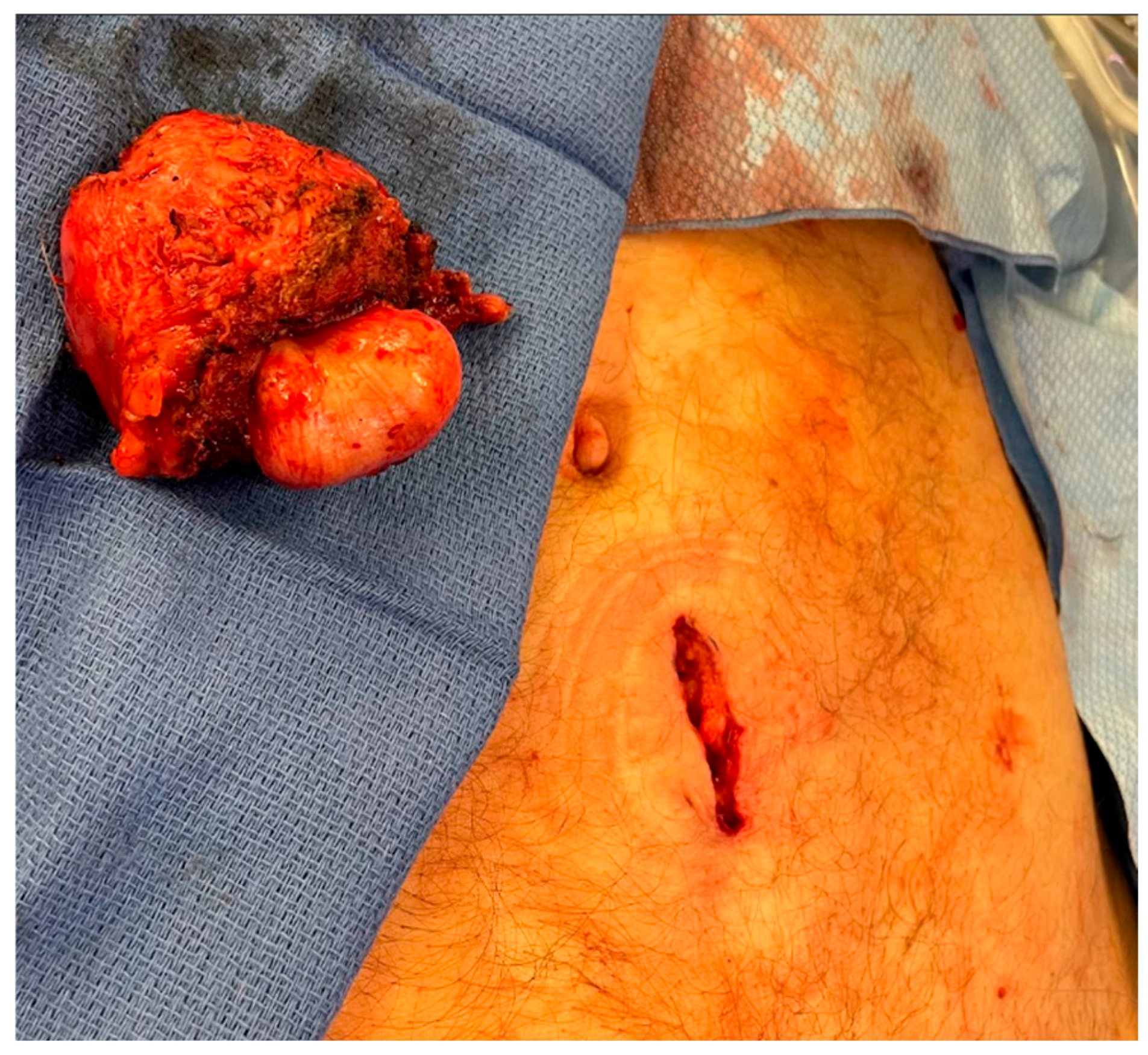
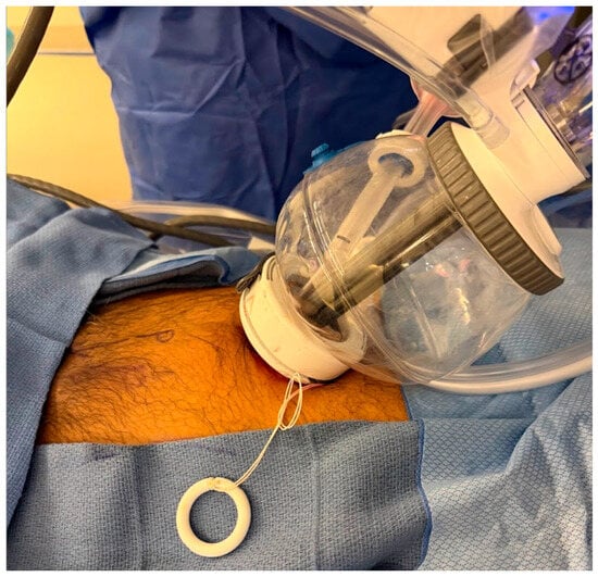
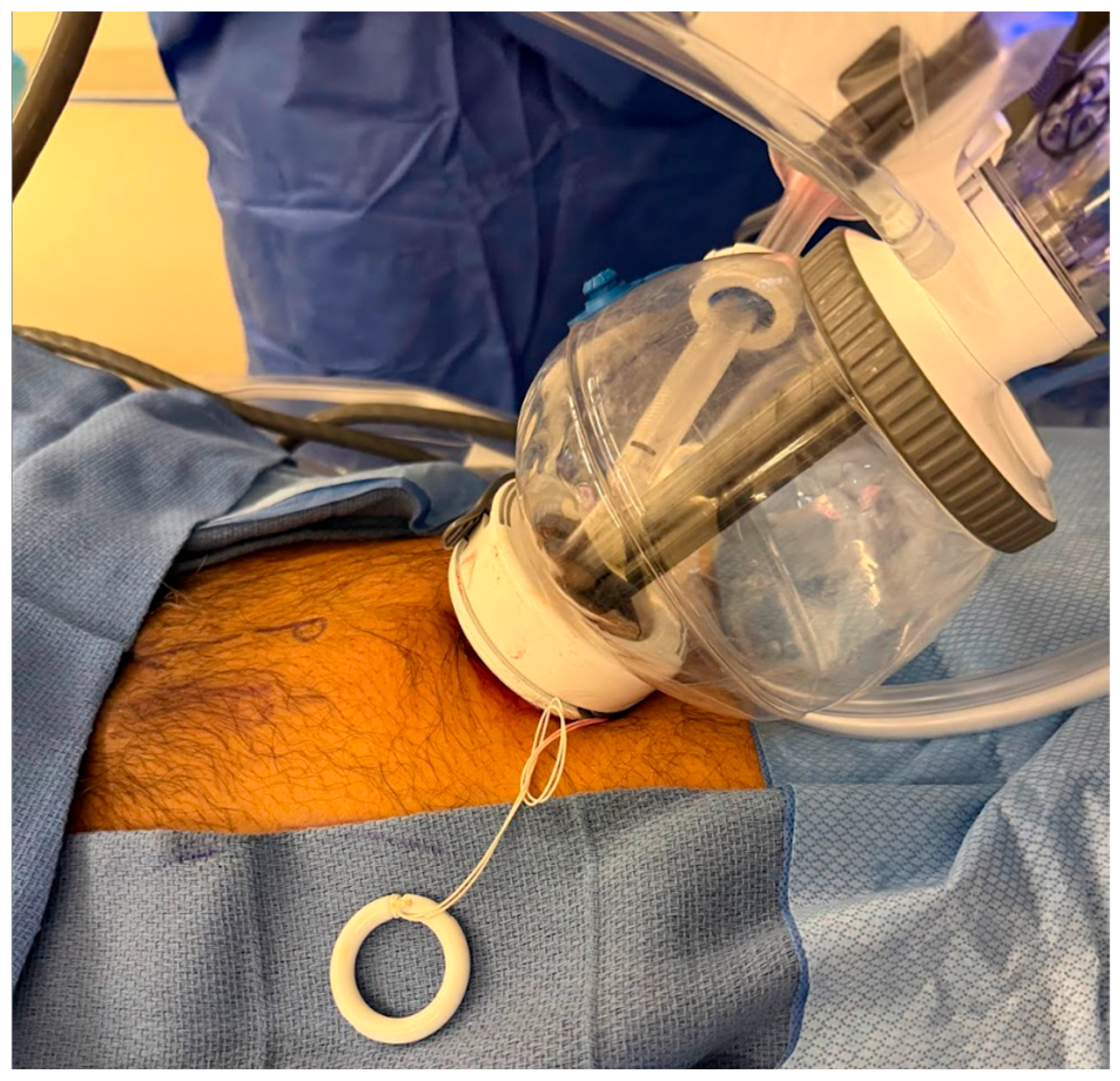
The access strategy chosen for an SP procedure is itself an expression of the regionalization philosophy, because each incision site is selected to align the cannula directly with the relevant anatomic compartment rather than to optimize port geometry across a shared cavity. For kidney and ureteral tract surgery, the low anterior access (LAA) approach, also described as the Pfannenstiel-line or Single-Port Ahmed Modification (SPAM) incision, places a 2 to 3 cm incision approximately 2 to 3 finger-breadths above the anterior superior iliac spine, allowing the retroperitoneal space to be entered through finger dissection alone without entering the peritoneal cavity [3,7]. See Figure 1 and Figure 2. This access has been adopted at multiple institutions under slightly different names reflecting the same anatomic principle: a low anterior retroperitoneal incision in the supine patient that provides direct access to the kidney and ureteral tract while preserving the option to access the peritoneal cavity from the same site if needed without re-docking [7]. For pelvic surgery, an incision placed 2.5 cm infraumbilically can be used for extraperitoneal pelvic surgery, including radical prostatectomy. See Figure 3 and Figure 4. For transvesical procedures, a midline suprapubic incision approximately 2 to 3 finger-breadths above the pubic symphysis provides direct percutaneous access to the bladder dome, allowing all instruments to be introduced into the bladder lumen and the entire operative field to be contained within the transvesical compartment [5,8]. Across all of these access strategies, the unifying principle is the same: the incision is placed to reach the compartment, not to establish a cavity.
Figure 1.
Low Anterior Access Incision: Preoperative surface marking demonstrating the LAA incision site on the right lower abdomen. The thick oblique line indicates the proposed 2–3 cm incision placed approximately 2–3 finger-breadths above the anterior superior iliac spine (ASIS). This access point allows direct entry into the retroperitoneal space through finger dissection alone without entering the peritoneal cavity, serving as the single entry point for SP robotic kidney and ureteral surgery.
Figure 2.
SP Docked at the LAA site: The da Vinci SP surgical system docked at the LAA incision site in a supine patient, demonstrating the single-cannula entry point that provides direct retroperitoneal access without transperitoneal entry or patient repositioning. The compact external profile of the SP platform reflects the absence of distributed port geometry required by multiport systems.
Figure 3.
Infraumbilical incision following single-port extraperitoneal robotic radical prostatectomy: A prostate specimen is shown alongside the infraumbilical access site. This midline incision, placed 2.5 cm below the umbilicus, serves as the sole entry point for extraperitoneal SP pelvic surgery, including radical prostatectomy, without entry into the peritoneal cavity or Retzius space.
Figure 4.
SP Docked at the Infraumbilical Site: The da Vinci SP surgical system docked at the infraumbilical access site in a supine patient undergoing single-port extraperitoneal radical prostatectomy. The single cannula is seated at the midline infraumbilical incision, again demonstrating the SP’s compact docking profile that provides direct access to the extravesical pelvic compartment without peritoneal entry. A more inferior entry point 2–3 finger breadths above the pubic bone can be used for transvesical procedures.
5. Regional Applications of SP Surgery
5.1. The Pelvic Compartment
The prostate sits within the pelvic compartment bounded anteriorly by the Retzius space, posteriorly by the Denonvilliers’ fascia, and inferiorly by the pelvic floor. Conventional multiport radical prostatectomy, whether transperitoneal or extraperitoneal, requires disruption of some or all of these boundaries to achieve the working space needed for triangulated dissection. The Retzius space is routinely entered, with division of the puboprostatic ligaments, ligation of the dorsal venous complex, and incision of the endopelvic fascia. This anterior disruption of the pelvic architecture has long been understood to carry functional consequences, particularly for early continence recovery [9,10,11].
The Retzius-sparing approach to radical prostatectomy was introduced by Galfano and Bocciardi, who described approaching the prostate posteriorly through the pouch of Douglas and working along a completely intrafascial plane without any dissection of the anterior compartment containing the pubourethral ligaments, the Santorini plexus, and the neurovascular structures thought to govern continence and potency [9]. In a subsequent prospective series of 200 consecutive patients, this approach demonstrated that complete prostatectomy could be achieved while preserving these structures, with immediate continence of 92% and 90% in the early and late learning curve cohorts, respectively, and one-year continence of 96% in both groups [12]. These results established the conceptual foundation for compartment-preserving pelvic surgery: that avoiding the anterior pelvic space may allow for earlier functional recovery. Subsequent systematic reviews and large propensity-matched analyses have confirmed that the Retzius-sparing approach is associated with significantly earlier continence recovery compared to the standard technique, with equivalent oncologic outcomes [10,11].
The SP platform takes this compartment-preserving logic further by adding the transvesical dimension. Transvesical SP radical prostatectomy (SP-RARP) performs the entire procedure from within the bladder, entering percutaneously through a suprapubic incision, without any entry into the abdominal or Retzius space [8]. The Retzius space is preserved not by approaching from a posterior plane but by never approaching it at all. Both ureteral orifices are visualized upon bladder entry. Dissection proceeds posteriorly through the bladder neck to the seminal vesicles, then anteriorly and apically along the prostate, and vesicourethral anastomosis is completed from within the bladder lumen. The entire operative field is confined to the bladder–prostate unit, with the peritoneum, bowel, and anterior pelvic anatomy left entirely undisturbed.
SPARC data comparing transvesical and extraperitoneal SP-RARP in a large propensity-matched multi-institutional cohort have demonstrated that the transvesical approach is associated with superior recovery across almost every postoperative domain while maintaining equivalent oncologic safety [1,5]. The transvesical approach has also been extended to robotic-assisted simple prostatectomy (TV-RASP) for benign prostatic obstruction, where Kaouk et al. reported the first series of single-port transvesical prostate adenoma enucleation in patients with large glands, completing all cases without conversion and in the supine flat position with no bowel manipulation [4]. Subsequent experience with TV-RASP demonstrated the ability to treat prostates of virtually any size entirely within the bladder compartment [13]. The ability to perform this procedure entirely within the bladder compartment in supine position, without pneumoperitoneum or Trendelenburg, has further enabled the use of epidural anesthesia without mechanical ventilation, a perioperative advance made possible specifically by the fully compartmentalized operative environment of transvesical SP surgery [14].
The transvesical compartment logic extends to focal therapy as well. SP transvesical partial prostatectomy for localized prostate cancer, as described by Kaouk et al., allows targeted intraluminal resection of the prostate entirely within the bladder-prostate compartment, preserving uninvolved lobes and supporting structures [15]. Reconstruction of the bladder neck with complete epithelialization may facilitate faster recovery and reduced irritative symptoms compared with many endoscopic outlet procedures [16].
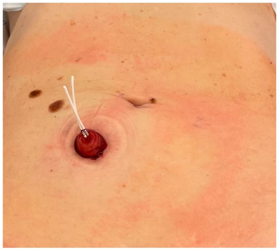
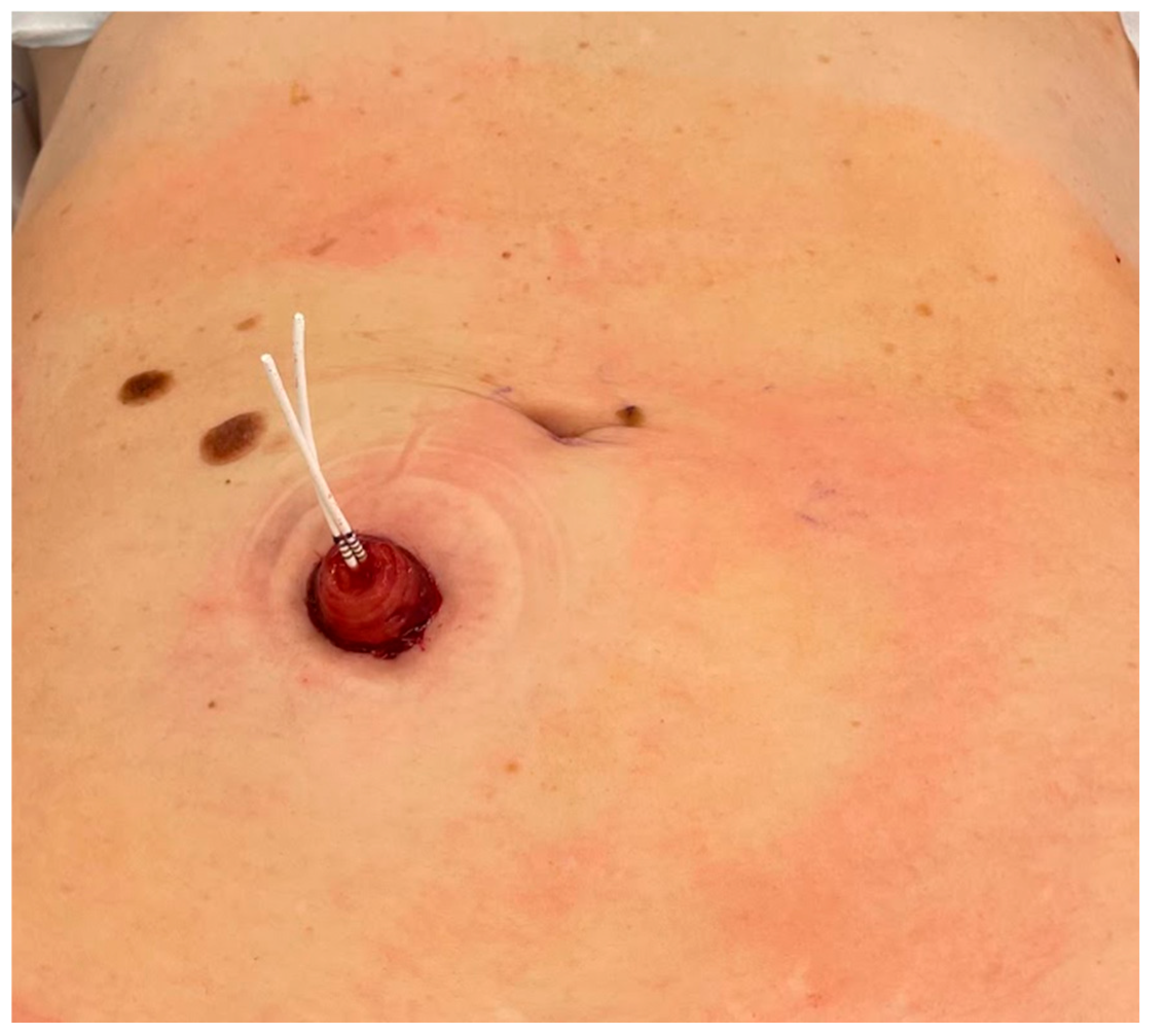
The regionalization concept also extends to radical cystectomy and urinary diversion. Early SP radical cystectomy experiences, including at our institution, have demonstrated that the stoma site itself can serve as the single-port access site, effectively eliminating the need for an additional extraction incision [17]. See Figure 5. In female patients, the vaginal canal has additionally been utilized as an auxiliary natural-orifice route for stapling and specimen extraction at our institution. With the introduction of the SP robotic stapler, similar incision-minimizing strategies are now becoming feasible in male patients as well. SP radical cystectomy with intracorporeal neobladder reconstruction has been performed through a single approximately 3-cm incision, further demonstrating the ability of the SP platform to consolidate extirpative and reconstructive surgery within a highly regionalized operative field [18]. Our team has also extended this concept to continent urinary diversion, including intracorporeal Indiana pouch reconstruction, highlighting the expanding applicability of SP compartment-based surgery to even the most complex extirpative and reconstructive procedures in urology.
Figure 5.
SP access site as stoma site following SP robotic radical cystectomy with intracorporeal urinary diversion. Postoperative appearance following single-port robotic radical cystectomy performed entirely through the stoma site incision: The urostomy with exiting ureteral stents is visible, with no separate abdominal incision required for port placement or specimen extraction. This approach consolidates the operative access point and the urinary diversion outlet into a single site, eliminating the additional extraction incision required by conventional robotic cystectomy.
5.2. The Superior Retroperitoneal Compartment: The Kidney as a Retroperitoneal Organ
The kidney is a retroperitoneal organ. It resides in a distinct anatomic compartment bounded by Gerota’s fascia, the psoas muscle, and the posterior abdominal wall that has no necessary relationship to the peritoneal cavity. Yet the dominant approach to robotic partial nephrectomy in the multiport era has been transperitoneal, not because the transperitoneal route is anatomically appropriate for the kidney, but because the geometric requirements of multiport robotics are more easily satisfied in the capacious peritoneal cavity than in the narrow retroperitoneal space. The multiport platform’s wide port spacing, substantial working distance requirements, and four independently docked arms make retroperitoneal surgery technically demanding, and adoption of a retroperitoneal approach with multiport robotics has historically remained limited [1,3].
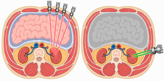
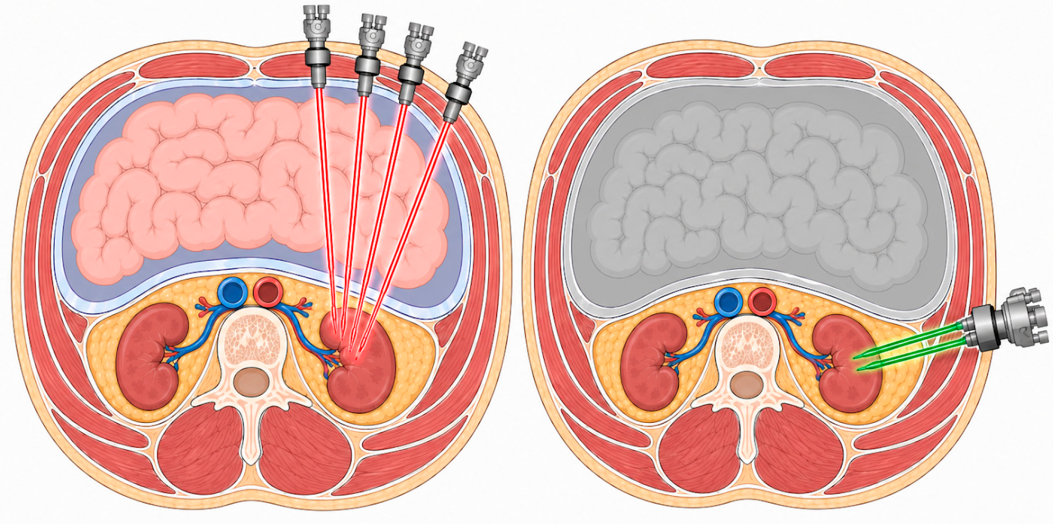
The SP platform changes this equation. Its single-arm architecture, designed for narrow confined spaces, is precisely suited to retroperitoneal geometry. As demonstrated in Figure 6, the SP platform enables access to the kidney via the retroperitoneum, as compared to the MP platform’s transperitoneal access. SPARC data reported by Raver et al. demonstrated this directly in a multi-institutional analysis showing that the adoption of SP robotic surgery was associated with a dramatic increase in the utilization of the retroperitoneal approach for partial nephrectomy, with the SP platform used more often with a retroperitoneal approach and the preference for this approach increasing over time as surgeons gained experience [3]. Retroperitoneal multiport partial nephrectomy also increased in the same institutions over the same period, suggesting that SP adoption normalizes a retroperitoneal access culture that extends beyond the SP platform itself.
Figure 6.
An axial schematic illustrating the concept of transperitoneal versus retroperitoneal renal surgeries.
The clinical rationale for treating the kidney as a retroperitoneal organ is straightforward: retroperitoneal access provides direct visualization of the renal hilum without navigating the colonic mesentery, eliminates bowel mobilization and its associated risks, allows a more direct approach to posterior renal masses, and avoids peritoneal contamination and its sequelae [3]. LAA/SPAM positioning allows the retroperitoneal space to be entered in the supine position through a small incision with finger dissection alone, simplifying both patient positioning and operative setup [7]. Data from SPARC and subsequent meta-analyses demonstrate broadly comparable perioperative and oncologic outcomes between SP and MP partial nephrectomy, although differences in warm ischemia time, blood loss, length of stay and transfusion rates have been recorded across studies [19,20,21,22]. Separately, SPARC adoption data reported by Raver et al. demonstrated that SP uptake was independently associated with a dramatic shift toward retroperitoneal access, with the retroperitoneal approach used in 52.8% of SP cases overall and increasing to 75% by 2023 [3].
The SP platform’s suitability for the retroperitoneum extends beyond partial nephrectomy to retroperitoneal radical nephrectomy and retroperitoneal nephroureterectomy. This principle also extends to retroperitoneal adrenalectomy, where the SP platform allows direct access to the adrenal gland without mobilization of intraperitoneal organs, further reinforcing the concept of organ-specific compartment surgery [23]. For radical nephroureterectomy, a procedure that historically required patient repositioning and robot re-docking to address both the kidney and the distal ureter in separate stages, the SP enables the kidney and ureter to be addressed in a continuous retroperitoneal sequence without repositioning, re-docking, or any new incision [1,3].
5.3. The Retroperitoneal Reconstructive Compartment: The Ureter as a Continuous Tract
The ureter presents a unique challenge for robotic reconstruction because it is a continuous structure extending from the ureteropelvic junction to the ureterovesical junction, traversing the retroperitoneum and pelvis across multiple anatomic zones. With multiport robotics, the location of the pathology dictated port placement configuration: proximal strictures required a lateral flank setup, distal strictures required a pelvic setup, and long-segment disease requiring work in multiple zones necessitated repositioning or re-docking. The fixed triangulated geometry of multiport systems makes the ureter difficult to address as a continuous structure from a single operative setup.
The SP platform simplifies ureteral surgery fundamentally because the robot can be relocated, redirecting its single arm to a new target angle without changing the access point or re-docking. Billah et al. describe this directly in the first SP reconstructive urologic surgery series, reporting on experience from Hackensack University Medical Center: Techniques described included Anderson–Hynes dismembered pyeloplasty for ureteropelvic junction obstruction, ureteroureterostomy for mid-ureteral stricture, buccal mucosa ureteroplasty for long-segment ureteral disease, ureteral reimplantation with or without Boari flap, and vesicovaginal fistula repair with omental interposition. These procedures were all accessed via the umbilicus, with the SP system relocated to direct the cannula toward upper, mid, or distal targets as the procedure required. For ureteral reconstruction specifically, Billah et al. described how the SP system allowed exclusive placement of the trocar via the umbilicus with the ability to easily reach the upper to distal ureter, contrasting this directly with the multiport era in which location and port placement strategy were critical and based on the location of the stricture [6]. Notably, this initial report features a transperitoneal approach, though our institution is actively adapting the same reconstructive procedures to a fully retroperitoneal approach via a LAA incision, with prospective data collection ongoing.
Given that there is evidence supporting that the SP platform can be used in urologic reconstruction in general, and evidence that the SP robot can be used entirely retroperitoneally and extraperitoneally in other urologic surgeries, conceptually, it would follow that a single-port system could be used extraperitoneally for urologic/ureteral reconstruction. However, there is very limited published evidence to support this. A 2025 study from the University of Illinois at Chicago demonstrated that ureteral reimplantations could be done extraperitoneally with the patient in a supine position [24]. This study found ureteral reimplantation using the SP platform had better outcomes in the extraperitoneal group, including shorter operative times, less blood loss, and shorter stays. Going forward, further investigation into the SP platform’s utility in urologic reconstruction and the impact of a retroperitoneal approach is warranted.
This continuous-tract access philosophy represents the regionalization concept applied longitudinally. Rather than organizing surgery around fixed port geometry, the SP’s dynamic relocation capability allows the surgeon to follow the anatomic course of the ureter as a single operative target, treating the ureter as its own compartment accessible along its full length from a single entry point. This retroperitoneal reconstructive philosophy also extends to stone surgery, including SP-assisted pyelolithotomy for complex and staghorn calculi, where direct access to the collecting system can be achieved without transperitoneal mobilization.
6. The Single-Port vs. Multiport Paradigm
The distinction between SP and multiport robotic surgery is not just about port number. It represents a fundamentally different operative mindset with consequences extending from patient positioning to surgical philosophy.
Multiport robotic surgery is triangulation-dependent, as was its predecessor modality laparoscopic surgery. The operative workspace is defined by the geometric relationships among multiple externally placed trocars converging on the target. This geometry requires adequate abdominal or pelvic volume and the ability to distribute ports in a configuration that allows each arm to move without collision [1]. The peritoneal cavity, with its ample volume and established anatomic landmarks, is the natural environment for this geometry, and transperitoneal access is its logical default.
SP robotic surgery is access-driven. The operative workspace is defined not by external port geometry but by the internal articulation of instruments from a single access point. The system creates its own triangulation intracorporeally, independent of external port placement. The access site can therefore be positioned to directly target the relevant anatomic compartment without requiring the peritoneal cavity as a working environment, thus allowing the re-retroperitonealization of nephroureteral surgeries. The fundamental shift is from port geometry toward anatomic region targeting.
Comparative data from SPARC and multiple propensity-matched single-institution analyses, including Hackensack experience with both prostatectomy and partial nephrectomy, have demonstrated that this approach produces equivalent oncologic and functional outcomes while reducing perioperative morbidity, including shorter hospital stays, reduced narcotic requirements, and reduced blood loss [3,4,5,19]. This is not the result of a smaller incision alone. It is the result of operating within a defined anatomic compartment rather than traversing a shared cavity to reach the target.
In most cases, patients who are candidates for MP robotic surgery are candidates for SP surgery. Co-morbidities may limit a patient’s overall surgical candidacy, and there are very few circumstances in which a patient would be a candidate for MP surgery and not SP. At our institution, there are few limitations in choosing candidates for SP surgery as we have successfully operated on patients with high BMI, high nephrometry scores, various comorbidities and extensive previous surgeries. Newer surgeons should be mindful of BMI when selecting patients for retroperitoneal partial nephrectomies, as well as upper pole tumors. Similarly, relatively large prostates and prostates with advanced disease can be challenging cases when learning SP surgery. In our experience, there are very few patient factors that deter us from SP reconstructive cases.
With the current surgical landscape heavily favoring MP systems, it is important to highlight the ways in which SP is superior, equivalent, and inferior so that surgeons are able to make informed decisions about which option is best for their practice in the short and long term. Practically speaking, there are several barriers to implementing an SP robotic system at new institutions. These barriers include platform cost and availability, credentialing requirements, and access to experienced proctors, in addition to institutional requirements such as alterations to operating room workflows. These concerns were central to an AUA debate on the topic, where critics questioned adoption given that SP robot-assisted prostatectomy is more costly than its MP counterpart and has a steep learning curve, begging the question of why such technology should be adopted. When first learning to use the SP platform it is estimated to take 30–150 cases to reach lowest achievable complication rates, depending on the procedure (30 cases for simple prostatectomy, 70 cases for partial nephrectomy, and 150 cases for radical prostatectomy) [25]. It is worth continuing to evaluate the learning curve in an era in which the system is becoming more widespread and introduced earlier in training.
There is some data that suggests MP platforms are superior to SP when it comes to warm ischemic time (WIT) in partial nephrectomies as mentioned in Table 1. A measured difference in WIT for SP and MP partial nephrectomy may not be clinically significant when the ischemic time remains within acceptable thresholds [26]. Additionally, the literature is heterogeneous and not every study finds a difference. A single-center propensity-score-matched analysis (30 SP matched 1:1 to 30 MP) found that after matching there were no significant differences between SP and MP approaches in estimated blood loss, ischemia time, transfusion rate, or intraoperative complications. This suggests that in experienced, high-volume hands with well-matched cases, the WIT gap is insignificant [27].
Table 1.
Systematic reviews and meta-analyses comparing single-port versus multiport robotic approaches.
7. Limitations
The SP platform’s advantages in compartment-based access come with inherent technical limitations that must be acknowledged. The most commonly cited is instrument crowding: because all instruments originate from a single 25-mm cannula, the operative geometry is fundamentally more constrained than with multiport surgery, and managing instrument collisions within a confined operative field requires deliberate spatial attention and familiarity with the SP Navigator display system [6]. In addition, SP may be less suitable in patients with exceptionally large, heavy tumors or target organs. The SP robot arms are slightly weaker compared to the MP system, which may make cases involving large kidneys or prostates more suitable for MP robotic surgery. Retraction capability is also reduced relative to the four-arm multiport geometry, which can be particularly challenging in cases with dense adhesions, bulky perinephric fat, or complex anatomy requiring sustained exposure. Various adjuncts including the ROSI suction system, Weck clips, and magnetic retractors have been described to compensate for these retraction limitations [6].
As with any newer medical intervention, long-term data on patient outcomes with the SP system are limited. Not only that, but the data being published on SP robotic surgery is coming from specialized, high-volume centers; therefore, our ability to generalize the results to routine practice is limited. Thus, it will be important to not only continue to follow patients throughout the lifespan as we continue to evaluate the SP system but to also evaluate the accessibility of the SP system to generate results that are more applicable to routine practice. Most of the current available literature on single-port prostatectomy and nephrectomy consists of single-center studies with small sample sizes and short-term follow up. More rigorous multi-center trials with randomization and long-term follow up are necessary to validate these initial studies and determine the clinical benefits across institutions and patient populations. It is also worth noting that, as with any medical innovation, it is important to consider that inequitable access for patients may result in health disparities and inequity in treatment or outcomes without diligent vigilance.
In total, these limitations are real but are not arguments against the regionalization paradigm. They are arguments for appropriate patient selection, structured training, and continued platform development. Adjunctive technologies such as valveless insufflation systems, including AirSeal and Lexion platforms, are increasingly important in SP surgery. Because many SP procedures are performed in confined extraperitoneal spaces, maintaining stable insufflation at lower pressures, often below 10 mmHg, may help reduce subcutaneous emphysema and carbon dioxide absorption while preserving adequate workspace exposure.
8. Future Directions
The regionalization paradigm points toward several important directions for the future of SP urologic surgery. Primarily, we propose conceptualizing robotic urologic surgeries via a regionalization framework. We define regionalization as the organization of a procedure around the anatomic compartment in which the target organ resides, rather than around the abdominal or pelvic cavity used as a passage to reach it. In doing so, formalization of compartment-specific training curricula can be prioritized. If the SP platform is enabling a different operative logic, then training programs must be designed around that logic rather than treating SP surgery as a variant of multiport technique. Curricula that explicitly address retroperitoneal anatomy and access, transvesical operative navigation, and the conceptual shift from port-geometry thinking to compartment targeting will be needed to realize the full potential of the platform. As SP is a newer surgical modality, there is limited data from outside institutions regarding its efficacy and overall patient outcomes. It is our hope that more institutions will evaluate the SP system and contribute to the field’s understanding of its impact.
The growth of opioid-sparing and ambulatory surgery models is a natural extension of the compartment-based approach. The consistent association of transvesical and retroperitoneal SP surgery with same-day discharge and reduced opioid requirements in SPARC data reflects not just a smaller incision but a fundamentally different relationship between the operative field and the systemic physiology of the patient [3,5]. The demonstration that transvesical SP surgery can be performed under epidural anesthesia without mechanical ventilation represents the most complete expression of this principle [14]. This is a major oncologic procedure that was performed without general anesthesia, made possible by an operative environment that does not violate the peritoneal cavity, does not require Trendelenburg positioning, and confines insufflation to the bladder lumen.
The expansion of transvesical applications beyond prostatectomy, including transvesical ureteral reimplantation, intravesical tumor work, and combined pelvic procedures in which the bladder serves as the operative corridor to multiple targets, represents a frontier that remains largely unexplored. Platform evolution with improved grip strength, extended-reach instruments, and newer instruments including staplers will further expand the range of compartments and procedures accessible via regionalized SP surgery.
9. Conclusions
The da Vinci SP robotic platform represents more than an incremental refinement of minimally invasive urologic surgery. It enables a fundamental reconceptualization of how urologic procedures are organized, a shift from the cavity-based, triangulation-dependent logic of multiport robotics toward a compartment-based operative philosophy in which each organ is approached directly within its own anatomic region. It is also important to recognize that the current da Vinci SP platform represents only the first generation of single-port robotic technology, whereas multiport robotic systems have already undergone multiple generations of iterative refinement. As instrumentation, visualization, retraction capability, and access technology continue to evolve, many of the present limitations of SP surgery will likely diminish, further expanding the feasibility and impact of compartment-based regionalized surgery.
Across the pelvic compartment, the retroperitoneal compartment, and the reconstructive ureteral corridor, the SP platform is demonstrating that peritoneal entry is not a surgical necessity. It is a historical default that arose from the geometric requirements of prior platforms and can be challenged when those geometric requirements no longer apply. There is growing clinical evidence that compartment-based access produces outcomes at least equivalent to and in many domains superior to those of transperitoneal surgery, while creating opportunities for new perioperative possibilities including opioid-free recovery or regional anesthesia for major oncologic procedures. This is not to say that the current predominant MP system and transperitoneal approach is approaching obsolescence, but further investigation is certainly warranted into regionalized SP urologic surgeries. Specifically, there is a need for future examination of the SP system and the utility of regionalization through prospective multicenter comparative studies, patient-reported outcomes, long-term oncologic data, standardized learning-curve analyses, and health-economic evaluations.
The long-term impact of this platform will likely be measured not in port counts or scar lengths but in its redefinition of surgical geography: the demonstration that urologic surgery can be organized around the organ’s own anatomic home rather than around the cavity through which it has traditionally been reached.
Author Contributions
Conceptualization, M.A., M.B. and A.S.; Methodology, A.S.; Investigation, O.U., A.S., J.O. and M.P.; Data Curation, A.S.; Writing—Original Draft Preparation, A.S.; Writing—Review and Editing, M.A., M.B., A.S., O.U., J.O. and M.P.; Visualization, J.O.; Supervision, M.A. and M.B. All authors have read and agreed to the published version of the manuscript.
Funding
This research received no external funding.
Institutional Review Board Statement
Not applicable.
Informed Consent Statement
Informed consent was obtained from all subjects involved in the study.
Data Availability Statement
No new data were created or analyzed in this study. Data sharing is not applicable to this article.
Acknowledgments
During the preparation of this manuscript, the authors used ChatGPT 6.1 (OpenAI) for assistance with grammar, sentence structure, and language refinement during final revisions. The authors reviewed and edited the output and take full responsibility for the content of this publication.
Conflicts of Interest
Dr. Mutahar Ahmed has served as a consultant for BioTissue Holdings Inc., Vascular Technology Incorporated (VTI), Ethicon, and Intuitive Surgical, Inc. The remaining authors (M.B., A.S., O.U., J.O., M.P.) declare that the research was conducted in the absence of any commercial or financial relationships that could be construed as a potential conflict of interest.
References
- Biasatti, A.; Soputro, N.A.; Porpiglia, F.; Perdonà, S.; Abdollah, F.; Nelson, R.; Rogers, C.; Zhao, L.C.; Ghazi, A.; Challacombe, B.; et al. The current landscape of single-port robotic surgery in urology. Nat. Rev. Urol. 2026, 23, 156–173. [Google Scholar] [CrossRef] [Scilit] [PubMed]
- Kaouk, J.H.; Haber, G.-P.; Autorino, R.; Crouzet, S.; Ouzzane, A.; Flamand, V.; Villers, A. A Novel Robotic System for Single-port Urologic Surgery: First Clinical Investigation. Eur. Urol. 2014, 66, 1033–1043. [Google Scholar] [CrossRef] [Scilit] [PubMed]
- Raver, M.; Ahmed, M.; Okhawere, K.E.; Saini, I.; Chaturvedi, R.; Patel, M.; Calvo, R.S.; Soputro, N.A.; Ramos, R.; Billah, M.; et al. Adoption of Single-Port Robotic Partial Nephrectomy Increases Utilization of the Retroperitoneal Approach: A Report from the Single-Port Advanced Research Consortium. J. Laparoendosc. Adv. Surg. Tech. 2025, 35, 131–137. [Google Scholar] [CrossRef] [Scilit] [PubMed]
- Kaouk, J.; Sawczyn, G.; Wilson, C.; Aminsharifi, A.; Fareed, K.; Garisto, J.; Lenfant, L. Single-Port Percutaneous Transvesical Simple Prostatectomy Using the SP Robotic System: Initial Clinical Experience. Urology 2020, 141, 173–177. [Google Scholar] [CrossRef] [Scilit] [PubMed]
- Soputro, N.A.; Hershenhouse, J.; Cole, S.; O’HAra, J.; Considine, J.; Nasir-Moin, M.; Khanmammadova, N.; Lee, Y.; Sauer-Calvo, R.; Kim, S.; et al. Single Port Robotic Radical Prostatectomy: A Propensity-matched Comparison of Transvesical Versus Extraperitoneal Approaches from the Single Port Advanced Research Consortium. Eur. Urol. Oncol. 2026; in press. [CrossRef] [Scilit] [PubMed]
- Billah, M.S.; Stifelman, M.; Munver, R.; Tsui, J.; Lovallo, G.; Ahmed, M. Single port robotic assisted reconstructive urologic surgery—With the da Vinci SP surgical system. Transl. Androl. Urol. 2020, 9, 870–878. [Google Scholar] [CrossRef] [Scilit] [PubMed]
- Billah, M.S.; Sheckley, F.; Nguyen, J.; Iarajuli, T.; Raver, M.; Rudnick, B.; Ahmed, M. Single Port Modified Partial Nephrectomy: Novel Simultaneous Access to Peritoneal and Retroperitoneal Partial Nephrectomy, Initial Clinical Experience. J. Endourol. 2024, 38, 444–449. [Google Scholar] [CrossRef] [Scilit] [PubMed]
- Kaouk, J.; Beksac, A.T.; Zeinab, M.A.; Duncan, A.; Schwen, Z.R.; Eltemamy, M. Single Port Transvesical Robotic Radical Prostatectomy: Initial Clinical Experience and Description of Technique. Urology 2021, 155, 130–137. [Google Scholar] [CrossRef] [Scilit]
- Galfano, A.; Ascione, A.; Grimaldi, S.; Petralia, G.; Strada, E.; Bocciardi, A.M. A New Anatomic Approach for Robot-Assisted Laparoscopic Prostatectomy: A Feasibility Study for Completely Intrafascial Surgery. Eur. Urol. 2010, 58, 457–461. [Google Scholar] [CrossRef] [Scilit] [PubMed]
- Checcucci, E.; Veccia, A.; Fiori, C.; Amparore, D.; Manfredi, M.; Di Dio, M.; Morra, I.; Galfano, A.; Autorino, R.; Bocciardi, A.M.; et al. Retzius-sparing robot-assisted radical prostatectomy vs the standard approach: A systematic review and analysis of comparative outcomes. BJU Int. 2020, 125, 8–16. [Google Scholar] [CrossRef] [Scilit] [PubMed]
- Lee, J.; Kim, H.Y.; Goh, H.J.; Heo, J.E.; Almujalhem, A.; Alqahtani, A.A.; Chung, D.Y.; Chang, K.; Choi, Y.D.; Rha, K.H. Retzius Sparing Robot-Assisted Radical Prostatectomy Conveys Early Regain of Continence over Conventional Robot-Assisted Radical Prostatectomy: A Propensity Score Matched Analysis of 1,863 Patients. J. Urol. 2020, 203, 137–144. [Google Scholar] [CrossRef] [Scilit] [PubMed]
- Galfano, A.; Di Trapani, D.; Sozzi, F.; Strada, E.; Petralia, G.; Bramerio, M.; Ascione, A.; Gambacorta, M.; Bocciardi, A.M. Beyond the Learning Curve of the Retzius-sparing Approach for Robot-assisted Laparoscopic Radical Prostatectomy: Oncologic and Functional Results of the First 200 Patients with ≥1 Year of Follow-up. Eur. Urol. 2013, 64, 974–980. [Google Scholar] [CrossRef] [Scilit] [PubMed]
- Zeinab, M.A.; Beksac, A.T.; Corse, T.; Talamini, S.; Morgantini, L.; Kaviani, A.; Ferguson, E.; Eltemamy, M.; Crivellaro, S.; Ahmed, M.; et al. The Multi-Institutional Experience in Single-Port Robotic Transvesical Simple Prostatectomy for Benign Prostatic Hyperplasia Management. J. Urol. 2022, 208, 369–378. [Google Scholar] [CrossRef] [Scilit] [PubMed]
- Kaouk, J.; Ferguson, E.; Ramos-Carpinteyro, R.; Chavali, J.; Geskin, A.; Cummings, K.C.; Perilla, M. Transvesical Percutaneous Access Allows for Epidural Anesthesia Without Mechanical Ventilation in Single-Port Robotic Radical and Simple Prostatectomy. Urology 2023, 175, 209–215. [Google Scholar] [CrossRef] [Scilit] [PubMed]
- Kaouk, J.H.; Ferguson, E.L.; Beksac, A.T.; Zeinab, M.A.; Kaviani, A.; Weight, C.; Haywood, S.; Eltemamy, M.; Purysko, A.; McKenney, J.K.; et al. Single-port Robotic Transvesical Partial Prostatectomy for Localized Prostate Cancer: Initial Series and Description of Technique. Eur. Urol. 2022, 82, 551–558. [Google Scholar] [CrossRef] [Scilit] [PubMed]
- Ramos, R.; Ferguson, E.; Zeinab, M.A.; Soputro, N.; Chavali, J.S.; Pedraza, A.M.; Schwen, Z.; Mikesell, C.; Kaouk, J. Single-port Transvesical Robot-Assisted Simple Prostatectomy: Surgical Technique and Clinical Outcomes. Eur. Urol. 2023, 85, 445–456. [Google Scholar] [CrossRef] [Scilit] [PubMed]
- Chen, G.; Crivellaro, S. Single-port robotic radical cystectomy with ileal conduit urinary diversion: Technique and review of the early outcomes in literature. Mini-Invasive Surg. 2021, 5, 54. [Google Scholar] [CrossRef] [Scilit]
- Ali, D.; Sawhney, R.; Billah, M.S.; Harrison, R.; Stifelman, M.; Lovallo, G.; Gopal, N.; Zaifman, J.; Ahsanuddin, S.; Lama-Tamang, T.; et al. Single-Port Robotic Radical Cystectomy with Intracorporeal Bowel Diversion: Initial Experience and Review of Surgical Outcomes. J. Endourol. 2022, 36, 216–223. [Google Scholar] [CrossRef] [Scilit] [PubMed]
- Okhawere, K.E.; Beksac, A.T.; Wilson, M.P.; Korn, T.G.; Meilika, K.N.; Harrison, R.; Morgantini, L.S.; Ahmed, M.; Mehrazin, R.; Abaza, R.; et al. A Propensity-Matched Comparison of the Perioperative Outcomes Between Single-Port and Multi-Port Robotic Assisted Partial Nephrectomy: A Report from the Single Port Advanced Research Consortium (SPARC). J. Endourol. 2022, 36, 1526–1531. [Google Scholar] [CrossRef] [Scilit] [PubMed]
- Nguyen, T.T.; Ngo, X.T.; Duong, N.X.; Dobbs, R.W.; Vuong, H.G.; Nguyen, D.-D.; Basilius, J.; Onder, N.K.; Mendiola, D.F.A.; Hoang, T.-D.; et al. Single-port vs multiport robot-assisted partial nephrectomy: A meta-analysis. J. Endourol. 2024, 38, 253–261. [Google Scholar] [CrossRef] [Scilit] [PubMed]
- Hu, A.; Lv, Z.; Chen, G.; Lin, Y.; Zhu, X.; Li, J.; Yu, X. Comparison of single-port versus multi-port robotic assisted partial nephrectomy: A systematic review and meta-analysis of perioperative and oncological outcomes. J. Robot. Surg. 2024, 18, 321. [Google Scholar] [CrossRef] [Scilit] [PubMed]
- He, S.; Liu, D.; Zhao, J.; Liu, Z.; Luo, N.; Li, B.; Wang, Y.; Zhang, J.; Zeng, H.; Sun, G. Single-port versus multi-port robotic-assisted partial nephrectomy in perioperative, oncological, and renal function outcomes: A systematic review and meta-analysis. J. Robot. Surg. 2026, 20, 392. [Google Scholar] [CrossRef] [Scilit] [PubMed]
- Kim, B.-C.; Kwon, D.; Pak, S.J.; Cho, J.W.; Kim, W.W.; Sung, T.-Y.; Chung, K.-W.; Lee, Y.-M. Safety and feasibility of single-port surgery for posterior retroperitoneal adrenalectomy using the da Vinci SP robotic system: A retrospective cohort study. Surg. Endosc. 2023, 37, 8269–8276. [Google Scholar] [CrossRef] [Scilit] [PubMed]
- Lambertini, L.; Avesani, G.; Haberal, H.B.; Anguiano, J.R.T.; Calvo, R.S.; Morgantini, L.; Minervini, A.; Crivellaro, S. Single-port Robot-assisted Ureteral Reimplantation via Low Anterior Access: Step-by-step Procedure and Preliminary Comparative Results. Eur. Urol. Open Sci. 2025, 78, 51–58. [Google Scholar] [CrossRef] [Scilit] [PubMed]
- Pellegrino, A.A.; Pellegrino, F.; Cannoletta, D.; Calvo, R.S.; Anguiano, J.T.; Morgantini, L.; Briganti, A.; Montorsi, F.; Crivellaro, S. Learning Curve for Single-port Robot-assisted Urological Surgery: Single-center Experience and Implications for Adoption. Eur. Urol. Focus 2025, 11, 136–141. [Google Scholar] [CrossRef] [Scilit] [PubMed]
- Okhawere, K.E.; Beksac, A.T.; Ferguson, E.; Zuluaga, L.; Saini, I.; Ucpinar, B.; Sauer, R.C.; Ahmed, M.; Mehrazin, R.; Abaza, R.; et al. Comparison of outcomes between single-port and multiport retroperitoneal robotic partial nephrectomy. Urol. Oncol. Semin. Orig. Investig. 2025, 43, 63.e1–63.e6. [Google Scholar] [CrossRef] [Scilit] [PubMed]
- Licari, L.C.; Bologna, E.; Franco, A.; Ditonno, F.; Manfredi, C.; Huang, J.; Latchamsetty, K.; Coogan, C.; Olweny, E.O.; Cherullo, E.E.; et al. Single-port vs multi-port robot-assisted partial nephrectomy: A single center propensity score-matched analysis. Eur. J. Surg. Oncol. 2024, 50, 108011. [Google Scholar] [CrossRef] [Scilit] [PubMed]
- Li, K.; Yu, X.; Yang, X.; Huang, J.; Deng, X.; Su, Z.; Wang, C.; Wu, T. Perioperative and oncologic outcomes of single-port vs multiport robot-assisted radical prostatectomy: A meta-analysis. J. Endourol. 2022, 36, 83–98. [Google Scholar] [CrossRef] [Scilit] [PubMed]
- Fahmy, O.; Fahmy, U.A.; Alhakamy, N.A.; Khairul-Asri, M.G. Single-port versus multiple-port robot-assisted radical prostatectomy: A systematic review and meta-analysis. J. Clin. Med. 2021, 10, 5723. [Google Scholar] [CrossRef] [Scilit] [PubMed]
- Nguyen, T.T.; Dobbs, R.W.; Vuong, H.G.; Quy, K.; Ngo, H.T.T.; Mai, A.T.; Tuyet, M.T.T.; Thai, M.S.; Tiong, H.Y.; Choi, S.Y.; et al. Single-port and multiport robot-assisted radical prostatectomy: A meta-analysis. Prostate Int. 2023, 11, 187–194. [Google Scholar] [CrossRef] [Scilit] [PubMed]
- Xiao, X.; Wang, M.; Liu, Y.; Wang, L.; You, C.; Yu, P.; Fang, Q.; Zhao, K.; Zhou, Z.; Tao, Y.; et al. Perioperative efficiency and clinical outcomes of single-port versus multi-port robot-assisted radical prostatectomy: An updated meta-analysis. J. Robot. Surg. 2025, 19, 492. [Google Scholar] [CrossRef] [Scilit] [PubMed]
- Shi, X.; Feng, D.; Han, P.; Wei, W. Upper urinary tract surgery through robotic single-port system vs multiport and laparoendoscopic single-site systems: A systematic review and meta-analysis. J. Endourol. 2023, 37, 542–550. [Google Scholar] [CrossRef] [Scilit] [PubMed]

Disclaimer/Publisher’s Note: The statements, opinions and data contained in all publications are solely those of the individual author(s) and contributor(s) and not of MDPI and/or the editor(s). MDPI and/or the editor(s) disclaim responsibility for any injury to people or property resulting from any ideas, methods, instructions or products referred to in the content. |
© 2026 by the authors. Licensee MDPI, Basel, Switzerland. This article is an open access article distributed under the terms and conditions of the Creative Commons Attribution (CC BY) license.