


Abstract
The prevalence of kidney stone (KS) disease and cardiometabolic diseases are simultaneously increasing in the United States. Dyslipidemia has been associated with an increased risk for KS formation and recurrence due to alterations in metabolism of purine and oxalate along with a defective urinary acidification mediated through insulin resistance. In addition, direct renal lipotoxicity, oxidative stress and crystal-induced inflammation also play a role. Although there are systematic reviews, metabolic analyses, cohort studies, and National Health and Nutrition Examination Survey (NHANES)-based studies on association of specific lipid phenotypes with urinary KS profiles, there is still insufficient evidence to include lipid profiles in metabolic evaluation for recurrent stone disease. Observational data suggest that lipid-lowering therapies may reduce stone risk. In this review, we provide a comprehensive overview of the epidemiological evidence linking dyslipidemia to KS disease, the underlying pathophysiology connecting dyslipidemia and stone formation along with the clinical implications for the metabolic evaluation, and the emerging data on pharmacologic therapies that may offer stone-protective benefits.
1. Introduction
Kidney stone (KS) disease poses a major public health challenge and a substantial financial burden on society, with a steadily rising prevalence. According to data from the 2015–2016 National Health and Nutrition Examination Survey (NHANES), KS affects approximately 10% of U.S. adults [1]. KS disease is also associated with an increased risk of cardiovascular disease (CVD), chronic kidney disease (CKD), and metabolic syndrome [2]. The simultaneous rise in the prevalence of cardio-kidney-metabolic syndrome among U.S. adults further suggests a potential link between these conditions. Dyslipidemia is associated with almost 1.5 times increased risk of KS disease [3], and a causal link has been suggested between the two conditions with shared risk factors [4], with several observational studies examining the associations between individual lipid phenotypes, urinary risk factors, and stone composition. A study involving 2442 KS formers showed that the association between dyslipidemia and KS risk was independent of other components of metabolic syndrome such as obesity and diabetes. The study also demonstrated a direct relationship between total cholesterol (TC), triglycerides (TG) and the presence of uric acid stones [5]. However, despite these studies and existing systematic reviews, current evidence remains insufficient to support the routine inclusion of lipid profiles in the metabolic evaluation of recurrent KS disease [6,7]. In this review, we provide a comprehensive overview of the mechanisms by which dyslipidemia promotes stone formation, the clinical evidence linking specific lipid abnormalities to stone risk and urine chemistry, and the emerging data on lipid-lowering therapies as a potential strategy for stone prevention.
2. Epidemiology
KS disease is common and affects 1 in 11 people in the U.S., with a higher prevalence among men than women [1,8]. Approximately 80% of stones are calcium-based, while uric acid stones account for about 10% of cases [9].
The risk of KS disease is strongly associated with metabolic syndrome. Kohjimoto et al. [10] demonstrated that the risk of developing KS is doubled in patients with four metabolic traits, namely obesity, hypertension, dyslipidemia, and diabetes, as compared to those with none of these traits. Focusing on lipid abnormalities within this metabolic profile, both hypertriglyceridemia and low high-density lipoprotein (HDL) cholesterol levels have been linked to increased KS risk [3,4,11,12], and hypertriglyceridemia is also associated with a higher risk of KS recurrence [13]. More recent NHANES data have similarly reported an association between dyslipidemia and KS disease, providing additional population-level evidence for this relationship [14].
3. Pathophysiology: How Dyslipidemia Promotes Stone Formation
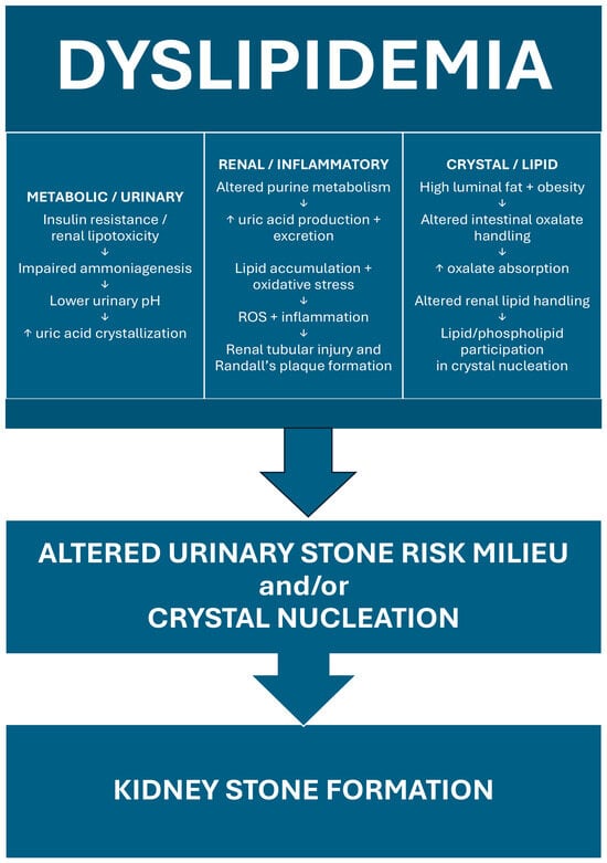
KS forms within a specific milieu when the balance between promoters [15,16] and inhibitors [17,18,19] of crystallization is disrupted and stone forming salts reach supersaturation. KS formation and dyslipidemia are connected via multiple interrelated mechanisms. These include alterations in purine and oxalate metabolism, defective urinary acidification through insulin resistance and renal lipotoxicity, oxidative stress and inflammation, as well as direct lipid participation in crystal nucleation [20] (Figure 1).
Figure 1.
Proposed mechanisms linking dyslipidemia with kidney stone formation; ROS, reactive oxygen species. ↓, leads to; ↑, increased.
3.1. Effects on Purine Metabolism
Dyslipidemia is associated with hyperuricemia [21], which in turn predisposes individuals to uric acid stone formation. Hyperuricemia develops due to an imbalance between hepatic production via xanthine oxidoreductase (XOR) and renal excretion via urate transporter 1 (URAT1), glucose transporter 9 (GLUT9), and adenosine triphosphate (ATP)-binding cassette subfamily G member 2 [22].
Dyslipidemia is linked with increased hepatic uric acid synthesis through several mechanisms. It impairs blood flow and oxygen carrying capacity, leading to tissue hypoxia [23,24]. Under hypoxic conditions, hypoxanthine is released from adipose tissues and serves as a substrate for uric acid production in the liver [25]. In the setting of dyslipidemia, hepatic uptake of hypoxanthine is further enhanced by fatty acid oxidation, followed by the upregulation of XOR via the activation of hypoxia-inducible factor 1α (HIF-1α) [26], ultimately leading to excessive uric acid production. Indeed, non-alcoholic fatty liver disease (NAFLD), which is closely linked to dyslipidemia, is strongly associated with subsequent development of hyperuricemia, with XOR serving as a key mediator of this relationship [21].
The relationship between dyslipidemia and uric acid is also bidirectional: uric acid itself has been associated with increased hepatic lipogenesis through mitochondrial reactive oxygen species (ROS) generation and nicotinamide adenine dinucleotide phosphate (NADPH) oxidase activation, further exacerbating dyslipidemia [27].
On the other hand, insulin resistance associated with dyslipidemia reduces renal excretion of uric acid via increased reabsorption through GLUT9 and URAT1 [28,29], leading to a lower fractional excretion of uric acid. However, the total amount of uric acid in the urine remains high due to the increased excretion load [30]. Furthermore, the resulting low urinary pH (discussed below) will promote uric acid crystallization even at a uric acid level as low as 200 mg/day [31].
3.2. Effects on Oxalate Metabolism
The urinary oxalate level depends on intestinal absorption, hepatic production, and renal excretion of oxalate. Dyslipidemia influences oxalate homeostasis through both obesity-dependent and obesity-independent pathways.
In the intestinal lumen, oxalate absorption occurs via solute carrier family 26 member 3 (SLC26A3), whereas oxalate secretion is mediated by solute carrier family 26 member 6 (SLC26A6) [32]. In the setting of dyslipidemia, higher luminal fat content [33] binds available calcium, reducing the formation of insoluble calcium oxalate complexes and thereby increasing the pool of free, absorbable oxalate [34]. Additionally, obesity, commonly present with dyslipidemia, suppresses SLC26A6-mediated secretion [35] and increases paracellular oxalate absorption due to reduced expression of tight junction proteins [36], further enhancing intestinal oxalate uptake and leading to a net increase in oxalate absorption.
The gut microbiome provides another link between dyslipidemia and oxalate metabolism in an obesity-independent manner. Colonization with Oxalobacter formigenes, an obligate anaerobe that degrades luminal oxalate, has been inversely associated with plasma [37] and urine [38] oxalate concentrations in KS formers. Although a direct causal relationship remains unproven, a complex interplay between the gut microbiota and lipid metabolism has been reported [39].
3.3. Effects on Urine pH
Low urinary pH is a major determinant of uric acid and plays a role in calcium oxalate stone formation. Dyslipidemia is associated with urinary acidification through two interconnected mechanisms: systemic insulin resistance and direct renal lipotoxicity.
Insulin resistance, which is highly prevalent in dyslipidemia, impairs renal ammoniagenesis and ammonium excretion. Under normal circumstances, insulin stimulates glutamine metabolism and promotes ammoniagenesis [40], thereby buffering urinary acid and raising urinary pH [41]. However, under conditions of insulin resistance, elevated free fatty acids compete with glutamine, reducing ammoniagenesis [42,43]. Additionally, impaired insulin response or renal tubular lipid accumulation by itself can directly inhibit sodium-hydrogen exchanger 3 (NHE3) activity and reduce ammonium excretion resulting in a more acidic urinary pH [44,45].
3.4. Oxidation and Inflammation
Dyslipidemia is associated with oxidative stress through multiple pathways, including lipid peroxidation, the generation of oxidized low-density lipoprotein (LDL), and the activation of NADPH oxidase, all of which increase the production of ROS [46] and can lead to lipotoxicity in renal tubular cells, thereby promoting the formation of Randall’s plaque [47,48,49,50]. However, direct evidence linking dyslipidemia to Randall’s plaque formation remains lacking.
3.5. Stone Matrix Composition
Beyond their effects on urinary KS promoters and inhibitors, lipids participate directly in stone matrix formation and crystal nucleation. All kidney stones have been shown to contain lipids, although lipid composition differs by stone type [51]. Furthermore, the organic matrix of calcium-based stones contains significantly more acidic and complexed phospholipids than uric acid or struvite stones [52].
Lipids promote crystal formation primarily through heterogeneous nucleation on tissue surfaces rather than spontaneously in solution [53]. Phospholipids promote crystal formation and phospholipase A2 hydrolyzes of membrane phospholipids to release fatty acids that facilitate crystal nucleation [52], which can subsequently grow into Randall’s plaques, especially in the presence of ROS-mediated tissue injury. In fact, fatty acid binding protein 4 (FABP4), a lipid chaperone expressed in renal papillary tissue that facilitates intracellular fatty acid transport, is downregulated in Randall’s plaques [54]. Notably, FABP4 knockout mice develop urinary crystals, providing direct mechanistic evidence linking impaired renal lipid handling to stone formation.
4. Lipid Phenotype, Urine Components, Stone Composition Connection
Several studies have examined the effects of lipid abnormalities on urinary constituents that contribute to KS formation, with each lipid phenotype demonstrating a distinctive urinary stone risk profile.
4.1. Hypertriglyceridemia
The evidence linking hypertriglyceridemia to KS risk has been most consistent among lipid abnormalities. Both Torricelli et al. [5] and Kang et al. [13] found that hypertriglyceridemia was significantly associated with a higher rate of uric acid stones, a finding that was well supported by the corresponding urinary stone risk profiles.
Torricelli et al. [5] performed a retrospective analysis of 2442 KS formers and found that patients with TG more than 150 mg/dL had significantly higher urinary sodium (179.4 vs. 163.8 mmol/24 h, p < 0.001), oxalate (50.1 vs. 48.0 mg/24 h, p = 0.046) and uric acid (549.3 vs. 501.9 mg/24 h, p < 0.001) levels and lower urinary pH (5.9 vs. 6.1, p < 0.001) than patients with TG less than 150 mg/dL. These associations remained significant after multivariate analysis controlling for age, sex, body mass index (BMI), diabetes, and hypertension.
Kang et al. [13] corroborated these findings in a separate cohort of 321 KS formers, demonstrating that patients with TG more than 150 mg/dL were associated with higher urinary calcium (196.5 ± 113.3 vs. 171 ± 98 mg per 24 h, p = 0.048), sodium (213.7 ± 81.8 vs. 186.9 ± 75.7 mmol per 24 h, p = 0.002), and uric acid (668.5 ± 270.2 vs. 595.1 ± 207.8 mg per 24 h, p = 0.048) levels than patients with TG less than 150 mg/dL. Notably, the association between hypertriglyceridemia and decreased urinary pH was significant only in the subgroup of uric acid stone formers (5.32 ± 0.43 vs. 5.64 ± 0.50, p = 0.013), suggesting that the pH-lowering effect may be more clinically relevant in this population.
4.2. Low HDL Cholesterol
Low HDL has also been associated with an increased risk of KS formation, although its effects on urinary KS risk profiles appeared to be mixed, affecting both inhibitors and promoters of KS formation.
Masterson et al. [12] studied a cohort of 52,184 KS formers and found that low HDL levels (45 mg/dL for men and 60 mg/dL for women) were significantly associated with increased stone risk on multivariate analysis (hazard ratio (HR) 1.3, 95% confidence interval (CI) 1.0–1.6, p = 0.02). However, the study did not report stone compositions. Kang et al. [13] further explored this association, and reported that patients with low HDL (40 mg/dL or less in men and 50 mg/dL or less in women) had an increased risk of uric acid stones. Mechanistically, low HDL levels (40 mg/dL or less in men and 50 mg/dL or less in women) were significantly associated with higher urinary sodium (176.9 vs. 164.4 mmol/24 h, p = 0.002), oxalate (50.3 vs. 47.8 mg/24 h, p = 0.025) and uric acid (540.9 vs. 504.5 mg/24 h, p < 0.001) levels and lower urinary pH (5.95 vs. 6.07, p < 0.001) [5].
4.3. High LDL Cholesterol
Unlike triglycerides and HDL, the relationship between LDL cholesterol and KS risk is paradoxical. Several studies suggested that elevated LDL may be associated with a more favorable urinary KS risk profile and convey a protective effect.
Kang et al. [13] found that KS formers with high LDL had lower urinary sodium (173.0 ± 64.7 vs. 200.5 ± 80.5 mmol per 24 h, p = 0.017) excretion. In their propensity-matched case–control study, Kang et al. [11] further demonstrated that high LDL cholesterolemia was significantly associated with reduced odds of KS formation (odds ratio (OR) = 0.610, p < 0.001). A recent study by Zhang et al. [55] corroborated these findings, showing that small dense LDL cholesterol (sdLDL-C) was inversely associated with KS prevalence in both a Chinese hospital cohort, which included 229 KS formers among 757 participants (OR = 0.53, 95% CI: 0.38–0.74), and a U.S. population-based cohort from NHANES, which included 882 KS formers among 9721 participants (OR = 0.68, 95% CI: 0.50–0.91).
However, not all studies have shown consistent results. Masterson et al. [12] found no association between LDL and stone risk in their large cohort of 52,184 participants, and Huang et al. [56], in a prospective case–control study of 114 patients, found that higher LDL (more than 100 mg/dL) was associated with significantly lower urinary citrate levels, indicating a higher KS risk. Similarly, when examining non-HDL cholesterol, which includes LDL cholesterol, Torricelli et al. [5] found that high non-HDL (more than 130 mg/dL) levels were significantly associated with higher urinary sodium (173.9 vs. 162.8 mmol/24 h, p < 0.001) excretions compared with patients with normal non-HDL levels.
These seemingly discordant findings may reflect the heterogeneity of LDL actions in vivo. The protective urinary profile associated with high LDL may be counterbalanced by its pro-oxidative and pro-inflammatory effects at the tissue level. The net effect of LDL on stone formation likely depends on the balance between these competing mechanisms.
4.4. High Total Cholesterol (TC)
The relationship between TC and KS risk is complex, with studies showing seemingly contradictory effects on risk of KS formation and urinary risk profile.
Kang et al. [11], in a propensity-matched case–control study, found that KS formers had lower TC levels than non-KS formers and that hypercholesterolemia was associated with reduced odds of KS (OR = 0.747, p = 0.003). In contrast, Torricelli et al. [5] found a strong association between elevated TC levels and a higher risk of uric acid stones (p = 0.006). Similarly, Besiroglu et al. [3] reported an association between elevated TC levels and KS risk in a meta-analysis.
Similarly, studies on urinary stone risk profiles have also been inconsistent. For example, Torricelli et al. [5] noted that a high TC (greater than 200 mg/dL) was associated with a significantly elevated urinary calcium level (245.6 vs. 199.0 mg per day, p = 0.036), suggesting increased risk. However, they also observed significantly higher urinary potassium (74.0 vs. 61.4 mg per day, p = 0.025) levels, which may indicate a protective effect. Huang et al. [56] reported that high TC was associated with low urinary citrate and that urinary citrate levels were inversely associated with calcium stone formation (calcium oxalate stone OR 0.970, 95% CI: 0.951–0.990, p = 0.003 and calcium phosphate stone OR 0.970, 95%CI: 0.953–0.987, p = 0.001), further supporting an increased risk of KS formation. In contrast, Kang et al. [13] did not find any associations between TC and urinary risk parameters in their cohort involving 321 KS formers.
Like LDL, these seemingly discordant findings may reflect the heterogeneity of total cholesterol as a biomarker. Total cholesterol encompasses both LDL and non-LDL fractions and can affect both promoters and inhibitors of KS formation. Thus, its net effect on stone risk likely depends on the relative contribution of each component and the overall balance between these opposing factors.
5. Lipid-Lowering Therapy and Potential Effects on Stone Risk
While dietary modifications remain the cornerstone for management of KS disease and dyslipidemia [57], lipid-lowering therapy should be used according to established indications for dyslipidemia and cardiovascular risk. Whether these therapies provide a secondary benefit by reducing KS risk remains uncertain.
5.1. Statins
Statins can alter the urinary stone milieu. Liu et al. [58] prospectively compared 78 male KS formers with 30 controls and classified them by American College of Cardiology/American Heart Association guidelines into high- or low-risk CVD. They noted that administrating atorvastatin 20 mg daily for 12 weeks among high-risk participants with KS significantly increased urine citrate (p < 0.001), reduced urinary uric acid (p < 0.001), and raised urine pH from 5.8 ± 0.2 to 6.4 ± 0.5 (p < 0.001). However, there were also increases in urinary calcium (p = 0.053) and oxalate levels (p = 0.037), which ultimately led to a neutral effect on the supersaturations of calcium oxalate and calcium phosphate. This study was limited by a lack of stone composition analysis, although the majority of stones were assumed to be calcium-based. These findings are mechanistically compatible with the proposed effects of dyslipidemia on urinary acidification and uric acid handling; however, whether these changes translate into reduced stone formation or recurrence remains uncertain.
Several retrospective studies have demonstrated an association between statin use and reduced KS risk. Sur et al. [59] noted a protective effect of statins (OR 0.51, 95% CI: 0.46–0.57, p < 0.001) among 57,232 KS formers with dyslipidemia over a 10 year period without effect modification by gender. Cohen et al. [60] subsequently confirmed these findings in their large cohort involving 101,259 KS formers and found a significantly lower risk of KS formation (OR 0.57, 95% CI: 0.53–0.60, p < 0.01) among statin users.
So far, no randomized controlled trial (RCT) has been conducted to evaluate statins specifically for KS prevention. The existing evidence is limited by their observational design and the inability to determine whether the protective effect of statins is mediated by their lipid-lowering effect, pleiotropic effects, or both. Prospective trials are needed to determine whether statins have a clinically meaningful effect on KS risk.
5.2. Non-Statin Lipid-Lowering Agents
Evidence for other lipid-lowering agents in the context of nephrolithiasis is limited. Fibrates effectively lower triglycerides and raise HDL, but no studies have directly evaluated their effect on stone formation. Fibrates are also not recommended for CVD risk reduction in patients with CKD by the Kidney Disease: Improving Global Outcomes (KDIGO) guidelines [61], limiting their use in clinical practice.
5.3. Omega-3 Fatty Acids
Omega-3 fatty acids have yielded mixed results with regard to KS risk. In animal models, fish oil supplementation significantly reduced urinary calcium and oxalate excretions [62]. This was subsequently confirmed in humans by Yasui et al. [63], who noted that supplementation with 1800 mg/day of eicosapentaenoic acid in 88 calcium KS formers over 3 months led to a significant reduction in urinary calcium excretion among those with hypercalciuria (332 ± 86 mg to 275 ± 132 mg, p < 0.005), whereas no significant changes were observed in the normocalciuric subgroup (defined as urine calcium < 250 mg/day). Subsequently, Ortiz-Alvarado et al. [64] reported significant reductions in urinary calcium excretion (329 to 247 mg/day, p < 0.0001), oxalate excretion (45.4 to 32.9 mg/day, p = 0.0004), and calcium oxalate supersaturation (9.73 to 3.68, p = 0.001) in a retrospective cohort of 29 hypercalciuric KS formers supplemented with 1200 mg/day of fish oil. They also observed a significant improvement in urinary citrate levels (732 to 940 mg/day, p = 0.0005). However, this study may be confounded by the concurrent dietary interventions, making it difficult to determine the independent effect of fish oil supplementation. The observed reductions in urinary calcium and oxalate are potentially relevant to the urinary risk pathways described above, particularly in patients with hypercalciuria, although available studies have not established a reduction in stone recurrence. Rothwell et al. [65], in their prospective clinical trial, demonstrated a significant reduction in urinary calcium levels (12.11 ± 0.96 mmol/day to 9.76 ± 0.71 mmol/day) among 18 hypercalciuric KS formers treated with 5 g daily of fish oil for 4 weeks. However, changes in stone burden were not reported.
In contrast, other studies have reported conflicting findings. Konya et al. [66] found that supplementation with 1800 mg/day of ethyl icosapentate for 6 months in 40 calcium KS formers did not reduce urinary calcium excretion; among the 25 patients with hypercalciuria, only nine responded and achieved normocalciuria. Similarly, a large prospective cohort analysis by Taylor et al. [67] involving 3956 KS formers from the Health Professionals Follow-Up Study and Nurses’ Health Studies failed to show a consistent association between dietary eicosapentaenoic acid/docosahexaenoic acid intake and incident KS risk over 36 years of follow-up.
Thus far, no RCT has evaluated omega-3 fatty acids using stone recurrence as a primary endpoint. The existing evidence is limited to small, uncontrolled studies examining surrogate urinary chemistry outcomes.
5.4. Ezetimibe
Ezetimibe reduces intestinal cholesterol absorption and has been shown to reduce 8-hydroxydeoxyguanosine, a marker of oxidative stress in the urine [68]. It also reduces the renal fat content among type 2 diabetic patients with early CKD and high renal parenchymal fat [69]. Given the proposed roles of renal lipotoxicity and oxidative stress in stone pathogenesis, these effects provide a mechanistic rationale for investigating whether ezetimibe influences stone risk; however, no clinical evidence currently supports this hypothesis, and further studies are needed to clarify its validity.
5.5. Bempedoic Acid
Bempedoic acid has been associated with hyperuricemia due to reduced renal excretion of uric acid via organic anion transporter 2 inhibition [70,71], but its effect on KS is not clear at this time.
5.6. Proprotein Convertase Subtilisin/Kexin Type 9 (PCSK9) Inhibitors
The effect of PCSK9 inhibitors on KS risk has not been studied extensively. In the Mendelian randomization study by Tan et al. [4], genetically proxied PCSK9 inhibition showed no significant association with urinary stone risk. Given that PCSK9 inhibitors primarily lower LDL cholesterol, whose association with KS risk remains inconsistent across existing studies, a meaningful stone-protective effect appears unlikely.
6. Clinical Implications
Metabolic Workup of Stone Formers: Integrating Lipid Assessment
The American Urological Association (AUA) guideline recommends an initial KS evaluation consisting of a detailed medical and dietary history, serum chemistries, and urinalysis, with 24 h urine testing reserved for a more comprehensive assessment in recurrent KS formers [7]. Additional metabolic testing is recommended for high-risk or recurrent stone formers. The 2025 European Association of Urology (EAU) guidelines similarly emphasize individualized management based on biochemical evaluation and stone composition [72]. Studies have further highlighted the importance of stone composition in clinical evaluation, including the potential use of non-contrast computed tomography characteristics to predict stone composition [73]. Similarly, clinical data have emphasized the role of stone analysis and metabolic evaluation in guiding subsequent kidney stone formation [74].
KS disease is now recognized as a complex systemic disorder that is strongly associated with metabolic syndrome. Due to the shared risk factors, the presence of KS disease itself can suggest an underlying ongoing silent atherosclerotic process, and the evaluation of cardio-kidney-metabolic risk factors, such as dyslipidemia, can be beneficial [75,76]. Given the growing evidence linking dyslipidemia to KS risk, lipid assessment may be clinically relevant in selected recurrent stone formers, particularly those with obesity, insulin resistance, metabolic syndrome, diabetes, or other features of cardiometabolic risk. However, routine lipid panel testing is not currently recommended as part of the stone-specific metabolic evaluation. In such patients, lipid testing may be considered as part of broader cardiovascular and metabolic risk assessment rather than as a routine test for KS disease itself. Whether identification and treatment of dyslipidemia can reduce stone recurrence remains uncertain and requires prospective clinical evaluation.
7. Summary
Dyslipidemia has been associated with an increased risk for KS formation and recurrence. Proposed mechanistic links include alterations in purine and oxalate metabolism, impaired urinary acidification secondary to insulin resistance, renal lipotoxicity, oxidative stress, crystal-induced inflammation, and the direct involvement of lipids in crystal nucleation. Despite the growing body of evidence linking dyslipidemia to KS formation and suggesting potential stone-protective effect of lipid-lowering therapies, critical knowledge gaps remain that limit the translation of these findings into clinical practice. Future mechanistic studies and large-scale clinical trials are needed to establish a causal relationship between dyslipidemia and KS disease and to determine whether lipid-lowering interventions can reduce stone recurrence. Further clinical studies are needed to determine whether lipid assessment or lipid-lowering therapy has a role in the prevention of recurrent KS disease.
Author Contributions
Conceptualization, S.S. and J.T.; data curation, S.S. and J.T.; writing—original draft preparation, S.S. and J.T.; writing—review and editing, S.S. and J.T. All authors have read and agreed to the published version of the manuscript.
Funding
Category 3 Brown Physicians Incorporated Academic Assessment Grant 8058418 (J.T.).
Institutional Review Board Statement
Not applicable.
Informed Consent Statement
Not applicable.
Data Availability Statement
No new data were created or analyzed in this study. Data sharing is not applicable to this article.
Conflicts of Interest
The authors declare no conflicts of interest.
References
- Chewcharat, A.; Curhan, G. Trends in the prevalence of kidney stones in the United States from 2007 to 2016. Urolithiasis 2021, 49, 27–39. [Google Scholar] [CrossRef] [Scilit] [PubMed]
- Bargagli, M.; Scoglio, M.; Howles, S.A.; Fuster, D.G. Kidney stone disease: Risk factors, pathophysiology and management. Nat. Rev. Nephrol. 2025, 21, 794–808. [Google Scholar] [CrossRef] [Scilit] [PubMed]
- Besiroglu, H.; Ozbek, E. Association between blood lipid profile and urolithiasis: A systematic review and meta-analysis of observational studies. Int. J. Urol. 2019, 26, 7–17. [Google Scholar] [CrossRef] [Scilit] [PubMed]
- Tan, Z.; Hong, J.; Sun, A.; Ding, M.; Shen, J. Causal effects of circulating lipids and lipid-lowering drugs on the risk of urinary stones: A Mendelian randomization study. Front. Endocrinol. 2023, 14, 1301163. [Google Scholar] [CrossRef] [Scilit] [PubMed]
- Torricelli, F.C.; De, S.K.; Gebreselassie, S.; Li, I.; Sarkissian, C.; Monga, M. Dyslipidemia and kidney stone risk. J. Urol. 2014, 191, 667–672. [Google Scholar] [CrossRef] [Scilit] [PubMed]
- Akram, M.; Jahrreiss, V.; Skolarikos, A.; Geraghty, R.; Tzelves, L.; Emilliani, E.; Davis, N.F.; Somani, B.K. Urological Guidelines for Kidney Stones: Overview and Comprehensive Update. J. Clin. Med. 2024, 13, 1114. [Google Scholar] [CrossRef] [Scilit] [PubMed]
- Pearle, M.S.; Goldfarb, D.S.; Assimos, D.G.; Curhan, G.; Denu-Ciocca, C.J.; Matlaga, B.R.; Monga, M.; Penniston, K.L.; Preminger, G.M.; Turk, T.M.; et al. Medical management of kidney stones: AUA guideline. J. Urol. 2014, 192, 316–324. [Google Scholar] [CrossRef] [Scilit] [PubMed]
- Scales, C.D., Jr.; Smith, A.C.; Hanley, J.M.; Saigal, C.S. Prevalence of kidney stones in the United States. Eur. Urol. 2012, 62, 160–165. [Google Scholar] [CrossRef] [Scilit] [PubMed]
- Shastri, S.; Patel, J.; Sambandam, K.K.; Lederer, E.D. Kidney Stone Pathophysiology, Evaluation and Management: Core Curriculum 2023. Am. J. Kidney Dis. 2023, 82, 617–634. [Google Scholar] [CrossRef] [Scilit] [PubMed]
- Kohjimoto, Y.; Sasaki, Y.; Iguchi, M.; Matsumura, N.; Inagaki, T.; Hara, I. Association of metabolic syndrome traits and severity of kidney stones: Results from a nationwide survey on urolithiasis in Japan. Am. J. Kidney Dis. 2013, 61, 923–929. [Google Scholar] [CrossRef] [Scilit] [PubMed]
- Kang, H.W.; Lee, S.K.; Kim, W.T.; Kim, Y.J.; Yun, S.J.; Lee, S.C.; Kim, W.J. Hypertriglyceridemia and low high-density lipoprotein cholesterolemia are associated with increased hazard for urolithiasis. J. Endourol. 2014, 28, 1001–1005. [Google Scholar] [CrossRef] [Scilit] [PubMed]
- Masterson, J.H.; Woo, J.R.; Chang, D.C.; Chi, T.; L’Esperance, J.O.; Stoller, M.L.; Sur, R.L. Dyslipidemia is associated with an increased risk of nephrolithiasis. Urolithiasis 2015, 43, 49–53. [Google Scholar] [CrossRef] [Scilit] [PubMed]
- Kang, H.W.; Seo, S.P.; Kim, W.T.; Kim, Y.J.; Yun, S.J.; Lee, S.C.; Kim, W.J. Hypertriglyceridemia is associated with increased risk for stone recurrence in patients with urolithiasis. Urology 2014, 84, 766–771. [Google Scholar] [CrossRef] [Scilit] [PubMed]
- Gao, M.; Liu, M.; Zhu, Z.; Chen, H. The association of dyslipidemia with kidney stone: Result from the NHANES 2007-2020. Int. Urol. Nephrol. 2024, 56, 35–44. [Google Scholar] [CrossRef] [Scilit] [PubMed]
- Tang, J.; McFann, K.; Chonchol, M. Dietary zinc intake and kidney stone formation: Evaluation of NHANES III. Am. J. Nephrol. 2012, 36, 549–553. [Google Scholar] [CrossRef] [Scilit] [PubMed]
- Ticinesi, A.; Nouvenne, A.; Maalouf, N.M.; Borghi, L.; Meschi, T. Salt and nephrolithiasis. Nephrol. Dial. Transplant. 2016, 31, 39–45. [Google Scholar] [CrossRef] [Scilit] [PubMed]
- Shringi, S.; Raker, C.A.; Tang, J. Dietary Magnesium Intake and Kidney Stone: The National Health and Nutrition Examination Survey 2011–2018. R. I. Med. J. 2023, 106, 20–25. [Google Scholar]
- Moe, O.W. Kidney stones: Pathophysiology and medical management. Lancet 2006, 367, 333–344. [Google Scholar] [CrossRef] [Scilit] [PubMed]
- Ferraro, P.M.; Mandel, E.I.; Curhan, G.C.; Gambaro, G.; Taylor, E.N. Dietary Protein and Potassium, Diet-Dependent Net Acid Load, and Risk of Incident Kidney Stones. Clin. J. Am. Soc. Nephrol. 2016, 11, 1834–1844. [Google Scholar] [CrossRef] [Scilit] [PubMed]
- Wen, Q.; Li, Q.; Zhou, H.; Su, B.; Ren, Y.; He, Q. Dysregulated lipid metabolism in kidney stone pathogenesis: A literature review of mechanistic insights and therapeutic implications. BMC Urol. 2025, 25, 203. [Google Scholar] [CrossRef] [Scilit] [PubMed]
- Xu, C.; Wan, X.; Xu, L.; Weng, H.; Yan, M.; Miao, M.; Sun, Y.; Xu, G.; Dooley, S.; Li, Y.; et al. Xanthine oxidase in non-alcoholic fatty liver disease and hyperuricemia: One stone hits two birds. J. Hepatol. 2015, 62, 1412–1419. [Google Scholar] [CrossRef] [Scilit] [PubMed]
- Furuhashi, M. New insights into purine metabolism in metabolic diseases: Role of xanthine oxidoreductase activity. Am. J. Physiol. Endocrinol. Metab. 2020, 319, E827–E834. [Google Scholar] [CrossRef] [Scilit] [PubMed]
- Radosinska, J.; Vrbjar, N. The role of red blood cell deformability and Na,K-ATPase function in selected risk factors of cardiovascular diseases in humans: Focus on hypertension, diabetes mellitus and hypercholesterolemia. Physiol. Res. 2016, 65, S43–S54. [Google Scholar] [CrossRef] [Scilit] [PubMed]
- Juhász, J. Dyslipidemia: Another brick in the wall. A feasible link in the OSA-cardiovascular disease axis. Sleep. Breath. 2014, 18, 5–6. [Google Scholar] [CrossRef] [Scilit] [PubMed]
- Nagao, H.; Nishizawa, H.; Tanaka, Y.; Fukata, T.; Mizushima, T.; Furuno, M.; Bamba, T.; Tsushima, Y.; Fujishima, Y.; Kita, S.; et al. Hypoxanthine Secretion from Human Adipose Tissue and its Increase in Hypoxia. Obesity 2018, 26, 1168–1178. [Google Scholar] [CrossRef] [Scilit] [PubMed]
- Liang, N.; Yuan, X.; Zhang, L.; Shen, X.; Zhong, S.; Li, L.; Li, R.; Xu, X.; Chen, X.; Yin, C.; et al. Fatty acid oxidation-induced HIF-1α activation facilitates hepatic urate synthesis through upregulating NT5C2 and XDH. Life Metab. 2024, 3, loae018. [Google Scholar] [CrossRef] [Scilit] [PubMed]
- Kushiyama, A.; Nakatsu, Y.; Matsunaga, Y.; Yamamotoya, T.; Mori, K.; Ueda, K.; Inoue, Y.; Sakoda, H.; Fujishiro, M.; Ono, H.; et al. Role of Uric Acid Metabolism-Related Inflammation in the Pathogenesis of Metabolic Syndrome Components Such as Atherosclerosis and Nonalcoholic Steatohepatitis. Mediat. Inflamm. 2016, 2016, 8603164. [Google Scholar] [CrossRef] [Scilit] [PubMed]
- Mandal, A.K.; Leask, M.P.; Estiverne, C.; Choi, H.K.; Merriman, T.R.; Mount, D.B. Genetic and Physiological Effects of Insulin on Human Urate Homeostasis. Front. Physiol. 2021, 12, 713710. [Google Scholar] [CrossRef] [Scilit] [PubMed]
- Fujii, W.; Yamazaki, O.; Hirohama, D.; Kaseda, K.; Kuribayashi-Okuma, E.; Tsuji, M.; Hosoyamada, M.; Kochi, Y.; Shibata, S. Gene-environment interaction modifies the association between hyperinsulinemia and serum urate levels through SLC22A12. J. Clin. Investig. 2025, 135, e186633. [Google Scholar] [CrossRef] [Scilit] [PubMed]
- Jalal, L.; Jaffery, Z.; Nasir, A.; Shoukat, W.; Khalid, A.; Irfan, A.; Oduoye, M.O.; Ozoemena, D.J.; Adesoye, I.M.; Oyeniyi, A.A. Exploring the pathophysiological link between obesity and kidney stone formation: Cost-effective dietary and lifestyle strategies for prevention. Ann. Med. Surg. 2025, 87, 6521–6527. [Google Scholar] [CrossRef] [Scilit] [PubMed]
- Bell, D.S. Beware the low urine pH--the major cause of the increased prevalence of nephrolithiasis in the patient with type 2 diabetes. Diabetes Obes. Metab. 2012, 14, 299–303. [Google Scholar] [CrossRef] [Scilit] [PubMed]
- Wang, J.; Wang, W.; Wang, H.; Tuo, B. Physiological and Pathological Functions of SLC26A6. Front. Med. 2020, 7, 618256. [Google Scholar] [CrossRef] [Scilit] [PubMed]
- Ahlberg, J.; Angelin, B.; Einarsson, K.; Hellström, K.; Leijd, B. Biliary lipid composition in normo- and hyperlipoproteinemia. Gastroenterology 1980, 79, 90–94. [Google Scholar] [CrossRef] [Scilit]
- Witting, C.; Langman, C.B.; Assimos, D.; Baum, M.A.; Kausz, A.; Milliner, D.; Tasian, G.; Worcester, E.; Allain, M.; West, M.; et al. Pathophysiology and Treatment of Enteric Hyperoxaluria. Clin. J. Am. Soc. Nephrol. 2021, 16, 487–495. [Google Scholar] [CrossRef] [Scilit] [PubMed]
- Amin, R.; Asplin, J.; Jung, D.; Bashir, M.; Alshaikh, A.; Ratakonda, S.; Sharma, S.; Jeon, S.; Granja, I.; Matern, D.; et al. Reduced active transcellular intestinal oxalate secretion contributes to the pathogenesis of obesity-associated hyperoxaluria. Kidney Int. 2018, 93, 1098–1107. [Google Scholar] [CrossRef] [Scilit] [PubMed]
- Bashir, M.; Meddings, J.; Alshaikh, A.; Jung, D.; Le, K.; Amin, R.; Ratakonda, S.; Sharma, S.; Granja, I.; Satti, M.; et al. Enhanced gastrointestinal passive paracellular permeability contributes to the obesity-associated hyperoxaluria. Am. J. Physiol. Gastrointest. Liver Physiol. 2019, 316, G1–G14. [Google Scholar] [CrossRef] [Scilit] [PubMed]
- Siener, R.; Bangen, U.; Sidhu, H.; Hönow, R.; von Unruh, G.; Hesse, A. The role of Oxalobacter formigenes colonization in calcium oxalate stone disease. Kidney Int. 2013, 83, 1144–1149. [Google Scholar] [CrossRef] [Scilit] [PubMed]
- Zhang, M.; Fan, L.; Li, J. Prediction of calcium oxalate kidney stones: A comprehensive analysis of clinical and gut microbiome characteristics. Medicine 2025, 104, e43103. [Google Scholar] [CrossRef] [Scilit] [PubMed]
- Guo, L.; Wang, Y.Y.; Wang, J.H.; Zhao, H.P.; Yu, Y.; Wang, G.D.; Dai, K.; Yan, Y.Z.; Yang, Y.J.; Lv, J. Associations of gut microbiota with dyslipidemia based on sex differences in subjects from Northwestern China. World J. Gastroenterol. 2022, 28, 3455–3475. [Google Scholar] [CrossRef] [Scilit] [PubMed]
- Nissim, I.; States, B.; Nissim, I.; Lin, Z.P.; Yudkoff, M. Hormonal regulation of glutamine metabolism by OK cells. Kidney Int. 1995, 47, 96–105. [Google Scholar] [CrossRef] [Scilit] [PubMed]
- Abate, N.; Chandalia, M.; Cabo-Chan, A.V., Jr.; Moe, O.W.; Sakhaee, K. The metabolic syndrome and uric acid nephrolithiasis: Novel features of renal manifestation of insulin resistance. Kidney Int. 2004, 65, 386–392. [Google Scholar] [CrossRef] [Scilit] [PubMed]
- Maalouf, N.M.; Cameron, M.A.; Moe, O.W.; Sakhaee, K. Novel insights into the pathogenesis of uric acid nephrolithiasis. Curr. Opin. Nephrol. Hypertens. 2004, 13, 181–189. [Google Scholar] [CrossRef] [Scilit] [PubMed]
- Maalouf, N.M.; Cameron, M.A.; Moe, O.W.; Sakhaee, K. Metabolic basis for low urine pH in type 2 diabetes. Clin. J. Am. Soc. Nephrol. 2010, 5, 1277–1281. [Google Scholar] [CrossRef] [Scilit] [PubMed]
- Bobulescu, I.A.; Dubree, M.; Zhang, J.; McLeroy, P.; Moe, O.W. Effect of renal lipid accumulation on proximal tubule Na+/H+ exchange and ammonium secretion. Am. J. Physiol.-Ren. Physiol. 2008, 294, F1315–F1322. [Google Scholar] [CrossRef] [Scilit] [PubMed]
- Bobulescu, I.A.; Dubree, M.; Zhang, J.; McLeroy, P.; Moe, O.W. Reduction of renal triglyceride accumulation: Effects on proximal tubule Na+/H+ exchange and urinary acidification. Am. J. Physiol. Ren. Physiol. 2009, 297, F1419–F1426. [Google Scholar] [CrossRef] [Scilit] [PubMed]
- Singh, R.; Devi, S.; Gollen, R. Role of free radical in atherosclerosis, diabetes and dyslipidaemia: Larger-than-life. Diabetes Metab. Res. Rev. 2015, 31, 113–126. [Google Scholar] [CrossRef] [Scilit] [PubMed]
- Ying, X.; Chen, Y.; Hao, Z.; Liu, H. The significance of reactive oxygen species in the formation of calcium oxalate stones and the protective effects of antioxidants on the kidneys. Front. Immunol. 2025, 16, 1540075. [Google Scholar] [CrossRef] [Scilit] [PubMed]
- Khan, S.R. Reactive oxygen species as the molecular modulators of calcium oxalate kidney stone formation: Evidence from clinical and experimental investigations. J. Urol. 2013, 189, 803–811. [Google Scholar] [CrossRef] [Scilit] [PubMed]
- Liu, Y.; Sun, Y.; Kang, J.; He, Z.; Liu, Q.; Wu, J.; Li, D.; Wang, X.; Tao, Z.; Guan, X.; et al. Role of ROS-Induced NLRP3 Inflammasome Activation in the Formation of Calcium Oxalate Nephrolithiasis. Front. Immunol. 2022, 13, 818625. [Google Scholar] [CrossRef] [Scilit] [PubMed]
- Khan, S.R.; Canales, B.K.; Dominguez-Gutierrez, P.R. Randall’s plaque and calcium oxalate stone formation: Role for immunity and inflammation. Nat. Rev. Nephrol. 2021, 17, 417–433. [Google Scholar] [CrossRef] [Scilit] [PubMed]
- Khan, S.R.; Shevock, P.N.; Hackett, R.L. Presence of lipids in urinary stones: Results of preliminary studies. Calcif. Tissue Int. 1988, 42, 91–96. [Google Scholar] [CrossRef] [Scilit] [PubMed]
- Khan, S.R.; Glenton, P.A.; Backov, R.; Talham, D.R. Presence of lipids in urine, crystals and stones: Implications for the formation of kidney stones. Kidney Int. 2002, 62, 2062–2072. [Google Scholar] [CrossRef] [Scilit] [PubMed]
- Fasano, J.M.; Khan, S.R. Intratubular crystallization of calcium oxalate in the presence of membrane vesicles: An in vitro study. Kidney Int. 2001, 59, 169–178. [Google Scholar] [CrossRef] [Scilit] [PubMed]
- Taguchi, K.; Chen, L.; Usawachintachit, M.; Hamamoto, S.; Kang, M.; Sugino, T.; Unno, R.; Tzou, D.T.; Sherer, B.A.; Okada, A.; et al. Fatty acid-binding protein 4 downregulation drives calcification in the development of kidney stone disease. Kidney Int. 2020, 97, 1042–1056. [Google Scholar] [CrossRef] [Scilit] [PubMed]
- Zhang, Y.; Qin, H.; Yang, J.; Shen, X.; Hao, Z.; Liang, C. Serum small dense LDL cholesterol is inversely associated with kidney stone prevalence: Evidence from two independent cross-sectional studies. Front. Endocrinol. 2026, 17, 1816344. [Google Scholar] [CrossRef] [Scilit] [PubMed]
- Huang, H.S.; Liao, P.C.; Liu, C.J. Calcium Kidney Stones are Associated with Increased Risk of Carotid Atherosclerosis: The Link between Urinary Stone Risks, Carotid Intima-Media Thickness, and Oxidative Stress Markers. J. Clin. Med. 2020, 9, 729. [Google Scholar] [CrossRef] [Scilit] [PubMed]
- Lin, B.B.; Lin, M.E.; Huang, R.H.; Hong, Y.K.; Lin, B.L.; He, X.J. Dietary and lifestyle factors for primary prevention of nephrolithiasis: A systematic review and meta-analysis. BMC Nephrol. 2020, 21, 267. [Google Scholar] [CrossRef] [Scilit] [PubMed]
- Liu, C.J.; Huang, H.S. Statins significantly alter urinary stone-related urine biochemistry in calcium kidney stone patients with dyslipidemia. Int. J. Urol. 2020, 27, 839–844. [Google Scholar] [CrossRef] [Scilit] [PubMed]
- Sur, R.L.; Masterson, J.H.; Palazzi, K.L.; L’Esperance, J.O.; Auge, B.K.; Chang, D.C.; Stoller, M.L. Impact of statins on nephrolithiasis in hyperlipidemic patients: A 10-year review of an equal access health care system. Clin. Nephrol. 2013, 79, 351–355. [Google Scholar] [CrossRef] [Scilit] [PubMed]
- Cohen, A.J.; Adamsky, M.A.; Nottingham, C.U.; Pruitt, J.; Lapin, B.; Wang, C.H.; Park, S. Impact of Statin Intake on Kidney Stone Formation. Urology 2019, 124, 57–61. [Google Scholar] [CrossRef] [Scilit] [PubMed]
- KDIGO. KDIGO Clinical Practice Guidelines for Lipid Management in CKD. Kidney Int. Suppl. 2013, 3, 259–305. [Google Scholar] [CrossRef] [Scilit]
- Buck, A.C.; Davies, R.L.; Harrison, T. The protective role of eicosapentaenoic acid [EPA] in the pathogenesis of nephrolithiasis. J. Urol. 1991, 146, 188–194. [Google Scholar] [CrossRef] [Scilit] [PubMed]
- Yasui, T.; Tanaka, H.; Fujita, K.; Iguchi, M.; Kohri, K. Effects of eicosapentaenoic acid on urinary calcium excretion in calcium stone formers. Eur. Urol. 2001, 39, 580–585. [Google Scholar] [CrossRef] [Scilit] [PubMed]
- Ortiz-Alvarado, O.; Miyaoka, R.; Kriedberg, C.; Leavitt, D.A.; Moeding, A.; Stessman, M.; Monga, M. Omega-3 fatty acids eicosapentaenoic acid and docosahexaenoic acid in the management of hypercalciuric stone formers. Urology 2012, 79, 282–286. [Google Scholar] [CrossRef] [Scilit] [PubMed]
- Rothwell, P.J.; Green, R.; Blacklock, N.J.; Kavanagh, J.P. Does fish oil benefit stone formers? J. Urol. 1993, 150, 1391–1394. [Google Scholar] [CrossRef] [Scilit] [PubMed]
- Konya, E.; Tsuji, H.; Umekawa, T.; Kurita, T.; Iguchi, M. Effect of ethyl icosapentate on urinary calcium and oxalate excretion. Int. J. Urol. 2000, 7, 361–365. [Google Scholar] [CrossRef] [Scilit] [PubMed]
- Taylor, E.N.; Stampfer, M.J.; Curhan, G.C. Fatty acid intake and incident nephrolithiasis. Am. J. Kidney Dis. 2005, 45, 267–274. [Google Scholar] [CrossRef] [Scilit] [PubMed]
- Nakamura, T.; Sato, E.; Fujiwara, N.; Kawagoe, Y.; Ueda, Y.; Suzuki, T.; Ueda, S.; Adachi, H.; Okuda, S.; Yamagishi, S. Ezetimibe decreases serum levels of asymmetric dimethylarginine (ADMA) and ameliorates renal injury in non-diabetic chronic kidney disease patients in a cholesterol-independent manner. Pharmacol. Res. 2009, 60, 525–528. [Google Scholar] [CrossRef] [Scilit] [PubMed]
- Heinrich, N.S.; Pedersen, R.P.; Vestergaard, M.B.; Lindberg, U.; Andersen, U.B.; Haddock, B.; Hansen, T.W.; Fornoni, A.; Larsson, H.B.W.; Rossing, P. Evaluation of the effects of ezetimibe on albuminuria and kidney fat in individuals with type 2 diabetes and chronic kidney disease. Diabetes Obes. Metab. 2023, 25, 2605–2615. [Google Scholar] [CrossRef] [Scilit] [PubMed]
- Cicero, A.F.G.; Pontremoli, R.; Fogacci, F.; Viazzi, F.; Borghi, C. Effect of Bempedoic Acid on Serum Uric Acid and Related Outcomes: A Systematic Review and Meta-analysis of the available Phase 2 and Phase 3 Clinical Studies. Drug Saf. 2020, 43, 727–736. [Google Scholar] [CrossRef] [Scilit] [PubMed]
- Nissen, S.E.; Lincoff, A.M.; Brennan, D.; Ray, K.K.; Mason, D.; Kastelein, J.J.P.; Thompson, P.D.; Libby, P.; Cho, L.; Plutzky, J.; et al. Bempedoic Acid and Cardiovascular Outcomes in Statin-Intolerant Patients. N. Engl. J. Med. 2023, 388, 1353–1364. [Google Scholar] [CrossRef] [Scilit] [PubMed]
- Skolarikos, A.; Geraghty, R.; Somani, B.; Tailly, T.; Jung, H.; Neisius, A.; Petřík, A.; Kamphuis, G.M.; Davis, N.; Bezuidenhout, C.; et al. European Association of Urology Guidelines on the Diagnosis and Treatment of Urolithiasis. Eur. Urol. 2025, 88, 64–75. [Google Scholar] [CrossRef] [Scilit] [PubMed]
- Doğan, Ç.; Şahin, M.F.; Sencer Köroğlu, M.; Orbeği, O.; Tuncer, F.B.; Yazıcı, C.M. Preoperative Prediction of Stone Composition Using Hounsfield Units in Non-Contrast CT Imaging: A Single-Center Study from Turkey. Nam. Kem. Med. J. 2025, 13, 384–388. [Google Scholar] [CrossRef] [Scilit]
- Şahin, M.F.; Yazici, C.M.; Özcan, R.; Doğan, Ç.; Akgül, M. The Significance of Stone Analysis, Metabolic Evaluation and Their Effect on Metaphylaxis: The Results from Tekirdağ Province. Nam. Kem. Med. J. 2025, 13, 100–107. [Google Scholar] [CrossRef] [Scilit]
- Gambaro, A.; Lombardi, G.; Caletti, C.; Ribichini, F.L.; Ferraro, P.M.; Gambaro, G. Nephrolithiasis: A Red Flag for Cardiovascular Risk. J. Clin. Med. 2022, 11, 5512. [Google Scholar] [CrossRef] [Scilit] [PubMed]
- Devarajan, A. Cross-talk between renal lithogenesis and atherosclerosis: An unveiled link between kidney stone formation and cardiovascular diseases. Clin. Sci. 2018, 132, 615–626. [Google Scholar] [CrossRef] [Scilit] [PubMed]
Disclaimer/Publisher’s Note: The statements, opinions and data contained in all publications are solely those of the individual author(s) and contributor(s) and not of MDPI and/or the editor(s). MDPI and/or the editor(s) disclaim responsibility for any injury to people or property resulting from any ideas, methods, instructions or products referred to in the content. |
© 2026 by the authors. Licensee MDPI, Basel, Switzerland. This article is an open access article distributed under the terms and conditions of the Creative Commons Attribution (CC BY) license.