


Abstract
Liver fibrosis is the common pathway through which chronic liver injury progresses to cirrhosis, portal hypertension, liver failure, hepatocellular carcinoma, and systemic complications. Its burden is increasing worldwide, driven mainly by metabolic dysfunction-associated steatotic liver disease, alcohol-related liver disease, viral hepatitis, and cardiometabolic comorbidity. Current evidence supports a clinically practical approach centered on early risk recognition, non-invasive fibrosis assessment, etiologic treatment, lifestyle and metabolic risk reduction, and timely referral of patients with suspected advanced fibrosis. Although advanced cirrhosis may remain only partly reversible, fibrosis can regress when the injurious stimulus is controlled, making prevention of progression a realistic therapeutic goal. This review provides a clinically actionable framework for the assessment, management, and prevention of liver fibrosis, integrating current insights into epidemiological trends, etiopathogenesis, non-invasive and portal-hypertension assessment, sex-specific effects, hepatic and extrahepatic outcomes, and treatment strategies. It highlights the potential for fibrosis regression when the underlying etiologic factor is controlled and emphasizes the stages (F0–F2) at which reversibility is most achievable. Additionally, the paper outlines key research priorities to address current knowledge gaps in biomarker discovery, precision medicine, and artificial intelligence-assisted risk stratification, while defining priorities for personalized screening, multidisciplinary care, and combination antifibrotic research.
Keywords:
cirrhosis; fibrosis regression; liver fibrosis; HCC; MASLD; non-invasive biomarkers; sex differences 1. Definitions, Burden and Aims
Liver fibrosis is the shared consequence of chronic hepatic injury, most often related to MASLD, alcohol-related liver disease, and chronic viral hepatitis [1]. Its clinical importance lies in its capacity to progress to cirrhosis, portal hypertension, liver failure, and HCC, while also signaling increased systemic risk [2].
Biologically, fibrosis reflects maladaptive wound healing, with extracellular matrix accumulation driven mainly by activated hepatic stellate cells and amplified by inflammation, endothelial dysfunction, and persistent injury [3,4]. In clinical practice, staging is increasingly based on non-invasive serum and imaging tests rather than biopsy alone [5].
Advanced fibrosis identifies patients at risk of liver failure, transplantation, portal hypertension, and HCC [6,7]. It also functions as a marker of systemic disease, especially in MASLD, where it is associated with mortality, cardiovascular disease, CKD, extrahepatic cancer, and type 2 diabetes (T2D) [1,8,9]. The burden of liver fibrosis is increasing globally, as shown by the total global deaths from cirrhosis by year, which increased from 1990 through 2017 [10]. Moreover, the prevalence of advanced fibrosis and cirrhosis is considerable and increasing worldwide, with significant geographic variation [11].
Population studies show that undiagnosed fibrosis is clinically relevant and costly, particularly when cirrhosis complications develop [12,13]. These data support proactive case-finding in high-risk groups and earlier intervention before irreversible decompensation occurs. Given this complex background, our objective is twofold:
- To provide a clinically actionable framework for the assessment, management, and prevention of liver fibrosis, with an emphasis on early risk recognition, non-invasive fibrosis assessment, etiologic treatment, lifestyle and metabolic risk reduction, and timely referral of patients with suspected advanced fibrosis.
- To outline key research priorities that address current gaps in knowledge, including biomarker discovery, precision medicine, and artificial intelligence-assisted risk stratification.
2. Prevalence and Risk Factors of Liver Fibrosis
Recent meta-analytic evidence, summarized in Table 1 [11,14,15,16,17,18,19,20], supports that liver fibrosis and cirrhosis occur in 3.3% and 1.3% of the general population [11].
Table 1.
Meta-analytic evidence regarding the prevalence of liver fibrosis and cirrhosis.
Available evidence indicates that fibrosis prevalence varies geographically and by diagnostic method, with higher rates reported in North America and Australia than in Europe and Asia [14,15]. Risk is increased by obesity, T2D, alcohol use, psoriasis, HIV, IBD, insulin resistance, aging, smoking, and methotrexate exposure [16,17,18,19,20,21,22,23,24,25,26,27,28]. Conversely, inverse associations between coffee consumption, antiplatelet therapy, statin exposure, and fibrosis prevalence or progression have been reported, but these findings are mainly derived from observational studies and should be considered hypothesis-generating because residual confounding, confounding by indication, healthy-user effects, and reverse causation cannot be excluded [19,20,26,27,28]. These exposures should therefore not be interpreted as proven antifibrotic interventions, and antiplatelet agents or statins should not be used solely for fibrosis prevention in the absence of established clinical indications. However, clinically these factors support targeted screening and earlier hepatology referral for patients most likely to benefit from prevention and treatment.
Overall, age, sex, lifestyle, comorbidities, and drug exposure can guide precision triage for fibrosis testing, referral, and early management aimed at preventing advanced liver disease.
3. Etiopathogenesis of Liver Fibrosis
Liver fibrosis results from sustained injury, inflammation, stellate cell activation, vascular remodeling, and systemic metabolic stress [29,30,31] (Figure 1). For clinical practice, the key implication is that fibrosis is dynamic: progression can often be slowed, and regression may occur when the underlying driver is controlled. Mechanistic insights are therefore most useful when they identify actionable targets for prevention, risk stratification, and treatment.
Figure 1.
Main mechanisms of liver fibrosis. Chronic liver injury from metabolic, alcohol-related, viral, autoimmune, cholestatic, or toxic causes triggers hepatocyte stress, inflammatory signaling, and hepatic stellate cell activation. These processes increase extracellular matrix deposition, vascular remodeling, and tissue stiffness, promoting progression toward cirrhosis. Systemic factors such as insulin resistance, gut-derived signals, adipose inflammation, and genetic susceptibility modulate risk and help explain why fibrosis prevention requires both liver-directed and cardiometabolic management.
Thus, fibrogenesis should be interpreted as a treatable continuum rather than a fixed endpoint; reversibility of liver fibrosis strictly depending on etiology, fibrosis stage, and duration of injury. The most relevant clinical applications are early removal of injury, aggressive management of metabolic and alcohol-related risks, use of non-invasive monitoring, and development of combination therapies that target inflammation, stellate cell activation, and matrix remodeling [32,33,34,35,36,37,38,39,40,41,42,43,44,45,46,47,48,49,50,51,52,53,54,55,56,57,58,59,60,61,62,63,64,65,66,67].
4. Assessment of Liver Fibrosis in Practice and Research
Although biopsy remains informative, its invasiveness, sampling variability, and under-staging risk limit routine use [68,69,70,71]. Clinical care now relies mainly on non-invasive tests to identify patients at risk for advanced fibrosis and cirrhosis.
Liver Fibrosis Assessment in Clinical Practice
The fibrosis-4 (FIB-4) index, calculated using age, platelet count, and levels of AST and ALT aminotransferases, is utilized in primary care and endocrinology settings to rule out advanced fibrosis due to its high negative predictive value [72,73,74].
In practice, FIB-4 is useful as a first-line risk tool in primary care, especially for MASLD, followed by elastography when results are indeterminate or elevated [72,73,74]. FIB-4 < 1.3 indicates low risk, 1.3–2.67 requires further evaluation, and >2.67 suggests high risk of advanced fibrosis (≥F3) and need for specialist referral [73]. This sequential strategy helps prevent delayed recognition of advanced disease. Patients with indeterminate or high FIB-4 scores are subsequently triaged to receive a secondary non-invasive modality, primarily transient elastography, to measure liver stiffness [75].
FIB-4 performance, however, varies with age, requiring careful interpretation and age-adapted thresholds when appropriate [76,77]. Individuals with FIB-4 < 1.3, or <2.0 if aged ≥65 years, may remain in primary care with risk-based reassessment. Those with FIB-4 ≥ 1.3, or ≥2.0 if aged ≥65 years, should undergo secondary testing using locally available modalities [73].
The aspartate aminotransferase (AST)-to-platelet index (APRI), NFS, AST/alanine aminotransferase (ALT) ratio, vibration-controlled transient elastography (VCTE), and magnetic resonance elastography (MRE) may complement risk assessment depending on availability, patient characteristics, and clinical urgency [5,78,79,80,81].
For example, patients with indeterminate or high FIB-4 results should undergo elastography. VCTE values < 8 kPa generally indicate low risk, whereas ≥12–15 kPa suggests advanced fibrosis, with higher values supporting cirrhosis assessment depending on the clinical context [73]. Patients with concordant high-risk non-invasive results should therefore be referred to hepatology evaluation, surveillance planning, and treatment of the underlying cause. This approach links fibrosis assessment directly to prevention of cirrhosis, decompensation, and HCC.
Assessment has also shifted from describing histology to predicting clinically meaningful outcomes, especially compensated advanced chronic liver disease, clinically significant portal hypertension (CSPH), and decompensation [82,83,84,85,86,87,88,89,90,91,92,93,94,95,96,97]. Hepatic venous pressure gradient (HVPG) and non-invasive portal hypertension tools help guide prognosis, surgical risk, pharmacologic treatment, TIPS decisions, and clinical trial endpoints, as summarized in Table 2.
Table 2.
Indications and cut-off values for HVPG measurement.
5. Sex Differences in Liver Fibrosis
Sex modifies fibrosis risk, progression, and clinical expression. Women may have lower MASLD prevalence but higher risk of advanced fibrosis once MASLD is present, while cardiometabolic risk factors appear especially important in women [98,99]. Sex differences also extend to clinical outcomes: men generally experience higher rates of long-term liver-related complications, including hepatocellular carcinoma, decompensation, and transplantation [100], whereas women with MASLD may experience a greater relative impact of associated cardiovascular events [101]. Emerging evidence also suggests that response to some therapies, including glucagon-like peptide-1 (GLP-1) receptor agonists, FGF21 analogues such as pegozafermin, and pioglitazone, may vary by sex, although further confirmation is needed [102].
These differences raise the possibility that sex-specific approaches may improve fibrosis risk stratification. For example, sex-specific thresholds have been proposed for some metabolic scores, such as the fatty liver index (FLI), although FLI primarily reflects steatosis risk rather than fibrosis severity [103]. In contrast, commonly used fibrosis scores, including FIB-4, NFS, and APRI, apply uniform thresholds despite incorporating variables influenced by sex, such as platelet count, albumin, and body composition [103]. Whether sex-specific calibration of these tools can improve fibrosis detection remains an important area for future research. Overall, hormonal, immune, metabolic, behavioral, and genetic factors probably interact to shape sex differences in liver fibrosis [102,104,105,106,107,108,109,110,111,112], supporting consideration of sex in risk assessment, patient counseling, and future antifibrotic trial design.
6. Hepatic Outcomes: Cirrhosis, Liver Failure, and Hepatocellular Carcinoma
Fibrosis drives the principal hepatic outcomes of chronic liver disease: cirrhosis, decompensation, liver failure, and HCC [32]. The clinical priority is to identify advanced fibrosis before complications occur.
6.1. Progression to Cirrhosis
Cirrhosis reflects advanced fibrosis with architectural distortion, vascular remodeling, and portal hypertension [113,114,115]. Preventing progression to this stage remains the most effective strategy for reducing morbidity and mortality.
6.2. Role of Fibrosis in Liver Failure
Fibrosis contributes to liver failure by replacing functional parenchyma, impairing oxygen and nutrient exchange, and increasing the risk of ascites, encephalopathy, and variceal bleeding [116,117,118,119,120,121,122]. Once decompensation occurs, survival declines sharply, underscoring the need for early diagnosis and intervention.
Systemic inflammation and portal hypertension further identify patients at high risk of decompensation and organ failure [123,124,125,126,127,128,129,130,131]. HVPG thresholds help translate fibrosis severity into actionable risk stratification.
6.3. Role of Fibrosis in Hepatocellular Carcinoma
Advanced fibrosis and cirrhosis are major risk factors for HCC, with most cases arising in cirrhotic liver [132]. Therefore, fibrosis staging should trigger appropriate surveillance, risk reduction, and continued follow-up even after apparent etiologic control [133,134,135,136,137]. In clinical practice, the value of fibrosis assessment is its ability to identify patients who need intensified prevention, surveillance, and treatment before irreversible complications develop.
7. Extrahepatic Outcomes: Cardiovascular Risk, Chronic Kidney Disease, Extrahepatic Cancers and Dementia
7.1. Cardiovascular Risk
Liver fibrosis is a clinically meaningful marker of increased cardiovascular risk, particularly in MASLD, where fibrosis severity is more strongly associated with adverse outcomes than steatosis alone [138,139]. Because fibrosis is associated with subclinical atherosclerosis and major cardiovascular events [140,141], advanced fibrosis should prompt aggressive management of blood pressure, lipids, diabetes, obesity, smoking, and other modifiable cardiovascular risks.
7.2. Chronic Kidney Disease
Fibrosis is also associated with chronic kidney disease (CKD), particularly among individuals with MASLD and T2D [8,142]. This association supports integrated hepatic and renal risk assessment in patients with advanced fibrosis. Bidirectional liver–kidney associations suggest that CKD should raise suspicion for fibrosis progression, while advanced fibrosis should prompt renal monitoring and cardiovascular risk reduction [143,144].
7.3. Extrahepatic Cancers
Advanced fibrosis has been associated with an increased risk of certain extrahepatic malignancies [145,146,147]. While causality remains uncertain, fibrosis can help identify patients who may benefit from strict adherence to standard cancer prevention and screening programs. These observations reinforce the view of fibrosis as a potential systemic risk marker and support multidisciplinary care for patients with advanced liver disease [148].
7.4. Dementia
Evidence linking liver fibrosis to cognitive decline and dementia suggests that advanced fibrosis may contribute to neurocognitive risk through liver–brain and cardiometabolic pathways [149,150]. Although further studies are needed, in patients with advanced fibrosis, attention to control of vascular and metabolic risk factors and cognitive health may be particularly important, especially given the increasing burden of MASLD and an aging population [151].
8. Principles of Medical Treatment of Liver Fibrosis
Treatment of liver fibrosis begins with removal or control of the cause of injury, but increasingly also targets fibrosis as an active and modifiable process [152,153]. Clinically, management should combine etiologic therapy, metabolic risk reduction, surveillance for complications, and emerging antifibrotic approaches (Figure 2).
Figure 2.
Clinical treatment framework for liver fibrosis. Established management focuses on removing or controlling the underlying cause of liver injury, including alcohol abstinence, antiviral or immunosuppressive therapy when indicated, and weight loss with cardiometabolic risk optimization in MASLD, together with surveillance and treatment of portal hypertension, hepatocellular carcinoma, and other cirrhosis-related complications. By contrast, direct antifibrotic strategies targeting hepatic stellate-cell activation, inflammation, metabolic pathways, or extracellular-matrix remodeling remain investigational. Future approaches may combine such agents and use biomarkers to tailor treatment to etiology, fibrosis stage, and molecular or metabolic phenotype.
8.1. Etiology-Directed Therapy as the Foundation
The most effective antifibrotic strategy remains control of the primary disease: antiviral therapy for HBV/HCV, alcohol cessation, immunosuppression for autoimmune hepatitis, and metabolic risk reduction in MASLD [152,153,154]. Clinical experience after HCV eradication and effective metabolic intervention shows that fibrosis can regress (Table 3), although regression may be incomplete in advanced disease [154,155]. For example, studies have demonstrated that patients with early-stage fibrosis (F0–F2) may achieve substantial or near-complete regression of fibrosis following sustained virological response (SVR) to antiviral therapy, whereas most patients with advanced fibrosis (F3–F4) may only achieve partial regression or stabilization of fibrosis [154,155]. Similarly, lifestyle modifications such as weight loss and improved glycemic control in MASLD patients can lead to significant fibrosis regression, particularly in those with early-stage disease [68,69,70,71].
Table 3.
Stages of liver fibrosis and potential for regression.
Emerging therapies target stellate cell activation, inflammation, metabolic pathways, and matrix remodeling, but single-pathway approaches have often been insufficient [156,157,158,159,160,161,162,163,164,165,166,167,168,169]. Therefore, combination strategies that address both the underlying cause of injury and the fibrogenic pathways are likely to be more effective in achieving fibrosis regression across all stages of disease [170].
Fibrosis regression is a well-documented phenomenon that occurs when the underlying etiologic factor driving liver injury is removed or controlled [171,172,173]. The mechanisms underlying fibrosis regression involve the degradation of accumulated extracellular matrix (ECM) components and the resolution of fibrogenic signaling pathways. Key cellular and molecular processes include:
- Deactivation of hepatic stellate cells (HSCs): Upon removal of the injurious stimulus, HSCs undergo apoptosis or revert to a quiescent phenotype, reducing ECM production and promoting matrix degradation through the action of matrix metalloproteinases (MMPs) [32,33,34,35,36,37,38,39,40,41].
- Degradation of fibrotic tissue: The balance between fibrogenesis and fibrolysis shifts toward degradation of the ECM. MMPs, particularly MMP-1, MMP-8, and MMP-13, play a crucial role in degrading collagen types I and III, the primary components of fibrotic tissue [42,43,44,45].
- Reduction of inflammatory signaling: Chronic inflammation is a key driver of fibrogenesis. Removal of the etiologic factor (e.g., alcohol cessation, viral eradication, or metabolic improvement) leads to a reduction in inflammatory cytokines such as transforming growth factor-beta (TGF-β), interleukin-1 (IL-1), and tumor necrosis factor-alpha (TNF-α), thereby decreasing HSC activation [46,47,48,49].
- Restoration of liver architecture: As fibrotic tissue is degraded, normal liver architecture begins to be restored. This process is more feasible in earlier stages of fibrosis (F0–F2) and becomes progressively more challenging in advanced fibrosis (F3–F4) due to architectural distortion and vascular remodeling [50,51,52,53].
The reversibility of fibrosis is tightly linked to the stage of fibrosis at the time of intervention. Experimental and clinical studies have demonstrated that fibrosis regression is most achievable in the early stages (F0–F2), whereas advanced fibrosis (F3) and cirrhosis (F4) are less reversible due to the extent of architectural distortion and the presence of irreversible changes such as nodule formation and portal hypertension [54,55,56,57]. However, even in advanced stages, partial regression and clinical improvement can occur, particularly in patients with compensated cirrhosis [58,59,60,61].
The likelihood and extent of fibrosis regression are influenced by several clinical de- terminants, including etiology, duration and severity of injury, comorbidities, age, sex, and concurrent medications.
- Etiology of liver disease: The underlying cause of liver injury plays a critical role. For example, fibrosis regression is more pronounced and achievable in patients with viral hepatitis following successful antiviral therapy (e.g., direct-acting antivirals for HCV or tenofovir for HBV) compared to patients with alcohol-related liver disease, where fibrosis regression is slower and dependent on sustained alcohol abstinence [62,63,64,171,172,173].
- Duration and severity of injury: Longer durations of liver injury and higher stages of fibrosis at the time of intervention are associated with reduced potential for complete regression. Patients with shorter durations of disease and lower fibrosis stages (F0–F2) exhibit greater regression potential [65,66,67].
- Comorbidities and metabolic factors: Metabolic dysfunction, particularly insulin resistance and obesity, impairs the resolution of fibrosis. Conversely, weight loss and improvement in metabolic parameters (e.g., glycemic control, lipid profile) are associated with enhanced fibrosis regression [58,59].
- Age and sex: Age-related differences in fibrotic responses and sex-specific hormonal influences may affect regression. For instance, younger patients and women may exhibit better regression potential due to more robust regenerative capacity and hormonal influences on fibrogenesis, respectively [76,77,98,99,102,104,105,106,107,108,109,110,111,112,174].
- Concurrent medications: Statin and antiplatelet exposure has been associated with lower fibrosis burden or slower progression in some observational studies, but these findings are susceptible to residual confounding, confounding by indication, differences in cardiometabolic risk management, healthy-user effects, and reverse causation [19,26,27,28]. Accordingly, these agents should be used according to established cardiovascular or other clinical indications rather than prescribed solely as antifibrotic therapy.
8.2. Emerging Antifibrotic Therapies Under Clinical Investigation
Several therapies targeting metabolic dysfunction, inflammation, and fibrogenic pathways have reached late-stage clinical development in MASH. These studies have provided important evidence that liver fibrosis is a modifiable disease process and can improve with pharmacological treatment, although current therapies remain incomplete and are most effective when combined with lifestyle modification and management of metabolic risk factors.
Resmetirom, a selective thyroid hormone receptor-β agonist, was the first approved therapy for MASH with moderate-to-advanced fibrosis. In the Phase 3 MAESTRO-NASH trial, resmetirom improved liver fibrosis and reduced MASH activity, leading to U.S. Food and Drug Administration (FDA) approval in 2024 for adults with noncirrhotic MASH and F2–F3 fibrosis [175].
Semaglutide, a GLP-1 receptor agonist widely used for diabetes and obesity, has also demonstrated beneficial effects on liver histology. In the Phase 3 ESSENCE trial, semaglutide improved MASH resolution and reduced fibrosis without worsening of steatohepatitis [176]. Based on these findings, in August 2025, the FDA granted accelerated approval to semaglutide (as Wegovy 2.4 mg) for the treatment of adults with noncirrhotic MASH with moderate to advanced liver fibrosis (consistent with stages F2–F3), to be used in combination with a reduced-calorie diet and increased physical activity.
Other incretin-based therapies, including dual GLP-1/glucagon receptor agonists such as survodutide, are also being investigated and have shown promising results [177].
Efruxifermin, an engineered form of the hormone fibroblast growth factor 21 (FGF21), is another investigational treatment for MASH-related fibrosis. In the Phase 2b HARMONY trial, weekly treatment with efruxifermin improved liver fibrosis after 96 weeks in F2–F3 patients without worsening of MASH [178]. The SYMMETRY trial [179] further evaluated efruxifermin in patients with compensated MASH-related cirrhosis (F4). Although the benefit was not significant after 36 weeks, longer treatment for 96 weeks led to greater fibrosis improvement compared with placebo, suggesting that even compensated cirrhosis may improve with prolonged therapy [179]. These findings support further potential treatments even for patients with F4 fibrosis.
Lanifibranor, a pan-peroxisome proliferation-activated receptor (PPAR) agonist targeting metabolic and inflammatory pathways, has also shown improvement in fibrosis in Phase 2b trials and continues to be evaluated in larger studies [180].
Because MASH involves multiple interacting pathways, future treatment strategies may benefit from combinations of drugs with complementary mechanisms. Approaches targeting bile acid metabolism, lipid synthesis, inflammation, and the gut microbiome are currently under investigation, alongside newer agents such as fatty acid synthase inhibitors [152]. However, whether combination therapy will provide superior long-term outcomes compared with single-agent treatment remains to be established.
Overall, recent clinical trials have changed the concept of liver fibrosis from an irreversible consequence of chronic liver injury to a potentially treatable condition. However, current therapies provide meaningful but incomplete benefits, highlighting the need for personalized approaches that combine metabolic control, antifibrotic strategies, and selection of treatments according to individual disease mechanisms. Figure 3 illustrates the clinical sequelae of progressive fibrosis together with major points of therapy.
Figure 3.
From chronic liver injury to clinical outcomes and points of therapeutic interventions. Schematic progression of chronic liver injury through inflammation, hepatic stellate-cell activation and extracellular-matrix deposition to progressive fibrosis and compensated or decompensated cirrhosis (background red color). Major clinical consequences include portal hypertension, variceal bleeding, ascites, infection, hepatic encephalopathy, kidney dysfunction, liver failure, and hepatocellular carcinoma. Intervention points span treatment of the underlying cause, prevention and surveillance, portal-pressure reduction, complication-specific therapy, oncologic management, and liver transplantation. Fibrosis may regress—particularly at earlier stages—when the injurious stimulus is removed.
9. Research Agenda
Future research should prioritize clinically useful prevention and treatment strategies for fibrosis, especially in MASLD, alcohol-related liver disease, viral hepatitis, and multimorbidity. Critical knowledge gaps persist in biomarker discovery for early detection and monitoring of fibrosis progression, precision medicine approaches that integrate genetic, metabolic, and etiologic heterogeneity, and the development of artificial intelligence-assisted tools for risk stratification and treatment optimization. A critical area of focus should be the elucidation of mechanisms underlying fibrosis regression and the identification of clinical determinants that predict successful regression, including the stages of fibrosis at which reversibility is achievable. Studies investigating combination therapies that target both the underlying cause of injury and fibrogenic pathways are urgently needed to enhance regression outcomes, particularly in patients with advanced fibrosis (F3–F4). The central goal is to detect fibrosis earlier, prevent progression to cirrhosis, and translate mechanistic discoveries into interventions that improve patient outcomes. Key priorities include:
- Development and validation of novel biomarkers (serum, imaging, and omics-based) for early detection, staging, and monitoring of fibrosis regression.
- Implementation of precision medicine frameworks that stratify patients by etiology, genetic risk (e.g., PNPLA3, TM6SF2), metabolic phenotype, and sex-specific factors.
- Validation of AI-assisted risk stratification models that integrate multi-omics data, clinical parameters, and longitudinal trajectories to predict fibrosis progression and treatment response.
- Testing of combination antifibrotic therapies in precision medicine-guided clinical trials.
- Implementation studies linking primary care with hepatology using digital health tools and AI-enabled decision support.
Beyond these priorities, public health strategies targeting obesity, diabetes, alcohol use, and viral hepatitis remain essential to reduce the general population burden of cirrhosis.
10. Conclusions
In conclusion, liver fibrosis is both a driver of advanced liver disease and a practical marker of systemic risk. While significant progress has been made in understanding fibrosis pathogenesis and developing non-invasive assessment tools, critical knowledge gaps remain in biomarker discovery for early detection and longitudinal monitoring, precision medicine approaches to tailor antifibrotic therapies, and the integration of artificial intelligence for risk stratification and treatment optimization. The available evidence supports early identification of at-risk individuals, non-invasive staging, aggressive treatment of underlying causes, and the potential for fibrosis regression, particularly in early-stage disease (F0–F2), through removal of the etiologic factor and targeted antifibrotic strategies. Prevention of progression to cirrhosis and surveillance for complications remain critical goals, and translating these principles into routine care can reduce liver-related morbidity, extrahepatic complications, and the global burden of chronic liver disease. Future research must prioritize the development of novel biomarkers that predict fibrosis progression and regression, implementation of precision medicine frameworks that account for etiologic, genetic, and metabolic heterogeneity, and validation of AI-driven models for early diagnosis, risk stratification, and treatment selection.
Author Contributions
Conceptualization, A.L., M.J. and R.W.; methodology, A.L., M.J. and R.W.; writing—original draft preparation, A.L., M.J. and R.W.; writing—review and editing, A.L., M.J. and R.W.; visualization, A.L., M.J. and R.W.; supervision, A.L., M.J. and R.W. All authors have read and agreed to the published version of the manuscript.
Funding
This research received no external funding.
Institutional Review Board Statement
Not applicable.
Informed Consent Statement
Not applicable.
Data Availability Statement
No new data were created or analyzed in this study. Data sharing is not applicable to this article.
Acknowledgments
The authors are grateful to Sabine Weiskirchen (IFMPEGKC) for preparing Figure 1 and Figure 2 for this article. Microsoft 365 Copilot (last updated on 2026-08-04) was used to improve the English-language presentation and to generate Figure 3 based on the manuscript text. The authors critically reviewed and revised all AI-generated output and assume full responsibility for the content of the submitted manuscript.
Conflicts of Interest
R.W. is Associate Editor of Livers. However, he was not involved in the peer-review process, the selection of reviewers, or the editorial decision regarding the publication of this manuscript. All other authors declare no conflicts of interest.
Abbreviations
The following abbreviations are used in this manuscript:
| ALT | Alanine aminotransferase |
| APRI | Aspartate aminotransferase (AST)-to-platelet index |
| AST | Aspartate aminotransferase |
| CKK | Chronic kidney disease |
| CSPH | Clinically significant portal hypertension |
| DAMP(s) | Damage-associated molecular pattern(s) |
| ECM | Extracellular matrix |
| FDA | Food and Drug Administratio |
| FGF21 | Fibroblast growth factor 21 |
| Fib-4 | Fibrosis-4 |
| GLP-1 | Glucagon-like peptide-1 |
| HBV | Hepatitis B virus |
| HCC | Hepatocellular carcinoma |
| HCV | Hepatitis C virus |
| HSC(s) | Hepatic stellate cell(s) |
| HVPG | Hepatic venous pressure gradient |
| LPS(s) | Lipopolysaccharides |
| LSEC(s) | Liver sinusoidal endothelial cell(s) |
| MASLD | Metabolic dysfunction-associated steatotic liver disease |
| MMP(s) | Matrix metalloproteinase(s) |
| MRE | Magnetic resonance elastography |
| NK | Natural killer |
| PDGF | Platelet-derived growth factor |
| PNPLA3 | Patatin-like phospholipase domain-containing protein 3 |
| PPAR | Peroxisome proliferation-activated receptor |
| ROS | Reactive oxygen species |
| SVR | Sustained virological response |
| T2D | Type 2 diabetes |
| TGF-β | Transforming growth factor-β |
| TIMP(s) | Tissue inhibitors of metalloproteinases |
| TM6SF2 | Transmembrane 6 superfamily, member 2 |
| VCTE | Vibration-controlled transient elastography |
References
- Lonardo, A.; Ballestri, S.; Baffy, G.; Weiskirchen, R. Liver fibrosis as a barometer of systemic health by gauging the risk of extrahepatic disease. Metab. Target Organ Damage 2024, 4, 41. [Google Scholar] [CrossRef] [Scilit]
- Nair, D.G.; Nair, D.B.; Weiskirchen, R. Reconstructing Liver Fibrosis: 3D Human Models, Microbiome Interfaces, and Therapeutic Innovation. Curr. Issues Mol. Biol. 2026, 48, 165. [Google Scholar] [CrossRef] [Scilit] [PubMed] [PubMed Central]
- Bataller, R.; Brenner, D.A. Liver fibrosis. J. Clin. Investig. 2005, 115, 209–218, Erratum in J. Clin. Investig. 2005, 115, 1100. https://doi.org/10.1172/JCI24282C1. [Google Scholar] [CrossRef] [PubMed]
- Somnay, K.; Wadgaonkar, P.; Sridhar, N.; Roshni, P.; Rao, N.; Wadgaonkar, R. Liver Fibrosis Leading to Cirrhosis: Basic Mechanisms and Clinical Perspectives. Biomedicines 2024, 12, 2229. [Google Scholar] [CrossRef] [Scilit] [PubMed]
- Lonardo, A.; Jamalinia, M.; Weiskirchen, R. Recent advances in metabolic dysfunction-associated steatotic liver disease. Eurasian J. Med. Oncol. 2026, 10, 026070085. [Google Scholar] [CrossRef] [Scilit]
- Zampino, R.; Patauner, F.; Durante-Mangoni, E. Clinical trajectories in liver cirrhosis: An evidence-based reappraisal for the internist. Eur. J. Intern Med. 2025, 136, 31–36. [Google Scholar] [CrossRef] [Scilit] [PubMed]
- Juanola, A.; Pose, E.; Ginès, P. Liver Cirrhosis: Ancient disease, new challenge. Med. Clin. 2025, 164, 238–246, (In Spanish with English abstract). [Google Scholar] [CrossRef] [Scilit] [PubMed]
- Lonardo, A. Association of NAFLD/NASH, and MAFLD/MASLD with chronic kidney disease: An updated narrative review. Metab. Target Organ Damage 2024, 4, 16. [Google Scholar] [CrossRef] [Scilit]
- Mantovani, A.; Petracca, G.; Beatrice, G.; Tilg, H.; Byrne, C.D.; Targher, G. Non-alcoholic fatty liver disease and risk of incident diabetes mellitus: An updated meta-analysis of 501 022 adult individuals. Gut 2021, 70, 962–969. [Google Scholar] [CrossRef] [Scilit] [PubMed]
- Institute for Health Metrics and Evaluation. Global Burden of Disease Study 2017; Institute for Health Metrics and Evaluation: Seattle, WA, USA, 2018; Available online: http://ghdx.healthdata.org/gbd-results-tool (accessed on 25 June 2026).
- Zamani, M.; Alizadeh-Tabari, S.; Ajmera, V.; Singh, S.; Murad, M.H.; Loomba, R. Global Prevalence of Advanced Liver Fibrosis and Cirrhosis in the General Population: A Systematic Review and Meta-analysis. Clin. Gastroenterol. Hepatol. 2025, 23, 1123–1134. [Google Scholar] [CrossRef] [Scilit] [PubMed]
- Graupera, I.; Thiele, M.; Castera, L.; Pera, G.; Piano, S.; Sòria, A.; Fabrellas, N.; Toran, P.; Chacon, C.; Bech, K.T.; et al. Prevalence of liver fibrosis in the general population (the LiverScreen project): A multinational European cohort study. Lancet 2026, 407, 1448–1458. [Google Scholar] [CrossRef] [Scilit] [PubMed]
- Kanwal, F.; Nelson, R.; Liu, Y.; Kramer, J.R.; Hernaez, R.; Cholankeril, G.; Rana, A.; Flores, A.; Smith, D.; Cao, Y.; et al. Cost of Care for Patients With Cirrhosis. Am. J. Gastroenterol. 2024, 119, 497–504. [Google Scholar] [CrossRef] [Scilit] [PubMed]
- Owrangi, S.; Paik, J.M.; Golabi, P.; de Avila, L.; Hashida, R.; Nader, A.; Paik, A.; Henry, L.; Younossi, Z.M. Meta-Analysis: Global Prevalence and Mortality of Cirrhosis in Metabolic Dysfunction-Associated Steatotic Liver Disease. Aliment. Pharmacol. Ther. 2025, 61, 433–443. [Google Scholar] [CrossRef] [Scilit] [PubMed]
- Kim, H.Y.; Yu, J.H.; Chon, Y.E.; Kim, S.U.; Kim, M.N.; Han, J.W.; Lee, H.A.; Jin, Y.J.; An, J.; Choi, M.; et al. Prevalence of clinically significant liver fibrosis in the general population: A systematic review and meta-analysis. Clin. Mol. Hepatol. 2024, 30, S199–S213. [Google Scholar] [CrossRef] [Scilit] [PubMed]
- Navarro, P.; Gutiérrez-Ramírez, L.; Tejera-Muñoz, A.; Arias, Á.; Lucendo, A.J. Systematic Review and Meta-Analysis: Prevalence of Non-Alcoholic Fatty Liver Disease and Liver Fibrosis in Patients with Inflammatory Bowel Disease. Nutrients 2023, 15, 4507. [Google Scholar] [CrossRef] [Scilit] [PubMed]
- Yongpisarn, T.; Namasondhi, A.; Iamsumang, W.; Rattanakaemakorn, P.; Suchonwanit, P. Liver fibrosis prevalence and risk factors in patients with psoriasis: A systematic review and meta-analysis. Front. Med. 2022, 9, 1068157. [Google Scholar] [CrossRef] [Scilit] [PubMed]
- Lyu, H.; Tang, H.; Liang, Y.; Huang, S.; Wang, Y.; Huang, W.; Zhou, Y. Alcohol Consumption and Risk of Liver Fibrosis in People Living With HIV: A Systematic Review and Meta-Analysis. Front. Immunol. 2022, 13, 841314. [Google Scholar] [CrossRef] [Scilit] [PubMed]
- Thongtan, T.; Deb, A.; Vutthikraivit, W.; Laoveeravat, P.; Mingbunjerdsuk, T.; Islam, S.; Islam, E. Antiplatelet therapy associated with lower prevalence of advanced liver fibrosis in non-alcoholic fatty liver disease: A systematic review and meta-analysis. Indian J. Gastroenterol. 2022, 41, 119–126. [Google Scholar] [CrossRef] [Scilit] [PubMed]
- Ebadi, M.; Ip, S.; Bhanji, R.A.; Montano-Loza, A.J. Effect of Coffee Consumption on Non-Alcoholic Fatty Liver Disease Incidence, Prevalence and Risk of Significant Liver Fibrosis: Systematic Review with Meta-Analysis of Observational Studies. Nutrients 2021, 13, 3042. [Google Scholar] [CrossRef] [Scilit] [PubMed]
- Marti-Aguado, D.; Calleja, J.L.; Vilar-Gomez, E.; Iruzubieta, P.; Rodríguez-Duque, J.C.; Del Barrio, M.; Puchades, L.; Rivera-Esteban, J.; Perelló, C.; Puente, A.; et al. Low-to-moderate alcohol consumption is associated with increased fibrosis in individuals with metabolic dysfunction-associated steatotic liver disease. J. Hepatol. 2024, 81, 930–940. [Google Scholar] [CrossRef] [Scilit] [PubMed]
- Hu, S.; Wang, D.; Yu, Q.; Chen, Z.; Lu, W.; Meng, Y.; Peng, X.; Liu, L.; Wan, H.; Shen, J. Metabolic Dysfunction-Associated Steatotic Liver Disease and Liver Fibrosis are Associated with Advanced Cardiovascular-Kidney-Metabolic Syndrome in Chinese and US Populations. Diabetes Metab. Syndr. Obes. 2025, 18, 4699–4713. [Google Scholar] [CrossRef] [Scilit] [PubMed]
- Njei, B.; Abdu, M.; Al-Ajlouni, Y.A.; Mohamed, M.F.; Deng, Y.; Osta, E.G.; Kanmounye, U.S.; Vilarinho, S.; Dranoff, J.; Lim, J.K.; et al. Insulin resistance, metabolic dysfunction-associated steatotic liver disease, and advanced liver fibrosis in lean US adults: A population-based study. Bayl. Univ. Med. Cent. Proc. 2025, 38, 637–645. [Google Scholar] [CrossRef] [Scilit] [PubMed]
- Kariyama, K.; Kawanaka, M.; Nouso, K.; Wakuta, A.; Shiota, S.; Kurisu, A.; Sugiyama, A.; Akita, T.; Kumada, T.; Tanaka, J.; et al. Identification of risk groups for advanced liver fibrosis in the general population using the Fibrosis-3 index. JGH Open 2024, 8, e70010. [Google Scholar] [CrossRef] [Scilit] [PubMed]
- El-Azab, G.; Elkhouly, E.; Abouyoussef, R.; Nagdy, H. Smoking and liver diseases: An updated review of pathogenesis, progression, and therapeutic implications. Clin. Exp. Med. 2025, 26, 51. [Google Scholar] [CrossRef] [Scilit] [PubMed]
- Simon, T.G.; King, L.Y.; Zheng, H.; Chung, R.T. Statin use is associated with a reduced risk of fibrosis progression in chronic hepatitis C. J. Hepatol. 2015, 62, 18–23. [Google Scholar] [CrossRef] [Scilit] [PubMed]
- Kamal, S.; Khan, M.A.; Seth, A.; Cholankeril, G.; Gupta, D.; Singh, U.; Kamal, F.; Howden, C.W.; Stave, C.; Nair, S.; et al. Beneficial Effects of Statins on the Rates of Hepatic Fibrosis, Hepatic Decompensation, and Mortality in Chronic Liver Disease: A Systematic Review and Meta-Analysis. Am. J. Gastroenterol. 2017, 112, 1495–1505. [Google Scholar] [CrossRef] [Scilit] [PubMed]
- Schreiner, A.D.; Zhang, J.; Petz, C.A.; Moran, W.P.; Koch, D.G.; Marsden, J.; Bays, C.; Mauldin, P.D.; Gebregziabher, M. Statin prescriptions and progression of advanced fibrosis risk in primary care patients with MASLD. BMJ Open Gastroenterol. 2024, 11, e001404. [Google Scholar] [CrossRef] [Scilit] [PubMed]
- Acharya, P.; Chouhan, K.; Weiskirchen, S.; Weiskirchen, R. Cellular Mechanisms of Liver Fibrosis. Front. Pharmacol. 2021, 12, 671640. [Google Scholar] [CrossRef] [Scilit] [PubMed]
- Wang, Z.; Cao, Z.; Dong, Y.; Hong, Y.; Zuo, L.; Wang, H. Hepatocyte-hepatic stellate cell interactions in liver fibrosis: Mechanisms and therapeutic implications. Hepatol. Commun. 2026, 10, e0893. [Google Scholar] [CrossRef] [Scilit] [PubMed]
- Horn, P.; Tacke, F. Metabolic reprogramming in liver fibrosis. Cell Metab. 2024, 36, 1439–1455. [Google Scholar] [CrossRef] [Scilit] [PubMed]
- Roehlen, N.; Crouchet, E.; Baumert, T.F. Liver Fibrosis: Mechanistic Concepts and Therapeutic Perspectives. Cells 2020, 9, 875. [Google Scholar] [CrossRef] [Scilit] [PubMed]
- Shojaie, L.; Iorga, A.; Dara, L. Cell Death in Liver Diseases: A Review. Int. J. Mol. Sci. 2020, 21, 9682. [Google Scholar] [CrossRef] [Scilit] [PubMed]
- Gan, C.; Cai, Q.; Tang, C.; Gao, J. Inflammasomes and Pyroptosis of Liver Cells in Liver Fibrosis. Front. Immunol. 2022, 13, 896473. [Google Scholar] [CrossRef] [Scilit] [PubMed]
- Ye, Z.; Zhang, N.; Lei, H.; Yao, H.; Fu, J.; Zhang, N.; Xu, L.; Zhou, G.; Liu, Z.; Lv, Y. Immunogenic necroptosis in liver diseases: Mechanisms and therapeutic potential. J. Mol. Med. 2023, 101, 1355–1363. [Google Scholar] [CrossRef] [Scilit] [PubMed]
- Lurje, I.; Gaisa, N.T.; Weiskirchen, R.; Tacke, F. Mechanisms of organ fibrosis: Emerging concepts and implications for novel treatment strategies. Mol. Asp. Med. 2023, 92, 101191. [Google Scholar] [CrossRef] [Scilit] [PubMed]
- Yang, M.; Vanderwert, E.; Kimchi, E.T.; Staveley-O’Carroll, K.F.; Li, G. The Important Roles of Natural Killer Cells in Liver Fibrosis. Biomedicines 2023, 11, 1391. [Google Scholar] [CrossRef] [Scilit] [PubMed]
- Khomich, O.; Ivanov, A.V.; Bartosch, B. Metabolic Hallmarks of Hepatic Stellate Cells in Liver Fibrosis. Cells 2019, 9, 24. [Google Scholar] [CrossRef] [Scilit] [PubMed]
- Dewidar, B.; Meyer, C.; Dooley, S.; Meindl-Beinker, A.N. TGF-β in Hepatic Stellate Cell Activation and Liver Fibrogenesis-Updated 2019. Cells 2019, 8, 1419. [Google Scholar] [CrossRef] [Scilit] [PubMed]
- Mostafa, S.; Shetab Boushehri, M.A.; Ezzat, A.A.; Weiskirchen, R.; Lamprecht, A.; Mansour, S.; Tammam, S.N. Targeted Delivery of Anti-TGF-β1-siRNA Using PDGFR-β Peptide-Modified Chitosan Nanoparticles for the Treatment of Liver Fibrosis. Mol. Pharm. 2025, 22, 6741–6758. [Google Scholar] [CrossRef] [Scilit] [PubMed]
- Nishikawa, K.; Osawa, Y.; Kimura, K. Wnt/β-Catenin Signaling as a Potential Target for the Treatment of Liver Cirrhosis Using Antifibrotic Drugs. Int. J. Mol. Sci. 2018, 19, 3103. [Google Scholar] [CrossRef] [Scilit] [PubMed]
- Sharip, A.; Kunz, J. Mechanosignaling via Integrins: Pivotal Players in Liver Fibrosis Progression and Therapy. Cells 2025, 14, 266. [Google Scholar] [CrossRef] [Scilit] [PubMed]
- Du, K.; Maeso-Díaz, R.; Oh, S.H.; Wang, E.; Chen, T.; Pan, C.; Xiang, K.; Dutta, R.K.; Wang, X.F.; Chi, J.T.; et al. Targeting YAP-mediated HSC death susceptibility and senescence for treatment of liver fibrosis. Hepatology 2023, 77, 1998–2015. [Google Scholar] [CrossRef] [Scilit] [PubMed]
- Yin, X.; Peng, J.; Gu, L.; Liu, Y.; Li, X.; Wu, J.; Xu, B.; Zhuge, Y.; Zhang, F. Targeting glutamine metabolism in hepatic stellate cells alleviates liver fibrosis. Cell Death Dis. 2022, 13, 955. [Google Scholar] [CrossRef] [Scilit] [PubMed]
- Poisson, J.; Lemoinne, S.; Boulanger, C.; Durand, F.; Moreau, R.; Valla, D.; Rautou, P.E. Liver sinusoidal endothelial cells: Physiology and role in liver diseases. J. Hepatol. 2017, 66, 212–227. [Google Scholar] [CrossRef] [Scilit] [PubMed]
- Banales, J.M.; Huebert, R.C.; Karlsen, T.; Strazzabosco, M.; LaRusso, N.F.; Gores, G.J. Cholangiocyte pathobiology. Nat. Rev. Gastroenterol. Hepatol. 2019, 16, 269–281. [Google Scholar] [CrossRef] [Scilit] [PubMed]
- Iwaisako, K.; Jiang, C.; Zhang, M.; Cong, M.; Moore-Morris, T.J.; Park, T.J.; Liu, X.; Xu, J.; Wang, P.; Paik, Y.H.; et al. Origin of myofibroblasts in the fibrotic liver in mice. Proc. Natl. Acad. Sci. USA 2014, 111, E3297–E3305. [Google Scholar] [CrossRef] [Scilit] [PubMed]
- So, J.; Kim, A.; Lee, S.H.; Shin, D. Liver progenitor cell-driven liver regeneration. Exp. Mol. Med. 2020, 52, 1230–1238. [Google Scholar] [CrossRef] [Scilit] [PubMed]
- Li, Y.; Wang, J.; Asahina, K. Mesothelial cells give rise to hepatic stellate cells and myofibroblasts via mesothelial-mesenchymal transition in liver injury. Proc. Natl. Acad. Sci. USA 2013, 110, 2324–2329. [Google Scholar] [CrossRef] [Scilit] [PubMed]
- Wang, S.; Link, F.; Han, M.; Chaudhary, R.; Asimakopoulos, A.; Liebe, R.; Yao, Y.; Hammad, S.; Dropmann, A.; Krizanac, M.; et al. The Interplay of TGF-β1 and Cholesterol Orchestrating Hepatocyte Cell Fate, EMT, and Signals for HSC Activation. Cell. Mol. Gastroenterol. Hepatol. 2024, 17, 567–587. [Google Scholar] [CrossRef] [Scilit] [PubMed]
- Eslam, M.; Newsome, P.N.; Sarin, S.K.; Anstee, Q.M.; Targher, G.; Romero-Gomez, M.; Zelber-Sagi, S.; Wai-Sun Wong, V.; Dufour, J.F.; Schattenberg, J.M.; et al. A new definition for metabolic dysfunction-associated fatty liver disease: An international expert consensus statement. J. Hepatol. 2020, 73, 202–209. [Google Scholar] [CrossRef] [Scilit] [PubMed]
- Weiskirchen, R.; Lonardo, A. Obesity, Metabolic Dysfunction-Associated Steatotic Liver Disease and Hepatocellular Carcinoma: How Molecular Changes Impact Cellular Functions. Biocell 2026, 50, 1. [Google Scholar] [CrossRef] [Scilit]
- Liu, R.; Li, Y.; Zheng, Q.; Ding, M.; Zhou, H.; Li, X. Epigenetic modification in liver fibrosis: Promising therapeutic direction with significant challenges ahead. Acta Pharm. Sin. B 2024, 14, 1009–1029. [Google Scholar] [CrossRef] [Scilit] [PubMed]
- Ichim, C.; Boicean, A.; Anderco, P.; Todor, S.B.; Hașegan, A.; Bîrsan, S.; Bîrluțiu, V. MicroRNAs in Liver Cirrhosis: Diagnostic and Therapeutic Perspectives-A Comprehensive Review. J. Pers. Med. 2025, 15, 376. [Google Scholar] [CrossRef] [Scilit] [PubMed]
- Zhao, M.; Qi, Q.; Liu, S.; Huang, R.; Shen, J.; Zhu, Y.; Chai, J.; Zheng, H.; Wu, H.; Liu, H. MicroRNA-34a: A Novel Therapeutic Target in Fibrosis. Front. Physiol. 2022, 13, 895242. [Google Scholar] [CrossRef] [Scilit] [PubMed]
- Lonardo, A.; Weiskirchen, R. Insulin Resistance at the Crossroads of Metabolic Inflammation, Cardiovascular Disease, Organ Failure and Cancer. Biomolecules 2025, 15, 1745. [Google Scholar] [CrossRef] [Scilit] [PubMed]
- Albillos, A.; de Gottardi, A.; Rescigno, M. The gut-liver axis in liver disease: Pathophysiological basis for therapy. J. Hepatol. 2020, 72, 558–577. [Google Scholar] [CrossRef] [Scilit] [PubMed]
- Parola, M.; Pinzani, M. Liver fibrosis in NAFLD/NASH: From pathophysiology towards diagnostic and therapeutic strategies. Mol. Asp. Med. 2024, 95, 101231. [Google Scholar] [CrossRef] [Scilit] [PubMed]
- Kisseleva, T.; Brenner, D. Molecular and cellular mechanisms of liver fibrosis and its regression. Nat. Rev. Gastroenterol. Hepatol. 2021, 18, 151–166. [Google Scholar] [CrossRef] [Scilit] [PubMed]
- Roeb, E. Matrix metalloproteinases and liver fibrosis (translational aspects). Matrix Biol. 2018, 68–69, 463–473. [Google Scholar] [CrossRef] [Scilit] [PubMed]
- Troeger, J.S.; Mederacke, I.; Gwak, G.Y.; Dapito, D.H.; Mu, X.; Hsu, C.C.; Pradere, J.P.; Friedman, R.A.; Schwabe, R.F. Deactivation of hepatic stellate cells during liver fibrosis resolution in mice. Gastroenterology 2012, 143, 1073–1083.e22. [Google Scholar] [CrossRef] [Scilit] [PubMed]
- Dobie, R.; Wilson-Kanamori, J.R.; Henderson, B.E.P.; Smith, J.R.; Matchett, K.P.; Portman, J.R.; Wallenborg, K.; Picelli, S.; Zagorska, A.; Pendem, S.V.; et al. Single-Cell Transcriptomics Uncovers Zonation of Function in the Mesenchyme during Liver Fibrosis. Cell Rep. 2019, 29, 1832–1847.e8. [Google Scholar] [CrossRef] [Scilit] [PubMed]
- Weiskirchen, R.; Weiskirchen, S.; Tacke, F. Organ and tissue fibrosis: Molecular signals, cellular mechanisms and translational implications. Mol. Asp. Med. 2019, 65, 2–15. [Google Scholar] [CrossRef] [Scilit] [PubMed]
- Yoneda, M.; Kobayashi, T.; Iwaki, M.; Nogami, A.; Saito, S.; Nakajima, A. Nonalcoholic Fatty Liver Disease as a Systemic Disease and the Need for Multidisciplinary Care. Gut Liver 2023, 17, 843–852. [Google Scholar] [CrossRef] [Scilit] [PubMed]
- Saidi, A.N.; Theel, W.B.; de Jong, V.D.; van Mil, S.R.; van der Lely, A.J.; Grobbee, D.E.; Apers, J.; Beek, E.V.Z.; Castro Cabezas, M. Liver Fibrosis as a Predictor of Cardiovascular Risk in Patients with Severe Obesity. J. Clin. Med. 2025, 14, 8532. [Google Scholar] [CrossRef] [Scilit] [PubMed]
- Jamalinia, M.; Lonardo, A.; Weiskirchen, R. Sex and Gender Differences in Liver Fibrosis: Pathomechanisms and Clinical Outcomes. Fibrosis 2024, 2, 10006. [Google Scholar] [CrossRef] [Scilit]
- Zhang, X.; Chang, K.M.; Yu, J.; Loomba, R. Unraveling Mechanisms of Genetic Risks in Metabolic Dysfunction-Associated Steatotic Liver Diseases: A Pathway to Precision Medicine. Annu. Rev. Pathol. 2025, 20, 375–403. [Google Scholar] [CrossRef] [Scilit] [PubMed]
- Colapietro, F.; Viganò, M.; Cerini, F.; Plebani, R.; Savino, A.; Calabrese, M.P.; Marra, P.; Martins de Mattos, K.D.; Taboni, S.; Cosenza, A.; et al. Incidence and Predictors of Complications Following Percutaneous Liver Biopsy: A Large Italian Multicentre Study. Liver Int. 2025, 45, e70078. [Google Scholar] [CrossRef] [Scilit] [PubMed]
- Sumida, Y.; Nakajima, A.; Itoh, Y. Limitations of liver biopsy and non-invasive diagnostic tests for the diagnosis of nonalcoholic fatty liver disease/nonalcoholic steatohepatitis. World J. Gastroenterol. 2014, 20, 475–485. [Google Scholar] [CrossRef] [Scilit] [PubMed]
- Karlgren, S.; Hagström, H.; Nilsson, H. High Heterogeneity of Several Liver Histology Parameters in Patients With Metabolic Dysfunction-Associated Steatotic Liver Disease Demonstrated by Biopsies From 7 Segments. Clin. Gastroenterol. Hepatol. 2025. [Google Scholar] [CrossRef] [Scilit] [PubMed]
- Ratziu, V.; Charlotte, F.; Heurtier, A.; Gombert, S.; Giral, P.; Bruckert, E.; Grimaldi, A.; Capron, F.; Poynard, T.; LIDO Study Group. Sampling variability of liver biopsy in nonalcoholic fatty liver disease. Gastroenterology 2005, 128, 1898–1906. [Google Scholar] [CrossRef] [Scilit] [PubMed]
- Tsochatzis, E.A. Screening for liver fibrosis—Sequential non-invasive testing works best. J. Hepatol. 2023, 79, 263–265. [Google Scholar] [CrossRef] [Scilit] [PubMed]
- Castera, L.; Rinella, M.E.; Tsochatzis, E.A. Noninvasive Assessment of Liver Fibrosis. N. Engl. J. Med. 2025, 393, 1715–1729. [Google Scholar] [CrossRef] [Scilit] [PubMed]
- Wattacheril, J.J.; Abdelmalek, M.F.; Lim, J.K.; Sanyal, A.J. AGA Clinical Practice Update on the Role of Noninvasive Biomarkers in the Evaluation and Management of Nonalcoholic Fatty Liver Disease: Expert Review. Gastroenterology 2023, 165, 1080–1088. [Google Scholar] [CrossRef] [Scilit] [PubMed]
- Canivet, C.M.; Ongaro, M.; Conquet, N.; Spahr, L.; Goossens, N. Validation of the American Association for the Study of Liver Disease/European Association for the Study of the Liver Multistep Screening Strategies for Metabolic Dysfunction-associated Steatotic Liver disease. Gastro Hep Adv. 2025, 4, 100747. [Google Scholar] [CrossRef] [Scilit] [PubMed] [PubMed Central]
- Sung, S.; Al-Karaghouli, M.; Tam, M.; Wong, Y.J.; Jayakumar, S.; Davyduke, T.; Ma, M.; Abraldes, J.G. Age-dependent differences in FIB-4 predictions of fibrosis in patients with MASLD referred from primary care. Hepatol. Commun. 2024, 9, e0609. [Google Scholar] [CrossRef] [Scilit] [PubMed]
- Kaltenbach, M. Why Are Non-Invasive Risk Scores Such as FIB-4 Used in Clinical Practice? 2022. Available online: https://www.aasld.org/liver-fellow-network/core-series/why-series/why-are-non-invasive-risk-scores-such-fib-4-used (accessed on 25 June 2026).
- Amernia, B.; Moosavy, S.H.; Banookh, F.; Zoghi, G. FIB-4, APRI, and AST/ALT ratio compared to FibroScan for the assessment of hepatic fibrosis in patients with non-alcoholic fatty liver disease in Bandar Abbas, Iran. BMC Gastroenterol. 2021, 21, 453. [Google Scholar] [CrossRef] [Scilit] [PubMed]
- Chan, W.K.; Treeprasertsuk, S.; Goh, G.B.-B.; Fan, J.-G.; Song, M.J.; Charatcharoenwitthaya, P.; Duseja, A.; Dan, Y.-Y.; Imajo, K.; Nakajima, A.; et al. Optimizing use of nonalcoholic fatty liver disease fibrosis score, fibrosis-4 score, and liver stiffness measurement to identify patients with advanced fibrosis. Clin. Gastroenterol. Hepatol. 2019, 17, 2570–2580.e37. [Google Scholar] [CrossRef] [Scilit] [PubMed]
- Kazi, I.N.; Kuo, L.; Tsai, E. Noninvasive Methods for Assessing Liver Fibrosis and Steatosis. Gastroenterol. Hepatol. 2024, 20, 21–29. [Google Scholar]
- Castera, L.; Friedrich-Rust, M.; Loomba, R. Noninvasive assessment of liver disease in patients with non-alcoholic fatty liver disease. Gastroenterology 2019, 156, 1264–1281.e4. [Google Scholar] [CrossRef] [Scilit] [PubMed]
- Segna, D.; Mendoza, Y.P.; Lange, N.F.; Rodrigues, S.G.; Berzigotti, A. Non-invasive tools for compensated advanced chronic liver disease and portal hypertension after Baveno VII—An update. Dig. Liver Dis. 2023, 55, 326–335. [Google Scholar] [CrossRef] [Scilit] [PubMed]
- de Franchis, R.; Baveno VI Faculty. Expanding consensus in portal hypertension: Report of the Baveno VI Consensus Workshop: Stratifying risk and individualizing care for portal hypertension. J. Hepatol. 2015, 63, 743–752. [Google Scholar] [CrossRef] [Scilit] [PubMed]
- Vanderschueren, E.; van der Merwe, S.; Laleman, W. Understanding clinically significant portal hypertension: An in-depth look at pathogenesis, diagnosis and treatment. Ann. Gastroenterol. 2025, 38, 380–391. [Google Scholar] [CrossRef] [Scilit] [PubMed]
- Smith, C.; Babich, M. Understanding New Nomenclature in Advanced Chronic Liver Disease. 2025. Available online: https://practicalgastro.com/2025/09/15/understanding-new-nomenclature-in-advanced-chronic-liver-disease/ (accessed on 25 June 2026).
- Bolognesi, M.; Di Pascoli, M.; Sacerdoti, D. Clinical role of non-invasive assessment of portal hypertension. World J. Gastroenterol. 2017, 23, 1–10. [Google Scholar] [CrossRef] [Scilit] [PubMed]
- Mandorfer, M.; Abraldes, J.G.; Berzigotti, A. Non-invasive assessment of portal hypertension: Liver stiffness and beyond. JHEP Rep. 2024, 7, 101300. [Google Scholar] [CrossRef] [Scilit] [PubMed]
- Sathawane, A.; Khobragade, H.; Pal, S. Correlation of Hepatic Venous Pressure Gradient Level With Clinical and Endoscopic Parameters in Decompensated Chronic Liver Disease. Cureus 2023, 15, e51154. [Google Scholar] [CrossRef] [Scilit] [PubMed]
- Berzigotti, A.; Reig, M.; Abraldes, J.G.; Bosch, J.; Bruix, J. Portal Hypertension and the Outcome of Surgery for Hepatocellular Carcinoma in Compensated Cirrhosis: A Systematic Review and Meta-analysis. Hepatology 2015, 61, 526–536. [Google Scholar] [CrossRef] [Scilit] [PubMed]
- Lu, Q.; Leong, S.; Lee, K.A.; Patel, A.; Chua, J.M.E.; Venkatanarasimha, N.; Lo, R.H.; Irani, F.G.; Zhuang, K.D.; Gogna, A.; et al. Hepatic venous-portal gradient (HVPG) measurement: Pearls and pitfalls. Br. J. Radiol. 2021, 94, 20210061. [Google Scholar] [CrossRef] [Scilit] [PubMed]
- Hong, W.K.; Kim, M.Y.; Baik, S.K.; Shin, S.Y.; Kim, J.M.; Kang, Y.S.; Lim, Y.L.; Kim, Y.J.; Cho, Y.Z.; Hwang, H.W.; et al. The usefulness of non-invasive liver stiffness measurements in predicting clinically significant portal hypertension in cirrhotic patients: Korean data. Clin. Mol. Hepatol. 2013, 19, 370–375. [Google Scholar] [CrossRef] [Scilit] [PubMed]
- Xu, X.; Liu, J.; Zhu, Y.; Rui, F.; Wu, C.; Li, J. Spleen stiffness measurement as a non-invasive assessment in patients with portal hypertension. eGastroenterology 2024, 2, e100031. [Google Scholar] [CrossRef] [Scilit] [PubMed]
- Qi, X.; Zhang, X.; Li, Z.; Hui, J.; Xiang, Y.; Chen, J.; Zhao, J.; Li, J.; Qi, F.Z.; Xu, Y. HVPG signature: A prognostic and predictive tool in hepatocellular carcinoma. Oncotarget 2016, 7, 62789–62796. [Google Scholar] [CrossRef] [Scilit] [PubMed][Green Version]
- de Franchis, R.; Bosch, J.; Garcia-Tsao, G.; Reiberger, T.; Ripoll, C.; Baveno VII Faculty. Baveno VII—Renewing consensus in portal hypertension. J. Hepatol. 2022, 76, 959–974, Erratum in J. Hepatol. 2022, 77, 271. https://doi.org/10.1016/j.jhep.2022.03.024. [Google Scholar] [CrossRef] [Scilit] [PubMed]
- Kaplan, D.E.; Ripoll, C.; Thiele, M.; Fortune, B.E.; Simonetto, D.A.; Garcia-Tsao, G.; Bosch, J. AASLD Practice Guidance on risk stratification and management of portal hypertension and varices in cirrhosis. Hepatology 2024, 79, 1180–1211. [Google Scholar] [CrossRef] [Scilit] [PubMed]
- Bettinger, D.; Berzigotti, A.; Mandorfer, M.; Ripoll, C.; Labenz, C.; Zizer, E.; Bruns, T.; De Gottardi, A.; Emrich, J.; Engelmann, C.; et al. Transjugular diagnostic procedures in hepatology: Indications, techniques and interpretation. JHEP Rep. 2025, 7, 101437. [Google Scholar] [CrossRef] [Scilit] [PubMed]
- Bruni, A.; Colecchia, L.; Dajti, E.; Barbara, G.; Azzaroli, F. New practice guidelines on risk stratification and management of portal hypertension: Towards a personalized multidisciplinary approach. Hepatobiliary Surg. Nutr. 2025, 14, 282–285. [Google Scholar] [CrossRef] [Scilit] [PubMed]
- Balakrishnan, M.; Patel, P.; Dunn-Valadez, S.; Dao, C.; Khan, V.; Ali, H.; El-Serag, L.; Hernaez, R.; Sisson, A.; Thrift, A.P.; et al. Women Have a Lower Risk of Nonalcoholic Fatty Liver Disease but a Higher Risk of Progression vs Men: A Systematic Review and Meta-analysis. Clin. Gastroenterol. Hepatol. 2021, 19, 61–71.e15. [Google Scholar] [CrossRef] [Scilit] [PubMed]
- Albhaisi, S.; Kim, S.; Terrault, N.; Dodge, J.L. Sex-Specific Cardiometabolic Profiles and Severity of Liver Fibrosis. JAMA Netw. Open 2026, 9, e260863. [Google Scholar] [CrossRef] [Scilit] [PubMed]
- Shi, Y.; Zhang, X.; Wong, T.; Yan, T.; Henry, L.; Cheung, R.; Nguyen, M.H. Sex Differences in Risk of Adverse Liver Events in Patients With Cirrhosis. JAMA Netw. Open 2025, 8, e2523674. [Google Scholar] [CrossRef] [Scilit] [PubMed] [PubMed Central]
- Jamalinia, M.; Zare, F.; Mantovani, A.; Targher, G.; Lonardo, A. Metabolic dysfunction-associated steatotic liver disease and sex-specific risk of fatal and non-fatal cardiovascular events: A meta-analysis. Diabetes Obes. Metab. 2025, 27, 5171–5181. [Google Scholar] [CrossRef] [Scilit] [PubMed]
- Jamalinia, M.; Saeian, S.; Nikkhoo, N.; Nazerian, A.; Lankarani, K.B. Sex and gender differences in MASLD: Pathophysiological mechanisms, clinical implications, and future directions. Metab. Target Organ Damage 2025, 5, 60. [Google Scholar] [CrossRef] [Scilit]
- Novielli, F.; De Matteis, C.; Moschetta, A.; Crudele, L. Non-invasive assessment of liver fibrosis in MASLD: The need of sex-adjusted scores. Expert Rev. Gastroenterol. Hepatol. 2025, 19, 1081–1100. [Google Scholar] [CrossRef] [Scilit] [PubMed]
- Jaruvongvanich, V.; Sanguankeo, A.; Riangwiwat, T.; Upala, S. Testosterone, Sex Hormone-Binding Globulin and Nonalcoholic Fatty Liver Disease: A Systematic Review and Meta-Analysis. Ann. Hepatol. 2017, 16, 382–394. [Google Scholar] [CrossRef] [Scilit]
- Kumarendran, B.; O’Reilly, M.W.; Manolopoulos, K.N.; Toulis, K.A.; Gokhale, K.M.; Sitch, A.J.; Wijeyaratne, C.N.; Coomarasamy, A.; Arlt, W.; Nirantharakumar, K. Polycystic ovary syndrome, androgen excess, and the risk of nonalcoholic fatty liver disease in women: A longitudinal study based on a United Kingdom primary care database. PLoS Med. 2018, 15, e1002542. [Google Scholar] [CrossRef] [Scilit] [PubMed]
- Burra, P.; Zanetto, A.; Schnabl, B.; Reiberger, T.; Montano-Loza, A.J.; Asselta, R.; Karlsen, T.H.; Tacke, F. Hepatic immune regulation and sex disparities. Nat. Rev. Gastroenterol. Hepatol. 2024, 21, 869–884, Erratum in Nat. Rev. Gastroenterol. Hepatol. 2024, 21, 887. https://doi.org/10.1038/s41575-024-01007-x. [Google Scholar] [CrossRef] [Scilit] [PubMed]
- Booijink, R.; Ramachandran, P.; Bansal, R. Implications of innate immune sexual dimorphism for MASLD pathogenesis and treatment. Trends Pharmacol. Sci. 2024, 45, 614–627. [Google Scholar] [CrossRef] [Scilit] [PubMed]
- Cooper, K.M.; Delk, M.; Devuni, D.; Sarkar, M. Sex differences in chronic liver disease and benign liver lesions. JHEP Rep. 2023, 5, 100870. [Google Scholar] [CrossRef] [Scilit] [PubMed]
- Cherubini, A.; Rosso, C.; Della Torre, S. Sex-specific effects of PNPLA3 I148M. Liver Int. 2025, 45, e16088. [Google Scholar] [CrossRef] [Scilit] [PubMed]
- Vilar-Gomez, E.; Pirola, C.J.; Sookoian, S.; Wilson, L.A.; Liang, T.; Chalasani, N. The Protection Conferred by HSD17B13 rs72613567 Polymorphism on Risk of Steatohepatitis and Fibrosis May Be Limited to Selected Subgroups of Patients With NAFLD. Clin. Transl. Gastroenterol. 2021, 12, e00400. [Google Scholar] [CrossRef] [Scilit] [PubMed]
- Jamalinia, M.; Targher, G.; Lonardo, A. X-chromosome gene dosage shapes MASLD beyond sex hormones. Trends Endocrinol. Metab. 2026. [Google Scholar] [CrossRef] [Scilit] [PubMed]
- Jamalinia, M.; Zare, F.; Mantovani, A.; Targher, G.; Lonardo, A. Meta-Analysis: Liver Disease Burden and Associated Factors in Turner Syndrome. Aliment. Pharmacol. Ther. 2026, 63, 468–482. [Google Scholar] [CrossRef] [Scilit] [PubMed]
- Sharma, B.; John, S. Hepatic Cirrhosis. In StatPearls [Internet]; StatPearls Publishing: Treasure Island, FL, USA, 2026. Available online: https://www.ncbi.nlm.nih.gov/books/NBK482419/ (accessed on 25 June 2026).
- DeLeve, L.D. Liver sinusoidal endothelial cells in hepatic fibrosis. Hepatology 2015, 61, 1740–1746, Erratum in Hepatology 2015, 62, 326. https://doi.org/10.1002/hep.27878. [Google Scholar] [CrossRef] [Scilit] [PubMed]
- Tsochatzis, E.A.; Bosch, J.; Burroughs, A.K. Liver cirrhosis. Lancet 2014, 383, 1749–1761. [Google Scholar] [CrossRef] [Scilit] [PubMed]
- Qing, Z.; Huang, H.; Yang, S.; Lin, J.; Zeng, Z.; Duan, J.; Yuan, B.; Ming, T. Hypoxia maintains the fenestration of liver sinusoidal endothelial cells and promotes their proliferation through the SENP1/HIF-1α/VEGF signaling axis. Biochem. Biophys. Res. Commun. 2021, 540, 42–50. [Google Scholar] [CrossRef] [Scilit] [PubMed]
- Guicciardi, M.E.; Malhi, H.; Mott, J.L.; Gores, G.J. Apoptosis and necrosis in the liver. Compr. Physiol. 2013, 3, 977–1010. [Google Scholar] [CrossRef] [Scilit] [PubMed]
- Haep, N.; Florentino, R.M.; Squires, J.E.; Bell, A.; Soto-Gutierrez, A. The Inside-Out of End-Stage Liver Disease: Hepatocytes are the Keystone. Semin. Liver Dis. 2021, 41, 213–224. [Google Scholar] [CrossRef] [Scilit] [PubMed]
- McCoy, M.S.; Angeli, P.; Trebicka, J. Acute and non-acute decompensation of liver cirrhosis. Liver Int. 2025, 45, e15861. [Google Scholar] [CrossRef] [Scilit] [PubMed]
- Perri, G.A. Ascites in patients with cirrhosis. Can. Fam. Physician 2013, 59, 1297–1299. [Google Scholar] [CrossRef] [Scilit] [PubMed]
- Jayakumar, A.R.; Norenberg, M.D. Hyperammonemia in Hepatic Encephalopathy. J. Clin. Exp. Hepatol. 2018, 8, 272–280. [Google Scholar] [CrossRef] [Scilit] [PubMed]
- Dib, N.; Oberti, F.; Calès, P. Current management of the complications of portal hypertension: Variceal bleeding and ascites. Can. Med. Assoc. J. 2006, 174, 1433–1443. [Google Scholar] [CrossRef] [Scilit] [PubMed]
- Arroyo, V.; Angeli, P.; Moreau, R.; Jalan, R.; Clària, J.; Trebicka, J.; Fernández, J.; Gustot, T.; Caraceni, P.; Bernardi, M.; et al. The systemic inflammation hypothesis: Towards a new paradigm of acute decompensation and multiorgan failure in cirrhosis. J. Hepatol. 2021, 74, 670–685. [Google Scholar] [CrossRef] [Scilit] [PubMed]
- Martell, M.; Coll, M.; Ezkurdia, N.; Raurell, I.; Genescà, J. Physiopathology of splanchnic vasodilation in portal hypertension. World J. Hepatol. 2010, 2, 208–220. [Google Scholar] [CrossRef] [Scilit] [PubMed]
- Kronsten, V.T.; Shawcross, D.L. Clinical Implications of Inflammation in Patients with Cirrhosis. Am. J. Gastroenterol. 2025, 120, 65–74. [Google Scholar] [CrossRef] [Scilit] [PubMed]
- Liu, H.; Wang, S.; Wang, J.; Guo, X.; Song, Y.; Fu, K.; Gao, Z.; Liu, D.; He, W.; Yang, L.L. Energy metabolism in health and diseases. Signal Transduct. Target Ther. 2025, 10, 69. [Google Scholar] [CrossRef] [Scilit] [PubMed]
- Do, L.H.; Da Costa, R.T.; Solesio, M.E. Effects of nutrients and diet on mitochondrial dysfunction: An opportunity for therapeutic approaches in human disease. Biomed. Pharmacother. 2025, 191, 118493. [Google Scholar] [CrossRef] [Scilit] [PubMed]
- Kumar, R.; Kumar, S.; Prakash, S.S. Compensated liver cirrhosis: Natural course and disease-modifying strategies. World J. Methodol. 2023, 13, 179–193. [Google Scholar] [CrossRef] [Scilit] [PubMed]
- Chaulagain, R.P.; Dinislam, K.; Shrestha, Y.; Yadav, D.K.; Ali, A. Advancing Diagnosis of Liver Cirrhosis: Why Non-invasive Methods Are the Future? Cureus 2025, 17, e99071. [Google Scholar] [CrossRef] [Scilit] [PubMed]
- Paternostro, R.; Kwanten, W.J.; Hofer, B.S.; Semmler, G.; Bagdadi, A.; Luzko, I.; Hernández-Gea, V.; Graupera, I.; García-Pagán, J.C.; Saltini, D.; et al. Hepatic venous pressure gradient predicts risk of hepatic decompensation and liver-related mortality in patients with MASLD. J. Hepatol. 2024, 81, 827–836. [Google Scholar] [CrossRef] [Scilit] [PubMed]
- Vaishnav, M.; Biswas, S.; Anand, A.; Pathak, P.; Swaroop, S.; Aggarwal, A.; Arora, U.; Elhence, A.; Gamanagatti, S.; Goel, A.; et al. Hepatic Venous Pressure Gradient Predicts Further Decompensation in Cirrhosis Patients with Acute Esophageal Variceal Bleeding. Diagnostics 2023, 13, 2385. [Google Scholar] [CrossRef] [Scilit] [PubMed]
- Asafo-Agyei, K.O.; Samant, H. Hepatocellular Carcinoma. In StatPearls [Internet]; StatPearls Publishing: Treasure Island, FL, USA, 2026. Available online: https://www.ncbi.nlm.nih.gov/books/NBK559177/ (accessed on 25 June 2026).
- Ai, J.; Li, H.; Zhang, M.; Liu, J.; Liu, L.; Sun, C. Mechanical Microenvironment in Tumor Immune Evasion: Bidirectional Regulation Between Matrix Stiffness and Immune Cells and Its Therapeutic Implications. Int. J. Biol. Sci. 2026, 22, 280–307. [Google Scholar] [CrossRef] [Scilit] [PubMed]
- Refolo, M.G.; Messa, C.; Guerra, V.; Carr, B.I.; D’Alessandro, R. Inflammatory Mechanisms of HCC Development. Cancers 2020, 12, 641. [Google Scholar] [CrossRef] [Scilit] [PubMed]
- Zhang, M.; Zhang, B. Extracellular matrix stiffness: Mechanisms in tumor progression and therapeutic potential in cancer. Exp. Hematol. Oncol. 2025, 14, 54. [Google Scholar] [CrossRef] [Scilit] [PubMed]
- Yin, Y.; Feng, W.; Chen, J.; Chen, X.; Wang, G.; Wang, S.; Xu, X.; Nie, Y.; Fan, D.; Wu, K.; et al. Immunosuppressive tumor microenvironment in the progression, metastasis, and therapy of hepatocellular carcinoma: From bench to bedside. Exp. Hematol. Oncol. 2024, 13, 72. [Google Scholar] [CrossRef] [Scilit] [PubMed]
- Liu, X.; Zhang, J.; Yi, T.; Li, H.; Tang, X.; Liu, D.; Wu, D.; Li, Y. Decoding tumor angiogenesis: Pathways, mechanisms, and future directions in anti-cancer strategies. Biomark. Res. 2025, 13, 62. [Google Scholar] [CrossRef] [Scilit] [PubMed]
- Sanyal, A.J.; Husain, M.; Diab, C.; Mangla, K.K.; Shoeb, A.; Lingvay, I.; Tapper, E.B. Cardiovascular disease in patients with metabolic dysfunction-associated steatohepatitis compared with metabolic dysfunction-associated steatotic liver disease and other liver diseases: A systematic review. Am. Heart J. Plus. 2024, 41, 100386. [Google Scholar] [CrossRef] [Scilit] [PubMed]
- Ekstedt, M.; Hagström, H.; Nasr, P.; Fredrikson, M.; Stål, P.; Kechagias, S.; Hultcrantz, R. Fibrosis stage is the strongest predictor for disease-specific mortality in NAFLD after up to 33 years of follow-up. Hepatology 2015, 61, 1547–1554. [Google Scholar] [CrossRef] [Scilit] [PubMed]
- Jamalinia, M.; Zare, F.; Lankarani, K.B. Systematic review and meta-analysis: Association between liver fibrosis and subclinical atherosclerosis in nonalcoholic fatty liver disease. Aliment. Pharmacol. Ther. 2023, 58, 384–394. [Google Scholar] [CrossRef] [Scilit] [PubMed]
- Nabi, O.; Spaak, J.; Bergström, G.; Engström, G.; Johan Östgren, C.; Malinovschi, A.; Kullberg, J.; Blomberg, A.; Jernberg, T.; Andersson, D.P.; et al. Prevalence and risk factors for metabolic dysfunction-associated steatotic liver disease in Sweden: Insights from the SCAPIS cohort. J. Intern. Med. 2026, 299, 481–501. [Google Scholar] [CrossRef] [Scilit] [PubMed]
- Seo, D.H.; Suh, Y.J.; Cho, Y.; Ahn, S.H.; Seo, S.; Hong, S.; Lee, Y.H.; Choi, Y.J.; Lee, E.; Kim, S.H. Advanced Liver Fibrosis Is Associated with Chronic Kidney Disease in Patients with Type 2 Diabetes Mellitus and Nonalcoholic Fatty Liver Disease. Diabetes Metab. J. 2022, 46, 630–639. [Google Scholar] [CrossRef] [Scilit] [PubMed]
- He, Y.; Zhang, F.; Zhang, Z.; Zhang, X.; Zhong, Y. Bidirectional association between chronic liver disease and chronic kidney disease: A longitudinal study based on CHARLS 2011–2020 data. Ann. Hepatol. 2026, 31, 102115. [Google Scholar] [CrossRef] [Scilit] [PubMed]
- Goh, R.S.J.; Koh, J.; Utami Intaran, M.A.; Chin, Y.; Kong, G.; Chong, B.; Chia, J.; Chan, M.Y.; Mehta, A.; Muthiah, M.; et al. Population-Based Study on the Coexistence of Metabolic Dysfunction-Associated Steatotic Liver Disease and Chronic Kidney Disease. J. Am. Heart Assoc. 2025, 14, e041834. [Google Scholar] [CrossRef] [Scilit] [PubMed]
- Tai, J.; Hsu, C.W.; Chen, W.T.; Yang, S.S.; Chiu, C.H.; Chien, R.N.; Chang, M.L. Association of liver fibrosis with extrahepatic cancer in steatotic liver disease patients with PNPLA3 I148M GG genotype. Cancer Sci. 2024, 115, 564–574. [Google Scholar] [CrossRef] [Scilit] [PubMed]
- Zelber-Sagi, S.; Schonmann, Y.; Weinstein, G.; Yeshua, H. Liver Fibrosis Marker FIB-4 Is Associated With Hepatic and Extrahepatic Malignancy Risk in a Population-Based Cohort Study. Liver Int. 2025, 45, e70139. [Google Scholar] [CrossRef] [Scilit] [PubMed]
- Abutaleb, A.; Almario, J.A.; Alghsoon, S.; Yoon, J.A.; Gheysens, K.; Kottilil, S.; Wilson, E. Higher Levels of Fibrosis in a Cohort of Veterans with Chronic Viral Hepatitis are Associated with Extrahepatic Cancers. J. Clin. Exp. Hepatol. 2021, 11, 195–200. [Google Scholar] [CrossRef] [Scilit] [PubMed]
- Haimi, M.; Baker, F.A.; Vinker, S.; Safadi, R.; Israel, A. Association Between High FIB-4 Score and the Risk of Malignancy Development and Mortality: A Retrospective Longitudinal Case-Control Study. Dig. Dis. Sci. 2025, 70, 3525–3537. [Google Scholar] [CrossRef] [Scilit] [PubMed]
- Jamalinia, M.; Weiskirchen, R.; Lonardo, A. Liver Fibrosis and the Risks of Impaired Cognition and Dementia: Mechanisms, Evidence, and Clinical Implications. Med. Sci. 2026, 14, 44. [Google Scholar] [CrossRef] [Scilit] [PubMed]
- Jamalinia, M.; Zare, F.; Lonardo, A. Liver Fibrosis and Risk of Incident Dementia in the General Population: Systematic Review With Meta-Analysis. Health Sci. Rep. 2025, 8, e71530. [Google Scholar] [CrossRef] [Scilit] [PubMed]
- Lim, T.S.; Chung, S.J.; Jeon, J.; Kim, J.K.; Kim, J. The Influence of Metabolic Dysfunction-Associated Steatotic Liver Disease and Body Mass Index on the Incidence of Alzheimer Disease: A Nationwide Cohort Study. Gut Liver 2026, 20, 107–116. [Google Scholar] [CrossRef] [Scilit] [PubMed]
- Cerrito, L.; Galasso, L.; Iaccarino, J.; Pizzi, A.; Termite, F.; Esposto, G.; Borriello, R.; Ainora, M.E.; Gasbarrini, A.; Zocco, M.A. Present and Future Perspectives in the Treatment of Liver Fibrosis. Pharmaceuticals 2025, 18, 1321. [Google Scholar] [CrossRef] [Scilit] [PubMed]
- Zeng, X.; Huang, D.; Zhu, Z.; Cai, Q.; Yang, Y.; Lu, H.; Chen, J. Mechanism-guided drug development and treatment for liver fibrosis: A clinical perspective. Front. Pharmacol. 2025, 16, 1574385. [Google Scholar] [CrossRef] [Scilit] [PubMed]
- Martín-Carmona, J.; Corona-Mata, D.; Téllez, F.; Navarrete Lorite, M.N.; Barroso, I.; Alados, J.C.; Palacios Muñoz, R.; de Los Santos, I.; Vera-Méndez, F.J.; Imaz, A.; et al. Long-term liver stiffness dynamics after sustained virological response in patients with HIV/HCV co-infection and advanced fibrosis. AIDS 2026, 40, 151–159. [Google Scholar] [CrossRef] [Scilit] [PubMed]
- Schon, H.T.; Weiskirchen, R. Silent inflammation: A hidden cause of liver fibrosis. Front. Pharmacol. 2026, 17, 1676534. [Google Scholar] [CrossRef] [Scilit] [PubMed]
- Ying, H.Z.; Chen, Q.; Zhang, W.Y.; Zhang, H.H.; Ma, Y.; Zhang, S.Z.; Fang, J.; Yu, C.H. PDGF signaling pathway in hepatic fibrosis pathogenesis and therapeutics (Review). Mol. Med. Rep. 2017, 16, 7879–7889. [Google Scholar] [CrossRef] [Scilit] [PubMed]
- Du, K.; Jun, J.H.; Dutta, R.K.; Diehl, A.M. Plasticity, heterogeneity, and multifunctionality of hepatic stellate cells in liver pathophysiology. Hepatol. Commun. 2024, 8, e0411. [Google Scholar] [CrossRef] [Scilit] [PubMed]
- Zhang, L.; Liu, T.; Shen, H.; Liu, B.; Mei, Y.; Liu, Y.; Chen, S.; Ma, X.; He, C.; Hu, T.; et al. Advanced strategies based on nanomedicine for Liver fibrosis treatment. Int. J. Pharm. 2026, 688, 126437. [Google Scholar] [CrossRef] [Scilit] [PubMed]
- Ratziu, V.; Sanyal, A.; Harrison, S.A.; Wong, V.W.; Francque, S.; Goodman, Z.; Aithal, G.P.; Kowdley, K.V.; Seyedkazemi, S.; Fischer, L.; et al. Cenicriviroc Treatment for Adults With Nonalcoholic Steatohepatitis and Fibrosis: Final Analysis of the Phase 2b CENTAUR Study. Hepatology 2020, 72, 892–905. [Google Scholar] [CrossRef] [Scilit] [PubMed]
- Gilgenkrantz, H.; Sayegh, R.A.; Lotersztajn, S. Immunoregulation of Liver Fibrosis: New Opportunities for Antifibrotic Therapy. Annu. Rev. Pharmacol. Toxicol. 2025, 65, 281–299. [Google Scholar] [CrossRef] [Scilit] [PubMed]
- Havranek, B.; Loh, R.; Torre, B.; Redfield, R.; Halegoua-DeMarzio, D. Glucagon-like peptide-1 receptor agonists improve metabolic dysfunction-associated steatotic liver disease outcomes. Sci. Rep. 2025, 15, 4947. [Google Scholar] [CrossRef] [Scilit] [PubMed]
- Wen, Y.Q.; Zou, Z.Y.; Zhao, G.G.; Zhang, M.J.; Zhang, Y.X.; Wang, G.H.; Shi, J.J.; Wang, Y.Y.; Song, Y.Y.; Wang, H.X.; et al. FXR activation remodels hepatic and intestinal transcriptional landscapes in metabolic dysfunction-associated steatohepatitis. Acta Pharmacol. Sin. 2024, 45, 2313–2327. [Google Scholar] [CrossRef] [Scilit] [PubMed]
- Li, N.; Wu, S.; Li, X.; Yan, M.; Ding, Y.; Zhang, L.; Brenner, D.A.; Liu, X.; Kisseleva, T. Peroxisome Proliferator-Activated Receptor Agonist IVA337 Alleviates Inflammation and Fibrosis in MASH by Restoring Lipid Homeostasis. Am. J. Pathol. 2025, 195, 1822–1838. [Google Scholar] [CrossRef] [Scilit] [PubMed]
- Mitropoulou, G.; Kompoura, V.; Saffioti, F.; Mavroeidis, V.K. The Role of Matrix Metalloproteinases in Liver Function and Disease. Front. Biosci. 2025, 30, 27127. [Google Scholar] [CrossRef] [Scilit] [PubMed]
- Tagliaferro, M.; Marino, M.; Basile, V.; Pocino, K.; Rapaccini, G.L.; Ciasca, G.; Basile, U.; Carnazzo, V. New Biomarkers in Liver Fibrosis: A Pass through the Quicksand? J. Pers. Med. 2024, 14, 798. [Google Scholar] [CrossRef] [Scilit] [PubMed]
- Schwabe, R.F.; Tacke, F.; Sugimoto, A.; Friedman, S.L. Antifibrotic therapies for metabolic dysfunction-associated steatotic liver disease. JHEP Rep. 2025, 7, 101421. [Google Scholar] [CrossRef] [Scilit] [PubMed]
- Salvati, A.; Poelstra, K. Drug Targeting and Nanomedicine: Lessons Learned from Liver Targeting and Opportunities for Drug Innovation. Pharmaceutics 2022, 14, 217. [Google Scholar] [CrossRef] [Scilit] [PubMed] [PubMed Central]
- Thakral, N.; Desalegn, H.; Diaz, L.A.; Cabrera, D.; Loomba, R.; Arrese, M.; Arab, J.P. A Precision Medicine Guided Approach to the Utilization of Biomarkers in MASLD. Semin. Liver Dis. 2024, 44, 273–286. [Google Scholar] [CrossRef] [Scilit] [PubMed]
- Weiskirchen, R. Hepatoprotective and Anti-fibrotic Agents: It’s Time to Take the Next Step. Front. Pharmacol. 2016, 6, 303. [Google Scholar] [CrossRef] [Scilit] [PubMed]
- Ioannou, G.N.; Beste, L.A.; Green, P.K.; Singal, A.G.; Tapper, E.B.; Waljee, A.K.; Sterling, R.K.; Feld, J.J.; Kaplan, D.E.; Taddei, T.H.; et al. Increased Risk for Hepatocellular Carcinoma Persists Up to 10 Years After HCV Eradication in Patients With Baseline Cirrhosis or High FIB-4 Scores. Gastroenterology 2019, 157, 1264–1278.e4. [Google Scholar] [CrossRef] [Scilit] [PubMed]
- Terrault, N.A.; Lok, A.S.F.; McMahon, B.J.; Chang, K.M.; Hwang, J.P.; Jonas, M.M.; Brown, R.S., Jr.; Bzowej, N.H.; Wong, J.B. Update on prevention, diagnosis, and treatment of chronic hepatitis B: AASLD 2018 hepatitis B guidance. Hepatology 2018, 67, 1560–1599. [Google Scholar] [CrossRef] [Scilit] [PubMed]
- Bhattacharya, D.; Aronsohn, A.; Price, J.; Lo Re, V.; AASLD-IDSA HCV Guidance Panel. Hepatitis C Guidance 2023 Update: AASLD-IDSA Recommendations for Testing, Managing, and Treating Hepatitis C Virus Infection. Clin. Infect. Dis. 2023, ciad319. [Google Scholar] [CrossRef] [Scilit] [PubMed]
- Weiskirchen, R.; Lonardo, A. How ‘miracle’ weight-loss semaglutide promises to change medicine but can we afford the expense? Br. J. Pharmacol. 2025, 182, 1651–1670. [Google Scholar] [CrossRef] [Scilit] [PubMed]
- Campos-Murguía, A.; Ruiz-Margáin, A.; González-Regueiro, J.A.; Macías-Rodríguez, R.U. Clinical assessment and management of liver fibrosis in non-alcoholic fatty liver disease. World J. Gastroenterol. 2020, 26, 5919–5943. [Google Scholar] [CrossRef] [Scilit] [PubMed]
- Harrison, S.A.; Bedossa, P.; Guy, C.D.; Schattenberg, J.M.; Loomba, R.; Taub, R.; Labriola, D.; Moussa, S.E.; Neff, G.W.; Rinella, M.E.; et al. A Phase 3, Randomized, Controlled Trial of Resmetirom in NASH with Liver Fibrosis. N. Engl. J. Med. 2024, 390, 497–509. [Google Scholar] [CrossRef] [Scilit] [PubMed]
- Sanyal, A.J.; Newsome, P.N.; Kliers, I.; Østergaard, L.H.; Long, M.T.; Kjær, M.S.; Cali, A.M.G.; Bugianesi, E.; Rinella, M.E.; Roden, M.; et al. Phase 3 Trial of Semaglutide in Metabolic Dysfunction-Associated Steatohepatitis. N. Engl. J. Med. 2025, 392, 2089–2099. [Google Scholar] [CrossRef] [Scilit] [PubMed]
- Kaplan, L.M.; Startseva, E.; le Roux, C.W.; Wharton, S.; Bozkurt, B.; Mazo, D.F.; von Schlippenbach, J.; González Maldonado, S.; Ajaz Hussain, S.; Neff, G.W.; et al. Survodutide in adults with obesity and metabolic dysfunction-associated steatotic liver disease: SYNCHRONIZE-MASLD, a randomized, double-blind, placebo-controlled phase 3 trial. Nat. Med. 2026, 32, 2948–2958. [Google Scholar] [CrossRef] [Scilit] [PubMed] [PubMed Central]
- Noureddin, M.; Frias, J.P.; Neff, G.W.; Lucas, K.J.; Behling, C.; Bedossa, P.; Dubourg, J.; Chan, D.; Burch, M.; Fong, E.; et al. Safety and efficacy of once-weekly efruxifermin versus placebo in metabolic dysfunction-associated steatohepatitis (HARMONY): 96-week results from a multicentre, randomised, double-blind, placebo-controlled, phase 2b trial. Lancet 2025, 406, 719–730. [Google Scholar] [CrossRef] [Scilit] [PubMed]
- Noureddin, M.; Rinella, M.E.; Chalasani, N.P.; Neff, G.W.; Lucas, K.J.; Rodriguez, M.E.; Rudraraju, M.; Patil, R.; Behling, C.; Burch, M.; et al. Efruxifermin in Compensated Liver Cirrhosis Caused by MASH. N. Engl. J. Med. 2025, 392, 2413–2424. [Google Scholar] [CrossRef] [Scilit] [PubMed]
- Francque, S.M.; Bedossa, P.; Ratziu, V.; Anstee, Q.M.; Bugianesi, E.; Sanyal, A.J.; Loomba, R.; Harrison, S.A.; Balabanska, R.; Mateva, L.; et al. A Randomized, Controlled Trial of the Pan-PPAR Agonist Lanifibranor in NASH. N. Engl. J. Med. 2021, 385, 1547–1558. [Google Scholar] [CrossRef] [Scilit] [PubMed]
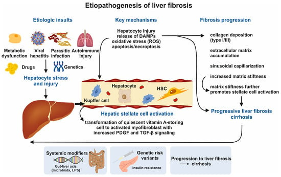
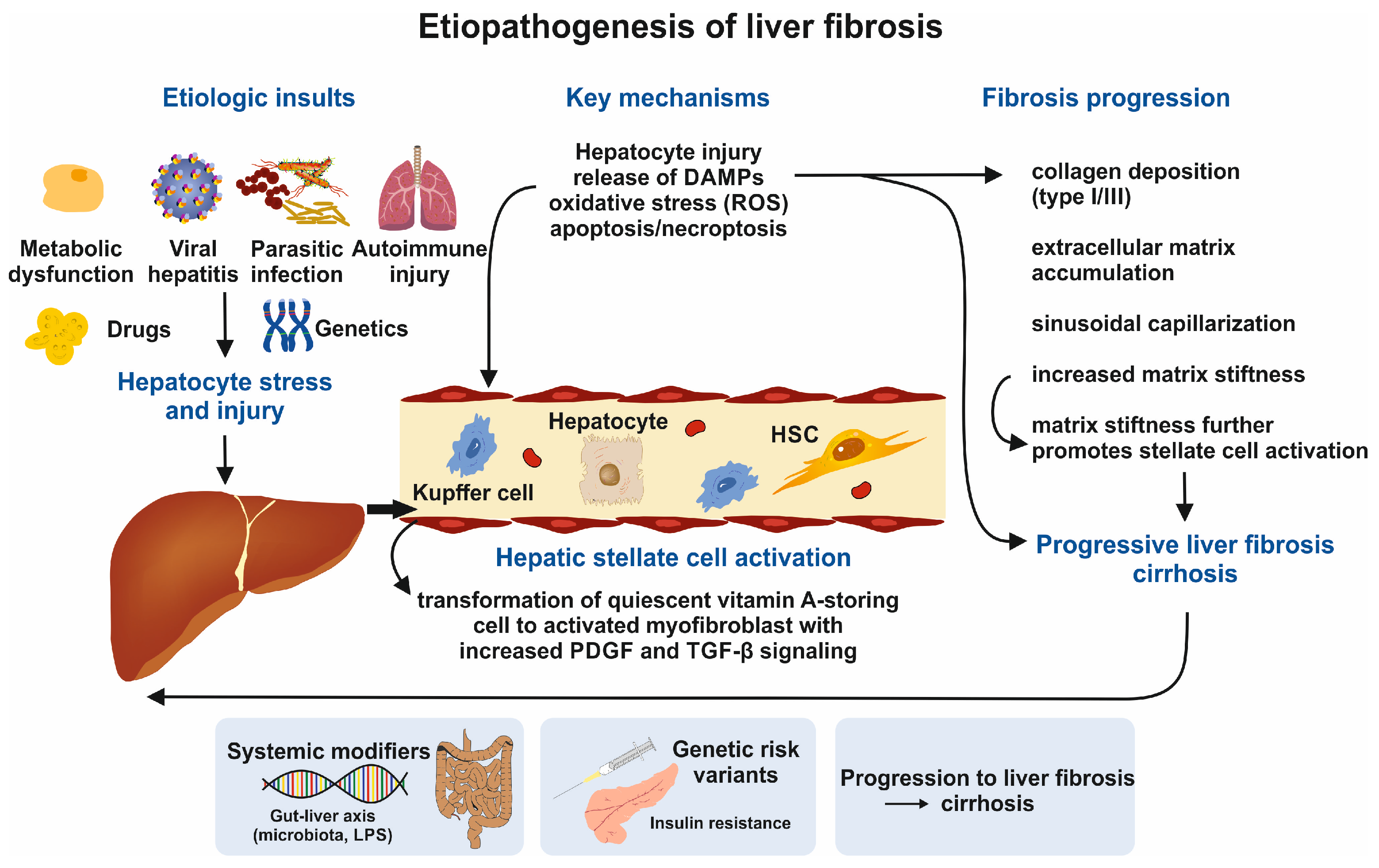
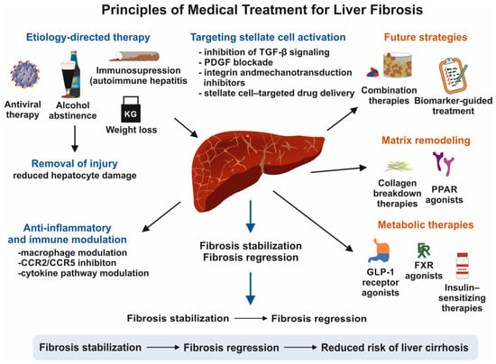
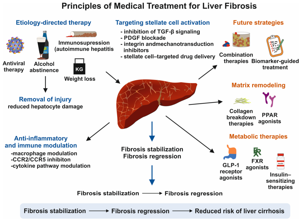
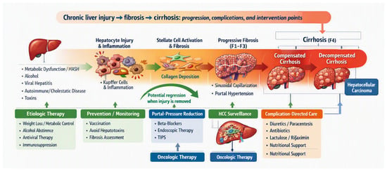
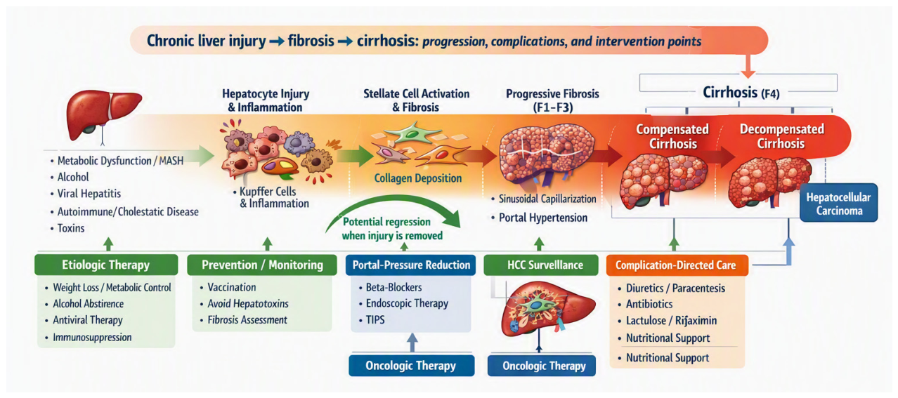

Disclaimer/Publisher’s Note: The statements, opinions and data contained in all publications are solely those of the individual author(s) and contributor(s) and not of MDPI and/or the editor(s). MDPI and/or the editor(s) disclaim responsibility for any injury to people or property resulting from any ideas, methods, instructions or products referred to in the content. |
© 2026 by the authors. Licensee MDPI, Basel, Switzerland. This article is an open access article distributed under the terms and conditions of the Creative Commons Attribution (CC BY) license.