


Abstract
Primary sclerosing cholangitis (PSC) is a rare cholangiopathy strongly associated with inflammatory bowel disease (IBD), particularly ulcerative colitis. PSC-IBD defines a clinically quiescent but biologically aggressive colitis phenotype, characterized by extensive yet often asymptomatic mucosal inflammation and disproportionately elevated risks of colorectal cancer—approximately 3- to 5-fold higher than in IBD alone—and cholangiocarcinoma (CCA), with IBD comorbidity representing an independent risk factor for hepatopancreatobiliary malignancy. The pathogenesis remains incompletely understood but involves genetic susceptibility, immune dysregulation—including aberrant lymphocyte trafficking and an imbalance between T helper 17 (Th17) and regulatory T (Treg) cells—intestinal barrier dysfunction, and gut–liver axis perturbations involving alterations in the microbiota and bile acid homeostasis. Within the biliary tree, chronic inflammation activates peribiliary glands (PBGs), which harbor stem/progenitor cells. PBG hyperplasia may contribute to periductal fibrosis through Hedgehog signaling and epithelial-to-mesenchymal transition and may represent a key step in PSC-associated cholangiocarcinogenesis. The true burden of PSC-IBD is likely underestimated, as PSC may remain clinically silent for years. Bidirectional screening is therefore essential: all patients with PSC should undergo ileocolonoscopy with biopsies regardless of symptoms, whereas patients with IBD and cholestatic liver biochemistry—particularly elevated gamma-glutamyl transferase or alkaline phosphatase—should undergo magnetic resonance cholangiopancreatography. No medical therapy has demonstrated a clear ability to alter the natural history of PSC, and liver transplantation remains the only definitive treatment for advanced disease. This review integrates current evidence on the epidemiology, pathophysiology, and management of PSC-IBD, critically examining unmet needs in timely diagnosis, mechanistic understanding, and therapeutic development, with particular attention to non-invasive biomarkers, microbiota-directed strategies, individualized risk stratification, and disease-modifying endpoints.
1. Introduction
Primary sclerosing cholangitis (PSC) is a chronic cholangiopathy characterized by progressive inflammation and fibrosis of the intrahepatic and extrahepatic bile ducts, leading to multifocal strictures, cholestasis, cirrhosis, and an increased risk of hepatobiliary and colorectal malignancies [1,2]. PSC is strongly associated with inflammatory bowel disease (IBD), and their coexistence defines a distinctive clinical phenotype, including extensive but often clinically mild colitis and a higher colorectal cancer risk than IBD alone [3,4,5]. Whether PSC-IBD represents a single disease process or a distinct clinical phenotype arising from partially shared pathogenic mechanisms remains unresolved. The occurrence of PSC in the absence of IBD, the persistence of hepatobiliary disease after colectomy, and the imperfect correlation between intestinal and hepatobiliary activity argue against considering PSC merely an extraintestinal manifestation of IBD [3]. Importantly, 20–30% of patients with PSC never develop IBD, and among those with PSC without IBD, the male predominance is much lower and the clinical course may differ, suggesting that gut-independent pathogenic pathways may operate in a substantial subset of patients [2,6]. A cross-disease genetic analysis of five chronic inflammatory diseases found that the strong comorbidity between PSC and IBD is best explained by biological pleiotropy—shared genetic effects acting through partially overlapping pathways—rather than by heterogeneity or diagnostic misclassification, and concluded that PSC-IBD is “likely the result of a unique disease, which is genetically distinct from classical inflammatory bowel disease phenotypes” [7]. These observations underscore that PSC encompasses substantial biological heterogeneity, and that the relationship between PSC and IBD cannot be adequately captured by any single pathogenic model. This distinction is further supported by genetic and molecular evidence. The largest genome-wide association study of PSC demonstrated that the genome-wide genetic correlation between PSC and ulcerative colitis is only modest, and that between PSC and Crohn’s disease is negligible, with approximately half of PSC susceptibility loci showing little or no association with IBD and instead overlapping with autoimmune diseases such as type 1 diabetes and celiac disease [8,9]. At the mucosal level, single-cell transcriptomic and spatial analyses have revealed that the PSC-UC colon harbors distinct region-specific microbial communities and an enrichment of activated CD8 T and γδ T cells in the right colon, even in the absence of histological inflammation, indicating that PSC-UC and UC are fundamentally different at the tissue level [10]. Moreover, the development of de novo IBD in 14–30% of patients within 10 years after liver transplantation (LT) for PSC further challenges a simple cause-and-effect relationship between intestinal and hepatobiliary disease [11,12]. Conversely, the aberrant hepatic expression of MAdCAM-1 and CCL25—normally restricted to the gut—and the consequent recruitment of α4β7-expressing mucosal lymphocytes to the liver provide a mechanistic link between intestinal and biliary inflammation; however, the failure of vedolizumab (anti-α4β7) to improve liver biochemistry despite effective control of IBD suggests that gut lymphocyte trafficking alone does not fully explain PSC pathogenesis [9,13,14].
Current evidence supports a role for the gut–liver axis in PSC-IBD, involving genetic susceptibility, immune dysregulation, intestinal barrier dysfunction, microbial factors, and altered bile acid homeostasis [3,15,16,17]. Microbial alterations are closely linked to changes in intestinal barrier function, mucosal immunity, and bile acid metabolism. Whether they contribute to PSC pathogenesis or result from cholestasis and intestinal inflammation remains unclear. The pathophysiology can be conceptualized as a triad of gut dysbiosis, altered bile acid metabolism, and immune-mediated biliary injury, in which bile acids shape the gut microbiome while gut microbes alter bile acid profiles, creating a bidirectional and potentially self-reinforcing cycle [17]. However, this conceptual framework, while heuristically useful, should be recognized as a working model rather than an established mechanism. The relative contribution of each component—dysbiosis, bile acid perturbation, and immune-mediated biliary injury—likely varies across PSC phenotypes (large-duct vs. small-duct, with vs. without IBD, pediatric vs. adult), and the directionality and hierarchy of these interactions remain largely hypothetical [18,19]. Disruption of the intestinal barrier allows microbial products to enter the portal circulation, stimulating hepatic immune cells and triggering biliary inflammation through pathways involving FXR and TGR5 signaling [20]. Notably, the dysbiosis in PSC extends beyond bacteria: patients with PSC harbor a distinct fungal gut microbiota (mycobiome), characterized by increased Exophiala and decreased Saccharomyces species, with a marked disruption of bacteria–fungi interkingdom correlation networks, suggesting that host–mycobiome interactions represent an underappreciated component of pathogenesis [21]. Preliminary evidence from fecal microbiota transplantation studies—in which FMT increased bacterial diversity and engraftment correlated with decreased alkaline phosphatase levels—provides early support for a causal role of the microbiome, although clinical responses have been modest and inconsistent [22,23]. Advances in imaging, molecular biology, and microbiome research have improved understanding of disease mechanisms, yet major gaps persist between pathophysiological insights and effective clinical translation [1,15,17,24,25].
Despite growing awareness, the natural history of PSC-IBD remains unpredictable, therapeutic options are limited, and the risk of malignancy remains substantial [1,2,5]. This review is organized around five interconnected domains of unmet need in PSC-IBD: (1) diagnostic gaps, including underdiagnosis of subclinical disease, limitations of current imaging and histological criteria, and the absence of validated non-invasive biomarkers; (2) mechanistic gaps, particularly the incomplete understanding of the gut–liver axis, the relative contributions of microbiota, bile acids, and immune trafficking, and the lack of actionable therapeutic targets derived from pathogenetic insights; (3) therapeutic gaps, including the absence of any disease-modifying therapy for PSC, the dissociation between biochemical improvement and clinical benefit, and the limited evidence base for IBD-directed therapies on cholangiopathy outcomes; (4) surveillance gaps, including inconsistencies among guideline recommendations, the uncertain cost-effectiveness of intensive protocols, and the lack of validated risk-stratification tools to individualize monitoring; and (5) research design gaps, including the reliance on surrogate endpoints, the scarcity of large-scale multicenter trials, and the need for composite, clinically meaningful outcome measures. Each section of this review presents the current evidence within the framework of these unmet needs, aiming to identify priorities that should guide future research and clinical strategies.
2. Literature Search
This narrative review was based on a focused search of PubMed/MEDLINE and Embase, last updated in July 2026. Search terms included “primary sclerosing cholangitis”, “inflammatory bowel disease”, “PSC-IBD”, “ulcerative colitis”, “Crohn’s disease”, “microbiome”, “cholangiocarcinoma”, “surveillance”, “liver transplantation”, and “clinical trials”. Priority was given to international guidelines, systematic reviews and meta-analyses, randomized trials, prospective cohort studies, and key mechanistic studies relevant to PSC-IBD. Reference lists of selected articles and guidelines were also reviewed to identify additional relevant publications. No formal systematic-review methodology or quantitative synthesis was applied.
3. Epidemiology of the PSC-IBD Association
PSC is a rare disease with marked geographic variation in incidence. In Europe, incidence ranges from approximately 0.1 per 100,000 in Southern countries to over 1.5 per 100,000 in Northern regions [26,27]. IBD is reported in 20–80% of PSC patients, with substantial geographic variability [28]. This association is likely underestimated because colonoscopy has not been systematically performed in all PSC cohorts [3]. Guidelines recommend colonoscopy with random biopsy at PSC diagnosis and regular surveillance thereafter due to the high colorectal cancer (CRC) risk [1,2]. While compliance with the first colonoscopy is high (89–92%), adherence to subsequent procedures after a negative result drops markedly (40%), leading to underdiagnosis of IBD in PSC patients [29]. Histological assessment often detects subclinical colonic inflammation missed by endoscopy alone. In a cohort of PSC patients undergoing ileocolonoscopy with segmental biopsies, inflammatory changes were detected significantly more often on histology than on endoscopy (89% vs. 47%), indicating that a substantial proportion of inflammation may be missed in the absence of routine random biopsies [30]. Similarly, pediatric PSC-IBD frequently shows subclinical inflammation despite clinical remission [31].
While IBD may be underestimated in PSC (Figure 1), the true prevalence of PSC among patients with IBD is also uncertain. Reported prevalence ranges from 2% to 14%, reflecting differences in diagnostic approaches and populations. However, an additional major determinant is IBD subtype: PSC is consistently more prevalent in UC than in CD, with intermediate or higher estimates reported in IBD-unclassified. In a systematic review and meta-analysis, the pooled prevalence of PSC was 2.16% in IBD overall, 2.47% in UC, 0.96% in Crohn’s disease (CD), and 5.01% in IBD-unclassified [4]. Prevalence increases when imaging such as magnetic resonance cholangiopancreatography (MRCP) is systematically performed, reflecting the long subclinical phase of PSC. This prolonged silent phase makes underdiagnosis particularly likely in IBD populations [3,4]. Notably, Lunder et al. found PSC about 3 times more often in long-term IBD when MRCP is performed outside stringent clinical indications [32].
Figure 1.
Underestimation of PSC–IBD and its impact on epidemiological estimates. Diagnosed PSC–IBD represents only a fraction of the true disease burden. Long subclinical phases and incomplete case findings—particularly when colonoscopy and systematic biopsies are not routinely performed—contribute to underdiagnosis and to wide variation in reported prevalence across cohorts and regions.
A UK study shows that the prevalence of PSC-IBD is rising faster than that of IBD alone. This trend cannot be attributed solely to improved diagnostic capability, suggesting a true increase in disease burden [33]. The projected prevalence in the UK in 2027 is estimated to reach 13.3 per 100,000 population, up from 5.7 per 100,000 in 2015. This growing burden will likely place additional demands on national healthcare systems [33]. Geographic differences should also be considered, as most population-based data derive from Western countries. Recent Asian data suggest a lower prevalence of PSC among patients with IBD and a milder presentation in more recent diagnostic eras, while data from China also indicate lower PSC prevalence than in Western populations and marked regional variation [34,35]. A multinational study across six Asian countries (n = 51,314 IBD patients) reported a PSC prevalence of 0.92% in IBD overall (1.4% in UC and 0.13% in CD), substantially lower than the pooled Western estimate of 2.16% [35]. In more recent diagnostic eras, Asian patients with PSC-IBD presented with fewer symptoms, lower alkaline phosphatase (ALP) levels, and better liver function scores, while the increased use of MRCP was associated with earlier detection and milder disease severity [35]. Data from South Asia are even more limited. In a large Chinese population-based study of over 44,000 patients with IBD, PSC was uncommon, occurring in approximately 0.5% of patients with UC and 0.2% of those with CD [36]. A scoping review of IBD in South Asia similarly found PSC to be rare among Indian IBD cohorts, although specific prevalence estimates were not reported [37]. In Japan, a 2019 nationwide hospital-based survey estimated the point prevalence of PSC at 1.80 per 100,000 population, nearly double the 0.95 per 100,000 reported in a 2007 survey, with a shift in the male-to-female ratio from 1:1.4 to 1:0.88 [38]. A single-center Japanese study reported a cumulative PSC risk of 3.0% in patients with UC and a PSC-UC comorbidity rate of 53.6%, substantially higher than the 34% reported in the earlier nationwide survey, suggesting that PSC-UC may be increasing in Japan in parallel with the rising prevalence of UC [39]. These data suggest that the epidemiology of PSC-IBD in East Asia may be evolving and that earlier estimates may underestimate the true burden. These differences may reflect genetic background, environmental exposures, IBD phenotype, diagnostic intensity, and healthcare access. Longitudinal temporal trends also vary by region. While the UK study projects a rising prevalence of PSC-IBD, the Asian multinational study found that more recent diagnostic eras were associated with milder presentations and longer time to LT, suggesting that increased diagnostic intensity, particularly greater MRCP availability, may be shifting the clinical spectrum toward earlier and less severe disease in some populations [35]. In Japan, the near-doubling of PSC prevalence between 2007 and 2016 may reflect both a true increase in incidence and improved diagnostic ascertainment [38].
Mortality differences further support the prognostic impact of PSC in patients with IBD. Population-based data consistently demonstrate that PSC-IBD is associated with substantially worse outcomes than IBD alone. In a nationwide English cohort of 284,560 incident IBD cases, development of PSC was associated with a 3.2-fold increased risk of death (HR 3.20; p < 0.001), with the greatest mortality difference observed in patients diagnosed with IBD before the age of 40 years (incidence rate ratio > 7), compared with those diagnosed after the age of 60 years (incidence rate ratio < 1.5). LT and PSC-related events accounted for approximately 75% of clinical events in the younger group versus 31% in the older group [5]. Two large database studies using propensity-matched designs confirmed the adverse prognostic impact of concomitant PSC in IBD, reporting mortality hazard ratios of 1.69 (95% CI 1.46–1.96) [40] and 2.26 (95% CI 1.85–2.75) [41], respectively. Although the difference in effect size likely reflects variations in matching strategies and outcome definitions, both studies consistently demonstrated increased mortality, hospitalization, and colorectal cancer risk in PSC-IBD. A population-based pediatric cohort from the EPIMAD Registry found that cancers were 28 times more frequent (SIR 27.9; 95% CI 7.0–111.7) and death was 13 times more frequent (SMR 13.3; 95% CI 3.3–53.4) in pediatric-onset IBD-PSC compared with the general population, whereas no increased risk was observed in pediatric IBD without PSC [42]. Interestingly, a recent systematic review and meta-analysis comparing PSC with IBD versus PSC alone found that PSC-IBD was associated with a lower risk of all-cause mortality or LT compared with PSC alone (HR 0.77; 95% CI 0.62–0.97), but with an increased risk of hepatopancreatobiliary cancer (HR 2.37; 95% CI 1.35–4.15) [43]. The lower all-cause mortality in PSC-IBD may reflect earlier diagnosis and more intensive hepatobiliary surveillance in patients identified through IBD follow-up, whereas the increased hepatopancreatobiliary cancer risk may reflect the additive carcinogenic effects of chronic colonic and biliary inflammation. These findings suggest that the prognostic impact of IBD in PSC is complex and direction-dependent. Data specifically separating colorectal cancer-related mortality from overall cancer-related mortality in PSC-IBD remain limited, representing an important gap for future population-based studies [44].
Unmet need. The true burden of PSC-IBD remains uncertain due to geographic heterogeneity in diagnostic intensity, inconsistent use of MRCP screening in IBD populations, and the absence of prospective population-based registries that systematically capture both hepatobiliary and intestinal disease dimensions. Standardized, internationally harmonized case-finding strategies—including systematic MRCP in high-risk IBD subgroups—are needed to define the actual prevalence and inform healthcare resource planning.
4. Definition and Diagnostic Framework of PSC-IBD
The non-random co-occurrence of PSC and IBD, their shared but incomplete genetic architecture, and the distinctive intestinal inflammatory phenotype observed in PSC all support the concept that PSC-IBD represents more than the incidental coexistence of two unrelated conditions. However, the precise nature of this relationship remains unresolved. Three competing models have been proposed: (a) PSC as an extraintestinal manifestation of IBD; (b) PSC and IBD as two distinct diseases that share a common genetic and environmental susceptibility, leading to a dual phenotype; and (c) PSC-IBD as a unified disease process driven by shared gut–liver pathogenic mechanisms. Each model is supported by some evidence but contradicted by other observations [1,3]. A fourth perspective has been proposed by Ellinghaus et al., who suggested that PSC-IBD may represent a genetically unique disease entity distinct from both classical PSC and classical IBD, based on cross-disease pleiotropy analysis [7]. This model is not mutually exclusive with the others but highlights that the genetic architecture of PSC-IBD may not be reducible to a simple combination of PSC and IBD susceptibility. The coexistence of these competing frameworks reflects the genuine biological complexity of the PSC-IBD relationship and argues against premature adoption of any single unifying model.
At the hepatic level, PSC is a chronic large bile duct cholangiopathy affecting the intra- and extrahepatic bile ducts, leading to progressive fibrosis, multifocal stricturing, and ultimately to biliary cirrhosis and its complications [1,2,45]. At the intestinal level, PSC is most frequently associated with UC and less commonly with CD. However, in the setting of PSC, IBD often exhibits distinctive clinical and endoscopic features, supporting the concept of a PSC-associated IBD phenotype that does not entirely overlap with conventional UC or CD (IBD-unclassified) [3,30,31]. Nevertheless, conventional IBD categories remain necessary for treatment decisions, surgical planning, cancer surveillance, and comparison across studies, even if they do not fully capture the peculiar pattern of intestinal inflammation observed in PSC. A structured diagnostic approach also requires exclusion of PSC mimickers and secondary sclerosing cholangitis, which are discussed in detail in the differential diagnosis section.
The persistence of PSC after colectomy, the development of de novo IBD after LT, and the failure of IBD-directed therapies to modify cholangiopathy all argue against a simple unified model [3]. Conversely, the aberrant hepatic expression of gut-homing molecules, including MAdCAM-1 and CCL25, and the distinctive mucosal immune profile of PSC-UC support at least partially shared pathogenic mechanisms [10,13]. Until this question is resolved, PSC-IBD is best regarded as a clinically distinct phenotype—or, more precisely, a spectrum of phenotypes—whose underlying pathogenic architecture remains incompletely defined and may differ across patient subgroups according to age at onset, ductal involvement, IBD subtype, and autoimmune overlap features [6,18]. Recognizing PSC-IBD as a clinically distinct phenotype, regardless of which pathogenic model ultimately prevails, has implications for diagnosis, surveillance, and therapeutic strategies [3,15,17].
In PSC-IBD, diagnostic accuracy depends on multidisciplinary integration rather than reliance on isolated findings. PSC-IBD diagnosis relies on the integration of clinical, biochemical, radiological, and histological data. Histology should therefore be interpreted within the broader clinical and diagnostic context. When detached from clinical presentation, biochemical patterns, serological profiles, cholangiographic findings, and genetic predisposition, liver biopsy may lead to diagnostic misinterpretation.
Unmet need. Despite growing recognition of PSC-IBD as a distinct phenotype, no consensus diagnostic criteria specific to PSC-IBD exist. The current reliance on conventional IBD classification systems, including the Montreal and Paris classifications, does not capture the distinctive intestinal inflammatory pattern observed in PSC, while the absence of validated non-invasive biomarkers for early PSC detection in IBD populations limits timely diagnosis. The development of PSC-IBD-specific diagnostic algorithms integrating clinical, biochemical, cholangiographic, and molecular data represents a priority.
5. PSC-IBD in the Pediatric Setting
PSC-IBD in children raises the question of whether it represents a distinct phenotype or an early phase of a broader pathogenic continuum. Kellermayer et al. proposed a “developmental window of opportunity” and an “Early PSC-IBD-AIH overlap,” although the biological relationship between pediatric and adult disease remains incompletely defined. Their proposal—that all PSC originates from a complex “Early PSC-IBD-AIH overlap” along an age-dependent continuum—remains a hypothesis rather than established evidence. The authors themselves describe this framework as speculative, and prospective longitudinal data bridging pediatric and adult cohorts are lacking to confirm or refute it [46].
In children, PSC-IBD more often presents a prominent inflammatory phenotype affecting both the liver and intestine. Autoimmune liver features and autoimmune hepatitis (AIH) overlap are more common than in adults. This contrasts with adult disease, where fibrotic and stenosing features predominate. Moreover, small-duct PSC (sdPSC) appears to be relatively more common in children, further reinforcing the concept of an early-stage disease, in which inflammatory injury precedes the development of overt cholangiographic strictures. In early disease, histological bile duct injury may precede detectable imaging abnormalities [1,31,47,48].
Whether these differences define a separate nosological entity or are best understood as temporal and biological gradients within the same disease spectrum remains an open question. The largest pediatric cohort study, including 781 children, found that small-duct PSC and PSC-IBD were associated with a favorable prognosis, and some pediatric patients with small-duct PSC may never progress to large-duct disease [49]. In mixed-age cohorts of small-duct PSC, approximately 23% of patients progress to large-duct disease over 5–14 years [2]. Whether this estimate applies specifically to pediatric PSC remains uncertain. The transition from inflammation to irreversible bile duct fibrosis is neither abrupt nor uniform, but appears directionally consistent across age groups [1,3,47].
This perspective has potential therapeutic implications. The inflammatory predominance in children may represent a therapeutic window before irreversible fibrosis develops [1,31,47]. In adults, diagnosis often occurs later, when fibrotic changes are already established.
Overall, pediatric PSC-IBD may represent an earlier and more inflammatory presentation within the PSC spectrum, rather than a fully separate disease entity. However, the higher prevalence of AIH overlap in children—reported in up to 35% of cases according to the American Association for the Study of Liver Diseases (AASLD) guidance—may reflect a genuinely distinct immunopathogenic mechanism rather than simply an earlier disease stage [2]. A multicenter ESPGHAN study of very early-onset PSC-IBD, diagnosed before 6 years of age, found that 92% of patients met criteria for PSC-AIH overlap, compared with 67.5% of older children. Baseline PSC characteristics were otherwise similar, although biliary complications developed earlier in the very early-onset group [50]. These findings suggest that the immunopathogenic landscape of pediatric PSC-IBD may be qualitatively different from that of adult disease, rather than merely shifted along a temporal continuum. Prospective studies are needed to clarify its relationship with adult fibrotic and stenosing disease. Timely diagnosis, appropriate surveillance, and the development of stage-adapted therapeutic strategies are essential.
Unmet need. Prospective longitudinal studies bridging pediatric and adult cohorts are lacking. Whether the inflammatory predominance in children represents a true therapeutic window—and whether early intervention can prevent progression to irreversible fibrosis—remains untested. Pediatric-specific clinical trials and validated transition-of-care protocols are urgently needed.
6. Small-Duct Primary Sclerosing Cholangitis and Its Association with IBD
SdPSC is a variant characterized by inflammation confined to intrahepatic bile ducts with normal MRCP findings [1,51]. The AASLD guidance and the international cohort by Weismüller et al. report that approximately 10% of patients have small-duct disease, whereas pediatric series have reported frequencies of 13–24% [2,6,47,49]. The AASLD guidance explicitly states that whether small-duct PSC represents a separate entity or an early or mild form of PSC remains controversial [2]. Available evidence is conflicting. A dual-center magnetic resonance imaging (MRI) follow-up study found that more than half of patients with sdPSC developed cholangiographic changes consistent with large-duct PSC over a mean interval of 10.6 years, supporting the early-stage hypothesis [52]. Conversely, the multicenter study by Björnsson et al. found that only 22.9% of patients progressed to large-duct disease over a median follow-up of 7.4 years and that cholangiocarcinoma did not occur unless progression to large-duct involvement had taken place [53]. Furthermore, a recent study showed that isolated intrahepatic PSC has clinical characteristics and outcomes similar to those of small-duct PSC, suggesting that ductal phenotype may define prognostically meaningful subgroups within the PSC spectrum [54]. The diagnostic and prognostic landscape of sdPSC therefore remains incompletely understood, highlighting a significant unmet need in clinical hepatology. Diagnosis relies primarily on liver histology, as imaging is non-diagnostic (Figure 2). Histological findings may include periductal fibrosis, ductopenia, and ductular reaction. None of these features is pathognomonic, complicating clinical decision-making [45]. Therefore, sdPSC should not be regarded as a single homogeneous entity, but rather as a diagnostic category that may include early-stage PSC and other small-duct cholangiopathies that may mimic PSC. In this setting, the coexistence of IBD may substantially increase diagnostic confidence [45].
Figure 2.
Diagnostic approach to small-duct primary sclerosing cholangitis (sdPSC). Abbreviations: PSC, primary sclerosing cholangitis; sdPSC, small-duct primary sclerosing cholangitis; IBD, inflammatory bowel disease; ANA, antinuclear antibodies; AMA, antimitochondrial antibodies; MRCP, magnetic resonance cholangiopancreatography. Secondary causes are grouped as: immune-mediated (IgG4-related cholangitis); ischemic (critical illness, hepatic artery thrombosis, intra-arterial chemotherapy); infectious (HIV/AIDS cholangiopathy, recurrent pyogenic cholangitis, parasitic disease); iatrogenic/traumatic (post-cholecystectomy or anastomotic biliary injury); and obstructive or neoplastic (choledocholithiasis, cholangiocarcinoma, diffuse metastatic or infiltrative disease).
IBD is reported in up to 80% of patients with sdPSC, with a distribution that slightly differs from that observed in large-duct PSC. While UC remains predominant (approximately 78%), CD accounts for a larger proportion of cases (around 21%) compared to its frequency in PSC, suggesting a potentially distinct immunological phenotype [51,53]. Available data suggest that the risk of CRC may be lower than in large-duct PSC; nevertheless, patients with concomitant IBD should continue colorectal cancer surveillance according to PSC-IBD recommendations [1,2,55]. The substantially lower risk of hepatobiliary malignancy has direct implications for surveillance. The AGA Clinical Practice Update recommends against routine cholangiocarcinoma surveillance in patients with small-duct PSC, unless progression to large-duct disease occurs [56]. Therefore, whether sdPSC represents a distinct entity or an early phase of conventional PSC is not merely a nosological question but directly influences surveillance intensity and risk communication. In the absence of IBD, the diagnosis of sdPSC becomes more uncertain and should prompt careful exclusion of alternative causes of small-duct cholangiopathy. Histological features of sdPSC may overlap with genetic cholangiopathies, including the spectrum of ABCB4-related disease [57], as well as other genetic biliary disorders. Therefore, genetic testing with an appropriate cholestasis genes panel is strongly recommended in patients without concomitant IBD [2,57].
Overlap with AIH adds another layer of diagnostic complexity, particularly in pediatric populations, where up to 52% of children with large-duct PSC (ldPSC) fulfill criteria for autoimmune sclerosing cholangitis (ASC). These patients often present with elevated alanine aminotransferase (ALT) and immunoglobulin G (IgG) levels and may benefit from immunosuppressive therapy. However, distinguishing overlap forms from “pure” PSC remains a clinical challenge, and standard criteria may not fully capture this [47,48,58].
In summary, while sdPSC is often perceived as a benign or indolent variant, it represents a diagnostically complex and frequently IBD-associated form of PSC. Integration of histology with clinical context is essential for accurate diagnosis and management.
Unmet need. The natural history of sdPSC remains insufficiently characterized, with limited data on long-term progression rates, predictors of evolution to large-duct disease, and malignancy risk. The absence of pathognomonic histological features and the overlap with genetic cholestasis/cholangiopathies create diagnostic uncertainty, particularly in patients without IBD. Prospective registries and standardized histological criteria for sdPSC are needed.
7. Pathogenesis of PSC-IBD: Microbiota, Genetics, and Immune Mechanisms
Understanding the mechanisms underlying inflammation and fibrosis in PSC represents one of the most critical unmet needs in the field, as the absence of validated therapeutic targets is a direct consequence of incomplete pathogenetic knowledge. Several hypotheses attempt to explain these mechanisms, but none has yet been translated into effective therapy. A systematic review comparing immune activation patterns in PSC (with or without IBD) and IBD alone found that PSC is characterized by upregulation of T helper 17 (Th17) responses and downregulation of regulatory T-cell (Treg) function, a profile that distinguishes PSC from IBD-only and suggests that PSC-specific immune dysregulation operates at least partly independently of intestinal inflammation [59]. This finding reinforces the view that PSC and IBD, even when co-occurring, involve non-identical immune mechanisms. The close relationship between PSC and IBD has raised doubts that PSC was only an extraintestinal manifestation of IBD and that isolated PSC is a separate entity. Several pathogenic hypotheses support partially shared gut–liver mechanisms between PSC and IBD, although PSC-IBD should not be interpreted as a uniform disease process [3]. One of the most relevant pathogenetic hypotheses suggests that IBD may be the primary trigger of the disease [60]. Dysregulation of the immune system and impairment of the intestinal barrier at the level of the biliary tract constitute central elements. The gut microbiota plays a crucial role in pathogenesis, highlighting the close association between these diseases, although its contribution has yet to be fully elucidated [61]. In addition, recent advances in genome-wide association studies (GWAS) are providing a deeper understanding of the molecular mechanisms involved, although the overall pathogenesis remains largely poorly understood [8,62].
7.1. An Integrative Framework
Conceptually, the pathogenesis of PSC-IBD can be organized as a sequential but bidirectional cascade operating along the gut–liver axis (Supplementary Figure S1) [17,20,63]. In genetically susceptible individuals, environmental triggers—including microbial shifts and altered bile acid profiles—disrupt intestinal barrier integrity, enabling translocation of microbial products, including lipopolysaccharide and pathobiont-derived outer membrane vesicles, into the portal circulation [64,65]. In parallel, gut-primed lymphocytes expressing α4β7 integrin and CCR9 are aberrantly recruited to the liver through ectopic hepatic expression of MAdCAM-1 and CCL25, establishing a cellular bridge between intestinal and biliary inflammation [9,66]. Within the liver, these convergent signals—microbial products via TLR4/NLRP3 pathways and recruited immune cells via adaptive and innate mechanisms—activate cholangiocytes, which transition from passive targets of injury to active effectors through the acquisition of a senescence-associated secretory phenotype (SASP) and the secretion of profibrogenic mediators including IL-6, IL-8, CCL2, and PAI-1 [67,68,69]. Senescent cholangiocytes, in turn, engage portal fibroblasts—the first responders to biliary injury and the primary collagen-producing cells in cholestatic fibrosis—generating the characteristic concentric “onion-skin” periductal fibrosis [70,71,72]. Simultaneously, cholestasis itself amplifies the cycle: retained bile acids alter gut microbial composition, further disrupt the intestinal barrier, and directly promote cholangiocyte injury and proliferation, creating a self-reinforcing loop [73,74]. Over time, the sustained fibroinflammatory microenvironment—characterized by chronic cholangiocyte activation, SASP-driven paracrine signaling, peri-biliary gland hyperplasia, and accumulation of DNA damage—may provide the substrate for neoplastic transformation and chol-angiocarcinoma development [75,76,77]. This integrated framework, while supported by converging lines of evidence from human tissue studies, preclinical models, and single-cell analyses, remains a working model: the temporal sequence, the relative weight of each component, and the points at which the cascade becomes self-sustaining independently of the initiating trigger are incompletely defined. The subsections that follow examine each component of this framework in detail.
7.2. Intestinal Microbiota
Several lines of evidence support a role for intestinal dysbiosis in PSC pathogenesis. Current models propose that environmental and genetic factors disrupt the biliary mucosal barrier and bile acid homeostasis, promoting biliary fibrosis and cholangiocarcinogenesis [15]. Within the integrated gut–liver framework outlined above, dysbiosis operates both as a potential initiating factor—by generating proinflammatory metabolites and disrupting barrier integrity—and as a downstream consequence of cholestasis-induced changes in bile acid composition, illustrating the bidirectional nature of the pathogenic cascade.
PSC is associated with an enrichment of potentially pro-inflammatory bacteria such as Veillonella, Streptococcus, Enterococcus, and a reduction in beneficial short-chain fatty acid producers [16,61] (Table 1). Metagenomic analyses have shown that PSC is associated with reduced microbial diversity and functional alterations involving bile acid and amino acid metabolism, which correlate with adverse liver-related outcomes [78,79]. Microbiome alterations in PSC extend beyond the intestine, as distinct salivary signatures have been described, partly overlapping with fecal dysbiosis and independent of IBD comorbidity [61]. Beyond bacterial communities, non-bacterial components of the intestinal microbiome may also be relevant. Altered fungal communities have been described in PSC [21,80], whereas the intestinal virome remains largely unexplored in PSC and PSC-IBD. Evidence from IBD suggests that disease-associated changes in virome composition and phage–bacteria interactions may influence mucosal immune activation and epithelial barrier integrity [81,82]. Gut virome dysbiosis in IBD is primarily driven by an expansion of Caudovirales bacteriophages and a loss of mucosal virome diversity, with disrupted phage–bacteria ecological networks that may perpetuate dysbiosis [83,84].
Bacteriophages can also directly modulate mucosal immunity: experimental studies have demonstrated that phage DNA stimulates IFN-γ production via the nucleotide-sensing receptor TLR9, and that phage expansion exacerbates colitis through this pathway, with mucosal IFN-γ levels correlating with bacteriophage abundance in ulcerative colitis patients [85]. In addition to bacteriophages, eukaryotic viruses may contribute to IBD-associated immune dysregulation. Metagenomic studies have reported enrichment of Anelloviridae—particularly Torque teno virus (TTV) species—in the fecal virome of patients with Crohn’s disease, with TTV abundance linked to immune disequilibrium and autoimmunity-related molecular mimicry [86]. Furthermore, members of the Herpesviridae family, including Epstein–Barr virus and cytomegalovirus, are frequently detected in the colonic mucosa of IBD patients at rates significantly higher than in immunocompetent controls, and have been identified as risk factors for IBD development and exacerbation [87,88,89]. Whether analogous virome alterations—involving bacteriophages, Anelloviridae, or Herpesviridae—contribute to hepatobiliary inflammation or disease progression in PSC-IBD remains unknown and represents an important area for future investigation. To further elucidate the role of the gut-liver axis in the pathogenesis of PSC, Liao et al. demonstrated in a mouse model of Mdr2-/- deficiency that the loss of the phospholipid transporter Mdr2 triggers a cholestatic response, inducing intrahepatic sclerosing cholangitis. By analyzing the Mdr2-/- microbiota, they highlighted a reduced species diversity and significant alterations in the Lachnospiraceae family, which plays a key role in secondary bile acid formation, as well as increased NLRP3 inflammasome activation in Mdr2-/- dysbiotic mice, leading to disrupted intestinal barrier integrity and translocation of endotoxins into the portal vein [74]. Recent metagenomic data in PSC-IBD patients confirmed enrichment of specific bile acid-modifying taxa and alterations in secondary bile acid profiles correlating with liver fibrosis [79].
These findings provide a biological rationale for microbiota-targeted interventions, discussed in the therapeutic section.
In summary, PSC is associated with reduced gut microbial diversity and distinct taxonomic and functional profiles. Although reduced microbial diversity is a hallmark of IBD, PSC-associated dysbiosis should not be interpreted solely as a consequence of colonic inflammation [16]. Several studies have reported overlapping microbial alterations in patients with PSC with and without IBD, while showing differences from IBD-alone cohorts [16,78]. These findings suggest that the microbial signature of PSC may be at least partly disease-specific and linked to gut–liver axis perturbations, altered bile acid metabolism, and cholestasis. Whether these microbial alterations contribute to PSC pathogenesis, represent a consequence of liver disease, or act as disease modifiers remains unclear.
Unmet need. The causal role of dysbiosis in PSC pathogenesis remains unproven. Current evidence is largely cross-sectional and cannot distinguish cause from consequence. Longitudinal studies with serial microbiome sampling before and after PSC diagnosis, combined with functional validation in gnotobiotic and humanized mouse models, are needed to establish causality and identify actionable microbial targets.
7.3. Genetic Risk
PSC is approximately ten times more common among first-degree relatives of patients, and genetic studies have identified several risk loci, highlighting its hereditary component [3]. Within the integrated pathogenic framework, genetic susceptibility—particularly HLA variants and non-HLA loci involved in immune regulation—is best understood as the background upon which environmental and microbial triggers act, determining individual vulnerability to the gut–liver cascade described above. However, there is limited genetic overlap between PSC (with or without IBD) and IBD. A much stronger genetic correlation exists between CD and UC. About half of the PSC-associated genes overlap with those linked to IBD, and genetic studies estimate a relatively low comorbidity rate of around 1.6%. This is considerably lower than the roughly 60% comorbidity observed clinically in patients with UC [8]. This disparity indicates that common genetic variants alone cannot explain the high PSC-IBD comorbidity. A Mendelian randomization study corroborated a causal association between genetically predicted PSC and UC but found no significant causal evidence for an association between PSC and CD, further supporting the specificity of the PSC-UC genetic link and arguing against a uniform PSC-IBD genetic architecture [90]. These findings are consistent with the observation by Ji et al. that approximately half of PSC susceptibility loci show little or no association with IBD and instead overlap with autoimmune diseases such as type 1 diabetes and celiac disease, suggesting that PSC pathogenesis involves immune pathways that are not captured by IBD-centric models [8]. Rare variants might play a role, as GWAS may not fully capture shared genetic influences. Additionally, PSC shares susceptibility loci with other autoimmune diseases, pointing to an autoimmune component in its pathogenesis and aligning with the increased prevalence of autoimmune disorders in PSC patients [62].
Previous studies have demonstrated a strong association between PSC and specific HLA haplotypes, suggesting a potential genetic role in its pathogenesis. Key alleles, including HLA-B8, -DR3, -DR2, -DR6, and -DQ, have been linked to increased susceptibility [55]. Schrumpf et al. observed higher frequencies of HLA-B8 and -DR3 in patients with both PSC and UC compared to those with UC alone [91]. Further analyses also identified HLA-DR2 as significant in HLA-DR3-negative cases [92]. Additional investigations into markers such as MICA5.1 and MICB24 support this genetic association [93]. While these findings indicate a genetic association, additional research is needed to better define the precise role of these markers in PSC.
GWASs have identified several genetic loci associated with PSC through SNP markers, with large patient samples providing strong statistical power. Srivastava et al. found associations between PSC and loci 3p21, 10p15 (IL2RA), and 4q27 (IL-2/IL-21), highlighting the significant role of the IL-2/IL2RA pathway in PSC. Other identified loci include 1p36 (MNEL1, TNFRSF1), 19q13 (FUT2), 2q37 (GPR35), 18q21 (TCF4), and 2q13 (BCL2L11), with the latter involved in apoptosis of autoreactive T cells [94]. Additionally, another study identified two non-HLA loci, 2p16 (REL) and 9q34 (CARD9), associated with PSC, which play roles in inflammatory signaling and innate immune activation [95]. These findings further support the hypothesis of a genetic predisposition to PSC, with several loci influencing immune regulation and inflammation. However, further research is needed to fully understand the functional implications of these genetic associations.
Unmet need. Despite the identification of numerous risk loci, the functional consequences of most PSC-associated genetic variants remain unknown. The gap between genetic discovery and mechanistic understanding limits the translation of GWAS findings into diagnostic or therapeutic applications. Functional genomic studies, including CRISPR-based perturbation screens in cholangiocyte and immune cell models, and integration of genetic data with single-cell transcriptomic and epigenomic profiling, are needed to bridge this gap.
7.4. Immunological Factors
The close association between PSC and IBD supports an immune-mediated component; however, the precise nature of this immune dysregulation remains incompletely defined [96].
Histopathological studies demonstrate portal tract infiltration by memory T lymphocytes in PSC [97], mirroring immune features reported in some extra-intestinal manifestations of IBD [98]. PSC may occur in patients with IBD irrespective of clinically active colitis and may persist after colectomy, suggesting that ongoing intestinal inflammation is not required to sustain the hepatic disease process [3,99]. De novo IBD has also been described after LT, even under systemic immunosuppression, often with a milder clinical course [3,12]. Collectively, these observations argue against models in which persistent bacterial translocation or luminal toxins from the inflamed gut are the sole drivers of PSC.
Aberrant lymphocyte homing to the liver has been proposed as a key link between intestinal inflammation and biliary injury. In PSC-IBD, hepatic endothelium may express adhesion molecules typically associated with mucosal immune trafficking, including VAP-1 and MAdCAM-1, thereby permitting recruitment of gut-primed T cells to portal areas. Experimental and immunohistochemical studies have supported this concept, suggesting that effector lymphocytes activated within intestinal lymphoid tissue can persist as memory cells and subsequently localize to the liver through the portal circulation [98]. Although this trafficking model provides a mechanistic bridge between gut and liver immunity, it does not account for the predominance of PSC in ulcerative colitis compared with Crohn’s disease, nor does it fully explain why immune cell recruitment evolves into progressive fibro-obliterative cholangiopathy rather than self-limited inflammation.
Beyond adaptive immunity, accumulating evidence implicates innate immune dysfunction at the level of cholangiocytes. Biliary epithelial cells are continuously exposed to gut-derived microbial products through portal circulation. Pattern recognition receptors (PRRs), particularly toll-like receptors (TLRs), enable cholangiocytes to detect pathogen-associated molecular patterns, such as lipopolysaccharide (LPS) from Gram-negative bacteria and lipoteichoic acid from Gram-positive bacteria. In PSC, this regulatory mechanism appears to be impaired [100,101]. Experimental data show enhanced TLR expression and activation of downstream signaling pathways, including the MyD88/IRAK complex, particularly in advanced disease stages. This heightened responsiveness is associated with increased production of pro-inflammatory cytokines such as interferon-γ and TNF-α, potentially amplifying local inflammation and promoting fibrogenesis. However, early-stage PSC livers showed low levels of pro-inflammatory markers, suggesting that the hyperactive immune response in PSC is more pronounced in advanced stages. The association of PSC with IBD may increase endotoxin exposure and enhance TLR-mediated signaling, though these findings are not exclusive to PSC [101]. In addition to soluble microbial products, gut pathobiont-derived outer membrane vesicles (OMVs) have recently been identified as a novel mechanism linking intestinal dysbiosis to biliary fibrosis. In both preclinical Mdr2-/- models and patients with PSC-IBD, translocation of OMVs to the liver correlated with enhanced bacterial sensing and accumulation of the NLRP3 inflammasome. Using ductal organoids, the pro-inflammatory and pro-fibrogenic properties of OMVs were attributed to signaling pathways dependent on TLR4 and NLRP3-gasdermin-D, with confirmed immunostimulatory effects on macrophages and hepatic stellate cells [64]. The transition from periductal inflammation to progressive biliary fibrosis involves multiple converging mechanisms. Persistent cholangiocyte activation may induce a senescence-associated secretory phenotype (SASP), characterized by secretion of IL-6, IL-8, CCL2, PAI-1, and other profibrogenic mediators, thereby sustaining a periductal fibroinflammatory niche [67,102,103]. PSC cholangiocytes express significantly higher levels of the senescence markers p16INK4a and γH2A.x compared with other liver diseases, and SASP induction can be triggered by biologically relevant biliary constituents, including oxysterols and LPS, through N-Ras activation [67]. Furthermore, telomere dysfunction—with telomere attrition and reduced TERT expression in PSC bile ducts—has been identified as a mechanistic driver of cholangiocyte senescence and biliary fibrosis, and androgen therapy targeting telomerase rescued liver fibrosis in a genetic mouse model of PSC [102]. Senescent cholangiocytes can also induce senescence in bystander cholangiocytes, potentially amplifying the fibrogenic response across the biliary tree [104]. Within this periductal niche, portal fibroblasts—rather than hepatic stellate cells—appear to be the first responders to biliary injury and the primary collagen-producing cells in cholestatic fibrosis, generating extracellular matrix in a characteristic concentric “onion-skin” pattern around interlobular bile ducts [70]. Recent single-cell transcriptomic studies have identified Clec3b+ portal fibroblasts as a distinct subset that rapidly expands after biliary injury and gives rise to the bulk of portal myofibroblasts, with their activation governed by a KLF4/periostin (POSTN) axis [72]. In parallel, IL-17 production by intrahepatic γδ T cells—stimulated by translocated gut bacteria such as Lactobacillus gasseri—has been shown to promote periductal fibrosis in Mdr2-/- mice, and neutralization of either IL-17A or γδ TCR significantly reduced hepatic fibrosis [105]. IL-17-expressing lymphocytes localize to peribiliary regions in human PSC, and the IL-17/Th17 axis is increasingly recognized as a key profibrogenic pathway through its interactions with cholangiocytes, neutrophils, and hepatic stellate cells [106]. The Th17 response in PSC appears to be driven, at least in part, by pathogen stimulation: PBMCs from PSC patients manifest significantly higher frequencies of Th17 cells after stimulation with bacteria and fungi isolated from bile fluid compared with healthy controls, with the highest Th17 induction observed after stimulation with Candida albicans—a pathogen linked to disease progression—and selective stimulation of TLR5 and TLR7 [106]. Monocytes from PSC patients produce significantly more IL-1β and IL-6 upon microbial stimulation than controls, and these cytokines are known drivers of Th17 differentiation; moreover, microbe-activated monocytes induce cholangiocytes to secrete CCL20 and CCL2, chemokines that recruit additional Th17 cells and monocytes into portal tracts, establishing a self-amplifying inflammatory loop [107]. These observations provide a mechanistic link between innate pattern recognition through TLR signaling and adaptive Th17 polarization in PSC. The reciprocal relationship between Th17 and regulatory T (Treg) cells adds further complexity. Single-cell RNA sequencing of intra-hepatic T cells has revealed an expanded population of liver-resident naive-like CD4+ T cells in PSC that are transcriptionally predisposed to Th17 polarization, suggesting that the Th17 bias in PSC may be imprinted at the level of T-cell priming within the liver microenvironment [108].
Bile acids themselves may contribute to this Th17/Treg imbalance. In a murine model of sclerosing cholangitis, bile acids were shown to repress Treg suppressor function and polarize Tregs toward a Th17 phenotype via S1PR-STAT3 signaling; conversely, IBAT inhibitor-mediated reduction of hepatic bile acid concentration promoted hepatic Treg expansion and diminished liver injury and fibrosis [109]. This finding provides a direct mechanistic link between cholestasis and immune dysregulation, suggesting that the cholestasis amplification loop described in the integrative framework may be partly mediated through bile acid-driven Th17/Treg imbalance.
A recent narrative review has further characterized the IL-17/Th17 axis as an emerging pathogenic frontier in PSC, noting that IL-17 has context-dependent roles in tissue homeostasis and host defense that complicate its therapeutic targeting [110]. A comprehensive discussion of individual T-cell subsets warrants a dedicated review; here, the Th17/Treg axis is highlighted as a critical node linking innate and adaptive immunity in the PSC-IBD pathogenic cascade, and its interplay with bile acid metabolism represents a promising area for therapeutic investigation.
The detection of autoantibodies in PSC patients is common, but their specificity is low, and there is no solid evidence that auto-antibodies are the primary cause of PSC. Immunosuppressive therapies targeting adaptive immunity have not demonstrated efficacy in altering the natural history of PSC [1,2]. Autoantibodies found in PSC include anti-biliary epithelial cell (BEC) antibodies, pANCA, ANA, smooth muscle actin, and anticardiolipin antibodies. The prevalence of pANCA in PSC patients with UC ranges from 33% to 83%, while in PSC patients with CD, it is much lower (0–27%) [111,112].
Xu et al. found significantly higher levels of anti-BEC antibodies in PSC patients (63%) compared to PBC (37%) and AIH (16%), while controls had only 8% [113]. Ge et al. reported anti-BEC antibodies in 55.6% of patients with PSC undergoing LT [114]. These antibodies might develop due to end-stage liver disease or suggest a greater likelihood of progression to advanced liver disease in these patients [113,114]. PR3-ANCA, an atypical ANCA, has been linked to PSC. Mahler et al. detected PR3-ANCA in 31.1% of UC patients’ sera compared to 1.9% in CD patients [115]. Stinton et al. found PR3-ANCA in 38.5% of PSC patients compared to 10.6% in controls (p < 0.0001), but no association was found between PR3-ANCA and IBD type [116]. The presence of anti-Saccharomyces cerevisiae antibodies (ASCA) in PSC patients has been reported but does not predict IBD onset or type [55]. Autoantibodies in PSC likely reflect an abnormal immune response rather than being a pathogenic factor. Taken together, the evidence reviewed in the preceding subsections supports a model in which intestinal barrier dysfunction, microbial and bile acid alterations, immune-cell trafficking, and cholangiocyte activation act as interconnected rather than independent mechanisms within the gut–liver axis. Intestinal dysbiosis and barrier disruption provide the upstream signals—microbial products, outer membrane vesicles, and altered bile acid profiles—that reach the liver via the portal circulation. Aberrant lymphocyte homing delivers gut-primed effector cells to the biliary epithelium. Cholangiocyte activation and senescence generate a periductal fibroinflammatory niche that recruits and activates portal fibroblasts, driving progressive biliary fibrosis. Strictures of large bile ducts, reduced bile flow, increased biliary pressure, and alterations in bile composition associated with cholestasis may further amplify fibrosis progression independently of immune-mediated injury. Finally, the sustained inflammatory and cholestatic milieu may promote neoplastic transformation through DNA damage, epithelial-to-mesenchymal transition, and peribiliary stem cell niche activation, linking the fibrogenic cascade to cholangiocarcinogenesis [75,76]. However, this integrated model should be interpreted with caution: most evidence derives from cross-sectional human studies or preclinical models that do not fully recapitulate human PSC, and the hierarchy and temporal sequence of these mechanisms remain hypothetical rather than established.
Unmet need. The failure of conventional immunosuppressive therapies to modify PSC progression underscores the incomplete understanding of the immune mechanisms driving biliary fibrosis. Whether the key pathogenic events occur at the level of adaptive immunity, innate cholangiocyte responses, or the senescence–fibrosis axis remains unclear. Identifying the dominant immune pathway—and the disease stage at which it is most amenable to intervention—is essential for rational therapeutic development.
Table 1.
Gut microbiota alterations in PSC and PSC–IBD. Summary of key human and experimental studies investigating gut (and oral) microbiota alterations in primary sclerosing cholangitis (PSC) and PSC associated with inflammatory bowel disease (PSC–IBD). The table reports study populations, PSC phenotypes, biological samples analyzed, main microbial compositional changes, and their reported clinical or functional associations. Arrows indicate relative enrichment (↑) or depletion (↓) compared with control groups. Abbreviations: BCAA, branched-chain amino acids; HC, healthy controls; IBD, inflammatory bowel disease; PSC, primary sclerosing cholangitis;; Th17, T helper 17 cells; UC, ulcerative colitis; 16S rRNA, 16S ribosomal RNA.
8. Diagnosis of PSC-IBD
The diagnosis of PSC-IBD requires the exclusion of other causes of liver disease and characterization of IBD (Figure 3A,B). Initial assessment should include a careful review of drug exposure, liver biochemistry, viral hepatitis screening, autoimmune serology including antimitochondrial antibodies (AMA), antinuclear antibodies (ANA), anti-smooth muscle antibodies (ASMA), and disease-specific ANA when appropriate (sp100 and gp210), serum IgG, and high-quality biliary imaging with MRCP [1,51] (Figure 3A). Common causes of liver test abnormalities in IBD include drug-induced liver injury (DILI), viral hepatitis, metabolic dysfunction-associated steatotic liver disease (MASLD), and AIH [1,45,51,130] (Figure 3B). However, the differential diagnosis mainly relies on distinguishing PSC from other cholangiopathies, overlap syndromes, and conditions that may mimic multifocal biliary stricturing [1,45,51].
Figure 3.
Diagnostic approach (A,B) and follow-up in PSC and IBD (C). Proposed diagnostic pathways: (A) PSC–IBD in patients presenting with cholestatic liver disease/cholangiopathy; (B) PSC in patients with established IBD. (C) Suggested follow-up strategy in PSC–IBD based on periodic liver biochemistry and imaging reassessment in the presence of new or worsening cholestasis. Abbreviations: PSC, primary sclerosing cholangitis; IBD, inflammatory bowel disease; UC, ulcerative colitis; ALP, alkaline phosphatase; GGT, gamma-glutamyl transferase; LFTs, liver function tests; MRCP, magnetic resonance cholangiopancreatography; ERCP, endoscopic retrograde cholangiopancreatography; EUS, endoscopic ultrasound.
High-quality MR-based diagnostics are central to the diagnostic work-up. In difficult or borderline cases, suboptimal MRCP acquisition or non-expert interpretation may lead to both overdiagnosis and underdiagnosis of PSC [1,24,51].
Secondary causes are grouped as: immune-mediated (IgG4-related cholangitis); ischemic (critical illness, hepatic artery thrombosis, intra-arterial chemotherapy); infectious (HIV/AIDS cholangiopathy, recurrent pyogenic cholangitis, parasitic disease); iatrogenic/traumatic (post-cholecystectomy or anastomotic biliary injury); and obstructive or neoplastic (choledocholithiasis, cholangiocarcinoma, diffuse metastatic or infiltrative disease). In addition, advanced cirrhosis may complicate cholangiographic interpretation [1,51].
IgG4-associated cholangitis (IAC) is one of the most important mimickers of PSC. The differential diagnosis may be particularly difficult in IAC type 2, which can involve both intrahepatic and extrahepatic bile ducts and resemble PSC radiologically [131]. The presence of autoimmune pancreatitis or other IgG4-related disease manifestations strongly supports IAC [131,132]. However, serum IgG4 should be interpreted with caution, as mild-to-moderate IgG4 elevation may also occur in PSC and is not diagnostic per se. Conversely, markedly elevated IgG4 levels, particularly values greater than four times the upper limit of normal, strongly support IAC in the appropriate clinical context [1]. Diagnosis should be assessed according to the HISORt framework, integrating histology, imaging, serology, other organ involvement, and response to steroid therapy. In selected cases with persistent diagnostic uncertainty, a carefully monitored steroid trial may help distinguish IAC, which is usually steroid-responsive, from PSC [131,132,133].
Caroli disease may resemble PSC clinically and radiologically, and early-stage disease can be difficult to distinguish. Caroli disease is a ductal plate malformation frequently accompanied by congenital hepatic fibrosis and renal cystic disease [134,135]. Radiologically, Caroli disease shows diffuse saccular and fusiform intrahepatic duct dilatation with a peripheral funnel-shaped sign, normal intervening ducts, and a normal common bile duct, together with the central dot sign on contrast-enhanced CT/MRI. High-quality MRCP combined with cross-sectional imaging therefore usually distinguishes the two, though an equivocal study should be repeated at an experienced center [134,135].
In patients without concomitant IBD or with atypical clinical features, genetic cholestasis/cholangiopathies should be considered. ABCB4-related disease may mimic small-duct PSC or classic PSC, e.g., low phospholipids associate cholestasis (LPAC), but genetic evaluation should not be limited to ABCB4 alone. In selected cases, an appropriate cholestasis/cholangiopathy genes panel should be considered to exclude genetic genetic disorders presenting with PSC-like features. This is particularly relevant in patients with early-onset disease, family history of cholestatic liver disease, recurrent cholestasis, disproportionate biliary abnormalities, or absence of typical PSC-associated features such as inflammatory bowel disease [2,55,57].
PSC-AIH overlap should be considered when cholangiographic features of PSC coexist with convincing biochemical, serological, and histological features of autoimmune hepatitis [1]. However, this diagnosis remains challenging and is prone to overdiagnosis, because PSC and AIH may share several non-specific features. Hypergammaglobulinemia, positive autoantibodies, and elevated serum transaminases may occur in both conditions; similarly, mild interface hepatitis may be observed in liver biopsies from patients with classical PSC.
When MRCP is normal or inconclusive, liver biopsy is indicated. Biopsy helps identify sdPSC, AIH, or other parenchymal disease [1,51,136].
Liver biopsy is essential when PSC-AIH overlap is suspected, since the diagnosis of AIH cannot be established without histological assessment in this setting. Features supporting the coexistence of AIH include a predominantly hepatocellular pattern of liver injury disproportionate to cholestasis, clearly elevated IgG, compatible autoantibodies, and moderate-to-severe interface hepatitis with lymphoplasmacytic portal inflammation [1,58]. In any case of AIH diagnosis followed by a PSC diagnosis, the AIH diagnosis should be carefully reassessed.
The use of AIH scoring systems requires caution. Although revised International Autoimmune Hepatitis Group criteria have classified a proportion of patients with PSC as having PSC-AIH overlap, these scores were not designed to diagnose variant syndromes, and resulting estimates should therefore be interpreted as approximate rather than definitive [58,137]. The distinction has therapeutic relevance because the AIH component may respond to immunosuppressive therapy, whereas classical PSC generally does not. Nevertheless, immunosuppression should be reserved for patients with convincing AIH features to avoid unnecessary treatment in patients with classical PSC [1].
In patients with IBD, drug-induced autoimmune-like hepatitis, particularly during or after exposure to anti-TNF-α agents, should also be excluded before diagnosing PSC-AIH overlap [138,139].
Unmet need. The differential diagnosis of PSC remains challenging, particularly in distinguishing PSC-AIH overlap from classical PSC, and sdPSC from genetic cholangiopathies. Overdiagnosis of overlap syndromes may lead to unnecessary immunosuppression, while underdiagnosis of genetic cholangiopathies delays appropriate management. Validated diagnostic algorithms incorporating cholangiographic phenotype, histological features, autoimmune serology, and genetic testing are needed to reduce diagnostic misclassification.
9. Malignancy Risk and Surveillance in PSC-IBD
The coexistence of PSC and IBD profoundly modifies malignancy risk, defining a distinct oncological profile compared with PSC or IBD alone. In this setting, cancer susceptibility reflects the combined influence of biliary disease phenotype and chronic intestinal inflammation [140,141]. In a nationwide population-based cohort from England, PSC-IBD was associated with an increased risk of colorectal cancer compared with IBD alone (HR 2.43), with a particularly marked excess in patients diagnosed with IBD before the age of 40 years. Hepatobiliary and pancreatic malignancies were also increased, especially cholangiocarcinoma (HR 28.46), while elevated risks were also reported for hepatocellular, pancreatic, and gallbladder cancer [5]. However, these estimates should be interpreted cautiously because cancer risk varies according to study design, surveillance intensity, IBD subtype, PSC phenotype, disease duration, dominant strictures, and cirrhosis [51,141,142,143].
Cholangiocarcinoma (CCA) represents the most clinically relevant malignancy associated with PSC-IBD [2,142]. While the annual incidence of CCA in unselected PSC cohorts ranges between 0.6% and 1.5%, accumulating evidence suggests that this risk is largely driven by patients with concomitant IBD, particularly UC [2,142]. CCA frequently occurs early in the disease course, with a substantial proportion of cases diagnosed within the first year after PSC recognition [2]. Importantly, CCA risk is heterogeneous across PSC phenotypes: patients with dominant biliary strictures exhibit the highest incidence, whereas CCA is rare in sdPSC and in PSC–AIH overlap syndromes [2,51,142]. In contrast, CD and the absence of IBD are associated with a lower biliary malignancy risk, suggesting that concomitant UC may identify a subgroup with a particularly elevated risk of biliary carcinogenesis [142].
CRC constitutes a major component of the malignancy burden in PSC-IBD and occurs more frequently than in IBD without PSC [140,144]. CRC in this setting typically develops at a younger age and is characterized by a right-sided and multifocal distribution, often arising in the context of extensive but clinically mild colitis [5]. This dissociation between inflammatory activity and neoplastic risk limits the reliability of symptom-based or activity-driven risk stratification and supports phenotype-specific surveillance strategies initiated at the time of PSC diagnosis, irrespective of apparent intestinal disease severity [5].
Surveillance in PSC-IBD should be structured, lifelong, and phenotype-driven, integrating hepatobiliary and colonic monitoring (Figure 3C). While all major guidelines agree on the need for structured surveillance, they differ in important aspects of implementation. These differences are summarized in Table 2 and discussed by domain below.
Table 2.
Comparison of surveillance recommendations across major guidelines for PSC and PSC-IBD. Abbreviations: AASLD, American Association for the Study of Liver Diseases; ACG, American College of Gastroenterology; AGA, American Gastroenterological Association; BSG, British Society of Gastroenterology; CCA, cholangiocarcinoma; CT, computed tomography; ERCP, endoscopic retrograde cholangiopancreatography; EASL, European Association for the Study of the Liver; FISH, fluorescence in situ hybridization; GB, gallbladder; HCC, hepatocellular carcinoma; HD, high-definition; HPB, hepatopancreato-biliary; LGD, low-grade dysplasia; MRCP, magnetic resonance cholangiopancreatography; MRI, magnetic resonance imaging; PSC, primary sclerosing cholangitis; SD, standard definition; US, ultrasound; AFP, alpha-fetoprotein; CA19-9, carbohydrate antigen 19-9; IBD, inflammatory bowel disease.
9.1. Hepatobiliary Imaging
AASLD recommends annual abdominal imaging, preferably MRI/MRCP, for surveillance of CCA and gallbladder carcinoma, and explicitly states that surveillance is not recommended for patients with PSC under 18 years of age or with small-duct PSC [2]. The European Association for the Study of the Liver (EASL) supports at least yearly ultrasound (US) and/or MRI/MRCP in ldPSC but does not specify MRI/MRCP as the preferred modality [1]. The British Society of Gastroenterology (BSG) and UK-PSC guidelines recommend annual US for gallbladder surveillance and MRI/MRCP when clinically indicated, but does not mandate annual MRCP for all patients [145]. The American Gastroenterological Association (AGA) Clinical Practice Update recommends imaging by US, computed tomography (CT), or MRI/MRCP every 6 to 12 months, offering the broadest range of modality options [56]. Recent prospective data from a Swedish nationwide cohort (n = 512) found that yearly CA19-9 and MRI/MRCP surveillance followed by ERCP was ineffective in detecting CCA early enough to support long-term survival, with median survival for patients diagnosed with CCA of only 13 months despite surveillance [146]. Conversely, a retrospective Australian cohort study found that MRCP surveillance was associated with improved overall 5-year survival (94% vs. 69%) compared with non-surveillance, although it did not detect early CCA or improve CCA-specific survival [147]. A multicenter comparison of three surveillance strategies—scheduled ERCP, annual MRI/MRCP, and on-demand ERCP—found that scheduled ERCP with individual risk stratification was associated with the best overall prognosis, although no differences in liver-related deaths were observed between strategies [148]. These conflicting data underscore the uncertainty surrounding the optimal imaging modality and interval for hepatobiliary surveillance.
9.2. CA19-9
AASLD includes CA19-9 as an optional component of annual surveillance (“with or without serum CA19-9”), noting that a cutoff of 129 U/mL achieves 78% sensitivity and 98% specificity, whereas a cutoff of 20 U/mL achieves 78% sensitivity but only 67% specificity [2]. The AGA similarly recommends CA19-9 every 6–12 months in combination with imaging [56]. EASL does not recommend CA19-9 for routine surveillance [1]. BSG/UK-PSC guidance is also cautious regarding routine CA19-9 use [145]. Importantly, up to one third of PSC patients with elevated CA19-9 may not have CCA, and up to 10% of the population do not express CA19-9 due to Lewis antigen negativity [2]. CA19-9 elevations may also reflect cholangitis, cholestasis, or relevant biliary strictures rather than malignancy, and should therefore be interpreted only in the context of imaging and clinical evolution. Importantly, CA19-9 exhibits high between-subject variation in PSC, with an index of individuality of 0.37, suggesting that population-based fixed cutoffs may be suboptimal. A longitudinal analysis of 513 PSC patients demonstrated that CA19-9 levels began to rise significantly 3–6 months before clinical diagnosis of biliary tract cancer, and that over 90% of patients who developed cancer showed an increase exceeding the calculated reference change value of 46.23% in the year prior to diagnosis, while CA19-9 remained stable in cancer-free patients [149]. These findings suggest that serial longitudinal monitoring with individualized reference values may outperform single fixed-cutoff measurements, and that shorter screening intervals, such as every 3 months, may warrant investigation in high-risk patients. However, a prospective Swedish study of 512 unselected PSC patients found that yearly CA19-9 and MRI/MRCP surveillance followed by ERCP was ineffective in detecting CCA early enough to support long-term survival, with a median survival of only 13 months among the 2% of patients diagnosed with CCA during follow-up [146]. These data highlight the urgent need for improved diagnostic methods and individualized surveillance strategies for PSC-related CCA.
9.3. Dominant or Relevant Strictures
AASLD recommends that intraductal tissue sampling for cytology and FISH should be performed routinely during ERCP for relevant strictures [2]. EASL and BSG/UK-PSC similarly recommend ERCP with tissue sampling when dominant strictures are identified, but differ in the threshold for triggering ERCP [1,145]. All guidelines agree that new or worsening cholestasis, recurrent cholangitis, or clinical deterioration should prompt earlier MRCP and/or ERCP with tissue sampling to evaluate relevant or dominant strictures and exclude malignancy.
9.4. Gallbladder Lesions
AASLD recommends cholecystectomy for gallbladder polyps >8 mm, preferably at an experienced center in patients with advanced disease, with monitoring by US every 6 months for polyps ≤8 mm [2]. The AGA notes that EASL and AASLD previously recommended cholecystectomy regardless of polyp size, whereas the American College of Gastroenterology (ACG) suggests cholecystectomy only for polyps >8 mm [56,150]. BSG/UK-PSC guidance emphasizes annual gallbladder ultrasound, with gallbladder polyps or mass lesions requiring specialist hepatopancreatobiliary assessment [145]. This discrepancy reflects the varying rates of neoplasia reported in small polyps and the 40% risk of postoperative complications following cholecystectomy in patients with advanced PSC [2].
9.5. Colonoscopic Surveillance
All guidelines recommend ileocolonoscopy with biopsies from all colonic segments, including the terminal ileum, at the time of PSC diagnosis, regardless of symptoms or the presence of endoscopic lesions. If IBD is confirmed, regular surveillance colonoscopy is required, generally annually, or every 1–2 years in selected lower-risk cases—specifically, patients with persistently quiescent disease, no prior dysplasia, and no family history of colorectal cancer—although the evidence base for risk-stratified interval extension in PSC-IBD specifically remains limited. PSC, active inflammation, and increasing age have been identified as independent risk factors for advanced colorectal neoplasia in IBD, and dysplasia is more often endoscopically invisible in PSC-IBD than in non-PSC IBD, underscoring the need for caution when considering interval extension in this population [151]. The National Comprehensive Cancer Network (NCCN) Colorectal Cancer Screening Guidelines classify PSC as a “higher risk” feature warranting annual colonoscopy follow-up, with 2–5-year intervals reserved only for lower-risk patients without PSC, active inflammation, or family history of CRC diagnosed before age 50 [152].
9.6. Chromoendoscopy
AASLD recommends chromoendoscopy when only standard-definition colonoscopy is available, and for surveillance of biopsy-proven invisible low-grade dysplasia [2]. The ACG recommends chromoendoscopy as the preferred modality for annual surveillance in PSC-IBD [150]. The AGA recommends high-definition colonoscopy complemented with one modality of chromoendoscopy with targeted biopsy sampling [153]. There is no consensus on whether chromoendoscopy is superior to high-definition white-light endoscopy specifically in the PSC-IBD population, and the choice should consider local expertise and availability.
9.7. Small-Duct PSC
AASLD explicitly states that CCA and gallbladder carcinoma surveillance is not recommended for patients with small-duct PSC but recommends monitoring by MRI/MRCP every 3–5 years for the development of large-duct disease [2]. EASL and BSG/UK-PSC do not provide specific surveillance recommendations for small-duct PSC [1,145]. This gap is clinically relevant because small-duct PSC carries a substantially lower risk of CCA, but approximately 23% of patients progress to large-duct disease over 5–14 years, at which point standard surveillance protocols would apply.
9.8. PSC-Related Cirrhosis
AASLD recommends that patients with PSC-related cirrhosis should undergo hepatocellular carcinoma (HCC) surveillance consistent with current AASLD guidelines [2]. The AGA recommends HCC surveillance with US, CT, or MRI, with or without alpha-fetoprotein (AFP), every 6 months [56]. EASL and BSG/UK-PSC similarly recommend standard cirrho-sis-based HCC surveillance pathways [1,145]. All guidelines agree on 6-monthly imaging for HCC in cirrhotic PSC patients.
9.9. PSC Without IBD
In patients with PSC but no IBD at initial evaluation, annual colonoscopic surveillance is not routinely recommended; repeat diagnostic colonoscopy may be considered every 3–5 years or whenever symptoms suggestive of IBD occur.
Unmet need. Current surveillance strategies are based on expert consensus rather than prospective evidence of clinical benefit. Key uncertainties include the optimal surveillance interval, the role of CA19-9 in routine monitoring, the cost-effectiveness of annual MRCP in all PSC phenotypes, and the absence of validated risk-stratification tools to individualize surveillance intensity. Prospective studies comparing risk-adapted versus uniform surveillance protocols, and incorporating novel biomarkers of early neoplastic transformation, are needed to optimize resource allocation and improve early cancer detection.
10. Advances in Early Detection of PSC in IBD
Recent advances in diagnostic tools have improved the early identification of PSC in patients with IBD. MRCP is the preferred non-invasive imaging technique, providing high sensitivity for detecting biliary abnormalities, including strictures and ductal irregularities. Recent innovations, such as contrast-enhanced MRCP and diffusion-weighted imaging, have further improved diagnostic accuracy by enabling the detection of subtle changes in the bile ducts and liver parenchyma [24].
A quantitative MRCP-based score combining biliary strictures and spleen size has been shown to predict medium-term outcomes more accurately than conventional radiological scores, suggesting potential utility for patient monitoring and clinical trial design [25].
Advanced endoscopy plays a crucial role in the evaluation of biliary strictures in PSC [154,155]. Endoscopic ultrasound (EUS) may help identify early biliary alterations by providing morphological assessment of bile duct wall thickening, which may be useful in selected patients with suspected PSC or indeterminate strictures [156]. EUS is the modality of choice in the evaluation of distal biliary strictures suspicious for malignancy, where tissue sampling by fine-needle biopsy (FNB) ensures high diagnostic accuracy while minimizing the risk of neoplastic seeding [157]. Furthermore, EUS-FNB has proven superior to CT and MRI for lymph node staging, a crucial parameter in determining surgical resectability or eligibility for LT [158]. In cases of dominant strictures where biliary drainage is required, ERCP is the procedure of choice, as it effectively combines biliary drainage with the possibility of tissue sampling through brushing or targeted biopsies performed under the guidance of cholangioscopy [157,159]. When both EUS and ERCP are clinically indicated, a same-session approach may improve diagnostic yield in the characterization of indeterminate biliary strictures [160]. Finally, the integration of molecular techniques such as fluorescence in situ hybridization, or FISH, for the detection of chromosomal aneuploidy, and next-generation sequencing, or NGS, for the analysis of multi-target mutations on extracted DNA, enhances the diagnostic yield of biliary tissue sampling (via brushing or biopsy), ensuring an early diagnosis of cholangiocarcinoma even in complex clinical contexts such as PSC [161,162].
Unmet need. No single non-invasive test reliably detects early PSC in asymptomatic IBD patients or predicts progression from subclinical to clinically significant disease. The integration of advanced imaging, molecular tissue sampling, and—if validated—AI-assisted tools into a unified early-detection pathway remains an aspirational rather than an evidence-based goal.
11. Artificial Intelligence: Investigational Applications
Artificial intelligence (AI) and machine learning offer potential tools for pattern recognition and predictive modeling that could support diagnostic assessment. Unlike established imaging modalities such as MRCP and endoscopic techniques including ERCP and cholangioscopy, which have defined diagnostic accuracy and validated roles in clinical practice guidelines, AI-based approaches in PSC remain at an early, investigational stage. AI algorithms trained on large datasets may analyze imaging and clinical data to support the detection of early-stage PSC. Data-driven approaches have also been explored to support disease recognition and patient stratification in the PSC-IBD setting, ranging from conventional clinical–genetic prediction models to artificial intelligence-based image analysis [163,164]. Among the latter, DeePSC, a deep-learning model applied to two-dimensional MRCP images, achieved high accuracy for the automated classification of PSC-compatible findings in both internal and external test sets [163]. However, no AI tool has yet been validated for routine diagnosis or risk stratification specifically in PSC-IBD. It is important to distinguish these experimental technologies from validated molecular diagnostic advances—such as fluorescence in situ hybridization (FISH) for polysomy detection in biliary strictures or next-generation sequencing panels for cholangiocarcinoma surveillance—which, although still evolving, have a more established evidence base supporting their clinical application [161,162].
Despite these preliminary developments, significant challenges remain before AI can transition from investigational to clinical use in PSC-IBD. Current limitations include small and heterogeneous training datasets, lack of multicenter external validation, absence of prospective studies demonstrating clinical utility, and potential biases arising from imbalanced cohorts in rare diseases such as PSC. Access to cutting-edge technologies, such as advanced MRCP and AI-driven tools, may also be limited by cost and infrastructure, particularly in resource-constrained settings. Furthermore, integration into routine clinical practice requires rigorous external validation, standardization of input data and outcome definitions, regulatory approval, and demonstration that AI-assisted decisions improve patient-relevant outcomes beyond the current standard of care. Addressing these gaps will be essential to maximizing the impact of early detection efforts and reducing the burden of undiagnosed PSC in patients with IBD. If externally validated in prospective multicenter studies, these tools may complement—but not replace—traditional clinical assessment and established diagnostic modalities, supporting earlier recognition and more individualized follow-up. Until such evidence is available, AI-based tools should be regarded as research instruments rather than clinical decision-support systems ready for routine use.
12. Therapeutic Management of PSC-IBD
The absence of any disease-modifying therapy for PSC represents the single most important unmet need in the field. In PSC-IBD, treatment must address both the biliary disease and the intestinal inflammation; however, effective control of colitis does not necessarily translate into improvement of PSC, and no IBD-directed therapy has convincingly modified the course of the cholan-giopathy. This therapeutic gap is compounded by the repeated failure to translate biochemical improvement into clinically meaningful disease modification, and by the lack of validated surrogate endpoints that reliably predict long-term outcomes. Most PSC trials have instead targeted cholestasis, bile acid signaling, microbiota, inflammation, or fibrosis, with limited success. Therapeutic strategies targeting bile acid signaling, including FXR agonists, FGF19 analogues, PPAR agonists, and bile acid-based therapies, have demonstrated biochemical effects on cholestatic markers, most notably alkaline phosphatase, in early-phase studies [165,166,167]. However, the International PSC Study Group consensus concluded that no biomarker currently exceeds level 3 validation as a surrogate endpoint in PSC, and that there are insufficient data to support level 2 validation for any surrogate endpoint [168]. This limitation applies to ALP, histology, transient elastography, and bilirubin alike, and means that biochemical improvement in any PSC trial should be interpreted as a pharmacodynamic signal rather than as evidence of disease modification until validated against hard clinical outcomes. To date, no pharmacological agent has demonstrated a proven benefit on fibrosis progression, biliary complications, hepatobiliary malignancy, transplantation, quality of life, or survival.
12.1. Bile Acid-Based Therapies and Lessons from UDCA
Ursodeoxycholic acid (UDCA) remains widely used despite lack of proven disease-modifying benefit [1]. Randomized controlled trials and meta-analyses have consistently shown that UDCA improves cholestatic liver biochemistry but does not confer benefit on survival, LT, symptoms, histology, or risk of CCA [1,2]. Standard-dose UDCA (13–15 mg/kg/day) improves serum liver tests without affecting symptoms, histology, or long-term outcomes, while higher doses (≥28–30 mg/kg/day) are associated with increased serious adverse events and worse clinical outcomes, despite biochemical improvement [169].
These findings underscore the limitations of ALP as a surrogate endpoint in PSC. In a prospective analysis of 234 patients with large-duct PSC enrolled in a phase 2b trial, Trivedi et al. demonstrated that median per-patient ALP variation was approximately 12% over 12 weeks and 20% over 48–96 weeks, with spontaneous reductions of >40% observed in 10.9–17.9% of patients without any therapeutic intervention. Critically, serum ALP activity did not associate with fibrosis progression or development of cirrhosis over a 2-year period, whereas the Enhanced Liver Fibrosis (ELF) score at multiple timepoints did predict fibrosis progression [170]. These data indicate that ALP-based endpoints in PSC trials are vulnerable to both spontaneous fluctuation and a disconnect from fibrosis outcomes, and that fibrosis-specific biomarkers such as the ELF score—which is strongly associated with transplant-free survival in PSC and has less serial variability than ALP [2]—may provide more reliable measures of disease progression. Observational and withdrawal studies in both adult and pediatric PSC have shown biochemical deterioration and worsening of pruritus after UDCA discontinuation, supporting continued use of moderate doses in clinical practice despite the absence of evidence for benefit on hard endpoints [171,172]. Data on UDCA for chemoprevention of colorectal neoplasia in PSC-IBD are conflicting, with randomized trials and meta-analyses showing no overall reduction in neoplasia risk, although subgroup analyses suggest a possible protective effect limited to low-dose regimens and advanced lesions [173,174]. Evidence for a protective effect against CCA is similarly limited and derives mainly from observational cohorts rather than controlled trials [2]. Overall, UDCA exemplifies the dissociation between biochemical improvement and true disease modification in PSC, highlighting the need for future trials to demonstrate effects on clinically meaningful outcomes, fibrosis progression, biliary disease progression, and transplant-free survival rather than relying solely on changes in ALP or other cholestatic biomarkers [175].
12.2. Antibiotics and Microbiota-Targeted Therapies
Guidelines do not recommend routine antibiotic therapy except for bacterial cholangitis [1]. However, small studies suggest that vancomycin may influence key pathogenic mechanisms, including gut microbiota composition, bile acid metabolism, and immune modulation [23,176,177]. These studies reported improvements in cholestatic biomarkers, Mayo Risk Score, and cholangiographic features [23,176,178]. However, these data derive from two small, randomized trials (n = 29 and n = 35) and open-label case series with follow-up limited to 12 weeks, and no study has demonstrated an effect of vancomycin on fibrosis progression, transplant-free survival, biliary complications, or malignancy risk. The largest retrospective analysis from the Pediatric PSC Consortium (n = 264) found that neither UDCA nor oral vancomycin was associated with improvements in biochemistries, fibrosis, or clinical outcomes compared with observation. Accordingly, the AASLD Practice Guidance concludes that there is insufficient evidence to recommend oral vancomycin for the treatment of PSC, and results of ongoing randomized trials (NCT03710122) are awaited [2]. In pediatric PSC-IBD, vancomycin has been associated with improvement or normalization of markers of intestinal inflammation, including fecal calprotectin and endoscopic activity [179]. These IBD-directed benefits, while clinically relevant, should not be conflated with modification of the underlying cholangiopathy, as no study has shown that vancomycin-associated improvement in intestinal inflammation translates into reduced fibrosis progression or improved liver-related outcomes. Available studies suggest an acceptable safety profile, including during long-term use and post-transplant settings [23,176,180].
12.3. IBD-Directed Therapies and Their Impact on PSC
Corticosteroids are ineffective for PSC except in overlap syndromes [181,182,183].
The most extensively studied biologics include anti-TNF-α agents, adhesion molecule antagonists, and Janus kinase (JAK) inhibitors. Importantly, these meta-analyses assessed only biochemical and imaging surrogates; no controlled trial of anti-TNF agents or ve-dolizumab has evaluated fibrosis progression, transplant-free survival, or biliary complications as primary or secondary endpoints in PSC [184]. More encouraging data, although needing confirmation through larger studies, concern the use of oral Janus kinase inhibitors such as tofacitinib in patients affected by PSC-IBD. Tofacitinib has been shown to reduce the transaminase levels and cholestasis biomarkers, but it is necessary to confirm that this significant biochemical improvement is associated with improvement of PSC-related abnormalities [185,186]. While these biochemical improvements are encouraging, it remains essential to demonstrate that JAK inhibitor-associated reductions in liver enzymes translate into measurable effects on fibrosis, biliary disease progression, or clinical outcomes before inferring disease modification [187].
The increasing availability of biosimilars for infliximab, adalimumab, and ustekinumab has substantially changed the landscape of biologic therapy in IBD. Randomized controlled trials, including the NOR-SWITCH study and the VOLTAIRE-CD trial, have demonstrated that switching from originator to biosimilar biologics is not associated with differences in efficacy, safety, immunogenicity, or treatment persistence [188]. Real-world registry data have confirmed comparable remission rates and healthcare utilization between biosimilar and originator initiators [189]. The ACG and AGA guidelines now endorse biosimilars as interchangeable with originator products, with multiple FDA-approved biosimilars available for infliximab, adalimumab, and ustekinumab, including two ustekinumab biosimilars with interchangeable status [190,191]. Biosimilar adoption has been associated with at least a 30% reduction in treatment costs, potentially improving access to biologic therapy in resource-constrained settings [190]. In parallel, subcutaneous formulations of infliximab (CT-P13 SC) and vedolizumab have been approved for maintenance therapy in both CD and UC, demonstrating pharmacokinetic, efficacy, and safety profiles comparable to their intravenous counterparts while enabling self-administration and reducing infusion-related healthcare resource utilization [192,193]. Beyond currently available formulations, oral delivery systems for biologics are under preclinical and early clinical investigation, including enteric-coated capsules, nanoparticle-based platforms, and inflammation-targeting delivery systems, although none has yet reached clinical use [194,195,196]. Despite these advances in IBD treatment delivery, the impact of biosimilars and novel formulations on PSC-specific outcomes remains unproven, as no study has specifically evaluated whether improved treatment access, adherence, or drug exposure translates into modification of cholangiopathy progression in PSC-IBD.
12.4. Fecal Microbiota Transplantation
Fecal microbiota transplantation (FMT) is effective in selected IBD settings, although data in PSC are limited [23]. FMT has shown benefits in two case reports involving recurrent acute bacterial cholangitis [197,198]. Additionally, in a pilot study by Allegretti et al., FMT in PSC was found to be safe and associated with early and sustained increases in bacterial diversity, which might be linked to the improved ALP levels observed in PSC patients [22]. These findings remain at the level of microbiome engraftment and biochemical response; no FMT study in PSC has assessed fibrosis, biliary progression, or clinical endpoints [22,197,198].
12.5. Colectomy, Ileal Pouch–Anal Anastomosis, and Pouchitis in PSC-IBD
Colectomy may be required in PSC-IBD for medically refractory colitis, colorectal cancer, or dysplasia not amenable to endoscopic management. However, colectomy should not be considered a liver-directed intervention, as available evidence does not show a consistent effect on PSC progression [99]. This supports the concept that cholangiopathy may persist independently of ongoing colonic inflammation. Accordingly, colectomy should be guided by intestinal disease activity, dysplasia or cancer risk, liver disease stage, transplant candidacy, and patient preferences, rather than by the expectation of modifying cholangiopathy.
Restorative proctocolectomy with ileal pouch–anal anastomosis (IPAA) remains feasible in selected patients with PSC-UC, but PSC modifies pouch outcomes. Compared with UC alone, PSC-UC is associated with worse pouch outcomes after IPAA, including higher rates of any pouchitis, chronic pouchitis, and pouch failure, as shown in a meta-analysis of 11 studies [199]. These data suggest that IPAA should not be considered contraindicated in PSC-UC, but that its expected benefits must be balanced against a higher burden of pouch-related inflammatory complications.
Recent studies suggest that pouchitis in PSC-UC may not simply be more frequent, but also phenotypically distinct. PSC-associated pouchitis appears more likely to be chronic and treatment-refractory, with higher rates of prepouch ileitis and more severe pouch inflammation than conventional UC-associated pouchitis [200]. This supports the view that pouch inflammation in PSC-IBD may be part of the broader gut–liver inflammatory phenotype rather than a purely postoperative complication. Although PSC-associated pouchitis may have distinctive features, treatment currently follows standard pouchitis algorithms, with antibiotics as first-line therapy and escalation to chronic or cycling antibiotics, corticosteroids, or advanced IBD therapies such as vedolizumab in chronic antibiotic-dependent or antibiotic-refractory cases [201].
The relationship between IPAA and LT adds further complexity. Available data suggest that pouchitis is common in patients with PSC-IBD undergoing LT, particularly when IPAA precedes transplantation, although IPAA does not appear to uniformly compromise pouch or graft survival [202,203]. Surgical planning should therefore be individualized and discussed within a multidisciplinary pathway involving IBD specialists, colorectal surgeons, and transplant hepatologists. Overall, colectomy and IPAA should be regarded as intestinal and oncological interventions in PSC-IBD, not as strategies to modify PSC progression.
12.6. Liver Transplantation in PSC-IBD
The progression of PSC is variable; about 40% of patients develop end-stage liver disease (ESLD), making LT the only curative therapeutic option [204]. PSC accounts for 5% of all liver transplants in Europe [205]. Listing and the prioritization for LT are usually based on Model for End-Stage Liver Disease (MELD) score or the sodium-adjusted MELD (MELD-Na) score. However, MELD and MELD-Na perform less well in PSC than in other causes of ESLD [204]. To address this limitation, exception points may be considered for PSC-specific complications not adequately captured by MELD, such as recurrent bacterial cholangitis or severe, intractable pruritus [203]. In patients with PSC and high-grade biliary dysplasia confirmed by cytology or ductal histology, listing for LT may be considered on an individualized basis within a multidisciplinary setting and according to local resources and allocation policies [203].
The choice of biliary anastomosis during transplantation for PSC is typically at the surgeon’s discretion. Two techniques can be used for bile duct reconstruction: hepaticojejunostomy and duct-to-duct anastomosis. Both techniques show comparable outcomes in terms of long-term survival, PSC recurrence, and biliary strictures [203,204,206]. Duct-to-duct anastomosis showed a lower risk of ascending cholangitis because it maintains more normal anatomy of the biliary tree, preserving the sphincter of Oddi function, and allowing easier endoscopic access to the biliary tree when needed [203,204,206].
Unmet need. MELD-based allocation inadequately captures the disease burden of PSC, and no validated PSC-specific allocation model exists. The optimal biliary reconstruction technique, the role of pre-transplant neoadjuvant strategies for occult CCA, and the prevention of PSC recurrence after transplantation remain unresolved. Disease-specific allocation policies and prospective studies comparing surgical strategies are needed.
12.7. Recurrence and Immunosuppression
After LT, PSC patients have favorable outcomes, with 1-, 5-, and 10-year survival rates of approximately 87%, 79%, and 70%, respectively [207].
PSC recurrence occurs in approximately 17% of patients, typically around 5 years after LT, and is more frequent in younger patients with UC [204,207]. Early PSC recurrence significantly reduces long-term graft survival [207].
Immunosuppressive regimens should be individualized in PSC transplant recipients [203].
Both early and late acute rejection are associated with PSC recurrence [204,207]. Accordingly, initial triple immunosuppression based on tacrolimus is often recommended, followed by prolonged dual therapy [203].
After LT, UC may remain stable, improve, or worsen, with post-transplant worsening reported in approximately 25% of patients [11]. Additionally, PSC patients without pre-existing IBD may develop de novo IBD after LT with a 10-year cumulative risk of 25.4% [11].
A recent Italian nationwide study on LT for PSC further highlighted the oncological complexity of the post-transplant setting, reporting a non-negligible prevalence of occult CCA identified on explanted livers, which was associated with poor post-transplant outcomes and represented a relevant cause of early and intermediate-term mortality [208].
These observations suggest that neoplastic transformation may already be ongoing at the time of transplantation and support the hypothesis of a diffuse biliary field cancerization in PSC, potentially providing a biological rationale for surgical strategies aimed at minimizing the persistence of recipient biliary epithelium, such as bilio-digestive reconstruction, although direct evidence is currently lacking [1,208].
12.8. Management of IBD After Transplantation
Active IBD before transplantation is associated with a higher risk of post-transplant colitis flares [203]. Active IBD before and/or after LT increases the risk of post-transplant complications, such as hepatic artery thrombosis, graft loss, and mainly PSC recurrence [203,209].
Achieving mucosal healing in PSC-IBD is crucial, particularly for patients who may require LT or retransplantation.
Corticosteroids and 5-aminosalicylic acids (5-ASA) may induce remission, while 5-ASA and azathioprine are effective in maintaining remission both before and after LT without adversely affecting post-operative outcomes or increasing PSC-related cancer risks [1,203,210]. Although mycophenolate mofetil is clearly effective in preventing rejection after LT, its usefulness in maintaining IBD remission in PSC transplant patients appears less certain, and azathioprine should be favored over mycophenolate [203,210].
Vedolizumab has shown clinical efficacy in patients with IBD after LT, although post-transplant infections have been reported; therefore, caution is recommended in patients with unstable liver graft function [203,211].
Limited case-series data suggest that JAK inhibitors may be feasible for the treatment of IBD after LT in patients receiving standard anti-rejection therapy, but their safety and efficacy require confirmation in larger studies [212].
Immunomodulatory therapies may increase the risk of opportunistic infections and malignancies [213,214,215,216]. Therefore, it is advisable to minimize the immunosuppressive load in transplant recipients to balance the risk of rejection, the risk of IBD worsening, and the risk of complications.
Retrospective studies link tacrolimus, but not ciclosporin, with increased IBD progression and de novo IBD post-transplant, but the evidence is not strong and may reflect changes in transplant practices [203].
Patients with PSC-UC have a higher lifetime risk of CRC before and after LT compared with UC alone [217,218]. According to current guidelines, colectomy should be considered in PSC-UC patients with resectable neoplasia or high-risk dysplasia, including flat, invisible, and/or multiple low-grade dysplastic lesions, considering the risk of progression [219,220,221].
Unmet need. The optimal immunosuppressive strategy that balances graft protection, IBD control, and oncological risk in PSC transplant recipients remains undefined. Prospective studies comparing immunosuppressive regimens with respect to IBD activity, PSC recurrence, and malignancy incidence are needed. The role of emerging therapies such as JAK inhibitors in the post-transplant setting requires dedicated evaluation.
12.9. Emerging Therapeutic Strategies in PSC
The absence of disease-modifying therapies for PSC has led to the investigation of multiple pharmacological pathways involved in cholestasis, bile acid signaling, inflammation, and fibrogenesis (Table 3). Therapeutic strategies targeting bile acid signaling, including FXR agonists, FGF19 analogues, PPAR agonists, and bile acid-based therapies, have demonstrated biochemical effects on cholestatic markers, most notably alkaline phosphatase, in early-phase studies [165,166,222,223]. Among these, norursodeoxycholic acid (norUDCA) has recently shown positive phase 3 results on a combined primary endpoint, but its place in clinical practice will depend on full peer-reviewed results, regulatory assessment, durability of response, safety, and evidence of benefit beyond bio-chemical endpoints [167]. Conversely, the phase 3 PRIMIS trial of the FXR agonist cilofexor was terminated early for futility, underscoring the limitations of pathway-driven approaches based mainly on surrogate endpoints [224]. The PRIMIS trial is particularly instructive because it was the only PSC trial to use histology-based liver fibrosis progression (Ludwig classification stage increase ≥1 at week 96) as its primary endpoint—chosen in conjunction with FDA guidance—yet fibrosis progression occurred in 31% of cilofexor-treated patients versus 33% on placebo (treatment difference −1.4%; p = 0.42), with no differences in ELF score or liver stiffness measurement [224]. Conversely, the NGM282 (aldafermin) phase 2 trial did not meet its primary ALP endpoint but significantly improved fibrosis biomarkers—ELF score and Pro-C3—that independently predict transplant-free survival in PSC [222]. These contrasting results illustrate a fundamental challenge: biochemical improvement does not guarantee histological or clinical benefit, and conversely, agents that fail on biochemical endpoints may still modulate fibrosis. This dissociation argues strongly for composite trial endpoints that integrate biochemical, fibrosis, imaging, and clinical outcome measures rather than relying on any single surrogate.
Table 3.
Selected pharmacological agents evaluated in clinical trials for primary sclerosing cholangitis. Abbreviations: ALP, alkaline phosphatase; CCR, C-C chemokine receptor; FGF19, fibroblast growth factor 19; FXR, farnesoid X receptor; HVPG, hepatic venous pressure gradient; LOXL2, lysyl oxidase-like 2; PPAR, peroxisome proliferator-activated receptor; ULN, upper limit of normal.
Microbiota-directed therapies beyond antibiotics and FMT are also being explored, reflecting the broader evolution of microbiome therapeutics from empirical interventions toward rationally designed, mechanism-based strategies [228]. These emerging approaches can be broadly categorized into three domains: (i) targeted elimination of candidate pathobionts, (ii) restoration of beneficial microbial communities, and (iii) modulation of microbial metabolites. Phage-based approaches represent the most advanced pathobiont-directed strategy in PSC. Ichikawa et al. developed a lytic phage cocktail targeting PSC-derived Klebsiella pneumoniae, demonstrating that both oral and intravenous administration suppressed K. pneumoniae colonization and attenuated hepatic Th17 responses, liver inflammation, and disease severity in hepatobiliary injury-prone mice, without off-target dysbiosis [229]. These findings provide proof-of-concept for precision antimicrobial strategies in PSC, although clinical translation requires demonstration of safety, sustained pathobiont suppression, and clinical benefit in human trials.
Defined microbial consortia—rationally designed communities of characterized bacterial strains—represent a second emerging strategy. Unlike FMT, which transfers an undefined and variable microbial community, defined consortia offer standardized composition, reproducible manufacturing, and the potential for functional design targeting specific metabolic deficiencies. In pre-clinical colitis models, functionally designed synthetic consortia have demonstrated efficacy comparable to FMT in counteracting dysbiosis [230]. In PSC specifically, colonization of antibiotic-pretreated Mdr2-/- mice with short-chain fatty acids (SCFA)-producing Lachnospiraceae reduced liver fibrosis, inflammation, and pathobiont translocation, providing a rationale for consortia enriched in protective taxa [231]. The regulatory framework for such products is evolving, with the FDA defining live biotherapeutic products (LBPs) as a distinct category requiring rigorous clinical development [232]. However, no defined consortium has yet entered clinical testing in PSC.
Modulation of microbial metabolites constitutes a third therapeutic avenue. SCFAs, particularly butyrate, are depleted in PSC and have demonstrated anti-inflammatory and antifibrotic properties in preclinical liver disease models, including improvement of intestinal barrier function and reduction in hepatic inflammation [233,234]. Postbiotics—defined as preparations of inanimate microorganisms and/or their components—and microbial metabolite supplementation strategies are under investigation in various liver diseases, although none has been tested specifically in PSC [235]. Additionally, next-generation probiotics featuring improved colonization and safety profiles, as well as genetically engineered bacteria designed to deliver anti-inflammatory molecules in situ, are being developed for IBD and may eventually be explored in PSC-IBD, though these remain at the preclinical or early clinical stage [236,237]. Overall, these microbiota-directed strategies remain experimental and have no established role in current clinical practice. Their translation to PSC-IBD will require disease-specific clinical trials demonstrating safety, sustained microbiome modulation, and improvement in clinically meaningful endpoints beyond biochemical markers [238].
Overall, current therapeutic development in PSC remains promising but uncertain, and future trials should prioritize endpoints that capture biliary disease progression, fibrosis, liver-related complications, quality of life, and transplant-free survival. In parallel, the growing emphasis on precision medicine in IBD—including biomarker-guided therapy selection, therapeutic drug monitoring, and multi-omic patient profiling—highlights the need to develop analogous individualized approaches for PSC-IBD, where treatment decisions must account for both intestinal and hepatobiliary disease dimensions [239]. A core outcome set initiative for PSC clinical trials is currently underway, reflecting a paradigm shift from biochemistry-based endpoints toward histological assessment of liver fibrosis, imaging-based biomarkers, and patient-reported outcome measures [240]. The ELF score has emerged as a particularly promising candidate: it is strongly associated with transplant-free survival independently of the Mayo Risk Score, has substantially less serial variability than ALP, and predicted fibrosis progression and PSC-related clinical events in the simtuzumab trial [241,242]. Until a validated composite endpoint is established, future trials should incorporate multiple complementary measures—including fibrosis biomarkers (ELF, Pro-C3), imaging (MRCP-based scores, elastography), histology, patient-reported outcomes, and clinical events—to enable post hoc assessment of which endpoints best capture true disease modification.
Unmet need. The development of effective therapies for PSC is hampered by fundamental research design limitations: reliance on ALP as a primary endpoint despite its substantial spontaneous variability and lack of association with fibrosis progression over 2 years [170]; short trial durations insufficient to capture fibrosis progression or clinical events; small sample sizes inherent to a rare disease; and the absence of validated composite endpoints that integrate biliary, fibrotic, and clinical outcomes. The International PSC Study Group concluded that no surrogate endpoint currently exceeds level 3 validation [168], and a core outcome set initiative is underway to harmonize trial endpoints [240]. International consensus on trial design—including agreement on primary endpoints, enrichment strategies, minimum follow-up duration, and the incorporation of fibrosis biomarkers and patient-reported outcomes—is a prerequisite for meaningful therapeutic progress.
13. Conclusions and Future Perspectives
PSC associated with IBD represents a clinically distinct phenotype—or, more precisely, a heterogeneous group of phenotypes—with specific epidemiological, pathological, and clinical features that vary according to ductal involvement, IBD subtype, age of onset, and autoimmune overlap [3,6]. The close relationship between PSC and IBD influences disease presentation, cancer risk, and long-term outcomes, underscoring the need for coordinated hepatological and gastroenterological care. Whether PSC-IBD ultimately proves to be a single disease process, a genetically unique entity distinct from both classical PSC and classical IBD [7], or a convergent phenotype arising from partially overlapping but non-identical pathogenic mechanisms [3] remains one of the central unresolved questions in the field. Future mechanistic and longitudinal studies should be designed to test these competing models rather than assuming any one of them, and should explicitly account for the biological heterogeneity across PSC subtypes—including small-duct disease, pediatric-onset disease, and PSC without IBD—that current evidence increasingly reveals.
Several gaps remain, particularly in the early diagnosis of subclinical disease, individual risk stratification, and the design of trials based on clinically meaningful outcomes rather than biochemical response alone. Table 4 summarizes the main priorities for future research. Five overarching unmet needs emerge from this review and should be prioritized in future research and clinical practice: (1) diagnostic: development of validated non-invasive biomarkers and standardized screening strategies for early PSC detection in IBD populations; (2) mechanistic: functional validation of candidate pathogenic pathways—including microbiota–bile acid interactions, cholangiocyte senescence, and immune-cell trafficking—to identify actionable therapeutic targets; (3) therapeutic: design of clinical trials powered for clinically meaningful endpoints, including fibrosis progression, biliary complications, and transplant-free survival; (4) surveillance: prospective evaluation of risk-adapted surveillance protocols incorporating novel biomarkers of neoplastic transformation; and (5) research design: international consensus on trial methodology, including composite endpoints, enrichment strategies, and minimum follow-up duration for PSC-specific studies.
Further progress in this field will require structured, sustained collaboration among hepatologists, gastroenterologists, microbiologists, immunologists, radiologists, pathologists, and translational researchers. The complexity of PSC-IBD—spanning chronic cholangiopathy, intestinal inflammation, immune dysregulation, microbiome perturbation, and a high oncological burden—exceeds the scope of any single specialty and demands integrated multidisciplinary care models. Dedicated PSC-IBD clinics or joint hepatology–gastroenterology pathways, ideally embedded within academic centers with access to advanced imaging, endoscopy, and translational research infrastructure, may facilitate coordinated surveillance, timely therapeutic decisions, and enrollment in clinical trials. Multidisciplinary tumor boards involving hepatobiliary surgeons, transplant hepatologists, oncologists, and interventional endoscopists are essential for the management of indeterminate biliary strictures and suspected malignancy. Equally, collaborative research networks integrating clinical cohorts with multi-omic platforms—including metagenomics, metabolomics, and immunophenotyping—will be critical to advancing mechanistic understanding and identifying actionable therapeutic targets. Despite improvements in diagnostic strategies, PSC-IBD remains frequently underdiagnosed, and no medical therapy has yet been shown to modify disease progression. Careful phenotypic characterization, including the identification of small-duct disease and pediatric forms, together with appropriate surveillance strategies, may contribute to improved patient management. Achieving these goals will require not only scientific advances but also organizational commitment to multidisciplinary care delivery, including shared clinical protocols, joint training programs, and patient registries that capture both hepatobiliary and intestinal disease dimensions longitudinally.
Future studies should assess whether the integration of intestinal inflammatory activity with cholestatic and fibrosis markers, cholangiographic phenotype, and oncological risk can improve patient stratification and support a more individualized management of PSC-IBD. In IBD, the concept of biomarker-guided treatment selection has gained increasing support. The CALM trial demonstrated that timely therapy escalation guided by fecal calprotectin and C-reactive protein, rather than symptoms alone, improved rates of deep remission and mucosal healing in Crohn’s disease [243,244]. The STRIDE-II consensus further endorsed normalization of serum and fecal inflammatory markers as intermediate treatment targets within a treat-to-target framework [245]. More recently, a multidimensional conceptual framework for dynamic clinical profiling of IBD patients has been proposed, integrating disease complexity, patient frailty, extraintestinal manifestations, patient preferences, and sustainability into an iterative, personalized treatment strategy [246].
In PSC-IBD, however, the application of precision medicine remains in its infancy. The unique challenge lies in the need to integrate intestinal inflammatory biomarkers with cholestatic and fibrosis markers, cholangiographic phenotype, and oncological risk—domains that are not captured by current IBD-specific treat-to-target algorithms. Proteomic profiling approaches aimed at identifying PSC-IBD-specific biomarkers are under investigation and may contribute to refining disease stratification in the future [247]. Whether composite, disease-specific profiling strategies can improve therapeutic decision-making and long-term outcomes in PSC-IBD remains to be determined and should be a priority for future research.
The development of validated non-invasive biomarkers for PSC remains a critical priority. The Enhanced Liver Fibrosis (ELF) score is strongly associated with transplant-free survival independently of the Mayo Risk Score, has substantially less serial variability than ALP, and predicted fibrosis progression and PSC-related clinical events in the simtuzumab trial [170,241,242]. Pro-C3, a marker of type III collagen formation, has shown moderate diagnostic ability for advanced fibrosis (AUROC 0.73–0.78) and predicted PSC-related clinical events (AUROC 0.70–0.71) in the same trial [242]. Newer clinical prediction models—including PREsTo (C-statistic 0.90 for hepatic decompensation), the UK-PSC score (C-statistic 0.85 for 10-year transplant-free survival), and the Amsterdam–Oxford model—have outperformed the revised Mayo Risk Score [248,249]. However, none of these models can predict cholangiocarcinoma, which can occur at any disease stage [2]. Proteomic profiling approaches aimed at identifying PSC-IBD-specific biomarkers are under investigation and may contribute to refining disease stratification in the future [247]. Prospective validation of these biomarkers—individually and in combination—across diverse populations and disease stages is needed before they can be incorporated into clinical practice or trial enrichment strategies.
13.1. Quantitative Imaging
Quantitative MRCP-based metrics represent a promising avenue for objective disease monitoring. The qMRCP-PSC score, combining biliary stricture count and spleen size, outperformed the semiquantitative Anali score (C-statistic 0.78 vs. 0.64) in predicting medium-term hepatobiliary complications [25]. A systematic review of MRCP+ metrics confirmed their association with validated clinical prediction models and adverse outcomes, with AUC values ranging from 0.65 to 0.87 [250]. The complementary use of MRI-based scores and vibration-controlled transient elastography has been shown to separate patients into low-, medium-, and high-risk groups with 5-year adverse outcome rates of 8%, 16%, and 38%, respectively [251]. However, these tools require external validation in multicenter prospective cohorts and standardization of acquisition protocols before they can serve as trial endpoints or clinical decision-support tools.
13.2. Standardized Trial Endpoints and Patient-Reported Outcomes
The International PSC Study Group concluded that no surrogate endpoint currently exceeds level 3 validation [168], and a core outcome set (COS) initiative is underway to harmonize trial endpoints through a four-stage process involving systematic literature review, stakeholder interviews, international Delphi survey, and consensus meeting [240]. This initiative reflects a paradigm shift from biochemistry-based endpoints toward histological assessment, imaging-based biomarkers, and patient-reported outcome measures (PROs). Several PSC-specific PRO instruments are now available or under development: the PSC PRO, a 42-item self-administered instrument with two modules (Symptoms and Impact of Symptoms), has demonstrated good psychometric properties including internal consistency (Cronbach alphas 0.84–0.94) and discriminant validity [252]; the CLDQ-PSC, a 24-item health-related quality of life instrument with five domains (emotional function, fatigue, symptoms, worry, and sleep), has shown discrimination between patients with and without cirrhosis or its complications [253]; and additional symptom-specific measures are being developed by the PSC Partners Seeking A Cure organization and UK-based groups [254]. The AASLD Practice Guidance has identified the development of a PSC-specific PRO tool that encompasses the entire patient experience—including abdominal pain, pruritus, and fatigue—as a key research priority [2]. Incorporating validated PROs as co-primary or key secondary endpoints in future trials would ensure that therapeutic benefit is assessed from the patient’s perspective, not solely through biochemical or imaging surrogates.
13.3. Pediatric Cohorts and Small-Duct PSC
The natural history of pediatric PSC and small-duct PSC remains insufficiently charac-terized, and both populations are systematically underrepresented in clinical trials. The largest pediatric cohort study, including 781 children, demonstrated that nearly half of children develop an adverse liver outcome after 10 years, with small-duct PSC and PSC-IBD associated with more favorable prognosis [49]. However, whether the inflammatory predominance in children represents a true therapeutic window—and whether early intervention can prevent progression to irreversible fibrosis—remains untested. For small-duct PSC, approximately 23% of patients progress to large-duct disease over 5–14 years [2], but predictors of progression are unknown, and surveillance recommendations are largely absent from EASL and BSG/UK-PSC guidelines. Prospective longitudinal registries bridging pediatric and adult cohorts, with standardized phenotyping and serial biomarker collection, are needed to define the natural history of these subgroups and to identify patients who may benefit from early therapeutic intervention. Pediatric-specific clinical trials and validated transition-of-care protocols are urgently needed.
13.4. Longitudinal Multi-Omics
Current mechanistic evidence in PSC-IBD is largely cross-sectional and cannot distinguish cause from consequence. A pilot integrative analysis combining colonic gene expression, gut microbiota, and immune infiltration data in PSC-IBD identified upregulation of bile acid signaling pathways and dysregulation of colonic bile acid homeostasis compared with UC, with multi-omic integration revealing networks involved in bile acid homeostasis and cancer regulation [255]. More recently, single-cell transcriptomic and spatial analyses have revealed that the colon of patients with PSC and ulcerative colitis (PSC-UC) harbors distinct region-specific microbial communities and an enrichment of activated CD8 T and γδ T cells in the right colon, even in the absence of histological inflammation [10]. However, these studies are limited by small sample sizes and cross-sectional design. Longitudinal studies integrating metagenomics, metabolomics—particularly bile acid profiling—immunophenotyping, and clinical data, with serial sampling before and after PSC diagnosis, during disease progression, and in response to therapeutic interventions, are essential to establish causal relationships, identify actionable therapeutic targets, and develop composite biomarker panels for disease monitoring and trial enrichment.
Table 4.
Priority unmet needs and research priorities in PSC–IBD. This table summarizes the main diagnostic, mechanistic, surveillance, and therapeutic gaps discussed in the review and the corre-sponding priorities for future research. Abbreviations: ALP, alkaline phosphatase; CCA, cholangiocarcinoma; CLDQ-PSC, Chronic Liver Disease Questionnaire–Primary Sclerosing Cholangitis; COS, core outcome set; CRC, colorectal cancer; ctDNA, circulating tumor DNA; ELF, Enhanced Liver Fibrosis; IBD, inflammatory bowel disease; MRI, magnetic resonance imaging; MRCP, magnetic resonance cholangiopancrea-tography; MRCP+, quantitative MRCP analysis platform; PREsTo, Primary Sclerosing Cholangitis Risk Estimate Tool; Pro-C3, N-terminal propeptide of type III collagen; PRO, patient-reported outcome; PSC, primary sclerosing cholangitis; qMRCP-PSC, quantitative MRCP-based PSC score; RCT, randomized controlled trial.
Supplementary Materials
The following supporting information can be downloaded at: https://www.mdpi.com/article/10.3390/livers6040075/s1. Supplementary Figure S1. Integrated gut–liver axis framework in PSC-IBD.
Author Contributions
V.C. was responsible for the general conception and design of the review. V.C. and A.V. prepared the manuscript. S.D.C. and F.F. prepared specific sections. All authors contributed to the literature search, interpretation, drafting of the manuscript, and critical revision for important intellectual content. A.V. and V.C. prepared the revised version, including updated literature search, integration of new references, and manuscript restructuring. All authors have read and agreed to the published version of the manuscript.
Funding
V.C. and D.A. were funded by Next Generation Europe Grant PE 6 FONDAZIONE HEAL ITALIA ‘Health Extended Alliance for Innovative Therapies, Advanced Lab-research and Integrated Approaches of Precision Medicine’ PE_00000019-CUP B53C22004000006, by Next Generation Europe Grant: Rome Technopole Flagship 4 (FP 4)-Development, innovation and cer-tification of medical and non-medical devices for health (Decreto MUR del 23 giugno 2022 prot. n. 105; codice ECS 00000024), by the European Union-Next Generation EU, Mission 4, Component 2, CUP B93D21010860004, Spoke 3, by PNC 0000001 D3 4 Health,-CUP B53C22006120001, The National Plan for Complementary Investments to the NRRP, Funded by the European Union–Next Generation EU, and by PRIN 2022 (project n. 20222J7W2K).
Institutional Review Board Statement
Not applicable.
Informed Consent Statement
Not applicable.
Data Availability Statement
No new data were created or analyzed in this study. Data sharing is not applicable to this article.
Acknowledgments
During the preparation of this manuscript, the authors used an AI-assisted language model to support language editing and text clarity. The authors critically reviewed and edited all AI-assisted output and take full responsibility for the final content. During the preparation of this manuscript, Microsoft Copilot was used to generate Supplementary Figure S1. The figure was subsequently carefully reviewed and revised by V.C. to ensure the accuracy of its scientific content and consistency with the manuscript. The authors take full responsibility for the content of the figure.
Conflicts of Interest
The authors declare no conflicts of interest.
References
- European Association for the Study of the Liver. EASL Clinical Practice Guidelines on Sclerosing Cholangitis. J. Hepatol. 2022, 77, 761–806. [Google Scholar] [CrossRef] [Scilit] [PubMed]
- Bowlus, C.L.; Arrivé, L.; Bergquist, A.; Deneau, M.; Forman, L.; Ilyas, S.I.; Lunsford, K.E.; Martinez, M.; Sapisochin, G.; Shroff, R.; et al. AASLD Practice Guidance on Primary Sclerosing Cholangitis and Cholangiocarcinoma. Hepatology 2023, 77, 659–702. [Google Scholar] [CrossRef] [Scilit] [PubMed]
- van Munster, K.N.; Bergquist, A.; Ponsioen, C.Y. Inflammatory Bowel Disease and Primary Sclerosing Cholangitis: One Disease or Two? J. Hepatol. 2024, 80, 155–168. [Google Scholar] [CrossRef] [Scilit] [PubMed]
- Barberio, B.; Massimi, D.; Cazzagon, N.; Zingone, F.; Ford, A.C.; Savarino, E.V. Prevalence of Primary Sclerosing Cholangitis in Patients with Inflammatory Bowel Disease: A Systematic Review and Meta-Analysis. Gastroenterology 2021, 161, 1865–1877. [Google Scholar] [CrossRef] [Scilit] [PubMed]
- Trivedi, P.J.; Crothers, H.; Mytton, J.; Bosch, S.; Iqbal, T.; Ferguson, J.; Hirschfield, G.M. Effects of Primary Sclerosing Cholangitis on Risks of Cancer and Death in People with Inflammatory Bowel Disease, Based on Sex, Race, and Age. Gastroenterology 2020, 159, 915–928. [Google Scholar] [CrossRef] [Scilit] [PubMed]
- Weismüller, T.J.; Trivedi, P.J.; Bergquist, A.; Imam, M.; Lenzen, H.; Ponsioen, C.Y.; Holm, K.; Gotthardt, D.; Färkkilä, M.A.; Marschall, H.-U.; et al. Patient Age, Sex, and Inflammatory Bowel Disease Phenotype Associate with Course of Primary Sclerosing Cholangitis. Gastroenterology 2017, 152, 1975–1984.e8. [Google Scholar] [CrossRef] [Scilit] [PubMed]
- Ellinghaus, D.; Jostins, L.; Spain, S.L.; Corber, A.; Bethune, J.; Han, B.; Park, Y.R.; Raychaudhuri, S.; Pouget, J.G.; Hübenthal, M.; et al. Analysis of Five Chronic Inflammatory Diseases Identifies 27 New Associations and Highlights Disease-Specific Patterns at Shared Loci. Nat. Genet. 2016, 48, 510–518. [Google Scholar] [CrossRef] [Scilit] [PubMed]
- Ji, S.-G.; Juran, B.D.; Mucha, S.; Folseraas, T.; Jostins, L.; Melum, E.; Kumasaka, N.; Atkinson, E.J.; Schlicht, E.M.; Liu, J.Z.; et al. Genome-Wide Association Study of Primary Sclerosing Cholangitis Identifies New Risk Loci and Quantifies the Genetic Relationship with Inflammatory Bowel Disease. Nat. Genet. 2017, 49, 269–273. [Google Scholar] [CrossRef] [Scilit] [PubMed]
- Hirschfield, G.M.; Karlsen, T.H.; Lindor, K.D.; Adams, D.H. Primary Sclerosing Cholangitis. Lancet 2013, 382, 1587–1599. [Google Scholar] [CrossRef] [Scilit] [PubMed]
- Tearle, J.L.E.; Sviriaeva, E.; Zhang, F.; Jackson, K.J.L.; Kaye, J.; Tavakoli, P.; Koentgen, S.; Warren, J.; Liang, R.R.; Malhotra, P.; et al. Primary Sclerosing Cholangitis Displays Distinct Colonic Mucosa Topography Yet a Shared Mast Cell State with Ulcerative Colitis. Nat. Commun. 2026. online ahead of print. [Google Scholar] [CrossRef] [Scilit] [PubMed]
- Mouchli, M.A.; Singh, S.; Boardman, L.; Bruining, D.H.; Lightner, A.L.; Rosen, C.B.; Heimbach, J.K.; Hasan, B.; Poterucha, J.J.; Watt, K.D.; et al. Natural History of Established and De Novo Inflammatory Bowel Disease After Liver Transplantation for Primary Sclerosing Cholangitis. Inflamm. Bowel Dis. 2018, 24, 1074–1081. [Google Scholar] [CrossRef] [Scilit] [PubMed]
- Singh, S.; Loftus, E.V.; Talwalkar, J.A. Inflammatory Bowel Disease after Liver Transplantation for Primary Sclerosing Cholangitis. Am. J. Gastroenterol. 2013, 108, 1417–1425. [Google Scholar] [CrossRef] [Scilit] [PubMed]
- Grant, A.J.; Lalor, P.F.; Hübscher, S.G.; Briskin, M.; Adams, D.H. MAdCAM-1 Expressed in Chronic Inflammatory Liver Disease Supports Mucosal Lymphocyte Adhesion to Hepatic Endothelium. Hepatology 2001, 33, 1065–1072. [Google Scholar] [CrossRef] [Scilit] [PubMed]
- Lynch, K.D.; Chapman, R.W.; Keshav, S.; Montano-Loza, A.J.; Mason, A.L.; Kremer, A.E.; Vetter, M.; de Krijger, M.; Ponsioen, C.Y.; Trivedi, P.J.; et al. Effects of Vedolizumab in Patients with Primary Sclerosing Cholangitis and Inflammatory Bowel Diseases. Clin. Gastroenterol. Hepatol. 2020, 18, 179–187.e6. [Google Scholar] [CrossRef] [Scilit] [PubMed]
- Bragazzi, M.C.; Venere, R.; Vignone, A.; Alvaro, D.; Cardinale, V. Role of the Gut-Liver Axis in the Pathobiology of Cholangiopathies: Basic and Clinical Evidence. Int. J. Mol. Sci. 2023, 24, 6660. [Google Scholar] [CrossRef] [Scilit] [PubMed]
- Dean, G.; Hanauer, S.; Levitsky, J. The Role of the Intestine in the Pathogenesis of Primary Sclerosing Cholangitis: Evidence and Therapeutic Implications. Hepatology 2020, 72, 1127–1138. [Google Scholar] [CrossRef] [Scilit] [PubMed]
- Shah, A.; Macdonald, G.A.; Morrison, M.; Holtmann, G. Targeting the Gut Microbiome as a Treatment for Primary Sclerosing Cholangitis: A Conceptional Framework. Am. J. Gastroenterol. 2020, 115, 814–822. [Google Scholar] [CrossRef] [Scilit] [PubMed]
- Cornillet, M.; Geanon, D.; Bergquist, A.; Björkström, N.K. Immunobiology of Primary Sclerosing Cholangitis. Hepatology 2025, 82, 911–926. [Google Scholar] [CrossRef] [Scilit] [PubMed]
- Manns, M.P.; Bergquist, A.; Karlsen, T.H.; Levy, C.; Muir, A.J.; Ponsioen, C.; Trauner, M.; Wong, G.; Younossi, Z.M. Primary Sclerosing Cholangitis. Nat. Rev. Dis. Prim. 2025, 11, 17. [Google Scholar] [CrossRef] [Scilit] [PubMed]
- Fousekis, F.S.; Mpakogiannis, K.; Lianos, G.D.; Antonelli, E.; Bassotti, G.; Katsanos, K.H. Gut-Liver Axis, Microbiota, Bile Acids, and Immune Response in Pathogenesis of Primary Sclerosing Cholangitis: An Overview. J. Clin. Med. 2025, 14, 7817. [Google Scholar] [CrossRef] [Scilit] [PubMed]
- Lemoinne, S.; Kemgang, A.; Ben Belkacem, K.; Straube, M.; Jegou, S.; Corpechot, C.; Saint-Antoine IBD Network; Chazouillères, O.; Housset, C.; Sokol, H. Fungi Participate in the Dysbiosis of Gut Microbiota in Patients with Primary Sclerosing Cholangitis. Gut 2020, 69, 92–102. [Google Scholar] [CrossRef] [Scilit] [PubMed]
- Allegretti, J.R.; Kassam, Z.; Carrellas, M.; Mullish, B.H.; Marchesi, J.R.; Pechlivanis, A.; Smith, M.; Gerardin, Y.; Timberlake, S.; Pratt, D.S.; et al. Fecal Microbiota Transplantation in Patients with Primary Sclerosing Cholangitis: A Pilot Clinical Trial. Am. J. Gastroenterol. 2019, 114, 1071–1079. [Google Scholar] [CrossRef] [Scilit] [PubMed]
- Bogatic, D.; Bryant, R.V.; Lynch, K.D.; Costello, S.P. Systematic Review: Microbial Manipulation as Therapy for Primary Sclerosing Cholangitis. Aliment Pharmacol. Ther. 2023, 57, 23–36. [Google Scholar] [CrossRef] [Scilit] [PubMed]
- Selvaraj, E.A.; Culver, E.L.; Bungay, H.; Bailey, A.; Chapman, R.W.; Pavlides, M. Evolving Role of Magnetic Resonance Techniques in Primary Sclerosing Cholangitis. World J. Gastroenterol. 2019, 25, 644–658. [Google Scholar] [CrossRef] [Scilit] [PubMed]
- Cristoferi, L.; Porta, M.; Bernasconi, D.P.; Leonardi, F.; Gerussi, A.; Mulinacci, G.; Palermo, A.; Gallo, C.; Scaravaglio, M.; Stucchi, E.; et al. A Quantitative MRCP-Derived Score for Medium-Term Outcome Prediction in Primary Sclerosing Cholangitis. Dig. Liver Dis. 2023, 55, 373–380. [Google Scholar] [CrossRef] [Scilit] [PubMed]
- Barner-Rasmussen, N.; Pukkala, E.; Jussila, A.; Färkkilä, M. Epidemiology, Risk of Malignancy and Patient Survival in Primary Sclerosing Cholangitis: A Population-Based Study in Finland. Scand. J. Gastroenterol. 2020, 55, 74–81. [Google Scholar] [CrossRef] [Scilit] [PubMed]
- Carbone, M.; Kodra, Y.; Rocchetti, A.; Manno, V.; Minelli, G.; Gerussi, A.; Ronca, V.; Malinverno, F.; Cristoferi, L.; Floreani, A.; et al. Primary Sclerosing Cholangitis: Burden of Disease and Mortality Using Data from the National Rare Diseases Registry in Italy. Int. J. Environ. Res. Public Health 2020, 17, 3095. [Google Scholar] [CrossRef] [Scilit] [PubMed]
- Trivedi, P.J.; Bowlus, C.L.; Yimam, K.K.; Razavi, H.; Estes, C. Epidemiology, Natural History, and Outcomes of Primary Sclerosing Cholangitis: A Systematic Review of Population-Based Studies. Clin. Gastroenterol. Hepatol. 2022, 20, 1687–1700.e4. [Google Scholar] [CrossRef] [Scilit] [PubMed]
- Walmsley, M.; Tornai, D.; Cazzagon, N.; Leburgue, A.; Mrzljak, A.; Lenzen, H.; Carbone, M.; Madaleno, J.; Lleo, A.; Junge, N.; et al. Patient-Reported Quality of Care in Primary Sclerosing Cholangitis. Liver Int. 2023, 43, 1654–1662. [Google Scholar] [CrossRef] [Scilit] [PubMed]
- Jørgensen, K.K.; Grzyb, K.; Lundin, K.E.A.; Clausen, O.P.F.; Aamodt, G.; Schrumpf, E.; Vatn, M.H.; Boberg, K.M. Inflammatory Bowel Disease in Patients with Primary Sclerosing Cholangitis: Clinical Characterization in Liver Transplanted and Nontransplanted Patients. Inflamm. Bowel Dis. 2012, 18, 536–545. [Google Scholar] [CrossRef] [Scilit] [PubMed]
- Ricciuto, A.; Hansen, B.E.; Ngo, B.; Aloi, M.; Walters, T.D.; Church, P.C.; Mazurek, A.; Khan, M.; Carman, N.; Siddiqui, I.; et al. Primary Sclerosing Cholangitis in Children with Inflammatory Bowel Diseases Is Associated with Milder Clinical Activity but More Frequent Subclinical Inflammation and Growth Impairment. Clin. Gastroenterol. Hepatol. 2020, 18, 1509–1517.e7. [Google Scholar] [CrossRef] [Scilit] [PubMed]
- Lunder, A.K.; Hov, J.R.; Borthne, A.; Gleditsch, J.; Johannesen, G.; Tveit, K.; Viktil, E.; Henriksen, M.; Hovde, Ø.; Huppertz-Hauss, G.; et al. Prevalence of Sclerosing Cholangitis Detected by Magnetic Resonance Cholangiography in Patients with Long –Term Inflammatory Bowel Disease. Gastroenterology 2016, 151, 660–669.e4. [Google Scholar] [CrossRef] [Scilit] [PubMed]
- Crothers, H.; Ferguson, J.; Quraishi, M.N.; Cooney, R.; Iqbal, T.H.; Trivedi, P.J. Past, Current, and Future Trends in the Prevalence of Primary Sclerosing Cholangitis and Inflammatory Bowel Disease across England (2015–2027): A Nationwide, Population-Based Study. Lancet Reg. Health Eur. 2024, 44, 101002. [Google Scholar] [CrossRef] [Scilit] [PubMed]
- Xu, X.; Meng, T.; Shi, L.; Duan, W.; Niu, J.; Ding, H.; Xie, W.; Zhou, L.; Wang, B.; Li, J.; et al. Prevalence and Clinical Profiles of Primary Sclerosing Cholangitis in China: Data from Electronic Medical Records and Systematic Literature Retrieval. J. Autoimmun. 2024, 147, 103264. [Google Scholar] [CrossRef] [Scilit] [PubMed]
- Akiyama, S.; Park, S.H.; Baek, J.E.; Kanai, S.; Takahara, N.; Kasuga, R.; Nakamoto, N.; Takagi, Y.; Shinzaki, S.; Weng, M.T.; et al. Prevalence and Outcomes of Primary Sclerosing Cholangitis in Inflammatory Bowel Disease: A Multinational Study Across Asia. Clin. Gastroenterol. Hepatol. 2025. online ahead of print. [Google Scholar] [CrossRef] [Scilit] [PubMed]
- Xu, Y.; Wu, K.; Liu, P.; Ni, C.; Li, W.; Jiang, H.; Wu, C.; Liu, T.; Ren, J.; Zhao, Y. Prevalence, Risk Factors, and Clinical Outcomes of Extraintestinal Manifestations in Inflammatory Bowel Disease: A Population-Based Study in China. Eur. J. Gastroenterol. Hepatol. 2026. online ahead of print. [Google Scholar] [CrossRef] [Scilit] [PubMed]
- Shenoy, S.; Jena, A.; Levinson, C.; Sharma, V.; Deepak, P.; Aswani-Omprakash, T.; Sebastian, S.; Colombel, J.-F.; Agrawal, M. Inflammatory Bowel Disease in South Asia: A Scoping Review. Lancet Gastroenterol. Hepatol. 2025, 10, 259–274. [Google Scholar] [CrossRef] [Scilit] [PubMed]
- Tanaka, A.; Takikawa, H.; Miwa, H.; Shimosegawa, T.; Onji, M.; Koike, K. Nationwide Survey for Primary Sclerosing Cholangitis and IgG4-Related Sclerosing Cholangitis in Japan. Hepatol. Res. 2019, 49, 891–898. [Google Scholar] [CrossRef] [Scilit] [PubMed]
- Kumagai, J.; Taida, T.; Ogasawara, S.; Nakagawa, T.; Iino, Y.; Shingyoji, A.; Ishikawa, K.; Akizue, N.; Yamato, M.; Takahashi, K.; et al. Clinical characteristics and outcomes of primary sclerosing cholangitis and ulcerative colitis in Japanese patients. PLoS ONE 2018, 13, e0209352. [Google Scholar] [CrossRef] [Scilit] [PubMed]
- D’Amico, F.; Allocca, M.; Lusetti, F.; Parigi, T.L.; Rusconi, F.; Hernandez, G.; Segovia-Hilara, A.; Solitano, V.; Zilli, A.; Furfaro, F.; et al. Primary Sclerosing Cholangitis Worsens Prognosis in Patients with Inflammatory Bowel Disease: A Propensity-Matched Cohort Study. United Eur. Gastroenterol. J. 2025, 13, 1776–1786. [Google Scholar] [CrossRef] [Scilit] [PubMed]
- Niu, C.; Zhang, J.; Bhatta, P.; Zhu, K.; Jadhav, N.; Okolo, P.I.; Mushtaq, A.; Daglilar, E.S. Coexisting Inflammatory Bowel Disease in Primary Sclerosing Cholangitis Is Associated with Higher Colorectal Cancer and Transplant Risk. J. Intern. Med. 2025, 298, 626–636. [Google Scholar] [CrossRef] [Scilit] [PubMed]
- Godet, M.-L.; Sarter, H.; Gottrand, F.; Saoudi, C.; Louvet, A.; Fumery, M.; Savoye, G.; Leroyer, A.; Gower-Rousseau, C.; Ley, D.; et al. Prognosis of Pediatric-Onset Inflammatory Bowel Disease Associated with Primary Sclerosing Cholangitis: A Population-Based Study. J. Pediatr. Gastroenterol. Nutr. 2025, 81, 1180–1188. [Google Scholar] [CrossRef] [Scilit] [PubMed]
- Nguyen, B.; Quon, S.; Chahal, D. Comparative Outcomes of Primary Sclerosing Cholangitis with and Without Comorbid Inflammatory Bowel Disease: A Systematic Review and Meta-Analysis. Dig. Dis. Sci. 2026. online ahead of print. [Google Scholar] [CrossRef] [Scilit] [PubMed]
- Leung, K.K.; Li, W.; Hansen, B.; Gulamhusein, A.; Lapointe-Shaw, L.; Shaheen, A.A.; Ricciuto, A.; Benchimol, E.I.; Flemming, J.A.; Hirschfield, G.M. Primary Sclerosing Cholangitis-Inflammatory Bowel Disease: Epidemiology, Mortality, and Impact of Diagnostic Sequence. JHEP Rep. 2025, 7, 101272. [Google Scholar] [CrossRef] [Scilit] [PubMed]
- Cazzagon, N.; Sarcognato, S.; Catanzaro, E.; Bonaiuto, E.; Peviani, M.; Pezzato, F.; Motta, R. Primary Sclerosing Cholangitis: Diagnostic Criteria. Tomography 2024, 10, 47–65. [Google Scholar] [CrossRef] [Scilit] [PubMed]
- Kellermayer, R.; Carbone, M.; Horvath, T.D.; Szigeti, R.G.; Buness, C.; Hirschfield, G.M.; Lewindon, P.J. Identifying a Therapeutic Window of Opportunity for People Living with Primary Sclerosing Cholangitis: Embryology and the Overlap of Inflammatory Bowel Disease with Immune-Mediated Liver Injury. Hepatology 2025, 82, 949–959. [Google Scholar] [CrossRef] [Scilit] [PubMed]
- van Rheenen, P.F.; Kolho, K.; Russell, R.K.; Aloi, M.; Deganello, A.; Hussey, S.; Junge, N.; De Laffolie, J.; Deneau, M.R.; Fitzpatrick, E.; et al. Primary Sclerosing Cholangitis in Children with Inflammatory Bowel Disease: An ESPGHAN Position Paper from the Hepatology Committee and the IBD Porto Group. J. Pediatr. Gastroenterol. Nutr. 2025, 80, 374–393. [Google Scholar] [CrossRef] [Scilit] [PubMed]
- Mieli-Vergani, G.; Vergani, D.; Baumann, U.; Czubkowski, P.; Debray, D.; Dezsofi, A.; Fischler, B.; Gupte, G.; Hierro, L.; Indolfi, G.; et al. Diagnosis and Management of Pediatric Autoimmune Liver Disease: ESPGHAN Hepatology Committee Position Statement. J. Pediatr. Gastroenterol. Nutr. 2018, 66, 345–360. [Google Scholar] [CrossRef] [Scilit] [PubMed]
- Deneau, M.R.; El-Matary, W.; Valentino, P.L.; Abdou, R.; Alqoaer, K.; Amin, M.; Amir, A.Z.; Auth, M.; Bazerbachi, F.; Broderick, A.; et al. The Natural History of Primary Sclerosing Cholangitis in 781 Children: A Multicenter, International Collaboration. Hepatology 2017, 66, 518–527. [Google Scholar] [CrossRef] [Scilit] [PubMed]
- Catassi, G.; D’Arcangelo, G.; Norsa, L.; Bramuzzo, M.; Hojsak, I.; Kolho, K.-L.; Romano, C.; Gasparetto, M.; Di Giorgio, A.; Hussey, S.; et al. Outcome of Very Early Onset Inflammatory Bowel Disease Associated with Primary Sclerosing Cholangitis: A Multicenter Study From the Pediatric IBD Porto Group of ESPGHAN. Inflamm. Bowel Dis. 2024, 30, 1662–1669. [Google Scholar] [CrossRef] [Scilit] [PubMed]
- Nguyen, C.M.; Kline, K.T.; Stevenson, H.L.; Khan, K.; Parupudi, S. Small Duct Primary Sclerosing Cholangitis: A Discrete Variant or a Bridge to Large Duct Disease, a Practical Review. World J. Hepatol. 2022, 14, 495–503. [Google Scholar] [CrossRef] [Scilit] [PubMed]
- Ringe, K.I.; Bergquist, A.; Lenzen, H.; Kartalis, N.; Manns, M.P.; Wacker, F.; Grigoriadis, A. Clinical Features and MRI Progression of Small Duct Primary Sclerosing Cholangitis (PSC). Eur. J. Radiol. 2020, 129, 109101. [Google Scholar] [CrossRef] [Scilit] [PubMed]
- Björnsson, E.; Olsson, R.; Bergquist, A.; Lindgren, S.; Braden, B.; Chapman, R.W.; Boberg, K.M.; Angulo, P. The Natural History of Small-Duct Primary Sclerosing Cholangitis. Gastroenterology 2008, 134, 975–980. [Google Scholar] [CrossRef] [Scilit] [PubMed]
- Are, V.S.; Gromski, M.A.; Akisik, F.; Vilar-Gomez, E.; Lammert, C.; Ghabril, M.; Vuppalanchi, R.; Chalasani, N. Primary Sclerosing Cholangitis Limited to Intrahepatic Bile Ducts Has Distinctly Better Prognosis. Dig. Dis. Sci. 2024, 69, 1421–1429. [Google Scholar] [CrossRef] [Scilit] [PubMed]
- Da Cunha, T.; Vaziri, H.; Wu, G.Y. Primary Sclerosing Cholangitis and Inflammatory Bowel Disease: A Review. J. Clin. Transl. Hepatol. 2022, 10, 531–542. [Google Scholar] [CrossRef] [Scilit] [PubMed]
- Bowlus, C.L.; Lim, J.K.; Lindor, K.D. AGA Clinical Practice Update on Surveillance for Hepatobiliary Cancers in Patients with Primary Sclerosing Cholangitis: Expert Review. Clin. Gastroenterol. Hepatol. 2019, 17, 2416–2422. [Google Scholar] [CrossRef] [Scilit] [PubMed]
- Gotthardt, D.; Runz, H.; Keitel, V.; Fischer, C.; Flechtenmacher, C.; Wirtenberger, M.; Weiss, K.H.; Imparato, S.; Braun, A.; Hemminki, K.; et al. A Mutation in the Canalicular Phospholipid Transporter Gene, ABCB4, Is Associated with Cholestasis, Ductopenia, and Cirrhosis in Adults. Hepatology 2008, 48, 1157–1166. [Google Scholar] [CrossRef] [Scilit] [PubMed]
- Boberg, K.M.; Chapman, R.W.; Hirschfield, G.M.; Lohse, A.W.; Manns, M.P.; Schrumpf, E. International Autoimmune Hepatitis Group. Overlap Syndromes: The International Autoimmune Hepatitis Group (IAIHG) Position Statement on a Controversial Issue. J. Hepatol. 2011, 54, 374–385. [Google Scholar] [CrossRef] [Scilit] [PubMed]
- Moniruzzaman, M.; Shah, A.; Fairlie, T.; Keely, S.; Burns, G.L.; Talley, N.; Holtmann, G. Immune Activation in Primary Sclerosing Cholangitis: A Systematic Review and Comparative Analysis with Inflammatory Bowel Diseases. United Eur. Gastroenterol. J. 2025, 13, 1740–1753. [Google Scholar] [CrossRef] [Scilit] [PubMed]
- Karlsen, T.H.; Folseraas, T.; Thorburn, D.; Vesterhus, M. Primary Sclerosing Cholangitis—A Comprehensive Review. J. Hepatol. 2017, 67, 1298–1323. [Google Scholar] [CrossRef] [Scilit] [PubMed]
- Horst, L.J.; Kempski, J.; Walmsley, M.; Huber, S.; Schramm, C. PSC and Colitis: A Complex Relationship. Hepatology 2025, 82, 960–984. [Google Scholar] [CrossRef] [Scilit] [PubMed]
- Folseraas, T.; Liaskou, E.; Anderson, C.A.; Karlsen, T.H. Genetics in PSC: What Do the “Risk Genes” Teach Us? Clin. Rev. Allergy Immunol. 2015, 48, 154–164. [Google Scholar] [CrossRef] [Scilit] [PubMed]
- Jalan-Sakrikar, N.; Anwar, A.A.; Ali, A.; Nasser-Ghodsi, N.; Felzen, A.; Huebert, R.C.; LaRusso, N.F.; O’Hara, S.P. Cholangiocyte Biology in Primary Sclerosing Cholangitis and Other Cholangiopathies: Pathogenesis, Clinical Insights, and Experimental Tools. Physiol. Rev. 2026, 106, 1913–1954. [Google Scholar] [CrossRef] [Scilit] [PubMed]
- Dorner, H.; Stolzer, I.; Mattner, J.; Kaminski, S.; Leistl, S.; Edrich, L.M.; Schwendner, R.; Hobauer, J.; Sebald, A.; Leikam, S.; et al. Gut Pathobiont-Derived Outer Membrane Vesicles Drive Liver Inflammation and Fibrosis in Primary Sclerosing Cholangitis-Associated Inflammatory Bowel Disease. Gastroenterology 2024, 167, 1183–1197.e16. [Google Scholar] [CrossRef] [Scilit] [PubMed]
- Neurath, M.F.; Artis, D.; Becker, C. The Intestinal Barrier: A Pivotal Role in Health, Inflammation, and Cancer. Lancet Gastroenterol. Hepatol. 2025, 10, 573–592. [Google Scholar] [CrossRef] [Scilit] [PubMed]
- de Krijger, M.; Wildenberg, M.E.; de Jonge, W.J.; Ponsioen, C.Y. Return to Sender: Lymphocyte Trafficking Mechanisms as Contributors to Primary Sclerosing Cholangitis. J. Hepatol. 2019, 71, 603–615. [Google Scholar] [CrossRef] [Scilit] [PubMed]
- Tabibian, J.H.; O’Hara, S.P.; Splinter, P.L.; Trussoni, C.E.; LaRusso, N.F. Cholangiocyte Senescence by Way of N-Ras Activation Is a Characteristic of Primary Sclerosing Cholangitis. Hepatology 2014, 59, 2263–2275. [Google Scholar] [CrossRef] [Scilit] [PubMed]
- Lazaridis, K.N.; LaRusso, N.F. Primary Sclerosing Cholangitis. N. Engl. J. Med. 2016, 375, 1161–1170. [Google Scholar] [CrossRef] [Scilit] [PubMed]
- Jalan-Sakrikar, N.; Guicciardi, M.E.; O’Hara, S.P.; Azad, A.; LaRusso, N.F.; Gores, G.J.; Huebert, R.C. Central Role for Cholangiocyte Pathobiology in Cholestatic Liver Diseases. Hepatology 2025, 82, 834–854. [Google Scholar] [CrossRef] [Scilit] [PubMed]
- Villesen, I.F.; Daniels, S.J.; Leeming, D.J.; Karsdal, M.A.; Nielsen, M.J. Review Article: The Signalling and Functional Role of the Extracellular Matrix in the Development of Liver Fibrosis. Aliment Pharmacol. Ther. 2020, 52, 85–97. [Google Scholar] [CrossRef] [Scilit] [PubMed]
- Nishio, T.; Hu, R.; Koyama, Y.; Liang, S.; Rosenthal, S.B.; Yamamoto, G.; Karin, D.; Baglieri, J.; Ma, H.-Y.; Xu, J.; et al. Activated Hepatic Stellate Cells and Portal Fibroblasts Contribute to Cholestatic Liver Fibrosis in MDR2 Knockout Mice. J. Hepatol. 2019, 71, 573–585. [Google Scholar] [CrossRef] [Scilit] [PubMed]
- Lei, L.; Zhao, C.; Gao, W.; Liu, G.; Liu, M.; Lv, G.; Fan, Z.; Cheng, Y.; Li, J.; Kong, F.; et al. Clec3b+ Fibroblasts Are the Primary Effectors of Portal Fibrosis Following Activation via a KLF4/Periostin Axis. Nat. Commun. 2026, 17, 5875. [Google Scholar] [CrossRef] [Scilit] [PubMed]
- Calzadilla, N.; Comiskey, S.M.; Dudeja, P.K.; Saksena, S.; Gill, R.K.; Alrefai, W.A. Bile Acids as Inflammatory Mediators and Modulators of Intestinal Permeability. Front. Immunol. 2022, 13, 1021924. [Google Scholar] [CrossRef] [Scilit] [PubMed]
- Liao, L.; Schneider, K.M.; Galvez, E.J.C.; Frissen, M.; Marschall, H.-U.; Su, H.; Hatting, M.; Wahlström, A.; Haybaeck, J.; Puchas, P.; et al. Intestinal Dysbiosis Augments Liver Disease Progression via NLRP3 in a Murine Model of Primary Sclerosing Cholangitis. Gut 2019, 68, 1477–1492. [Google Scholar] [CrossRef] [Scilit] [PubMed]
- Chung, B.K.; Karlsen, T.H.; Folseraas, T. Cholangiocytes in the Pathogenesis of Primary Sclerosing Cholangitis and Development of Cholangiocarcinoma. Biochim Biophys. Acta Mol. Basis Dis. 2018, 1864, 1390–1400. [Google Scholar] [CrossRef] [Scilit] [PubMed]
- Carpino, G.; Cardinale, V.; Folseraas, T.; Overi, D.; Grzyb, K.; Costantini, D.; Berloco, P.B.; Di Matteo, S.; Karlsen, T.H.; Alvaro, D.; et al. Neoplastic Transformation of the Peribiliary Stem Cell Niche in Cholangiocarcinoma Arisen in Primary Sclerosing Cholangitis. Hepatology 2019, 69, 622–638. [Google Scholar] [CrossRef] [Scilit] [PubMed]
- Wu, N.; Bayatpour, S.; Hylemon, P.B.; Aseem, S.O.; Brindley, P.J.; Zhou, H. Gut Microbiome and Bile Acid Interactions: Mechanistic Implications for Cholangiocarcinoma Development, Immune Resistance, and Therapy. Am. J. Pathol. 2025, 195, 397–408. [Google Scholar] [CrossRef] [Scilit] [PubMed]
- Kummen, M.; Thingholm, L.B.; Rühlemann, M.C.; Holm, K.; Hansen, S.H.; Moitinho-Silva, L.; Liwinski, T.; Zenouzi, R.; Storm-Larsen, C.; Midttun, Ø.; et al. Altered Gut Microbial Metabolism of Essential Nutrients in Primary Sclerosing Cholangitis. Gastroenterology 2021, 160, 1784–1798.e0. [Google Scholar] [CrossRef] [Scilit] [PubMed]
- Leibovitzh, H.; Nayeri, S.; Borowski, K.; Hernandez-Rocha, C.; Lee, S.-H.; Turpin, W.; Stempak, J.M.; Sandhu, I.; Milgrom, R.; Smith, M.I.; et al. Inflammatory Bowel Disease Associated with Primary Sclerosing Cholangitis Is Associated with an Altered Gut Microbiome and Bile Acid Profile. J. Crohns Colitis 2024, 18, 1957–1966. [Google Scholar] [CrossRef] [Scilit] [PubMed]
- Balderramo, D.C.; Romagnoli, P.A.; Granlund, A.V.B.; Catalan-Serra, I. Fecal Fungal Microbiota (Mycobiome) Study as a Potential Tool for Precision Medicine in Inflammatory Bowel Disease. Gut Liver 2023, 17, 505–515. [Google Scholar] [CrossRef] [Scilit] [PubMed]
- Norman, J.M.; Handley, S.A.; Baldridge, M.T.; Droit, L.; Liu, C.Y.; Keller, B.C.; Kambal, A.; Monaco, C.L.; Zhao, G.; Fleshner, P.; et al. Disease-Specific Alterations in the Enteric Virome in Inflammatory Bowel Disease. Cell 2015, 160, 447–460. [Google Scholar] [CrossRef] [Scilit] [PubMed]
- Clooney, A.G.; Sutton, T.D.S.; Shkoporov, A.N.; Holohan, R.K.; Daly, K.M.; O’Regan, O.; Ryan, F.J.; Draper, L.A.; Plevy, S.E.; Ross, R.P.; et al. Whole –Virome Analysis Sheds Light on Viral Dark Matter in Inflammatory Bowel Disease. Cell Host Microbe 2019, 26, 764–778.e5. [Google Scholar] [CrossRef] [Scilit] [PubMed]
- Tun, H.M.; Peng, Y.; Massimino, L.; Sin, Z.Y.; Parigi, T.L.; Facoetti, A.; Rahman, S.; Danese, S.; Ungaro, F. Gut Virome in Inflammatory Bowel Disease and Beyond. Gut 2024, 73, 350–360. [Google Scholar] [CrossRef] [Scilit] [PubMed]
- Zuo, T.; Lu, X.-J.; Zhang, Y.; Cheung, C.P.; Lam, S.; Zhang, F.; Tang, W.; Ching, J.Y.L.; Zhao, R.; Chan, P.K.S.; et al. Gut Mucosal Virome Alterations in Ulcerative Colitis. Gut 2019, 68, 1169–1179. [Google Scholar] [CrossRef] [Scilit] [PubMed]
- Gogokhia, L.; Buhrke, K.; Bell, R.; Hoffman, B.; Brown, D.G.; Hanke-Gogokhia, C.; Ajami, N.J.; Wong, M.C.; Ghazaryan, A.; Valentine, J.F.; et al. Expansion of Bacteriophages Is Linked to Aggravated Intestinal Inflammation and Colitis. Cell Host Microbe 2019, 25, 285–299.e8. [Google Scholar] [CrossRef] [Scilit] [PubMed]
- Kong, C.; Liu, G.; Kalady, M.F.; Jin, T.; Ma, Y. Dysbiosis of the Stool DNA and RNA Virome in Crohn’s Disease. J. Med. Virol. 2023, 95, e28573. [Google Scholar] [CrossRef] [Scilit] [PubMed]
- Daryani, N.E.; Jazayeri, S.M.; Izadi, N.; Ahmadi, H.; Bannazadeh Baghi, H.; Shirmohammadi, M.; Sabbaghian, M.; Shekarchi, A.A.; Sadeghipour Marvi, S.; Azadi, A.; et al. Characterizing the Gut Virome in Ulcerative Colitis and Crohn’s Disease: Signatures of Disease Severity. Virol. J. 2026, 23, 46. [Google Scholar] [CrossRef] [Scilit] [PubMed]
- Vásquez, J.-N.; Doncel, P.; Camacho, J.; Ruiz, E.; Recio, V.; Tarragó, D. Targeted Virome Deep Sequencing Reveals Frequent Herpesvirus Detection in Intestinal Biopsies of Inflammatory Bowel Disease Patients. PLoS ONE 2025, 20, e0337322. [Google Scholar] [CrossRef] [Scilit] [PubMed]
- Marongiu, L.; Venturelli, S.; Allgayer, H. Involvement of HHV-4 (Epstein–Barr Virus) and HHV-5 (Cytomegalovirus) in Inflammatory Bowel Disease and Colorectal Cancer: A Meta-Analysis. Cancers 2022, 14, 5085. [Google Scholar] [CrossRef] [Scilit] [PubMed]
- Dong, X.; Gong, L.-L.; Hong, M.-Z.; Pan, J.-S. Investigating the Shared Genetic Architecture between Primary Sclerosing Cholangitis and Inflammatory Bowel Diseases: A Mendelian Randomization Study. BMC Gastroenterol. 2024, 24, 77. [Google Scholar] [CrossRef] [Scilit] [PubMed]
- Schrumpf, E.; Fausa, O.; Førre, O.; Dobloug, J.H.; Ritland, S.; Thorsby, E. HLA Antigens and Immunoregulatory T Cells in Ulcerative Colitis Associated with Hepatobiliary Disease. Scand. J. Gastroenterol. 1982, 17, 187–191. [Google Scholar] [CrossRef] [Scilit] [PubMed]
- Noguchi, K.; Kobayashi, M.; Yagihashi, A.; Yoshida, Y.; Terasawa, K.; Konno, A.; Ichida, F.; Venek, M.; Iwatsuki, S.; Starzl, T.E.; et al. HLA Antigens in Primary Sclerosing Cholangitis. Transpl. Proc. 1992, 24, 2775–2776. [Google Scholar]
- Wiencke, K.; Spurkland, A.; Schrumpf, E.; Boberg, K.M. Primary Sclerosing Cholangitis Is Associated to an Extended B8-DR3 Haplotype Including Particular MICA and MICB Alleles. Hepatology 2001, 34, 625–630. [Google Scholar] [CrossRef] [PubMed]
- Srivastava, B.; Mells, G.F.; Cordell, H.J.; Muriithi, A.; Brown, M.; Ellinghaus, E.; Franke, A.; UK-PSC Consortium; Karlsen, T.H.; Sandford, R.N.; et al. Fine Mapping and Replication of Genetic Risk Loci in Primary Sclerosing Cholangitis. Scand. J. Gastroenterol. 2012, 47, 820–826. [Google Scholar] [CrossRef] [Scilit] [PubMed]
- Janse, M.; Lamberts, L.E.; Franke, L.; Raychaudhuri, S.; Ellinghaus, E.; Boberg, K.M.; Melum, E.; Folseraas, T.; Schrumpf, E.; Bergquist, A.; et al. Three Ulcerative Colitis Susceptibility Loci Are Associated with Primary Sclerosing Cholangitis and Indicate a Role for IL2, REL, and CARD9. Hepatology 2011, 53, 1977–1985. [Google Scholar] [CrossRef] [Scilit] [PubMed]
- Kekilli, M.; Tunc, B.; Beyazit, Y.; Kurt, M.; Onal, I.K.; Ulker, A.; Haznedaroglu, I.C. Circulating CD4+CD25+ Regulatory T Cells in the Pathobiology of Ulcerative Colitis and Concurrent Primary Sclerosing Cholangitis. Dig. Dis. Sci. 2013, 58, 1250–1255. [Google Scholar] [CrossRef] [Scilit] [PubMed]
- Ponsioen, C.Y.; Kuiper, H.; Ten Kate, F.J.; van Milligen de Wit, M.; van Deventer, S.J.; Tytgat, G.N. Immunohistochemical Analysis of Inflammation in Primary Sclerosing Cholangitis. Eur. J. Gastroenterol. Hepatol. 1999, 11, 769–774. [Google Scholar] [CrossRef] [Scilit] [PubMed]
- Grant, A.J.; Lalor, P.F.; Salmi, M.; Jalkanen, S.; Adams, D.H. Homing of Mucosal Lymphocytes to the Liver in the Pathogenesis of Hepatic Complications of Inflammatory Bowel Disease. Lancet 2002, 359, 150–157. [Google Scholar] [CrossRef] [Scilit] [PubMed]
- Ong, J.; Bath, M.F.; Swift, C.; Al-Naeeb, Y. Does Colectomy Affect the Progression of Primary Sclerosing Cholangitis? A Systematic Review and Meta-Analysis. Gastroenterol. Hepatol. Bed Bench 2018, 11, 277–283. [Google Scholar] [PubMed]
- Harada, K.; Nakanuma, Y. Biliary Innate Immunity: Function and Modulation. Mediat. Inflamm. 2010, 2010, 373878. [Google Scholar] [CrossRef] [Scilit] [PubMed]
- Mueller, T.; Beutler, C.; Picó, A.H.; Shibolet, O.; Pratt, D.S.; Pascher, A.; Neuhaus, P.; Wiedenmann, B.; Berg, T.; Podolsky, D.K. Enhanced Innate Immune Responsiveness and Intolerance to Intestinal Endotoxins in Human Biliary Epithelial Cells Contributes to Chronic Cholangitis. Liver Int. 2011, 31, 1574–1588. [Google Scholar] [CrossRef] [Scilit] [PubMed]
- Jalan-Sakrikar, N.; Anwar, A.; Yaqoob, U.; Gan, C.; Lagnado, A.B.; Wixom, A.Q.; Jurk, D.; Huebert, R.C. Telomere Dysfunction Promotes Cholangiocyte Senescence and Biliary Fibrosis in Primary Sclerosing Cholangitis. JCI Insight 2023, 8, e170320. [Google Scholar] [CrossRef] [Scilit] [PubMed]
- Ceci, L.; Gaudio, E.; Kennedy, L. Cellular Interactions and Crosstalk Facilitating Biliary Fibrosis in Cholestasis. Cell Mol. Gastroenterol. Hepatol. 2024, 17, 553–565. [Google Scholar] [CrossRef] [Scilit] [PubMed]
- Guicciardi, M.E.; Trussoni, C.E.; LaRusso, N.F.; Gores, G.J. The Spectrum of Reactive Cholangiocytes in Primary Sclerosing Cholangitis. Hepatology 2020, 71, 741–748. [Google Scholar] [CrossRef] [Scilit] [PubMed]
- Tedesco, D.; Thapa, M.; Chin, C.Y.; Ge, Y.; Gong, M.; Li, J.; Gumber, S.; Speck, P.; Elrod, E.J.; Burd, E.M.; et al. Alterations in Intestinal Microbiota Lead to Production of Interleukin 17 by Intrahepatic γδ T-Cell Receptor-Positive Cells and Pathogenesis of Cholestatic Liver Disease. Gastroenterology 2018, 154, 2178–2193. [Google Scholar] [CrossRef] [Scilit] [PubMed]
- Katt, J.; Schwinge, D.; Schoknecht, T.; Quaas, A.; Sobottka, I.; Burandt, E.; Becker, C.; Neurath, M.F.; Lohse, A.W.; Herkel, J.; et al. Increased T Helper Type 17 Response to Pathogen Stimulation in Patients with Primary Sclerosing Cholangitis. Hepatology 2013, 58, 1084–1093. [Google Scholar] [CrossRef] [Scilit] [PubMed]
- Kunzmann, L.K.; Schoknecht, T.; Poch, T.; Henze, L.; Stein, S.; Kriz, M.; Grewe, I.; Preti, M.; Hartl, J.; Pannicke, N.; et al. Monocytes as Potential Mediators of Pathogen-Induced Th17 Differentiation in Patients with Primary Sclerosing Cholangitis (PSC). Hepatology 2020, 72, 1310–1326. [Google Scholar] [CrossRef] [Scilit] [PubMed]
- Poch, T.; Krause, J.; Casar, C.; Liwinski, T.; Glau, L.; Kaufmann, M.; Ahrenstorf, A.E.; Hess, L.U.; Ziegler, A.E.; Martrus, G.; et al. Single-Cell Atlas of Hepatic T Cells Reveals Expansion of Liver-Resident Naive-Like CD4+ T Cells in Primary Sclerosing Cholangitis. J. Hepatol. 2021, 75, 414–423. [Google Scholar] [CrossRef] [Scilit] [PubMed]
- Kudira, R.; Yang, Z.F.; Osuji, I.; Damen, M.S.M.A.; vom Hofe, A.Y.; Singh, M.; Karns, R.; Bariya, P.; Pfuhler, L.; Mullen, M.; et al. Bile Acids Engage the SIPR-STAT3 Signaling Axis to Modulate Regulatory T Cell Responses in Fibrosing Cholangiopathies. J. Hepatol. 2025, 83, 1128–1141. [Google Scholar] [CrossRef] [Scilit] [PubMed]
- Elzubeir, A.; Patel, M.; Rushbrook, S. The Interleukin-17–T Helper 17 Axis in Primary Sclerosing Cholangitis: A Narrative Review of an Emerging Pathogenic Frontier. Autoimmun. Rev. 2026, 25, 104095. [Google Scholar] [CrossRef] [Scilit] [PubMed]
- Seibold, F.; Weber, P.; Klein, R.; Berg, P.A.; Wiedmann, K.H. Clinical Significance of Antibodies against Neutrophils in Patients with Inflammatory Bowel Disease and Primary Sclerosing Cholangitis. Gut 1992, 33, 657–662. [Google Scholar] [CrossRef] [Scilit] [PubMed]
- Lo, S.K.; Fleming, K.A.; Chapman, R.W. Prevalence of Anti-Neutrophil Antibody in Primary Sclerosing Cholangitis and Ulcerative Colitis Using an Alkaline Phosphatase Technique. Gut 1992, 33, 1370–1375. [Google Scholar] [CrossRef] [Scilit] [PubMed]
- Xu, B.; Broome, U.; Ericzon, B.-G.; Sumitran-Holgersson, S. High Frequency of Autoantibodies in Patients with Primary Sclerosing Cholangitis That Bind Biliary Epithelial Cells and Induce Expression of CD44 and Production of Interleukin 6. Gut 2002, 51, 120–127. [Google Scholar] [CrossRef] [Scilit] [PubMed]
- Ge, X.; Ericzon, B.G.; Nowak, G.; Öhrström, H.; Broomé, U.; Sumitran-Holgersson, S. Are Preformed Antibodies to Biliary Epithelial Cells of Clinical Importance in Liver Transplantation? Liver Transpl. 2003, 9, 1191–1198. [Google Scholar] [CrossRef] [Scilit] [PubMed]
- Mahler, M.; Bogdanos, D.P.; Pavlidis, P.; Fritzler, M.J.; Csernok, E.; Damoiseaux, J.; Bentow, C.; Shums, Z.; Forbes, A.; Norman, G.L. PR3-ANCA: A Promising Biomarker for Ulcerative Colitis with Extensive Disease. Clin. Chim. Acta 2013, 424, 267–273. [Google Scholar] [CrossRef] [Scilit] [PubMed]
- Stinton, L.M.; Bentow, C.; Mahler, M.; Norman, G.L.; Eksteen, B.; Mason, A.L.; Kaplan, G.G.; Lindkvist, B.; Hirschfield, G.M.; Milkiewicz, P.; et al. PR3-ANCA: A Promising Biomarker in Primary Sclerosing Cholangitis (PSC). PLoS ONE 2014, 9, e112877. [Google Scholar] [CrossRef] [Scilit] [PubMed]
- Rossen, N.G.; Fuentes, S.; Boonstra, K.; D’Haens, G.R.; Heilig, H.G.; Zoetendal, E.G.; de Vos, W.M.; Ponsioen, C.Y. The Mucosa-Associated Microbiota of PSC Patients Is Characterized by Low Diversity and Low Abundance of Uncultured Clostridiales II. J. Crohns Colitis 2015, 9, 342–348. [Google Scholar] [CrossRef] [Scilit] [PubMed]
- Kevans, D.; Tyler, A.D.; Holm, K.; Jørgensen, K.K.; Vatn, M.H.; Karlsen, T.H.; Kaplan, G.G.; Eksteen, B.; Gevers, D.; Hov, J.R.; et al. Characterization of Intestinal Microbiota in Ulcerative Colitis Patients with and Without Primary Sclerosing Cholangitis. J. Crohns Colitis 2016, 10, 330–337. [Google Scholar] [CrossRef] [Scilit] [PubMed]
- Torres, J.; Bao, X.; Goel, A.; Colombel, J.-F.; Pekow, J.; Jabri, B.; Williams, K.M.; Castillo, A.; Odin, J.A.; Meckel, K.; et al. The Features of Mucosa-Associated Microbiota in Primary Sclerosing Cholangitis. Aliment. Pharmacol. Ther. 2016, 43, 790–801. [Google Scholar] [CrossRef] [Scilit] [PubMed]
- Quraishi, M.N.; Sergeant, M.; Kay, G.; Iqbal, T.; Chan, J.; Constantinidou, C.; Trivedi, P.; Ferguson, J.; Adams, D.H.; Pallen, M.; et al. The Gut-Adherent Microbiota of PSC-IBD Is Distinct to That of IBD. Gut 2017, 66, 386–388. [Google Scholar] [CrossRef] [Scilit] [PubMed]
- Sabino, J.; Vieira-Silva, S.; Machiels, K.; Joossens, M.; Falony, G.; Ballet, V.; Ferrante, M.; Van Assche, G.; van der Merwe, S.; Vermeire, S.; et al. Primary Sclerosing Cholangitis Is Characterised by Intestinal Dysbiosis Independent from IBD. Gut 2016, 65, 1681–1689. [Google Scholar] [CrossRef] [Scilit] [PubMed]
- Bajer, L.; Kverka, M.; Kostovcik, M.; Macinga, P.; Dvorak, J.; Stehlikova, Z.; Brezina, J.; Wohl, P.; Spicak, J.; Drastich, P. Distinct Gut Microbiota Profiles in Patients with Primary Sclerosing Cholangitis and Ulcerative Colitis. World J. Gastroenterol. 2017, 23, 4548–4558. [Google Scholar] [CrossRef] [Scilit] [PubMed]
- Iwasawa, K.; Suda, W.; Tsunoda, T.; Oikawa-Kawamoto, M.; Umetsu, S.; Inui, A.; Fujisawa, T.; Morita, H.; Sogo, T.; Hattori, M. Characterisation of the Faecal Microbiota in Japanese Patients with Paediatric-Onset Primary Sclerosing Cholangitis. Gut 2017, 66, 1344–1346. [Google Scholar] [CrossRef] [Scilit] [PubMed]
- Kummen, M.; Holm, K.; Anmarkrud, J.A.; Nygård, S.; Vesterhus, M.; Høivik, M.L.; Trøseid, M.; Marschall, H.-U.; Schrumpf, E.; Moum, B.; et al. The Gut Microbial Profile in Patients with Primary Sclerosing Cholangitis Is Distinct from Patients with Ulcerative Colitis Without Biliary Disease and Healthy Controls. Gut 2017, 66, 611–619. [Google Scholar] [CrossRef] [Scilit] [PubMed]
- Torres, J.; Palmela, C.; Brito, H.; Bao, X.; Ruiqi, H.; Moura-Santos, P.; Pereira da Silva, J.; Oliveira, A.; Vieira, C.; Perez, K.; et al. The Gut Microbiota, Bile Acids and Their Correlation in Primary Sclerosing Cholangitis Associated with Inflammatory Bowel Disease. United Eur. Gastroenterol. J. 2018, 6, 112–122. [Google Scholar] [CrossRef] [Scilit] [PubMed]
- Nakamoto, N.; Sasaki, N.; Aoki, R.; Miyamoto, K.; Suda, W.; Teratani, T.; Suzuki, T.; Koda, Y.; Chu, P.-S.; Taniki, N.; et al. Gut Pathobionts Underlie Intestinal Barrier Dysfunction and Liver T Helper 17 Cell Immune Response in Primary Sclerosing Cholangitis. Nat. Microbiol. 2019, 4, 492–503. [Google Scholar] [CrossRef] [Scilit] [PubMed]
- Rühlemann, M.C.; Liwinski, T.; Heinsen, F.-A.; Bang, C.; Zenouzi, R.; Kummen, M.; Thingholm, L.B.; Tempel, M.; Lieb, W.; Karlsen, T.H.; et al. Consistent Alterations in Faecal Microbiomes of Patients with Primary Sclerosing Cholangitis Independent of Associated Colitis. Aliment Pharmacol. Ther. 2019, 50, 580–589. [Google Scholar] [CrossRef] [Scilit] [PubMed]
- Vieira-Silva, S.; Sabino, J.; Valles-Colomer, M.; Falony, G.; Kathagen, G.; Caenepeel, C.; Cleynen, I.; van der Merwe, S.; Vermeire, S.; Raes, J. Quantitative Microbiome Profiling Disentangles Inflammation- and Bile Duct Obstruction-Associated Microbiota Alterations across PSC/IBD Diagnoses. Nat. Microbiol. 2019, 4, 1826–1831. [Google Scholar] [CrossRef] [Scilit] [PubMed]
- Lapidot, Y.; Amir, A.; Ben-Simon, S.; Veitsman, E.; Cohen-Ezra, O.; Davidov, Y.; Weiss, P.; Bradichevski, T.; Segev, S.; Koren, O.; et al. Alterations of the Salivary and Fecal Microbiome in Patients with Primary Sclerosing Cholangitis. Hepatol. Int. 2021, 15, 191–201. [Google Scholar] [CrossRef] [Scilit] [PubMed]
- Rojas-Feria, M.; Castro, M.; Suárez, E.; Ampuero, J.; Romero-Gómez, M. Hepatobiliary Manifestations in Inflammatory Bowel Disease: The Gut, the Drugs and the Liver. World J. Gastroenterol. 2013, 19, 7327–7340. [Google Scholar] [CrossRef] [Scilit] [PubMed]
- Kamisawa, T.; Nakazawa, T.; Tazuma, S.; Zen, Y.; Tanaka, A.; Ohara, H.; Muraki, T.; Inui, K.; Inoue, D.; Nishino, T.; et al. Clinical Practice Guidelines for IgG4-Related Sclerosing Cholangitis. J. Hepatobiliary Pancreat. Sci. 2019, 26, 9–42. [Google Scholar] [CrossRef] [Scilit] [PubMed]
- Ohara, H.; Okazaki, K.; Tsubouchi, H.; Inui, K.; Kawa, S.; Kamisawa, T.; Tazuma, S.; Uchida, K.; Hirano, K.; Yoshida, H.; et al. Clinical Diagnostic Criteria of IgG4-Related Sclerosing Cholangitis 2012. J. Hepatobiliary Pancreat. Sci. 2012, 19, 536–542. [Google Scholar] [CrossRef] [Scilit] [PubMed]
- Beuers, U.; Trampert, D.C. IgG4-Related Cholangitis. Semin. Liver Dis. 2025, 45, 381–396. [Google Scholar] [CrossRef] [Scilit] [PubMed]
- Levy, A.D.; Rohrmann, C.A., Jr.; Murakata, L.A.; Lonergan, G.J. Caroli’s Disease: Radiologic Spectrum with Pathologic Correlation. AJR Am. J. Roentgenol. 2002, 179, 1053–1057. [Google Scholar] [CrossRef] [Scilit] [PubMed]
- Yonem, O.; Bayraktar, Y. Clinical Characteristics of Caroli’s Disease. World J. Gastroenterol. 2007, 13, 1930–1933. [Google Scholar] [CrossRef] [Scilit] [PubMed]
- Krones, E.; Graziadei, I.; Trauner, M.; Fickert, P. Evolving Concepts in Primary Sclerosing Cholangitis. Liver Int. 2012, 32, 352–369. [Google Scholar] [CrossRef] [Scilit] [PubMed]
- van Buuren, H.R.; van Hoogstraten, H.J.E.; Terkivatan, T.; Schalm, S.W.; Vleggaar, F.P. High Prevalence of Autoimmune Hepatitis among Patients with Primary Sclerosing Cholangitis. J. Hepatol. 2000, 33, 543–548. [Google Scholar] [CrossRef] [Scilit] [PubMed]
- Adar, T.; Mizrahi, M.; Pappo, O.; Scheiman-Elazary, A.; Shibolet, O. Adalimumab-Induced Autoimmune Hepatitis. J. Clin. Gastroenterol. 2010, 44, e20–e22. [Google Scholar] [CrossRef] [Scilit] [PubMed]
- Rodrigues, S.; Lopes, S.; Magro, F.; Cardoso, H.; Horta e Vale, A.M.; Marques, M.; Mariz, E.; Bernardes, M.; Lopes, J.; Carneiro, F.; et al. Autoimmune Hepatitis and Anti-Tumor Necrosis Factor Alpha Therapy: A Single Center Report of 8 Cases. World J. Gastroenterol. 2015, 21, 7584–7588. [Google Scholar] [CrossRef] [Scilit] [PubMed]
- Abdalla, M.; Eberhardson, M.; Landerholm, K.; Andersson, R.E.; Myrelid, P. Impact of Inflammatory Bowel Disease and Primary Sclerosing Cholangitis on Colorectal Cancer Risk: National Cohort Study. Clin. Gastroenterol. Hepatol. 2026, 24, 503–511. [Google Scholar] [CrossRef] [Scilit] [PubMed]
- Pitoni, D.; Dal Buono, A.; Gabbiadini, R.; Ronca, V.; Colapietro, F.; Pugliese, N.; Ribaldone, D.G.; Bezzio, C.; Lleo, A.; Armuzzi, A. Navigating Neoplasm Risk in Inflammatory Bowel Disease and Primary Sclerosing Cholangitis. Cancers 2025, 17, 2165. [Google Scholar] [CrossRef] [Scilit] [PubMed]
- Båve, A.L.; Bergquist, A. Neoplastic Risk for Liver and Colon in Primary Sclerosing Cholangitis. Hepatoma Res. 2021, 7, 58. [Google Scholar] [CrossRef] [Scilit]
- Hilscher, M.B.; Tabibian, J.H.; Carey, E.J.; Gostout, C.J.; Lindor, K.D. Dominant Strictures in Primary Sclerosing Cholangitis: A Multicenter Survey of Clinical Definitions and Practices. Hepatol. Commun. 2018, 2, 836–844. [Google Scholar] [CrossRef] [Scilit]
- Soetikno, R.M.; Lin, O.S.; Heidenreich, P.A.; Young, H.S.; Blackstone, M.O. Increased Risk of Colorectal Neoplasia in Patients with Primary Sclerosing Cholangitis and Ulcerative Colitis: A Meta-Analysis. Gastrointest. Endosc. 2002, 56, 48–54. [Google Scholar] [CrossRef] [Scilit] [PubMed]
- Chapman, M.H.; Thorburn, D.; Hirschfield, G.M.; Webster, G.G.J.; Rushbrook, S.M.; Alexander, G.; Collier, J.; Dyson, J.K.; Jones, D.E.; Patanwala, I.; et al. British Society of Gastroenterology and UK-PSC Guidelines for the Diagnosis and Management of Primary Sclerosing Cholangitis. Gut 2019, 68, 1356–1378. [Google Scholar] [CrossRef] [Scilit] [PubMed]
- Villard, C.; Friis-Liby, I.; Rorsman, F.; Said, K.; Warnqvist, A.; Cornillet, M.; Kechagias, S.; Nyhlin, N.; Werner, M.; Janczewska, I.; et al. Prospective Surveillance for Cholangiocarcinoma in Unselected Individuals with Primary Sclerosing Cholangitis. J. Hepatol. 2023, 78, 604–613. [Google Scholar] [CrossRef] [Scilit] [PubMed]
- Zhang, G.D.; Attree, C.; Irani, N.; Tibballs, J.M.; Ferguson, J.; Calzadilla Bertot, L.; Wallace, M.; Smith, B.; Garas, G.; Jeffrey, G.; et al. Annual Magnetic Resonance Cholangiopancreatography Surveillance Outcomes in Individuals with Primary Sclerosing Cholangitis. Intern. Med. J. 2026. online ahead of print. [Google Scholar] [CrossRef] [Scilit] [PubMed]
- Barner-Rasmussen, N.; Molinaro, A.; Mol, B.; Ponsioen, C.; Bergquist, A.; Kautiainen, H.; Färkkilä, M.A. Surveillance of Primary Sclerosing Cholangitis—A Comparison of Scheduled or On-Demand ERCP with Annual MRI Surveillance: A Multicenter Study. Endoscopy 2025, 57, 431–440. [Google Scholar] [CrossRef] [Scilit] [PubMed]
- Wannhoff, A.; Brune, M.; Knierim, J.; Weiss, K.H.; Rupp, C.; Gotthardt, D.N. Longitudinal Analysis of CA19-9 Reveals Individualised Normal Range and Early Changes before Development of Biliary Tract Cancer in Patients with Primary Sclerosing Cholangitis. Aliment. Pharmacol. Ther. 2019, 49, 769–778. [Google Scholar] [CrossRef] [Scilit] [PubMed]
- Lindor, K.D.; Kowdley, K.V.; Harrison, M.E.; American College of Gastroenterology. ACG Clinical Guideline: Primary Sclerosing Cholangitis. Am. J. Gastroenterol. 2015, 110, 646–659. [Google Scholar] [CrossRef] [Scilit] [PubMed]
- Shah, S.C.; ten Hove, J.R.; Castaneda, D.; Palmela, C.; Mooiweer, E.; Colombel, J.-F.; Harpaz, N.; Ullman, T.A.; van Bodegraven, A.A.; Jansen, J.M.; et al. High Risk of Advanced Colorectal Neoplasia in Patients with Primary Sclerosing Cholangitis Associated with Inflammatory Bowel Disease. Clin. Gastroenterol. Hepatol. 2018, 16, 1106–1113.e3. [Google Scholar] [CrossRef] [Scilit] [PubMed]
- National Comprehensive Cancer Network. NCCN Clinical Practice Guidelines in Oncology: Colorectal Cancer Screening, Version 2.2026; NCCN: Plymouth Meeting, PA, USA, 2026. [Google Scholar]
- Murthy, S.K.; Feuerstein, J.D.; Nguyen, G.C.; Velayos, F.S. AGA Clinical Practice Update on Endoscopic Surveillance and Management of Colorectal Dysplasia in Inflammatory Bowel Diseases: Expert Review. Gastroenterology 2021, 161, 1043–1051.e4. [Google Scholar] [CrossRef] [Scilit] [PubMed]
- Chapman, R.; Fevery, J.; Kalloo, A.; Nagorney, D.M.; Boberg, K.M.; Shneider, B.; Gores, G.J. Diagnosis and Management of Primary Sclerosing Cholangitis. Hepatology 2010, 51, 660–678. [Google Scholar] [CrossRef] [Scilit] [PubMed]
- Cadamuro, M.; Al-Taee, A.; Gonda, T.A. Advanced Endoscopy Meets Molecular Diagnosis of Cholangiocarcinoma. J. Hepatol. 2023, 78, 1063–1072. [Google Scholar] [CrossRef] [Scilit] [PubMed]
- Lutz, H.H.; Wasmuth, H.E.; Streetz, K.; Tacke, F.; Koch, A.; Luedde, T.; Trautwein, C.; Tischendorf, J.J.W. Endoscopic Ultrasound as an Early Diagnostic Tool for Primary Sclerosing Cholangitis: A Prospective Pilot Study. Endoscopy 2012, 44, 934–939. [Google Scholar] [CrossRef] [Scilit] [PubMed]
- Cholangiocarcinoma Working Group. Italian Clinical Practice Guidelines on Cholangiocarcinoma—Part I: Classification, Diagnosis and Staging. Dig. Liver Dis. 2020, 52, 1282–1293. [Google Scholar] [CrossRef] [Scilit] [PubMed]
- Malikowski, T.; Levy, M.J.; Gleeson, F.C.; Storm, A.C.; Vargas, E.J.; Topazian, M.D.; Abu Dayyeh, B.K.; Iyer, P.G.; Rajan, E.; Gores, G.J.; et al. Endoscopic Ultrasound/Fine Needle Aspiration Is Effective for Lymph Node Staging in Patients with Cholangiocarcinoma. Hepatology 2020, 72, 940–948. [Google Scholar] [CrossRef] [Scilit] [PubMed]
- Moura, D.T.H.; de Moura, E.G.H.; Matuguma, S.E.; Dos Santos, M.E.; Moura, E.T.H.; Baracat, F.I.; Artifon, E.L.; Cheng, S.; Bernardo, W.M.; Chacon, D.; et al. EUS-FNA versus ERCP for Tissue Diagnosis of Suspect Malignant Biliary Strictures: A Prospective Comparative Study. Endosc. Int. Open 2018, 6, E769–E777. [Google Scholar] [CrossRef] [Scilit] [PubMed]
- Troncone, E.; Gadaleta, F.; Paoluzi, O.A.; Gesuale, C.M.; Formica, V.; Morelli, C.; Roselli, M.; Savino, L.; Palmieri, G.; Monteleone, G.; et al. Endoscopic Ultrasound Plus Endoscopic Retrograde Cholangiopancreatography Based Tissue Sampling for Diagnosis of Proximal and Distal Biliary Stenosis Due to Cholangiocarcinoma: Results from a Retrospective Single-Center Study. Cancers 2022, 14, 1730. [Google Scholar] [CrossRef] [Scilit] [PubMed]
- Brooks, C.; Gausman, V.; Kokoy-Mondragon, C.; Munot, K.; Amin, S.P.; Desai, A.; Kipp, C.; Poneros, J.; Sethi, A.; Gress, F.G.; et al. Role of Fluorescent In Situ Hybridization, Cholangioscopic Biopsies, and EUS-FNA in the Evaluation of Biliary Strictures. Dig. Dis. Sci. 2018, 63, 636–644. [Google Scholar] [CrossRef] [Scilit] [PubMed]
- Arechederra, M.; Rullán, M.; Amat, I.; Oyon, D.; Zabalza, L.; Elizalde, M.; Latasa, M.U.; Mercado, M.R.; Ruiz-Clavijo, D.; Saldaña, C.; et al. Next-Generation Sequencing of Bile Cell-Free DNA for the Early Detection of Patients with Malignant Biliary Strictures. Gut 2022, 71, 1141–1151. [Google Scholar] [CrossRef] [Scilit] [PubMed]
- Ragab, H.; Westhaeusser, F.; Ernst, A.; Yamamura, J.; Fuhlert, P.; Zimmermann, M.; Sauerbeck, J.; Shenas, F.; Özden, C.; Weidmann, A.; et al. DeePSC: A Deep Learning Model for Automated Diagnosis of Primary Sclerosing Cholangitis at Two-Dimensional MR Cholangiopancreatography. Radiol. Artif. Intell. 2023, 5, e220160. [Google Scholar] [CrossRef] [Scilit] [PubMed]
- Wang, M.-H.; Friton, J.J.; Rebert, N.; Monroe, K.; Nix, B.D.; Fiocchi, C.; Raffals, L.E.; Leighton, J.A.; Pasha, S.F.; Picco, M.F.; et al. Novel Genetic Risk Variants and Clinical Predictors Associated with Primary Sclerosing Cholangitis in Patients with Ulcerative Colitis. Clin. Transl. Gastroenterol. 2023, 14, e00615. [Google Scholar] [CrossRef] [Scilit] [PubMed]
- Fickert, P.; Hirschfield, G.M.; Denk, G.; Marschall, H.-U.; Altorjay, I.; Färkkilä, M.; Schramm, C.; Spengler, U.; Chapman, R.; Bergquist, A.; et al. norUrsodeoxycholic Acid Improves Cholestasis in Primary Sclerosing Cholangitis. J. Hepatol. 2017, 67, 549–558. [Google Scholar] [CrossRef] [Scilit] [PubMed]
- Trauner, M.; Fuchs, C.D. Novel Therapeutic Targets for Cholestatic and Fatty Liver Disease. Gut 2022, 71, 194–209. [Google Scholar] [CrossRef] [Scilit] [PubMed]
- Trauner, M.; Trivedi, P.J.; Denk, G.; Färkkilä, M.; Schirmacher, P.; Hübscher, S.G.; Dill, M.; Villadsen, G.E.; Berg, C.P.; Jørgensen, K.K.; et al. LBO-001 Norucholic Acid for the Treatment of Primary Sclerosing Cholangitis: 96-Week Analysis of a Pivotal Phase 3 Trial. J. Hepatol. 2025, 82, S9. [Google Scholar] [CrossRef] [Scilit]
- Ponsioen, C.Y.; Chapman, R.W.; Chazouillères, O.; Hirschfield, G.M.; Karlsen, T.H.; Lohse, A.W.; Pinzani, M.; Schrumpf, E.; Trauner, M.; Gores, G.J. Surrogate Endpoints for Clinical Trials in Primary Sclerosing Cholangitis: Review and Results from an International PSC Study Group Consensus Process. Hepatology 2016, 63, 1357–1367. [Google Scholar] [CrossRef] [Scilit] [PubMed]
- Lindor, K.D.; Kowdley, K.V.; Luketic, V.A.C.; Harrison, M.E.; McCashland, T.; Befeler, A.S.; Harnois, D.; Jorgensen, R.; Petz, J.; Keach, J.; et al. High-Dose Ursodeoxycholic Acid for the Treatment of Primary Sclerosing Cholangitis. Hepatology 2009, 50, 808–814. [Google Scholar] [CrossRef] [Scilit] [PubMed]
- Trivedi, P.J.; Muir, A.J.; Levy, C.; Bowlus, C.L.; Manns, M.P.; Lu, X.; Crans, G.; Chung, C.; Subramanian, G.M.; Myers, R.P.; et al. Inter- and Intra-Individual Variation, and Limited Prognostic Utility, of Serum Alkaline Phosphatase in a Trial of Patients with Primary Sclerosing Cholangitis. Clin. Gastroenterol. Hepatol. 2021, 19, 1248–1257. [Google Scholar] [CrossRef] [Scilit] [PubMed]
- Wunsch, E.; Trottier, J.; Milkiewicz, M.; Raszeja-Wyszomirska, J.; Hirschfield, G.M.; Barbier, O.; Milkiewicz, P. Prospective Evaluation of Ursodeoxycholic Acid Withdrawal in Patients with Primary Sclerosing Cholangitis. Hepatology 2014, 60, 931–940. [Google Scholar] [CrossRef] [Scilit] [PubMed]
- Black, D.D.; Mack, C.; Kerkar, N.; Miloh, T.; Sundaram, S.S.; Anand, R.; Gupta, A.; Alonso, E.; Arnon, R.; Bulut, P.; et al. A Prospective Trial of Withdrawal and Reinstitution of Ursodeoxycholic Acid in Pediatric Primary Sclerosing Cholangitis. Hepatol. Commun. 2019, 3, 1482–1495. [Google Scholar] [CrossRef] [Scilit] [PubMed]
- Pardi, D.S.; Loftus, E.V.; Kremers, W.K.; Keach, J.; Lindor, K.D. Ursodeoxycholic Acid as a Chemopreventive Agent in Patients with Ulcerative Colitis and Primary Sclerosing Cholangitis. Gastroenterology 2003, 124, 889–893. [Google Scholar] [CrossRef] [Scilit] [PubMed]
- Singh, S.; Khanna, S.; Pardi, D.S.; Loftus, E.V., Jr.; Talwalkar, J.A. Effect of Ursodeoxycholic Acid Use on the Risk of Colorectal Neoplasia in Patients with Primary Sclerosing Cholangitis and Inflammatory Bowel Disease: A Systematic Review and Meta-Analysis. Inflamm. Bowel Dis. 2013, 19, 1631–1638. [Google Scholar] [CrossRef] [Scilit] [PubMed]
- Olsson, R.; Boberg, K.M.; de Muckadell, O.S.; Lindgren, S.; Hultcrantz, R.; Folvik, G.; Bell, H.; Gangsøy-Kristiansen, M.; Matre, J.; Rydning, A.; et al. High-Dose Ursodeoxycholic Acid in Primary Sclerosing Cholangitis: A 5-Year Multicenter, Randomized, Controlled Study. Gastroenterology 2005, 129, 1464–1472. [Google Scholar] [CrossRef] [Scilit] [PubMed]
- Ali, A.H.; Damman, J.; Shah, S.B.; Davies, Y.; Hurwitz, M.; Stephen, M.; Lemos, L.M.; Carey, E.J.; Lindor, K.D.; Buness, C.W.; et al. Open-Label Prospective Therapeutic Clinical Trials: Oral Vancomycin in Children and Adults with Primary Sclerosing Cholangitis. Scand. J. Gastroenterol. 2020, 55, 941–950. [Google Scholar] [CrossRef] [Scilit] [PubMed]
- Shah, A.; Holtmann, G. Editorial: Is It Time to Include Oral Vancomycin in Therapeutic Guidelines for Primary Sclerosing Cholangitis-Associated Inflammatory Bowel Disease? Aliment. Pharmacol. Ther. 2024, 59, 1290–1291. [Google Scholar] [CrossRef] [Scilit] [PubMed]
- Shah, A.; Crawford, D.; Burger, D.; Martin, N.; Walker, M.; Talley, N.J.; Tallis, C.; Jones, M.; Stuart, K.; Keely, S.; et al. Effects of Antibiotic Therapy in Primary Sclerosing Cholangitis with and Without Inflammatory Bowel Disease: A Systematic Review and Meta-Analysis. Semin. Liver Dis. 2019, 39, 432–441. [Google Scholar] [CrossRef] [Scilit] [PubMed]
- Ricciuto, A.; Liu, K.; El-Matary, W.; Amin, M.; Amir, A.Z.; Aumar, M.; Auth, M.; Di Guglielmo, M.D.; Druve Tavares Fagundes, E.; Rodrigues Ferreira, A.; et al. Oral Vancomycin Is Associated with Improved Inflammatory Bowel Disease Clinical Outcomes in Primary Sclerosing Cholangitis-Associated Inflammatory Bowel Disease (PSC-IBD): A Matched Analysis from the Paediatric PSC Consortium. Aliment. Pharmacol. Ther. 2024, 59, 1236–1247. [Google Scholar] [CrossRef] [Scilit] [PubMed]
- Hey, P.; Lokan, J.; Johnson, P.; Gow, P. Efficacy of Oral Vancomycin in Recurrent Primary Sclerosing Cholangitis Following Liver Transplantation. BMJ Case Rep. 2017, 2017, bcr2017221165. [Google Scholar] [CrossRef] [Scilit] [PubMed]
- Boberg, K.M.; Egeland, T.; Schrumpf, E. Long-Term Effect of Corticosteroid Treatment in Primary Sclerosing Cholangitis Patients. Scand. J. Gastroenterol. 2003, 38, 991–995. [Google Scholar] [CrossRef] [Scilit] [PubMed]
- Angulo, P.; Batts, K.P.; Jorgensen, R.A.; LaRusso, N.A.; Lindor, K.D. Oral Budesonide in the Treatment of Primary Sclerosing Cholangitis. Am. J. Gastroenterol. 2000, 95, 2333–2337. [Google Scholar] [CrossRef] [PubMed]
- van Hoogstraten, H.J.; Vleggaar, F.P.; Boland, G.J.; van Steenbergen, W.; Griffioen, P.; Hop, W.C.; van Hattum, J.; van Berge Henegouwen, G.P.; Schalm, S.W.; van Buuren, H.R. Budesonide or Prednisone in Combination with Ursodeoxycholic Acid in Primary Sclerosing Cholangitis: A Randomized Double-Blind Pilot Study. Belgian-Dutch PSC Study Group. Am. J. Gastroenterol. 2000, 95, 2015–2022. [Google Scholar] [CrossRef] [Scilit] [PubMed]
- Huynh, D.; Rubtsov, D.; Khaing, M.M. Efficacy of Biologics in the Treatment of Primary Sclerosing Cholangitis Associated With Inflammatory Bowel Disease: A Systematic Review and Meta-Analysis. Cureus 2024, 16, e56182. [Google Scholar] [CrossRef] [Scilit] [PubMed]
- Schregel, I.; Ramos, G.P.; Ioannou, S.; Culver, E.; Färkkilä, M.; Schramm, C. International PSC Study Group. Evaluation of Tofacitinib in Primary Sclerosing Cholangitis and Associated Colitis: A Multicenter, Retrospective Study. Clin. Gastroenterol. Hepatol. 2023, 21, 3448–3450.e3. [Google Scholar] [CrossRef] [Scilit] [PubMed]
- Wang, Y.; Wan, Z.; Jin, R.; Xu, T.; Ouyang, Y.; Wang, B.; Ruan, G.; Bai, X. Tofacitinib for Extraintestinal Manifestations of Inflammatory Bowel Disease: A Literature Review. Int. Immunopharmacol. 2022, 105, 108517. [Google Scholar] [CrossRef] [Scilit] [PubMed]
- Soriano, A.; Maggi, D.; Kagramanova, A.; Molnar, T.; Halasz, R.; Desmedt, V.; Holvoet, T.; Savarino, E.V.; Barberio, B.; Juillerat, P.; et al. P0591 Janus Kinase Inhibitors Improve Liver Biochemistry in Patients with Primary Sclerosing Cholangitis and Inflammatory Bowel Disease: An ECCO CONFER Study. J. Crohns Colitis 2025, 19, i1176–i1177. [Google Scholar] [CrossRef] [Scilit]
- Balogh, F.; Angyal, D.; Varga, A.; Gonczi, L.; Lontai, L.; Ilias, A.; Lakatos, P.L. Efficacy and Safety of Biosimilars in Gastroenterology: A Focus on Inflammatory Bowel Disease Management. Expert Opin. Biol. Ther. 2025, 25, 967–978. [Google Scholar] [CrossRef] [Scilit] [PubMed]
- Moura, C.S.; Etingin, A.; Lukusa, L.; Singh, H.; Narula, N.; Targownik, L.E.; Leung, Y.; Zezos, P.; Polewiczowska-Nowak, B.; Afif, W.; et al. Comparable Remission and Health Care Use in Real-World Inflammatory Bowel Disease Patients Initiating Originator Biologics vs Biosimilars. World J. Gastroenterol. 2026, 32, 114580. [Google Scholar] [CrossRef] [Scilit] [PubMed]
- Lichtenstein, G.R.; Loftus, E.V., Jr.; Afzali, A.; Long, M.D.; Barnes, E.L.; Isaacs, K.L.; Ha, C.Y. ACG Clinical Guideline: Management of Crohn’s Disease in Adults. Am. J. Gastroenterol. 2025, 120, 1225–1264. [Google Scholar] [CrossRef] [Scilit] [PubMed]
- Singh, S.; Loftus, E.V., Jr.; Limketkai, B.N.; Haydek, J.P.; Agrawal, M.; Scott, F.I.; Ananthakrishnan, A.N.; AGA Clinical Guidelines Committee. AGA Living Clinical Practice Guideline on Pharmacological Management of Moderate-to-Severe Ulcerative Colitis. Gastroenterology 2024, 167, 1307–1343. [Google Scholar] [CrossRef] [Scilit] [PubMed]
- Fierens, L.; Liefferinckx, C.; Hoefkens, E.; Lobatòn, T.; Dreesen, E.; the DIAMOND Platform from the Belgian IBD Research and Development (BIRD) Group; Sabino, J.; Ferrante, M. Introduction of Subcutaneous Infliximab CT-P13 and Vedolizumab in Clinical Practice: A Multi-Stakeholder Position Statement Highlighting the Need for Post-Marketing Studies. J. Crohns Colitis 2022, 16, 1059–1069. [Google Scholar] [CrossRef] [Scilit] [PubMed]
- Feig, V.R.; Zhang, S.; Patel, A.; Santos, B.; Kang, Z.; Wasan, S.; Beloqui, A.; Traverso, G. Designing for Medication Adherence in Inflammatory Bowel Disease: Multi-Disciplinary Approaches for Self-Administrable Biotherapeutics. EClinicalMedicine 2024, 77, 102850. [Google Scholar] [CrossRef] [Scilit] [PubMed]
- Yasmin, F.; Najeeb, H.; Shaikh, S.; Hasanain, M.; Naeem, U.; Moeed, A.; Koritala, T.; Hasan, S.; Surani, S. Novel Drug Delivery Systems for Inflammatory Bowel Disease. World J. Gastroenterol. 2022, 28, 1922–1933. [Google Scholar] [CrossRef] [Scilit] [PubMed]
- Yang, J.; Li, D.; Zhang, M.; Lin, G.; Hu, S.; Xu, H. From the Updated Landscape of the Emerging Biologics for IBDs Treatment to the New Delivery Systems. J. Control Release 2023, 361, 568–591. [Google Scholar] [CrossRef] [Scilit] [PubMed]
- Aljabri, A.; Soliman, G.M.; Ramadan, Y.N.; Medhat, M.A.; Hetta, H.F. Biosimilars versus Biological Therapy in Inflammatory Bowel Disease: Challenges and Targeting Strategies Using Drug Delivery Systems. Clin. Exp. Med. 2025, 25, 107. [Google Scholar] [CrossRef] [Scilit] [PubMed]
- Ramos-Martínez, A.; Múñez, E.; Del-Campo, R.; Nieto-Fernández, A.; Gonzalez-Haba, M.; Calderón-Parra, J. Fecal Microbiota Transplantation as a Preventive Treatment for Recurrent Acute Cholangitis. IDCases 2024, 37, e02025. [Google Scholar] [CrossRef] [Scilit] [PubMed]
- Philips, C.A.; Augustine, P.; Phadke, N. Healthy Donor Fecal Microbiota Transplantation for Recurrent Bacterial Cholangitis in Primary Sclerosing Cholangitis—A Single Case Report. J. Clin. Transl. Hepatol. 2018, 6, 438–441. [Google Scholar] [CrossRef] [Scilit] [PubMed]
- Barnes, E.L.; Holubar, S.D.; Herfarth, H.H. Systematic Review and Meta-analysis of Outcomes After Ileal Pouch-anal Anastomosis in Primary Sclerosing Cholangitis and Ulcerative Colitis. J. Crohns Colitis 2021, 15, 1272–1278. [Google Scholar] [CrossRef] [Scilit] [PubMed]
- Quinn, K.P.; Urquhart, S.A.; Janssens, L.P.; Lennon, R.J.; Chedid, V.G.; Raffals, L.E. Primary Sclerosing Cholangitis-Associated Pouchitis: A Distinct Clinical Phenotype. Clin. Gastroenterol. Hepatol. 2022, 20, e964–e973. [Google Scholar] [CrossRef] [Scilit] [PubMed]
- Barnes, E.L.; Agrawal, M.; Syal, G.; Ananthakrishnan, A.N.; Cohen, B.L.; Haydek, J.P.; Al Kazzi, E.S.; Eisenstein, S.; Hashash, J.G.; Sultan, S.S.; et al. AGA Clinical Practice Guideline on the Management of Pouchitis and Inflammatory Pouch Disorders. Gastroenterology 2024, 166, 59–85. [Google Scholar] [CrossRef] [Scilit] [PubMed]
- Wang, S.; Farokhian, A.; Shen, B. Clinical Association between Inflammatory Bowel Disease and Primary Sclerosing Cholangitis: What Changes after Colectomy and Liver Transplantation? eGastroenterology 2025, 3, e100199. [Google Scholar] [CrossRef] [Scilit] [PubMed]
- Carbone, M.; Della Penna, A.; Mazzarelli, C.; De Martin, E.; Villard, C.; Bergquist, A.; Line, P.D.; Neuberger, J.M.; Al-Shakhshir, S.; Trivedi, P.J.; et al. Liver Transplantation for Primary Sclerosing Cholangitis (PSC) with or Without Inflammatory Bowel Disease (IBD)-A European Society of Organ Transplantation (ESOT) Consensus Statement. Transpl. Int. 2023, 36, 11729. [Google Scholar] [CrossRef] [Scilit] [PubMed]
- Saner, F.H.; Frey, A.; Stüben, B.-O.; Hoyer, D.P.; Willuweit, K.; Daniel, M.; Rashidi-Alavieh, J.; Treckmann, J.W.; Schmidt, H.H. Transplantation for Primary Sclerosing Cholangitis: Outcomes and Recurrence. J. Clin. Med. 2023, 12, 3405. [Google Scholar] [CrossRef] [Scilit] [PubMed]
- Adam, R.; Karam, V.; Cailliez, V.; O’Grady, J.G.; Mirza, D.; Cherqui, D.; Klempnauer, J.; Salizzoni, M.; Pratschke, J.; Jamieson, N.; et al. 2018 Annual Report of the European Liver Transplant Registry (ELTR)—50-Year Evolution of Liver Transplantation. Transpl. Int. 2018, 31, 1293–1317. [Google Scholar] [CrossRef] [Scilit] [PubMed]
- Shamsaeefar, A.; Shafiee, M.; Nikeghbalian, S.; Kazemi, K.; Mansorian, M.; Motazedian, N.; Afshinnia, F.; Geramizadeh, B.; Malekhosseini, S.A. Biliary Reconstruction in Liver Transplant Patients with Primary Sclerosing Cholangitis, Duct-to-Duct or Roux-En-Y? Clin. Transpl. 2017, 31, e12964. [Google Scholar] [CrossRef] [Scilit] [PubMed]
- Visseren, T.; Erler, N.S.; Polak, W.G.; Adam, R.; Karam, V.; Vondran, F.W.R.; Ericzon, B.-G.; Thorburn, D.; IJzermans, J.N.M.; Paul, A.; et al. Recurrence of Primary Sclerosing Cholangitis after Liver Transplantation—Analysing the European Liver Transplant Registry and Beyond. Transpl. Int. 2021, 34, 1455–1467. [Google Scholar] [CrossRef] [Scilit] [PubMed]
- Morelli, M.C.; Gambato, M.; Martini, S.; Carrai, P.; Toniutto, P.; Giannelli, V.; Donato, F.; Lenci, I.; Pasulo, L.; Mazzarelli, C.; et al. Trends in Liver Transplantation for Primary Sclerosing Cholangitis. Dig. Liver Dis. 2024, 56, 1343–1349. [Google Scholar] [CrossRef] [Scilit] [PubMed]
- Visseren, T.; Erler, N.S.; Heimbach, J.K.; Eaton, J.E.; Selzner, N.; Gulamhusein, A.; van der Heide, F.; Porte, R.J.; van Hoek, B.; Alwayn, I.P.J.; et al. Inflammatory Conditions Play a Role in Recurrence of PSC after Liver Transplantation: An International Multicentre Study. JHEP Rep. 2022, 4, 100599. [Google Scholar] [CrossRef] [Scilit] [PubMed]
- Nannegari, V.; Roque, S.; Rubin, D.T.; Quera, R. A Review of Inflammatory Bowel Disease in the Setting of Liver Transplantation. Gastroenterol. Hepatol. 2014, 10, 626–630. [Google Scholar]
- Spadaccini, M.; Aghemo, A.; Caprioli, F.; Lleo, A.; Invernizzi, F.; Danese, S.; Donato, M.F. Safety of Vedolizumab in Liver Transplant Recipients: A Systematic Review. United Eur. Gastroenterol. J. 2019, 7, 875–880. [Google Scholar] [CrossRef] [Scilit] [PubMed]
- Con, D.; Hilley, P.; Chin, S.; Corte, C.; Hafeez, B.; Testro, A.; De Cruz, P.; Choy, M.; Srinivasan, A. Safety and Effectiveness of Janus Kinase Inhibitors in the Management of Inflammatory Bowel Disease Following Liver Transplantation. J. Crohns Colitis 2024, 18, 1505–1509. [Google Scholar] [CrossRef] [Scilit] [PubMed]
- Altwegg, R.; Combes, R.; Laharie, D.; De Ledinghen, V.; Radenne, S.; Conti, F.; Chazouilleres, O.; Duvoux, C.; Dumortier, J.; Leroy, V.; et al. Effectiveness and Safety of Anti-TNF Therapy for Inflammatory Bowel Disease in Liver Transplant Recipients for Primary Sclerosing Cholangitis: A Nationwide Case Series. Dig. Liver Dis. 2018, 50, 668–674. [Google Scholar] [CrossRef] [Scilit] [PubMed]
- Olmedo-Martín, R.V.; Amo-Trillo, V.; González-Grande, R.; Tenorio-González, E.; Sánchez-García, O.; de la Cruz-Lombardo, J.; Rodrigo-López, J.M.; Jiménez-Pérez, M. Efficacy and Safety of Anti-TNF-α Agents in Inflammatory Bowel Disease After Liver Transplant: A Case Series. Transpl. Proc. 2018, 50, 619–622. [Google Scholar] [CrossRef] [Scilit] [PubMed]
- Russell, M.D.; Stovin, C.; Alveyn, E.; Adeyemi, O.; Chan, C.K.D.; Patel, V.; Adas, M.A.; Atzeni, F.; Ng, K.K.H.; Rutherford, A.I.; et al. JAK Inhibitors and the Risk of Malignancy: A Meta-Analysis across Disease Indications. Ann. Rheum. Dis. 2023, 82, 1059–1067. [Google Scholar] [CrossRef] [Scilit] [PubMed]
- Westerouen van Meeteren, M.J.; Hayee, B.; Inderson, A.; van der Meulen, A.E.; Altwegg, R.; van Hoek, B.; Pageaux, G.P.; Stijnen, T.; Stein, D.; Maljaars, P.W.J. Safety of Anti-TNF Treatment in Liver Transplant Recipients: A Systematic Review and Meta-Analysis. J. Crohns Colitis 2017, 11, 1146–1151. [Google Scholar] [CrossRef] [Scilit] [PubMed]
- Wang, R.; Leong, R.W. Primary Sclerosing Cholangitis as an Independent Risk Factor for Colorectal Cancer in the Context of Inflammatory Bowel Disease: A Review of the Literature. World J. Gastroenterol. 2014, 20, 8783–8789. [Google Scholar] [CrossRef] [Scilit] [PubMed]
- Angelico, R.; Siragusa, L.; Blasi, F.; Bellato, V.; Mineccia, M.; Lolli, E.; Monteleone, G.; Sica, G.S. Colorectal Cancer in Ulcerative Colitis after Liver Transplantation for Primary Sclerosing Cholangitis: A Systematic Review and Pooled Analysis of Oncological Outcomes. Discov. Oncol. 2024, 15, 529. [Google Scholar] [CrossRef] [Scilit] [PubMed]
- Lamb, C.A.; Kennedy, N.A.; Raine, T.; Hendy, P.A.; Smith, P.J.; Limdi, J.K.; Hayee, B.; Lomer, M.C.E.; Parkes, G.C.; Selinger, C.; et al. British Society of Gastroenterology Consensus Guidelines on the Management of Inflammatory Bowel Disease in Adults. Gut 2019, 68, s1–s106. [Google Scholar] [CrossRef] [Scilit] [PubMed]
- Brown, S.R.; Fearnhead, N.S.; Faiz, O.D.; Abercrombie, J.F.; Acheson, A.G.; Arnott, R.G.; Clark, S.K.; Clifford, S.; Davies, R.J.; Davies, M.M.; et al. The Association of Coloproctology of Great Britain and Ireland Consensus Guidelines in Surgery for Inflammatory Bowel Disease. Colorectal Dis. 2018, 20, 3–117. [Google Scholar] [CrossRef] [Scilit] [PubMed]
- Poritz, L.S.; Koltun, W.A. Surgical Management of Ulcerative Colitis in the Presence of Primary Sclerosing Cholangitis. Dis. Colon Rectum 2003, 46, 173–178. [Google Scholar] [CrossRef] [Scilit] [PubMed]
- Hirschfield, G.M.; Chazouillères, O.; Drenth, J.P.; Thorburn, D.; Harrison, S.A.; Landis, C.S.; Mayo, M.J.; Muir, A.J.; Trotter, J.F.; Leeming, D.J.; et al. Effect of NGM282, an FGF19 Analogue, in Primary Sclerosing Cholangitis: A Multicenter, Randomized, Double-Blind, Placebo-Controlled Phase II Trial. J. Hepatol. 2019, 70, 483–493. [Google Scholar] [CrossRef] [Scilit] [PubMed]
- Kowdley, K.V.; Vuppalanchi, R.; Levy, C.; Floreani, A.; Andreone, P.; LaRusso, N.F.; Shrestha, R.; Trotter, J.; Goldberg, D.; Rushbrook, S.; et al. A Randomized, Placebo-Controlled, Phase II Study of Obeticholic Acid for Primary Sclerosing Cholangitis. J. Hepatol. 2020, 73, 94–101. [Google Scholar] [CrossRef] [Scilit] [PubMed]
- Trauner, M.; Levy, C.; Tanaka, A.; Goodman, Z.; Thorburn, D.; Joshi, D.; Salminen, K.; Yimam, K.; Isayama, H.; Montano-Loza, A.J.; et al. Cilofexor in Non-Cirrhotic Primary Sclerosing Cholangitis (PRIMIS): A Randomised, Double-Blind, Multicentre, Placebo-Controlled, Phase 3 Trial. Lancet Gastroenterol. Hepatol. 2026, 11, 46–58. [Google Scholar] [CrossRef] [Scilit] [PubMed]
- Muir, A.J.; Levy, C.; Janssen, H.L.A.; Montano-Loza, A.J.; Shiffman, M.L.; Caldwell, S.; Luketic, V.; Ding, D.; Jia, C.; McColgan, B.J.; et al. Simtuzumab for Primary Sclerosing Cholangitis: Phase 2 Study Results with Insights on the Natural History of the Disease. Hepatology 2019, 69, 684–698. [Google Scholar] [CrossRef] [Scilit] [PubMed]
- Eksteen, B.; Bowlus, C.L.; Montano-Loza, A.J.; Lefebvre, E.; Fischer, L.; Vig, P.; Martins, E.B.; Ahmad, J.; Yimam, K.K.; Pockros, P.J.; et al. Efficacy and Safety of Cenicriviroc in Patients with Primary Sclerosing Cholangitis: PERSEUS Study. Hepatol. Commun. 2021, 5, 478–490. [Google Scholar] [CrossRef] [Scilit] [PubMed]
- Levy, C.; Abouda, G.F.; Bilir, B.M.; Bonder, A.; Bowlus, C.L.; Campos-Varela, I.; Cazzagon, N.; Chandok, N.; Cheent, K.; Cortez-Pinto, H.; et al. Safety and Efficacy of Elafibranor in Primary Sclerosing Cholangitis: The ELMWOOD Phase II Randomized-Controlled Trial. J. Hepatol. 2026, 84, 74–85. [Google Scholar] [CrossRef] [Scilit] [PubMed]
- Gulliver, E.L.; Young, R.B.; Chonwerawong, M.; D’Adamo, G.L.; Thomason, T.; Widdop, J.T.; Rutten, E.L.; Rossetto Marcelino, V.; Bryant, R.V.; Costello, S.P.; et al. Review Article: The Future of Microbiome-Based Therapeutics. Aliment. Pharmacol. Ther. 2022, 56, 192–208. [Google Scholar] [CrossRef] [Scilit] [PubMed]
- Ichikawa, M.; Nakamoto, N.; Kredo-Russo, S.; Weinstock, E.; Weiner, I.N.; Khabra, E.; Ben-Ishai, N.; Inbar, D.; Kowalsman, N.; Mordoch, R.; et al. Bacteriophage Therapy Against Pathological Klebsiella pneumoniae Ameliorates the Course of Primary Sclerosing Cholangitis. Nat. Commun. 2023, 14, 3261. [Google Scholar] [CrossRef] [Scilit] [PubMed]
- Kurt, F.; Leventhal, G.E.; Spalinger, M.R.; Anthamatten, L.; Rogalla von Bieberstein, P.; Menzi, C.; Reichlin, M.; Meola, M.; Rosenthal, F.; Rogler, G.; et al. Co-Cultivation Is a Powerful Approach to Produce a Robust Functionally Designed Synthetic Consortium as a Live Biotherapeutic Product (LBP). Gut Microbes 2023, 15, 2177486. [Google Scholar] [CrossRef] [Scilit] [PubMed]
- Awoniyi, M.; Wang, J.; Ngo, B.; Meadows, V.; Tam, J.; Viswanathan, A.; Lai, Y.; Montgomery, S.; Farmer, M.; Kummen, M.; et al. Protective and Aggressive Bacterial Subsets and Metabolites Modify Hepatobiliary Inflammation and Fibrosis in a Murine Model of PSC. Gut 2023, 72, 671–685. [Google Scholar] [CrossRef] [Scilit] [PubMed]
- Bloom, P.P.; Bajaj, J.S. The Current and Future State of Microbiome Therapeutics in Liver Disease. Am. J. Gastroenterol. 2024, 119, S36–S41. [Google Scholar] [CrossRef] [Scilit] [PubMed]
- Pant, K.; Venugopal, S.K.; Lorenzo Pisarello, M.J.; Gradilone, S.A. The Role of Gut Microbiome-Derived Short-Chain Fatty Acid Butyrate in Hepatobiliary Diseases. Am. J. Pathol. 2023, 193, 1455–1467. [Google Scholar] [CrossRef] [Scilit] [PubMed]
- Pohl, K.; Moodley, P.; Dhanda, A. The Effect of Increasing Intestinal Short-Chain Fatty Acid Concentration on Gut Permeability and Liver Injury in the Context of Liver Disease: A Systematic Review. J. Gastroenterol. Hepatol. 2022, 37, 1498–1506. [Google Scholar] [CrossRef] [Scilit] [PubMed]
- Yilmaz, Y. Postbiotics as Antiinflammatory and Immune-Modulating Bioactive Compounds in Metabolic Dysfunction-Associated Steatotic Liver Disease. Mol. Nutr. Food Res. 2024, 68, e2400754. [Google Scholar] [CrossRef] [Scilit] [PubMed]
- Kern, L.; Tofield, A.; Frame, J.; Elinav, E. Next-Generation Probiotics: An Outlook into Current Applications and Future Developments. Nat. Rev. Microbiol. 2026, 24, 539–558. [Google Scholar] [CrossRef] [Scilit] [PubMed]
- Quiroga-Centeno, A.C.; Atanasova, K.; Ebert, M.P.; Thomann, A.K.; Reindl, W. Emerging Microbiome-Directed Therapies in Inflammatory Bowel Disease: Beyond Diet Modification and FMT. Semin. Immunopathol. 2025, 47, 42. [Google Scholar] [CrossRef] [Scilit] [PubMed]
- Park, K.; Kim, J.Y.; Roh, S.W.; Savaliya, B.P.; Turner, C.A.; Pirius, N.E.; LaRusso, N.F.; Song, K.; O’Hara, S.P. Microbial Monotherapy with Leuconostoc sp. LB-P8 Improves Inflammation and Fibrosis in Mouse Primary Sclerosing Cholangitis by Inhibiting Transforming Growth Factor β1/SMAD Signaling. Cell Mol. Gastroenterol. Hepatol. 2026, 20, 101789. [Google Scholar] [CrossRef] [Scilit] [PubMed]
- Schreiber, S.; Aden, K.; Tran, F.; Rosenstiel, P. Rise of Precision Medicine: Can It Deliver on Its Promise in IBD? Gut 2026, 75, 176–188. [Google Scholar] [CrossRef] [Scilit] [PubMed]
- Hussain, N.; Ma, C.; Hirschfield, G.; Walmsley, M.; Hanford, P.; Vesterhus, M.; Kowdley, K.; Bergquist, A.; Ponsioen, C.; Levy, C.; et al. Protocol for the Development of a Core Outcome Set for Clinical Trials in Primary Sclerosing Cholangitis. BMJ Open 2024, 14, e080143. [Google Scholar] [CrossRef] [Scilit] [PubMed]
- Vesterhus, M.; Hov, J.R.; Holm, A.; Schrumpf, E.; Nygård, S.; Godang, K.; Andersen, I.M.; Naess, S.; Thorburn, D.; Saffioti, F.; et al. Enhanced Liver Fibrosis Score Predicts Transplant-Free Survival in Primary Sclerosing Cholangitis. Hepatology 2015, 62, 188–197. [Google Scholar] [CrossRef] [Scilit] [PubMed]
- Thorburn, D.; Leeming, D.J.; Barchuk, W.T.; Wang, Y.; Lu, X.; Malkov, V.A.; Ito, K.L.; Bowlus, C.L.; Levy, C.; Goodman, Z.; et al. Serologic Extracellular Matrix Remodeling Markers Are Related to Fibrosis Stage and Prognosis in a Phase 2b Trial of Simtuzumab in Patients with Primary Sclerosing Cholangitis. Hepatol. Commun. 2024, 8, e0467. [Google Scholar] [CrossRef] [Scilit] [PubMed]
- Colombel, J.F.; Panaccione, R.; Bossuyt, P.; Lukas, M.; Baert, F.; Vaňásek, T.; Danalioglu, A.; Novacek, G.; Armuzzi, A.; Hébuterne, X.; et al. Effect of Tight Control Management on Crohn’s Disease (CALM): A Multicentre, Randomised, Controlled Phase 3 Trial. Lancet 2017, 390, 2779–2789. [Google Scholar] [CrossRef] [Scilit] [PubMed]
- Baumgart, D.C.; Le Berre, C. Newer Biologic and Small-Molecule Therapies for Inflammatory Bowel Disease. N. Engl. J. Med. 2021, 385, 1302–1315. [Google Scholar] [CrossRef] [Scilit] [PubMed]
- Turner, D.; Ricciuto, A.; Lewis, A.; D’Amico, F.; Dhaliwal, J.; Griffiths, A.M.; Bettenworth, D.; Sandborn, W.J.; Sands, B.E.; Reinisch, W.; et al. STRIDE-II: An Update on the Selecting Therapeutic Targets in Inflammatory Bowel Disease (STRIDE) Initiative of the International Organization for the Study of IBD (IOIBD): Determining Therapeutic Goals for Treat-to-Target Strategies in IBD. Gastroenterology 2021, 160, 1570–1583. [Google Scholar] [CrossRef] [Scilit] [PubMed]
- Privitera, G.; Allocca, M.; Antonioli, L.; Calabrese, E.; Caprioli, F.A.; Castiglione, F.; Danese, S.; Daperno, M.; Dragoni, G.; Fantini, M.C.; et al. Dynamic Profiling in Inflammatory Bowel Disease: A Manifesto for Personalized Care. Autoimmun. Rev. 2026, 25, 104068. [Google Scholar] [CrossRef] [Scilit] [PubMed]
- Fabian, O.; Bajer, L.; Macinga, P.; Brezina, J.; Hlavaty, M.; Drastich, P.; Wohl, P.; Harant, K.; Talacko, P.; Sticova, E.; et al. Identifying Biomarkers for Diagnosis and Disease Activity Monitoring in PSC-IBD and UC Through Proteomic Profiling: A Prospective, Biomarker Discovery Single-Center Study Protocol. PLoS ONE 2026, 21, e0341885. [Google Scholar] [CrossRef] [Scilit] [PubMed]
- Eaton, J.E.; Vesterhus, M.; McCauley, B.M.; Atkinson, E.J.; Schlicht, E.M.; Juran, B.D.; Gossard, A.A.; LaRusso, N.F.; Gores, G.J.; Karlsen, T.H.; et al. Primary Sclerosing Cholangitis Risk Estimate Tool (PREsTo) Predicts Outcomes of the Disease: A Derivation and Validation Study Using Machine Learning. Hepatology 2020, 71, 214–224. [Google Scholar] [CrossRef] [Scilit] [PubMed]
- Schmeltzer, P.A.; Russo, M.W. Systematic Review of Prognostic Models Compared to the Mayo Risk Score for Primary Sclerosing Cholangitis. J. Clin. Med. 2021, 10, 4476. [Google Scholar] [CrossRef] [Scilit] [PubMed]
- Vuppalanchi, R.; Akisik, F.; Pansini, M.; Culver, E.L.; Tanaka, A.; Pratt, D.S.; Shumbayawonda, E.; Harisinghani, M. The Prognostic Utility of Quantitative Magnetic Resonance Cholangiopancreatography in Patients with PSC: A Systematic Review with Structured Evidence Synthesis. J. Comput. Assist. Tomogr. 2026, 50, 365–375. [Google Scholar] [CrossRef] [Scilit] [PubMed]
- Cazzagon, N.; Lemoinne, S.; El Mouhadi, S.; Trivedi, P.J.; Gaouar, F.; Kemgang, A.; Ben Belkacem, K.; Floreani, A.; Hirschfield, G.; Chretien, Y.; et al. The Complementary Value of Magnetic Resonance Imaging and Vibration-Controlled Transient Elastography for Risk Stratification in Primary Sclerosing Cholangitis. Am. J. Gastroenterol. 2019, 114, 1878–1885. [Google Scholar] [CrossRef] [Scilit] [PubMed]
- Younossi, Z.M.; Afendy, A.; Stepanova, M.; Racila, A.; Nader, F.; Gomel, R.; Safer, R.; Lenderking, W.R.; Skalicky, A.; Kleinman, L.; et al. Development and Validation of a Primary Sclerosing Cholangitis-Specific Patient-Reported Outcomes Instrument: The PSC PRO. Hepatology 2018, 68, 155–165. [Google Scholar] [CrossRef] [Scilit] [PubMed]
- Younossi, Z.M.; Stepanova, M.; Younossi, I.; Racila, A. Development and Validation of a Primary Sclerosing Cholangitis-Specific Health-Related Quality of Life Instrument: CLDQ-PSC. Hepatol. Commun. 2023, 7, e0049. [Google Scholar] [CrossRef] [Scilit] [PubMed]
- Bowlus, C.L.; Evon, D.M. Editorial: When and What to Ask—Capturing the Symptoms Experienced by Patients with PSC. Aliment. Pharmacol. Ther. 2025, 61, 1826–1827. [Google Scholar] [CrossRef] [Scilit] [PubMed]
- Quraishi, M.N.; Acharjee, A.; Beggs, A.D.; Horniblow, R.; Tselepis, C.; Gkoutos, G.V.; Ghosh, S.; Rossiter, A.E.; Loman, N.; van Schaik, W.; et al. A Pilot Integrative Analysis of Colonic Gene Expression, Gut Microbiota, and Immune Infiltration in Primary Sclerosing Cholangitis-Inflammatory Bowel Disease: Association of Disease with Bile Acid Pathways. J. Crohns Colitis 2020, 14, 935–947. [Google Scholar] [CrossRef] [Scilit] [PubMed]
- Ponsioen, C.Y.; Lindor, K.D.; Mehta, R.; Dimick-Santos, L. Design and Endpoints for Clinical Trials in Primary Sclerosing Cholangitis. Hepatology 2018, 68, 1174–1188. [Google Scholar] [CrossRef] [Scilit] [PubMed]
- Evon, D.M.; Reeve, B.B. Assessing Patient-Reported Outcomes in Primary Sclerosing Cholangitis: An Update. Curr. Opin. Gastroenterol. 2025, 41, 59–66. [Google Scholar] [CrossRef] [Scilit] [PubMed]
- Valentino, P.L.; Wiggins, S.; Harney, S.; Raza, R.; Lee, C.K.; Jonas, M.M. The Natural History of Primary Sclerosing Cholangitis in Children: A Large Single-Center Longitudinal Cohort Study. J. Pediatr. Gastroenterol. Nutr. 2016, 63, 603–609. [Google Scholar] [CrossRef] [Scilit] [PubMed]
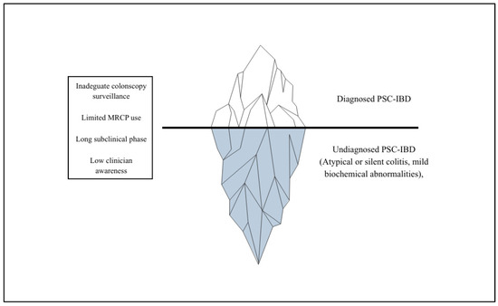
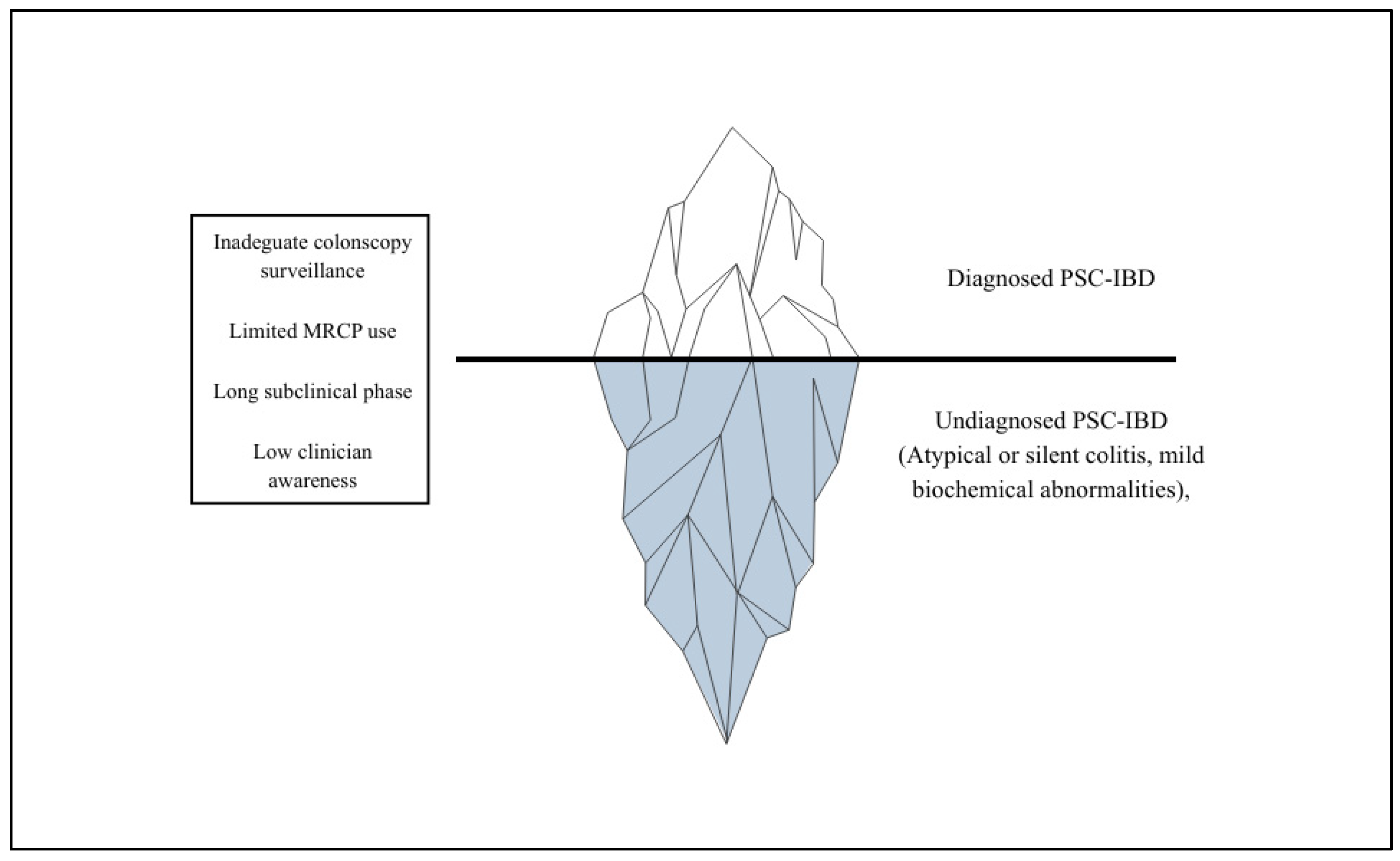
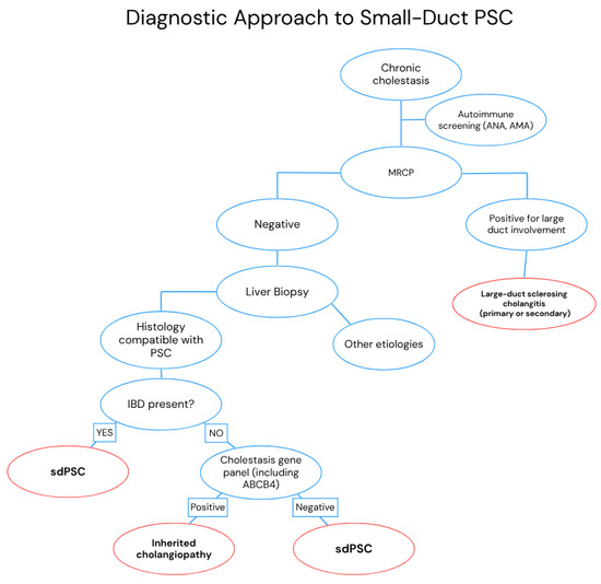
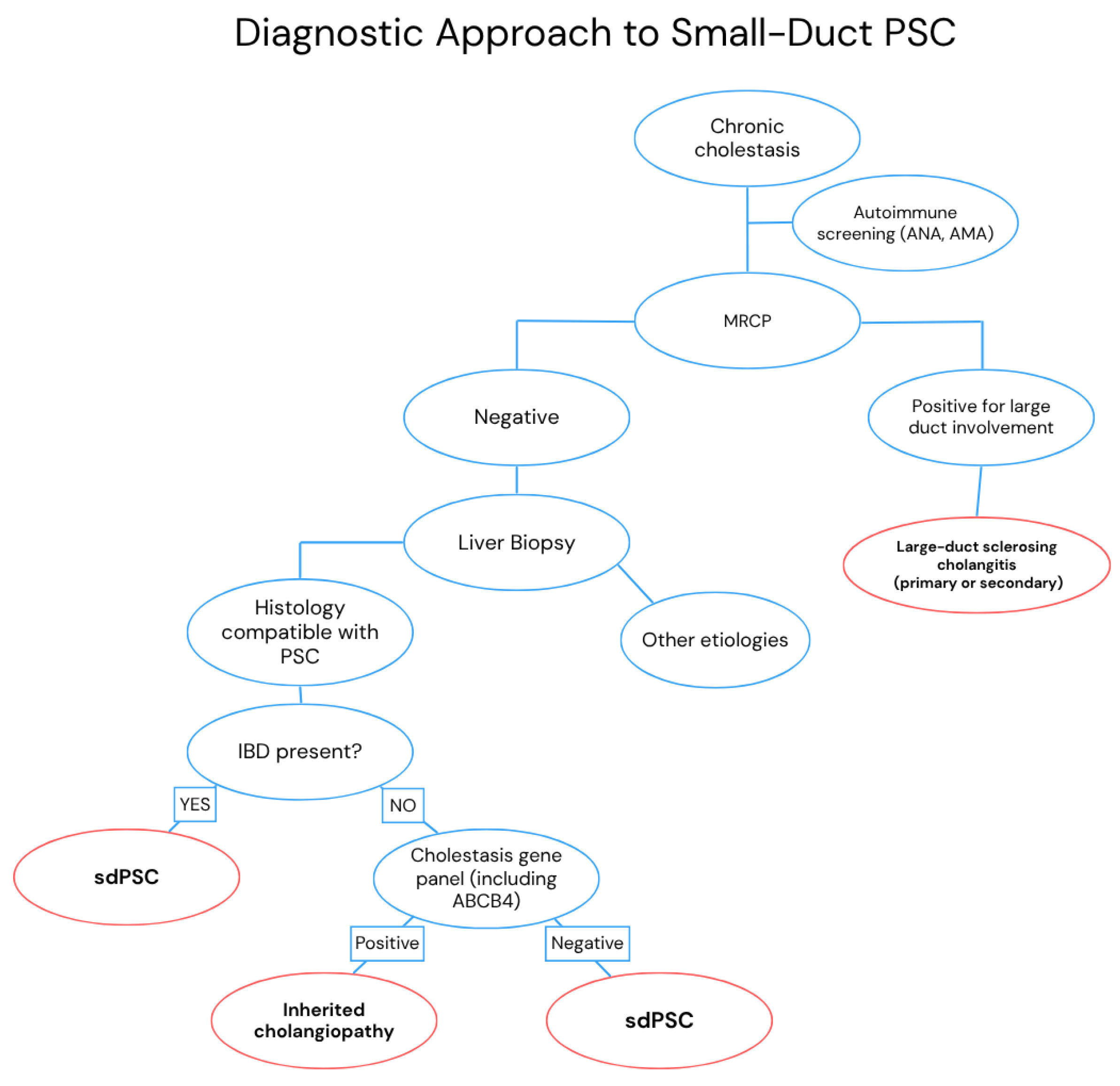
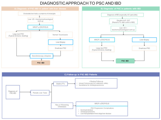

Disclaimer/Publisher’s Note: The statements, opinions and data contained in all publications are solely those of the individual author(s) and contributor(s) and not of MDPI and/or the editor(s). MDPI and/or the editor(s) disclaim responsibility for any injury to people or property resulting from any ideas, methods, instructions or products referred to in the content. |
© 2026 by the authors. Licensee MDPI, Basel, Switzerland. This article is an open access article distributed under the terms and conditions of the Creative Commons Attribution (CC BY) license.