


Abstract
Background/Objectives: Metabolically dysregulated-associated steatotic liver disease (MASLD) is a complex, multifactorial disorder characterised by hepatic lipid accumulation, insulin resistance, oxidative stress, and dysfunction of the gut–liver axis. Given its intricate pathophysiology, multi-target nutritional strategies represent a promising complementary approach. This study aimed to evaluate the biological effects of a multi-component nutraceutical formulation using an integrated in vitro platform replicating intestinal, hepatic, and pancreatic–liver interactions. Methods: The formulation was tested on Caco-2 intestinal cells to assess cell viability, transepithelial electrical resistance (TEER), probiotic functional properties, and glucose absorption. Intestinally processed metabolites were then applied to HepaRG liver cells under hyperglycemic (glucose) or lipotoxic conditions (oleic acid/palmitic acid) to analyse lipid accumulation, cholesterol biomarkers (HMGR, LDL), bile acid production, and cellular damage (ALT, AST). Finally, a pancreas–liver co-culture model (EndoC-βH5 and HepaRG) was employed to investigate insulin secretion and downstream hepatic metabolic signalling (IRS1, GLUT2, glycogen). Results: The formulation preserved intestinal barrier integrity and enhanced probiotic functionality, including aggregation and hydrophobicity. In hepatic models, the treatment significantly reduced intracellular lipid accumulation and triglycerides, while increasing bile acid production and improving cholesterol profiles. Under steatotic stress, it lowered transaminase levels and downregulated lipogenic signalling. In the pancreas–liver axis model, the formulation restored glucose-stimulated insulin secretion and improved hepatic metabolic signalling by increasing IRS1 levels and glycogen synthesis, indicating enhanced insulin sensitivity. Conclusions: These findings support the biological plausibility of a multi-target nutraceutical approach for MASLD. The formulation demonstrates coordinated beneficial effects on intestinal barrier function, hepatic lipid management, and glucose metabolism, providing a strong rationale for further clinical investigation.
1. Introduction
Non-alcoholic fatty liver disease (NAFLD), recently redefined as part of the spectrum of metabolic dysfunction-associated steatotic liver disease (MASLD), is one of the most common chronic liver disorders worldwide and is increasingly recognised in paediatric populations, alongside rising childhood obesity rates [1]. Recent international consensus statements have introduced the terms MAFLD (metabolic dysfunction-associated fatty liver disease) and MASLD (metabolic dysfunction-associated steatotic liver disease) to more accurately reflect the condition’s metabolic basis. These nomenclatures have been proposed to accurately reflect the disease’s metabolic origin. The aetiology of non-alcoholic steatohepatitis (NASH) is recognised as multifactorial, resulting from a synergistic interaction between hormonal, dietary, and environmental determinants against a background of genetic susceptibility [2].
Recently, identifying specific gene polymorphisms, such as PNPLA3, TM6SF2, MBOAT7, and GCKR, has enabled more accurate risk assessment of progression to more severe phenotypes. MASLD in younger individuals is not a static condition and may progress to steatohepatitis (MASH), advanced fibrosis, and early chronic liver complications, highlighting the importance of timely metabolic intervention [3].
The initial factors contributing to pathological progression can be identified in the chronic low-grade inflammatory state (low-grade inflammation), which serves as a unifying element between visceral obesity, metabolic syndrome (MetS) and liver damage [4]. In paediatrics, this dysfunction can manifest early: as early as age 3 years, the expansion of visceral adipose tissue induces remodelling of the immune microenvironment, characterised by infiltration of pro-inflammatory macrophages (M1). These secrete systemic cytokines, such as tumor necrosis factor alpha (TNF-α) and interleukin-6 (IL-6), which interfere with the insulin signalling cascade through the inhibitory phosphorylation of serine residues on the insulin receptor substrates 1 and 2 (IRS1 and IRS2) [5,6,7].
Hepatic insulin resistance is a central driver of altered glucose and lipid homeostasis. Despite compensatory hyperinsulinaemia, the liver maintains uncontrolled endogenous glucose production and increases de novo lipogenesis. The substantial increase in circulating free fatty acid (FFA) levels, resulting from heightened activity of transporters such as CD36, leads to saturation of hepatocyte metabolic capacity. The resulting lipid overload has been shown to induce endoplasmic reticulum stress, mitochondrial dysfunction, and overproduction of reactive oxygen species (ROS). These phenomena ultimately lead to lipotoxicity, cellular apoptosis and the activation of fibrogenesis processes [8,9].
The multifactorial nature of MASLD suggests the need for integrated strategies that can simultaneously modulate insulin resistance, oxidative stress, and intestinal barrier dysfunction. This therapeutic approach aligns with the findings of the most recent systematic reviews and meta-analyses, which highlight the clinical efficacy of multicomponent nutraceutical interventions and dietary polyphenols in improving hepatic steatosis, insulin sensitivity, and systemic metabolic dysfunction in MASLD [10,11,12]. An examination of these concurrent pathophysiological processes elucidates how the formulations under scrutiny link extensive clinical observations to meticulous cellular mechanisms.
In this context, selected phytocompounds and probiotic strains may offer a biologically plausible approach to targeting interconnected metabolic pathways involved in disease progression. Insulin resistance is a key driver of the development of metabolic syndrome-associated liver disease (MASLD), even in paediatric age groups. It contributes to de novo hepatic lipogenesis and to the progression of steatosis to more severe inflammatory forms [10,13].
Myo-inositol, in combination with white mulberry extract titrated in 1-deoxynojirimycin (1-DNJ), plays a pivotal role in this process. Myo-inositol serves as a precursor to intracellular second messengers involved in insulin signalling, thereby enhancing insulin sensitivity and glucose and lipid metabolism [14]. Evidence from clinical and experimental studies indicates that myo-inositol supplementation may enhance metabolic parameters associated with insulin resistance and lipid profile in paediatric populations with obesity [15]. Concurrently, the presence of 1-DNJ in mulberry exerts a potent competitive inhibition of intestinal α-glucosidases, thereby attenuating carbohydrate absorption and reducing postprandial glycaemic peaks. This effect contributes to the limitation of compensatory hyperinsulinemia and the activation of hepatic lipogenesis [16].
To counteract hepatocyte lipotoxicity and endoplasmic reticulum stress, supplementation with silymarin (extracted from Silybum marianum) and a balanced mixture of tocotrienols and tocopherols (vitamin E) is another promising nutraceutical approach. Silymarin has been widely investigated for its hepatoprotective properties, including antioxidant, anti-inflammatory and antifibrotic effects, and experimental models of metabolic liver disease demonstrate its ability to modulate endoplasmic reticulum stress pathways and reduce lipid accumulation in hepatocytes [17,18]. Furthermore, studies in NAFLD models show that silymarin administration attenuates the expression of ER-stress markers such as 78-kDa glucose-regulated protein (GRP78) and X-box binding protein 1 (XBP-1), supporting its role in mitigating ER stress-mediated hepatocellular injury [19]. In parallel, vitamin E, which includes both tocopherols and tocotrienols, acts as a potent lipid-soluble antioxidant that neutralises ROS and limits lipid peroxidation in hepatocyte membranes, thereby reducing oxidative stress associated with metabolic liver disease [20].
Finally, modulation of the gut–liver axis represents a crucial frontier in the treatment of metabolic liver diseases. The utilisation of specific probiotic strains, including Lactobacillus rhamnosus (L. rhamnosus) and Bifidobacterium species, has been associated with improvements in gut microbiota composition and intestinal barrier integrity. Experimental and clinical studies suggest that probiotic supplementation can enhance the expression of tight junction proteins and restore intestinal epithelial barrier function, thereby reducing intestinal permeability and limiting the translocation of bacterial endotoxins into the portal circulation, a key mechanism implicated in the pathogenesis of NAFLD [21,22]. Moreover, specific strains such as L. rhamnosus GG have demonstrated the ability to attenuate hepatic lipid accumulation and improve metabolic parameters in experimental models of NAFLD, highlighting the potential role of targeted probiotic therapy in modulating disease progression through gut microbiota–mediated mechanisms [23]. Consistent with these findings, systematic reviews and meta-analyses indicate that probiotic or synbiotic interventions may improve hepatic steatosis, inflammatory markers, and liver enzymes in individuals with NAFLD, supporting the concept that modulation of the gut microbiome represents a promising therapeutic strategy for metabolic liver disease [24].
In contrast to conventional nutraceutical methodologies, which predominantly emphasise the efficacy of individual ingredients in isolated-cell models, this study arose from the need to appraise a multicomponent formulation through a systems approach that can concurrently consider the multiple sites implicated in the pathogenesis of MASLD. To achieve this objective, a sequential in vitro platform was developed. This platform is designed to simulate the passage of the formulation across the intestinal barrier and the subsequent release of digested, bioavailable metabolites into liver and pancreatic models. This approach also represents a methodological advancement compared with conventional gut–liver or pancreas–liver co-culture systems. In contrast to the simultaneous or bidirectional models, in which different cell types are exposed to the same medium and stimuli, the sequential design ensures a more physiologically relevant progression of exposure. Specifically, only metabolites processed in the intestine are transferred to downstream compartments, thereby avoiding direct, non-physiological exposure of liver and pancreatic cells to the original formulation. Furthermore, the platform integrates multiple organ axes into a single experimental flow, enabling parallel investigation of both direct effects on the liver and pancreas–liver metabolic crosstalk. This configuration offers a more accurate reflection of inter-organ communication under postprandial conditions and facilitates a more profound mechanistic understanding than conventional dual systems.
Accordingly, the present study aimed to investigate the biological effects of an integrated nutraceutical formulation containing probiotics, insulin-sensitising compounds, antioxidant micronutrients, and botanical extracts using a sequential multi-compartment in vitro platform. Specifically, the study evaluated whether the formulation could modulate intestinal barrier integrity, glucose handling, hepatic lipid accumulation, transaminase levels, and key molecular markers involved in lipid uptake, storage, and insulin signalling. Using interconnected intestinal, hepatic, and pancreatic–liver models, this work sought to provide mechanistic evidence supporting a multi-target nutritional strategy relevant to MASLD-related metabolic dysfunction. Although the present experimental models do not specifically reproduce paediatric physiology, paediatric MASLD represents one of the clinical contexts in which early metabolic modulation strategies may be of increasing interest.
2. Materials and Methods
2.1. Agents’ Preparation
The study formulation was diluted 1:2000 to replicate the intended human in vitro exposure, following previously described approaches [25,26]. Briefly, the formulation was prepared by combining 500 mg of myo-inositol, 100 mg of choline bitartrate, 100 mg of white mulberry extract, 62.5 mg of milk thistle extract, 50 mg of tocotrienols–tocopherols from rice, 27 mg of vitamin E acetate 50% (equivalent to 13.5 mg of vitamin E), 10 mg (equivalent to 1 × 10⁹ CFU) of Bifidobacterium animalis (B. animalis), 2.86 mg (equivalent to 1 × 10⁹ CFU) of L. rhamnosus, and 6 mg of vitamin D3 100,000 IU/g (equivalent to 15 µg of vitamin D3).
After homogenization of the powder, the mixture was dissolved in 15 mL of phenol red-free Dulbecco’s Modified Eagle Medium (DMEM; Merck Life Science, Rome, Italy) supplemented with 2 mM L-glutamine and 1% penicillin–streptomycin (initial concentration of 57.3 mg/mL) and subsequently diluted in phenol red-free DMEM supplemented with 2 mM L-glutamine and 1% penicillin–streptomycin to reach the final concentration of 0.05× in the well. Colony-forming units (CFU) were determined by conventional plate count techniques [27,28] prior to preparation, after dilution, and after the exposure duration. Viability tests were performed for both B. animalis and L. rhamnosus in the presence and absence of antibiotics. Since no significant differences in CFU counts were detected in the presence of penicillin–streptomycin, antibiotics were maintained throughout the cell culture experiments to prevent contamination and preserve probiotic viability.
To validate the selection of the working concentration used in the study, a preliminary dose–response evaluation of the complete nutraceutical formulation was conducted using the dilutions documented in Table 1. This formulation is referred to throughout the manuscript as the nutraceutical formulation.
Table 1.
We report the concentrations of each component of the study formulation for the tested dilution. These concentrations have been verified against the latest scientific literature.
For intestinal viability experiments, a 10% sodium dodecyl sulphate (SDS) solution (Merck Life Science, Rome, Italy) was used as a positive control for damage [29].
A positive control was included in each phase of the study to ensure the validity and accuracy of the results. Specifically, for the hyperglycaemic (hypercholesterolemic) condition, red yeast rice (RYR) was used at a concentration of 1 µM [30]; for the condition of hepatic steatosis, obeticholic acid (OCA) was used at a concentration of 10 µM [31]; finally, for the hyperglycaemic condition, metformin was used at a concentration of 1 mM [32].
2.2. Preparation of Metabolic and Steatotic Stress Conditions
To reproduce hyperglycaemic metabolic stress, cells were exposed to glucose (Merck Life Science, Rome, Italy) at a final concentration of 30 mM for 24 h, as previously reported in hepatic in vitro models designed to induce hyperglycaemia-related metabolic dysfunction, oxidative stress, and insulin-resistance-associated alterations [33]. This condition was used as an operational model of high-glucose (hyperglycaemia) metabolic stress rather than to specifically dissect glucose-dependent effects from osmolarity-dependent effects.
To induce hepatic steatosis and lipotoxic stress, hepatic cells were treated with a free fatty acid (FFA) mixture composed of oleic acid and palmitic acid complexed to fatty-acid-free bovine serum albumin (BSA). The oleic acid/palmitic acid (OA/PA) mixture was prepared at a molar ratio of 2:1 and used at final concentrations of 0.4 mM and 0.2 mM for 24 h, as previously described in vitro steatosis models [34]. Therefore, the OA/PA-induced lipotoxicity model was employed to assess the nutraceutical formulation’s response, with BSA serving as the technical vehicle.
2.3. Cell Lines and Culture Conditions
Caco-2 human intestinal epithelial cells (American Type Culture Collection, ATCC, Manassas, VA, USA) were cultured in Advanced DMEM/Advanced Nutrient F-12 Ham’s medium (Adv-DMEM/F12; Gibco®, Thermo Fisher, Waltham, MA, USA), supplemented with 10% foetal bovine serum (FBS), 2 mM L-glutamine, and 1% penicillin–streptomycin. Cells were maintained at 37 °C in a humidified 5% CO2 incubator. For experiments, Caco-2 cells were used between passages 26 and 32 to preserve paracellular permeability and transport properties [35].
For viability assays, Caco-2 cells were seeded at 1 × 10⁴ cells/well in 96-well plates. For intestinal barrier experiments, probiotic-related assays, and glucose uptake analyses, cells were seeded at 2 × 10⁴ cells/insert on 6.5 mm Transwell® inserts with 0.4 µm pore-size polycarbonate membranes (Corning® Costar®, Merck Life Science, Rome, Italy) in 24-well plates [36].
Human pancreatic β-cells, designated EndoC-βH5, were obtained from Human Cell Design (Paris, France) and maintained as previously described [37] using Ulti-β1® medium, specifically formulated to preserve human β-cell functional identity. For viability assays, cells were seeded at 1 × 10⁴ cells/well in 96-well plates. For pancreas–liver co-culture experiments, 1.5 × 10⁵ cells were seeded onto Transwell® inserts and allowed to stabilise for approximately 7 days to ensure glucose-dependent insulin secretion [38].
Human HepaRG cells (from Merck Life Science, Rome, Italy) were used as a differentiated hepatic model to more closely recapitulate hepatocyte-like metabolic functions relevant to MASLD-related lipid and glucose metabolism. Cells were cultured according to the supplier’s recommendations in William’s E medium supplemented with 10% FBS, 2 mM L-glutamine, 1% penicillin–streptomycin, hydrocortisone hemisuccinate, and insulin-containing differentiation supplements, and were maintained at 37 °C in a humidified 5% CO2 incubator [39]. For viability assays, HepaRG cells were seeded at 1 × 10⁴ cells/well in 96-well plates. For lipid accumulation, triglyceride levels, 3-hydroxy-3-methylglutaryl-CoA reductase (HMGR), low-density lipoprotein (LDL) levels, bile acid production, alanine aminotransferas (ALT), aspartate aminotransferase (AST), lactate dehydrogenase (LDH), cluster of differentiation 36 (CD36), sterol regulatory element-binding protein 1 (SREBP-1), peroxisome proliferator-activated receptor gamma (PPARγ), perilipin, IRS1, glycogen synthesis, glucose uptake, glucose transporter 2 (GLUT2) analyses, and AMP-activated protein kinase (AMPK) signalling cells were seeded at 1 × 10⁶ cells/well in 6-well plates or according to the requirements of each assay.
2.4. Experimental Design
All experiments were conducted using established, commercially available human cell lines under normal laboratory conditions at biosafety level 2 (BSL-2). All experimental procedures complied with applicable institutional biosafety regulations and standard laboratory safety guidelines.
The study was organised into four sequential experimental phases: an intestinal phase, a hepatic metabolic stress phase, a hepatic steatosis/lipotoxicity phase, and a pancreas–liver axis phase (Figure 1).
Figure 1.
Schematic representation of the sequential multi-compartment in vitro platform. The experimental workflow comprised an intestinal phase using differentiated Caco-2 cells cultured on Transwell® inserts to generate the intestinally processed metabolite, followed by its application to three downstream models: hepatic metabolic stress, hepatic steatosis/lipotoxicity, and the pancreas–liver axis. Across the models, cell viability, intestinal barrier integrity, glucose handling, lipid accumulation, transaminase levels, insulin secretion, and key molecular markers of lipid and glucose metabolism were evaluated.
In the intestinal phase, differentiated Caco-2 cells cultured on Transwell® inserts were treated apically with the nutraceutical formulation, diluted 1:2000, for 1–6 h (concentration evaluated after a dose–response study in Figure A1, Appendix A). Cell viability was evaluated using the MTT assay, and intestinal barrier integrity was assessed by measuring transepithelial electrical resistance (TEER). In addition, the aggregation and hydrophobicity of the probiotic component of the formulation were assessed, together with intestinal glucose uptake.
The basolateral medium collected from the intestinal model after 6 h of treatment was used as an intestinally processed metabolite and subsequently applied to hepatic cells. Specifically, 100 µL of basolateral medium was collected at the end of the 6 h exposure period and directly transferred to the downstream experimental models without further dilution or normalisation. In addition, control and damage-control groups received the same volume (100 µL) of basolateral medium collected from the corresponding intestinal control conditions and processed using the same experimental protocols. This sequential approach was used to more closely reproduce intestinal processing of the formulation prior to hepatic exposure.
In the hepatic metabolic stress phase, HepaRG cells were exposed to 30 mM glucose for 24 h to reproduce hyperglycaemic stress. After stress induction, cells were treated with the intestinally processed metabolite for 24 h. Cell viability, lipid accumulation, triglyceride levels, HMGR, LDL levels, and bile acid production were then evaluated.
In the hepatic steatosis/lipotoxicity phase, HepaRG cells were exposed to an OA/PA FFA mixture complexed to BSA to induce lipid accumulation and lipotoxic stress. Following damage induction, cells were treated with the intestinally processed metabolite and analysed for cell viability, ALT, AST, LDH, CD36, SREBP-1, PPARγ, perilipin and pAMPK/AMPK ratio.
In the pancreas–liver axis phase, EndoC-βH5 pancreatic β-cells were cultured in the apical compartment of Transwell® inserts, while HepaRG cells were cultured in the basolateral compartment to reproduce pancreas–liver metabolic crosstalk. Pancreatic cells were exposed to hyperglycaemic stress using 30 mM glucose for 24 h and subsequently treated with the intestinally processed metabolite. β-cell viability and insulin secretion were assessed. The pancreatic conditioned medium was then used to evaluate downstream hepatic glucose metabolism, including, insulin production,IRS1 levels, glycogen production, glucose uptake, and GLUT2 expression.
In each study condition, the efficacy of the nutraceutical mix was evaluated in a comparative analysis with various positive controls.
2.5. Intestinal Barrier Model
The intestinal barrier model was established using Caco-2 cells cultured on Transwell® inserts for 21–28 days to achieve full differentiation and polarisation, resulting in a continuous epithelial monolayer with functional tight junctions and apical microvilli [40,41,42,43]. Barrier integrity was monitored by measuring TEER with an EVOM3 system. TEER values ≥ 400 Ω·cm2 were considered indicative of a fully developed, functionally competent epithelial barrier. Before treatment, the apical and basolateral media were adjusted to mimic physiological conditions, with apical and basolateral pHs of approximately 6.5 and 7.4, respectively. TEER stability was confirmed before initiating the experiments.
2.6. Pancreas–Liver Co-Culture Model
The pancreas–liver axis model was established using a Transwell® co-culture system. EndoC-βH5 pancreatic β-cells were seeded in the apical compartment and allowed to stabilise, while HepaRG cells were cultured in the basolateral compartment [44]. After stabilisation, the pancreatic insert was positioned above the hepatic compartment to enable intercellular metabolic communication. Hyperglycaemic conditions were induced by exposure to 30 mM glucose [45]. Co-cultures were maintained in phenol red- and FBS-free medium during treatment to avoid analytical interference, thereby enabling the investigation of pancreatic insulin secretion and downstream hepatic glucose metabolism.
2.7. Cell Viability
Cell viability was assessed using the MTT assay [46]. After treatment, cells were incubated for 2 h at 37 °C in phenol red-free medium containing MTT. Absorbance was measured at 570 and 690 nm using an Infinite 200 Pro MPlex microplate reader (Tecan, Männedorf, Switzerland). Results are expressed as mean percentage ± SD relative to untreated control cells, based on five independent biological experiments performed in technical triplicate.
2.8. Surface Hydrophobicity and Aggregation Activity
Surface hydrophobicity and aggregation activity were evaluated using previously described methods [47]. Phosphate-buffered saline (PBS) served as the control. For the hydrophobicity assay, 0.4 mL of xylene was added to 4 mL of probiotic suspension, the mixture was vortexed and then allowed to stand for 15 min to permit phase separation. Absorbance was measured at 600 nm. Surface hydrophobicity was calculated as follows:
Surface hydrophobicity = [(OD600 control − OD600 test)/OD600 control] × 100%
For the aggregation assay, 0.1 mL of probiotic suspension was combined with 2.9 mL of PBS, and absorbance at 600 nm was recorded immediately and after 2 h of incubation. Aggregation activity was calculated as follows:
where OD600 (0) represents the absorbance at time zero and OD600 (t) represents the absorbance after 2 h. All measurements were performed in triplicate.
Aggregation activity = [(OD600 (0) − OD600 (t))/OD600 (0)] × 100%
2.9. Glucose Uptake Assay
Glucose uptake was measured using a Glucose Uptake Colourimetric Assay Kit (Merck Life Science, Rome, Italy) [45]. Absorbance was recorded at 412 nm at 5-min intervals with an Infinite 200 Pro MPlex microplate reader. Data are expressed as mean percentage ± SD relative to untreated control cells, based on five independent biological experiments conducted in technical triplicate.
2.10. Insulin Quantification
Insulin levels were quantified using the Human HS-INS high-sensitivity insulin Acquant® ELISA Kit (FineTest, Wuhan, China) following the manufacturer’s instructions [48]. Absorbance was measured at 450 nm with an Infinite 200 Pro MPlex microplate reader. A standard calibration curve was generated from insulin concentrations ranging from 7.813 to 500 pg/mL. Results are expressed as mean ± SD relative to untreated control cells, based on five independent biological experiments performed in technical triplicate.
2.11. Lipid Accumulation Assay
Total lipid accumulation was assessed using the Hepatic Lipid Accumulation Assay Kit (Abcam, Cambridge, UK) following the manufacturer’s protocol [46]. After treatment, cells were fixed, washed, and stained with Oil Red O working solution for 20 min at room temperature. Stained cells were visualised under a Leica DM1000 microscope (Wetzlar, Hesse, Germany). For quantitative analysis, dye extraction solution was added, and absorbance was measured at 490–520 nm using an Infinite 200 Pro MPlex microplate reader. Lipid accumulation is expressed relative to untreated control cells.
2.12. Triglyceride Quantification
Triglyceride levels were measured using a colourimetric assay according to the manufacturer’s instructions [49]. Briefly, 10 µL of each sample was added to the appropriate wells, followed by 150 µL of reaction mixture. After 60 min of incubation, absorbance was measured at 540 nm using an Infinite 200 Pro MPlex microplate reader. Results were calculated from a standard curve spanning 3.13 to 200 mg/dL and expressed as mean ± SD relative to untreated control cells, based on five independent biological experiments performed in technical triplicate.
2.13. HMGR Quantification
HMGR levels were measured using the HMGR ELISA Kit (LSBio, Seattle, DC, USA) according to the manufacturer’s instructions [50]. After treatment, cells were lysed and incubated with biotinylated detection antibody and HRP-streptavidin, followed by TMB substrate and stop solution. Absorbance was measured at 450 nm using an Infinite 200 Pro MPlex microplate reader. HMGR concentrations were determined from a standard curve spanning 0.625 to 40 ng/mL. Data are presented as mean ± SD from five independent biological experiments performed in technical triplicate.
2.14. LDL Levels Determination
LDL levels were measured using LDLC colourimetric assay kits based on the cholesterol oxidase/phenol aminophenazone method (Elabscience, Wuhan, China) following the manufacturer’s protocol [51]. Absorbance was recorded at 546 nm using an Infinite 200 Pro MPlex microplate reader. Results were normalised to protein content and expressed relative to untreated control cells, based on five independent biological experiments performed in technical triplicate.
2.15. Bile Acid Determination
Bile acid production by hepatic cells was assessed using a fluorometric total bile acid assay, following established protocols [52]. Cells were washed with cold PBS, lysed by sonication in cold 1× PBS, and centrifuged at 10,000× g for 10 min at 4 °C. Bile acids were quantified by resorufin fluorescence, with excitation at 560 nm and emission at 590 nm, using an Infinite 200 Pro MPlex microplate reader. Data are expressed as mean ± SD, normalised to protein content and reported relative to untreated controls.
2.16. Transaminase and LDH Assays
ALT and AST levels were measured using colourimetric assay kits according to the manufacturer’s instructions [53]. Hepatic cells exposed to steatotic/lipotoxic conditions were homogenised in assay buffer and centrifuged at 15,000× g for 10 min. The reaction mixture was added, and absorbance was recorded at 570 nm using the Infinite 200 Pro MPlex microplate reader. Plates were protected from light and incubated at 37 °C, with readings collected every 5 min. Results were expressed as mean ± SD relative to untreated control cells.
LDH release was assessed as an index of cytotoxicity. Briefly, prepared LDH reagent was added to each well containing hepatic cells under steatotic/lipotoxic conditions. Plates were gently agitated and incubated at room temperature for 10 min. Absorbance was measured at 500 nm using the Infinite 200 Pro MPlex microplate reader. LDH release was calculated relative to control and total lysis wells.
2.17. CD36 Quantification
CD36 levels in hepatic cell culture supernatants were quantified using a Human CD36/SR-B3 ELISA Kit (Thermo Fisher, Milan, Italy) following the manufacturer’s instructions [34]. Absorbance was measured at 450 nm with an Infinite 200 Pro MPlex microplate reader. CD36 concentrations were determined by interpolation from a standard calibration curve spanning 2 to 500 ng/mL and were expressed as mean ± SD relative to untreated control cells, based on five independent biological experiments performed in technical triplicate. The results were interpreted as an extracellular/soluble CD36-related signal.
2.18. IRS1 Quantification
IRS1 levels in hepatic cell lysates under hyperglycaemic conditions were measured using an ELISA kit (NovusBio, San Diego, CA, USA) following the manufacturer’s instructions [36]. Samples were incubated with a biotinylated detection antibody and an HRP conjugate, then with a substrate reagent. Absorbance was measured at 450 nm using the Infinite 200 Pro MPlex microplate reader. Concentrations were calculated from a standard curve spanning 0.16 to 10 ng/mL and expressed relative to untreated control cells.
2.19. Glycogen Measurement
Glycogen content in hepatic cell lysates under hyperglycaemic conditions was assessed using a Glycogen Assay Kit (Merck Life Science, Rome, Italy) [54]. Absorbance was measured at 570 nm with the Infinite 200 Pro MPlex microplate reader. Results are expressed as mean ± SD relative to untreated control cells, based on five independent biological experiments conducted in technical triplicate.
2.20. Western Blot
Hepatic cells were lysed on ice in Complete Tablet Buffer (Roche, Basel, Switzerland) supplemented with 2 mM Na3VO4, 1 mM phenylmethylsulfonyl fluoride (PMSF), and protease/phosphatase inhibitors (Merck Life Science, Rome, Italy). Protein concentration was determined, and 40 µg of total protein was separated by 10% SDS-polyacrylamide gel electrophoresis (PAGE) and transferred to polyvinylidene fluoride (PVDF) membranes. Membranes were incubated overnight at 4 °C with the following primary antibodies: anti-SREBP-1, anti-PPARγ, anti-perilipin, anti-GLUT2, anti-pAMPK and anti-AMPK (all 1:500, Santa Cruz, CA, USA). β-actin (1:1000, Merck Life Science, Rome, Italy) served as the loading control. Additionally, prior to calculating the ratio of the two protein forms (p-AMPK and AMPK), the initial step involved normalizing their expressions on β-actin. Bands were detected using appropriate secondary antibodies and quantified by densitometric analysis. Results are expressed as mean ± SD relative to untreated control cells.
2.21. Statistical Analysis
All experiments were performed with five independent biological replicates, each conducted in technical triplicate. For statistical analysis, the technical triplicates were averaged, with each biological replicate contributing an independent value. Consequently, the statistical unit was the biological experiment, rather than the technical replicates. Statistical analyses were conducted using GraphPad Prism 10.6.1. Results are presented as mean ± SD. Statistical significance was assessed by one-way analysis of variance (ANOVA) with Bonferroni’s post hoc test. A p-value < 0.05 was considered statistically significant.
3. Results
3.1. Intestinal Safety, Cell Viability, and Barrier Integrity of the Nutraceutical Formulation
As an initial step, the nutraceutical formulation was evaluated in an intestinal model to exclude potential cytotoxic effects and to assess epithelial barrier integrity over an exposure period of 1 to 6 h. As shown in Figure 2A, the viability of untreated control cells remained stable at approximately 100% throughout the experimental interval. In contrast, the positive damage control (10% SDS) significantly reduced cell viability, with values consistently below 50% compared with untreated cells (p < 0.05).
Figure 2.
Intestinal safety, cell viability, and barrier integrity of the nutraceutical formulation. (A) Cell viability was assessed by MTT assay after 1–6 h of treatment. (B) Transepithelial electrical resistance (TEER) was measured using the EVOM3 system over the same treatment interval. Control = untreated cells; Positive Control = 10% SDS solution; Nutraceutical formulation = 1:2000 dilution. * p < 0.05 vs. Positive Control; # p < 0.05 vs. Control. Data are expressed as the mean ± SD of five independent experiments, performed in triplicate.
Treatment with the nutraceutical formulation maintained cell viability above 70% at all tested time points, indicating no relevant cytotoxic or irritant effects under the experimental conditions. The highest viability values were observed after 4 h of treatment, significantly higher than those of the positive control (p < 0.05).
A similar pattern was observed for epithelial barrier integrity (Figure 2B). Untreated Caco-2 monolayers maintained TEER values of approximately 410 Ω·cm2 ± 50 during the entire observation period, whereas the positive damage control reduced TEER values below the 400 Ω·cm2 threshold considered indicative of preserved monolayer integrity. In contrast, treatment with the nutraceutical formulation maintained or modestly increased TEER values compared with untreated controls throughout the treatment interval (p < 0.05), while remaining significantly higher than the positive control. These findings indicate that the formulation did not impair intestinal barrier integrity and may support epithelial monolayer stability.
3.2. Probiotic-Related Functional Properties and Intestinal Glucose Uptake
Subsequently, the aggregation and surface hydrophobicity of the nutraceutical formulation were evaluated at the intestinal level and compared with those of the individual probiotic strains it contained. As shown in Figure 3A, both individual probiotic strains displayed significantly greater aggregation capacity than untreated controls (approximately 46% for B. animalis and 36% for L. rhamnosus; p < 0.05 vs. control). Notably, the complete nutraceutical formulation further enhanced aggregation activity, increasing by approximately 18% compared with B. animalis alone (p < 0.046) and by 35% compared with L. rhamnosus alone (p < 0.046).
Figure 3.
Probiotic-related functional properties and intestinal glucose uptake in the in vitro intestinal model. (A) Aggregation activity. * p < 0.05 vs. Control; # p < 0.046 vs. individual probiotic strains. (B) Surface hydrophobicity. * p < 0.05 vs. Control; # p < 0.041 vs. individual probiotic strains. (C) Glucose uptake assessed by colourimetric assay under hyperglycaemic conditions. Control = untreated cells; Glucose Control = 30 mM glucose; Nutraceutical formulation = 1:2000 dilution. * p < 0.004 vs. Glucose Control; # p < 0.05 vs. Control. Data are expressed as the mean ± SD of five independent experiments, performed in triplicate.
A similar trend was observed for surface hydrophobicity (Figure 3B). The individual probiotic strains exhibited significantly higher hydrophobicity than untreated controls (p < 0.05), whereas the complete formulation further increased hydrophobicity by approximately 21.5% relative to B. animalis and 39% relative to L. rhamnosus (p < 0.041).
Intestinal glucose uptake was then assessed under hyperglycaemic conditions. As reported in Figure 3C, the nutraceutical formulation significantly increased glucose uptake compared with untreated controls (by about 29%, p < 0.05) and markedly improved glucose uptake compared with cells exposed to glucose stress alone (by about 76%, p < 0.004). These findings suggest that the formulation may favourably modulate probiotic functional behaviour and intestinal glucose handling under metabolically adverse conditions.
3.3. Effects of the Nutraceutical Formulation on Hepatic Cells Under Metabolic Dysfunction
To evaluate the nutraceutical formulation’s potential to modulate pathways associated with metabolic dysfunction, hepatic cells were exposed to 30 mM glucose, and cell viability, lipid accumulation, triglyceride levels, and cholesterol-related parameters were subsequently assessed.
As shown in Figure 4A, hyperglycaemic metabolic stress significantly reduced hepatic cell viability compared with untreated controls (p < 0.05). Treatment with the nutraceutical formulation markedly improved viability, restoring values close to those of the control group, with an approximate 47% recovery relative to glucose-stressed cells (p < 0.05).
Figure 4.
Effects of the nutraceutical formulation on hepatic cells under hyperglycaemic metabolic stress. (A) Cell viability assessed by MTT assay. * p < 0.035 vs. Hyperglycaemia. (B) Triglyceride levels determined by colourimetric/ELISA assay. * p < 0.005 vs. Hyperglycaemia; # p < 0.046 vs. Control; α p < 0.043 vs. Hyperglycaemia + RYR. (C) Intracellular lipid accumulation quantified by a specific assay. * p < 0.001 vs. Hyperglycaemia; # p < 0.016 vs. Control; α p < 0.012 vs. Hyperglycaemia + RYR. (D) HMGR levels measured by ELISA. * p < 0.002 vs. Hyperglycaemia; # p < 0.039 vs. Control; α p < 0.05 vs. Hyperglycaemia + RYR. (E) LDL levels determined by colourimetric assay. * p < 0.0015 vs. Hyperglycaemia; # p < 0.05 vs. Control; α p < 0.047 vs. Hyperglycaemia + RYR. (F) Bile acid production quantified by fluorometric assay. * p < 0.0005 vs. Hyperglycaemia; # p < 0.05 vs. Control; α p < 0.042 vs. Hyperglycaemia + RYR. Control = untreated cells; Hyperglycaemic metabolic stress = induction with 30 mM glucose; Nutraceutical formulation = 1:2000 dilution. Data are expressed as the mean ± SD of five independent experiments, performed in triplicate.
Triglyceride content and intracellular lipid accumulation were then evaluated. As reported in Figure 4B,C, hyperglycaemic metabolic stress significantly increased both triglyceride levels and lipid accumulation compared with untreated controls (p < 0.05). In contrast, treatment with the nutraceutical formulation significantly attenuated these alterations, reducing triglycerides and lipid accumulation by approximately 45% and 54%, respectively, compared with hyperlipidic-stressed cells (p < 0.05). In addition, in order to verify the actual effect, a comparison was made between the nutraceutical formulation and an RYR extract. In the present study, the results showed that the nutraceutical formulation modulated triglyceride levels (approximately 15% compared with RYR, p < 0.043) and lipid accumulation (approximately 23.5% compared with RYR, p < 0.012) in a manner similar to RYR, suggesting a favourable influence on hepatic lipid homeostasis.
To further investigate modulation of cholesterol-related pathways, HMGR, LDL levels, and bile acid production were assessed. Figure 4D,E show that hyperglycaemic metabolic stress significantly increased HMGR and LDL-associated signals compared with untreated controls (increase of approximately 19.5% and 17.3%, respectively; p < 0.05). Following treatment with the nutraceutical formulation, both parameters were significantly reduced compared with hyperlipidic-stressed cells (by approximately 52% and 54%, respectively), approaching control values. Additionally, the nutraceutical formulation demonstrated favourable modulation of cholesterol-related parameters. Specifically, there was a descriptive reduction of approximately 27% in HMGR levels (p < 0.05) and 31% in LDL levels (p < 0.047) relative to the RYR reference condition in the in vitro setting tested. Finally, bile acid production was markedly impaired under hyperglycaemic metabolic stress, showing an approximate 78% reduction compared with untreated controls (Figure 4F, p < 0.05). Treatment with the nutraceutical formulation significantly increased bile acid production by approximately 74% relative to hyperglycaemic metabolic stress conditions (p < 0.005), with an additional descriptive increase of about 25% relative to the RYR reference condition (p < 0.042). Collectively, these findings indicate favourable modulation of hepatic viability, lipid handling, and cholesterol-related metabolism under metabolic stress.
3.4. Effects of the Nutraceutical Formulation Under Hepatic Steatosis and Lipotoxic Stress Conditions
The beneficial effects of the nutraceutical formulation were further evaluated in a hepatic steatosis/lipotoxicity model induced by exposure to the OA/PA free fatty acid mixture. Particular attention was given to cell viability and hepatocellular injury markers, including ALT and AST, as excessive intracellular lipid accumulation is a key feature of MASLD-related hepatic damage.
As shown in Figure 5A, exposure to the FFA mixture (hepatic steatosis) significantly reduced hepatic cell viability compared with untreated controls (p < 0.05). In parallel, hepatic steatosis significantly increased ALT (Figure 5B) and AST (Figure 5C) levels relative to untreated cells (p < 0.05), confirming hepatocellular injury under these conditions.
Figure 5.
Hepatocellular injury analysis following treatment with the nutraceutical formulation under steatotic/lipotoxic conditions. (A) Cell viability was assessed by the MTT assay. * p < 0.0002 vs. Hepatic steatosis. (B) ALT levels are measured by a specific colourimetric/ELISA assay. * p < 0.0001 vs. Hepatic steatosis; # p < 0.011 vs. Control; α p < 0.012 vs. Hepatic steatosis + OCA. (C) AST levels measured by a specific colourimetric/ELISA assay. * p < 0.0001 vs. Hepatic steatosis; # p < 0.05 vs. Control; α p < 0.0042 vs. Hepatic steatosis + OCA. Control = untreated cells; Hepatic steatosis = induction with a mixture of oleic acid and palmitic acid in a 2:1 ratio; Nutraceutical formulation = 1:2000 dilution. Data are expressed as the mean ± SD of five independent experiments, performed in triplicate.
Treatment with the nutraceutical formulation significantly improved cell viability, restoring values close to those observed in untreated controls, with an approximate 48% recovery relative to the hepatic steatosis cell group (p < 0.05). In addition, ALT and AST levels were markedly reduced following treatment, decreasing by approximately 65% and 63%, respectively, compared with the hepatic steatosis cell group (p < 0.05), with values approaching those of the control group. In addition, OCA was included as a reference compound to provide context for the steatotic/lipotoxic model’s response. In this experimental setting, the nutraceutical formulation reduced ALT and AST levels by approximately 50% (p < 0.012) and 70% (p < 0.0042), respectively, compared to the OCA reference condition.
Overall, these findings indicate that the formulation may attenuate hepatocellular damage and preserve viability under conditions of lipid overload and lipotoxic stress (hepatic steatosis).
3.5. Effects of the Nutraceutical Formulation on Lipid Uptake, Synthesis, and Storage Pathways Under Hepatic Steatosis Conditions
To investigate the mechanisms underlying the observed reduction in hepatic lipid burden, key regulators of fatty acid uptake, lipogenesis, and lipid droplet storage were evaluated. Specifically, CD36, a membrane transporter involved in fatty acid uptake; SREBP-1, a major transcriptional regulator of de novo lipogenesis; PPARγ, a transcription factor associated with triglyceride storage; and perilipin, a structural protein involved in lipid droplet stabilisation, were analysed.
As shown in Figure 6A, induction of hepatic steatosis resulted in a significant increase in CD36 levels compared with untreated controls (p < 0.05). Treatment with the nutraceutical formulation significantly reduced CD36 expression by approximately 52.5% relative to steatotic/lipotoxic cells (p < 0.027), with values approaching those of the control group. Relative to the OCA reference condition, the nutraceutical formulation showed a reduction of approximately 25% in CD36 levels (p < 0.031). This suggests favourable modulation of fatty acid uptake in this in vitro model.
Figure 6.
Effects of the nutraceutical formulation on hepatic lipid metabolism pathways under steatotic/lipotoxic conditions. (A) CD36 levels measured by ELISA. * p < 0.0001 vs. Hepatic steatosis; # p < 0.027 vs. Control; α p < 0.031 vs. Hepatic steatosis + OCA. (B) SREBP-1 expression was assessed by Western blot analysis. * p < 0.0001 vs. Hepatic steatosis; # p < 0.011 vs. Control; α p < 0.022 vs. Hepatic steatosis + OCA. (C) PPARγ expression assessed by Western blot analysis. * p < 0.0002 vs. Hepatic steatosis; # p < 0.036 vs. Control; α p < 0.05 vs. Hepatic steatosis + OCA. (D) Perilipin expression was assessed by Western blot analysis. * p < 0.0002 vs. Hepatic steatosis; # p < 0.028 vs. Control; α p < 0.008 vs. Hepatic steatosis + OCA. (E) Ratio pAMPK/AMPK expression was assessed by Western blot analysis. * p < 0.0001 vs. Hepatic steatosis; # p < 0.024 vs. Control; α p < 0.015 vs. Hepatic steatosis + OCA. (F) Representative Western blot bands. Control = untreated cells; Hepatic steatosis = induction with a mixture of oleic acid and palmitic acid in a 2:1 ratio; Nutraceutical formulation = 1:2000 dilution. Data are expressed as the mean ± SD of five independent experiments, performed in triplicate.
Similarly, SREBP-1 expression was markedly increased in the hepatic steatosis condition compared with untreated controls (Figure 6B, p < 0.05). Treatment with the formulation significantly attenuated this response, reducing SREBP-1 levels by approximately 55% compared with steatotic/lipotoxic cells (p < 0.05), again approaching control values. Nutraceutical formulations also reduced SREBP-1 levels by about 26% (p < 0.022) compared to the OCA reference condition, suggesting they can modulate signals that control fat production in conditions of excess fat or lipotoxicity. PPARγ (Figure 6C) and perilipin (Figure 6D) were also significantly elevated under steatotic/lipotoxic conditions compared with untreated cells (increases of approximately 38% and 24%, respectively; p < 0.05). In contrast, treatment with the nutraceutical formulation restored both markers to basal levels, with approximate recoveries of 59.5% for PPARγ and 69% for perilipin relative to the hepatic steatosis cell group (p < 0.05). Moreover, the nutraceutical formulation diminished levels of PPARγ and perilipin by approximately 31% (p < 0.05) and 82% (p < 0.008), respectively, relative to the OCA reference condition, thereby indicating a favourable modulation of lipid storage-related pathways in this experimental model.
In conclusion, to establish the efficacy of the nutraceutical formulation in restoring the hepatic energy sensor, the post-treatment expression of pAMPK/AMPK was investigated (Figure 6E). The data demonstrated that the nutraceutical formulation was capable of restoring pAMPK expression compared with the damage induced by hepatic steatosis (approximately 52.5%, p < 0.05). Additionally, the formulation increased pAMPK/AMPK expression by approximately 30% (p < 0.015), relative to the OCA reference condition, suggesting that AMPK-related metabolic signalling may have recovered under the tested conditions. Collectively, these findings suggest that the formulation may favourably modulate complementary pathways involved in hepatic fatty acid uptake, lipid synthesis, and intracellular lipid storage.
3.6. Effects of the Nutraceutical Formulation on the Pancreas–Liver Axis Under Hyperglycaemic Conditions
In the final phase of the study, a pancreas–liver co-culture model was established under hyperglycaemic conditions to investigate whether the nutraceutical formulation could modulate glucose homeostasis via inter-organ metabolic crosstalk.
Initially, pancreatic β-cell viability was assessed (Figure 7A). Exposure to glucose stress (hyperglycaemia) significantly reduced pancreatic cell viability compared with untreated controls (approximately 52% reduction, p < 0.05). In contrast, treatment with the nutraceutical formulation significantly improved viability, with an approximate 49% recovery relative to glucose-stressed cells (p < 0.05), restoring values close to those of the control group.
Figure 7.
Effects of the nutraceutical formulation on the pancreas–liver axis under hyperglycaemic conditions. (A) Pancreatic β-cell viability assessed by MTT assay. * p < 0.0002 vs. Hyperglycaemia. (B) Insulin secretion measured by high-sensitivity ELISA. * p < 0.001 vs. Hyperglycaemia. Control = untreated cells; Hyperglycaemia = induction with 30 mM glucose; Nutraceutical formulation = 1:2000 dilution. Data are expressed as the mean ± SD of five independent experiments, performed in triplicate.
The effect of treatment on pancreatic insulin secretion was then evaluated (Figure 7B). Hyperglycaemic stress significantly reduced insulin production compared with untreated controls (approximately 46% reduction, p < 0.05). Following treatment with the nutraceutical formulation, insulin secretion was significantly restored, showing an approximate 52% increase compared with glucose-stressed cells (p < 0.05).
In both parameters analysed, the nutraceutical formulation was found to match the effects exerted by metformin, with no significant differences observed between the two treatments.
These findings indicate that the formulation may preserve pancreatic β-cell function and insulin secretory capacity under metabolically adverse conditions.
3.7. Effects of the Nutraceutical Formulation on Downstream Hepatic Glucose Metabolism Within the Pancreas–Liver Axis
To further evaluate the downstream metabolic consequences of pancreatic modulation, hepatic cell viability and key markers of insulin signalling and glucose metabolism were assessed in the basolateral hepatic compartment of the pancreas–liver co-culture system.
As shown in Figure 8A, exposure to hyperglycaemic conditions significantly reduced hepatic cell viability compared with untreated controls (p < 0.05). Treatment with the nutraceutical formulation significantly improved viability, yielding an approximately 48% increase relative to glucose-stressed cells (p < 0.0002) and restoring values close to those of the control group.
Figure 8.
Effects of the nutraceutical formulation on downstream hepatic glucose metabolism within the pancreas–liver axis under hyperglycaemic conditions. (A) Hepatic cell viability assessed by MTT assay. * p < 0.0002 vs. Hyperglycaemia. (B) IRS1 levels measured by ELISA. * p < 0.007 vs. Hyperglycaemia; # p < 0.05 vs. Control. (C) Glycogen content was quantified by a specific assay. * p < 0.0042 vs. Hyperglycaemia. (D) Glucose uptake was determined by colourimetric assay. * p < 0.001 vs. Hyperglycaemia. (E) GLUT2 expression was assessed by Western blot analysis. * p < 0.0003 vs. Hyperglycaemia. (F) Representative Western blot bands. Control = untreated cells; Hyperglycaemia = induction with 30 mM glucose; Nutraceutical formulation = 1:2000 dilution. Data are expressed as the mean ± SD of five independent experiments, performed in triplicate.
4. Discussion
IRS1 levels and glycogen production were then assessed as markers of insulin responsiveness and intracellular glucose storage. As reported in Figure 8B,C, glucose stress significantly reduced IRS1 levels and glycogen content compared with untreated controls (reduction of approximately 57.5% and 47%, respectively; p < 0.05). In contrast, treatment with the nutraceutical formulation significantly improved both parameters, yielding approximately 51% and 46% increases in IRS1 (p < 0.007) and glycogen production (p < 0.0042), respectively, relative to glucose-stressed cells, returning values toward those of untreated controls.
To further investigate hepatic glucose handling, glucose uptake (Figure 8D) and GLUT2 expression (Figure 8E,F) were subsequently evaluated. Under hyperglycaemic conditions, glucose uptake and GLUT2 levels were significantly reduced compared with untreated controls (reduction of approximately 35% and 51%, respectively; p < 0.05). Treatment with the nutraceutical formulation significantly reversed these alterations, restoring GLUT2 expression to control levels (an increase of approximately 47% vs. glucose-stressed cells, p < 0.001) and increasing glucose uptake by approximately 32% compared with glucose-stressed cells (p < 0.0003).
Notably, across all analysed parameters, the nutraceutical formulation showed effects comparable to metformin, suggesting similar efficacy in mitigating hyperglycaemia-induced hepatic alterations.
Overall, these findings suggest that the formulation may favourably modulate hepatic insulin signalling and glucose utilisation within an integrated pancreas–liver metabolic model.
NAFLD is the most common chronic liver disease worldwide, with prevalence steadily increasing in the paediatric population [55]. It is characterised by excessive lipid accumulation in the liver, not associated with alcohol consumption, and is closely linked to obesity, insulin resistance, and metabolic syndrome [56]. Its pathogenesis is multifactorial and involves dysfunction of the gut–liver axis, oxidative stress, and alterations in glucose and lipid metabolism [57]. Without early intervention, the disease may progress to more severe forms, including steatohepatitis and liver fibrosis [58]. The results of the present study demonstrate that the nutraceutical combination, referred to as the Nutraceutical mix, can exert an integrated, coordinated, multi-target effect on the key pathophysiological mechanisms underlying MASLD development. This aspect is particularly relevant given the multifactorial nature of the disease, characterised by a complex interplay among insulin resistance, dysregulated lipid metabolism, oxidative stress, and alterations in the gut–liver axis [59].
In the intestinal model, the formulation demonstrated a favourable safety profile, preserving cell viability and enhancing epithelial barrier integrity, as evidenced by increased TEER values. This finding is particularly relevant, as impairment of the intestinal barrier is one of the earliest events in MASLD pathogenesis, promoting endotoxin translocation and activation of hepatic inflammatory responses [60]. In this context, the observed effect suggests that the nutraceutical combination may help restore a functional intestinal environment, thereby reducing one of the main triggers of systemic inflammation. In parallel, the increased aggregation and hydrophobicity observed in the intestinal model indicate an enhanced ability to interact with the epithelium, which may translate into greater overall functional efficacy of the formulation. This behaviour suggests that the effect of the combination may extend beyond the contributions of the individual components, supporting a coordinated interaction that could enhance overall biological properties. Another important finding was that Caco-2 cells took up more glucose when they were in a high-blood-sugar state. Specifically, the observed increase may indicate distinct intracellular glucose management and metabolic utilisation by intestinal cells. However, as Morus alba extracts are generally associated with reduced intestinal glucose digestion and transport, further studies on glucose transporter expression and transepithelial flow are needed to clarify this mechanism. Given that insulin resistance is a major driver of MASLD progression, the combination’s ability to act at the intestinal level is strategically important [61,62].
In the hepatic model of metabolic syndrome, the formulation markedly reduced lipid accumulation and triglyceride levels, accompanied by favourable modulation of cholesterol-related pathways. The reduction in HMGR and LDL levels, together with increased bile production, suggests coordinated regulation of lipid metabolism, potentially shifting towards cholesterol elimination rather than accumulation. This finding is consistent with the concept that targeting multiple metabolic pathways may be a useful complementary strategy for modulating hepatic steatosis.
In the steatosis-induced model, the nutraceutical combination demonstrated a significant hepatoprotective effect, as evidenced by restored cell viability and reduced liver damage markers, such as ALT and AST. These findings suggest that the formulation can counteract lipotoxicity and oxidative stress, which are key drivers in the progression from simple steatosis to more advanced liver disease [63,64]. Particularly relevant is the modulation observed in key regulators of lipid metabolism. The reduction in CD36, SREBP-1, PPAR-γ, and perilipin levels indicates a coordinated effect of the combination that simultaneously limits lipid uptake, synthesis, and storage in hepatocytes. This finding suggests that the formulation may broadly modulate the lipogenic cascade, interrupting the mechanisms that lead to pathological lipid accumulation in the liver.
The analysis of the pancreas–liver axis further supported the formulation’s metabolic profile. Under hyperglycaemic conditions, the Mix restored pancreatic cell viability and insulin secretion, and significantly improved hepatic parameters related to glucose metabolism, including IRS1, glycogen, GLUT2, and glucose uptake. These results indicate a global effect on insulin sensitivity and interorgan metabolic communication, both of which are relevant to pathways implicated in MASLD progression. At the molecular level, the nutraceutical formulation has been observed to enhance hepatic insulin sensitivity by modulating IRS1, GLUT2, and glycogen synthesis. In conditions of lipotoxicity and hyperglycaemia, the accumulation of diacylglycerols and ceramides activates PKC, leading to inhibitory phosphorylation of IRS1 and impairment of the PI3K/Akt pathway. This results in reduced insulin signalling and hepatic glycogen synthesis [65,66]. The restoration of IRS1 observed in the experimental model is consistent with the recovery of the downstream insulin cascade and the regulation of glycogen synthase kinase 3 (GSK3) activity, and consequently hepatic glycogen synthesis [66]. Concurrently, GLUT2 normalisation has been shown to enhance hepatic glucose homeostasis. Moreover, increased glycogen synthesis has been identified as a functional indicator of restored insulin sensitivity. Glucose diversion to glycogen synthesis has been shown to decrease the availability of substrates for de novo lipogenesis. This is thought to contribute to the decrease in lipid accumulation and hepatic triglyceride levels observed in the experimental model. These findings are consistent with known mechanisms of insulin metabolic regulation [67]. Moreover, incorporating condition-specific positive controls in each experimental phase enhanced the robustness and interpretability of the results. Despite the metabolic stress model being derived from exposure to elevated glucose levels, this condition was specifically utilised to replicate the glucose-induced dysregulation of hepatic cholesterol homeostasis and the ensuing hypercholesterolemic phenotype previously delineated in in vitro investigations [33,68,69]. Therefore, red yeast rice (RYR) was selected as a positive control due to its well-known cholesterol-lowering activity and relevance to the metabolic pathways investigated in this model. The use of RYR in the hyperglycaemic metabolic stress model, OCA in the steatosis model, and metformin in the hyperglycemic pancreas–liver axis model confirmed the responsiveness of each system to established interventions. The utilisation of these reference compounds facilitated the contextualisation of the nutraceutical formulation within established pharmacological frameworks. It is noteworthy that the nutraceutical combination exhibited comparable or even superior efficacy across several parameters, thereby supporting its potential as a complementary multi-target strategy in MASLD-associated conditions.
An important added value of our study is the demonstrated ‘matrix effect’, which optimises probiotic functionality. Compared with strains tested individually, the complete formulation significantly increased both the aggregation activity and the surface hydrophobicity of the probiotics. The evident synergistic interaction with phytocompounds and micronutrients provides a more robust therapeutic strategy than traditional isolated microbiome modulation.
Furthermore, our results highlight the formulation’s comprehensive, multi-pathway regulatory potential, setting it apart from many existing nutraceutical options described in the literature, which typically target a single hallmark of MASLD pathogenesis [70,71,72]. Specifically, our formulation exhibits a coordinated mechanism involving the preservation of intestinal tight junction integrity, optimisation of glucose management, downregulation of key hepatic lipogenic pathways (SREBP-1 and PPARγ), restoration of insulin secretion in pancreatic β cells and enhancement of downstream hepatic metabolic signalling (IRS1 and GLUT2). The scientific relevance of this multi-target efficacy is reinforced by direct validation against established therapeutic and reference standards, which confirms that a multi-component approach can match or even surpass the modulation of critical metabolic parameters in MASLD-related conditions.
Although these findings were generated in controlled in vitro systems and therefore require confirmation in vivo and in clinical settings, they provide mechanistic support for further translational investigation of multi-target nutritional approaches in MASLD.
This study has some limitations, particularly regarding the use of in vitro results in the clinical context of MASLD. The multi-compartmental platform is a big step forward methodically, as it prevents cells from being exposed to the crude formulation and allows for more controlled, sequential assessment. But the experimental system still can’t fully reproduce the multisystemic complexity of the human disease. Notably, the model does not incorporate the dynamic interaction with visceral adipose tissue and the contribution of chronic low-grade inflammation mediated by M1 macrophages, which are central to the progression of systemic insulin resistance, especially in early-onset paediatric settings. Furthermore, static or transwell models cannot fully simulate human pharmacokinetic and pharmacodynamic processes, including intestinal absorption times, effective bioavailability, systemic distribution, first-pass hepatic metabolism, and clearance mechanisms for multi-botanical compounds. Consequently, although the results provide solid proof of principle for the formulation’s multi-target efficacy and robust biological plausibility, well-designed, controlled clinical studies are needed to confirm its true therapeutic relevance in MASLD and its translation to patients. In addition, the contribution of individual formulation components was not investigated separately, and the study was not designed to formally assess synergistic interactions. Despite this being a limitation, the individual ingredients included in the formulation have been previously described in the literature about their relevance to glucose metabolism, insulin sensitivity, oxidative stress, hepatic lipid management and modulation of the gut–liver axis. The innovative aspect of this study was therefore the evaluation of the final multicomponent formulation within a sequential in vitro gut–liver and pancreas–liver platform, rather than the recharacterisation of each component in isolation. Furthermore, inflammatory and fibrotic pathways were not extensively explored and should be investigated in future studies together with in vivo and clinical validation. Overall, the data suggest that the observed biological activity of the formulation may derive from the combined contributions of its components, enabling simultaneous modulation of multiple pathophysiological pathways. This integrated approach appears particularly relevant in the context of MASLD, where the complexity of the underlying mechanisms may benefit from multifactorial complementary strategies rather than single-target interventions.
5. Conclusions
The findings of the present study indicate that the nutraceutical formulation exerts a coordinated, multi-target effect on several pathways associated with Metabolic dysfunction-associated steatotic liver disease. Specifically, the formulation was shown to preserve intestinal barrier integrity, modulate glucose homeostasis, reduce hepatic lipid accumulation, attenuate markers of hepatocellular injury, and improve parameters related to insulin signalling within the pancreas–liver axis.
The simultaneous modulation of these interconnected mechanisms supports the biological plausibility of a complementary nutritional strategy targeting multiple aspects of MASLD pathophysiology. In this context, the formulation may represent an innovative multi-component approach worthy of further translational investigation. The use of specific positive controls enhances the validity of experimental models, facilitates comparison with reference compounds, and supports the biological relevance of the observed effects.
However, because the current results were obtained in controlled in vitro models, they cannot fully capture the systemic complexity of human MASLD. Furthermore, this study did not specifically investigate the contributions of individual formulation components, formal synergistic interactions, or inflammatory or fibrotic pathways. Consequently, these results should be interpreted as exploratory and hypothesis-generating. Further mechanistic validation, together with in vivo studies and well-designed clinical trials, will be necessary to confirm the approach’s efficacy, define its optimal conditions of use, and clarify its translational relevance.
Author Contributions
Conceptualization, R.G. and F.U.; methodology, R.G., S.M., F.P. and F.U.; software, R.G., S.M., F.P. and F.U.; validation, S.M. and F.U.; formal analysis, F.U.; investigation, F.U.; resources, F.U.; data curation, F.U.; writing—original draft preparation, R.G., S.M., F.P. and F.U.; visualization, F.U.; supervision, F.U.; project administration, F.U.; funding acquisition, F.U. All authors have read and agreed to the published version of the manuscript.
Funding
This research received no external funding.
Institutional Review Board Statement
Not applicable.
Informed Consent Statement
Not applicable.
Data Availability Statement
The data supporting the findings of this study are available from the corresponding author upon request. This includes the raw experimental data and statistical results. The Physiology Laboratory securely stores the data to ensure traceability and long-term preservation.
Acknowledgments
The authors thank Smartfarma S.R.L. for providing the experimental materials used in this study. The tested formulation was provided as a laboratory research prototype and was not a finished product marketed or commercially available at the time of the experiments. Smartfarma S.R.L. had no role in study design, data collection, data analysis, interpretation of results, preparation of the manuscript, or the decision to submit the manuscript for publication.
Conflicts of Interest
R. G. and F. P. are both employees of Noivita Srls. F. U. is the founder of Noivita Srls. The formulation evaluated in this study was an experimental laboratory prototype developed for research purposes only; it was not a finished product that was marketed, registered or commercially available at the time of the experiments. Smartfarma S.R.L. provided the experimental material used in the study but was not involved in the design of the study, the collection, analysis or interpretation of the data, the writing of the manuscript, or the decision to publish the results. Noivita Srls was not involved in the analysis of the data, the interpretation of the results, or the decision to publish. The authors declare that the study was conducted as a preclinical in vitro investigation, and that the results should not be interpreted as promotional or marketing claims for a marketed product.
Abbreviations
The following abbreviations are used in this manuscript:
| ALT | Alanine aminotransferase |
| AMPK | AMP-activated protein kinase |
| ANOVA | Analysis of variance |
| AST | Aspartate aminotransferase |
| B. animalis | Bifidobacterium animalis |
| BSA | Bovine serum albumin |
| CD36 | Cluster of differentiation 36 |
| CFU | Colony-forming units |
| CO2 | Carbon dioxide |
| DMEM | Dulbecco’s Modified Eagle Medium |
| ELISA | Enzyme-linked immunosorbent assay |
| FBS | Foetal bovine serum |
| FFA | Free fatty acids |
| GRP78 | 78-kDa glucose-regulated protein |
| GLUT-2 | Glucose transporter type 2 |
| HMGR | 3-hydroxy-3-methylglutaryl-coenzyme A reductase |
| HRP | Horseradish peroxidase |
| IL-6 | Interleukin-6 |
| IRS1 | Insulin receptor substrate 1 |
| L. rhamnosus | Lactobacillus rhamnosus |
| LDH | Lactate dehydrogenase |
| LDL | Low-density lipoprotein |
| MAFLD | Metabolic dysfunction-associated fatty liver disease |
| MASH | Metabolic dysfunction-associated steatohepatitis |
| MASLD | Metabolic dysfunction-associated steatotic liver disease |
| MetS | Metabolic syndrome |
| MTT | 3-(4,5-dimethylthiazol-2-yl)-2,5-diphenyl tetrazolium bromide |
| NAFLD | Non-alcoholic fatty liver disease |
| NASH | Non-alcoholic steatohepatitis |
| OA | Oleic acid |
| OCA | Obeticholic acid |
| PA | Palmitic acid |
| PBS | Phosphate-buffered saline |
| PMSF | phenylmethylsulfonyl fluoride |
| pAMPK | Phosphorylated AMP-activated protein kinase |
| PPARγ | Peroxisome proliferator-activated receptor gamma |
| PVDF | Polyvinylidene difluoride |
| ROS | Reactive oxygen species |
| RYR | Red yeast rice |
| SD | Standard deviation |
| SDS | Sodium dodecyl sulphate |
| SDS-PAGE | Sodium dodecyl sulphate polyacrylamide gel electrophoresis |
| SREBP-1 | Sterol regulatory element-binding protein 1 |
| TEER | Transepithelial electrical resistance |
| TNF-α | Tumor necrosis factor alpha |
| TMB | Tetramethylbenzidine |
| XBP-1 | X-box binding protein 1 |
| 1-DNJ | 1-deoxynojirimycin |
Appendix A
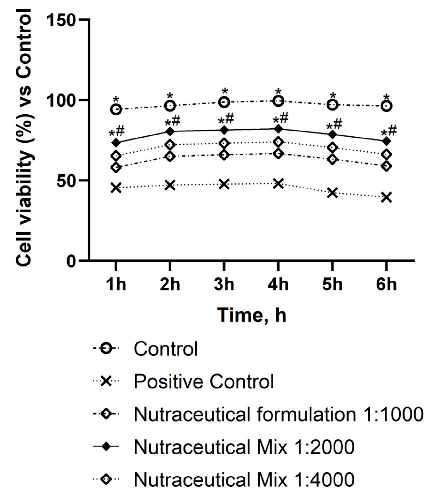
As shown in Figure A1, all dilutions of the Nutraceutical Mix were found to be safe for the intestinal monolayer, with viability remaining above 50% and higher than the positive control (SDS 10%). The 1:2000 dilution performed particularly well, exerting effects similar to the untreated control (p < 0.05) and outperforming all other concentrations over the entire treatment interval.
Figure A1.
Dose–response study of nutraceutical formulation evaluated by MTT test on Caco-2 cells after 1 h to 6 h of treatment. p < 0.05 vs. Control; * p < 0.05 vs. Positive Control (SDS 10%); # p < 0.05 vs. other concentration tested.
References
- Younossi, Z.M.; Golabi, P.; Paik, J.M.; Henry, A.; Van Dongen, C.; Henry, L. The global epidemiology of nonalcoholic fatty liver disease (NAFLD) and nonalcoholic steatohepatitis (NASH): A systematic review. Hepatology 2023, 77, 1335–1347. [Google Scholar] [CrossRef] [Scilit] [PubMed]
- Rupasinghe, K.; Hind, J.; Hegarty, R. Updates in metabolic dysfunction-associated fatty liver disease (MAFLD) in children. J. Pediatr. Gastroenterol. Nutr. 2023, 77, 583–591. [Google Scholar] [CrossRef] [Scilit]
- Nobili, V.; Alisi, A.; Valenti, L.; Miele, L.; Feldstein, A.E.; Alkhouri, N. NAFLD in children: New genes, new diagnostic modalities and new drugs. Nat. Rev. Gastroenterol. Hepatol. 2019, 16, 517–530. [Google Scholar] [CrossRef] [Scilit] [PubMed]
- Podeanu, M.-A.; Ionele, C.M.; Sandu, R.E.; Rogoveanu, I.; Stepan, M.D.; Niculescu, C.E.; Cazacu, S.-M.; Vintilescu, Ș.B. Obesity, metabolic syndrome and MASLD in children: Inflammation as the missing link—A short narrative review. Life 2026, 16, 310. [Google Scholar] [CrossRef] [Scilit] [PubMed]
- Engin, A. Reappraisal of Adipose Tissue Inflammation in Obesity. Adv. Exp. Med. Biol. 2024, 1460, 297–327. [Google Scholar] [CrossRef] [Scilit] [PubMed]
- Kojta, I.; Chacińska, M.; Błachnio-Zabielska, A. Obesity, bioactive lipids, and adipose tissue inflammation in insulin resistance. Nutrients 2020, 12, 1305. [Google Scholar] [CrossRef] [Scilit] [PubMed]
- Calcaterra, V.; Regalbuto, C.; Porri, D.; Pelizzo, G.; Mazzon, E.; Vinci, F.; Zuccotti, G.; Fabiano, V.; Cena, H. Inflammation in obesity-related complications in children: The protective effect of diet and its potential role as a therapeutic agent. Biomolecules 2020, 10, 1324. [Google Scholar] [CrossRef] [Scilit] [PubMed]
- Shaunak, M.; Byrne, C.D.; Davis, N.; Afolabi, P.; Faust, S.N.; Davies, J.H. Non-alcoholic fatty liver disease and childhood obesity. Arch. Dis. Child. 2021, 106, 3–8. [Google Scholar] [PubMed]
- Zhang, C.; Zheng, M.; Bai, R.; Chen, J.; Yang, H.; Luo, G. Molecular mechanisms of lipid droplets–mitochondria coupling in obesity and metabolic syndrome: Insights and pharmacological implications. Front. Physiol. 2024, 15, 1491815. [Google Scholar] [CrossRef] [Scilit] [PubMed]
- Vrentzos, E.; Pavlidis, G.; Korakas, E.; Kountouri, A.; Pliouta, L.; Dimitriadis, G.D.; Lambadiari, V. Nutraceutical Strategies for Metabolic Dysfunction-Associated Steatotic Liver Disease (MASLD): A Path to Liver Health. Nutrients 2025, 17, 1657. [Google Scholar] [CrossRef] [Scilit] [PubMed]
- Hu, Z.; He, Z.; Wang, Y.; Chu, Z.; Zhou, Y.; Li, W.; Luo, F. Targeting the gut-liver axis with dietary polyphenols to ameliorate metabolic dysfunction-associated steatotic liver disease: Advances in molecular mechanisms. Crit. Rev. Food Sci. Nutr. 2026, 66, 1813–1835. [Google Scholar] [CrossRef] [Scilit] [PubMed]
- Starvaggi, J.; Di Chio, C.; De Luca, F.; Previti, S.; Zappalà, M.; Ettari, R. Selected Nutraceuticals in Metabolic Syndrome: Molecular Mechanisms and Clinical Implications. Biomedicines 2026, 14, 646. [Google Scholar] [CrossRef] [Scilit] [PubMed]
- Song, K.; Lee, E.; Lee, H.S.; Youn, Y.H.; Baik, S.J.; Shin, H.J.; Chae, H.W.; Lee, J.W.; Kwon, Y.J. Identification of pediatric MASLD using insulin resistance indices. JHEP Rep. 2025, 7, 101419. [Google Scholar] [CrossRef] [Scilit] [PubMed]
- Long, L.; Huang, Q.; Song, T.; Dai, Z. Myo-inositol rescued insulin resistance and dyslipidemia in db/db mice. J. Appl. Biomed. 2024, 22, 74–80. [Google Scholar] [CrossRef] [Scilit] [PubMed]
- Antoniotti, V.; Partenope, C.; Solito, A.; Mancioppi, V.; Baima, J.; Medina, F.; Dimarakis, S.; Agostini, A.; Sista, M.T.; Monzani, A.; et al. Efficacy of myo-inositol and zinc on insulin resistance in a paediatric population with obesity. Diabetes Obes. Metab. 2025, 27, 1932–1939. [Google Scholar] [CrossRef] [Scilit] [PubMed]
- Street, M.E.; Casadei, F.; Di Bari, E.R.; Ferraboschi, F.; Montani, A.G.; Shulhai, A.-M.; Esposito, S. The role of nutraceuticals and probiotics in addition to lifestyle intervention in the management of childhood obesity—Part 1: Metabolic changes. Nutrients 2025, 17, 1630. [Google Scholar] [CrossRef] [Scilit] [PubMed]
- Malekpour-Dehkordi, Z.; Nourbakhsh, M.; Shahidi, M.; Sarraf, N.; Sharifi, R. Silymarin diminishes oleic acid-induced lipid accumulation in HepG2 cells by modulating the expression of endoplasmic reticulum stress markers. J. Herb. Med. 2022, 33, 100565. [Google Scholar] [CrossRef] [Scilit]
- Wal, P.; Yadav, S.; Jha, S.K.; Singh, A.; Bhargavi, B.; Shivaram, R.; Imran, M.; Aziz, N. Role of natural compounds in non-alcoholic fatty liver diseases (NAFLD): A mechanistic approach. Egypt. Liver J. 2025, 15, 50. [Google Scholar] [CrossRef] [Scilit]
- Sahin, E.; Bagci, R.; Bektur Aykanat, N.E.; Kacar, S.; Sahinturk, V. Silymarin attenuated nonalcoholic fatty liver disease through the regulation of endoplasmic reticulum stress proteins GRP78 and XBP-1 in mice. J. Food Biochem. 2020, 44, e13194. [Google Scholar] [CrossRef] [Scilit] [PubMed]
- Chiurazzi, M.; Cacciapuoti, N.; Di Lauro, M.; Nasti, G.; Ceparano, M.; Salomone, E.; Guida, B.; Lonardo, M.S. The synergic effect of a nutraceutical supplementation associated to a Mediterranean hypocaloric diet in a population of overweight/obese adults with NAFLD. Nutrients 2022, 14, 4750. [Google Scholar] [CrossRef] [Scilit] [PubMed]
- Xue, L.; He, J.; Gao, N.; Lu, X.; Li, M.; Wu, X.; Liu, Z.; Jin, Y.; Liu, J.; Xu, J.; et al. Probiotics may delay the progression of nonalcoholic fatty liver disease by restoring the gut microbiota structure and improving intestinal endotoxemia. Sci. Rep. 2017, 7, 45176. [Google Scholar] [CrossRef] [Scilit] [PubMed]
- Adnan, M.L. The effect of probiotic supplementation as alternative therapy for NAFLD: A literature review. Eur. J. Transl. Clin. Med. 2021, 4, 75–85. [Google Scholar] [CrossRef] [Scilit]
- Jang, H.R.; Park, H.J.; Kang, D.; Chung, H.; Nam, M.H.; Lee, Y.; Park, J.H.; Lee, H.Y. A protective mechanism of probiotic Lactobacillus against hepatic steatosis via reducing host intestinal fatty acid absorption. Exp. Mol. Med. 2019, 51, 1–14. [Google Scholar] [CrossRef] [Scilit] [PubMed]
- Kaufmann, B.; Seyfried, N.; Hartmann, D.; Hartmann, P. Probiotics, prebiotics, and synbiotics in nonalcoholic fatty liver disease and alcohol-associated liver disease. Am. J. Physiol. Gastrointest. Liver Physiol. 2023, 325, G42–G61. [Google Scholar] [CrossRef] [Scilit] [PubMed]
- Galla, R.; Mulè, S.; Ferrari, S.; Molinari, C.; Uberti, F. Combined effects of diosmin, hesperidin, Ruscus aculeatus, Ananas comosus, and bromelain on endothelial function and gut barrier integrity in vitro. Int. J. Mol. Sci. 2025, 26, 10538. [Google Scholar] [CrossRef] [Scilit] [PubMed]
- Barakat, A.; Kramer, J.; de Souza Cristiane, C.; Lehr, C.M. In vitro–in vivo correlation: Shades on some non-conventional dosage forms. Dissolut. Technol. 2015, 22, 19–23. [Google Scholar] [CrossRef] [Scilit]
- Kodinova, S.; Dushkova, M.; Miteva-Petrova, M.; Yanakieva, V.; Petrov, S.; Denkova, Z. Production of probiotic Bulgarian yoghurts obtained from an ultrafiltered cow’s milk. Ir. J. Agric. Food Res. 2020, 59, 1–11. [Google Scholar]
- ISO 4833-1:2013; Microbiology of the Food Chain—Horizontal Method for the Enumeration of Microorganisms—Part 1: Colony Count at 30 °C by the Pour Plate Technique. International Organization for Standardization: Geneva, Switzerland, 2013.
- Welch, J.; Wallace, J.; Lansley, A.B.; Roper, C. Evaluation of the toxicity of sodium dodecyl sulphate (SDS) in the MucilAir™ human airway model in vitro. Regul. Toxicol. Pharmacol. 2021, 125, 105022. [Google Scholar] [CrossRef] [Scilit] [PubMed]
- Ruga, S.; Galla, R.; Penna, C.; Molinari, C.; Uberti, F. The Activity of Ten Natural Extracts Combined in a Unique Blend to Maintain Cholesterol Homeostasis—In Vitro Model. Int. J. Mol. Sci. 2022, 23, 3805. [Google Scholar] [CrossRef] [Scilit] [PubMed]
- Anfuso, B.; Tiribelli, C.; Adorini, L.; Rosso, N. Obeticholic acid and INT-767 modulate collagen deposition in a NASH in vitro model. Sci. Rep. 2020, 10, 1699. [Google Scholar] [CrossRef] [Scilit] [PubMed]
- Malekpour, K.; Hazrati, A.; Soudi, S.; Roshangar, L.; Pourfathollah, A.A.; Ahmadi, M. Combinational administration of mesenchymal stem cell-derived exosomes and metformin reduces inflammatory responses in an in vitro model of insulin resistance in HepG2 cells. Heliyon 2023, 9, e15489. [Google Scholar] [CrossRef] [Scilit] [PubMed]
- Mulè, S.; Galla, R.; Parini, F.; Musu, M.; Uberti, F. Hepatic cholesterol regulation through multi-botanical extract targeting of the PCSK9–LDLr–SREBP-2 axis in HepG2 cells. Biomedicines 2026, 14, 430. [Google Scholar] [CrossRef] [Scilit]
- Mulè, S.; Galla, R.; Parini, F.; Botta, M.; Ferrari, S.; Uberti, F. An in vitro gut–liver–adipose axis model to evaluate the anti-obesity potential of a novel probiotic–polycosanol combination. Foods 2025, 14, 2003. [Google Scholar] [CrossRef] [Scilit] [PubMed]
- Panse, N.; Gerk, P.M. The Caco-2 model: Modifications and enhancements to improve efficiency and predictive performance. Int. J. Pharm. 2022, 624, 122004. [Google Scholar] [CrossRef] [Scilit] [PubMed]
- Parini, F.; Galla, R.; Mulè, S.; Musu, M.; Uberti, F. A combined probiotic-Morus alba strategy enhances glucose homeostasis in an in vitro gut-pancreas-liver axis model: A preliminary mechanistic screening study. Int. J. Mol. Sci. 2026, 27, 2375. [Google Scholar] [CrossRef] [Scilit] [PubMed]
- Blanchi, B.; Taurand, M.; Colace, C.; Thomaidou, S.; Audeoud, C.; Fantuzzi, F.; Sawatani, T.; Gheibi, S.; Sabadell-Basallote, J.; Boot, F.W.J.; et al. EndoC-βH5 cells are storable and ready-to-use human pancreatic beta cells with physiological insulin secretion. Mol. Metab. 2023, 76, 101772. [Google Scholar] [CrossRef] [Scilit] [PubMed]
- Sabadell-Basallote, J.; Astiarraga, B.; Castano, C.; Ejarque, M.; Repollés-de-Dalmau, M.; Quesada, I.; Fernández-Veledo, S. SUCNR1 regola la secrezione di insulina e il glucosio aumenta la risposta succinata nelle persone con prediabete. J. Clin. Investig. 2024, 134, e173214. [Google Scholar] [PubMed]
- Frigerio, J.; Tedesco, E.; Benetti, F.; Insolia, V.; Nicotra, G.; Mezzasalma, V.; Campone, L. Anticholesterolemic activity of three vegetal extracts (Artichoke, Caigua, and Fenugreek) and their unique blend. Front. Pharmacol. 2021, 12, 726199. [Google Scholar] [CrossRef] [Scilit] [PubMed]
- FDA. Available online: https://www.fda.gov/media/117974/download (accessed on 23 March 2026).
- EMA. Available online: https://www.ema.europa.eu/en/documents/scientific-guideline/ich-m9-biopharmaceutics-classification-system-based-biowaivers-step-2b-first-version_en.pdf (accessed on 23 March 2026).
- Christides, T.; Wray, D.; McBride, R.; Fairweather, R.; Sharp, P. Iron bioavailability from commercially available iron supplements. Eur. J. Nutr. 2015, 54, 1345–1352. [Google Scholar] [CrossRef] [Scilit] [PubMed]
- Guha, S.; Alvarez, S.; Majumder, K. Transport of dietary anti-inflammatory peptide, γ-glutamyl valine (γ-EV), across the intestinal Caco-2 monolayer. Nutrients 2021, 13, 1448. [Google Scholar] [CrossRef] [Scilit] [PubMed]
- Qin, T.; Hu, S.; Kong, D.; Lakey, J.R.T.; de Vos, P. Pancreatic stellate cells support human pancreatic β-cell viability in vitro and enhance survival of immunoisolated human islets exposed to cytokines. Mater. Today Bio 2024, 27, 101129. [Google Scholar] [CrossRef] [Scilit] [PubMed]
- Galla, R.; Mulè, S.; Parini, F.; Uberti, F. Modulation of glucose metabolism along the intestine–pancreas–liver in vitro axis by mulberry, bilberry, and black currant extracts: A mechanistic approach. Nutrients 2026, 18, 811. [Google Scholar] [CrossRef] [Scilit] [PubMed]
- Huang, C.H.; Shiu, S.M.; Wu, M.T.; Chen, W.L.; Wang, S.G.; Lee, H.M. Monacolin K affects lipid metabolism through SIRT1/AMPK pathway in HepG2 cells. Arch. Pharm. Res. 2013, 36, 1541–1551. [Google Scholar] [CrossRef] [Scilit] [PubMed]
- Guan, C.; Chen, X.; Jiang, X.; Zhao, R.; Yuan, Y.; Chen, D.; Zhang, C.; Lu, M.; Lu, Z.; Gu, R. Studies of adhesion properties of six lactic acid bacteria isolated from the longevous population of China. RSC Adv. 2020, 10, 24234–24240. [Google Scholar] [CrossRef] [Scilit] [PubMed]
- El-Fadeal, N.M.A.; Saad, M.A.; Mehanna, E.T.; Atwa, H.; Abo-Elmatty, D.M.; Hosny, N. Association of CIITA (rs8048002) and CLEC2D (rs2114870) gene variants and type 1 diabetes mellitus. J. Diabetes Metab. Disord. 2024, 23, 1151–1162. [Google Scholar] [CrossRef] [Scilit] [PubMed]
- Upadhyay, R.K. Emerging risk biomarkers in cardiovascular diseases and disorders. J. Lipids 2015, 2015, 971453. [Google Scholar] [CrossRef] [Scilit] [PubMed]
- Mullen, P.J.; Lüscher, B.; Scharnagl, H.; Krähenbühl, S.; Brecht, K. Effect of simvastatin on cholesterol metabolism in C2C12 myotubes and HepG2 cells, and consequences for statin-induced myopathy. Biochem. Pharmacol. 2010, 79, 1200–1209. [Google Scholar] [CrossRef] [Scilit] [PubMed]
- LDLC Colorimetric Assay Kit (Cholesterol Oxidase/Phenol Aminophenazone Method) User Manual; Elabscience: Wuhan, China, 2021; Available online: https://file.elabscience.com/Manual/biochemical_kits/E-BC-K206-S-Elabscience.pdf (accessed on 30 January 2026).
- Shao, D.; Wang, Y.; Huang, Q.; Shi, J.; Yang, H.; Pan, Z.; Jin, M.; Zhao, H.; Xu, X. Cholesterol-lowering effects and mechanisms in view of bile acid pathway of resveratrol and resveratrol glucuronides. J. Food Sci. 2016, 81, H2841–H2848. [Google Scholar] [CrossRef] [Scilit] [PubMed]
- Brereton, M.F.; Rohm, M.; Shimomura, K.; Holland, C.; Tornovsky-Babeay, S.; Dadon, D.; Iberl, M.; Chibalina, M.V.; Lee, S.; Glaser, B.; et al. Hyperglycaemia induces metabolic dysfunction and glycogen accumulation in pancreatic β-cells. Nat. Commun. 2016, 24, 13496. [Google Scholar] [CrossRef] [Scilit] [PubMed]
- Li, J.; Huang, K.; Dibra, I.; Liu, Y.; Perrimon, N.; Simons, M. Desaturase-dependent secretory functions of hepatocyte-like cells control systemic lipid metabolism during starvation in Drosophila. Res. Sq. 2024, Preprint. [Google Scholar] [CrossRef] [Scilit] [PubMed]
- Wong, V.; Ekstedt, M.; Wong, G. Changing epidemiology, global trends and implications for outcomes of NAFLD. J. Hepatol. 2023, 79, 842–852. [Google Scholar] [CrossRef] [Scilit] [PubMed]
- Pouwels, S.; Sakran, N.; Graham, Y.; Leal, A.; Pintar, T.; Yang, W.; Kassir, R.; Singhal, R.; Mahawar, K.; Ramnarain, D. Non-alcoholic fatty liver disease (NAFLD): A review of pathophysiology, clinical management and effects of weight loss. BMC Endocr. Disord. 2022, 22, 63. [Google Scholar] [CrossRef] [Scilit] [PubMed]
- Vallianou, N.G.; Kounatidis, D.; Psallida, S.; Vythoulkas-Biotis, N.; Adamou, A.; Zachariadou, T.; Kargioti, S.; Karampela, I.; Dalamaga, M. NAFLD/MASLD and the gut-liver axis: From pathogenesis to treatment options. Metabolites 2024, 14, 366. [Google Scholar] [CrossRef] [Scilit] [PubMed]
- Fernando, D.H.; Forbes, J.M.; Angus, P.W.; Herath, C.B. Development and progression of non-alcoholic fatty liver disease: The role of advanced glycation end products. Int. J. Mol. Sci. 2019, 20, 5037. [Google Scholar] [CrossRef] [Scilit] [PubMed]
- Tilg, H.; Adolph, T.E.; Dudek, M.; Knolle, P. Non-alcoholic fatty liver disease: The interplay between metabolism, microbes and immunity. Nat. Metab. 2021, 3, 1596–1607. [Google Scholar] [CrossRef] [Scilit] [PubMed]
- Meroni, M.; Paolini, E.; Longo, M.; Piciotti, R.; Tria, G.; Fargion, S.; Fracanzani, A.L.; Dongiovanni, P. Recreating gut-liver axis during NAFLD onset by using a Caco-2/HepG2 co-culture system. Metab. Target Organ Damage 2022, 2, 4. [Google Scholar] [CrossRef] [Scilit]
- Dua, A.; Kumari, R.; Singh, M.; Kumar, R.; Pradeep, S.; Ojesina, A.I.; Kumar, R. Metabolic dysfunction-associated steatotic liver disease (MASLD): The interplay of gut microbiome, insulin resistance, and diabetes. Front. Med. 2025, 12, 1618275. [Google Scholar] [CrossRef] [Scilit] [PubMed]
- Bansal, S.K.; Bansal, M.B. Pathogenesis of MASLD and MASH—Role of insulin resistance and lipotoxicity. Aliment. Pharmacol. Ther. 2024, 59, S10–S22. [Google Scholar] [CrossRef] [Scilit] [PubMed]
- Chandrasekharnath, N.; Uma, A.; Mahalakshmi, Y.V. Polyherbal formulation mitigates high-fat diet-induced hepatic steatosis by targeting insulin resistance, hepatic lipid accumulation, and oxidative stress via modulation of the two-hit hypothesis. Comp. Clin. Pathol. 2025, 34, 1267–1281. [Google Scholar] [CrossRef] [Scilit]
- Rada, P.; González-Rodríguez, Á.; García-Monzón, C.; Valverde, Á.M. Understanding lipotoxicity in NAFLD pathogenesis: Is CD36 a key driver? Cell Death Dis. 2020, 11, 802. [Google Scholar] [CrossRef] [Scilit] [PubMed]
- Jornayvaz, F.R.; Shulman, G.I. Diacylglycerol activation of protein kinase Cε and hepatic insulin resistance. Cell Metab. 2012, 15, 574–584. [Google Scholar] [CrossRef] [Scilit] [PubMed]
- Lee, S.H.; Park, S.Y.; Choi, C.S. Insulin Resistance: From Mechanisms to Therapeutic Strategies. Diabetes Metab. J. 2022, 46, 15–37. [Google Scholar] [CrossRef] [Scilit] [PubMed]
- Petersen, M.C.; Shulman, G.I. Mechanisms of Insulin Action and Insulin Resistance. Physiol. Rev. 2018, 98, 2133–2223. [Google Scholar] [CrossRef] [Scilit] [PubMed]
- Mahdavi, A.; Bagherniya, M.; Fakheran, O.; Reiner, Ž.; Xu, S.; Sahebkar, A. Medicinal plants and bioactive natural compounds as inhibitors of HMG-CoA reductase: A literature review. BioFactors 2020, 46, 906–926. [Google Scholar] [CrossRef] [Scilit] [PubMed]
- Uberti, F.; Mulè, S.; Parini, F.; Musu, M.; Galla, R. Natural Extract Combination Modulates Intestinal Barrier and Hepatic Cholesterol via the Gut–Liver Axis In Vitro. Pharmaceutics 2026, 18, 328. [Google Scholar] [CrossRef] [Scilit] [PubMed]
- Azeemuddin, M.; Rafiq, M.; Anturlikar, S.D.; Sharath Kumar, L.M.; Patki, P.S.; Babu, U.V.; Shyam, R. Ex-tract of a polyherbal formulation ameliorates experimental nonalcoholic steatohepatitis. J. Tradit. Complement. Med. 2015, 6, 160–167. [Google Scholar] [CrossRef] [Scilit] [PubMed]
- Vibrys, A. Multi-Pathway Cholesterol Management with Policosanol, Monacolin K, Auricularia auricula-judae, and Pleurotus ostreatus: Evidence Review of AURI 25 by Zenius Labs™; Zenodo (CERN): Geneva, Switzerland, 2026. [Google Scholar]
- Yuan, H.; Jung, E.S.; Chae, S.W.; Jung, S.J.; Daily, J.W.; Park, S. Biomarkers for health functional foods in metabolic dysfunction-associated steatotic liver disorder (MASLD) prevention: An integrative analysis of network pharmacology, gut microbiota, and multi-omics. Nutrients 2024, 16, 3061. [Google Scholar] [CrossRef] [Scilit] [PubMed]
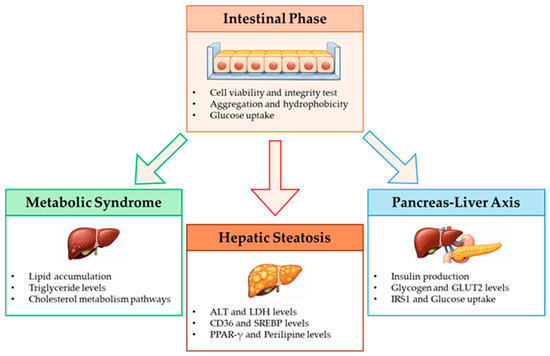
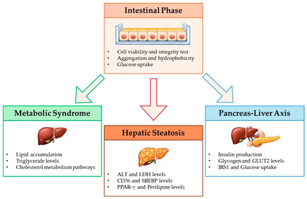
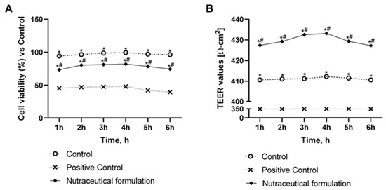
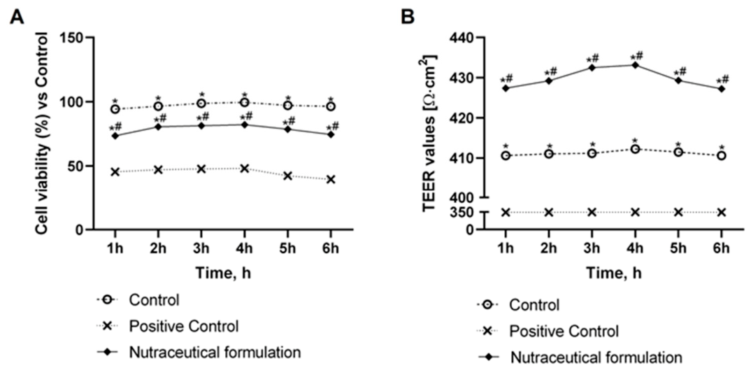
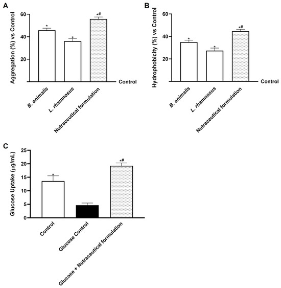
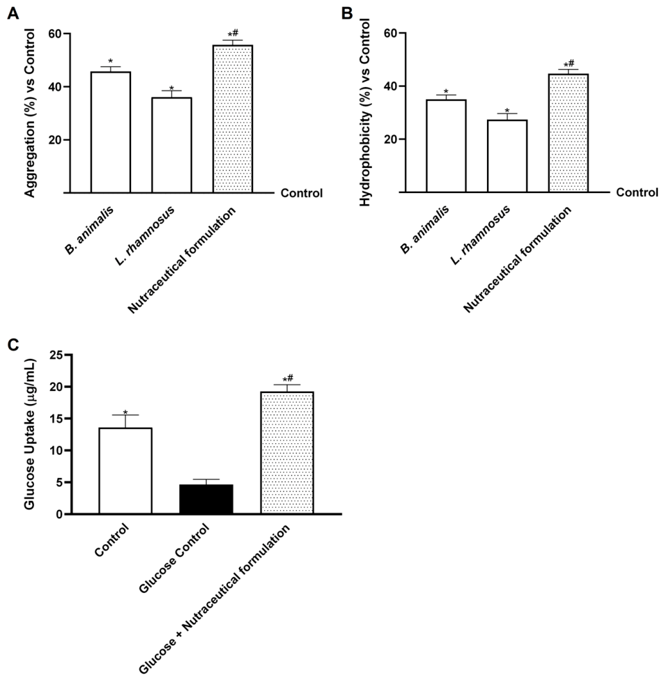
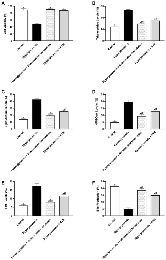
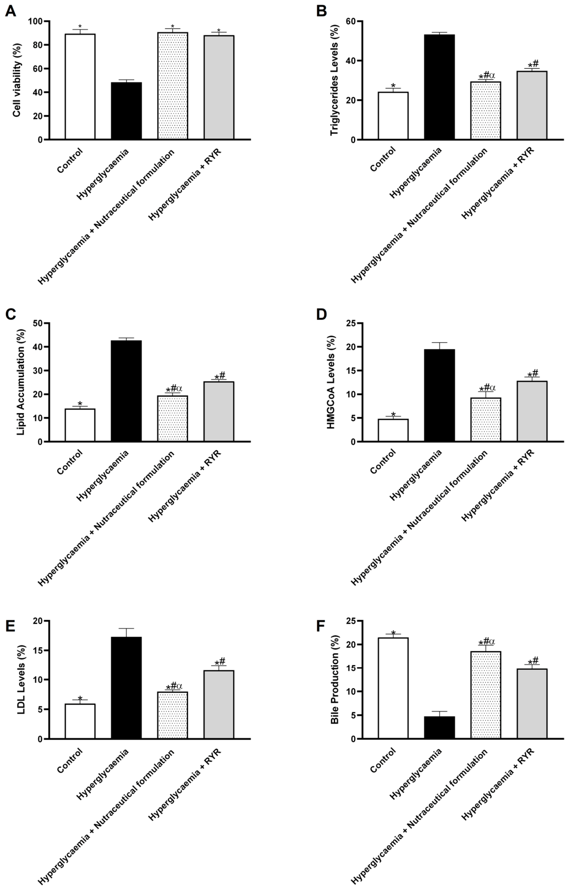
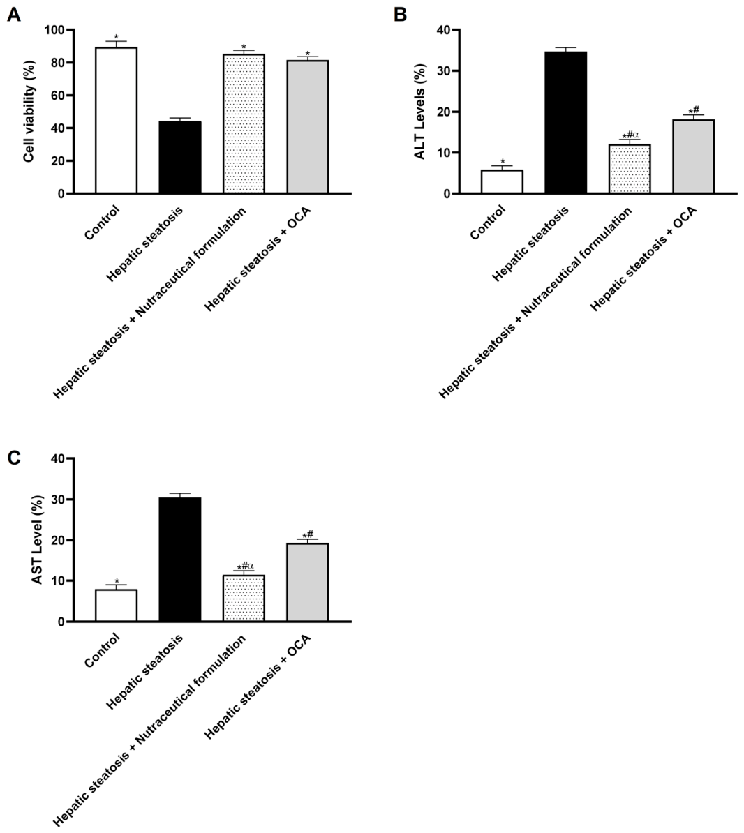
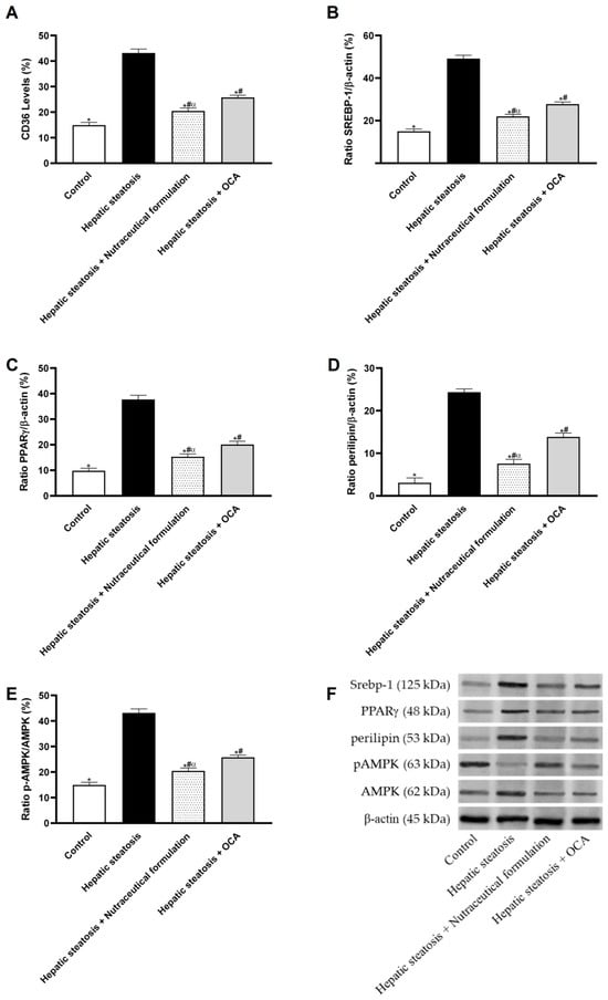
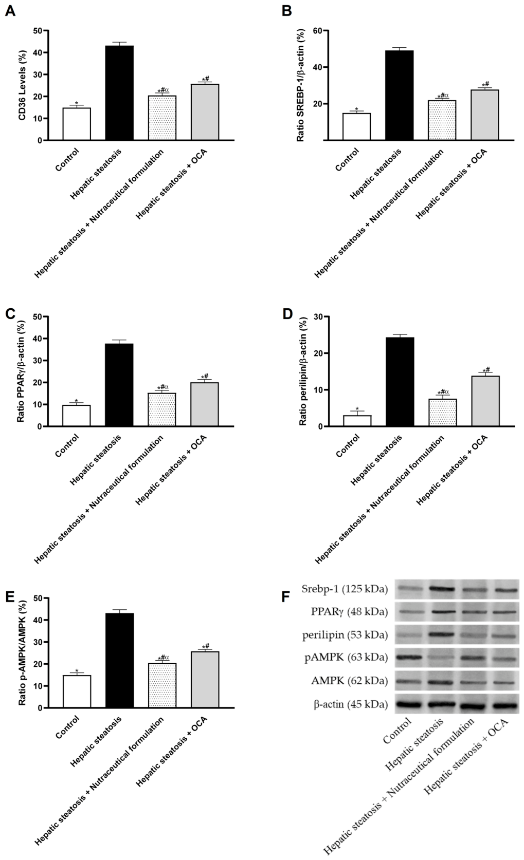
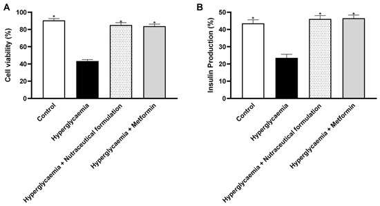
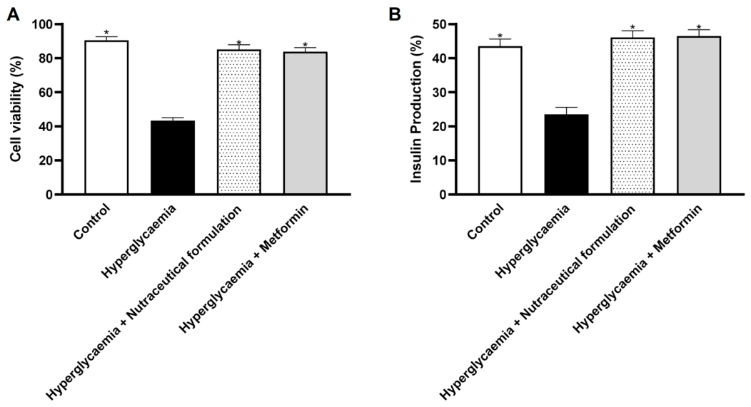
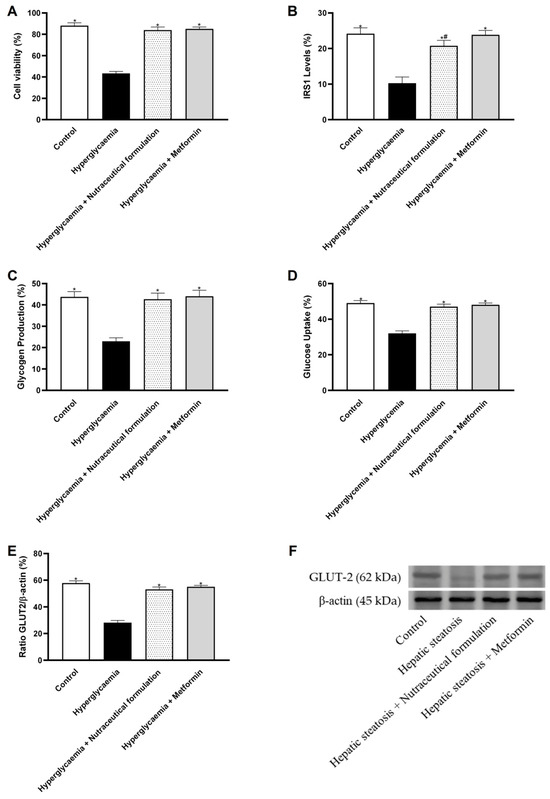
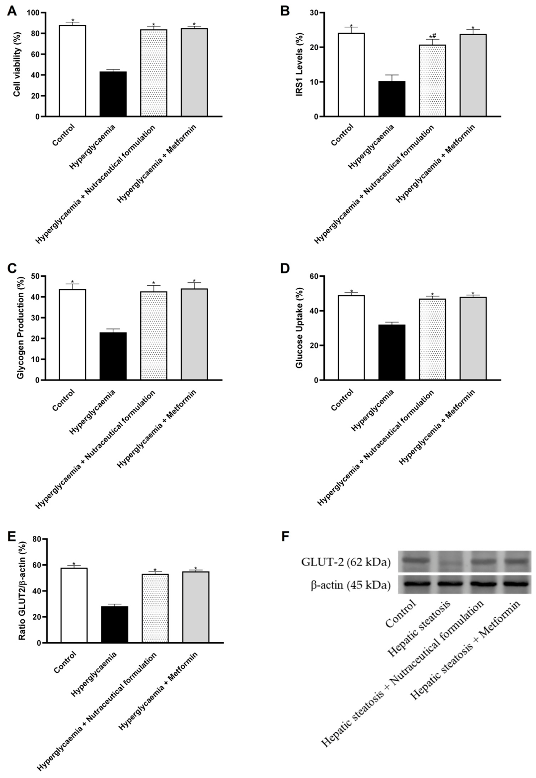

Disclaimer/Publisher’s Note: The statements, opinions and data contained in all publications are solely those of the individual author(s) and contributor(s) and not of MDPI and/or the editor(s). MDPI and/or the editor(s) disclaim responsibility for any injury to people or property resulting from any ideas, methods, instructions or products referred to in the content. |
© 2026 by the authors. Licensee MDPI, Basel, Switzerland. This article is an open access article distributed under the terms and conditions of the Creative Commons Attribution (CC BY) license.