The Hepatic Crown-like Structure: A Focal Point for Macrophage Evolution and Disease Response in Steatotic Liver Disease


Abstract
1. Introduction
2. Macrophage Ontogeny and Diversity in Steatotic Liver Disease
3. Origin and Compositional Diversity of Hepatic Crown-like Structures
3.1. Lipid-Associated Macrophages (LAMs)
3.2. LAM-like Kupffer Cells (LLKCs)
4. Factors That Modulate hCLS Development
4.1. Lipids
4.2. FXR
4.3. Inflammasome and NLRP3 Signaling
5. Role of hCLS in Steatosis Generation
6. Role of hCLS in Inflammation
6.1. Evidence for Pro-Inflammatory Function of CLS
6.2. Evidence for Anti-Inflammatory Function of CLS
7. Functional Role of hCLS in Fibrosis
7.1. Profibrotic Functions of hCLS
7.2. Role of CLS in Fibrosis Resolution
8. Conclusions
Author Contributions
Funding
Institutional Review Board Statement
Informed Consent Statement
Data Availability Statement
Conflicts of Interest
Abbreviations
| Axl | AXL receptor tyrosine kinase |
| Vsig4 | V-set immunoglobulin domain-containing 4 |
| Ccr2 | Chemokine (C-C motif) receptor 2 |
| Cd163 | Cluster of differentiation 163 |
| Cd36 | Cluster of differentiation 36 |
| Gpbar1 | G protein-coupled bile acid receptor 1 |
| Gpnmb | Glycoprotein non-metastatic melanoma protein B |
| Il1a | Interluekin-1 alpha |
| Itgal | Integrin subunit alpha L |
| Kdm5b | Lysine demethylase 5B |
| Ms4a3 | Membrane-spanning 4-domains subfamily A member 3 |
| Ms4a7 | Membrane-spanning 4-domains subfamily A member 7 |
| Nfkb1 | Nuclear factor NF-kappa-B p105 subunit |
| Nr1h4 | Nuclear receptor subfamily 1 group H member 4 |
| Ppar-gamma | Peroxisome proliferator-activated receptor gamma |
| Tgfb1 | Transforming growth factor beta 1 |
| Timd4 | T-cell membrane protein 4 |
| Trem2 | Triggering receptor expressed on myeloid cells 2 |
| Vcam1 | Vascular cell adhesion molecule 1 |
References
- Zhao, Y.; Bo, Y.; Zu, J.; Xing, Z.; Yang, Z.; Zhang, Y.; Deng, Y.; Liu, Y.; Zhang, L.; Yuan, X.; et al. Global Burden of Chronic Liver Disease and Temporal Trends: A Population-Based Analysis from 1990 to 2021 with Projections to 2050. Liver Int. 2025, 45, e70155. [Google Scholar] [CrossRef] [PubMed]
- Kim, D.; Bonham, C.A.; Konyn, P.; Cholankeril, G.; Ahmed, A. Mortality Trends in Chronic Liver Disease and Cirrhosis in the United States, Before and During COVID-19 Pandemic. Clin. Gastroenterol. Hepatol. 2021, 19, 2664–2666.e2. [Google Scholar] [CrossRef] [PubMed]
- Irfan, M.; Ahmad, R.; Ahmed, M.A.; Khan, A.M.; Aamir, Z.; Imran, R.; Ahmed, R. Trends in Liver Cirrhosis and Diabetes-Related Mortality Among Adults in the United States: A CDC WONDER Database Analysis (1999–2020). Life 2025, 15, 852. [Google Scholar] [CrossRef] [PubMed]
- Manikat, R.; Ahmed, A.; Kim, D. Current epidemiology of chronic liver disease. Gastroenterol. Rep. 2024, 12, goae069. [Google Scholar]
- Devarbhavi, H.; Asrani, S.K.; Arab, J.P.; Nartey, Y.A.; Pose, E.; Kamath, P.S. Global burden of liver disease: 2023 update. J. Hepatol. 2023, 79, 516–537. [Google Scholar] [CrossRef] [PubMed]
- Itoh, M.; Kato, H.; Suganami, T.; Konuma, K.; Marumoto, Y.; Terai, S.; Sakugawa, H.; Kanai, S.; Hamaguchi, M.; Fukaishi, T.; et al. Hepatic crown-like structure: A unique histological feature in non-alcoholic steatohepatitis in mice and humans. PLoS ONE 2013, 8, e82163. [Google Scholar] [CrossRef] [PubMed]
- Itoh, M.; Suganami, T.; Kato, H.; Kanai, S.; Shirakawa, I.; Sakai, T.; Goto, T.; Asakawa, M.; Hidaka, I.; Sakugawa, H.; et al. CD11c+ resident macrophages drive hepatocyte death-triggered liver fibrosis in a murine model of nonalcoholic steatohepatitis. JCI Insight 2017, 2, e92902. [Google Scholar] [CrossRef] [PubMed]
- Kanamori, Y.; Tanaka, M.; Itoh, M.; Ochi, K.; Ito, A.; Hidaka, I.; Sakaida, I.; Ogawa, Y.; Suganami, T. Iron-rich Kupffer cells exhibit phenotypic changes during the development of liver fibrosis in NASH. iScience 2021, 24, 102032. [Google Scholar] [CrossRef] [PubMed]
- Tada, Y.; Kasai, K.; Makiuchi, N.; Igarashi, N.; Kani, K.; Takano, S.; Honda, H.; Yanagibashi, T.; Watanabe, Y.; Usui-Kawanishi, F.; et al. Roles of Macrophages in Advanced Liver Fibrosis, Identified Using a Newly Established Mouse Model of Diet-Induced Non-Alcoholic Steatohepatitis. Int. J. Mol. Sci. 2022, 23, 13251. [Google Scholar] [CrossRef] [PubMed]
- Aoki, H.; Isobe, Y.; Yoshida, M.; Kang, J.X.; Maekawa, M.; Arita, M. Enzymatically-epoxidized docosahexaenoic acid, 19,20-EpDPE, suppresses hepatic crown-like structure formation and nonalcoholic steatohepatitis fibrosis through GPR120. Biochim. ET Biophys. Acta (BBA)-Mol. Cell Biol. Lipids 2023, 1868, 159275. [Google Scholar] [CrossRef]
- Daemen, S.; Gainullina, A.; Kalugotla, G.; He, L.; Chan, M.M.; Beals, J.W.; Liss, K.H.; Klein, S.; Feldstein, A.E.; Finck, B.N.; et al. Dynamic Shifts in the Composition of Resident and Recruited Macrophages Influence Tissue Remodeling in NASH. Cell Rep. 2022, 41, 111660. [Google Scholar] [CrossRef] [PubMed]
- Gale, R.P.; Sparkes, R.S.; Golde, D.W. Bone marrow origin of hepatic macrophages (Kupffer cells) in humans. Science 1978, 201, 937–938. [Google Scholar] [CrossRef] [PubMed]
- van Furth, R.; Cohn, Z.A. The origin and kinetics of mononuclear phagocytes. J. Exp. Med. 1968, 128, 415–435. [Google Scholar] [CrossRef] [PubMed]
- Schulz, C.; Gomez Perdiguero, E.; Chorro, L.; Szabo-Rogers, H.; Cagnard, N.; Kierdorf, K.; Prinz, M.; Wu, B.; Jacobsen, S.E.W.; Pollard, J.W.; et al. A lineage of myeloid cells independent of Myb and hematopoietic stem cells. Science 2012, 336, 86–90. [Google Scholar] [CrossRef] [PubMed]
- Stremmel, C.; Schuchert, R.; Wagner, F.; Thaler, R.; Weinberger, T.; Pick, R.; Mass, E.; Ishikawa-Ankerhold, H.C.; Margraf, A.; Hutter, S.; et al. Yolk sac macrophage progenitors traffic to the embryo during defined stages of development. Nat. Commun. 2018, 9, 75. [Google Scholar] [CrossRef] [PubMed]
- Gomez Perdiguero, E.; Klapproth, K.; Schulz, C.; Busch, K.; Azzoni, E.; Crozet, L.; Garner, H.; Trouillet, C.; de Bruijn, M.F.; Geissmann, F.; et al. Tissue-resident macrophages originate from yolk-sac-derived erythro-myeloid progenitors. Nature 2015, 518, 547–551. [Google Scholar] [PubMed]
- Ginhoux, F.; Greter, M.; Leboeuf, M.; Nandi, S.; See, P.; Gokhan, S.; Mehler, M.F.; Conway, S.J.; Ng, L.G.; Stanley, E.R.; et al. Fate mapping analysis reveals that adult microglia derive from primitive macrophages. Science 2010, 330, 841–845. [Google Scholar] [CrossRef] [PubMed]
- Hashimoto, D.; Chow, A.; Noizat, C.; Teo, P.; Beasley, M.B.; Leboeuf, M.; Becker, C.D.; See, P.; Price, J.; Lucas, D.; et al. Tissue-resident macrophages self-maintain locally throughout adult life with minimal contribution from circulating monocytes. Immunity 2013, 38, 792–804. [Google Scholar] [CrossRef] [PubMed]
- Klein, I.; Cornejo, J.C.; Polakos, N.K.; John, B.; Wuensch, S.A.; Topham, D.J.; Pierce, R.H.; Crispe, I.N. Kupffer cell heterogeneity: Functional properties of bone marrow derived and sessile hepatic macrophages. Blood 2007, 110, 4077–4085. [Google Scholar] [CrossRef] [PubMed]
- Tran, S.; Baba, I.; Poupel, L.; Dussaud, S.; Moreau, M.; Gelineau, A.; Marcelin, G.; Magréau-Davy, E.; Ouhachi, M.; Lesnik, P.; et al. Impaired Kupffer Cell Self-Renewal Alters the Liver Response to Lipid Overload during Non-alcoholic Steatohepatitis. Immunity 2020, 53, 627–640.e5. [Google Scholar] [CrossRef] [PubMed]
- Iwata, A.; Maruyama, J.; Natsuki, S.; Nishiyama, A.; Tamura, T.; Tanaka, M.; Shichino, S.; Seki, T.; Komai, T.; Okamura, T.; et al. Egr2 drives the differentiation of Ly6Chi monocytes into fibrosis-promoting macrophages in metabolic dysfunction-associated steatohepatitis in mice. Commun. Biol. 2024, 7, 681. [Google Scholar] [CrossRef] [PubMed]
- Miller, S.J.; Janssen, R.C.; Zhao, W.; Jonscher, K.R.; Zhong, H.; DeVette, C.I.; Friedman, J.E.; Zimmerman, K.A. Maternal western-style diet alters Kupffer cell proportion leading to metabolic dysfunction-associated steatotic liver disease when challenged with western diet in adulthood. Front Immunol. 2025, 16, 1698609. [Google Scholar] [CrossRef] [PubMed]
- Achalu, S.; Berry, R.; Wei, M.T.; Banerjee, S.; Ghanouni, P.; Kambham, N.; Kwo, P.Y. Immunoglobulin G4-Seronegative Autoimmune Cholangiopathy with Pancreatic and Hepatic Involvement Mimicking as Primary Sclerosing Cholangitis. ACG Case Rep. J. 2023, 10, e01044. [Google Scholar] [CrossRef] [PubMed]
- De Ponti, F.F.; Bujko, A.; Liu, Z.; Collins, P.J.; Schuermans, S.; Maueroder, C.; Amstelveen, S.; Thoné, T.; Martens, L.; McKendrick, J.G.; et al. Spatially restricted and ontogenically distinct hepatic macrophages are required for tissue repair. Immunity 2025, 58, 362–380.e10. [Google Scholar] [CrossRef] [PubMed]
- Karlmark, K.R.; Weiskirchen, R.; Zimmermann, H.W.; Gassler, N.; Ginhoux, F.; Weber, C.; Merad, M.; Luedde, T.; Trautwein, C.; Tacke, F. Hepatic recruitment of the inflammatory Gr1+ monocyte subset upon liver injury promotes hepatic fibrosis. Hepatology 2009, 50, 261–274. [Google Scholar] [CrossRef] [PubMed]
- Dambach, D.M.; Watson, L.M.; Gray, K.R.; Durham, S.K.; Laskin, D.L. Role of CCR2 in macrophage migration into the liver during acetaminophen-induced hepatotoxicity in the mouse. Hepatology 2002, 35, 1093–1103. [Google Scholar] [CrossRef] [PubMed]
- Hoffmann, J.; Fiser, K.; Liebetrau, C.; Staubach, N.; Kost, D.; Voss, S.; Heiden, A.Z.; Dörr, O.; Lipps, C.; Nef, H.M.; et al. High-Content Immunophenotyping and Hierarchical Clustering Reveal Sources of Heterogeneity and New Surface Markers of Human Blood Monocyte Subsets. Thromb. Haemost. 2020, 120, 141–155. [Google Scholar] [PubMed]
- Geissmann, F.; Jung, S.; Littman, D.R. Blood monocytes consist of two principal subsets with distinct migratory properties. Immunity 2003, 19, 71–82. [Google Scholar] [CrossRef] [PubMed]
- Sunderkotter, C.; Nikolic, T.; Dillon, M.J.; Van Rooijen, N.; Stehling, M.; Drevets, D.A.; Leenen, P.J. Subpopulations of mouse blood monocytes differ in maturation stage and inflammatory response. J. Immunol. 2004, 172, 4410–4417. [Google Scholar] [CrossRef] [PubMed]
- Sakai, M.; Troutman, T.D.; Seidman, J.S.; Ouyang, Z.; Spann, N.J.; Abe, Y.; Ego, K.M.; Bruni, C.M.; Deng, Z.; Schlachetzki, J.C.; et al. Liver-Derived Signals Sequentially Reprogram Myeloid Enhancers to Initiate and Maintain Kupffer Cell Identity. Immunity 2019, 51, 655–670.e8. [Google Scholar] [CrossRef] [PubMed]
- Bonnardel, J.; T’Jonck, W.; Gaublomme, D.; Browaeys, R.; Scott, C.L.; Martens, L.; Vanneste, B.; De Prijck, S.; Nedospasov, S.A.; Kremer, A.; et al. Stellate Cells, Hepatocytes, and Endothelial Cells Imprint the Kupffer Cell Identity on Monocytes Colonizing the Liver Macrophage Niche. Immunity 2019, 51, 638–654.e9. [Google Scholar] [CrossRef] [PubMed]
- Bleriot, C.; Dupuis, T.; Jouvion, G.; Eberl, G.; Disson, O.; Lecuit, M. Liver-resident macrophage necroptosis orchestrates type 1 microbicidal inflammation and type-2-mediated tissue repair during bacterial infection. Immunity 2015, 42, 145–158. [Google Scholar] [PubMed]
- Scott, C.L.; Zheng, F.; De Baetselier, P.; Martens, L.; Saeys, Y.; De Prijck, S.; Lippens, S.; Abels, C.; Schoonooghe, S.; Raes, G.; et al. Bone marrow-derived monocytes give rise to self-renewing and fully differentiated Kupffer cells. Nat. Commun. 2016, 7, 10321. [Google Scholar] [CrossRef] [PubMed]
- Xiong, X.; Kuang, H.; Ansari, S.; Liu, T.; Gong, J.; Wang, S.; Zhao, X.Y.; Ji, Y.; Li, C.; Guo, L.; et al. Landscape of Intercellular Crosstalk in Healthy and NASH Liver Revealed by Single-Cell Secretome Gene Analysis. Mol. Cell 2019, 75, 644–660.e5. [Google Scholar] [CrossRef] [PubMed]
- Seidman, J.S.; Troutman, T.D.; Sakai, M.; Gola, A.; Spann, N.J.; Bennett, H.; Bruni, C.M.; Ouyang, Z.; Li, R.Z.; Sun, X.; et al. Niche-Specific Reprogramming of Epigenetic Landscapes Drives Myeloid Cell Diversity in Nonalcoholic Steatohepatitis. Immunity 2020, 52, 1057–1074.e7. [Google Scholar] [CrossRef] [PubMed]
- Remmerie, A.; Martens, L.; Thone, T.; Castoldi, A.; Seurinck, R.; Pavie, B.; Roels, J.; Vanneste, B.; De Prijck, S.; Vanhockerhout, M.; et al. Osteopontin Expression Identifies a Subset of Recruited Macrophages Distinct from Kupffer Cells in the Fatty Liver. Immunity 2020, 53, 641–657.e14. [Google Scholar] [CrossRef] [PubMed]
- Guilliams, M.; Bonnardel, J.; Haest, B.; Vanderborght, B.; Wagner, C.; Remmerie, A.; Bujko, A.; Martens, L.; Thoné, T.; Browaeys, R.; et al. Spatial proteogenomics reveals distinct and evolutionarily conserved hepatic macrophage niches. Cell 2022, 185, 379–396.e38. [Google Scholar] [CrossRef] [PubMed]
- Jaitin, D.A.; Adlung, L.; Thaiss, C.A.; Weiner, A.; Li, B.; Descamps, H.; Lundgren, P.; Bleriot, C.; Liu, Z.; Deczkowska, A.; et al. Lipid-Associated Macrophages Control Metabolic Homeostasis in a Trem2-Dependent Manner. Cell 2019, 178, 686–698.e14. [Google Scholar] [CrossRef] [PubMed]
- Fallowfield, J.A.; Mizuno, M.; Kendall, T.J.; Constandinou, C.M.; Benyon, R.C.; Duffield, J.S.; Iredale, J.P. Scar-associated macrophages are a major source of hepatic matrix metalloproteinase-13 and facilitate the resolution of murine hepatic fibrosis. J. Immunol. 2007, 178, 5288–5295. [Google Scholar] [CrossRef] [PubMed]
- Cinti, S.; Mitchell, G.; Barbatelli, G.; Murano, I.; Ceresi, E.; Faloia, E.; Wang, S.; Fortier, M.; Greenberg, A.S.; Obin, M.S. Adipocyte death defines macrophage localization and function in adipose tissue of obese mice and humans. J. Lipid Res. 2005, 46, 2347–2355. [Google Scholar] [CrossRef] [PubMed]
- Li, W.; Yang, Y.; Yang, L.; Chang, N.; Li, L. Monocyte-derived Kupffer cells dominate in the Kupffer cell pool during liver injury. Cell Rep. 2023, 42, 113164. [Google Scholar] [CrossRef] [PubMed]
- Miura, K.; Yang, L.; van Rooijen, N.; Ohnishi, H.; Seki, E. Hepatic recruitment of macrophages promotes nonalcoholic steatohepatitis through CCR2. Am. J. Physiol. Gastrointest. Liver Physiol. 2012, 302, G1310–G1321. [Google Scholar] [CrossRef] [PubMed]
- Tanaka, M.; Ikeda, K.; Suganami, T.; Komiya, C.; Ochi, K.; Shirakawa, I.; Hamaguchi, M.; Nishimura, S.; Manabe, I.; Matsuda, T.; et al. Macrophage-inducible C-type lectin underlies obesity-induced adipose tissue fibrosis. Nat. Commun. 2014, 5, 4982. [Google Scholar] [CrossRef] [PubMed]
- Malavazos, A.E.; Di Vincenzo, A.; Iacobellis, G.; Basilico, S.; Dubini, C.; Morricone, L.; Menicanti, L.; Luca, T.; Giordano, A.; Castorina, S.; et al. The density of crown-like structures in epicardial adipose tissue could play a role in cardiovascular diseases. Eat. Weight Disord. 2022, 27, 2905–2910. [Google Scholar] [CrossRef] [PubMed]
- Maliniak, M.L.; Miller-Kleinhenz, J.; Cronin-Fenton, D.P.; Lash, T.L.; Gogineni, K.; Janssen, E.A.M.; McCullough, L.E. Crown-Like Structures in Breast Adipose Tissue: Early Evidence and Current Issues in Breast Cancer. Cancers 2021, 13, 2222. [Google Scholar] [CrossRef] [PubMed]
- Harris, A.R.; Hughes, J.D.; Lawrence, W.R.; Lenz, P.; Franklin, J.; Bhawsar, P.M.S.; Dorsey, T.H.; Rossi, E.L.; Pichardo, C.M.; Pichardo, M.S.; et al. Neighborhood Environment, DNA Methylation, and Presence of Crown-Like Structures of the Breast. JAMA Netw. Open 2025, 8, e2461334. [Google Scholar] [CrossRef] [PubMed]
- Chang, M.C.; Eslami, Z.; Ennis, M.; Goodwin, P.J. Crown-like structures in breast adipose tissue of breast cancer patients: Associations with CD68 expression, obesity, metabolic factors and prognosis. npj Breast Cancer 2021, 7, 97. [Google Scholar] [CrossRef] [PubMed]
- Liang, Y.L.; Lin, C.N.; Tsai, H.F.; Wu, P.Y.; Lin, S.H.; Hong, T.M.; Hsu, K.F. Omental Macrophagic “Crown-like Structures” Are Associated with Poor Prognosis in Advanced-Stage Serous Ovarian Cancer. Curr. Oncol. 2021, 28, 4234–4246. [Google Scholar] [CrossRef] [PubMed]
- Xu, R.; Vujic, N.; Bianco, V.; Reinisch, I.; Kratky, D.; Krstic, J.; Prokesch, A. Lipid-associated macrophages between aggravation and alleviation of metabolic diseases. Trends Endocrinol. Metab. 2024, 35, 981–995. [Google Scholar] [CrossRef] [PubMed]
- Ganguly, S.; Rosenthal, S.B.; Ishizuka, K.; Troutman, T.D.; Rohm, T.V.; Khader, N.; Aleman-Muench, G.; Sano, Y.; Archilei, S.; Soroosh, P.; et al. Lipid-associated macrophages’ promotion of fibrosis resolution during MASH regression requires TREM2. Proc. Natl. Acad. Sci. USA 2024, 121, e2405746121. [Google Scholar] [CrossRef] [PubMed]
- Hendrikx, T.; Porsch, F.; Kiss, M.G.; Rajcic, D.; Papac-Milicevic, N.; Hoebinger, C.; Goederle, L.; Hladik, A.; Shaw, L.E.; Horstmann, H.; et al. Soluble TREM2 levels reflect the recruitment and expansion of TREM2+ macrophages that localize to fibrotic areas and limit NASH. J. Hepatol. 2022, 77, 1373–1385. [Google Scholar] [CrossRef] [PubMed]
- Lessard, A.J.; LeBel, M.; Egarnes, B.; Prefontaine, P.; Theriault, P.; Droit, A.; Brunet, A.; Rivest, S.; Gosselin, J. Triggering of NOD2 Receptor Converts Inflammatory Ly6Chigh into Ly6Clow Monocytes with Patrolling Properties. Cell Rep. 2017, 20, 1830–1843. [Google Scholar] [CrossRef] [PubMed]
- Dempsey, J.L.; Savard, C.; Kothari, V.; Tang, J.; Lee, S.P.; Bornfeldt, K.E.; Carr, R.M.; Ioannou, G.N. Soluble Triggering Receptor Expressed on Myeloid Cells 2 is a Biomarker but Not a Mediator of Fibrosing Steatohepatitis. Gastro Hep Adv. 2026, 5, 100828. [Google Scholar] [CrossRef] [PubMed]
- Yu, W.; Zhang, Y.; Sun, L.; Huang, W.; Li, X.; Xia, N.; Chen, X.; Wikana, L.P.; Xiao, Y.; Chen, M.; et al. Myeloid Trem2 ameliorates the progression of metabolic dysfunction-associated steatotic liver disease by regulating macrophage pyroptosis and inflammation resolution. Metabolism 2024, 155, 155911. [Google Scholar] [CrossRef] [PubMed]
- Ji, P.X.; Chen, Y.X.; Ni, X.X.; Miao, Q.; Hua, J. Effect of triggering receptor expressed on myeloid cells 2-associated alterations on lipid metabolism in macrophages in the development of non-alcoholic fatty liver disease. J. Gastroenterol. Hepatol. 2024, 39, 369–380. [Google Scholar] [PubMed]
- Ramachandran, P.; Dobie, R.; Wilson-Kanamori, J.R.; Dora, E.F.; Henderson, B.E.P.; Luu, N.T.; Portman, J.R.; Matchett, K.P.; Brice, M.; Marwick, J.A.; et al. Resolving the fibrotic niche of human liver cirrhosis at single-cell level. Nature 2019, 575, 512–518. [Google Scholar] [CrossRef] [PubMed]
- De Ponti, F.F.; Liu, Z.; Scott, C.L. Understanding the complex macrophage landscape in MASLD. JHEP Rep. 2024, 6, 101196. [Google Scholar] [CrossRef] [PubMed]
- Helmy, K.Y.; Katschke, K.J., Jr.; Gorgani, N.N.; Kljavin, N.M.; Elliott, J.M.; Diehl, L.; Scales, S.J.; Ghilardi, N.; van Lookeren Campagne, M. CRIg: A macrophage complement receptor required for phagocytosis of circulating pathogens. Cell 2006, 124, 915–927. [Google Scholar] [CrossRef] [PubMed]
- Yang, C.Y.; Chen, J.B.; Tsai, T.F.; Tsai, Y.C.; Tsai, C.Y.; Liang, P.H.; Hsu, T.L.; Wu, C.Y.; Netea, M.G.; Wong, C.H.; et al. CLEC4F is an inducible C-type lectin in F4/80-positive cells and is involved in alpha-galactosylceramide presentation in liver. PLoS ONE 2013, 8, e65070. [Google Scholar] [CrossRef] [PubMed]
- Beattie, L.; Sawtell, A.; Mann, J.; Frame, T.C.M.; Teal, B.; de Labastida Rivera, F.; Brown, N.; Walwyn-Brown, K.; Moore, J.W.J.; MacDonald, S.; et al. Bone marrow-derived and resident liver macrophages display unique transcriptomic signatures but similar biological functions. J. Hepatol. 2016, 65, 758–768. [Google Scholar] [CrossRef] [PubMed]
- Bleriot, C.; Barreby, E.; Dunsmore, G.; Ballaire, R.; Chakarov, S.; Ficht, X.; De Simone, G.; Andreata, F.; Fumagalli, V.; Guo, W.; et al. A subset of Kupffer cells regulates metabolism through the expression of CD36. Immunity 2021, 54, 2101–2116 e2106. [Google Scholar] [CrossRef] [PubMed]
- Liu, Z.; Gu, Y.; Chakarov, S.; Bleriot, C.; Kwok, I.; Chen, X.; Shin, A.; Huang, W.; Dress, R.J.; Dutertre, C.A.; et al. Fate Mapping via Ms4a3-Expression History Traces Monocyte-Derived Cells. Cell 2019, 178, 1509–1525.e19. [Google Scholar] [CrossRef] [PubMed]
- Lin, B.; Zhang, H.; Zhu, P.; Chen, J.; Li, D.; Zhou, J.; Zhang, T.; Chen, Q.; Tang, C.; Song, X.; et al. Bone marrow-derived CD169+ macrophages promote autoimmune hepatitis by recruiting CCR2+ monocytes via secreting CCL12. Exp. Mol. Med. 2025, 57, 2930–2942. [Google Scholar] [CrossRef] [PubMed]
- Skytthe, M.K.; Pedersen, F.B.; Wernberg, C.W.; Chandran, V.I.; Krag, A.; Di Caterino, T.; Mandacaru, S.C.; Blagoev, B.; Lauridsen, M.M.; Detlefsen, S.; et al. Obese Patients with Nonalcoholic Fatty Liver Disease Have an Increase in Soluble Plasma CD163 and a Concurrent Decrease in Hepatic Expression of CD163. Gastro Hep Adv. 2023, 2, 711–720. [Google Scholar] [CrossRef] [PubMed]
- Kleiner, D.E.; Brunt, E.M.; Van Natta, M.; Behling, C.; Contos, M.J.; Cummings, O.W.; Ferrell, L.D.; Liu, Y.C.; Torbenson, M.S.; Unalp-Arida, A.; et al. Design and validation of a histological scoring system for nonalcoholic fatty liver disease. Hepatology 2005, 41, 1313–1321. [Google Scholar] [CrossRef] [PubMed]
- Gluchowski, N.L.; Becuwe, M.; Walther, T.C.; Farese, R.V., Jr. Lipid droplets and liver disease: From basic biology to clinical implications. Nat. Rev. Gastroenterol. Hepatol. 2017, 14, 343–355. [Google Scholar] [CrossRef] [PubMed]
- Schott, M.B.; Weller, S.G.; Schulze, R.J.; Krueger, E.W.; Drizyte-Miller, K.; Casey, C.A.; McNiven, M.A. Lipid droplet size directs lipolysis and lipophagy catabolism in hepatocytes. J. Cell Biol. 2019, 218, 3320–3335. [Google Scholar] [CrossRef] [PubMed]
- Singh, R.; Cuervo, A.M. Lipophagy: Connecting autophagy and lipid metabolism. Int. J. Cell Biol. 2012, 2012, 282041. [Google Scholar] [CrossRef] [PubMed]
- Loneker, A.E.; Alisafaei, F.; Kant, A.; Li, D.; Janmey, P.A.; Shenoy, V.B.; Wells, R.G. Lipid droplets are intracellular mechanical stressors that impair hepatocyte function. Proc. Natl. Acad. Sci. USA 2023, 120, e2216811120. [Google Scholar] [CrossRef] [PubMed]
- Mashek, D.G. Hepatic lipid droplets: A balancing act between energy storage and metabolic dysfunction in NAFLD. Mol. Metab. 2021, 50, 101115. [Google Scholar] [CrossRef] [PubMed]
- Mooli, R.G.R.; Ramakrishnan, S.K. Liver Steatosis is a Driving Factor of Inflammation. Cell Mol. Gastroenterol. Hepatol. 2022, 13, 1267–1270. [Google Scholar] [CrossRef] [PubMed]
- Ioannou, G.N.; Haigh, W.G.; Thorning, D.; Savard, C. Hepatic cholesterol crystals and crown-like structures distinguish NASH from simple steatosis. J. Lipid Res. 2013, 54, 1326–1334. [Google Scholar] [CrossRef] [PubMed]
- Sakuma, I.; Gaspar, R.C.; Nasiri, A.R.; Dufour, S.; Kahn, M.; Zheng, J.; LaMoia, T.E.; Guerra, M.T.; Taki, Y.; Kawashima, Y.; et al. Liver lipid droplet cholesterol content is a key determinant of metabolic dysfunction-associated steatohepatitis. Proc. Natl. Acad. Sci. USA 2025, 122, e2502978122. [Google Scholar] [CrossRef] [PubMed]
- Rada, P.; Gonzalez-Rodriguez, A.; Garcia-Monzon, C.; Valverde, A.M. Understanding lipotoxicity in NAFLD pathogenesis: Is CD36 a key driver? Cell Death Dis. 2020, 11, 802. [Google Scholar] [CrossRef] [PubMed]
- Yang, P.; Qin, H.; Li, Y.; Xiao, A.; Zheng, E.; Zeng, H.; Su, C.; Luo, X.; Lu, Q.; Liao, M.; et al. CD36-mediated metabolic crosstalk between tumor cells and macrophages affects liver metastasis. Nat. Commun. 2022, 13, 5782. [Google Scholar] [CrossRef] [PubMed]
- Cai, L.; Wang, Z.; Ji, A.; Meyer, J.M.; van der Westhuyzen, D.R. Scavenger receptor CD36 expression contributes to adipose tissue inflammation and cell death in diet-induced obesity. PLoS ONE 2012, 7, e36785. [Google Scholar] [CrossRef] [PubMed]
- Furumaki, H.; Matsuoka, S.; Sakurai, T.; Kohanawa, M.; Zhao, S.; Kuge, Y.; Tamaki, N.; Chiba, H. A novel murine model for non-alcoholic steatohepatitis developed by combination of a high-fat diet and oxidized low-density lipoprotein. Lab Investig. 2012, 92, 265–281. [Google Scholar] [CrossRef]
- Bieghs, V.; Wouters, K.; van Gorp, P.J.; Gijbels, M.J.; de Winther, M.P.; Binder, C.J.; Lutjohann, D.; Febbraio, M.; Moore, K.J.; van Bilsen, M.; et al. Role of scavenger receptor A and CD36 in diet-induced nonalcoholic steatohepatitis in hyperlipidemic mice. Gastroenterology 2010, 138, 2477–2486.e3. [Google Scholar] [CrossRef] [PubMed]
- Guan, Y.; Kim, Y.; Wang, Y.; Cho, Y.E.; Xiang, X.; Kim, S.J.; Yao, T.; Feng, D.; Hwang, S.; Gao, B. Adipocyte death promotes hepatic infiltration of S100A8+ macrophages and steatotic liver disease progression in mice. J. Clin. Investig. 2025, 135, e190635. [Google Scholar] [CrossRef] [PubMed]
- Clifford, B.L.; Sedgeman, L.R.; Williams, K.J.; Morand, P.; Cheng, A.; Jarrett, K.E.; Chan, A.P.; Brearley-Sholto, M.C.; Wahlström, A.; Ashby, J.W.; et al. FXR activation protects against NAFLD via bile-acid-dependent reductions in lipid absorption. Cell Metab. 2021, 33, 1671–1684.e4. [Google Scholar] [CrossRef] [PubMed]
- Morrison, M.C.; Verschuren, L.; Salic, K.; Verheij, J.; Menke, A.; Wielinga, P.Y.; Iruarrizaga-Lejarreta, M.; Gole, L.; Yu, W.; Turner, S.; et al. Obeticholic Acid Modulates Serum Metabolites and Gene Signatures Characteristic of Human NASH and Attenuates Inflammation and Fibrosis Progression in Ldlr-/- Leiden. Mice. Hepatol. Commun. 2018, 2, 1513–1532. [Google Scholar] [CrossRef] [PubMed]
- Wen, Y.Q.; Zou, Z.Y.; Zhao, G.G.; Zhang, M.J.; Zhang, Y.X.; Wang, G.H.; Shi, J.J.; Wang, Y.Y.; Song, Y.Y.; Wang, H.X.; et al. FXR activation remodels hepatic and intestinal transcriptional landscapes in metabolic dysfunction-associated steatohepatitis. Acta Pharmacol. Sin. 2024, 45, 2313–2327. [Google Scholar] [CrossRef] [PubMed]
- Pathak, P.; Xie, C.; Nichols, R.G.; Ferrell, J.M.; Boehme, S.; Krausz, K.W.; Patterson, A.D.; Gonzalez, F.J.; Chiang, J.Y.L. Intestine farnesoid X receptor agonist and the gut microbiota activate G-protein bile acid receptor-1 signaling to improve metabolism. Hepatology 2018, 68, 1574–1588. [Google Scholar] [CrossRef] [PubMed]
- Kasai, K.; Igarashi, N.; Tada, Y.; Kani, K.; Takano, S.; Yanagibashi, T.; Usui-Kawanishi, F.; Fujisaka, S.; Watanabe, S.; Ichimura-Shimizu, M.; et al. Impact of Vancomycin Treatment and Gut Microbiota on Bile Acid Metabolism and the Development of Non-Alcoholic Steatohepatitis in Mice. Int. J. Mol. Sci. 2023, 24, 4050. [Google Scholar] [CrossRef] [PubMed]
- Nagasaki, A.; Sakamoto, S.; Arai, T.; Kato, M.; Ishida, E.; Furusho, H.; Fujii, M.; Takata, T.; Miyauchi, M. Elimination of Porphyromonas gingivalis inhibits liver fibrosis and inflammation in NASH. J. Clin. Periodontol. 2021, 48, 1367–1378. [Google Scholar] [CrossRef] [PubMed]
- Lara-Reyna, S.; Caseley, E.A.; Topping, J.; Rodrigues, F.; Jimenez Macias, J.; Lawler, S.E.; McDermott, M.F. Inflammasome activation: From molecular mechanisms to autoinflammation. Clin. Transl. Immunol. 2022, 11, e1404. [Google Scholar] [CrossRef]
- Martinon, F.; Burns, K.; Tschopp, J. The inflammasome: A molecular platform triggering activation of inflammatory caspases and processing of proIL-beta. Mol. Cell 2002, 10, 417–426. [Google Scholar] [CrossRef] [PubMed]
- Taru, V.; Szabo, G.; Mehal, W.; Reiberger, T. Inflammasomes in chronic liver disease: Hepatic injury, fibrosis progression and systemic inflammation. J. Hepatol. 2024, 81, 895–910. [Google Scholar] [CrossRef] [PubMed]
- Ioannou, G.N.; Horn, C.L.; Kothari, V.; Yeh, M.M.; Shyu, I.; Lee, S.P.; Savard, C.E. Genetic deletion or pharmacologic inhibition of the Nlrp3 inflammasome did not ameliorate experimental NASH. J. Lipid Res. 2023, 64, 100330. [Google Scholar] [CrossRef] [PubMed]
- Wree, A.; McGeough, M.D.; Pena, C.A.; Schlattjan, M.; Li, H.; Inzaugarat, M.E.; Messer, K.; Canbay, A.; Hoffman, H.M.; Feldstein, A.E. NLRP3 inflammasome activation is required for fibrosis development in NAFLD. J. Mol. Med. 2014, 92, 1069–1082. [Google Scholar] [CrossRef] [PubMed]
- Stephenson, K.; Kennedy, L.; Hargrove, L.; Demieville, J.; Thomson, J.; Alpini, G.; Francis, H. Updates on Dietary Models of Nonalcoholic Fatty Liver Disease: Current Studies and Insights. Gene Expr. 2018, 18, 5–17. [Google Scholar] [CrossRef] [PubMed]
- Kaufmann, B.; Kui, L.; Reca, A.; Leszczynska, A.; Kim, A.D.; Booshehri, L.M.; Wree, A.; Friess, H.; Hartmann, D.; Broderick, L.; et al. Cell-specific Deletion of NLRP3 Inflammasome Identifies Myeloid Cells as Key Drivers of Liver Inflammation and Fibrosis in Murine Steatohepatitis. Cell Mol. Gastroenterol. Hepatol. 2022, 14, 751–767. [Google Scholar] [CrossRef] [PubMed]
- Diehl, K.L.; Vorac, J.; Hofmann, K.; Meiser, P.; Unterweger, I.; Kuerschner, L.; Weighardt, H.; Förster, I.; Thiele, C. Kupffer Cells Sense Free Fatty Acids and Regulate Hepatic Lipid Metabolism in High-Fat Diet and Inflammation. Cells 2020, 9, 2258. [Google Scholar] [CrossRef] [PubMed]
- Guilliams, M.; Svedberg, F.R. Does tissue imprinting restrict macrophage plasticity? Nat. Immunol. 2021, 22, 118–127. [Google Scholar] [CrossRef] [PubMed]
- Makiuchi, N.; Takano, S.; Tada, Y.; Kasai, K.; Igarashi, N.; Kani, K.; Kato, M.; Goto, K.; Matsuura, Y.; Ichimura-Shimizu, M.; et al. Dynamics of Liver Macrophage Subsets in a Novel Mouse Model of Non-Alcoholic Steatohepatitis Using C57BL/6 Mice. Biomedicines 2023, 11, 2659. [Google Scholar] [CrossRef] [PubMed]
- Zhou, L.; Qiu, X.; Meng, Z.; Liu, T.; Chen, Z.; Zhang, P.; Kuang, H.; Pan, T.; Lu, Y.; Qi, L.; et al. Hepatic danger signaling triggers TREM2+ macrophage induction and drives steatohepatitis via MS4A7-dependent inflammasome activation. Sci. Transl. Med. 2024, 16, eadk1866. [Google Scholar] [CrossRef] [PubMed]
- Wang, X.; Qiu, Z.; Zhong, Z.; Liang, S. TREM2-expressing macrophages in liver diseases. Trends Endocrinol. Metab. 2026, 37, 55–67. [Google Scholar] [CrossRef] [PubMed]
- Wang, X.; Wang, Y.; Yang, L.; Zhang, Y.; Yang, L. TREM2+ macrophages: A key role in disease development. Front. Immunol. 2025, 16, 1550893. [Google Scholar] [CrossRef] [PubMed]
- Liu, W.; Liu, Y.; Guo, X.; Cao, C.; Weng, S.; Peng, D. The TREM Receptor Family in Cardiovascular Diseases: Functions, Mechanisms and Therapeutic Perspectives. Int. Immunopharmacol. 2026, 172, 116167. [Google Scholar] [CrossRef] [PubMed]
- Che, Y.; Yu, Z.; Ji, S.; Yang, D. Decoding TREM2 Signaling Pathways: Linking Macrophage Glycolysis to Inflammatory Diseases in the CNS. Neurol. Neuroimmunol. Neuroinflamm. 2026, 13, e200527. [Google Scholar] [CrossRef] [PubMed]
- Li, Y.; Wang, Y.; Li, H.; Gao, R.; Zhang, W.; Yang, X. The role of the triggering receptor expressed on myeloid cells family in metabolic syndrome: A review. Cell. Signal. 2026, 144, 112509. [Google Scholar] [CrossRef] [PubMed]
- Dong, X.; Zhao, X.; Gao, J.; Bo, L.; Li, C.; Kong, Z.; Sun, W.; Xu, X.; Liu, Z.; Xiu, Q.; et al. Synthetic cleavage-resistant TREM2 boosts macrophage efferocytosis to treat inflammatory diseases. Cell Rep. Med. 2026, 7, 102580. [Google Scholar] [CrossRef] [PubMed]
- Fredrickson, G.; Florczak, K.; Barrow, F.; Mahmud, S.; Dietsche, K.; Wang, H.; Parthiban, P.; Hakeem, A.; Almutlaq, R.; Adeyi, O.; et al. TREM2 macrophages mediate the beneficial effects of bariatric surgery against MASH. Hepatology 2025, 81, 1776–1791. [Google Scholar] [PubMed]
- Zhong, H.; Liu, C.; Huang, Z.; Tan, P.; Chen, H.; Fu, W. Crosstalk between Hepatic Stellate Cells and Hepatic Macrophages in Metabolic Dysfunction-Associated Steatohepatitis. Am. J. Pathol. 2025, 195, 1040–1056. [Google Scholar] [CrossRef] [PubMed]
- Wang, W.M.; Xu, X.S.; Miao, C.M. Kupffer Cell-Derived TNF-α Triggers the Apoptosis of Hepatic Stellate Cells through TNF-R1/Caspase 8 due to ER Stress. BioMed Res. Int. 2020, 2020, 8035671. [Google Scholar] [CrossRef] [PubMed]
- Kisseleva, T.; Brenner, D. Molecular and cellular mechanisms of liver fibrosis and its regression. Nat. Rev. Gastroenterol. Hepatol. 2021, 18, 151–166. [Google Scholar] [PubMed]
- Chan, M.M.; He, L.; Finck, B.N.; Schilling, J.D.; Daemen, S. Cutting Edge: Hepatic Stellate Cells Drive the Phenotype of Monocyte-derived Macrophages to Regulate Liver Fibrosis in Metabolic Dysfunction-associated Steatohepatitis. J. Immunol. 2024, 213, 251–256. [Google Scholar] [CrossRef] [PubMed]
- Habibi, M.; Ferguson, D.; Eichler, S.J.; Chan, M.M.; Fu, C.; Pietka, T.A.; Bredemeyer, A.L.; LaPoint, A.; Shew, T.M.; He, M.; et al. A Critical Role for the Mitochondrial Pyruvate Carrier in Hepatic Stellate Cell Activation. Cell Mol. Gastroenterol. Hepatol. 2025, 19, 101517. [Google Scholar] [CrossRef] [PubMed]
- Schonfeld, M.; Nataraj, K.; Mah, S.; Zhong, W.; Weinman, S.A.; Tikhanovich, I. Acute Phase Response-driven Hepatic Niche Remodeling Promotes Fibrosis Resolution After Alcohol Cessation. Cell. Mol. Gastroenterol. Hepatol. 2025, 20, 101689. [Google Scholar] [CrossRef] [PubMed]
- Schonfeld, M.; O’Neil, M.; Weinman, S.A.; Tikhanovich, I. Alcohol-induced epigenetic changes prevent fibrosis resolution after alcohol cessation in miceresolution. Hepatology 2024, 80, 119–135. [Google Scholar] [PubMed]


| IMs | moKCs | emKCs |
|---|---|---|
| CD68 | CD68 | CD68 |
| F4/80 | F4/80 | F4/80 |
| IBA1 | IBA1 | IBA1 |
| CCR2 | CCR2 | VSIG4 |
| CX3CR1 | CX3CR1 | CLEC4F |
| LY6C2 | VSIG4 | CD163 |
| MINCLE/CLEC4E | CLEC4F | TIM4 |
| CLEC2 | CLEC2 | |
| CD169 | CD169 |
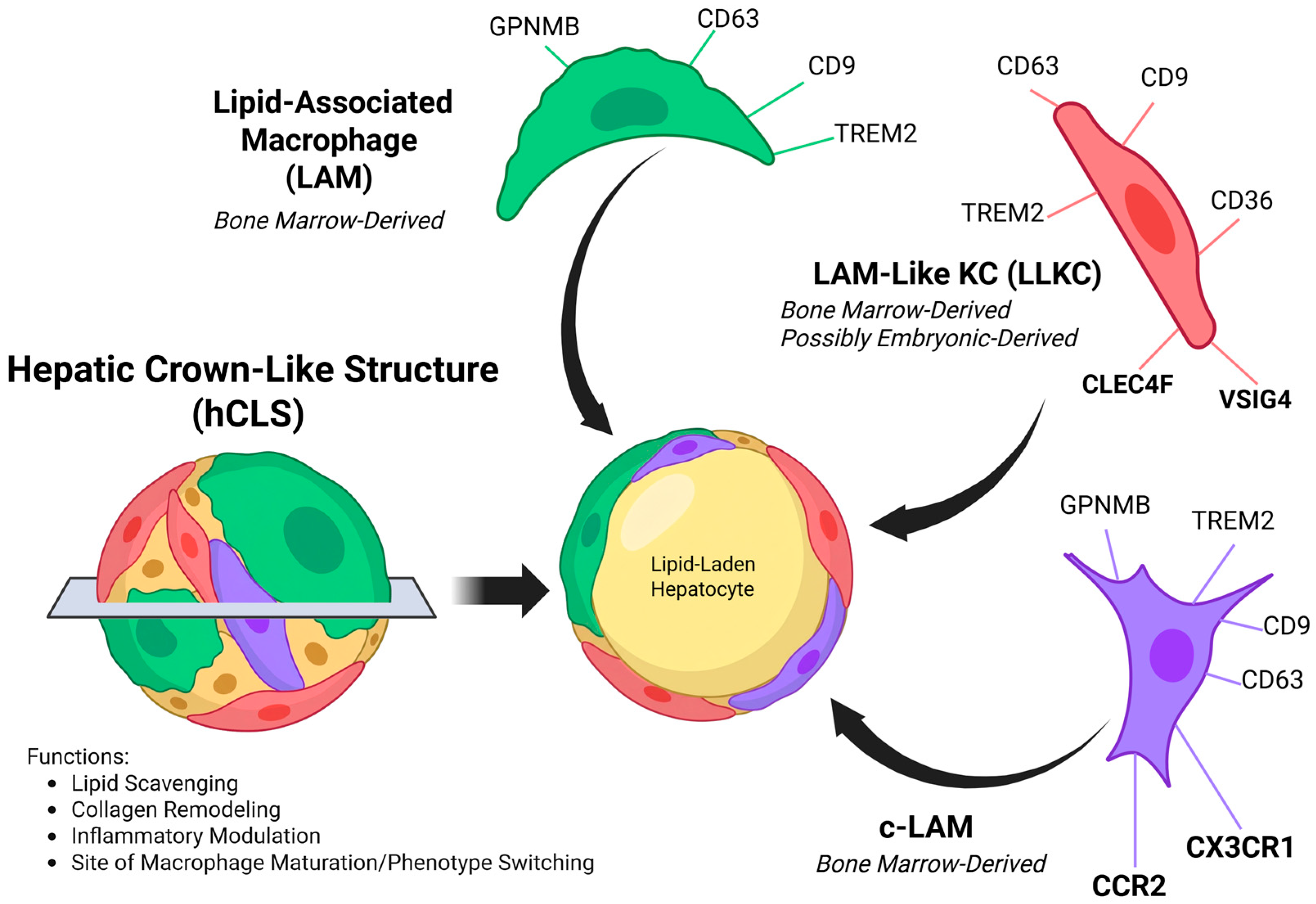
| LAMs | c-LAMs | LLKCs |
|---|---|---|
| CD11c | CD11c | CD11c |
| GPNMB | GPNMB | GPNMB |
| CD63 | CD63 | CD63 |
| CD9 | CD9 | CD9 |
| TREM2 | TREM2 | TREM2 |
| CD36 | CCR2 | CD36 |
| CX3CR1 | CLEC4F | |
| VSIG4 | ||
| CD169 |
Disclaimer/Publisher’s Note: The statements, opinions and data contained in all publications are solely those of the individual author(s) and contributor(s) and not of MDPI and/or the editor(s). MDPI and/or the editor(s) disclaim responsibility for any injury to people or property resulting from any ideas, methods, instructions or products referred to in the content. |
© 2026 by the authors. Licensee MDPI, Basel, Switzerland. This article is an open access article distributed under the terms and conditions of the Creative Commons Attribution (CC BY) license.
Share and Cite
Yuquimpo, K.; Dare, A.; Pulido Ruiz, I.A.; Weinman, S.A. The Hepatic Crown-like Structure: A Focal Point for Macrophage Evolution and Disease Response in Steatotic Liver Disease. Livers 2026, 6, 72. https://doi.org/10.3390/livers6040072
Yuquimpo K, Dare A, Pulido Ruiz IA, Weinman SA. The Hepatic Crown-like Structure: A Focal Point for Macrophage Evolution and Disease Response in Steatotic Liver Disease. Livers. 2026; 6(4):72. https://doi.org/10.3390/livers6040072
Chicago/Turabian StyleYuquimpo, Kyle, Ayobami Dare, Isabel Aranzazu Pulido Ruiz, and Steven A. Weinman. 2026. "The Hepatic Crown-like Structure: A Focal Point for Macrophage Evolution and Disease Response in Steatotic Liver Disease" Livers 6, no. 4: 72. https://doi.org/10.3390/livers6040072
APA StyleYuquimpo, K., Dare, A., Pulido Ruiz, I. A., & Weinman, S. A. (2026). The Hepatic Crown-like Structure: A Focal Point for Macrophage Evolution and Disease Response in Steatotic Liver Disease. Livers, 6(4), 72. https://doi.org/10.3390/livers6040072