


Abstract
Background: Early recurrence following curative-intent hepatectomy for colorectal liver metastases (CRLMs) remains a significant clinical challenge. This study investigates risk factors for recurrence within 6 and 12 months and evaluates the impact of salvage surgery on long-term survival. Methods: We conducted a retrospective cohort study of 109 patients who underwent liver resection for CRLMs between 2013 and 2024. The primary outcome was the identification of predictors for early recurrence using Cox proportional-hazards models. The secondary outcomes focused on overall survival (OS) stratified by the timing of recurrence and subsequent treatment. Results: High tumor burden (>4 metastases) was an independent predictor of recurrence at both 6 months (HR 3.526; p = 0.008) and 12 months (HR 3.115; p = 0.004). Intraoperative blood loss >1000 mL was significantly associated with 6-month recurrence (HR 3.356; p = 0.004) and 12-month recurrence (HR 2.171; p = 0.041). For the 12-month window, independent predictors included AJCC T3/T4 stage (HR 6.513; p = 0.011) and RAS mutation (HR 2.740; p = 0.006). Notably, patients with early recurrence who underwent salvage re-hepatectomy achieved 5-year OS rates that did not statistically differ from those without recurrence (p = 0.907 for <6 months; p = 0.433 for <12 months); however, these subgroup analyses are highly underpowered. Conclusions: High tumor burden (>4 metastases), RAS mutations, significant blood loss (>1000 mL), and primary tumor T3/T4 identify patients at high risk for early recurrence. While aggressive salvage re-hepatectomy is associated with prolonged survival in select patients, the non-significant p-values in our small salvage cohorts cannot be interpreted as evidence of survival equivalence. The observed survival benefits in the salvage cohort are heavily confounded by inherent selection biases, and therefore, the true extent of this ‘rescue’ effect must be interpreted with extreme caution and validated in larger, adequately powered multicenter studies.
1. Introduction
Surgical resection remains the only curative treatment for colorectal liver metastases (CRLMs), achieving 5-year survival rates exceeding 50% in highly selected cohorts. Despite ongoing advancements in operative techniques and perioperative care, postoperative recurrence remains a significant clinical challenge, affecting up to 75% of patients following a curative-intent hepatectomy. While technical resectability was historically the primary determinant of surgical eligibility, the contemporary paradigm has shifted toward establishing “oncological feasibility”. This transition acknowledges that poor long-term outcomes are frequently driven by occult micrometastases and aggressive tumor biology already present at the time of the initial operation [1,2].
The timing of disease recurrence provides critical insights into this biological behavior. “Early recurrence”, typically defined as disease reappearance within 6 to 12 months post-surgery, represents a distinct clinical entity inherently associated with a poor prognosis. However, the optimal threshold for defining early recurrence has remained a subject of ongoing debate, with previous studies proposing various cutoffs ranging from 6 to 24 months based on survival impacts [3]. Recent hazard function analyses indicate that the risk of recurrence peaks approximately six months postoperatively. Key drivers of this early failure include a high Tumor Burden Score (TBS), adverse molecular profiles (such as KRAS or BRAF mutations), and compromised surgical margins. Notably, recent data suggest that R1 margins (<1 mm) often serve as a surrogate marker for an infiltrative tumor phenotype rather than solely representing a technical deficiency, thereby compounding the risk of early oncologic failure [4,5].
The clinical implications of early recurrence are profound, directly correlating with significantly diminished overall survival and reduced eligibility for salvage interventions. Unlike late recurrences, which are often amenable to repeat hepatectomy, early failures frequently necessitate systemic chemotherapy and exhibit notable resistance to subsequent treatments [6]. Furthermore, evidence suggests that the risk of recurrence remains substantial even after secondary resections, underscoring the limitations of surgery alone for biologically aggressive disease. Despite this aggressive biology, recent large-scale cohorts have demonstrated that, if resectability can be achieved, salvage re-hepatectomy for early recurrence can independently and significantly improve survival, shifting the paradigm from purely palliative chemotherapy toward a more aggressive surgical approach when feasible [7,8]. Consequently, identifying reliable predictors of early recurrence is essential for optimizing patient selection, tailoring surveillance strategies, and determining the need for intensified adjuvant therapies. This study aimed to analyze the risk factors for early recurrence within 6 and 12 months post-surgery as the primary outcomes, and to evaluate overall survival following recurrence as the secondary outcome.
2. Methods
A retrospective cohort study was conducted utilizing data obtained from electronic medical records between May 2013 and July 2024. This study received approval from the Institutional Review Board (IRBRTA0036/2025); however, the requirement for written informed patient consent was waived due to the retrospective nature of the study design.
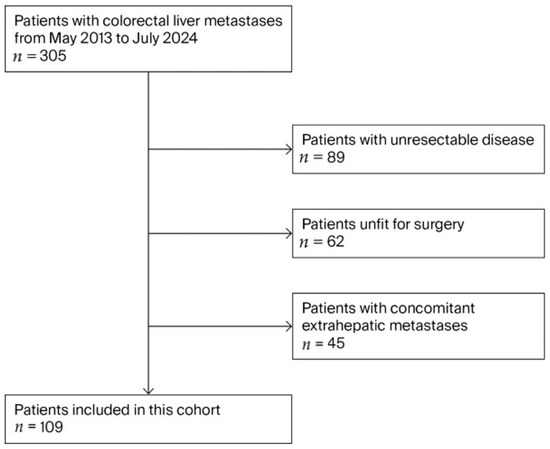
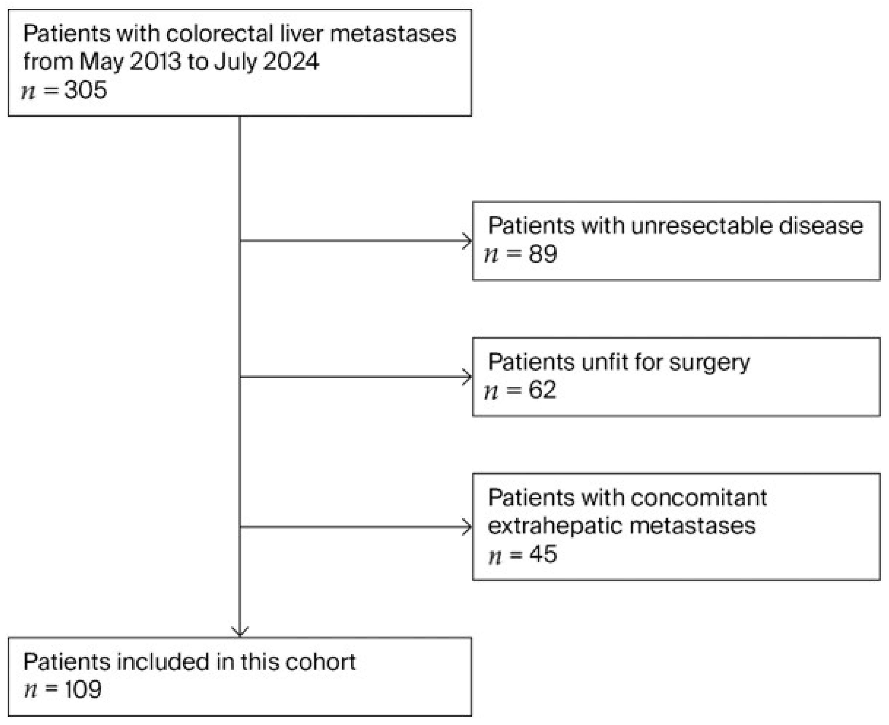
The inclusion criteria were (1) cases diagnosed with liver metastases of colorectal origin, confirmed by a diagnostic radiologist or by a pathologist in cases that underwent a liver biopsy, and (2) patients with resectable disease (adequate future liver remnant and the ability to achieve a negative resection margin). The exclusion criteria were (1) patients deemed medically unfit for surgery, (2) patients diagnosed with extrahepatic metastases prior to hepatectomy, and (3) patients with incomplete medical records (shown in Figure 1). The primary outcomes were the risk factors for early recurrence after surgery at 6 and 12 months, and the secondary outcomes were overall survival after recurrence.
Figure 1.
Flowchart of patient selection and study enrollment. Legend: This diagram illustrates the recruitment process for patients with colorectal liver metastases (CRLMs) from May 2013 to July 2024. Out of an initial 305 patients, 109 were included in the final cohort after excluding those with extrahepatic metastases (n = 45), those unfit for surgery (n = 62), and those with unresectable disease (n = 89).
2.1. Statistical Analysis
Continuous variables were evaluated using either Student’s t-test or the Mann–Whitney U test, depending on the data distribution; the results are reported as the mean ± standard deviation (SD) or the median with the interquartile range (IQR). For categorical variables, comparisons were performed using the Chi-square test or Fisher’s exact test, with data expressed as frequencies and percentages. Survival outcomes were assessed via the Kaplan–Meier method and compared using log-rank tests. Specifically, overall survival (OS) was defined as the time from surgery to death, while disease-free survival (DFS) measured the interval from surgery to the first recurrence. To identify risk factors impacting OS and DFS, hazard ratios (HRs) and 95% confidence intervals (CIs) were estimated using Cox proportional-hazards models. This time-to-event analytical framework was deliberately chosen over logistic regression to account for right-censoring (e.g., patients lost to follow-up) and to properly model the cumulative hazard of recurrence occurring up to the 6- and 12-month clinical landmarks. Patients lost to follow-up were censored during the analysis; at the 5-year mark, 21 patients (19.26%) had been lost to follow-up. Regarding missing data, patients with incomplete medical records for key clinicopathological variables were excluded during the initial enrollment phase (Figure 1), resulting in a complete-case analysis approach for the final cohort of 109 patients. The cutoff values for clinical variables, such as the number of metastases (>4) and tumor size (≥5 cm), were selected a priori based on established prognostic indices in the existing literature, notably the Fong Clinical Score and the JSHBPS nomogram (also known as the Beppu score), which is a clinical scoring tool developed by the Japanese Society of Hepato-Biliary-Pancreatic Surgery [9,10]. All analyses were conducted using STATA/IC 14.0 (StataCorp LP, College Station, TX, USA), with statistical significance set at a p-value < 0.05. To develop the prognostic risk score model for early recurrence within 12 months, key prognostic variables were selected based on the results of the multivariable Cox regression analysis. The scoring system was constructed by assigning weighted points to these independent risk factors, which were derived directly from their respective regression coefficients. The predictive performance and discriminative ability of the resulting cumulative risk score were evaluated using Receiver Operating Characteristic (ROC) curve analysis, reporting the Area Under the Curve (AUC) with a 95% confidence interval. Finally, the optimal threshold for clinical risk stratification was determined using the Youden index method for empirical cutpoint estimation.
2.2. Surgical Procedures
Surgical resection was performed under general anesthesia with low central venous pressure. A right subcostal incision, a midline extension, or an inverted-L incision was utilized. Hepatectomy was performed ensuring a resection margin of at least 1 cm beyond the tumor. Intraoperative ultrasonography was routinely used to estimate the number, size, and location of tumors, as well as their proximity to adjacent vessels; a resection margin line was mapped with an electronic scalpel on the liver surface under continuous intraoperative ultrasonographic guidance. The Cavitron ultrasonic aspiration (CUSA, Valleylab Corp, Boulder, CO, USA) or clamp-crushing technique was employed to dissect the liver parenchyma, and hemostasis was achieved using electric coagulation, argon beam units, titanium clips, and suturing. The Pringle maneuver was routinely performed, utilizing a clamp/unclamp time ratio of 15 min to 5 min. Patients remained hospitalized until their liver function approached normal levels and all adverse reactions or complications had entirely resolved.
2.3. Postoperative Management and Follow-Up
Patients were required to return to our department for follow-up appointments every 3 to 6 months post-treatment, excluding those who passed away or were lost to follow-up. Serum Carcinoembryonic Antigen (CEA) levels were measured, and magnetic resonance imaging (MRI) was performed at each visit. Chest CTs and bone scintigraphy were conducted in cases of suspected extrahepatic recurrence. Once a recurrence was definitively confirmed, a secondary treatment approach was proposed through a multidisciplinary team discussion involving surgeons, medical oncologists, pathologists, and radiologists; however, the patient’s preference remained the deciding factor. The therapeutic options included repeat liver resections or local ablative therapies (such as radiofrequency ablation or microwave ablation).
3. Results
3.1. Patient Demographics and Perioperative Characteristics
The patient demographics and clinical characteristics are summarized in Table 1. There were no statistically significant differences regarding gender, age, primary tumor location, or the type and distribution of metastases between the groups. However, a significant difference was observed regarding the number of tumors among patients experiencing recurrence within 6 months versus those within 12 months. Additionally, RAS mutation status differed significantly within the 12-month recurrence group. With the exception of intraoperative blood loss for the 6-month recurrence group, no significant differences were detected in the perioperative data between the early recurrence groups (<6 months and <12 months), as detailed in Table 1 and Table 2.
Table 1.
Demographic data in study cohort stratified by recurrence within 6 and within 12 months.
Table 2.
Perioperative data in the study cohort stratified by recurrence within 6 and within 12 months.
3.2. Predictors of Early Recurrence Within 6 and 12 Months
Univariate and multivariate hazard ratio analyses of risk factors for recurrence are presented in Table 3. A positive resection margin (R1 hepatic side) was significantly associated with recurrence within 6 months (HR, 2.721; 95% CI, 1.004–7.3747; p = 0.049). A tumor burden of more than four metastases was independently associated with recurrence within 6 months (HR, 3.526; 95% CI, 1.392–8.93; p = 0.008) and within 12 months (HR, 3.115; 95% CI, 1.446–6.711; p = 0.004). Intraoperative blood loss exceeding 1000 mL was significantly associated with recurrence within 6 months (HR, 3.356; 95% CI, 1.458–7.727; p = 0.004) and within 12 months (HR, 2.171; 95% CI, 1.031–4.57; p = 0.041). For recurrence within 12 months, independent risk factors included AJCC 8th edition T3/T4 stage (HR, 6.513; 95% CI, 1.539–27.566; p = 0.011) and RAS mutation (HR, 2.74; 95% CI, 1.328–5.655; p = 0.006).
Table 3.
Univariable and multivariable analyses of factors associated with recurrence within 6 months and within 12 months.
3.3. Predictors of Early Recurrence (Exploratory Risk Score for Early Recurrence Within 12 Months)
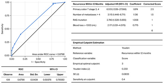
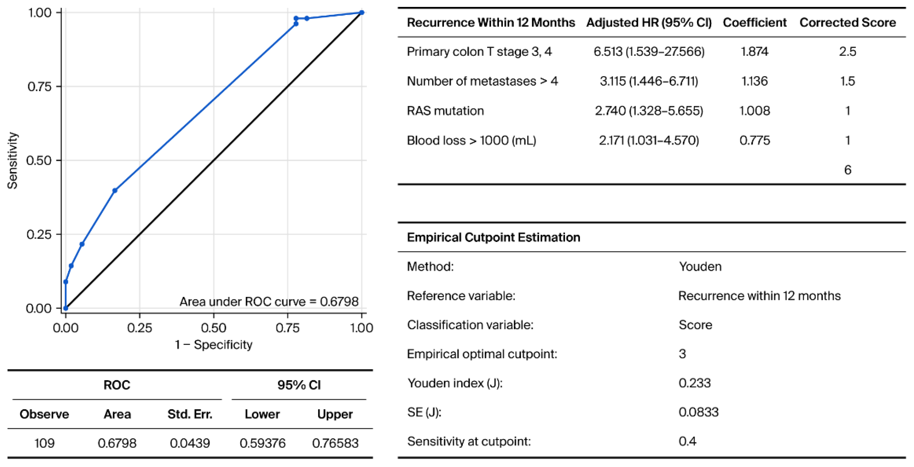
To further quantify the risk of early oncological failure, an exploratory prognostic risk score model was developed based on the multivariable analysis for recurrence within 12 months. The scoring system assigns weighted points to four key prognostic variables derived from their respective coefficients: primary colon T stage 3 or 4 (corrected score = 2.5), number of metastases greater than four (corrected score = 1.5), the presence of a RAS mutation (corrected score = 1), and intraoperative blood loss exceeding 1000 mL (corrected score = 1). This yields a maximum possible cumulative risk score of 6.
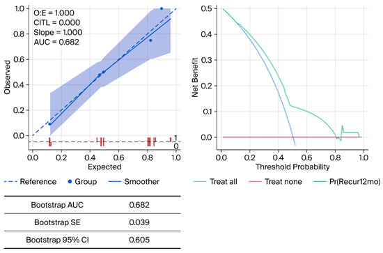
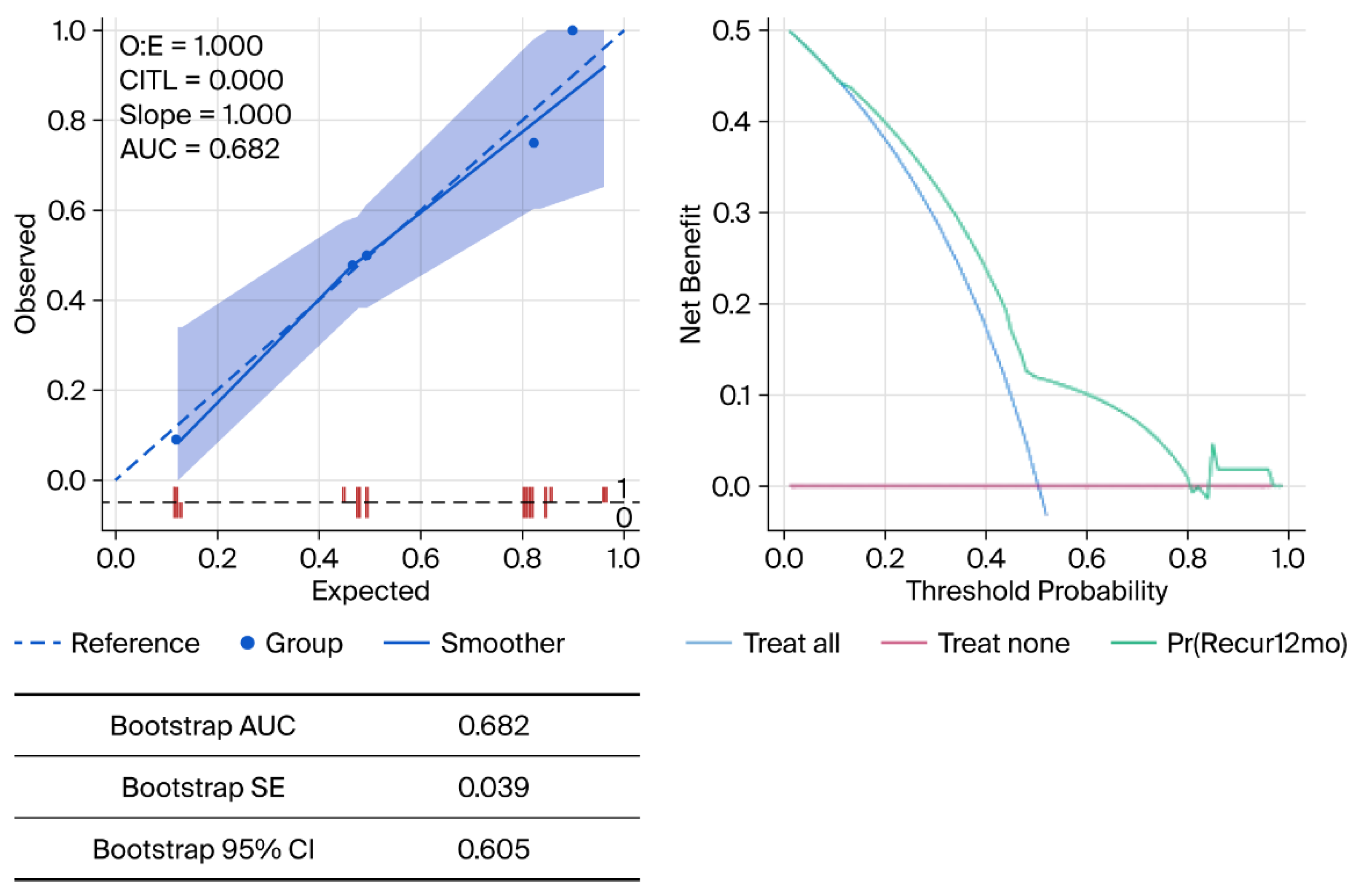
Receiver Operating Characteristic (ROC) curve analysis was performed to evaluate the model’s predictive performance, demonstrating an Area Under the Curve (AUC) of 0.6798 (95% CI: 0.59376–0.76583). Utilizing the Youden index method for empirical cutpoint estimation (J = 0.233), an optimal cutoff score of 3 was identified. At this cutpoint, the model’s sensitivity for predicting early recurrence within 12 months is 0.4 (Figure 2). To address the small sample size and mitigate the risk of overfitting, internal validation was performed using bootstrap resampling. The internal validation revealed a bootstrap-adjusted Area Under the Curve (AUC) of 0.682, with a standard error (SE) of 0.039 and a 95% confidence interval (CI) lower bound of 0.605. The calibration plot assesses the agreement between the model’s predicted probabilities and the actual observed recurrence rates, while the decision curve analysis (DCA) evaluates the net clinical benefit of utilizing this exploratory model across varying threshold probabilities (shown in Figure 2 and Figure 3).
Figure 2.
Risk scores for early recurrence within 12 months after curative surgery and decision curve analysis. Legend: The scoring system assigns weighted points to four key prognostic variables derived from their respective coefficients: primary colon T stage 3 or 4 (corrected score = 2.5), number of metastases greater than four (corrected score = 1.5), the presence of a RAS mutation (corrected score = 1), and intraoperative blood loss exceeding 1000 mL (corrected score = 1). This yields a maximum possible cumulative risk score of 6. Receiver Operating Characteristic (ROC) curve analysis was performed to evaluate the model’s predictive performance, demonstrating an Area Under the Curve (AUC) of 0.6798 (95% CI: 0.59376–0.76583). Utilizing the Youden index method for empirical cutpoint estimation (J = 0.233), an optimal cutoff score of 3 was identified. At this cutpoint, the model’s sensitivity for predicting early recurrence within 12 months is 0.4.
Figure 3.
Calibration and decision curve analysis after adding internal validation (bootstrap). Legend: This figure presents the calibration plot and decision curve analysis for the exploratory risk score model used to predict early recurrence within 12 months following curative-intent surgery. To address the small sample size and mitigate the risk of overfitting, internal validation was performed using bootstrap resampling. The internal validation revealed a bootstrap-adjusted Area Under the Curve (AUC) of 0.682, with a standard error (SE) of 0.039 and a 95% confidence interval (CI) lower bound of 0.605. The calibration plot assesses the agreement between the model’s predicted probabilities and the actual observed recurrence rates, while the decision curve analysis (DCA) evaluates the net clinical benefit of utilizing this exploratory model across varying threshold probabilities.
3.4. Survival Outcomes
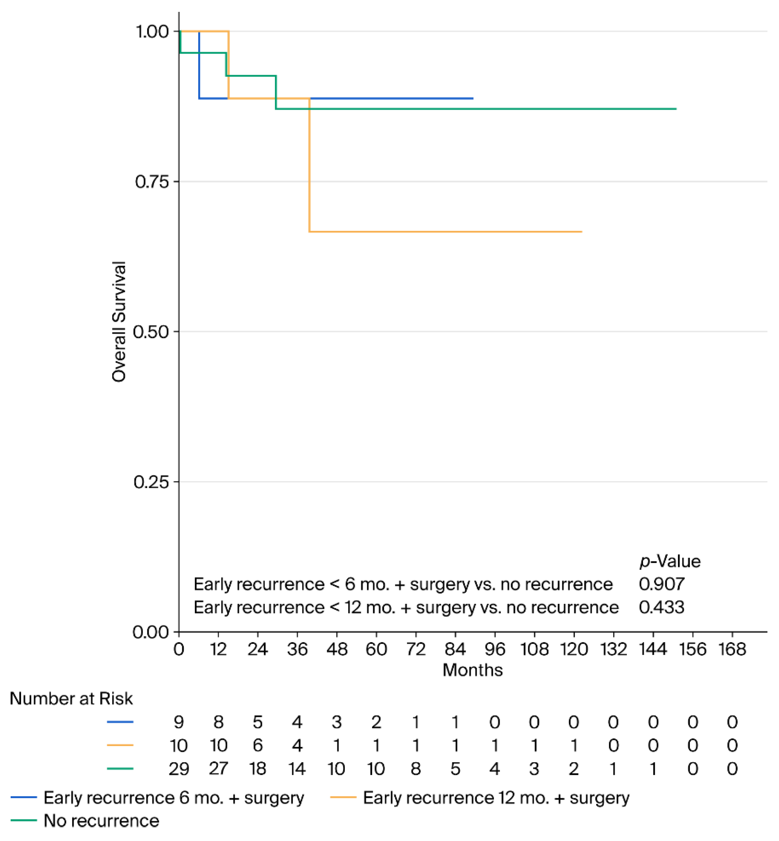
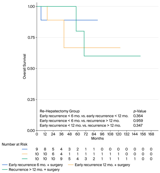
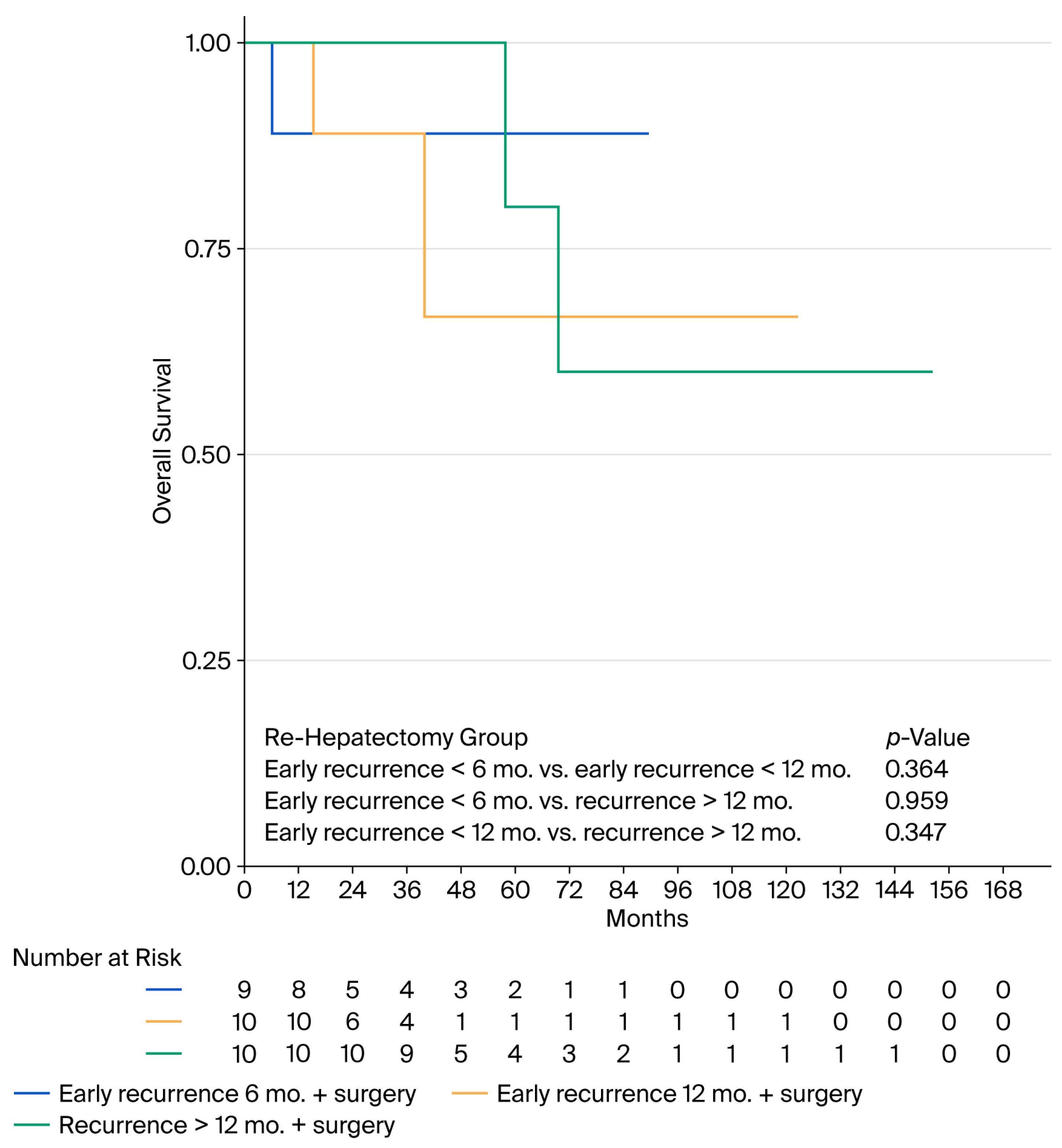
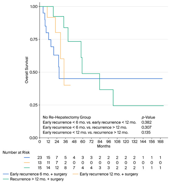
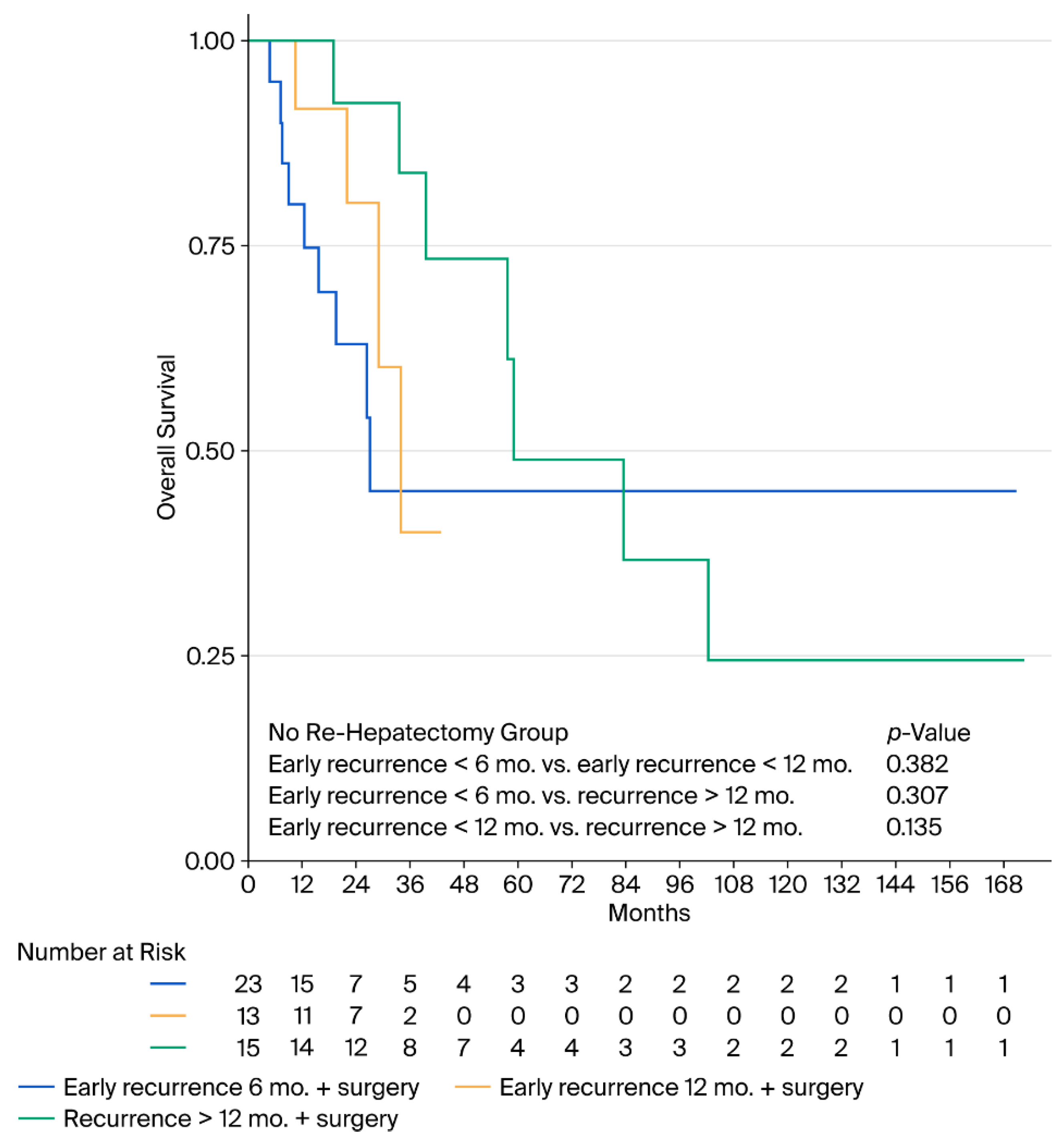
For the entire cohort, the 5-year overall survival (OS) rate was 64.91%, and the median survival limit was not reached. The 5-year disease-free survival (DFS) was 20.39%, with a median time to recurrence of 11.9 months. The recurrence rate was 0.058 (95% CI, 0.041–0.081) person-time at 6 months and 0.057 (95% CI, 0.044–0.074) person-time at 12 months. Notably, among patients who underwent salvage re-hepatectomy, there was no significant difference in OS between those with an early recurrence (within 6 or 12 months) and those with a late recurrence (after 12 months). Similarly, among patients who did not undergo re-hepatectomy, survival outcomes were comparably poor regardless of the precise timing of the recurrence (Figure 4, Figure 5 and Figure 6).
Figure 4.
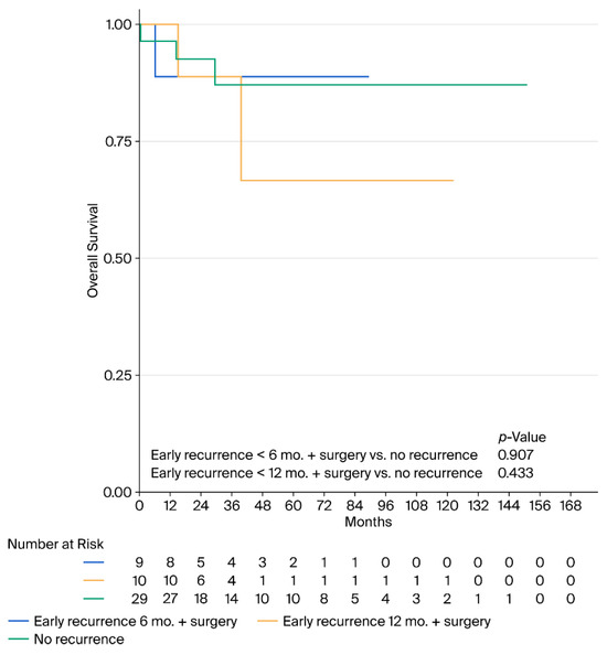
Kaplan–Meier survival curves of overall survival comparing no recurrence to salvage surgery groups. Legend: This figure compares the overall survival (OS) of patients who remained recurrence-free against those who underwent salvage re-hepatectomy after recurrence. Statistical analysis showed no significant difference in survival between the no-recurrence group and those with early recurrence at <6 months (p = 0.907) or <12 months (p = 0.433) who received secondary surgery.
Figure 5.
Kaplan–Meier curves of overall survival stratified by recurrence timing in the re-hepatectomy group. Legend: This figure displays OS for patients who underwent repeat liver resection. There were no statistically significant differences in survival outcomes based on the timing of the recurrence: early recurrence at <6 months vs. <12 months (p = 0.364), early recurrence at <6 months vs. late recurrence at >12 months (p = 0.959), and early recurrence at <12 months vs. late recurrence at >12 months (p = 0.347).
Figure 6.
Kaplan–Meier curves of overall survival stratified by recurrence timing in the no-re-hepatectomy group. Legend: This figure highlights the survival of patients with recurrent disease who did not undergo salvage surgery. Survival remained consistently poor regardless of whether the recurrence was early (<6 months or <12 months) or late (>12 months), with no significant difference observed between these subgroups (p = 0.382, p = 0.307, and p = 0.135, respectively).
4. Discussion
While the surgical management of colorectal liver metastases (CRLMs) has undoubtedly advanced, early recurrence remains a critical challenge, primarily reflecting aggressive tumor biology rather than mere technical failure. Our data demonstrate that specific clinicopathological factors reliably identify patients at a high risk for rapid disease recurrence. In our study, a high tumor burden (>4 metastases) emerged as the dominant independent predictor, increasing the risk of recurrence threefold at both 6 months (HR 3.526, p = 0.008) and 12 months (HR 3.115, p = 0.004). This finding closely aligns with classical prognostic models, such as the Fong Clinical Score, as well as more recent Tumor Burden Score (TBS) metrics. These previous studies assert that multiple visible lesions strongly reflect pervasive micrometastatic disease—consistent with the “seed and soil” hypothesis—rather than representing a localized surgical failure [9,11]. Furthermore, our multivariable analysis identified RAS mutation status as an independent predictor for recurrence within 12 months (HR 2.74, p = 0.006). This corroborates the foundational work of Vauthey et al. and Brudvik et al., who demonstrated that RAS-mutant clones exhibit enhanced infiltrative capacities, promote early systemic escape, and are consistently associated with poorer oncological outcomes [12,13].
The perioperative environment also proved critical in our cohort. Intraoperative blood loss exceeding 1000 mL was significantly associated with recurrence within 6 months and 12 months (HR 3.356, p = 0.004). As suggested by Suh et al., massive blood loss and subsequent transfusions can induce profound physiological stress and transient immunosuppression, thereby facilitating the rapid outgrowth of dormant tumor cells within a very short timeframe [14]. However, it is equally important to consider that significant blood loss may not be solely a direct causal driver of recurrence, but rather a surrogate marker for other adverse clinical scenarios. For instance, increased bleeding often correlates with greater operative complexity due to advanced tumor burden, challenging anatomical locations, or prolonged parenchymal transection. Furthermore, patients who have received extensive preoperative systemic therapies may suffer from chemotherapy-associated liver injury (CALI), which increases parenchymal friability and bleeding risk during surgery. Therefore, blood loss likely represents a composite metric reflecting both systemic surgical stress and inherently challenging baseline disease.
Advanced primary tumor T-stage (T3/T4; HR 6.513, p = 0.011) and positive resection margins (R1; HR 2.721, p = 0.049) were identified as key risk factors. The link between R1 margins and early failure in our data reinforces the contemporary paradigm supported by Perrin et al. and Gagniere et al. that positive margins often act as a surrogate for an infiltrative tumor phenotype extending beyond macroscopic boundaries, rather than merely reflecting surgical inadequacy [5,15,16]. Consequently, optimizing perioperative care to minimize complications is not merely a recovery metric but a fundamental component of a long-term oncologic strategy.
To translate these independent prognostic factors into a practical clinical tool, we formulated an exploratory risk score aimed at predicting early recurrence within 12 months following curative-intent surgery. By synthesizing the weighted impacts of advanced primary T-stage, high tumor burden, RAS mutational status, and significant intraoperative blood loss, the model provides a cumulative assessment of a patient’s recurrence risk. The model achieved an AUC of 0.6798 with a sensitivity of 0.4 at the optimal empirical cutpoint of 3. We acknowledge that these performance metrics reflect limited discriminative ability. Consequently, this model must be strictly interpreted as an exploratory tool rather than a definitive clinical instrument. While it highlights a particularly vulnerable patient cohort that may benefit from highly intensified postoperative surveillance protocols, the lack of internal validation (such as bootstrap resampling) and calibration analysis in this small dataset means that the score requires substantial refinement and rigorous external validation before it can be applied in routine clinical practice.
Regarding long-term outcomes, our entire cohort achieved a robust 5-year overall survival (OS) of 64.91%. A notable observation in the present study relates to the potential “rescue” effect of salvage re-hepatectomy. Specifically, Kaplan–Meier analyses revealed no statistically significant difference in OS between patients who remained recurrence-free and those who experienced early recurrence but subsequently underwent salvage surgery (p = 0.907 for <6 months and p = 0.433 for <12 months). However, it is imperative to interpret these findings with extreme caution. Because these subgroup analyses rely on very small cohorts (9 to 10 patients), they are inherently underpowered, and the non-significant p-values cannot be reliably interpreted as evidence of survival equivalence [7,17].
This observation strongly aligns with contemporary literature, such as the large cohort study by Watanabe et al. [8], which indicated that, although early recurrence often presents with aggressive disease characteristics, highly selected patients amenable to repeat resection experience a substantial survival benefit comparable to those suffering late recurrences. We recognize that the findings in our cohort are largely confirmatory when compared to extensive multicenter datasets such as the LiverMetSurvey or foundational meta-analyses by Viganò et al. [18]. The primary value of our study lies in validating that these established prognostic factors hold true in our specific setting, underscoring their universal applicability despite potential variations in healthcare resources, tumor biology, or specific patient populations. Additionally, our analysis evaluated perioperative parameters such as R1 parenchymal margins and intraoperative blood loss exceeding 1000 mL. While the importance of these factors has been discussed extensively in the existing literature, our findings confirm their prognostic relevance within our specific regional cohort, reinforcing the established principle that meticulous surgical technique and the minimization of operative stress are essential components of a successful oncologic strategy. Conversely, within our cohort, patients who did not undergo secondary surgery exhibited markedly lower survival trajectories regardless of when their recurrence occurred (p = 0.382 for <6 months vs. <12 months; p = 0.307 for <6 months vs. >12 months). Furthermore, survival did not significantly differ between patients recurring before versus after the 12-month mark, provided that they underwent a successful re-hepatectomy (p = 0.347). This validates the aggressive clinical policies advocated by Adam et al. and Wicherts et al.: surgical re-intervention should be strongly reconsidered whenever it is oncologically and technically feasible, because it remains the primary driver of mitigating the severe impact of aggressive tumor biology in recurrent CRLM [17,19].
Limitations: Despite the significant insights provided by our findings, this study possesses inherent limitations that must be acknowledged. First, as a retrospective cohort study conducted at a single institution, it carries an unavoidable risk of selection bias, meaning that our findings may not be entirely generalizable to other healthcare settings with different patient demographics or surgical volumes. Second, while our cohort was robust enough to identify key predictors of recurrence, the total sample size of 109 patients remains relatively small. This limitation renders our multivariable Cox regression models susceptible to statistical overfitting. Furthermore, our subgroup analyses evaluating long-term survival trajectories for patients undergoing salvage re-hepatectomy are based on very small sample sizes (e.g., 9–10 patients per group). Consequently, these survival comparisons lack the statistical power necessary to support definitive conclusions and must be interpreted with extreme caution. Similarly, while our proposed risk score highlights a vulnerable patient subset, its moderate discriminative ability (AUC 0.6798) and lack of external validation limit its immediate clinical utility without further testing in larger, independent cohorts. Additionally, it must be emphasized that patients who underwent salvage re-hepatectomy in our cohort represent a highly selected subgroup. These individuals inherently possess more favorable tumor biology, superior performance status, and more technically amenable recurrence patterns compared to patients who were relegated strictly to palliative systemic therapy. Consequently, the conclusion that salvage surgery achieves a survival rate comparable to that in patients without recurrence should be interpreted with caution, because this observation is heavily confounded by inherent selection biases. Therefore, while our data strongly supports the role of aggressive surgical intervention for early recurrence, future large-scale, multicenter prospective studies are absolutely necessary to fully validate these conclusions. Finally, our study period spanned over a decade, taking place from 2013 to 2024. Over these 11 years, there were substantial and continuous evolutions in systemic chemotherapy, including the introduction of highly effective targeted biologic therapies. Although our analysis accounted for whether patients received preoperative and adjuvant chemotherapy, the specific nuances, durations, and variations of these evolving medical regimens were not analyzed in depth. This evolving landscape of systemic treatment could serve as an unmeasured confounding factor influencing our long-term survival outcomes. Therefore, future studies tracking standardized contemporary chemotherapy regimens are warranted to fully contextualize these findings.
5. Conclusions
High tumor burden (>4 metastases), RAS mutations, significant blood loss (>1000 mL), and primary tumor T3/T4 identify patients at high risk for early recurrence. While aggressive salvage re-hepatectomy is associated with prolonged survival in select patients, the non-significant p-values in our small salvage cohorts cannot be interpreted as evidence of survival equivalence. The observed survival benefits in the salvage cohort are heavily confounded by inherent selection biases, and therefore, the true extent of this ‘rescue’ effect must be interpreted with extreme caution and validated in larger, adequately powered multicenter studies.
Author Contributions
P.B.: study conceptualization, design, data analysis, and writing the manuscript. N.T. and V.C.: data collection and interpretation. A.T.: data collection and critical review. P.F.: statistical analysis and editing. S.H.: study supervision and final approval. All authors have read and agreed to the published version of the manuscript.
Funding
This research received no external funding.
Institutional Review Board Statement
This study was approved by the Institutional Review Board of the Royal Thai Army Medical Department (IRBRTA0036/2025, approval date 10 January 2025).
Informed Consent Statement
Informed consent was not required due to the retrospective nature of the study, as per local legislation (IRBRTA0036/2025).
Data Availability Statement
The data are available from the corresponding author upon reasonable request.
Acknowledgments
During the preparation of this study, the authors used Google Gemini 3.1 Pro to create the graphical abstract. The authors have reviewed and edited the output and take full responsibility for the content of this publication.
Conflicts of Interest
The authors declare no conflicts of interest.
References
- Conticchio, M.; Uldry, E.; Hübner, M.; Digklia, A.; Fraga, M.; Sempoux, C.; Raisaro, J.L.; Fuks, D. Prognostic factors in colorectal liver metastases: An exhaustive review of the literature and future prospectives. Cancers 2025, 17, 2539. [Google Scholar] [CrossRef] [Scilit] [PubMed]
- Kawashima, J.; Akabane, M.; Endo, Y.; Woldesenbet, S.; Altaf, A.; Ruzzenente, A.; Popescu, I.; Kitago, M.; Poultsides, G.; Sasaki, K.; et al. Recurrence timing and risk following curative resection of colorectal liver metastases: Insights from a hazard function analysis. J. Surg. Oncol. 2024, 130, 1290–1298. [Google Scholar] [CrossRef] [Scilit] [PubMed]
- Imai, K.; Allard, M.A.; Benitez, C.C.; Vibert, E.; Cunha, A.S.; Cherqui, D.; Castaing, D.; Bismuth, H.; Baba, H.; Adam, R. Early Recurrence After Hepatectomy for Colorectal Liver Metastases: What Optimal Definition and What Predictive Factors? Oncologist 2016, 21, 887–894. [Google Scholar] [CrossRef] [Scilit] [PubMed]
- Margonis, G.A.; Buettner, S.; Andreatos, N.; Sasaki, K.; Ijzermans, J.N.M.; van Vugt, J.L.A.; Pawlik, T.M.; Choti, M.A.M.; Cameron, J.L.; He, J.; et al. Anatomical resections improve disease-free survival in patients with KRAS-mutated colorectal liver metastases. Ann. Surg. 2017, 266, 641–649. [Google Scholar] [CrossRef] [Scilit] [PubMed]
- Perrin, T.; McIntyre, S.M.; Ehrich, F.; Heiselman, J.; van der Stok, E.P.; Fiasconaro, M.; Gönen, M.; Shia, J.; Kingham, T.P.; Balachandran, V.P.; et al. The association between surgical margin status, recurrence patterns, and oncologic outcomes in patients with colorectal liver metastases. Ann Surg. 2025; online ahead of print. [CrossRef] [Scilit] [PubMed]
- Yoshizaki, Y.; Kawaguchi, Y.; Seki, Y.; Sasaki, S.; Miyata, A.; Nishioka, Y.; Ichida, A.; Akamatsu, N.; Kaneko, J.; Hasegawa, K. Risk of recurrence does not decrease in patients undergoing the second and third resection of colorectal liver metastases. HPB 2025, 27, 937–945. [Google Scholar] [CrossRef] [Scilit] [PubMed]
- Engstrand, J.; Strömberg, C.; Nilsson, H.; Freedman, J.; Jonas, E. The role of repeat liver resection for colorectal liver metastases: A systematic review and meta-analysis. Surg. Oncol. 2018, 27, 367–374. [Google Scholar] [CrossRef] [Scilit] [PubMed]
- Watanabe, G.; Mise, Y.; Ito, H.; Inoue, Y.; Ishizawa, T.; Takahashi, Y.; Saiura, A. Repeat Hepatectomy for Early Recurrence of Colorectal Liver Metastases-Prognostic Impacts Assessed from the Recurrence Pattern. World J. Surg. 2020, 44, 268–276. [Google Scholar] [CrossRef] [Scilit] [PubMed]
- Fong, Y.; Fortner, J.; Sun, R.L.; Brennan, M.F.; Blumgart, L.H. Clinical score for predicting recurrence after hepatic resection for metastatic colorectal cancer: Analysis of 1001 consecutive cases. Ann. Surg. 1999, 230, 309–318. [Google Scholar] [CrossRef] [Scilit] [PubMed]
- Beppu, T.; Yamamura, K.; Sakamoto, K.; Honda, G.; Kobayashi, S.; Endo, I.; Hasegawa, K.; Kotake, K.; Itabashi, M.; Hashiguchi, Y.; et al. Validation study of the JSHBPS nomogram for patients with colorectal liver metastases who underwent hepatic resection in the recent era—A nationwide survey in Japan. J. Hepatobiliary Pancreat. Sci. 2023, 30, 591–601. [Google Scholar] [CrossRef] [Scilit] [PubMed]
- Sasaki, K.; Morioka, D.; Conci, S.; Margonis, G.A.; Sawada, Y.; Ruzzenente, A.; Kumamoto, T.; Iacono, C.; Andreatos, N.; Guglielmi, A.; et al. The tumor burden score: A new “metro-ticket” prognostic tool for colorectal liver metastases based on tumor size and number of tumors. Ann. Surg. 2018, 267, 132–141. [Google Scholar] [CrossRef] [Scilit] [PubMed]
- Vauthey, J.N.; Zimmitti, G.; Kopetz, S.E.; Shindoh, J.; Chen, S.S.; Andreou, A.; Curley, S.A.; Aloia, T.A.; Maru, D.M. RAS Mutation Status Predicts Survival and Patterns of Recurrence in Patients Undergoing Hepatectomy for Colorectal Liver Metastases. Ann. Surg. 2013, 258, 619–627. [Google Scholar] [CrossRef] [Scilit] [PubMed]
- Brudvik, K.W.; Kopetz, S.E.; Li, L.; Conrad, C.; Aloia, T.A.; Vauthey, J.N. RAS mutation predicts positive resection margins and worse outcomes after resection of colorectal liver metastases. Ann. Surg. 2015, 262, 117–123. [Google Scholar] [CrossRef] [Scilit] [PubMed]
- Suh, S.W.; Lee, S.E.; Choi, Y.S. Influence of intraoperative blood loss on tumor recurrence after surgical resection in hepatocellular carcinoma. J. Pers. Med. 2023, 13, 1115. [Google Scholar] [CrossRef] [Scilit] [PubMed]
- Memon, S.; Liang, R.X. Impact of surgical margins on early recurrence after hepatic resection. HPB 2020, 22, S156. [Google Scholar] [CrossRef] [Scilit]
- Iwaki, K.; Kaihara, S.; Koyama, T.; Nakao, K.; Matsuda, S.; Toriguchi, K.; Kitamura, K.; Oshima, N.; Kondo, M.; Hashida, H.; et al. The Impact of KRAS Status on the Required Surgical Margin Width for Colorectal Liver Metastasis Resection. J. Clin. Med. 2023, 12, 2313. [Google Scholar] [CrossRef] [Scilit] [PubMed]
- Costa, P.F.; Coelho, F.F.; Jeismann, V.B.; Kruger, J.A.P.; Fonseca, G.M.; Cecconello, I.; Herman, P. Repeat hepatectomy for recurrent colorectal liver metastases: A comparative analysis of short- and long-term results. Hepatobiliary Pancreat. Dis. Int. 2022, 21, 162–167. [Google Scholar] [CrossRef] [Scilit] [PubMed]
- Viganò, L.; Capussotti, L.; Lapointe, R.; Barroso, E.; Hubert, C.; Giuliante, F.; Ijzermans, J.N.M.; Mirza, D.F.; Elias, D.; Adam, R. Early recurrence after liver resection for colorectal metastases: Risk factors, prognosis, and treatability. A report from the international dataset of liver metastases from colorectal cancer (LiverMetSurvey). Ann. Surg. Oncol. 2014, 21, 452–460. [Google Scholar] [CrossRef] [Scilit] [PubMed]
- Adam, R.; Bismuth, H.; Castaing, D.; Waechter, F.; Navarro, F.; Abascal, A.; Majno, P.; Engerran, L. Repeat hepatectomy for colorectal liver metastases. Ann. Surg. 1997, 225, 51–62. [Google Scholar] [CrossRef] [Scilit] [PubMed]

Disclaimer/Publisher’s Note: The statements, opinions and data contained in all publications are solely those of the individual author(s) and contributor(s) and not of MDPI and/or the editor(s). MDPI and/or the editor(s) disclaim responsibility for any injury to people or property resulting from any ideas, methods, instructions or products referred to in the content. |
© 2026 by the authors. Licensee MDPI, Basel, Switzerland. This article is an open access article distributed under the terms and conditions of the Creative Commons Attribution (CC BY) license.