


Abstract
Mucoadhesive drug delivery systems have emerged as a promising strategy to enhance the therapeutic efficacy of pharmaceuticals by improving drug residence time, bioavailability, and site-specific targeting. Among various materials investigated, biopolysaccharides have gained significant attention due to their biocompatibility, biodegradability, non-toxicity, and inherent mucoadhesive properties. Natural polymers such as chitosan, alginate, pectin, hyaluronic acid, and cellulose derivatives exhibit strong interactions with mucosal surfaces through hydrogen bonding, electrostatic interactions, and polymer chain entanglement. These properties enable prolonged drug retention at mucosal sites, controlled drug release, and enhanced permeation across biological barriers. Mucoadhesive biopolysaccharides have been explored for diverse routes of administration, including oral, buccal, nasal, ocular, vaginal, and pulmonary delivery. Furthermore, chemical modification and nanostructuring of these polymers have expanded their functionality, enabling targeted delivery of small molecules, proteins, peptides, and nucleic acids. This review highlights the mechanisms of mucoadhesion, key biopolysaccharides used in drug delivery, formulation approaches, and recent advances in their application as versatile platforms for novel therapeutic delivery systems. The continued development of mucoadhesive biopolysaccharide-based carriers holds substantial potential for improving treatment outcomes and patient compliance.
1. Introduction
The establishment of original and effective drug delivery systems that can navigate the difficulties posed by standard dosage forms is a major area of current pharmaceutical research. Poor bioavailability, brief residence time at the absorption site, quick drug clearance, and inconsistent drug delivery to maintain therapeutic benefit concentration are just a few of the drawbacks of traditional systems [1,2,3]. Particularly for long-term treatments, these restrictions lower patient adherence and therapeutic efficacy. Mucoadhesive drug delivery systems (MDDS) have garnered a lot of interest as a solution to these problems. These technologies offer regulated medication release, localized distribution, and extended retention by adhering to the mucosal surfaces of biological membranes [4,5]. Due to their enormous surface area, high vascularization, and capacity to avoid hepatic first-pass metabolism, the mucosal membranes of the oral cavity, nasal cavity, gastrointestinal tract, ocular surface, vagina, and rectum are especially appealing for drug delivery. However, physiological barriers such as mucus turnover, enzymatic degradation, and mucociliary clearance frequently limit medication delivery through mucosal channels, despite their potential. By making close, sustained contact with the mucous layer or epithelial surface, mucoadhesive systems get past these obstacles and improve medication stability and absorption [6,7]. The polymer and mucin glycoproteins interact mechanically, chemically, and physically as part of the mucoadhesion mechanism. Van der Waals forces, electrostatic attraction, hydrogen bonds, and polymer chain interpenetration are some examples of these interactions [8]. In recent years, biopolysaccharides which are naturally derived, high-molecular-weight carbohydrate polymers have emerged as highly promising materials for mucoadhesive formulations. Studies have focused on chitosan, alginate, hyaluronic acid, pectin, cellulose derivatives (e.g., HPMC, CMC), carrageenan, and xanthan gum. These polymers exhibit several desirable characteristics for biomedical use, such as biocompatibility, biodegradability, intrinsic mucoadhesive potential, versatility in formulation, sustainability and abundance [9]. The use of mucoadhesive biopolysaccharides offers multiple advantages in drug delivery, including enhanced absorption. They also facilitate the stabilization of labile biomolecules, such as peptides, proteins, and nucleic acids, by shielding them from enzymatic degradation, while enabling site-directed delivery and supporting well-regulated, sustained release profiles [10,11]. These collective attributes contribute to improved therapeutic performance by sustaining drug availability at the target site, thereby lowering the required dosing frequency and ultimately promoting better patient adherence. Moreover, advances in polymer chemistry and nanotechnology have enabled the modification and functionalization of natural polysaccharides to improve their mucoadhesive strength, mechanical stability, and drug-loading efficiency. For instance, thiolated chitosan derivatives show improved adhesion and permeation properties, while carboxymethylated cellulose enhances hydration and swelling behavior. Thus, mucoadhesive biopolysaccharides represent a promising and sustainable platform for the next generation of drug delivery systems, combining natural safety with advanced functionality. Their continued development is expected to play a pivotal role in achieving targeted, prolonged, and patient-friendly therapies across a broad range of therapeutic areas [2,9]. Therefore, this review aims to provide a comprehensive overview of mucoadhesive biopolysaccharide-based systems, emphasizing their mechanisms, design strategies, and expanding role as advanced platforms for effective and targeted drug delivery.
2. Structure of the Mucous Membrane
Previous investigations have offered detailed insights into the anatomical features and functional responsibilities of the oral mucosa, establishing a strong foundation for ongoing research in this domain [7]. However, a brief summary in this chapter is essential. According to light microscopy, epithelial maturation within the human oral mucosa varies considerably across different intraoral regions. Structurally, the oral mucosa comprises three principal components: the epithelium, the basement membrane, and the underlying connective tissue. Together, these layers maintain the structural integrity of the mucosal barrier, with the connective tissue providing essential support to the basement membrane (Figure 1).
Figure 1.
Composition of the oral mucosa.
The epithelial layer serves as the primary protective interface for deeper tissues and is broadly categorized into two forms. Keratinized epithelium is predominantly present in the hard palate and other mechanically resilient regions of the oral cavity, whereas non-keratinized epithelium characterizes more flexible areas such as the vestibule, lips, buccal mucosa, floor of the mouth, alveolar mucosa, and the mucosal lining of the soft palate [7]. Basal cells function as the proliferative source of new epithelial cells, which progressively differentiate, undergo morphological transitions, and enlarge as they migrate toward the mucosal surface. Comparative investigations have reported that the buccal epithelial thickness in humans, dogs, and rabbits typically ranges from approximately 500 to 800 micrometres [12]. A distinct layer is formed by the basement membrane. This layer lies between the epithelium and the connective tissue, providing essential mechanical support and maintaining firm adhesion to the underlying stromal matrix. The biomechanical characteristics of the oral mucosa are largely governed by the connective tissue compartment, which imparts resilience and structural stability. Within this context, the buccal epithelium is classified as non-keratinized, reflecting its flexible and compliant nature [13]. It is infused with tall, connective tissues shaped like a cone. These tissues, known as the lamina propria, are made up of smooth muscles, blood vessels, collagen fibers, and a supporting layer of connective tissues [14]. The oral mucosa receives a rich vascular supply primarily from branches of the external carotid artery. Within the buccal region, perfusion is maintained through contributions from the buccal artery and several terminal branches of the facial artery. In addition, the posterior superior alveolar artery and the infraorbital artery play key roles in delivering arterial blood to the inner cheek, ensuring adequate nourishment and functional integrity of the buccal mucosal tissues. A gel-like secretion known as mucus, composed primarily of water-insoluble glycoproteins, forms a continuous coating across the oral cavity. This mucous layer adheres to the apical surface of epithelial cells, where it functions as a protective interface, shielding the underlying tissues from mechanical stress, microbial invasion, and chemical irritation [7]. This material is characterized as a viscoelastic hydrogel composed predominantly of water (95–99%) and a smaller fraction (1–5%) of the aforementioned water-insoluble glycoproteins, accompanied by trace quantities of proteins, enzymes, electrolytes, and nucleic acids. Notably, its precise composition varies according to the anatomical site and the specific glands responsible for mucus secretion, reflecting functional adaptations across different regions of the body [15,16].
3. Adhesion/Bioadhesion/Mucoadhesion
From the first animal-derived pastes used in prehistoric cave drawings to the use of extremely strong epoxy glues used in aircraft manufacturing, adhesives have been a common part of human history. Moreover, bacterial adhesion and related processes impair the functionality of numerous indwelling biomaterials, frequently leading to hospital-acquired infections and medical device-related infections, which raise hospital expenses by more than $500 million yearly. Adhesion processes are also very significant in nature and have a variety of healthcare implications [8]. Adhesion mechanisms also have an impact on non-biomedical fields: biofilms are acquired by water purification, transportation, and storage systems, while marine organisms frequently colonize ship hulls and other stationary marine structures. The “fixing” of two surfaces to one another is the simplest definition of the process of adhesion [17]. The general physical/chemical phenomenon that causes two different materials to adhere to one another is called adhesion. The main mechanisms for biological interfaces and polymers are mechanical interlocking, electrostatic (donor–acceptor) interactions, interpenetration/diffusion, wetting/adsorption, and the formation of covalent or secondary bonds. It is essential to comprehend these mechanisms because biological surfaces, which are soft, hydrated, and dynamic, do not always respond well to tactics that increase bulk polymer adhesion [18]. Depending on the context, adhesion can have a wide variety of terminological subsets. It is commonly referred to as “bioadhesion” when adhesion takes place in a biological environment, and “mucoadhesion” when it happens on mucosal membranes. The binding of an artificial or natural polymer to a biological substrate is known as bioadhesion. The phrase mucoadhesion is frequently used when this substrate takes the form of a mucous layer [19]. Biopolymers are utilized for a variety of therapeutic applications and exhibit bioadhesive qualities. The two types of bioadhesive polymers are (i) specific, meaning they can stick to specific chemical structures in biological molecules, and (ii) non-specific, indicating that they have the ability to attach to the mucosal layer and cellular surfaces [20]. Pharmaceutical formulations that are bioadhesive are typically made to prolong the time spent in residence of drug compounds and localize the effect at the desired location in order to improve drug bioavailability. At the same time, they help design local drug delivery formulations, which improve bioavailability by avoiding metabolic pathways [10]. In biological contexts, bioadhesion is generally classified into three distinct categories. Type I bioadhesion describes interactions occurring between two biological entities, as seen in processes such as platelet aggregation or tissue repair. Type II bioadhesion involves the attachment of biological components to non-biological or synthetic surfaces; examples include cell adhesion to culture plates and the formation of microbial biofilms on prosthetic materials. Type III bioadhesion denotes the reverse scenario, wherein a synthetic or engineered material adheres to a biological substrate, such as dental sealants bonding to enamel or the attachment of synthetic hydrogels to soft tissues. In the field of drug delivery, the term bioadhesion refers to the capacity of a therapeutic carrier to establish intimate and prolonged contact with a designated biological surface. This interaction enables the delivery system to remain localized at the target site, thereby enhancing residence time and potentially improving the efficiency and precision of drug administration. The mucous layer on a tissue’s surface or epithelial tissue can both be considered biological surfaces. Mucoadhesion is the term for the phenomenon where adhesive attachment occurs to a mucus coat [6]. Mucoadhesion refers to the ability of natural or synthetic materials to bind to mucosal tissues through a range of interfacial physicochemical interactions. This phenomenon facilitates close and sustained contact with the mucosal surface, making it highly valuable for improving the retention and performance of drug delivery systems. This characteristic enables the targeted delivery of active biomolecules, such as proteins, peptides, and oligonucleotides, to particular body regions and helps get around a number of drawbacks of traditional drug delivery methods, including first-pass metabolism. When two substances, at least one of which is of biological origin, adhere to each other for a prolonged duration due to interfacial forces, it is termed mucoadhesion. It refers to a substance’s (biological or synthetic) capacity to stick to mucosal tissues in pharmaceutical applications, allowing the drug and its carrier to stay in close proximity to the mucous membrane. All things considered, mucoadhesion is a complicated process that includes polymer chain wetting, adsorption, and interpenetration at the interface. Hydrophilic polymers with mucoadhesive properties are commonly incorporated into pharmaceutical formulations alongside the active pharmaceutical ingredient (API) to facilitate localized drug delivery. By adhering to mucosal surfaces, these polymers help maintain the formulation at the intended site of action, thereby enhancing residence time and supporting more effective therapeutic outcomes. For localized drug delivery, the formulation will be “maintained” on a biological surface. The API will be made available near the site of action, which will increase its bioavailability [8]. The terms “bioadhesion” and “mucoadhesion” may be confused quite often. In mucoadhesion, the polymer is attached to the mucus surface (substrate), whereas in bioadhesion, it stays attached to the biological surface, which can be either epithelial tissue or the mucus coating on that tissue. Numerous theories have been put forth by research to explain the mechanism of mucoadhesion. These include cohesive theory, mechanical theory, adsorption theory, diffusion theory, wetting theory, and electronic theory [21].
4. Mechanism of Mucoadhesion
Mucoadhesive polymers interact with the mucin-rich surface of mucosal tissues through a sequence of three fundamental stages depicted in Figure 2.
Figure 2.
Schematic illustration of the mucoadhesion mechanism (Author drawn by using MS PowerPoint 2007).
As illustrated in Figure 2, there are three phases related to mucoadhesion: (I) contact, in which mucoadhesive polymers adhere to the mucous membrane; (II) interpenetration stage, in which the mucoadhesive polymer diffuses into the mucus layer; (III) consolidation, in which the polymers in the mucoadhesive formulation penetrate the mucous layer [6,21]. Understanding the three phases of mucoadhesion is necessary to comprehend how mucoadhesive polymers interact with the mucous membrane to improve adhesion and drug delivery [8]. Mucoadhesive drug delivery systems have numerous essential characteristics that enhance their efficacy in mucosal medication administration. An optimal mucoadhesive system must demonstrate robust adherence to the mucosal surface via intermolecular interactions, including hydrogen bonding, electrostatic attraction, van der Waals forces, and polymer chain interpenetration with mucin glycoproteins. Proper swelling and hydration capacity are crucial for ensuring close contact with the mucus layer and promoting polymer relaxation and diffusion. Moreover, appropriate molecular weight, chain flexibility, and functional group density substantially affect mucoadhesive strength and retention duration. These solutions must exhibit biocompatibility, biodegradability, and non-irritancy to guarantee safety during extended mucosal administration. These characteristics collectively allow mucoadhesive drug delivery systems to extend residence time at the administration site, enhance drug absorption and bioavailability, facilitate site-specific targeting, decrease dosing frequency, and ultimately improve therapeutic efficacy and patient compliance [8].
4.1. Stages of Mucoadhesion
Step 1: Contact Stage
The first close interaction between the mucoadhesive substance and the mucous membrane initiates the mucoadhesion process. Various methods can be used to establish this contact, depending on the administration site. Sometimes, like when a mucoadhesive delivery system is inserted and held in the oral cavity, ocular surface, or vaginal mucosa, the two surfaces are mechanically brought together. In other cases, deposition happens spontaneously due to aerodynamic or physiological causes. For example, processes such as inertial impaction, which filter out particulate matter from the airstream, enable particles to deposit onto the sticky mucus layer in the nasal cavity or bronchi of the respiratory tract [22,23].
Step 2: Interpenetration Stage
Following the initial contact between the polymer and the mucosal surface, the process advances to the interpenetration stage, which is widely regarded as the critical phase of mucoadhesion. At this point, the polymer chains begin to diffuse into the mucin network, setting the foundation for the formation of a stable adhesive interface. By this point, a semi-permanent adhesive network is formed as polymer chains and mucin glycoproteins start to diffuse and interpenetrate across the interface. According to the diffusion theory, mucoadhesion is established when the polymer chains migrate into the mucin matrix and subsequently engage in additional secondary interactions. This interchain entanglement contributes significantly to the development of a stable adhesive bond. Hydrogen bonds, van der Waals forces, and mutual chain entanglement act together to strengthen the interfacial contact and enhance mucoadhesive performance. Sufficient polymer flexibility, hydration, and contact time encourage chain mobility and deeper mucus layer penetration, enhancing the adhesive bond and extending the dosage form’s residence time [11,21].
Step 3: Consolidation Stage
After the Interpenetration stage, the consolidation stage entails the mucoadhesive polymers penetrating the mucous layer. It has been suggested that a second consolidation stage is necessary when strong or extended adhesion is needed, such as in larger formulations subjected to physiological stresses like blinking or mouth movements. When mucoadhesive materials are activated by moisture, they stick to solid or semi-dry mucosal surfaces the best during this stage. Moisture causes the polymer system to plasticize, increasing chain mobility and enabling the adhesive molecules to dissociate, reorient, and adapt to the mucosal surface. This helps the polymer and mucin form weak hydrogen and van der Waals bonds, creating an adhesive interface that is both flexible and stable enough to sustain extended contact in dynamic situations [22,23]. The consolidation of the mucoadhesive interface occurs through a combination of mechanical interlocking and chemical interactions. These mechanisms promote sustained propagation of adhesion, enabling the polymer to maintain a robust bond with the mucosal surface over time, as illustrated in Figure 3 [23].
Figure 3.
Phases of mucoadhesion (Reproduced from Ref. [23], IJPMR Publishing Press, 2017, open access journal).
Two theories are engaged in elucidating the consolidation steps: (i) diffusion and (ii) dehydration. Diffusion theory describes the reciprocal interaction between mucoadhesive polymers and mucin glycoproteins, which arises from the interpenetration of their respective chains and the subsequent establishment of secondary intermolecular bonds. This cooperative engagement strengthens the overall mucoadhesive interface. Dehydration theory states that in an aqueous environment, substances that engage with mucus gel and water occupy the dosage form in reaction to a concentration gradient until osmotic balance is achieved. This results in the formulation mixture and mucus on the mucous membrane maintaining contact for an extended duration. Consequently, instead of the intertwining of macromolecular chains, the flow of water is what strengthens the adhesive bonds [24,25].
4.2. Theories of Mucoadhesion
The process of mucosal adhesion is complex, and a number of ideas have been proposed that are crucial to adhesion [3].
The electronic theory (illustrated in Figure 4) suggests that mucoadhesion develops through electron transfer between the bioadhesive polymer and the mucin glycoprotein network. This exchange is driven by differences in their electronic structures, ultimately leading to the formation of an electrostatic interaction at the interface. Electrostatic attractive forces are created at the interface as a result of this electron exchange, creating an electrical double layer of opposing charges that aids in surface adhesion. Instead of being a secondary effect of joint strength, the resulting interfacial charge distribution not only starts the adhesive interaction but also strengthens the bond overall, suggesting that electrostatic forces are a major factor in mucoadhesion [3,26].
Figure 4.
Mechanism of mucoadhesion in accordance with electronic theory (Reproduced from Ref. [26], MDPI, 2022, open access journal).
According to the adsorption theory, mucoadhesion is mediated by the formation of intermolecular interactions, most notably hydrogen bonds and van der Waals forces, between the mucoadhesive polymer and the mucus layer. These attractive forces promote mutual affinity at the interface and contribute to the stabilization of the adhesive bond [3,10]. Adhesive materials attach to mucosal surfaces through a spectrum of weak intermolecular forces, such as van der Waals interactions, hydrophobic associations, and hydrogen bonding. Collectively, these interactions facilitate stable yet reversible adhesion at the biological interface. The sheer volume of interactions gives them tremendous adhesive strength even though they are secondary weak forces [21]. Hydrophobic effects might also be significant, particularly if the mucoadhesive polymers are amphiphilic. When the mucoadhesive polymer and mucins form strong covalent bonds, the adsorption theory also takes chemisorption into account [10]. Two different kinds of chemical bonding are at play here: (i) strong primary bonds, where covalent bonds are undesirable due to their permanent nature; (ii) weak secondary bonds, such as hydrophobic bonds, hydrogen bonds, van der Waals forces, and electrostatic forces. These are the most projecting surface interaction forms in adhesion because these interactions are relatively stable yet not permanent, and they can be disrupted with minimal energetic input [3].
The diffusion theory of mucoadhesion postulates that adhesion betide through the process of adhesion refers to the intertwining of polymer chains with the mucin network found in the mucus layer, resulting in the formation of a semi-permanent interfacial network structure. This process is driven by a concentration gradient between the polymer and mucin phases, enabling molecular chains from both surfaces to diffuse into each other to a sufficient depth for effective entanglement. The extent of interpenetration is influenced by multiple factors, including the polymer diffusion coefficient, chain flexibility, mucin mobility, contact time, mutual solubility, and similarity in chemical structure. For efficient bioadhesive bonding, the depth of interfusion is typically reported to range between 0.2 and 0.5 µm [3,27]. The extent of polymer penetration into the mucus layer can be estimated using the diffusion equation,
where t represents the contact time and Db denotes the diffusion coefficient of the mucoadhesive polymer within the mucus matrix. Optimal adhesive strength is achieved when the penetration depth approaches the characteristic length of the polymer chains. In general, a higher degree of structural compatibility between the polymer and the mucin network enhances their mutual solubility and promotes more effective diffusion, ultimately contributing to a stronger mucoadhesive interaction [11].
l = (tDb)½,
Possibly the most well-established theory of adhesion is the wetting theory. It works better with low-viscosity or liquid bioadhesives [7]. According to the wetting theory, the adhesive strength is mostly determined by the liquid’s capacity to spread across the biological surface in systems with low viscosity and strong affinity for the substrate. The contact angle, spreadability coefficient, and work of adhesion, the energy needed to separate the two phases, all correlate with adhesion strength, according to this theory (Figure 5). According to the hypothesis, the liquid distributes more uniformly as the contact angle gets closer to zero, increasing adhesion because of the larger contact area [4,11,26]. The distinction in surface energies of the liquid (γA) and biological substrate (γB) and their interfacial energy (γAB) is known as the spreadability coefficient (SAB), which is defined as SAB = γB – γA − γAB. Generally speaking, systems with smaller contact angles and greater individual surface energies have better interfacial interaction and superior adhesive strength, which enhances the mucoadhesive performance as a whole [27]. The energy needed to split the two phases is represented by the work of adhesion (WA), which may be calculated as follows:
wA = γA + γ − γAB
Figure 5.
The idea of wetting in relation to contact angle (Author drawn by using MS PowerPoint 2007).
According to this idea, increasing mucoadhesion requires lowering surface and interfacial energy and adjusting the contact angle [21].
According to the cohesive theory, mucoadhesion results not only from interfacial bonding with the mucosal surface but also from the internal cohesive strength and intermolecular forces inside the sticky substance itself. This perspective posits that mucoadhesion is chiefly governed by the cohesive interactions among polymer chains, together with the complementary interactions established between the polymer and mucin molecules. Together, these forces underpin the formation and stability of the mucoadhesive interface. In contrast to other explanatory models of mucoadhesion, this approach centers on the magnitude of force required to disrupt or separate the adhesive junction once it has been established, highlighting the mechanical resistance that must be overcome to detach the interacting surfaces. It further proposes that failure of the adhesive joint occurs directly at the interfacial region; however, in reality, it often occurs within the weaker material, indicating cohesive failure rather than adhesive failure. The internal molecular network and structural integrity are crucial for maintaining adhesion in rigid and semirigid mucoadhesive materials, which are most suited for this theory. The mechanisms of mucoadhesion can be broadly categorized into two groups: physical theories, which include the cohesive, diffusion, and wetting theories, and chemical theories, which include the electronic and adsorption theories [21,26,27]. This hypothesis characterizes the amount of force necessary to separate the two surfaces after an adhesive interface has been formed, thereby providing insight into the mechanical stability of the mucoadhesive bond. In the following equation, the fracture strength is equal to the adhesive strength:
σ = (E × ε/L)1/2
In this framework, ε represents the fracture energy, while σ denotes the corresponding fracture strength. The parameter L refers to the critical crack length, and E indicates the Young’s modulus of elasticity, which collectively describe the mechanical behavior of the adhesive joint under stress [5].
The mechanical theory of mucoadhesion posits that adhesion takes place when a liquid adhesive infiltrates and occupies the irregularities or micro-cracks found on a rough substrate surface, resulting in the creation of an interlocked structure that improves adhesive strength. A textured or uneven substrate enlarges the effective contact area between the polymer and the mucosal surface, while simultaneously enhancing viscoelastic and plastic energy dissipation during detachment phenomena viewed as more critical to adhesion performance than simple mechanical interlocking. In certain instances, the adhesive process may additionally be influenced by electron exchange between the mucosal surface and the mucoadhesive substrate, driven by disparities in their electronic configurations, which in turn leads to the formation of a bilayer. This electrostatic interaction generates extra alluring forces that contribute to overall adhesion strength. However, there has been debate regarding whether these electrostatic forces are the primary factor contributing to adhesion or merely a consequence of strong interfacial bonding. Overall, the mechanical theory emphasizes the role of surface roughness, interfacial energy dissipation, and possible electronic interactions in enhancing the adhesion between the mucoadhesive and the biological substrate [7,19,27].
Among the several theoretical models developed to elucidate mucoadhesion, the diffusion/interpenetration and adsorption theories are deemed most relevant to biopolysaccharide-based mucoadhesive systems. This occurs mainly because natural polysaccharides typically engage with mucosal surfaces by allowing flexible polymer chains to interpenetrate the mucin glycoprotein network, subsequently forming secondary intermolecular interactions, including hydrogen bonding, van der Waals forces, and hydrophobic associations. Moreover, electrostatic interactions may significantly boost mucoadhesion in charged polysaccharides such as chitosan, alginate, and carrageenan, since ionic attraction between polymer functional groups and negatively charged mucin residues improves interfacial adhesion. Despite the molecular significance of these ideas, the in vivo mucoadhesive efficacy of biopolysaccharide-based systems often diverges from in vitro forecasts owing to the intricate and dynamic characteristics of mucosal environments. Physiological factors, including continuous mucus turnover, mucociliary clearance, enzymatic degradation, pH fluctuations, variations in ionic strength, and competitive hydration by adjacent biological fluids, may diminish polymer–mucin interactions and decrease the overall residence time at the site of administration. Consequently, whereas conventional mucoadhesion theories provide a valuable mechanistic basis, their practical usefulness is limited unless the dynamic physiological factors of the target mucosal environment are included in formulation design and assessment methodologies.
The relevance of mucoadhesion theories depends on the physicochemical properties of the polysaccharide. Chitosan mostly adheres to electrical and adsorption theories due to the electrostatic interactions between protonated amino groups and negatively charged mucin. Conversely, alginate, pectin, and cellulose derivatives mostly demonstrate mucoadhesion via diffusion and adsorption processes that include hydrogen bonding and chain interpenetration. Furthermore, thiolated polysaccharides may establish covalent disulphide bonds with mucin, resulting in increased adhesion strength (Table 1).
Table 1.
Relationship between Mucoadhesive Polysaccharides and Predominant Adhesion Mechanisms.
5. Mucoadhesive Biopolysaccharides
The third major class of biopolymers, or carbohydrates, is polysaccharides, which are crucial for blood coagulation, fertilisation, immune system function, preventing pathogenesis, and therapeutic efficacy [28,29]. Polysaccharides influence a number of biological functions in cells, including cell signal transduction, lubrication, energy storage, and structural support. Although numerous naturally derived polymers exhibit inherent mucoadhesive behavior, the precise molecular pathways contributing to this phenomenon remain only partially elucidated [30]. Polysaccharides, the most abundant class of naturally occurring carbohydrates, are classified according to their chemical architecture, which is defined by monosaccharide units interconnected through glycosidic linkages [31]. As a reflection of their inherent chain flexibility, Menchicchi and co-workers systematically evaluated a diverse set of polysaccharides differing in their fundamental structural features, such as primary architecture, molecular weight, charge distribution, conformational behavior, and salt-induced coil compaction, to understand how these parameters influence their mucoadhesive performance. They conducted a systematic investigation of polysaccharide–mucin interactions by employing highly sensitive analytical instruments, tightly controlled experimental parameters, including the initial viscosities of both mucin and polysaccharide solutions, and an integrated dataset spanning multiple length scales. By combining macroscopic insights from micro-viscosimetry with nanostructural information obtained through scattering techniques, the researchers were able to comprehensively elucidate how molecular weight, charge characteristics, and chain contraction influence the mucoadhesive behaviour of polysaccharides. The study showed that dextran sulphate interacts with mucin through measurable alterations in viscosity, and that the molecular weight of the polysaccharide serves as a key determinant in regulating this behavior, influencing both its interaction with mucin and its overall mucoadhesive performance. These observations were consistent with results obtained from DLS and isothermal titration calorimetry, which similarly indicated that molecular-weight dependence also characterizes the interaction between alginate and mucin proteins. Despite the fact that MW is believed to be crucial in these interactions, chain flexibility controls how well high-MW polysaccharides can cause mucin to contract [32]. Polysaccharides may be categorized into distinct groups according to differences in their structural configurations or biological functions (Table 2). Broadly, they are classified into three principal types: structural polysaccharides such as cellulose and chitin; storage polysaccharides, including starch and glycogen; and gel-forming varieties, exemplified by alginic acid and mucopolysaccharides (Figure 6). These macromolecules may exist as linear or branched chains and can be classified further as ionic or nonionic (anionic or cationic). Moreover, they are capable of forming covalent linkages with other biomolecules, including peptides, amino acids, and lipids [33].
Table 2.
A list of a few polysaccharides from different sources [33].
Figure 6.
Sorting polysaccharides according to their physiological characteristics and the kind of monosaccharides that are used as building blocks (Author drawn by using CANVA 2.362).
The inherent structural rigidity and organized architecture of many natural polysaccharides, such as cellulose, enhance their capacity to entrap and retain food components, including starch [34]. Negatively charged polysaccharides comprise pectin, heparin, hyaluronic acid, and alginate, while chitosan serves as the primary example of a cationic polysaccharide; notably, pectin, pectinol, and alginate function as polyelectrolyte-based polymers, in contrast to the positively charged nature of chitosan [33].
Importantly, the mucoadhesive performance of biopolysaccharides is not solely dependent on their polymeric nature or molecular weight, but is profoundly influenced by the type, density, and spatial arrangement of functional groups present along the polymer backbone. Recent investigations have demonstrated that amino, hydroxyl, carboxyl, sulfate, and acetyl groups critically regulate the interaction of polysaccharides with mucin glycoproteins through hydrogen bonding, electrostatic attraction, hydration, and chain interpenetration mechanisms. These structure–function relationships are now considered central to understanding the overall mucoadhesive efficiency of natural polymers.
Among naturally derived polysaccharides, chitosan exhibits particularly strong mucoadhesive behavior owing to the presence of protonated amino groups, which establish electrostatic interactions with negatively charged sialic acid and sulfate residues within mucin. In addition, hydroxyl groups present along the chitosan backbone further contribute through hydrogen bonding, thereby stabilizing the adhesive interface. Studies have shown that the cationic nature, moderate flexibility, and linear conformation of chitosan collectively enhance mucosal penetration and prolong residence time compared with many neutral or anionic polysaccharides.
In contrast, alginate and pectin primarily depend on carboxyl-mediated hydrogen bonding and swelling behavior for mucoadhesion. The abundance of ionizable carboxyl groups improves hydration and polymer chain relaxation, facilitating deeper interpenetration into the mucus network. Menchicchi et al. further demonstrated that molecular weight and chain flexibility strongly influence alginate–mucin interactions, highlighting that highly hydrated and flexible chains generally produce superior mucoadhesive performance. However, excessive negative charge density may occasionally reduce adhesion due to electrostatic repulsion with negatively charged mucin domains.
Cellulose derivatives such as hydroxypropyl methylcellulose and carboxymethyl cellulose exhibit mucoadhesive properties mainly through extensive hydrogen bonding generated by hydroxyl and ether functionalities. The incorporation of carboxymethyl groups additionally improves swelling capacity and water retention, thereby enhancing polymer mobility and mucosal contact. Nevertheless, because these polymers lack strong ionic interactions, their adhesive strength is generally lower than that of chitosan-based systems.
Similarly, carrageenans possess sulfate groups that contribute to high hydration capacity, gel formation, and viscoelasticity, which collectively support prolonged mucosal retention. Although the precise molecular basis of carrageenan-mediated mucoadhesion remains insufficiently characterized, current evidence suggests that hydration-driven chain entanglement and secondary intermolecular interactions play dominant roles in their adhesive behavior. Hyaluronic acid, containing both hydroxyl and carboxyl functionalities, demonstrates remarkable hydration and viscoelastic properties, enabling the formation of stable hydrogel-like networks capable of maintaining intimate contact with mucosal tissues for extended durations.
Collectively, these findings indicate that the mucoadhesive efficiency of polysaccharides is governed not merely by polymer presence but by the specific functional groups responsible for intermolecular interactions with mucin. Polysaccharides containing amino functionalities generally exhibit stronger electrostatic interactions, whereas hydroxyl-, carboxyl-, and sulfate-rich polymers predominantly rely on hydrogen bonding, hydration, and polymer chain interpenetration. Therefore, understanding these molecular interactions provides a more rational framework for the design of advanced mucoadhesive drug delivery systems with enhanced residence time, controlled release characteristics, and improved therapeutic efficacy.
The mucoadhesive efficacy of polysaccharides is affected by their charge, functional groups, and ambient pH. Cationic polymers like chitosan often demonstrate enhanced mucoadhesion owing to electrostatic interactions with negatively charged mucin, especially in acidic to neutral environments. Conversely, anionic polymers like alginate and pectin mostly depend on hydrogen bonding and chain interpenetration for adherence. Consequently, the choice of a suitable polysaccharide should be determined by the physicochemical characteristics of the target mucosal environment and the specific therapeutic application (Table 3).
Table 3.
Comparative Mucoadhesive Characteristics of Major Polysaccharides.
Cationic polysaccharides often exhibit enhanced primary mucoadhesion due to electrostatic attraction with mucin, whereas anionic polymers depend more on secondary interactions and hydration-mediated interpenetration. Nonetheless, formulation-specific parameters like viscosity, crosslinking, swelling kinetics, and mucosal location may markedly affect practical adhesive efficacy.
5.1. Cellulose
Cellulose is considered one of the most abundant biomolecules in nature, accounting for more than half of the biomass produced by photosynthetic organisms and constituting the primary structural component of plant cell walls. Cellulose has long been recognized as a fundamental structural constituent of a wide range of plant-derived materials, including cotton, linen, wood, hemp, jute, kenaf, sugar beet pulp, various cereal straws, and flax. Sources of cellulose also include marine invertebrates that are part of the Ascite family, algae like Valonia and Microdicyon, along with bacteria such as Acetobacter [28,35]. The chemical reactivity of cellulose is strongly influenced by its molecular architecture, particularly the surface-accessible hydroxyl groups, which readily participate in both intra- and intermolecular hydrogen bonding [28,36]. The physical and chemical behavior of cellulose is largely dictated by the hydrogen-bonding networks that arise between adjacent polymer chains, which reinforce their linear arrangement. Despite this intrinsic structural strength, cellulose exhibits several limitations, including minimal solubility in common solvents, poor wrinkle resistance, and the absence of thermoplastic character. To overcome these constraints, deliberate chemical or physicochemical surface modifications are often required to improve its functional properties [37]. In most cases, the naturally produced fraction of cellulose cannot be transformed into a synthetic polymer [38]. Introducing functional groups onto the cellulose surface has been proposed as an effective strategy, as it allows deliberate modification of the material’s interfacial chemistry while preserving key attributes such as its capacity for self-assembly, controlled dispersion within polymeric matrices, and enhanced inter- and intraparticle bonding. Native cellulose and its chemically modified derivatives, including cellulose ethers, cellulose esters, and oxidized cellulose, are extensively utilized in a variety of drug-delivery platforms. Among the naturally derived cellulose ethers, sodium carboxymethyl cellulose (CMC), methylcellulose (MC), ethylcellulose (EC), hydroxypropyl cellulose (HPC), and hydroxypropyl methylcellulose (HPMC) represent key examples, each providing specific functional advantages for pharmaceutical formulation (Figure 7) [39].
Figure 7.
A range of cellulose ethers produced using various techniques (Author drawn by using MS PowerPoint 2007).
5.2. Hemicelluloses
Hemicelluloses, which constitute another essential component of plant cell walls, comprise a diverse group of polysaccharides, including mannans, xyloglucans, xylans, and mixed-linkage (1→3),(1→4)-β-glucans. These polymers form an interconnected matrix surrounding the cellulose microfibrils, contributing to the overall structural integrity and flexibility of the plant cell wall [40,41,42]. These polysaccharides are typically associated with cellulose microfibrils through noncovalent interactions such as hydrogen bonding and van der Waals forces. Beyond their structural role, hemicelluloses also function in cellular signaling and serve as metabolic reserves, highlighting their multifaceted contribution to plant physiology [43]. It represents the second most abundant renewable component of lignocellulosic biomass after cellulose, with an estimated global production surpassing 60 billion tons annually [44,45].
5.3. Chitosan
Chitosan, derived primarily from the deacetylation of chitin found in marine crustacean shells, is a naturally sourced polymer recognized for its biocompatibility and biodegradability. Owing to these attributes, it has been extensively utilized in areas like haemostasis, wound care, and numerous biomedical procedures. Notably, chitosan exhibits solubility in mildly acidic aqueous media, typically below a pH of 6.5, due to the protonation of its amino groups [46,47,48,49]. After cellulose, chitosan is the most prevalent polysaccharide used in mucoadhesive agents. Chitosan, a cationic mucoadhesive agent, is essentially a polysaccharide that is produced by deacetylating chitin. This is a copolymer of N-acetyl-glucosamine and glucosamine. Chitosan dissolves in weak, diluted acids but not in water. Chitosan’s low toxicity, biodegradation, and biocompatibility likely make it a desirable polymeric component. Numerous mechanisms are responsible for chitosan’s mucoadhesive properties. Because chitosan contains numerous hydroxyl and amino functional groups along its backbone, it readily forms hydrogen-bonding interactions with mucin glycoproteins, thereby contributing to its mucoadhesive behaviour [27,50]. Numerous research teams have examined the molecular-level mechanistic evidence between mucin and chitosan. Chitosan is a partially deacetylated linear polysaccharide consisting of repeating units of D-glucosamine and N-acetyl-D-glucosamine, joined by β-(1→4) glycosidic linkages. This structural arrangement underpins its characteristic physicochemical and biological properties [51]. The primary component of crustacean shells, chitin, undergoes alkaline deacetylation to produce chitosan, a random copolymer. The repeating unit of chitosan macromolecules contains multiple potential mucin-binding sites: At acidic pH, the glucosamine units within chitosan become predominantly protonated, while the polymer itself presents hydrophilic hydroxyl groups, hydrophobic acetyl moieties at non-deacetylated positions, and ionisable amino groups. The apparent pKa of chitosan generally falls between 6.1 and 7.0, though this range may vary slightly depending on the polymer’s source and the analytical method used [52]. The chitosan molecule’s cationic nature, linear conformation, and moderate flexibility determine the unique properties of the chitosan for biomedical applications [15,53].
5.4. Pectin
Pectin is widely recognized as one of the most commercially significant hydrocolloids, with broad application across numerous food products. Its functional versatility and biocompatibility have established it as an essential material within the food and related industrial sectors. Sunflower pectin represents a natural source of low-methoxyl pectin (approximately 50%), whereas commercially produced pectin is predominantly derived from citrus fruits and apples, which typically contain high-methoxyl pectin exceeding 50% [54].
5.5. Refined Starch
Refined starch appears as a white, odorless powder. Amylose and amylopectin molecules make up the polysaccharide. The relative contents of amylose and amylopectin vary substantially among starches derived from different botanical sources, reflecting species-specific differences in their biosynthetic pathways and structural organization; however, starch commonly comprises approximately 20–25% amylose and 75–80% amylopectin [55]. The primary polysaccharide responsible for energy storage in plants is starch, which is widely distributed throughout botanical tissues, occurring as granules in fruits, seeds, roots, tubers, stem pith, leaves, and rhizomes [56,57,58]. It makes up the majority of the carbohydrates consumed by humans, much like potatoes, rice, wheat, corn, and cassava [59].
5.6. Glycogen
Glycogen is a different type of carbohydrate that serves as an energy reserve. It resembles amylopectin and other storage polysaccharides [60]. but is more branching and compressed. Animal cells typically contain glycogen, a kind of glucose, in their cytoplasm. Despite its central role in physiological metabolic processes, glycogen is included here solely for completeness, as it holds no practical relevance for industrial applications [61].
5.7. Chitin
Chitin is an animal-derived, linear polysaccharide composed of repeating N-acetyl-D-glucosamine units, bearing both acetyl and amino functional groups. These structural features impart a notable degree of hydrophobicity to the polymer backbone and influence its overall physicochemical behavior. Owing to this structural rigidity and extensive intermolecular hydrogen bonding, chitin exhibits extremely limited solubility in water as well as in the majority of common organic solvents, a characteristic largely attributed to its extensive hydrogen-bonding network and crystalline structure. Instead, it dissolves only in a limited range of specialized solvent systems, such as dimethylacetamide supplemented with approximately 5% lithium chloride, as well as certain chloroalcohols combined with aqueous mineral acids. Functionally, chitin acts as a primary structural biopolymer and is broadly distributed in nature, constituting the exoskeletal framework of insects, crustaceans, and various other invertebrate species. It is also a key component of fungal cell walls, including the mycelial network and sporulating structures, highlighting its biological and ecological relevance [62,63].
5.8. Hyaluronic Acid
Hyaluronic acid, also known as hyaluronan, is a salient example of a mucopolysaccharide, a class of glycoprotein-associated polysaccharides commonly referred to as mucins. Unlike many polysaccharides restricted to specific tissues, hyaluronan is one of the few carbohydrate polymers widely distributed in vertebrate systems, with particularly high concentrations observed in developing embryos. Structurally, it is a linear, high-molecular-weight polysaccharide, typically reaching masses on the order of 7 × 10⁶ g/mol. Hyaluronan frequently associates with proteins through extensive hydrogen bonding, forming proteoglycan complexes that play central roles in maintaining extracellular matrix organization, hydration, and tissue viscoelasticity [64]. Consequently, its extraction and purification present considerable challenges due to its rigid structural organization and limited solubility. Chitin and its derivatives also exhibit a range of biological applications, including their use in wound management, where combinations with alginate have been shown to promote tissue repair during surgical interventions [65,66].
5.9. Alginates
Alginates, long-chain hydrophilic polymers obtained from seaweed, are characterized by the algae’s cell walls’ strength and flexibility. They have been consumed since 600 B.C., but Krefting successfully extracted pure alginate from seaweed only in 1896. In 1929, alginate first achieved commercial significance when it was adopted by the company Kelco as a stabilizing agent in ice cream formulations, marking its early industrial integration and highlighting its functional versatility in food systems. This early industrial application established alginate as a valuable hydrocolloid and facilitated its broader adoption across the food and pharmaceutical sectors [45,62]. Alginate is widely regarded as a safe and nutritionally relevant biopolymer, and its favourable physicochemical characteristics distinguish it from many other seaweed-derived polysaccharides. Its ability to undergo ion-mediated cross-linking, respond to pH variations, and exhibit excellent biocompatibility and biodegradability has supported its extensive use within the food, nutraceutical, and functional-ingredient sectors. Notably, alginate is unique among naturally occurring polysaccharides in that each of its monomeric units contains a carboxyl group, a feature that underpins its versatile gelling behaviour and broad industrial applicability [67].
5.10. Carrageenans
Carrageenans (CRGs), derived from red seaweeds, constitute a diverse group of linear sulfated galactans valued for their extensive structural variability and the corresponding range of physicochemical and biological properties they confer. Their primary backbone is composed of alternating 3-linked β-D-galactopyranose and 4-linked α-D-galactopyranose residues, which together define the characteristic structural motif of these polysaccharides. Variations in the sulfation pattern, the configuration of the repeating disaccharide units, and the presence or absence of 3,6-anhydrogalactose within the 4-linked position serve as key determinants for distinguishing among the principal carrageenan types. These structural distinctions underpin the functional diversity observed across CRG-based materials [68]. Compared with well-established mucoadhesive polymers such as chitosan, pectin, and alginate, the mucoadhesive behaviour of carrageenan (CRG) has been less extensively characterized. The specific structural motifs within different carrageenan types and their influence on adhesive performance remain insufficiently understood, creating gaps in the current structure–function relationship. Nevertheless, available evidence suggests that CRG-based matrices can facilitate the transport of macromolecular therapeutics across mucosal epithelia, an effect largely attributed to their inherent, albeit underexplored, mucoadhesive properties [30].
5.11. Guar Gum
Guar gum is a naturally occurring galactomannan polysaccharide derived from the endosperm of Cyamopsis tetragonoloba seeds. It comprises a linear β-(1→4)-linked mannose backbone with α-(1→6)-linked galactose side chains. Guar gum has significant mucoadhesive qualities owing to its elevated molecular weight, substantial hydroxyl functionality, and pronounced hydration capacity, facilitated by hydrogen bonding and chain tangling with mucin glycoproteins. Its elevated viscosity and swelling characteristics enhance extended residence duration and sustained drug release, making it a valuable excipient in buccal, gastric, and nasal mucoadhesive formulations [69].
5.12. Dextran
Dextran is a branched microbial polysaccharide mostly composed of α-(1→6)-linked D-glucose units, with sporadic α-(1→3) branching points. Native dextran has moderate mucoadhesive capabilities owing to its hydrophilic nature and flexible polymeric configuration; yet, its adhesive performance may be significantly enhanced by chemical derivatisation. Modified dextran derivatives, including oxidised, thiolated, and sulfated forms, exhibit improved interaction with mucin via increased hydrogen bonding and electrostatic interactions. Consequently, dextran-based materials have attracted increased attention in the development of mucoadhesive hydrogels, nanoparticles, and in situ gelling delivery systems [70].
5.13. Pullulan
Pullulan is a water-soluble extracellular polysaccharide synthesised by Aureobasidium pullulans, consisting of repeated maltotriose units linked by α-(1→6) glycosidic bonds. It is distinguished by superior film-forming ability, non-toxicity, biodegradability, and advantageous mechanical qualities. Despite natural pullulan’s relatively moderate mucoadhesion owing to its neutral characteristics, the abundance of hydroxyl groups facilitates hydrogen bonding with mucosal glycoproteins. Furthermore, the chemical modification of pullulan by thiolation, carboxymethylation, or catechol functionalisation has been shown to markedly enhance its mucoadhesive strength, hence broadening its use in buccal and oral transmucosal drug administration systems [71].
5.14. Chemically Modified and Thiolated Polysaccharides as Advanced Mucoadhesive Platforms
To overcome the inherent limitations of native polysaccharides, such as moderate mucoadhesive strength, pH-dependent performance, and rapid mucosal clearance, several chemical modification approaches have been investigated to improve their functional properties. Among these, thiolated polysaccharides have emerged as particularly promising due to their ability to form covalent disulfide bonds with cysteine-rich domains of mucin glycoproteins, thereby providing stronger and more prolonged mucoadhesion compared with conventional noncovalent systems. Thiolated derivatives of chitosan, alginate, and pectin have demonstrated enhanced mucoadhesive strength, improved permeation, and greater stability of encapsulated therapeutics. In addition, trimethylated chitosan derivatives exhibit improved aqueous solubility at neutral pH while retaining cationic mucoadhesive characteristics. Catechol-functionalized polysaccharides have also gained attention as wet-adhesive biomaterials owing to their robust adhesion under physiological conditions. Collectively, these modified polysaccharides represent an important advancement in the development of next-generation mucoadhesive drug delivery systems [47,72].
6. Mucoadhesive Systems
These dosage forms (Figure 8) are typically employed to administer drugs to epithelial surfaces, including buccal, sublingual, oral, ocular, nasal, vaginal, and pulmonary sites [4].
Figure 8.
Mucoadhesive dosage forms (Author drawn by using MS PowerPoint 2007).
6.1. Tablets
Buccal tablets are the most researched way to take drugs by mouth. Mucoadhesive tablets are compact, flat, often oval-shaped solid dosage forms intended for direct application to mucosal tissues. They are formulated to enable sustained residence at the site of administration, allowing for localized therapeutic effects or controlled systemic drug delivery. Upon contact with moisture, they hydrate, soften, and adhere to the mucous membrane, where they remain in place until the formulation dissolves or releases its active ingredient completely. Although conventional tablets can be engineered for controlled release, the incorporation of mucoadhesive functionalities provides distinct advantages. Their relatively large surface-area-to-volume ratio ensures intimate contact with the mucosa, enhancing drug absorption and improving bioavailability. Because these systems are intended to adhere to various mucosal tissues, they offer opportunities for both site-specific and systemic controlled drug delivery [3,4]. Using mucosal adhesive tablets on the gastric mucosa makes the medicine work better in one area. Mucosal adherent tablets are widely employed to facilitate prolonged drug release, reduce the frequency of medication administration, while enhancing patient adherence. The main problem with these tablets is that they don’t bend very well, which makes them hard for patients to accept when they have to take them for a long time [3].
6.2. Patches
A buccal patch is a multilayered laminate system consisting of an impermeable backing layer, a drug-loaded reservoir engineered for controlled release, and a mucoadhesive layer that facilitates secure attachment to the buccal mucosa. This configuration supports prolonged retention and consistent therapeutic delivery. This configuration facilitates the direct administration of therapeutics across the oral mucosal surface, functioning in a manner analogous to transdermal delivery systems while offering the advantage of bypassing hepatic first-pass metabolism [3]. Bioadhesive patches are made using two techniques: Buccal patches are commonly manufactured using either solvent casting or direct milling techniques. In the solvent-casting method, the polymer and drug are first dissolved or dispersed within a suitable solvent system and then evenly distributed onto a backing membrane. This process allows controlled film formation upon solvent evaporation, resulting in a uniform buccal patch with consistent drug content. After controlled solvent evaporation, the resulting film is cut into patches of the required dimensions. In contrast, the direct milling method involves thoroughly blending all formulation components in the dry state, compressing the mixture to achieve the desired film thickness, and subsequently cutting it into patches of standardized size and shape. These fabrication strategies allow precise control over drug loading, uniformity, and mechanical properties of the final dosage form. In order to prevent drug loss, promote unidirectional drug release, and lessen patch distortion and disintegration over time, an impermeable backing membrane is utilized. Buccal patches are superior to creams and ointments because they administer a predetermined dosage of medication to the area [73,74].
6.3. Buccal Films
For buccal drug delivery, a variety of bioadhesive dosage forms, including tablets, patches, films, discs, ointments, and gels, have been developed recently. Because of their greater flexibility, comfort, and patient acceptability, mucoadhesive films are frequently chosen over adhesive tablets and discs. In comparison with gels and ointments, which are readily removed by saliva and have comparatively short mucosal retention, they also provide more accurate dosing and a longer residence period. Additionally, mucoadhesive films provide an added advantage for local therapy in oral disorders by forming a protective barrier over the affected site. This protective layer reduces patient discomfort, shields the wound from mechanical irritation, and enhances overall therapeutic performance. An ideal mucoadhesive film should exhibit softness, elasticity, and flexibility to ensure patient comfort, while simultaneously possessing sufficient mechanical integrity to resist tearing or deformation during oral movements and routine physiological activity. Maintaining this balance is essential for sustained adhesion and consistent drug release throughout the intended application period. In addition to being soft, elastic, and flexible, the perfect mucoadhesive film should have enough mechanical strength to withstand breakage from mouth movements and still have enough mucoadhesive strength for long-term retention and the intended drug action [74,75].
6.4. Gels and Ointments
Gels and ointments are classified as semisolid dosage forms, being capable of spreading efficiently across the oral mucosa and promoting uniform deposition of the therapeutic agent over the target area. This ability to conform to irregular mucosal surfaces enhances initial drug coverage; however, their limited intrinsic mucoadhesive properties often result in poor retention at the application site. Consequently, the reduced residence time can compromise drug absorption and diminish overall therapeutic efficacy. Incorporating mucoadhesive excipients into these semisolid formulations can mitigate their inherently poor residence time by promoting stronger adhesion to the mucosal surface. This enhanced retention supports improved drug absorption and contributes to more effective therapeutic outcomes. However, compared with solid dose forms such as pills, patches, or films, drug dosing from these formulations could not be as precise. The development of mucoadhesive formulations has effectively mitigated one of the primary limitations of conventional gels, their inability to remain at the site of application for extended periods. Several mucoadhesive polymers, including sodium carboxymethylcellulose, carbopol, hyaluronic acid, and xanthan gum, are capable of undergoing a liquid-to-semisolid phase transition upon exposure to mucosal fluids. This in situ gelation enhances their ability to remain at the application site and supports sustained drug delivery. This transformation enhances their residence time and promotes a more sustained and controlled release of the therapeutic agent, thereby improving clinical performance. This enhances viscosity and allows for regulated and prolonged medication release. Hydrogels have emerged as promising platforms for buccal drug delivery because of their ability to hydrate, swell, and encapsulate drug molecules within their polymeric network. These properties enable a delayed and controlled release profile, governed by diffusion and/or gradual matrix erosion. Mucoadhesive gels increase drug penetration, prolong formulation retention in the oral cavity, and boost patient acceptability and therapeutic efficacy. One particularly significant clinical application of these formulations is in the management of periodontitis—an inflammatory and infectious condition characterized by the formation of periodontal pockets between the teeth and gingiva, which, if left untreated, can ultimately result in tooth loss. Mucoadhesive polymers have shown promise in enhancing local medication delivery and disease control when added to antibacterial gels that are simple to inject into periodontal pockets with a syringe. Furthermore, hydroxypropyl methylcellulose (HPMC) has been successfully used as a base for adhesive ointments, and highly viscous gels made from carbopol and hydroxypropylcellulose have been shown to adhere to oral tissues for up to eight hours, providing longer-lasting therapeutic effects and improved patient compliance [4,76]. Table 4 delineates the relationship between frequently used polymers and their optimal dose forms, highlighting the formulation-specific appropriateness of various mucoadhesive biopolysaccharides based on their physicochemical and functional attributes.
Table 4.
Correlation of Mucoadhesive Biopolysaccharides with Preferred Dosage Forms.
7. Mucoadhesive Drug Delivery Routes
Mucoadhesive dosage forms must be in close proximity to the absorbing surface to extend the duration of contact and prolong drug action [77]. Because mucosal tissues line several physiological sites, mucoadhesive polymers are widely employed in dosage forms intended for administration to the ocular conjunctiva, oral cavity, nasal passages, vaginal tract, and various regions of the gastrointestinal system. Their ability to interact with mucus layers and prolong residence time makes them particularly valuable for enhancing local and systemic drug delivery across these mucosal surfaces [78]. Each mucosal delivery route confers distinct advantages, determined by the interplay between the physicochemical attributes of the dosage form and the physiological features of the targeted mucosal surface. Although buccal and sublingual administration provide rapid onset of action by circumventing hepatic first-pass metabolism, these routes can present challenges related to taste, salivary flow, and interference from food intake, which may reduce patient comfort and adherence. Although microvilli may enhance absorption in the gastrointestinal tract, stability must be taken into account as they may trigger first-pass metabolism. Although they are challenging to administer, both the rectal and vaginal routes exhibit remarkable local drug absorption. Mucociliary clearance can remove the dosage form by draining it from the nasal and ocular sites. The suitability of mucoadhesive biopolysaccharides also varies according to the anatomical and physiological characteristics of the target mucosal route. A comparative summary is presented in Table 5.
Table 5.
Correlation of Mucoadhesive Biopolysaccharides with Drug Delivery Routes.
7.1. Buccal/Oral Cavity (Intra-Oral)
The surface area of the buccal cavity is approximately 50 cm in size [79]. Its accessibility makes it a preferred site for drug delivery because it circumvents local oral lesion treatments and hepatic first-pass metabolism [6,9]. Both the buccal and sublingual routes can circumvent first-pass metabolism, allowing for direct entry into the systemic circulation; however, buccal mucosa exhibits lower permeability than sublingual mucosa, a difference attributed in part to its thicker, smoother, and relatively immobile epithelial surface [6,77,78]. Consequently, mucoadhesive formulations tend to perform more effectively when applied to the buccal mucosa, where prolonged adhesion supports sustained and controlled drug release, ultimately enhancing bioavailability. In contrast, sublingual administration generally promotes rapid drug dissolution and absorption, making it less suitable for controlled-release applications. A film that is impermeable to water is usually included in the delivery system to prevent the drug from being washed away by saliva [6]. Drugs that exhibit short biological half-lives, limited permeability, susceptibility to enzymatic degradation, or require a sustained-release profile can particularly benefit from delivery via the oral mucosa. This route is advantageous because the non-keratinized epithelial lining of the oral cavity possesses markedly higher permeability than the skin, enabling more efficient drug absorption even for compounds with poor aqueous solubility [77]. Among other forms, buccal and sublingual medication delivery systems are available as tablets, films, or sprays.
7.2. Ocular Cavity (Eye Conjunctiva)
Topical administration is generally effective for targeting ocular structures such as the conjunctiva, cornea, anterior chamber, and iris, whereas systemic therapy is more commonly required for conditions involving the eyelids. The interaction of mucoadhesive polymers with ocular tissues is influenced by the distinct mucosal environments within the eye. These polymers tend to adhere more effectively to the conjunctival surface because it contains goblet cells that secrete mucins, creating a relatively static mucus layer. In contrast, the cornea lacks goblet cells and is covered by a more dynamic tear film, making polymer adhesion considerably less efficient. Long-term interaction with ocular tissues improves ophthalmic dosage forms [77]. However, drug retention within the ocular cavity is inherently limited because tear secretion, tear-film turnover, and blinking rapidly dilute and remove the administered formulation. These physiological clearance mechanisms markedly reduce ocular bioavailability. The incorporation of mucoadhesive ocular inserts or patches can help address these limitations by extending the residence time of the formulation on the ocular surface and facilitating a more sustained release of the therapeutic agent [9].
7.3. Reproductive Lumen (Vaginal and Rectal)
For both topical and systemic therapy, vaginal drug delivery offers notable advantages. By maintaining the formulation within the vaginal cavity for extended periods, both day and night, this route can reduce dosing frequency and circumvent hepatic first-pass metabolism, thereby improving therapeutic efficiency. Alongside vaginal administration, rectal delivery has been investigated as an alternative mucosal route, especially for drugs that necessitate improved absorption or localized therapeutic action within the lower gastrointestinal tract [9,77,78]. The vaginal epithelium contains extensive folds and micro-ridges on its luminal surface, features that substantially increase the available surface area for interaction with drug formulations. This anatomical complexity supports improved adhesion and facilitates more efficient drug absorption across the mucosal tissue [77]. Another consideration is the possibility of formulation migration within the vaginal or rectal lumen, which may alter the intended site of deposition and thereby limit the precision and effectiveness of drug delivery via these routes. Mucoadhesive polymers improve the therapeutic efficacy by reducing this migration. However, this route is often considered suboptimal, as the delivery system typically moves rapidly beyond the principal absorption site, thereby reducing the opportunity for effective drug uptake and diminishing overall therapeutic efficiency [9].
7.4. Nasal Cavity
The nasal cavity offers a favorable anatomical and histological environment for drug delivery. Comparable to the buccal mucosa, it provides an accessible surface for the design of mucoadhesive formulations intended to promote intimate contact with the absorptive mucosal membrane and improve drug uptake. This close interaction, coupled with the potential to prolong residence time at the administration site, has made nasal delivery an attractive approach for improving both local and systemic drug uptake [6]. Notably, the nasal mucosa provides a relatively large absorptive surface, with an estimated area of approximately 150–200 cm, which contributes to its suitability as a noninvasive route for drug delivery and has increased mucociliary activity when it comes to foreign particles. As previously discussed, mucin—the predominant glycoprotein constituent of mucus—possesses an overall negative charge, principally attributable to the sialic acid residues present along its molecular structure. In the nasal cavity, this mucin-rich environment interacts with mucoadhesive formulations while the underlying epithelial membrane, characterized by its high permeability, extensive vascularization, and efficient absorptive capacity, facilitates rapid systemic uptake. These anatomical and physiological attributes enable nasal administration to bypass hepatic first-pass metabolism, often yielding absorption rates and extents that approach those achieved through intravenous delivery [77].
7.5. Gastro-Intestinal (GI) Tract
Moreover, delivering drugs via the mucosal route bypasses the initial hepatic metabolism, which helps prevent the breakdown of intestinal enzymes. Thus, the advancement of mucoadhesive systems was hastened by the targeted delivery of a drug to particular gastrointestinal areas [9,77]. The major objective of oral use would be to enable once-daily dosing and to greatly extend the drug’s residence time for a localized effect. In the GI tract, significant studies have focused on changing the delivery transit time [9]. Consequently, the combination of extensive mucosal surface area and rich vascular perfusion enables mucoadhesive drug delivery systems to achieve rapid drug absorption and enhanced bioavailability. These characteristics make mucosal routes particularly advantageous for both local and systemic therapeutic applications [15]. Stimuli-responsive mucoadhesive systems have developed into sophisticated platforms that may regulate medication release in reaction to physiological cues such as pH, temperature, or ionic strength. These intelligent technologies boost site-specific medication delivery, extend retention, and improve treatment accuracy. Nanostructured mucoadhesive carriers, including nanoparticles, micelles, and nanogels, provide superior mucus penetration, better drug preservation, regulated release, and effective delivery of delicate therapies such as peptides and nucleic acids.
8. Current Challenges and Future Perspectives
Notwithstanding significant advancements, various constraints continue to impede the clinical application of mucoadhesive biopolysaccharide-based delivery systems, such as interindividual variability in mucus physiology, inadequate predictive capability of existing in vitro mucoadhesion models, batch-to-batch variability of natural polymers, and regulatory apprehensions regarding modified and nanoengineered formulations. Recent advancements in stimuli-responsive systems, functionalised polysaccharide derivatives, and nano-enabled mucoadhesive carriers have shown significant promise to surmount these obstacles by enhancing mucoadhesion, site-specific targeting, and controlled drug release. Future research should concentrate on standardised assessment methodology, scalable manufacturing strategies, and rigorous translational studies to expedite the clinical implementation of these advanced delivery systems.
9. Conclusions
Mucoadhesive polysaccharides have surfaced as potential candidates for creating advanced drug delivery systems due to their remarkable biocompatibility, intrinsic biodegradability, and natural capacity to interact with mucosal tissues. These properties collectively enable prolonged residence at mucosal sites and facilitate more efficient therapeutic delivery. These natural polymers, which come from things like chitosan, alginate, pectin, and hyaluronic acid, are a flexible way to distribute drugs in a regulated and localized way. Their capacity to increase the residence period of formulations at mucosal locations considerably enhances the bioavailability of both conventional and macromolecular medicinal agents. The mucoadhesive behavior of these polysaccharides arises from the capacity of their functional groups to establish multiple noncovalent interactions, including hydrogen bonding, electrostatic attraction, and van der Waals forces, with mucin glycoproteins. These molecular interactions collectively contribute to strong interfacial adhesion and prolonged retention on mucosal surfaces. Because of this characteristic, they can adhere firmly to epithelial tissues, increasing therapeutic efficacy and reducing the frequency of administration. These biopolymers can now be structurally modified to increase their mucoadhesive potential and drug-loading capabilities thanks to developments in polymer chemistry. Additionally, the implementation of mucoadhesive polysaccharides in oral, nasal, ocular, buccal, and vaginal delivery systems has increased due to their incorporation into nanocarriers, hydrogels, films, and microparticles. In addition to offering sustained release, these novel formulations shield delicate medications from enzymatic breakdown in mucosal environments; these multipurpose systems are opening the door to more individualized and patient-friendly treatment strategies. To address this need, researchers have focused on developing hybrid mucoadhesive systems capable of responding to environmental cues, such as pH, temperature, or ionic strength. These stimulus-responsive platforms are designed to adapt to local physiological conditions, thereby improving the precision and efficiency of mucoadhesion and drug release. Recent research is focused on combining polysaccharides with synthetic polymers to provide site-specific delivery and controlled release profiles, as these systems lessen systemic drawbacks and amplify overall treatment results. Moreover, the adaptability of these materials offers enormous promise for the establishment of personalized medicine in the future. To sum up, mucoadhesive polysaccharides offer a stable and environmentally beneficial platform for the creation of innovative therapeutic delivery systems. Their functionality will be further improved by ongoing developments in polymer engineering and formulation technology, allowing for the accurate, secure, and effective delivery of a variety of therapeutic agents. To close the gap between laboratory innovation and practical therapeutic applications, future research should prioritize translational studies and clinical evaluations.
Author Contributions
Conceptualization, R.B. and L.I.A.; validation, R.B. and L.I.A.; investigation, D.D. (Dipankar Das); S.S., T.S. and D.D. (Diana Deleu); data curation, D.D. (Dipankar Das), S.S., T.S. and D.D. (Diana Deleu); writing—original draft preparation, D.D. (Dipankar Das), S.S., T.S., R.B. and D.D. (Diana Deleu); writing—review and editing, L.I.A.; visualization, T.S.; R.B. and L.I.A.; supervision, R.B. and L.I.A.; project administration, R.B. All authors have read and agreed to the published version of the manuscript.
Funding
This research received no external funding.
Institutional Review Board Statement
Not applicable.
Data Availability Statement
No new data were created or analyzed in this study.
Acknowledgments
During the preparation of this manuscript, the author(s) used ChatGPT 5.5 to draw Figure 1. The authors have reviewed and edited the output and take full responsibility for the content of this publication.
Conflicts of Interest
The authors declare no conflicts of interest.
References
- Yu, L.; Luo, Z.; Chen, T.; Ouyang, Y.; Xiao, L.; Liang, S.; Peng, Z.; Liu, Y.; Deng, Y. Bioadhesive Nanoparticles for Local Drug Delivery. Int. J. Mol. Sci. 2022, 23, 2370. [Google Scholar] [CrossRef] [Scilit]
- Krishna, P.V.V.; Srinivasa, P.; Kumar, T.; Lakshmi, K.; Apparao, C. Advances and Challenges in Mucoadhesive Drug Delivery Systems: A Comprehensive Review. Asian J. Pharm. 2024, 18, 401. [Google Scholar] [CrossRef] [Scilit]
- Chauhan, V.; Agrawal, A.; Singh, U.K. A Comprehensive Review on Mucoadhesive Drug Delivery. J. Drug Deliv. Ther. 2022, 12, 199–209. [Google Scholar] [CrossRef] [Scilit]
- Alawdi, S.; Solanki, A.B. Mucoadhesive Drug Delivery Systems: A Review of Recent Developments. J. Sci. Res. Med. Biol. Sci. 2021, 2, 50–64. [Google Scholar] [CrossRef] [Scilit]
- Waghmare, P.; Pawar, S.; Pawar, A.; Ganthade, A.; Waghaskar, S.; Shaikh, M. Review on Mucoadhesive drug delivery system and natural polymers. Int. J. Pharm. Res. Appl. 2024, 9, 1016–1022. [Google Scholar] [CrossRef] [Scilit]
- Shaikh, R.; Singh, T.R.R.; Garland, M.J.; Woolfson, A.D.; Donnelly, R.F. Mucoadhesive drug delivery systems. J. Pharm. Bioallied Sci. 2011, 3, 89–100. [Google Scholar] [CrossRef] [Scilit]
- Salamat-Miller, N.; Chittchang, M.; Johnston, T.P. The use of mucoadhesive polymers in buccal drug delivery. Adv. Drug Deliv. Rev. 2005, 57, 1666–1691. [Google Scholar] [CrossRef] [Scilit]
- Andrews, G.P.; Laverty, T.P.; Jones, D.S. Mucoadhesive polymeric platforms for controlled drug delivery. Eur. J. Pharm. Biopharm. 2009, 71, 505–518. [Google Scholar] [CrossRef] [Scilit] [PubMed]
- Surendranath, M.; Parameswaran, R. Recent advances in functionally modified polymers for mucoadhesive drug delivery. J. Mater. Chem. B 2022, 10, 5913–5924. [Google Scholar] [CrossRef] [Scilit]
- Khutoryanskiy, V.V. Advances in Mucoadhesion and Mucoadhesive Polymers. Macromol. Biosci. 2011, 11, 748–764. [Google Scholar] [CrossRef] [Scilit] [PubMed]
- Carvalho, F.C.; Bruschi, M.L.; Evangelista, R.C.; Gremião, M.L.D. Mucoadhesive drug delivery systems. Braz. J. Pharm. Sci. 2010, 46, 1–17. [Google Scholar] [CrossRef] [Scilit]
- Madhav, N.V.S.; Semwal, R.; Semwal, D.K.; Semwal, R.B. Recent Trends in Oral Transmucosal Drug Delivery Systems: An Emphasis on the Soft Palatal Route. Expert Opin. Drug Deliv. 2012, 9, 629–647. [Google Scholar] [CrossRef] [Scilit]
- Bácskay, I.; Arany, P.; Fehér, P.; Józsa, L.; Vasvári, G.; Nemes, D.; Pető, Á.; Kósa, D.; Haimhoffer, Á.; Ujhelyi, Z.; et al. Bioavailability Enhancement and Formulation Technologies of Oral Mucosal Dosage Forms: A Review. Pharmaceutics 2025, 17, 148. [Google Scholar] [CrossRef] [Scilit] [PubMed]
- Huanbutta, K.; Kumar, T.; Kumar, S.; Kharb, V.; Sangnim, T.; Suwanpitak, K.; Singh, I. Oral Transmucosal Delivery: An In-Depth Review of Buccal Patch Fabrication, Innovations and Applications. Int. J. Appl. Pharm. 2026, 18, 119–129. [Google Scholar] [CrossRef] [Scilit]
- Sharma, R.; Kumar, S.; Malviya, R.; Prajapati, B.G.; Puri, D.; Limmatvapirat, S.; Sriamornsak, P. Recent Advances in Biopolymer-Based Mucoadhesive Drug Delivery Systems for Oral Application. J. Drug Deliv. Sci. Technol. 2024, 91, 105227. [Google Scholar] [CrossRef] [Scilit]
- Abrami, M.; Biasin, A.; Tescione, F.; Tierno, D.; Dapas, B.; Carbone, A.; Grassi, G.; Conese, M.; Di Gioia, S.; Larobina, D.; et al. Mucus Structure, Viscoelastic Properties, and Composition in Chronic Respiratory Diseases. Int. J. Mol. Sci. 2024, 25, 1933. [Google Scholar] [CrossRef] [Scilit]
- Bernkop-Schnürch, A.; Dünnhaupt, S. Chitosan-Based Drug Delivery Systems. Eur. J. Pharm. Biopharm. 2012, 81, 463–469. [Google Scholar] [CrossRef] [Scilit]
- Gong, L.; Xiang, L.; Zhang, J.; Chen, J.; Zeng, H. Fundamentals and Advances in the Adhesion of Polymer Surfaces and Thin Films. Langmuir 2019, 35, 15914–15936. [Google Scholar] [CrossRef] [Scilit]
- Bricotte, L.; Chougrani, K.; Alard, V.; Ladmiral, V.; Caillol, S. Adhesion Theories: A Didactic Review About a Century of Progress. Int. J. Adhes. Adhes. 2024, 132, 103673. [Google Scholar] [CrossRef] [Scilit]
- Kumar, K.; Dhawan, N.; Sharma, H.; Vaidya, S.; Vaidya, B. Bioadhesive Polymers: Novel Tool for Drug Delivery. Artif. Cells Nanomed. Biotechnol. 2014, 42, 274–283. [Google Scholar] [CrossRef] [Scilit]
- Subramanian, M. Mucoadhesive Delivery System: A Smart Way to Improve Bioavailability of Nutraceuticals. Foods 2021, 10, 1362. [Google Scholar] [CrossRef] [Scilit]
- Smart, J.D. The Basics and Underlying Mechanisms of Mucoadhesion. Adv. Drug Deliv. Rev. 2005, 57, 1556–1568. [Google Scholar] [CrossRef] [Scilit]
- Saini, H.K.; Nautiyal, U. Pioneering and Encouraging Approach—Mucoadhesive Drug Delivery System. Int. J. Pharm. Med. Res. 2017, 5, 455–463. [Google Scholar]
- Kaul, M. An Overview of Buccal Drug Delivery System. Int. J. Pharm. Res. 2021, 13, 1303–1321. [Google Scholar] [CrossRef] [Scilit]
- Gandhi, P.A. A Review Article on Mucoadhesive Buccal Delivery System. Int. J. Pharm. Res. Dev. 2011, 3, 159–173. [Google Scholar]
- De Lima, C.S.A.; Varca, J.P.R.O.; Alves, V.M.; Nogueira, K.M.; Cruz, C.P.C.; Rial-Hermida, M.I.; Kadłubowski, S.S.; Varca, G.H.C.; Lugão, A.B. Mucoadhesive Polymers and Their Applications in Drug Delivery Systems for the Treatment of Bladder Cancer. Gels 2022, 8, 587. [Google Scholar] [CrossRef] [Scilit]
- Anil, A.; Sudheer, P. Mucoadhesive Polymers: A Review. J. Pharm. Res. 2018, 17, 47–55. [Google Scholar] [CrossRef]
- Benalaya, I.; Alves, G.; Lopes, J.; Silva, L.R. A Review of Natural Polysaccharides: Sources, Characteristics, Properties, Food, and Pharmaceutical Applications. Int. J. Mol. Sci. 2024, 25, 1322. [Google Scholar] [CrossRef] [Scilit] [PubMed]
- Zhang, F.; Xu, S.; Wang, Z. Pre-Treatment Optimization and Properties of Gelatin from Freshwater Fish Scales. Food Bioprod. Process. 2011, 89, 185–193. [Google Scholar] [CrossRef] [Scilit]
- Yermak, I.M.; Davydova, V.N.; Volod’ko, A.V. Mucoadhesive Marine Polysaccharides. Mar. Drugs 2022, 20, 522. [Google Scholar] [CrossRef] [Scilit]
- D’Ayala, G.G.; Malinconico, M.; Laurienzo, P. Marine Derived Polysaccharides for Biomedical Applications: Chemical Modification Approaches. Molecules 2008, 13, 2069–2106. [Google Scholar] [CrossRef] [Scilit]
- Menchicchi, B.; Fuenzalida, J.P.; Hensel, A.; Swamy, M.J.; David, L.; Rochas, C.; Goycoolea, F.M. Biophysical Analysis of the Molecular Interactions between Polysaccharides and Mucin. Biomacromolecules 2015, 16, 924–935. [Google Scholar] [CrossRef] [Scilit]
- Liu, Z.; Jiao, Y.; Wang, Y.; Zhou, C.; Zhang, Z. Polysaccharides-Based Nanoparticles as Drug Delivery Systems. Adv. Drug Deliv. Rev. 2008, 60, 1650–1662. [Google Scholar] [CrossRef] [Scilit] [PubMed]
- Lovegrove, A.; Edwards, C.H.; De Noni, I.; Patel, H.; El, S.N.; Grassby, T.; Zielke, C.; Ulmius, M.; Nilsson, L.; Butterworth, P.J.; et al. Role of polysaccharides in food, digestion, and health. Crit. Rev. Food Sci. Nutr. 2017, 57, 237–253. [Google Scholar] [CrossRef] [Scilit] [PubMed]
- Pérez, S.; Mazeau, K. Conformations, Structures, and Morphologies of Celluloses. In Polysaccharides; CRC Press: Boca Raton, FL, USA, 2004. [Google Scholar] [CrossRef] [Scilit]
- Chen, T.; Sun, C.; Tian, X.; Jiang, X.; Zhang, M. Natural polysaccharide: Modification and application. Pap. Biomater. 2021, 6, 43–58. [Google Scholar] [CrossRef]
- Klemm, D.; Heublein, B.; Fink, H.-P.; Bohn, A. Cellulose: Fascinating Biopolymer and Sustainable Raw Material. Angew. Chem. Int. Ed. 2005, 44, 3358–3393. [Google Scholar] [CrossRef] [Scilit] [PubMed]
- Roy, D.; Semsarilar, M.; Guthrie, J.T.; Perrier, S. Cellulose Modification by Polymer Grafting: A Review. Chem. Soc. Rev. 2009, 38, 2046–2064. [Google Scholar] [CrossRef] [Scilit]
- Farrán, A.; Cai, C.; Sandoval, M.; Xu, Y.; Liu, J.; Hernáiz, M.J.; Linhardt, R.J. Green Solvents in Carbohydrate Chemistry: From Raw Materials to Fine Chemicals. Chem. Rev. 2015, 115, 6811–6853. [Google Scholar] [CrossRef] [Scilit]
- Chang, C.; Duan, B.; Cai, J.; Zhang, L. Superabsorbent Hydrogels Based on Cellulose for Smart Swelling and Controllable Delivery. Eur. Polym. J. 2010, 46, 92–100. [Google Scholar] [CrossRef] [Scilit]
- Zielke, C.; Stradner, A.; Nilsson, L. Characterization of Cereal β-Glucan Extracts: Conformation and Structural Aspects. Food Hydrocoll. 2018, 79, 218–227. [Google Scholar] [CrossRef] [Scilit]
- Hu, X.; Zhao, J.; Zhao, Q.; Zheng, J. Structure and Characteristic of β-Glucan in Cereal: A Review. J. Food Process. Preserv. 2015, 39, 3145–3153. [Google Scholar] [CrossRef] [Scilit]
- Kuki, H.; Yokoyama, R.; Kuroha, T.; Nishitani, K. Xyloglucan Is Not Essential for the Formation and Integrity of the Cellulose Network in the Primary Cell Wall Regenerated from Arabidopsis Protoplasts. Plants 2020, 9, 629. [Google Scholar] [CrossRef] [Scilit] [PubMed]
- Li, Z.; Pan, X. Strategies to Modify Physicochemical Properties of Hemicelluloses from Biorefinery and Paper Industry for Packaging Material. Rev. Environ. Sci. Biotechnol. 2018, 17, 47–69. [Google Scholar] [CrossRef] [Scilit]
- Rao, J.; Lv, Z.; Chen, G.; Peng, F. Hemicellulose: Structure, Chemical Modification, and Application. Prog. Polym. Sci. 2023, 140, 101675. [Google Scholar] [CrossRef] [Scilit]
- Aranaz, I.; Alcántara, A.R.; Civera, M.C.; Arias, C.; Elorza, B.; Heras Caballero, A.; Acosta, N. Chitosan: An Overview of Its Properties and Applications. Polymers 2021, 13, 3256. [Google Scholar] [CrossRef] [Scilit]
- Jawadi, Z.; Yang, C.; Haidar, Z.S.; Santa Maria, P.L.; Massa, S. Bio-Inspired Muco-Adhesive Polymers for Drug Delivery Applications. Polymers 2022, 14, 5459. [Google Scholar] [CrossRef] [Scilit]
- Araújo, F.; Magalhães, S.; Medronho, B.; Eivazi, A.; Dahlström, C.; Norgren, M.; Alves, L. Effect of Chitosan Properties and Dissolution State on Solution Rheology and Film Performance in Triboelectric Nanogenerators. Gels 2025, 11, 523. [Google Scholar] [CrossRef] [Scilit]
- Sogias, L.A.; Williams, A.C.; Khutoryanskiy, V.V. Why Is Chitosan Mucoadhesive? Biomacromolecules 2008, 9, 1837–1842. [Google Scholar] [CrossRef] [Scilit]
- Chatterjee, B.; Amalina, N.; Sengupta, P.; Mandal, U.K. Mucoadhesive Polymers and Their Mode of Action: A Recent Update. J. Appl. Pharm. Sci. 2017, 7, 195–203. [Google Scholar] [CrossRef] [Scilit]
- Younes, I.; Rinaudo, M. Chitin and Chitosan Preparation from Marine Sources: Structure, Properties and Applications. Mar. Drugs 2015, 13, 1133–1174. [Google Scholar] [CrossRef] [Scilit]
- Davydova, V.N.; Yermak, I.M. The Conformation of Chitosan Molecules in Aqueous Solutions. Biophysics 2018, 63, 501–511. [Google Scholar] [CrossRef] [Scilit]
- Guerini, M.; Condrò, G.; Perugini, P. Evaluation of the Mucoadhesive Properties of Chitosan-Based Microstructured Lipid Carriers (CH-MLC). Pharmaceutics 2022, 14, 170. [Google Scholar] [CrossRef] [Scilit] [PubMed]
- Basak, S.; Annapure, U.S. Trends in “Green” and Novel Methods of Pectin Modification—A Review. Carbohydr. Polym. 2022, 278, 118967. [Google Scholar] [CrossRef] [Scilit]
- Kajiwara, K.; Miyamoto, T. ChemInform Abstract: Progress in Structural Characterization of Functional Polysaccharides. ChemInform 2010, 30. [Google Scholar] [CrossRef] [Scilit]
- Zhang, J.; Zhai, A. Microstructure, Thermodynamics and Rheological Properties of Different Types of Red Adzuki Bean Starch. Qual. Assur. Saf. Crops Foods 2020, 12, 89–99. [Google Scholar] [CrossRef] [Scilit]
- Farooq, U.; Di Mattia, C.; Faieta, M.; Flamminii, F.; Pittia, P. Colloidal Properties and Stability of Olive Oil-in-Water Emulsions Stabilized by Starch Particles. Ital. J. Food Sci. 2021, 33, 4. [Google Scholar] [CrossRef] [Scilit]
- Bojarczuk, A.; Skąpska, S.; Mousavi Khaneghah, A.; Marszałek, K. Health Benefits of Resistant Starch: A Review of the Literature. J. Funct. Foods 2022, 93, 105094. [Google Scholar] [CrossRef] [Scilit]
- Tan, L.; Kong, L. Starch-Guest Inclusion Complexes: Formation, Structure, and Enzymatic Digestion. Crit. Rev. Food Sci. Nutr. 2020, 60, 780–789. [Google Scholar] [CrossRef] [Scilit]
- Yui, T.; Ogawa, K. X-Ray Diffraction Study of Polysaccharides. In Polysaccharides; CRC Press: Boca Raton, FL, USA, 2004. [Google Scholar] [CrossRef] [Scilit]
- Zhu, Y.; Delbianco, M.; Seeberger, P.H. Automated Assembly of Starch and Glycogen Polysaccharides. J. Am. Chem. Soc. 2021, 143, 9758–9768. [Google Scholar] [CrossRef] [Scilit]
- Ifuku, S.; Saimoto, H. Chitin Nanofibers: Preparations, Modifications, and Applications. Nanoscale 2012, 4, 3308–3318. [Google Scholar] [CrossRef] [Scilit]
- Kulkarni, V.; Butte, K.; Rathod, S. Natural Polymers—A Comprehensive Review. Int. J. Res. Pharm. Biomed. Sci. 2012, 3, 1597–1613. [Google Scholar]
- Guizzardi, S.; Uggeri, J.; Belletti, S.; Cattarini, G. Hyaluronate Increases Polynucleotides Effect on Human Cultured Fibroblasts. J. Cosmet. Dermatol. Sci. Appl. 2013, 3, 124. [Google Scholar] [CrossRef]
- Taravel, F.; Mazeau, K.; Tvaroska, I. Computer Modeling of Polysaccharide–Polysaccharide Interactions. In Polysaccharides; CRC Press: Boca Raton, FL, USA, 2004. [Google Scholar] [CrossRef] [Scilit]
- Zhang, S.; Dong, J.; Pan, R.; Xu, Z.; Li, M.; Zang, R. Structures, Properties, and Bioengineering Applications of Alginate and Hyaluronic Acid. Polymers 2023, 15, 2149. [Google Scholar] [CrossRef] [Scilit]
- Bi, D.; Yang, X.; Yao, L.; Hu, Z.; Li, H.; Xu, X.; Lu, J. Potential Food and Nutraceutical Applications of Alginate: A Review. Mar. Drugs 2022, 20, 564. [Google Scholar] [CrossRef] [Scilit]
- Jabeen, F.; Zil-E-Aimen; Ahmad, R.; Mir, S.; Awwad, N.S.; Ibrahium, H.A. Carrageenan: Structure, properties and applications with special emphasis on food science. RSC Adv. 2025, 15, 22035–22062. [Google Scholar] [CrossRef] [Scilit]
- Heikal, E.J.; Hammady, T.M.; Gad, S. Bioadhesive Drug Delivery Systems: A Review. Rec. Pharm. Biomed. Sci. 2023, 7, 25–40. [Google Scholar] [CrossRef] [Scilit]
- Gilhotra, R.M.; Ikram, M.; Srivastava, S.; Gilhotra, N. A Clinical Perspective on Mucoadhesive Buccal Drug Delivery Systems. J. Biomed. Res. 2014, 28, 81–97. [Google Scholar] [CrossRef] [Scilit] [PubMed]
- Boddupalli, B.M.; Mohammed, Z.N.K.; Ravinder Nath, A.; Banji, D. Mucoadhesive Drug Delivery System: An Overview. J. Adv. Pharm. Technol. Res. 2010, 1, 381–387. [Google Scholar] [CrossRef] [Scilit] [PubMed]
- Yadav, V.K.; Kumar, B. Mucoadhesive Polymers: Means of Improving the Mucoadhesive Properties of Drug Delivery System. J. Chem. Pharm. Res. 2010, 2, 418–432. [Google Scholar]
- Mansuri, S.; Kesharwani, P.; Jain, K.; Tekade, R.K.; Jain, N.K. Mucoadhesion: A Promising Approach in Drug Delivery System. React. Funct. Polym. 2016, 100, 151–172. [Google Scholar] [CrossRef] [Scilit]
- Hombach, J.; Bernkop-Schnürch, A. Mucoadhesive Drug Delivery Systems. In Mucoadhesive Drug Delivery Systems; Springer: Berlin/Heidelberg, Germany, 2010; pp. 251–266. [Google Scholar] [CrossRef] [Scilit]
- Lohani, A.; Chaudhary, G. Mucoadhesive Microspheres: A Novel Approach to Increase Gastroretention. Chron. Young Sci. 2012, 3, 121. [Google Scholar] [CrossRef] [Scilit]
- Pinnaratip, R.; Bhuiyan, M.S.A.; Meyers, K.; Rajachar, R.M.; Lee, B.P. Multifunctional Biomedical Adhesives. Adv. Healthc. Mater. 2019, 8, e1801568. [Google Scholar] [CrossRef] [Scilit] [PubMed]
- Mohammadinejad, R.; Kumar, A.; Ranjbar-Mohammadi, M.; Ashrafizadeh, M.; Han, S.S.; Khang, G.; Roveimiab, Z. Recent Advances in Natural Gum-Based Biomaterials for Tissue Engineering and Regenerative Medicine: A Review. Polymers 2020, 12, 176. [Google Scholar] [CrossRef] [Scilit]
- Sarkar, T.; Dutta, A.; Saha, G.; Hati, A.R.; Dhang, P.; Mukherjee, K.; Giri, T.K. Thiol Modification of Polysaccharides to Improve Mucoadhesive Properties for Buccal Drug Delivery Applications. Int. J. Pharm. 2025, 657, 125641. [Google Scholar] [CrossRef] [Scilit] [PubMed]
- Le, N.M.N.; Le-Vinh, B.; Friedl, J.D.; Jalil, A.; Kali, G.; Bernkop-Schnürch, A. Polyaminated Pullulan: A New Biodegradable and Cationic Pullulan Derivative for Mucosal Drug Delivery. Carbohydr. Polym. 2022, 280, 119032. [Google Scholar] [CrossRef] [Scilit]
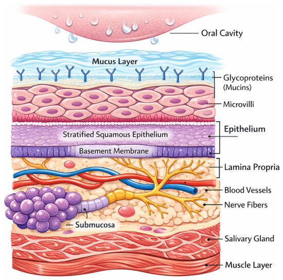
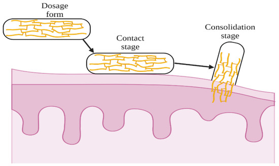
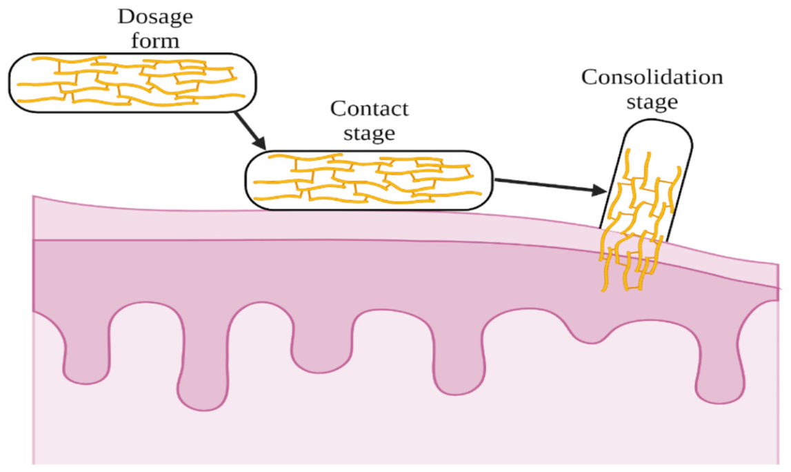
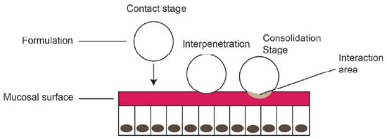
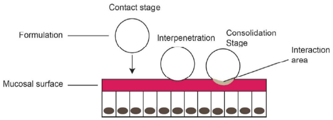
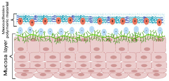
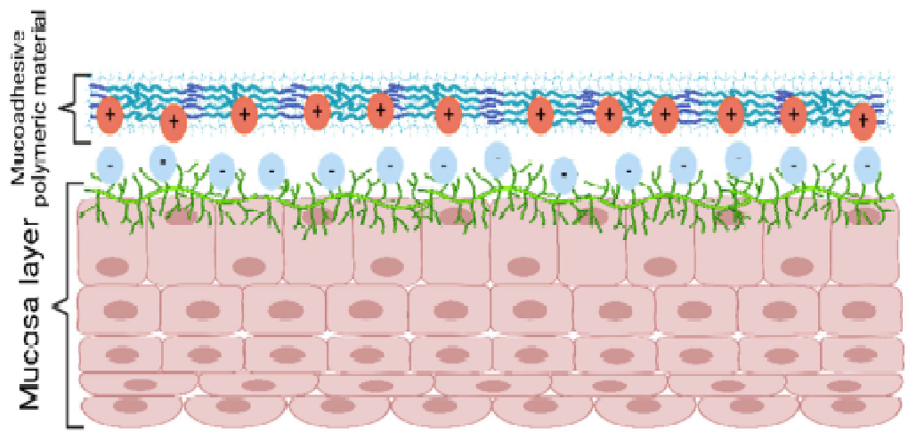
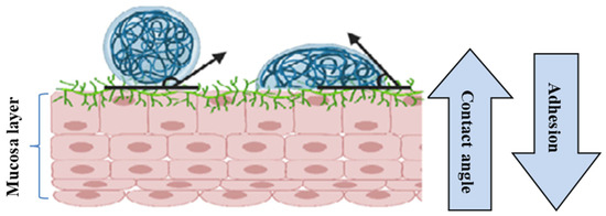
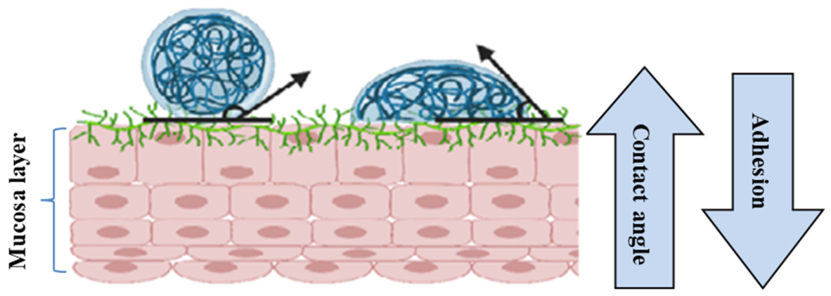
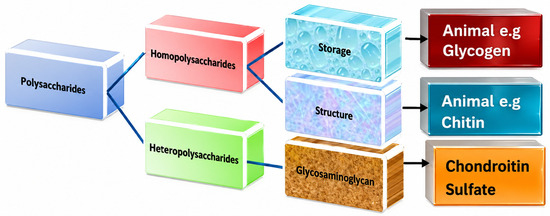
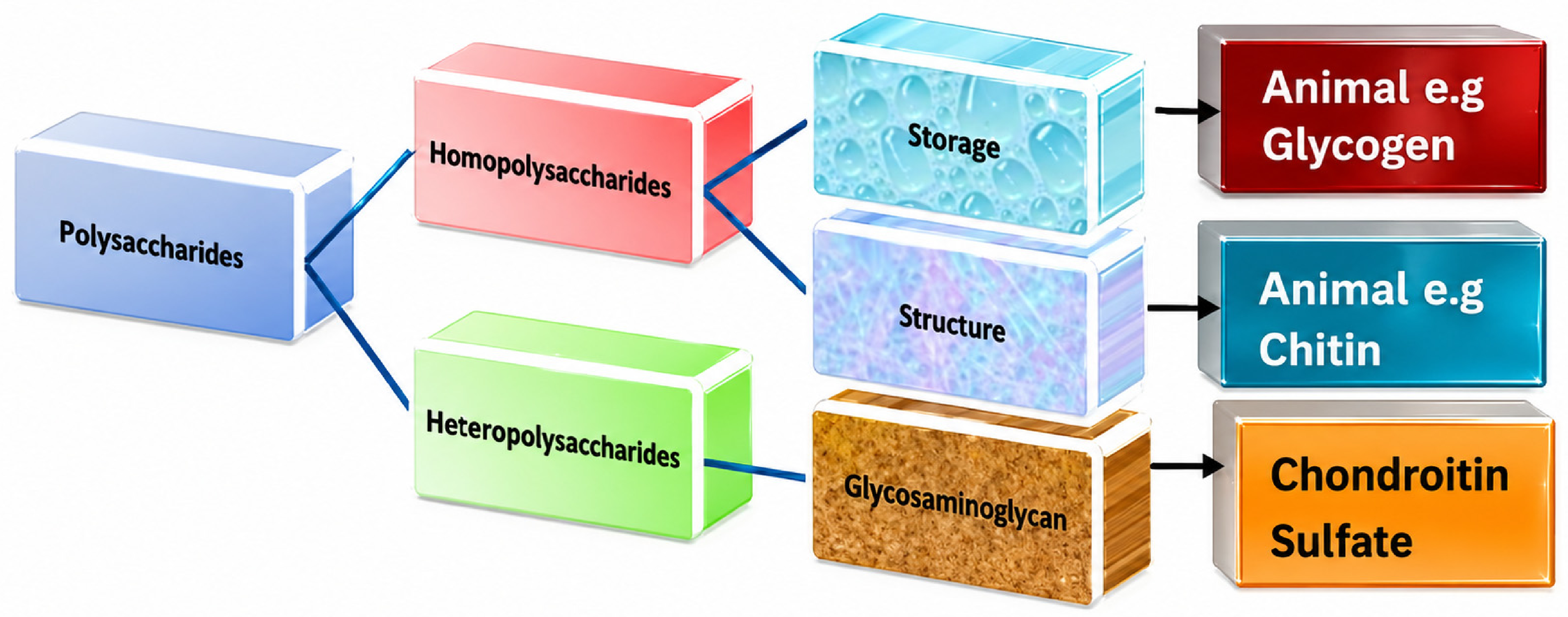
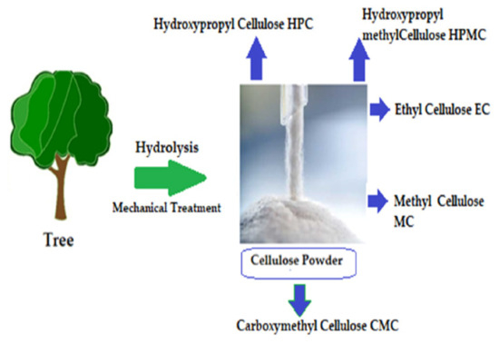
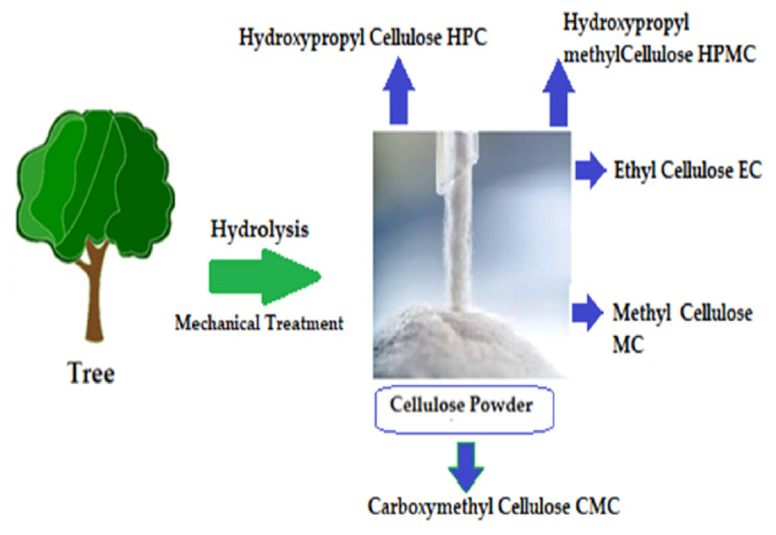
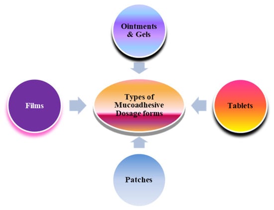
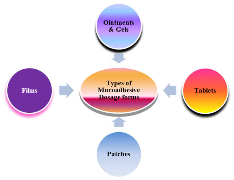

Disclaimer/Publisher’s Note: The statements, opinions and data contained in all publications are solely those of the individual author(s) and contributor(s) and not of MDPI and/or the editor(s). MDPI and/or the editor(s) disclaim responsibility for any injury to people or property resulting from any ideas, methods, instructions or products referred to in the content. |
© 2026 by the authors. Licensee MDPI, Basel, Switzerland. This article is an open access article distributed under the terms and conditions of the Creative Commons Attribution (CC BY) license.