


Abstract
Biofouling remains a critical challenge in membrane bioreactors (MBR), which is primarily caused by the production of extracellular polymeric substances (EPS) as an initial step in biofilm formation. This still limits their widespread application in wastewater treatment. In the past decades, much research has been carried out to understand and consequently reduce biofouling in MBR. More recent studies have focused primarily on inhibiting the release of EPS by applying quorum quenching (QQ) to control biofouling in MBR. This study presents the first investigation of the QQ potential of Rubellimicrobium mesophilum and its effects on biofilm inhibition by EPS reduction, which is demonstrated for MBR operated with submerged flat sheet (PTFE, PS) and hollow fibre polyvinylidene fluoride (PVDF) membranes operated in parallel for 114 days. The QQ effect has a significant impact on the reduction in biofilm thickness on PTFE membranes by 45% and on PS membranes by about 47%, respectively. Additionally, the performance of PVDF was improved by 287.5%. Similarly, the total protein concentration on the PTFE membranes was reduced by 57%, while on the PS membranes, the reduction was 78%. In mixed liquor, protein reduction was 55%, indicating its effectiveness in controlling biofouling over extended operation. The biofilm formation was monitored by measuring the biofilm thickness via fluorescence microscopy and by analyzing the protein and sugar content of the developing biofilm and of the mixed liquor. All parameters indicated decreasing biofilm formation with increasing amounts of entrapped QQ bacteria, while the removal efficiency of organic compounds and ammonia remained similar between all MBRs.
1. Introduction
Rising water demand is driven by population growth, inadequate management of available water resources, industrialization, and climate change. These factors result in groundwater depletion, water pollution, and drought, ultimately leading to global water scarcity [1]. As freshwater sources become progressively depleted, the reuse of treated wastewater presents a sustainable solution to address future water requirements [2]. Properly treated wastewater can fulfil the demand for water usage in different areas such as agriculture, irrigation, groundwater recharge, vehicle washing, toilet flushes, firefighting, and building construction activities, etc. [3]. However, ensuring high effluent quality is crucial for safe and effective reuse. Among the technologies enabling safe wastewater reuse, membrane-based treatment technologies, particularly membrane bioreactors (MBR), have gained significant attention due to their high-effluent quality and compact design.
MBR, which combines membrane separation with biological treatment, can achieve excellent effluent quality [4,5]. This technology is widely used in wastewater treatment due to its high removal efficiency and smaller footprint [6]. It is a single-step process that allows for operation at a high mixed liquor suspended solid (MLSS) concentration, which reduces the hydraulic retention time (HRT). This operational advantage makes MBR an attractive choice for implementation in wastewater treatment [7]. However, one of the most persistent issues limiting its broader adoption is membrane fouling. Fouling leads to rapid increases in transmembrane pressure (TMP), requiring frequent backwashing and chemical cleaning, which significantly increases operational costs and decreases membrane life span [8,9,10].
Biofouling is considered undesirable due to the accumulation of microorganisms on the surface of the membranes, which subsequently leads to the development of biofilms. In consequence, ongoing biofilm formation continuously increases the TMP on the surface of the membrane and thereby decreases the permeate flux over time [11]. To facilitate attachment, microorganisms secrete extracellular polymeric substances (EPS), which can contribute to modifying the surface charge and promoting adhesion [12]. Once EPS deposition begins, it promotes further microbial colonization and pore clogging, gradually leading to the formation of a dense biofilm and ultimately contributing to membrane fouling [13,14]. EPS, composed of polysaccharides, proteins, lipids, and nucleic acids, plays a crucial role in stabilizing the biofilm by forming a structural and protective matrix. However, in the MBR system, this EPS matrix can enhance microbial adhesion to the membrane surface, facilitate biofilm formation, and contribute to membrane biofouling [8,15]. Therefore, effective biofouling control management is essential to maintain system efficiency, extend membrane lifespan, and ensure consistent effluent quality [16].
In the initial stage of biofilm adhesion, one of the most essential microbial mechanisms is typically attributed to quorum sensing (QS) [17]. QS refers to the coordination of collective bacterial behaviors triggered by changes in population density. Through this mechanism, bacteria release and sense signaling molecules that increase in concentration as a function of cell density [18]. During this process, bacteria synthesize and secrete small signaling molecules, called autoinducers, to facilitate cell-to-cell communication [19,20]. As bacterial populations grow and the concentration of signaling molecules reaches a critical threshold, these molecules trigger the activation of specific genes that regulate several physiological processes, including biofilm formation and the expression of virulence factors [21,22]. Therefore, QS plays a crucial role in coordinating behavior that contributes to biofilm development. It is involved in all stages of membrane biofouling, encompassing microbial colonization within membrane pores and the accumulation of bacterial communities on membrane surfaces [21].
To address this issue, quorum quenching (QQ) has emerged as a promising biological approach for disrupting the QS signal pathway. Most QS signaling molecules produced by Gram-negative bacteria are classified as acyl-homoserine lactones (AHLs), which play a critical role in facilitating QS and biofilm formation. In contrast, QQ bacteria release enzymes that degrade these signaling molecules, thereby interrupting bacterial communication. As a result, EPS production decreases and biofilm adhesion on the membrane surface is reduced [23,24]. Over the past decade, significant research has focused on developing QQ-based strategies. These studies suggest that QQ implementation can extend the membrane filtration cycle by inhibiting QS, thereby effectively controlling biofouling in aerobic MBRs [25,26].
Nevertheless, a major challenge in implementing QQ strategies is maintaining the stability and effectiveness of QQ bacteria within the MBR biomass community. Due to the increase in MLSS in MBR, which easily reaches up to 20 g L−1, QQ bacteria can easily be outcompeted by indigenous QS signal-producing bacteria. To address this issue, various cell-entrapping techniques have been developed for the utilization of QQ bacteria. This includes microbial vessels, rotating microbial carrier frames and hydrophilic polymer media such as alginate beads and sheets. Immobilization not only protects QQ bacteria from microbial competition and predation but also prevents biomass washout during sludge wasting [27].
The strain Rhodococcus sp. BH4, known to produce AHL-lactonase enzymes that degrade AHL signal molecules by cleaving the lactone ring, has been extensively investigated for biofouling control in MBR treating synthetic or real municipal wastewater [28]. Previous studies reported that immobilized Rhodococcus sp. BH4 can significantly delay membrane fouling and extend filtration time by up to tenfold when encapsulated in alginate beads [28,29]. However, limited attention has been given to understanding how QQ influences the broader biofouling mechanism within the MBR system, including microbial community dynamics, EPS production, and biofilm structural developments. So, a comprehensive investigation of these interactions is still required to better understand the mechanisms by which QQ controls biofouling and to optimize its practical application in wastewater treatment systems.
Therefore, the main objective of this study was to investigate the effect of QQ on the structure and composition of biofilms. This was done by analyzing the biofilm thickness, membrane fouling and EPS production, with a particular focus on proteins and polysaccharides. The study included different types of polymer membranes, including hollow fiber and flat sheets, using different membrane materials such as polyvinylidene difluoride (PVDF), polytetrafluoroethylene (PTFE) and polysulfones (PS). A PVDF membrane was used for filtration, while PTFE and PS membranes were used to assess biofilm formation. To enhance its practical application, QQ bacteria were encapsulated in polyvinyl alcohol and sodium alginate (PVA-SA) beads, providing a stable and controlled release mechanism, in which immobilised bacteria release enzymes from the PVA-SA beads, which disrupt QS. This study hypothesizes that, for the first time, encapsulation of Rubellimicrobium mesophilum can disrupt QS, reduce EPS production, alter biofilm structure, and consequently mitigate membrane fouling. This approach provides comprehensive insights into the material-specific response to QQ treatment and its impact on fouling mitigation. Overall, this study gives new insights into the potential of QQ-based strategies for biofouling control in MBR.
2. Materials and Methods
2.1. Chemicals
Polyvinyl alcohol (PVA), 2270° (UNI-Chem, Lahore, Pakistan) with a purity of 99% were used. Sodium alginate was purchased from Fisher (Loughborough, UK). Boric acid was purchased from AppliChem (Darmstadt, Germany), calcium chloride (CaCl2) and sodium sulphate (Na2SO4) were purchased from Merck (Darmstadt, Germany). R2A broth medium was purchased from Neogen (Heywood, UK). The fluorescent dyes SYTO 9 and propidium iodide were both purchased from Invitrogen (Eugene, OR, USA).
2.2. MBR Design, Setup, and Operational Parameters
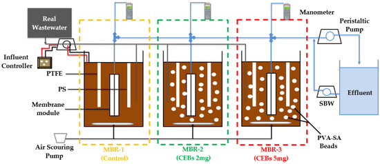
In this study, three MBRs were operated in parallel. All MBRs provided the same geometry and a working volume of 4 L, a hydraulic retention time (HRT) of 5.1 h and a target MLSS concentration of 8 g L−1. The MLSS concentration was determined according to the Standard Method by taking the initial weight of the filter paper, then filtering the MLSS sample, and drying it at 105 °C in an oven to calculate the final mass of residue per liter of sample. The MBRs were operated under different bead conditions using varying concentrations of QQ bacteria. MBR-1 was operated as a control reactor in two separate modes: (i) without any beads, (ii) with vacant beads, to distinguish membrane fouling caused by bead membrane physical interactions. In contrast, MBR-2 and MBR-3 were operated as QQ-MBR containing the QQ bacterial concentrations of 2 g L−1 and 5 g L−1, respectively. The bead filling ratio was set at 1% (v/v) of the total reactor volume. A schematic representation of the operational setup is shown in Figure 1. All MBRs were operated with municipal wastewater obtained from a local municipal treatment plant. The influent wastewater had an average pH of 7.8 ± 0.2, conductivity of 1520 ± 20.3 µS cm−1, and TOC of 113.5 ± 41.9 mg L−1 (mean ± SD, n = 5), with an organic loading rate (OLR) ranging from 1.5 to 1.7 kg COD m−3 × day−1 and a food to mass ratio (F/M) of 0.2 kg COD kg MLSS-1 × day−1. An aerator was installed at the bottom of the reactor to provide oxygen, with a flow rate of 3 L air min−1; with this configuration, dissolved oxygen (DO) was maintained at an average of 3.5 mg L−1 in the reactor. All three MBRs were equipped with hollow fiber PVDF ultrafiltration membranes (pore size 0.1 μm, PHILOS, Gwangmyeong-si, Republic of Korea). The membranes were operated at a constant flux of 27 LMH, and the same flux was maintained for all reactors during the different operational phases. The membranes were connected to a peristaltic pump (ISM833C, Odessa, TX, USA) to withdraw the treated wastewater as permeate. The filtration process was conducted over 10 min, followed by a 1 min standard backwash (SBW). The TMP was measured by a SPER scientific data logging manometer (840099, Limited Liability Company, San Bernardino, CA, USA). For monitoring the effect of QQ, flat sheet 0.2 µm microfiltration membranes out of PTFE (Sartorius, Göttingen, Germany) and PS (Alfa Laval Mid Europe GmbH, Hamburg, Germany) were additionally submerged in each MBR. These membranes were not operated for filtration, but for continuously monitoring the biofilm development on the surface of the submerged PTFE and PS membranes.
Figure 1.
Schematic diagram of MBR plant.
2.3. Immobilization of QQ Bacteria
QQ bacteria were entrapped within PVA-SA beads, and the pure strain Rubellimicrobium mesophilum (KCTC No. 22012) was used as the QQ bacterium. The bacteria were cultivated before immobilization in 1:5 diluted sterilized R2A broth, on an orbital shaker (Certomat MO II, Göttingen, Germany) at room temperature for 9 days. Thereafter, the bacterial suspension was centrifuged at 5000 rpm for 10 min at room temperature. The bacterial pellet was resuspended in 2 mL of deionize water and added to the PVA-SA solution to obtain final concentrations of 2 g L−1 and 5 g L−1 for MBR-2 and MBR-3, respectively, and mixed thoroughly to ensure uniform distribution. The prepared solution is ready to form cell-entrapping beads (CEBs) with lower (MBR-2) and higher concentration (MBR-3) of QQ Bacteria. The bacterial concentration was determined by the dry weight method after filtration and drying at 60 °C for 30 min.
2.4. Estimation of Membrane Fouling via Fluorescence Microscopy
Fluorescence microscopy (ECLIPSE LV100, Nikon, Chiyoda-ku, Japan) was used to assess membrane fouling by visualizing the accumulation of the microbial community on the surface of PTFE and PS membranes. Flat membrane samples were collected weekly to monitor the biofilm pattern. For this purpose, small pieces of membrane were cut, rinsed with tap water, placed on a glass slide, and stained using the LIVE/DEAD assay (BacLight TM Bacterial Viability Kits, Invitrogen, Karlsruhe, Germany) in which SYTO 9 stains live bacteria green and propidium iodide stains dead bacteria red. The prepared samples were then analyzed using three-dimensional (3D) imaging to determine the thickness of the biofilm at 40× magnification, providing a detailed qualitative assessment of membrane fouling.
2.5. Statistical Analysis
The statistical analysis was performed to assess biofilm thickness in all measurements in quintuplicate (n = 5 thickness points per sample) to ensure reliability. The data were presented as the mean ± standard deviation (SD). To evaluate the efficacy of the treatment and address the significance of the observed differences, a two-sample t-test was applied using OriginPro 2021 (OriginLab Corporation, Northampton, MA, USA). The analysis compared the biofilm thickness of the QQ-MBRs (MBR-2 and MBR-3) with the control (MBR-1), and differences between the QQ dose-dependent reactors were also evaluated. A significance level of p < 0.05 was used to determine statistically meaningful differences among the experimental conditions. The mean ± SD was also used to report water quality parameters, including COD, BOD, TOC, and ammonia.
2.6. Protein and Polysaccharide Extraction of EPS
For protein extraction, 1.5 cm long × 1.0 cm wide membrane samples of both PTFE and PS and 2 mL of sludge were taken from the reactors every week. The membrane pieces and sludge sample were rinsed three times with tap water and twice with potassium phosphate-buffered solution (pH 7) to remove loosely attached substances. Every time, the washing process was done in a centrifuge (FRESCO-17, Osterode, Germany) at 5000 rpm for 5 min. Then, the membrane pieces and sludge sample pellets were incubated in 1M NaOH for 15 min at room temperature, followed by lysing the sample at 100 °C for 5 min. Subsequently, they were cooled on ice and again centrifuged at 7000 rpm for 30 min [30]. Total protein concentration was measured using the Lowry method [31], with absorbance measured at 570 nm using a Beckman DU 600 spectrophotometer (Fullerton, CA, USA).
For EPS polysaccharide extraction, membrane samples of the same dimensions and sludge were used. The samples were placed in 4 mL of 1 mM Na-EDTA for 4 h, followed by sonication using a Jx-031S ultrasonic cleaner (Shenzhen, China) for 30 min using a water bath to facilitate the detachment of polysaccharide content from the samples. Then, the samples were incubated for an additional 3.5 h. After incubation, 2 mL of the sample was collected and centrifuged at 13,000 rpm for 30 min. The resulting supernatant was used for further analysis. The polysaccharide concentration was determined using the photometric phenol-sulfuric acid method [32]. The polysaccharide content was measured at 490 nm.
2.7. Analytical Method
The TOC was analysed using the TOC analyser DIMATOC-100 (Diamtech, Essen, Germany) according to DIN EN 12260 [33]. Ammonia was measured using a Shimadzu UV-2450 spectrophotometer (Tokyo, Japan) according to the European standard procedures DIN 38,406 E5 and EN ISO 6878:2004 [34,35]. The COD was measured according to DIN ISO 15705:2003-01 (Merk cell test) [36]. The biological oxygen demand (BOD) was measured using Respirometer BSBdigi from Selutec (Hechingen, Germany), according to EN ISO 9408 [37]. For COD, BOD, and TOC, measurements were performed once per sample, while ammonia (NH4+) was measured in duplicate for each sample using different dilution factors to ensure accuracy, providing the concentrations of pollutants in influent and effluent and thereby allowing assessment of reactor performance.
3. Results and Discussion
3.1. Microscopic Analysis of Biofilm Thickness on Flat Sheet Membranes
3.1.1. Impact of QQ on PTFE Membrane
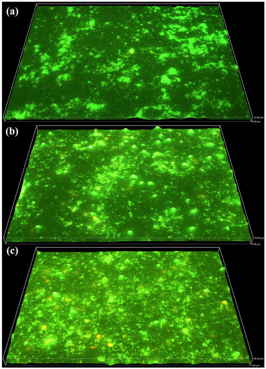
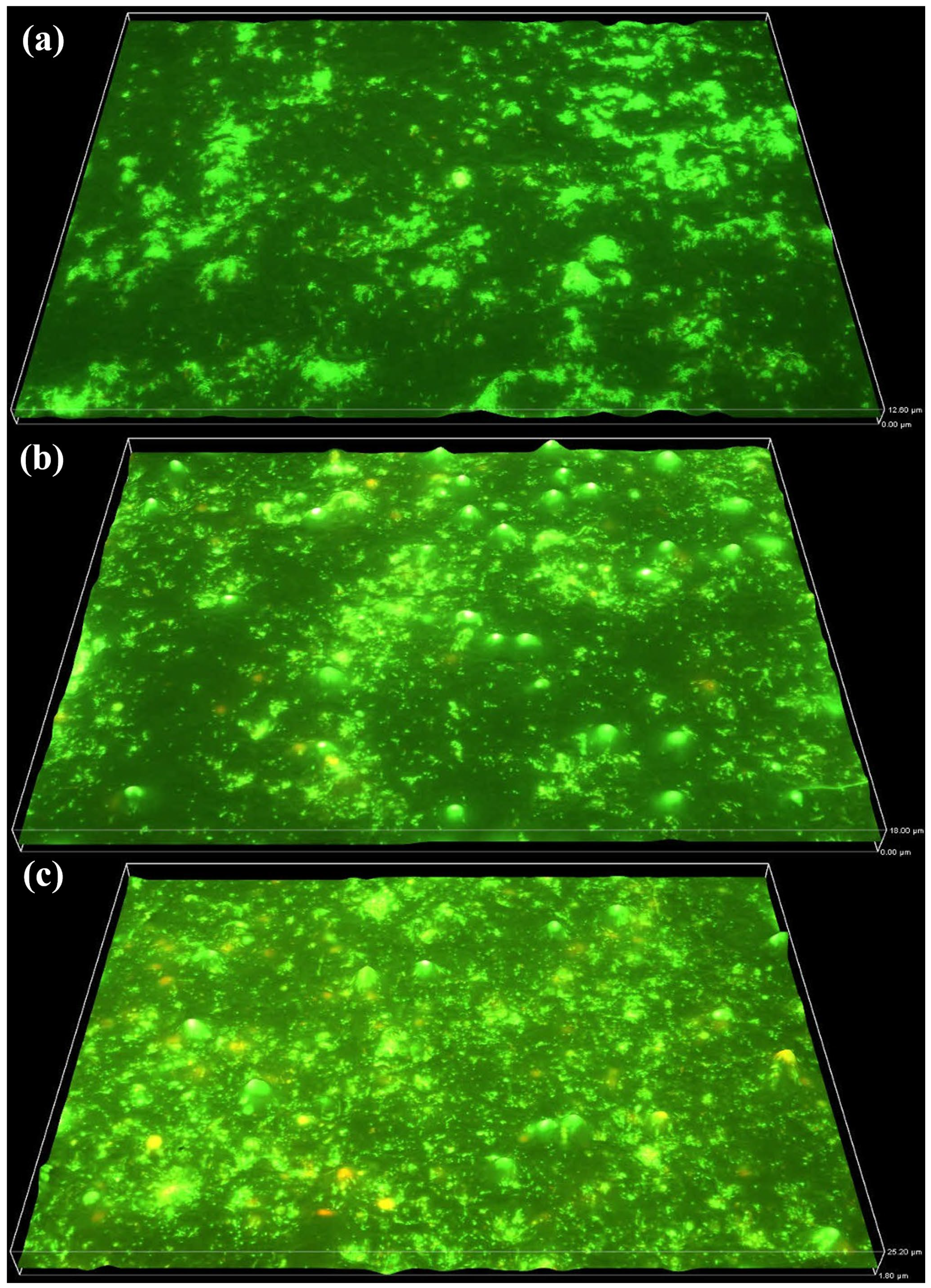
The biofilm thickness and distribution on the PTFE membrane were analysed using fluorescence microscopy. The results indicate a progressive increase in biofilm accumulation in the control reactor, with distinct spatial variations across the membrane surface. Over time, the biofilm thickness showed a continuous increase. As depicted in Figure 2a–c, the biofilm reached a thickness of 12.6 µm by week 6, 18 µm by week 9 and the highest recorded thickness of 25.2 µm by week 12. This progressive increase in biofilm accumulation indicates sustained microbial attachment and development on the PTFE membrane surface.
Figure 2.
Biofilm thickness on the PTFE membrane (MBR-1) increased from 0 µm to 12.6 µm by week 6 (a), 18 µm by week 9 (b) and to 25.2 µm by week 12 (c).
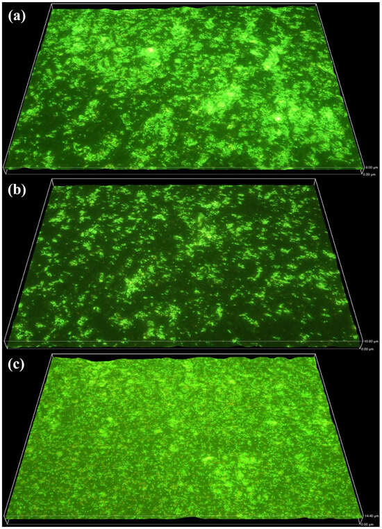
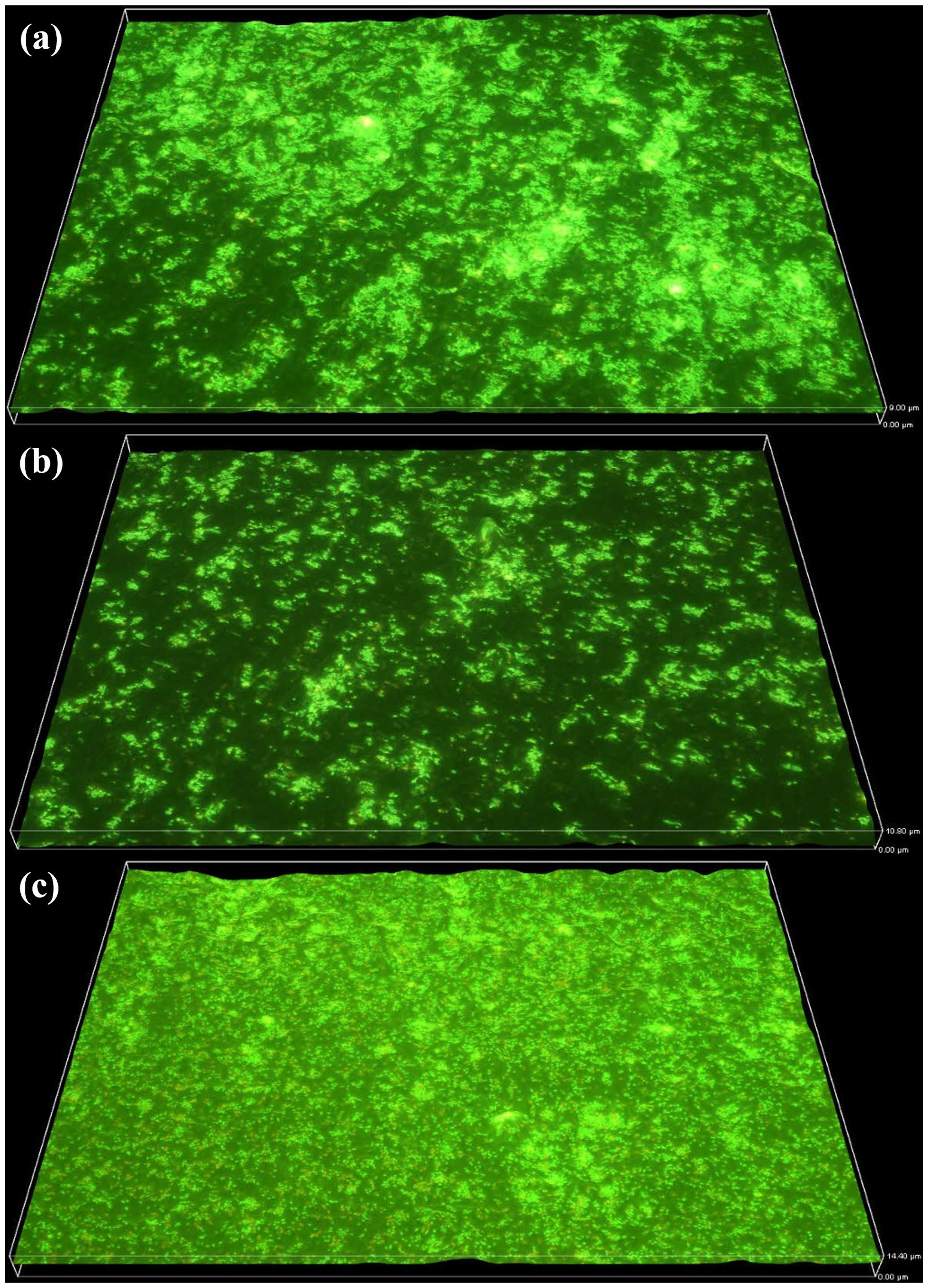
The anti-biofouling effects of QQ-MBR were assessed by comparing biofilm thickness over a 114-day growth period. The results demonstrated a continuous reduction in biofilm formation compared to the control reactor. In MBR-2, a gradual increase in biofilm thickness was observed. As shown in Figure 3a, the biofilm thickness reached 9 µm by week 6, followed by further growth to 10.8 µm by week 9 in Figure 3b, and ultimately to 14.4 µm by week 12 in Figure 3c. Although biofilm development persisted, the presence of QQ bacteria appeared to reduce its accumulation, indicating a potential QQ interference in QS pathways.
Figure 3.
Biofilm thickness on the PTFE membrane (MBR-2) increased from 0 µm to 9 µm by week 6 (a), 10.8 µm by week 9 (b) and 14.4 µm by week 12 (c).
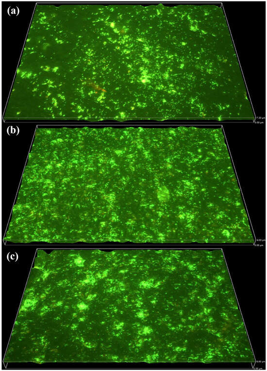
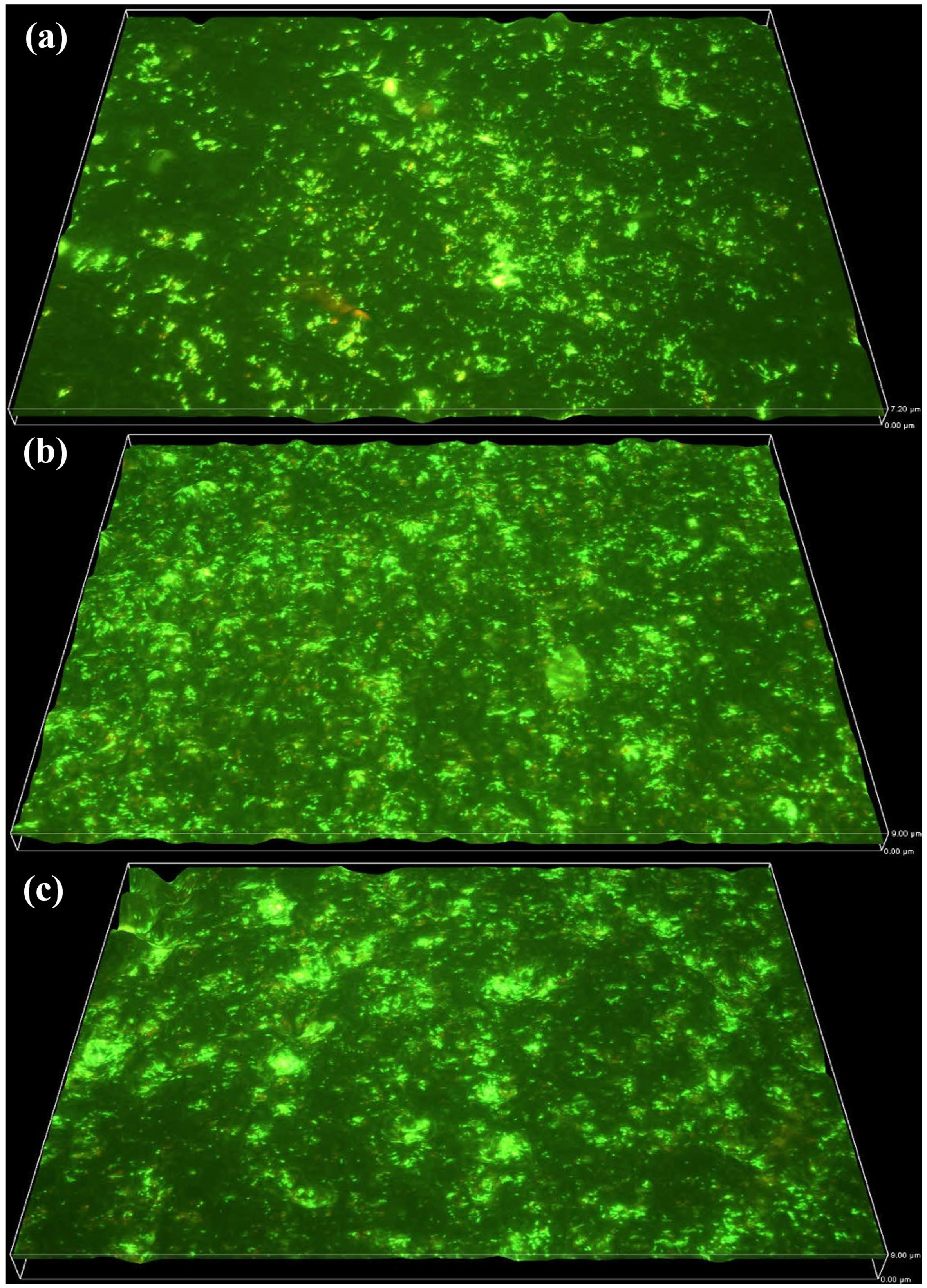
In MBR-3, a significant reduction in biofilm thickness on the surface of the PTFE membrane compared to the control and MBR-2 was observed. The biofilm thickness was measured at 7.2 µm by week 6 (Figure 4a), indicating the early stages of biofilm development. As the operation progressed, the biofilm thickness increased at a slow rate to reach 9 µm by week 9 (Figure 4b). The biofilm thickness remained consistent at 9 µm by week 12 (Figure 4c) due to the QQ intervention.
Figure 4.
Biofilm thickness on the PTFE membrane (MBR-3) increased from 0 µm to 7.2 µm by week 6 (a), 9 µm by week 9 (b) and 9 µm by week 12 (c).
Table 1 reveals a progressive increase in biofilm thickness in all three reactors from Week 6 to Week 12, indicating continuous biofilm development over time. MBR-1 exhibits the most significant growth, with biofilm thickness increasing from 12.24 ± 1.51 µm at Week 6 to 24.12 ± 2.73 µm by Week 12, while the QQ-MBR shows reduced biofilm accumulation compared to the control reactor. Both CEBs reactors (MBR-2) demonstrated a noticeably lower biofilm thickness throughout the experimental period, with MBR-3 successfully maintaining the lowest biofilm formation (6.84 ± 1.51 to 10.8 ± 0.99 µm), suggesting a dose-dependent inhibitory effect of CEBs on biofilm development.
Table 1.
Statistical analysis of biofilm thickness (µm)on PTFE membrane.
The statistical analysis of biofilm thickness on the PTFE membrane was performed using a two-sample t-test to compare the performance of MBR-2 and MBR-3 at weeks 6, 9, and 12. The analysis showed that the p-values were 0.096, 0.172, and 0.055, respectively. While the differences did not reach the threshold of statistical significance (p > 0.05), they were close to the significance level, particularly at week 12. The mean value of biofilm thickness consistently indicated that MBR-3 resulted in a thinner biofilm compared to MBR-2 across all time points. This consistency suggests that higher CEBs concentrations contribute to greater efficiency in inhibiting biofilm formation.
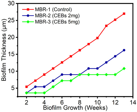
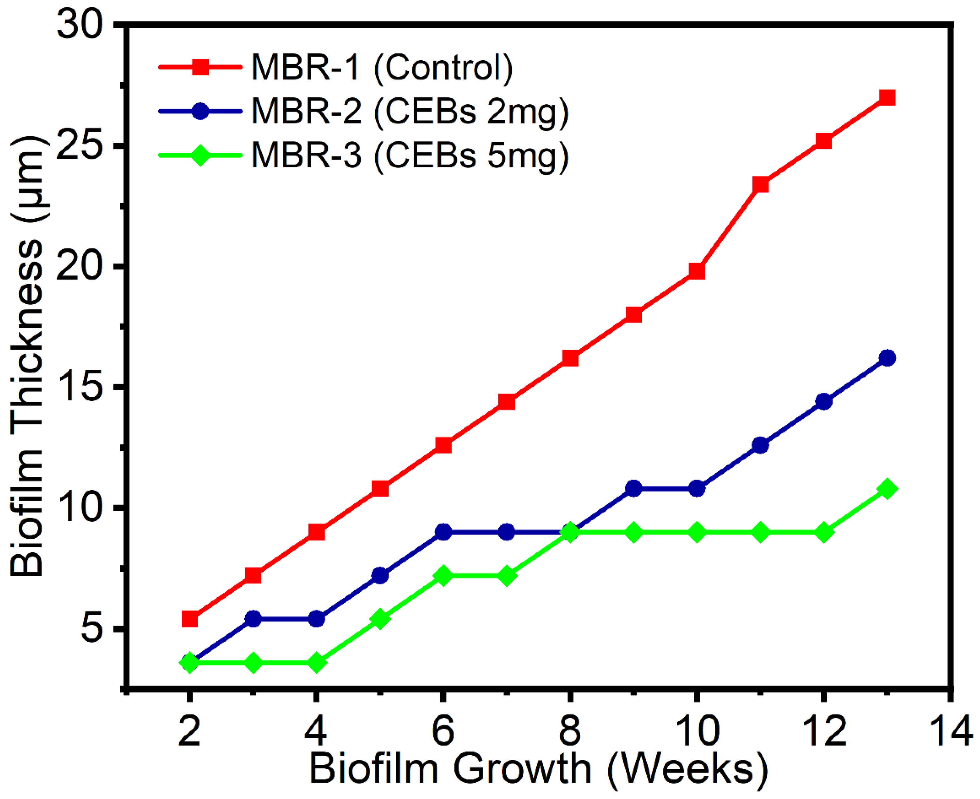
An overall comparison of biofilm growth patterns under three different conditions on the PTFE membrane was conducted. Figure 5 illustrates the biofilm thickness profile for the control MBR and QQ-MBR systems, highlighting the impact of QQ on biofilm formation. The results indicate that in the control MBR, Biofilm formation was observed from the beginning of the operation, initially measuring 5.4 µm, and over time, biofilm thickness exhibited a continuous increase throughout the operational period, ultimately reaching 27 µm in the final measurement. This consistent growth suggests that in the absence of QQ intervention, microbial attachment and accumulation proceeded unhindered, leading to significant biofouling. The persistent increase in thickness further indicates that QS activity was not inhibited, leading to microbial attachment, EPS production, and progressive biofilm maturation on the membrane surface. In contrast, the MBR-2 showed a notable reduction in biofilm thickness by 40% compared to the control MBR. This reduction suggests that the presence of QQ bacteria disrupted quorum sensing, thereby limiting bacterial communication and subsequent biofilm formation. The degradation of QS signaling molecules by QQ bacteria interferes with key regulatory pathways that control EPS secretion and the establishment of stable biofilms. A further enhancement in biofouling control was observed in MBR-3, where a 51.4% reduction in biofilm thickness was achieved compared to the control MBR. This suggests a dose-dependent effect, where a higher concentration of QQ bacteria led to more effective inhibition of biofilm development. This observation indicates that increasing the abundance of QQ bacteria enhances the degradation rate of signaling molecules and prevents the microbial population from reaching the critical threshold required for strong biofilm development. The plateau observed in biofilm growth within the MBR-3 system between weeks 8 and 12 can be attributed to the effective activity of QQ bacteria, which were actively degrading QS molecules, preventing biofilm formation during this period. This stabilization phase suggests that the QQ activity was sufficient to counter microbial growth, temporarily maintaining an equilibrium between biofilm formation and inhibition. However, the subsequent increase in biofilm thickness in week 13 suggests a reduced QQ efficiency or microbial adaptation.
Figure 5.
Biofilm thickness over time on PTFE membrane.
Previous research indicated that bacterial doses of 10 g L−1 and 15 g L−1 of PVA beads did not significantly enhance biofouling reduction, possibly due to excessive cell density leading to competition for nutrients or limited diffusion of QQ molecules. Therefore, 5 g L−1 was identified as the optimal dose for effective biofouling mitigation [38]. The findings of this study are consistent with these results, suggesting that an optimal QQ bacterial dosage can achieve more efficient degradation of QS molecules, even during long-term operation. The gradual biofilm increase in MBR-2 suggests that while the QQ approach is effective, biofouling mitigation is dose-dependent, warranting further optimization of bacterial concentration to enhance long-term antifouling performance. This gradual increase in thickness and eventual reduction in the biofilm suggest that the presence of QQ has a noticeable effect on controlling biofilm formation in the system [39,40]. These findings align with previous studies demonstrating that QQ strategies disrupt bacterial communication and ultimately limit biofilm establishment [41]. The statistical analysis further supports this trend, as the comparative evaluation between MBR-2 and MBR-3 consistently showed lower mean biofilm thickness in the system with the higher concentration of CEBs. Although the differences did not reach strict statistical significance, the values (week 12) approached the significance threshold, indicating a stronger tendency toward improved biofilm suppression with increasing QQ bacterial dosage. This trend strengthens the interpretation that the enhanced QQ activity in MBR-3 contributed to more effective inhibition of biofilm formation compared with MBR-2. Specifically, biofilm formation on the PTFE membrane was lower due to its higher hydrophobicity and smoother surface, which likely reduced microbial adhesion and subsequent biofilm expansion.
3.1.2. Effect of QQ on PS Membrane
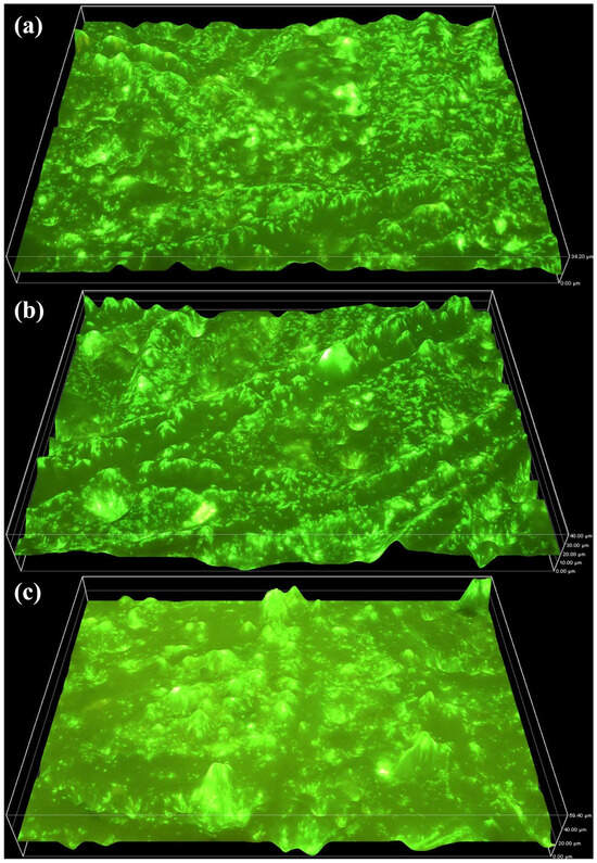
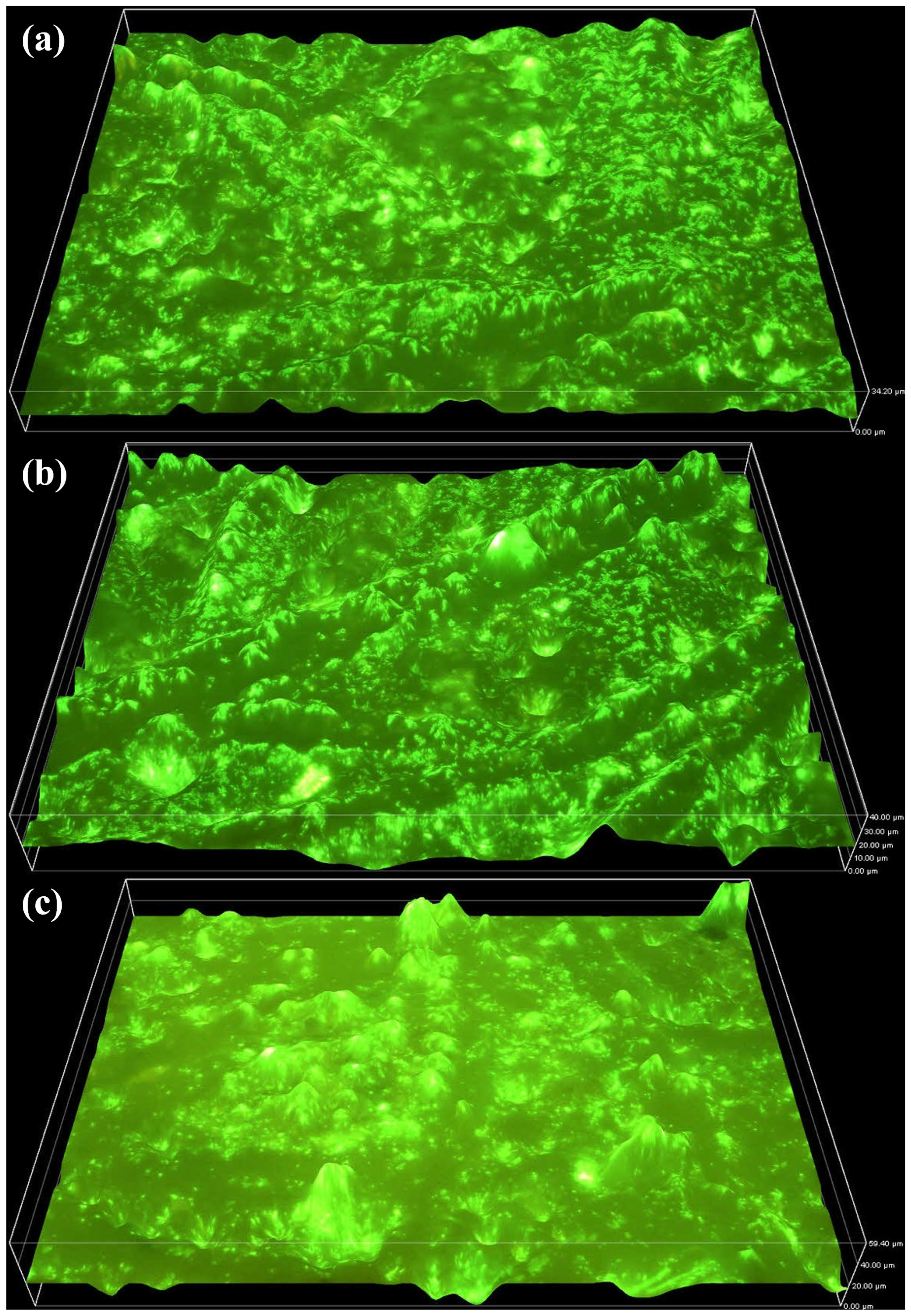
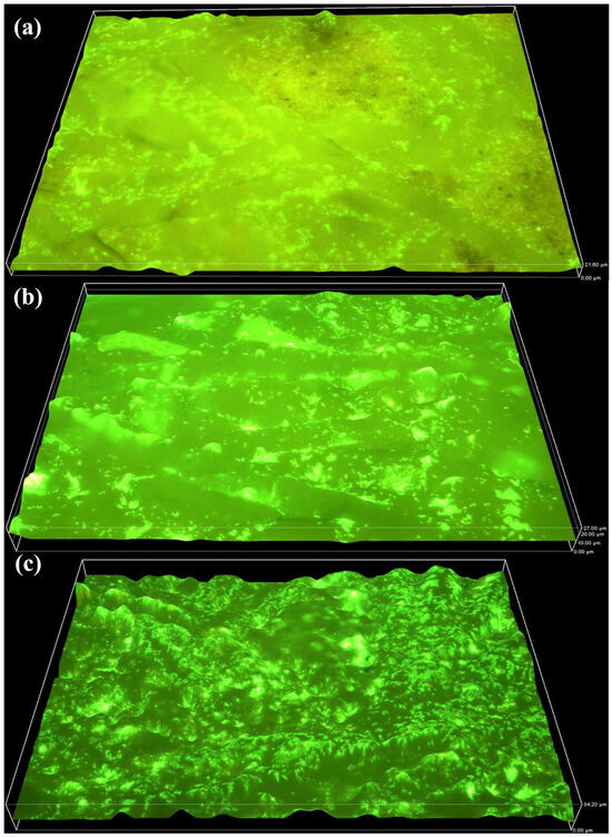
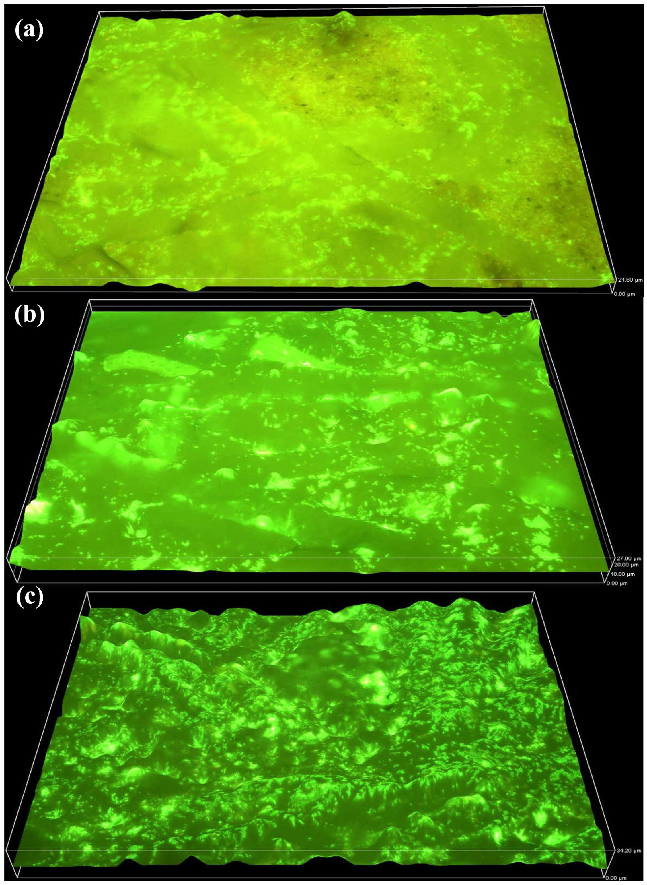
The biofilm formation on the PS membrane was analysed to understand its growth pattern, distribution, and structural changes. The results indicate that biofilm growth on the PS membrane was significantly higher compared to the PTFE membrane, likely due to differences in material properties and surface structure. At the beginning of the operation, biofilm thickness was greater on the PS membrane than on the PTFE membrane, indicating faster initial colonization by microorganisms. Over time, biofilm accumulation continued to increase, as shown in Figure 6a–c, where thickness measurements reached 34.2 µm by week 6, 40 µm by week 9, and 59.4 µm by week 12, respectively. This continuous increase suggests that the PS membrane provided a more favourable surface for microbial attachment and biofilm development compared to the PTFE membrane.
Figure 6.
Biofilm Growth on the PS membrane (MBR-1) increasing from 0 µm to 34.2 µm by week 6 (a), 40 µm by week 9 (b) and 59.4 µm by week 12 (c).
The results demonstrate a significant reduction in biofilm formation on the PS membrane due to the presence of QQ bacteria. Initially in MBR-2, the biofilm thickness was significantly lower compared to the control reactor, indicating an early inhibition of biofilm development. As the operation progressed, a gradual increase in biofilm thickness to 21.6 µm by week 6 (Figure 7a) and 27 µm by week 9 (Figure 7b). By the end of the operation, a final thickness of 34.2 µm was observed by week 12 (Figure 7c). Despite the increase over time, the overall biofilm accumulation remained significantly lower than in the control system, confirming that QQ bacteria effectively mitigated biofilm formation on the PS membrane.
Figure 7.
Biofilm Growth on the PS membrane (MBR-2) increasing from 0 µm to 21.6 µm by week 6 (a), 27 µm by week 9 (b) and 34.2 µm by week 12 (c).
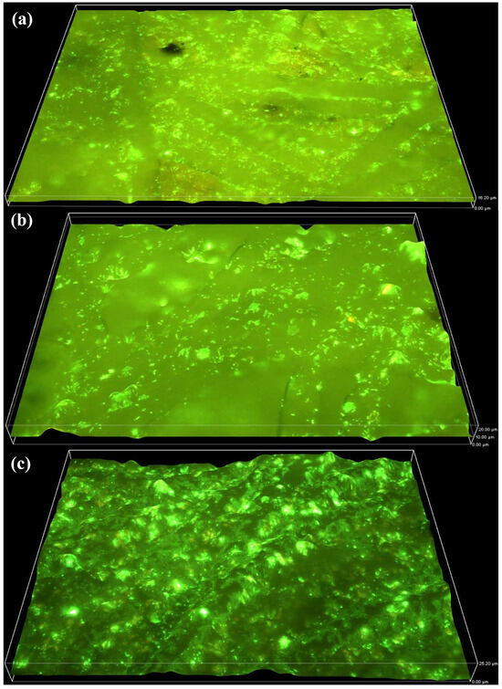
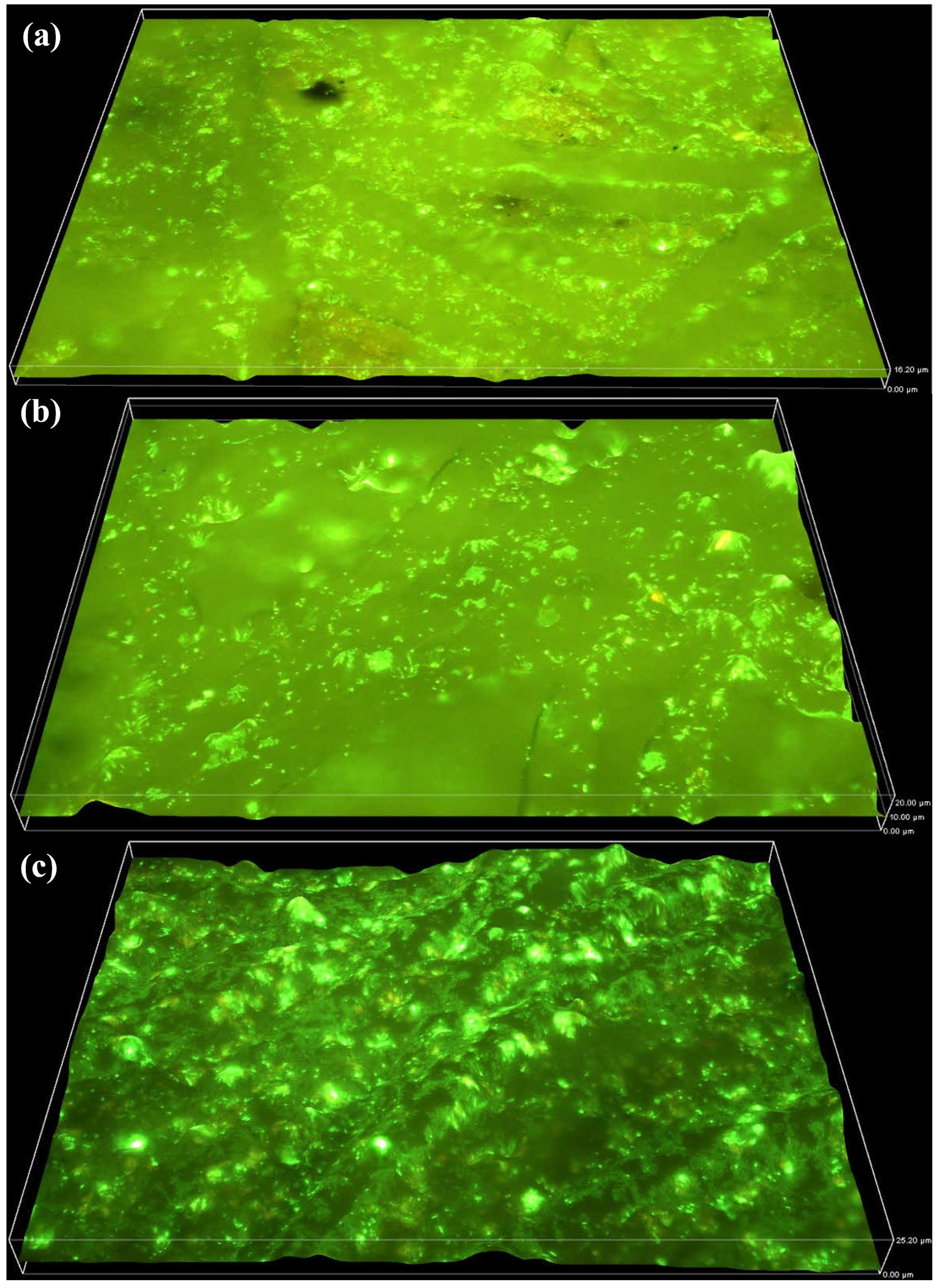
In MBR-3 with higher QQ dosage, a greater biofilm reduction was observed compared to the lower QQ dosage, showing the critical role of optimal QQ concentration in biofouling mitigation. At the beginning of the operation, there was only a minor difference in biofilm formation between the QQ-MBR-2 and QQ-MBR-3. However, as the operation progressed, a notable reduction in biofilm formation was observed. As illustrated in Figure 8a–c, biofilm thickness increased at a significantly slower rate, reaching 16.2 µm in week 6, 20 µm in week 9, and 25.2 µm in week 12, respectively. Results indicate that over the longer operation, the MBR-3 system exhibited superior biofilm control compared to lower dosages, effectively limiting biofilm thickness and mitigating membrane biofouling.
Figure 8.
Biofilm Growth on the PS membrane (MBR-3) increasing from 0 µm to 16.2 µm by week 6 (a) 20 µm by week 9 (b) and 25.2 µm by week 12 (c).
Table 2 presents the statistical evaluation of biofilm thickness on the PS membrane and supports the trends observed in the 3D imaging analysis. A marked and time-dependent increase in biofilm thickness was observed in the control MBR, reaching 55.76 ± 9 µm by Week 12, whereas the QQ-MBR exhibited consistently lower biofilm development across all sampling periods. At Week 6, CEBs reactors demonstrated a substantial reduction in biofilm thickness compared to the control, and this difference became more pronounced with prolonged operation. By Week 12, MBR-2 and MBR-3 exhibited biofilm thickness values of 33.84 ± 2.67 µm and 25.56 ± 1.51 µm, respectively.
Table 2.
Statistical analysis of biofilm thickness (µm)on PS membrane.
A two-sample t-test was performed to evaluate the reduction in biofilm thickness on PS membrane compared to the MBR-1. The statistical analysis confirmed a significant reduction in biofilm thickness for the MBR-2 and MBR-3, with p-values of 0.026 at week 6, 0.001 at week 9, and 0.001 at week 12, indicating progressively stronger statistical significance over time. These highly significant results (p < 0.05) demonstrate a clear inhibition of biofilm accumulation on PS membranes, with the efficiency of the increase in QQ bacteria suggesting that the increase in QQ dose effectively mitigated biofilm formation.
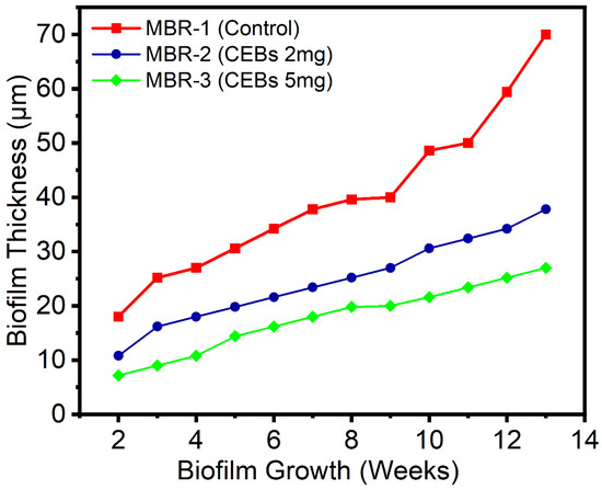
Biofilm thickness on PS membranes was monitored under three different operating conditions during long-term operation. Figure 9 illustrates the rapid increase in biofilm thickness in the control MBR from the beginning of the operation, with continuous growth reaching a maximum of 70 µm, indicating uninhibited biofouling progression. This continuous increase reflects the active QS signaling among microbial populations, enabling coordinated attachment and release of EPS content, which facilitates progressive biofilm maturation on the PS membrane. Additionally, the PS membrane exhibited greater biofilm formation due to its moderately hydrophobic nature and relatively rough surface morphology. Such surface characteristics promote stronger initial microbial adhesion and enhance the retention of attached cells, facilitating progressive biofilm development during long-term operation [42]. In contrast, the application of QQ bacteria significantly reduced biofilm accumulation. MBR-2 achieved a 38.2% reduction in biofilm thickness compared with the control, indicating moderate suppression of biofilm formation. A more pronounced inhibition was observed in MBR-3, where biofilm thickness decreased by 55.8% relative to the control. This reduction can be attributed to the disruption of acyl homoserine lactone signaling molecules, which limits EPS production and interferes with the biofilm maturation process [17]. These results demonstrate a clear dose-dependent effect of QQ bacteria on biofouling mitigation, with higher QQ concentrations providing more effective control of biofilm development. The statistical analysis further confirms a strong correlation between increasing QQ dosage and reduced biofilm formation. Moreover, the increasing statistical significance over the operational weeks suggests that the effect of QQ bacteria accumulates over time, highlighting their potential for sustained biofouling control and improved long-term MBR performance [43].
Figure 9.
Biofilm thickness over time on PS membrane.
3.1.3. Impact of QQ Bacteria on TMP
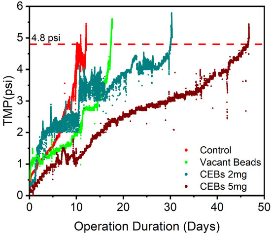
Figure 10 illustrates the TMP profiles corresponding to different operational conditions. A new membrane module was immersed in each MBR and operated until the membranes got fouled and TMP reached a maximum value (4.8 psi = 33 kPa), under steady-state treatment conditions. The TMP progression for the control MBR, which was operated without any beads and QQ intervention, showed a rapid and continuous increase over time. In this setup, membrane fouling occurred after 12 days, indicating the baseline operational performance of the membrane under conventional conditions. In contrast, the second operation, which was conducted with vacant beads, operated for a slightly longer period of 17.4 days due to the physical interaction between the beads and the membrane induced by the air-scouring effect, which resulted in a 45% improvement. In the third operation, a QQ strategy employing 2 g L−1 of QQ bacteria resulted in a notable improvement in membrane performance. The operation duration was extended to 30 days, showing a 150.4% improvement compared to the control. Operation four was operated with an increased QQ bacterial dosage of 5 g L−1. This system revealed the best performance: the membrane reached the fouling threshold in 46.5 days, representing a 287.5% improvement over the control.
Figure 10.
TMP Profile of different operating conditions.
This enhancement demonstrates the potential of QQ strategies in mitigating biofouling through QS disruption and a dose-dependent effectiveness of QQ bacteria in prolonging membrane lifespan and maintaining lower TMP levels. In this study, the membranes were operated at a constant flux of 27 LMH, which is comparable to the previously reported studies for the treatment of municipal wastewater [7,29,44]. Operating at a similar flux allows for a meaningful comparison of membrane fouling under different operating conditions, and a slower TMP also suggests that biofouling mitigation was achieved without reducing hydraulic load. Another factor that contributed to enhancing the longer operation duration of QQ-MBRs was physical cleaning. As the reactors contained a similar amount of beads, either operated with vacant beads or QQ beads, these beads provide mechanical scouring through the bead membrane physical interactions. This mechanical action effectively reduced microbial accumulation on the surface of the membrane. Therefore, the mechanical scouring effect provided a baseline for comparison between the reactor operated without beads and the reactors containing CEBs. This made it easier to assess the improvement resulting from both physical interactions and QQ effects. In addition, the QQ-MBRs differed in the amount of CEBs, which enzymatically disrupt AHL molecules essential for QS and biofilm maturation. This difference indicates that the improved performance in reactors with a higher bacterial dose resulted mainly from QQ rather than from mechanical scouring. There was also evidence that the slower increase in TMP in QQ-MBR, caused by QQ bacteria, reflected improved membrane performance proportional to the QQ dose, which highlights the potential of QQ-MBRs for practical application in wastewater treatment [45]. These factors clearly indicate that QQ activity disrupts the QS pathway, thereby suppressing EPS production. The results of this study are consistent with previous studies, which have reported that QQ significantly inhibits biofilm formation and reduces membrane fouling in MBR systems [38,46,47]. The results support the feasibility of QQ-based approaches for improving the lifespan of the membrane by reducing the accumulation of biofilm on its surface, maintaining membrane permeability, and decreasing the cleaning frequency, which ultimately lowers operational costs.
3.2. Distribution of EPS on Membrane Surface and in Mixed Liquor
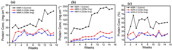
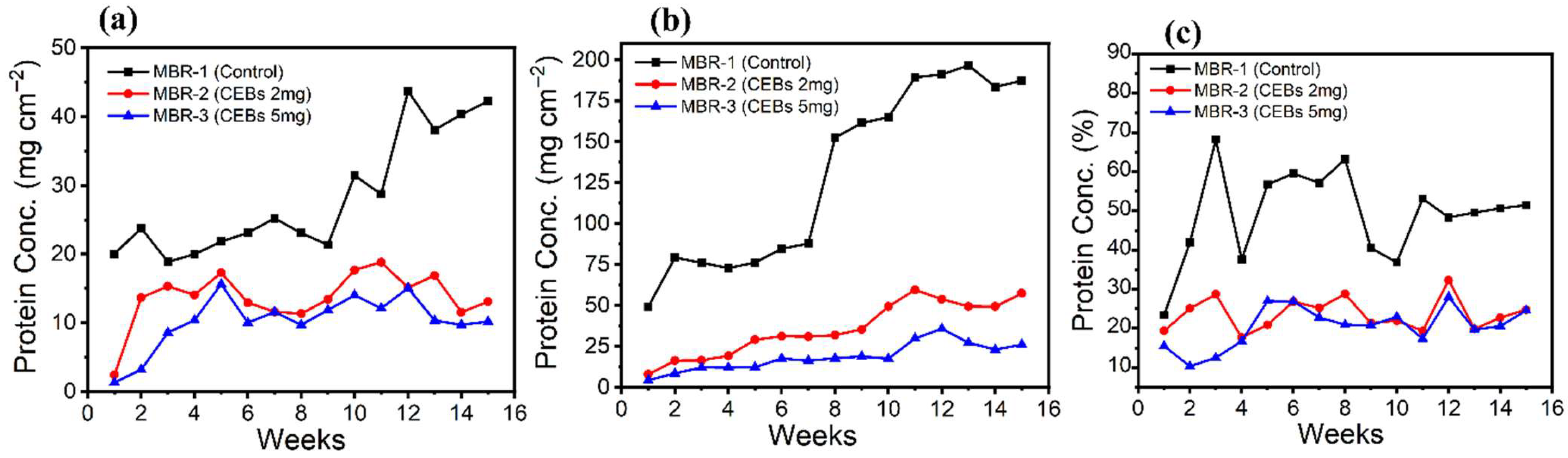
EPS consists of complex mixtures of polysaccharides, proteins, humic acids, nucleic acids, and various other substances. These molecules influence the physicochemical parameters of the activated sludge mixture and create a highly hydrated gel matrix that develops biofilm on the membrane surface. It also provide nutrients and a habitat for the bacteria on the membrane surface [48]. This work evaluated and analysed the primary components of EPS, specifically proteins and polysaccharides, due to their significant role in membrane biofouling. The EPS content was measured weekly to monitor protein concentration on the PTFE membrane. Figure 11a illustrates that in the initial stage, the total protein concentration in the control MBR was 20 mg cm−2, which progressively increased to 25.2 mg cm−2 at week 7 and 43.7 mg cm−2 at week 12, indicating a continuous accumulation of protein over time. However, the presence of QQ bacteria significantly reduced protein accumulation on the membrane surface. In MBR-2, the initial protein concentration was 2.4 mg cm−2, approximately 8 times lower than in the control. Over time, it increased to a maximum of 18.8 mg cm−2, indicating a significant reduction compared to the control. An even greater reduction was observed in MBR-3, where the initial protein concentration was 1.3 mg cm−2, and the maximum recorded value was 15.6 mg cm−2, which was lower than in MBR-2. Overall, the protein reduction on the PTFE membrane was 51.4% in MBR-2 and 63.6% in MBR-3 compared to the control MBR, highlighting the effectiveness of QQ bacteria in mitigating biofouling by limiting EPS-protein accumulation.
Figure 11.
Concentration of Protein on membranes and Sludge (a) On PTFE membrane (b) On PS membrane (c) In sludge.
The PS membrane provides a more favourable surface for bacterial attachment, accumulation, biofilm formation, and the subsequent release of total proteins. Figure 11b illustrates the protein concentration on the PS membrane over time. In the control MBR, the initial protein concentration was 49.4 mg cm−2, which progressively increased to 84.6 mg cm−2 by week 6 and further increased to 191.2 mg cm−2 by week 12, indicating a continuous rise in protein concentration. In contrast, the MBR-2 system exhibited a significant reduction in protein concentration, with an initial value of 7.9 mg cm−2, which was approximately seven times lower than that of the control MBR. Further reductions were observed at week 6, when it was 31.2 mg cm−2, and week 12 was 53.8 mg cm−2, demonstrating the efficacy of QQ treatment in the reduction in protein concentration. Similarly, the MBR-3 system showed an even greater reduction in protein concentration, with an initial reading of 4.3 mg cm−2, which was lower than that of MBR-2. By week 6 and week 12, protein concentrations in MBR-3 were recorded at 17.6 mg cm−2 and 35.8 mg cm−2, respectively, indicating a substantial reduction compared to both the control MBR and MBR-2. Overall, the MBR-2 system achieved a 72.5% reduction, while the MBR-3 system demonstrated an 85.7% reduction in protein concentration compared to the control MBR. Figure 11c shows the measured total protein concentration in the mixed liquor, which consistently revealed a higher protein content in the control MBR. compared to the QQ-MBR systems. The protein concentration of both QQ reactors was about the same, but when compared to the MBR-1, MBR-2 showed a 51.9% drop, while MBR-3 showed a slightly higher 58.4% drop.
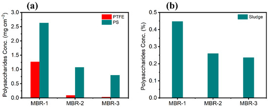
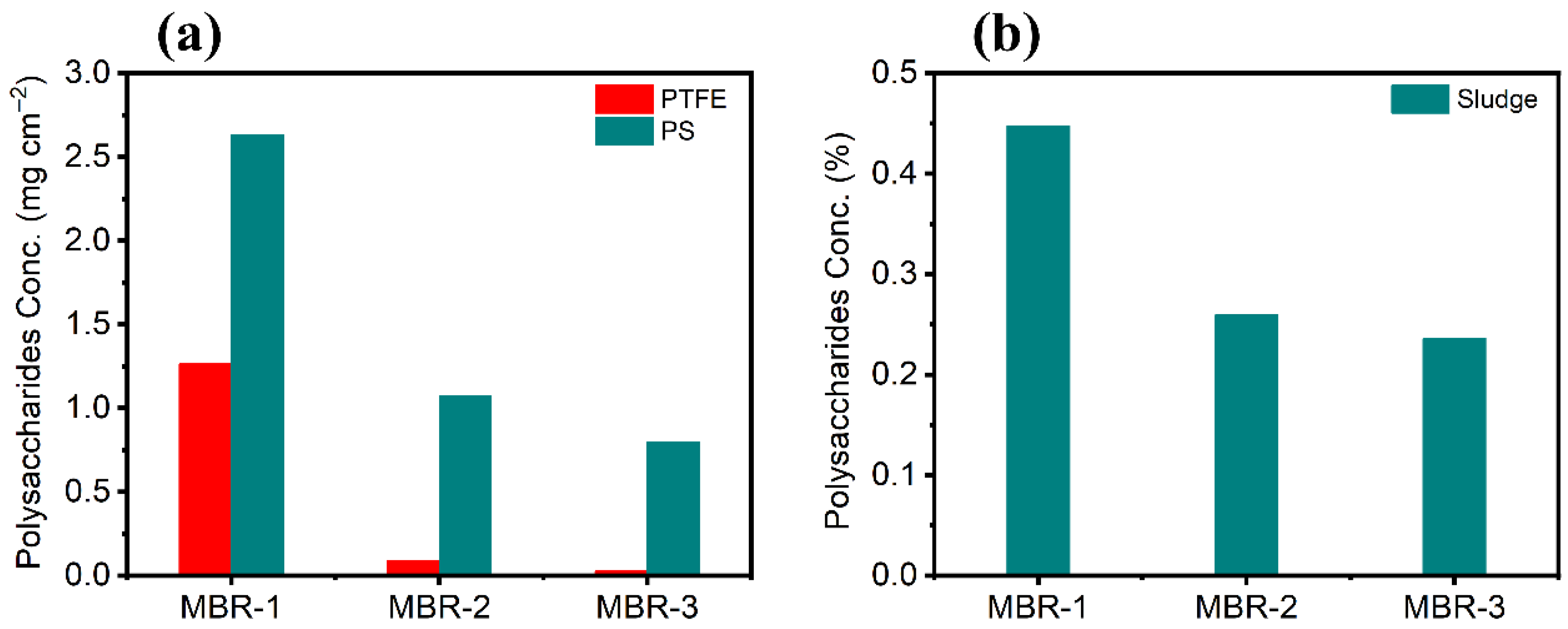
The content of EPS polysaccharides on the PTFE membrane exhibited an increasing trend throughout the operational period. Figure 12a demonstrates that in the control MBR, the average polysaccharide concentration was 1.3 mg cm−2, which was significantly higher compared to the systems incorporating the QQ technique. The MBR-2 system attained 0.09 mg cm−2, while the MBR-3 achieved 0.03 mg cm−2, indicating substantial reductions in EPS polysaccharides by 92.9% and 97.8%, respectively. The average polysaccharide concentration on the PS membrane was 2.6 mg cm−2 in the control operation, which was substantially higher compared to the QQ technique. In contrast, the MBR-2 and MBR-3 systems exhibited markedly lower polysaccharide concentrations of 1.07 mg cm−2 and 0.7 mg cm−2, respectively. This corresponds to a 59.3% reduction in MBR-2 and a 69.7% reduction in MBR-3. The polysaccharide concentration in the mixed liquor, as demonstrated in Figure 12b, showed a notable difference between the control and QQ systems. MBR-2 and MBR-3 demonstrated a 42% and 47.2% decrease in EPS polysaccharides, respectively. These findings highlight the effectiveness of QQ technology as a promising strategy for EPS reduction and biofouling mitigation in membrane bioreactors.
Figure 12.
Concentration of Polysaccharides on membranes and Sludge (a) On PTFE and PS (b) In sludge.
Total protein and polysaccharides are two important components that play a critical role in establishing biofilm. In this study, a reduction in protein and polysaccharide was observed on PTFE and PS membranes, as well as in the mixed liquor, with QQ intervention. The substantial decrease is directly linked to QQ activity, which was absent in the control operation, where the concentration of protein and polysaccharides remained high on the surface of membranes. In contrast, QQ-MBRs showed a significantly greater reduction in EPS [49]. The higher accumulation of EPS content on the surface of the PS membrane compared to the PTFE membrane can be attributed to differences in the physicochemical properties of the membrane materials. PTFE membranes generally exhibit a highly hydrophobic and relatively smooth surface, while PS membranes are comparatively less hydrophobic and possess a rougher surface morphology. These characteristics can promote stronger microbial interactions and surface attachment, thereby facilitating greater EPS accumulation on the PS membrane surface [50,51]. PS contains aromatic and sulfone functional groups, which can promote hydrophobic interactions, hydrogen bonding, and acid–base interactions with EPS constituents. EPS components, particularly proteins and polysaccharides, contain functional groups such as hydroxyl, carboxyl, and amine groups that can interact with membrane surfaces through these mechanisms, enhancing their adsorption and accumulation on the membrane surface [52,53]. The overall reduction in EPS was higher in the mixed liquor compared to the control. Although different QQ doses did not have a pronounced effect on EPS in the sludge, this was possibly because the sludge was kept suspended within the reactor [41]. In suspended sludge, EPS is mostly present as tightly bound EPS embedded inside the flocs, resulting in less being released into the mixed liquor. On the membrane surface, microbes produce more loosely bound EPS and release more, partly because the membrane surface is hydrophobic [54].
3.3. Removal Efficiency
The removal efficiency of organic compounds and ammonia was determined to check the reactor’s performance. Although all three reactors operated under different operation conditions in terms of without QQ and with different concentrations of QQ bacteria immobilization, the effluent quality remained consistent throughout the long-term operation. Table 3 shows that the average removal efficiencies across all reactors were 83.5 ± 5.7% for COD, 89.1 ± 3.7% for BOD, 77 ± 5.3% for TOC, and 89.7 ± 6.7% for ammonia, indicating stable biological treatment performance throughout the experimental period. These results indicate that QQ activity did not affect the biodegradability of organic compounds and ammonia. This suggests that QQ activity targeted QS signalling pathways without disrupting the basic metabolic processes of the microbial community. Therefore, the fouling control benefits of QQ can be achieved without compromising effluent quality, which is a critical consideration for long-term MBR operation. These findings are consistent with previous studies that have shown that effluent quality is not affected by the QQ strategy [29,45]. More detailed information on COD, BOD, TOC and NH3 values can be found in S1.
Table 3.
Removal Efficiency (%) of COD, BOD, TOC, and NH3 across three different operations.
4. Conclusions
The PVA-SA beads showed excellent bacterial immobilization and stability while ensuring sustained QQ activity for long-term operation. The presence of QQ bacteria significantly limited the biofilm thickness on both types of flat sheet membranes and delayed the increase in TMP, which extended the operation duration to 3.9 times, indicating that the membrane lifespan has been effectively enhanced compared to the control. Similarly, the EPS protein on the surface of the PTFE membrane was reduced by 2.8-fold, while a more pronounced reduction of 6.9-fold was observed on the PS membrane. In the sludge, the total protein concentration decreased by 2.4-fold. Moreover, EPS polysaccharides showed a similar reduction trend. MBR-3 exhibited the maximum mitigation effect on different polymeric membranes and even in sludge, which highlights the strong potential of the QQ strategy to minimize fouling-causing agents. The above-mentioned parameters indicate that Rubellimicrobium mesophilum plays an important role in disrupting QS signaling and reducing AHL concentrations, thereby ultimately mitigating biofouling. The results also revealed that the QQ dose plays a crucial role in the mitigation, with higher concentration demonstrating stronger anti-biofouling effects. Overall, these findings highlight the promising environmental implications of extending membrane lifespan and reducing the need for chemical cleaning, which can lower operational costs and improve the economic viability of the system. Moreover, the high effluent quality further enhances the potential for water reuse. These results further suggest that QQ-based biofouling control could support the development of more sustainable and energy-efficient MBR systems, particularly for long-term wastewater treatment and reuse applications.
Supplementary Materials
The following supporting information can be downloaded at: https://www.mdpi.com/article/10.3390/polysaccharides7020042/s1. Table S1: Removal Efficiency of COD, BOD, TOC, and NH3 across three different operations.
Author Contributions
Conceptualization, N.S. and M.M., Writing—original draft, N.S., Validation, N.S. and M.M., Methodology, N.S., M.M. and J.B., Investigation, N.S., Formal analysis, N.S., Visualization, review & editing, N.S., J-B., R.R., M.S.N. and M.M. Supervision and Project administration, M.M. All authors have read and agreed to the published version of the manuscript.
Funding
This research received no external funding.
Data Availability Statement
Data are contained within the article and Supplementary Materials.
Acknowledgments
The authors would like to thank the Chair of Water Treatment Biotechnology for providing all the necessary resources and sincerely express their gratitude to Shinho Chung for generously providing the PVDF membrane.
Conflicts of Interest
The authors declare that they have no known competing financial interests or personal relationships that could have appeared to influence the work reported in this paper.
References
- Phan, N.N.; Dang, N.H.T.; Le, K.H.; Duong, Y.H.; Pham, L.P.T. Performance Evaluation of MBR-RO Technology for Domestic Wastewater Treatment. In Proceedings of the IOP Conference Series: Earth and Environmental Science; Institute of Physics: London, UK, 2024; Volume 1383. [Google Scholar]
- Capodaglio, A.G. Fit-for-Purpose Urban Wastewater Reuse: Analysis of Issues and Available Technologies for Sustainable Multiple Barrier Approaches. Crit. Rev. Environ. Sci. Technol. 2020, 51, 1619–1666. [Google Scholar] [CrossRef] [Scilit]
- Kesari, K.K.; Soni, R.; Jamal, Q.M.S.; Tripathi, P.; Lal, J.A.; Jha, N.K.; Siddiqui, M.H.; Kumar, P.; Tripathi, V.; Ruokolainen, J. Wastewater Treatment and Reuse: A Review of Its Applications and Health Implications. Water Air Soil Pollut. 2021, 232, 208. [Google Scholar] [CrossRef] [Scilit]
- Qrenawi, L.I.; Rabah, F.K.J. Membrane Bioreactor (MBR) as a Reliable Technology for Wastewater Treatment: Review. J. Membr. Sci. Res. 2023, 9, 548826. [Google Scholar] [CrossRef]
- Sohail, N.; Martienssen, M. Mechanistic Insights into Quorum Quenching-Mediated Control of EPS and Biofilm Formation in Submerged MBR. Molecules 2026, 31, 1022. [Google Scholar] [CrossRef] [Scilit]
- Yu, H.; Du, C.; Qu, F.; He, J.; Rong, H. Efficient Biostimulants for Bacterial Quorum Quenching to Control Fouling in MBR. Chemosphere 2022, 286, 131689. [Google Scholar] [CrossRef] [Scilit]
- Sohail, N.; Ahmed, S.; Chung, S.; Nawaz, M.S. Performance Comparison of Three Different Reactors (MBBR, MBR and MBBMR) for Municipal Wastewater Treatment. Desalin. Water Treat. 2020, 174, 71–78. [Google Scholar] [CrossRef] [Scilit]
- Cui, Y.; Gao, H.; Yu, R.; Gao, L.; Zhan, M. Biological-Based Control Strategies for MBR Membrane Biofouling: A Review. Water Sci. Technol. 2021, 83, 2597–2614. [Google Scholar] [CrossRef] [Scilit]
- Islam, Z.U.; Ayub, M.; Chung, S.; Oh, H. Effect of Aeration Intensity on Performance of Lab-Scale Quorum-Quenching Membrane Bioreactor. Membranes 2022, 12, 289. [Google Scholar] [CrossRef] [Scilit]
- Weerasekara, N.A.; Choo, K.H.; Lee, C.H. Hybridization of Physical Cleaning and Quorum Quenching to Minimize Membrane Biofouling and Energy Consumption in a Membrane Bioreactor. Water Res. 2014, 67, 1–10. [Google Scholar] [CrossRef] [Scilit]
- Yi, K.; Ouyang, Y.; Huang, J.; Pang, H.; Liu, C.; Shu, W.; Ye, C.; Guo, J. Evaluating the Effect of Aeration Rate on Quorum Quenching Membrane Bioreactors: Performance of Activated Sludge, Membrane Fouling Behavior, and the Energy Consumption Analysis. J. Environ. Chem. Eng. 2023, 11, 109037. [Google Scholar] [CrossRef] [Scilit]
- Flemming, H.C.; Wingender, J. The Biofilm Matrix. Nat. Rev. Microbiol. 2010, 8, 623–633. [Google Scholar] [CrossRef] [Scilit] [PubMed]
- Flemming, H.-C. Microbial Biofouling: Unsolved Problems, Insufficient Approaches, and Possible Solutions. In Biofilm Highlights; Springer: Berlin/Heidelberg, Germany, 2011; pp. 81–109. [Google Scholar]
- Sohail, N.; Riedel, R.; Dorneanu, B.; Arellano-Garcia, H. Prolonging the Life Span of Membrane in Submerged MBR by the Application of Different Anti-Biofouling Techniques. Membranes 2023, 13, 217. [Google Scholar] [CrossRef] [Scilit] [PubMed]
- Dong, X.; Qin, J.; Zhang, Y.; Chen, X.; Ge, S. Mitigation of Membrane Biofouling Using Quorum-Quenching Bacteria in a Continuously Operated Membrane Bioreactor. Int. Biodeterior. Biodegrad. 2022, 166, 105339. [Google Scholar] [CrossRef] [Scilit]
- Du, X.; Shi, Y.; Jegatheesan, V.; Ul Haq, I. A Review on the Mechanism, Impacts and Control Methods of Membrane Fouling in MBR System. Membranes 2020, 10, 24. [Google Scholar] [CrossRef] [Scilit]
- Tabraiz, S.; Shamurad, B.; Petropoulos, E.; Quintela-Baluja, M.; Charlton, A.; Dolfing, J.; Sallis, P.J. Mitigation of Membrane Biofouling in Membrane Bioreactor Treating Sewage by Novel Quorum Quenching Strain of Acinetobacter Originating from a Full-Scale Membrane Bioreactor. Bioresour. Technol. 2021, 334, 125242. [Google Scholar] [CrossRef] [Scilit]
- Yu, H.; Qu, F.; Zhang, X.; Wang, P.; Li, G.; Liang, H. Effect of Quorum Quenching on Biofouling and Ammonia Removal in Membrane Bioreactor under Stressful Conditions. Chemosphere 2018, 199, 114–121. [Google Scholar] [CrossRef] [Scilit]
- Yu, H.; Lee, K.; Zhang, X.; Choo, K.H. In Situ versus Pre-Quorum Quenching of Microbial Signaling for Enhanced Biofouling Control in Membrane Bioreactors. J. Memb. Sci. 2019, 592, 117387. [Google Scholar] [CrossRef] [Scilit]
- Weerasekara, N.A.; Choo, K.H.; Lee, C.H. Biofouling Control: Bacterial Quorum Quenching versus Chlorination in Membrane Bioreactors. Water Res. 2016, 103, 293–301. [Google Scholar] [CrossRef] [Scilit]
- Wang, Y.; Feng, X.; Wang, W.; Shi, H.; Xiao, Z.; Jiang, C.; Xu, Y.; Zhang, X.; Ren, N. Application of Encapsulated Quorum Quenching Strain Acinetobacter Pittii HITSZ001 to a Membrane Bioreactor for Biofouling Control. Separations 2023, 10, 127. [Google Scholar] [CrossRef] [Scilit]
- Kamath, A.; Shukla, A.; Patel, D. Quorum Sensing and Quorum Quenching: Two Sides of the Same Coin. Physiol. Mol. Plant Pathol. 2023, 123, 101927. [Google Scholar] [CrossRef] [Scilit]
- Liu, J.; Sun, F.; Zhang, P.; Zhou, Y. Integrated Powdered Activated Carbon and Quorum Quenching Strategy for Biofouling Control in Industrial Wastewater Membrane Bioreactor. J. Clean. Prod. 2021, 279, 123551. [Google Scholar] [CrossRef] [Scilit]
- Jahangir, D.; Oh, H.S.; Kim, S.R.; Park, P.K.; Lee, C.H.; Lee, J.K. Specific Location of Encapsulated Quorum Quenching Bacteria for Biofouling Control in an External Submerged Membrane Bioreactor. J. Memb. Sci. 2012, 411–412, 130–136. [Google Scholar] [CrossRef] [Scilit]
- Waheed, H.; Mehmood, C.T.; Li, Y.; Yang, Y.; Xiao, Y. Genetic Insights Unraveling Quorum Quenching Potential of Indigenous Isolates from an Anaerobic Membrane Bioreactor. Sci. Total Environ. 2022, 811, 152349. [Google Scholar] [CrossRef] [Scilit] [PubMed]
- Pang, H.; Huang, J.; Li, X.; Yi, K.; Liu, Z.; Wang, X.; Zhang, W.; Zhang, C.; Liu, S. Control of Membrane Biofouling in MBR for Wastewater Treatment by Biohybrid Membrane with Superhydrophilic Self-QQ Function. J. Environ. Chem. Eng. 2024, 12, 114675. [Google Scholar] [CrossRef] [Scilit]
- Shu, N.N.; Park, H.; Shah, S.S.A.; Mameda, N.; Yoo, H.J.; Min, J.; Angelidaki, I.; Choo, K.H. Probiotic Strategy for Biofouling Control through Direct Injection of Quorum-Quenching Bacteria into Membrane Bioreactors. Chem. Eng. J. 2022, 438, 135572. [Google Scholar] [CrossRef] [Scilit]
- Huang, S.; Xu, B.; Ng, T.C.A.; He, M.; Shi, X.; Ng, H.Y. Feasibility of Implementing Quorum Quenching Technology to Mitigate Membrane Fouling in MBRs Treating Phenol-Rich Pharmaceutical Wastewater: Application of Rhodococcus Sp. BH4 and Quorum Quenching Consortium. Bioresour. Technol. 2022, 358, 127389. [Google Scholar] [CrossRef] [Scilit]
- Ahmed, S.; Chung, S.; Sohail, N.; Qazi, I.A.; Justin, A. Application of Cell Entrapping Beads for Quorum Quenching Technique in Submerged Membrane Bioreactor. Water Sci. Technol. 2020, 81, 744–752. [Google Scholar] [CrossRef] [Scilit]
- Peterson, G.L. A Simplification of the Protein Assay Method of Lowry et al. Which Is More Generally Applicable. Anal. Biochem. 1977, 83, 346–356. [Google Scholar] [CrossRef] [Scilit]
- Lowry, O.H.; Rosebrough, N.J.; Farr, A.L.; Randall, R.J. Protein measurement with the Folin phenol reagent. J. Biol. Chem. 1951, 193, 265–275. [Google Scholar] [CrossRef] [Scilit]
- Jiang, W.; Xia, S.; Liang, J.; Zhang, Z.; Hermanowicz, S.W. Effect of Quorum Quenching on the Reactor Performance, Biofouling and Biomass Characteristics in Membrane Bioreactors. Water Res. 2013, 47, 187–196. [Google Scholar] [CrossRef] [Scilit]
- SIST EN 12260:2003; Water Quality, Determination of Nitrogen, Determination of Bound Nitrogen (TNb), Following Oxidation to Nitrogen Oxides. DIN Media GmbH: Berlin, Germany, 2003.
- DIN 38406-5:1983-10; German Standard Methods for the Examination of Water, Waste Water and Sludge; Cations (Group E); Determination of Ammonia-Nitrogen (E 5). DIN Media GmbH: Berlin, Germany, 1983.
- DIN EN ISO 6878:2004-09; Water Quality—Determination of Phosphorus—Ammonium Molybdate Spectrometric Method (ISO 6878:2004); German Version EN ISO 6878:2004 2004. DIN Media GmbH: Berlin, Germany, 2004.
- DIN ISO 15705:2003-01; Water Quality—Determination of the Chemical Oxygen Demand Index (ST-COD)—Small-Scale Sealed Tube Method (ISO 15705:2002) 2003. DIN Media GmbH: Berlin, Germany, 2003.
- DIN EN ISO 9408:1999-12; Water Quality—Evaluation of Ultimate Aerobic Biodegradability of Organic Compounds in Aqueous Medium by Determination of Oxygen Demand in a Closed Respirometer. DIN Media GmbH: Berlin, Germany, 1999.
- Rose, J.; Chung, S.; Islam, Z.U.; Azhar, B.; Oh, H. Optimum Quorum Quenching Bacteria Concentration in the Better-Quality Cell Entrapping Beads to Control Biofouling in Membrane Bioreactor. Biofouling 2024, 40, 153–164. [Google Scholar] [CrossRef] [Scilit] [PubMed]
- Taiswa, A.; Andriolo, J.M.; Hailer, M.K.; Skinner, J.L. Quorum Quenching Nanofibers for Anti-Biofouling Applications. Coatings 2024, 14, 70. [Google Scholar] [CrossRef] [Scilit]
- Mukherjee, M.; Hu, Y.; Tan, C.H.; Rice, S.A.; Cao, B. Engineering a Light-Responsive, Quorum Quenching Biofilm to Mitigate Biofouling on Water Purification Membranes. Sci. Adv. 2018, 4, eaau1459. [Google Scholar] [CrossRef] [Scilit] [PubMed]
- Kampouris, I.D.; Karayannakidis, P.D.; Banti, D.C.; Sakoula, D.; Konstantinidis, D.; Yiangou, M.; Samaras, P.E. Evaluation of a Novel Quorum Quenching Strain for MBR Biofouling Mitigation. Water Res. 2018, 143, 56–65. [Google Scholar] [CrossRef] [Scilit]
- Li, Q.; Xu, Z.; Pinnau, I. Fouling of Reverse Osmosis Membranes by Biopolymers in Wastewater Secondary Effluent: Role of Membrane Surface Properties and Initial Permeate Flux. J. Memb. Sci. 2007, 290, 173–181. [Google Scholar] [CrossRef] [Scilit]
- Lee, K.; Park, Y.J.; Iqbal, T.; Park, H.; Jung, Y.G.; Shin, J.H.; Choo, K.H. Does Quorum Quenching Matter to Microbial Community Dynamics in Long-Term Membrane Bioreactor Operation? Water Res. 2023, 244, 120473. [Google Scholar] [CrossRef] [Scilit]
- Islam, Z.U.; Rose, J.; Ahmed, S.; Chung, S. Quorum Quenching Cell Entrapping Bead by Polyvinyl Alcohol Method for Biofouling Mitigation in Lab-Scale MBR. NUST J. Eng. Sci. 2020, 13, 28–36. [Google Scholar] [CrossRef] [Scilit]
- Wang, R.; An, Z.; Fan, L.; Zhou, Y.; Su, X.; Zhu, J.; Zhang, Q.; Chen, C.; Lin, H.; Sun, F. Effect of Quorum Quenching on Biofouling Control and Microbial Community in Membrane Bioreactors by Brucella sp. ZJ1. J. Environ. Manag. 2023, 339, 117961. [Google Scholar] [CrossRef] [Scilit]
- Shah, S.S.A.; Park, H.; Park, H.J.; Kim, J.; Angelidaki, I.; Lee, C.; Kim, J.; Choo, K.H. Polyelectrolyte-Silica Composite Quorum Quenching Biomedia as New Antifouling Agents for Anaerobic Membrane Bioreactor Treatment. Chem. Eng. J. 2023, 452, 139568. [Google Scholar] [CrossRef] [Scilit]
- Lee, S.; Park, S.K.; Kwon, H.; Lee, S.H.; Lee, K.; Nahm, C.H.; Jo, S.J.; Oh, H.S.; Park, P.K.; Choo, K.H.; et al. Crossing the Border between Laboratory and Field: Bacterial Quorum Quenching for Anti-Biofouling Strategy in an MBR. Environ. Sci. Technol. 2016, 50, 1788–1795. [Google Scholar] [CrossRef] [Scilit]
- Huang, J.; Gu, Y.; Zeng, G.; Yang, Y.; Ouyang, Y.; Shi, L.; Shi, Y.; Yi, K. Control of Indigenous Quorum Quenching Bacteria on Membrane Biofouling in a Short-Period MBR. Bioresour. Technol. 2019, 283, 261–269. [Google Scholar] [CrossRef] [Scilit]
- Huang, J.; Zhou, J.; Zeng, G.; Gu, Y.; Hu, Y.; Tang, B.; Shi, Y.; Shi, L. Biofouling Control and Sludge Properties Promotion through Quorum Quenching in Membrane Bioreactors at Two Aeration Intensities. Biotechnol. Lett. 2018, 40, 1067–1075. [Google Scholar] [CrossRef] [Scilit]
- Bousbih, S.; Belhadj Ammar, R.; Ben Amar, R.; Dammak, L.; Darragi, F.; Selmane, E. Synthesis and Evaluation of Asymmetric Mesoporous PTFE/Clay Composite Membranes for Textile Wastewater Treatment. Membranes 2021, 11, 850. [Google Scholar] [CrossRef] [Scilit]
- Nguyen, H.T.V.; Ngo, T.H.A.; Do, K.D.; Nguyen, M.N.; Dang, N.T.T.; Nguyen, T.T.H.; Vien, V.; Vu, T.A. Preparation and Characterization of a Hydrophilic Polysulfone Membrane Using Graphene Oxide. J. Chem. 2019, 2019, 3164373. [Google Scholar] [CrossRef] [Scilit]
- Drews, A. Membrane Fouling in Membrane Bioreactors—Characterisation, Contradictions, Cause and Cures. J. Memb. Sci. 2010, 363, 1–28. [Google Scholar] [CrossRef] [Scilit]
- Meng, F.; Zhang, S.; Oh, Y.; Zhou, Z.; Shin, H.S.; Chae, S.R. Fouling in Membrane Bioreactors: An Updated Review. Water Res. 2017, 114, 151–180. [Google Scholar] [CrossRef] [Scilit]
- Sheng, G.P.; Yu, H.Q.; Li, X.Y. Extracellular Polymeric Substances (EPS) of Microbial Aggregates in Biological Wastewater Treatment Systems: A Review. Biotechnol. Adv. 2010, 28, 882–894. [Google Scholar] [CrossRef] [Scilit] [PubMed]

Disclaimer/Publisher’s Note: The statements, opinions and data contained in all publications are solely those of the individual author(s) and contributor(s) and not of MDPI and/or the editor(s). MDPI and/or the editor(s) disclaim responsibility for any injury to people or property resulting from any ideas, methods, instructions or products referred to in the content. |
© 2026 by the authors. Licensee MDPI, Basel, Switzerland. This article is an open access article distributed under the terms and conditions of the Creative Commons Attribution (CC BY) license.