


Abstract
Despite the critical role of essential nutrients in maintaining overall health in freshwater organisms, the effects of nutritional deficiency remain largely unknown. This study investigated the biological and biochemical effects of single and combined deficiencies of boron (B), magnesium (Mg), and calcium (Ca), essential nutrients for physiological functions, in Daphnia magna. Biological responses included life-history parameters (survival, growth, and reproduction), while biochemical effects comprised catalase (CAT) and glutathione S-transferase (GST) activity, as markers of oxidative stress and detoxification, respectively. B deficiency conditions significantly affected survival, growth, reproduction, and CAT activity, while Mg-deficient conditions were associated with significantly reduced survival, long-term growth, reproduction, and CAT activity. Under Ca-deficient conditions, the most severe responses were observed, including reduced survival, growth, reproduction and increased GST activity. Combined B omission with Ca or Mg also produced pronounced adverse responses compared with the corresponding single-nutrient deficiency conditions. Fluorescence imaging revealed treatment-dependent differences in B-associated fluorescence, consistent with differences in B availability. Impaired life-history traits and elevated GST activity suggest cellular stress or activation of detoxification processes associated with reduced organismal performance. The findings demonstrate that nutrient deficiencies act as chronic stressors, highlighting the ecological importance of essential nutrient scarcity in freshwater ecosystems.
1. Introduction
In aquatic organisms, nutrient availability is a key determinant of physiological functions, survival, and reproductive performance. Among these, boron (B), magnesium (Mg), and calcium (Ca) are critical micronutrients whose deficiency can trigger significant biological and biochemical stress responses. Although the ecological consequences of chemical contamination and nutrient excess have been well studied, the biological impacts of nutrient deficiency remain underexplored. In freshwater environments, the availability of B, Mg and Ca is strongly influenced by the geological background, hydrological processes, and anthropogenic activities. In natural waters, B is generally present at low concentrations (<0.5 mg L−1) and is primarily regulated by geological inputs and weathering processes [1]. Water treatment processes, such as reverse osmosis, adsorption techniques, and electrocoagulation, can efficiently remove B to meet regulatory standards [2]. Consequently, the discharge of treated water into natural water bodies may contribute to reduced B concentrations. Despite the well-established importance of B in plants [3,4], it also regulates growth [5], embryonic development [6], cell membrane function, mineral metabolism, and antioxidant activity in animals [7,8]. In freshwater systems, Mg availability is mainly governed by geological processes, with dissolved Mg2+ primarily originating from bedrock weathering. However, Mg2+ rarely dominates the ionic composition of freshwater systems, as the dissolution of Mg-rich minerals generally occurs at relatively slow rates [9]. Moreover, in some freshwater lakes, dissolved Mg levels can decrease due to in situ formation of Mg-bearing minerals, which remove Mg from the water column and act as internal sinks [10]. Mg functions as a critical cofactor for enzymes and plays important roles in energy metabolism, neuromuscular function [11], and antioxidant defense [12,13]; however, the effects of its deficiency in freshwater zooplankton remain unclear. Ca deficiency can occur in soft-water systems due to reduced mineral weathering rates associated with declining sulfate deposition and reversed acidification [14,15]. Ca is an important structural and physiological component in cladocerans, required for exoskeleton formation, molting, muscle contraction, and ion regulation. As daphniids contain relatively high Ca concentrations [16], low Ca availability significantly affects growth, survival, and egg production in Daphnia sp., especially in soft-water habitats [15,17].
As a fundamental species in freshwater ecosystems, D. magna exhibits significant responses to environmental stressors. Furthermore, it serves as a link between primary producers and consumers, thus transferring energy within the aquatic food web. D. magna is a widely used model organism for ecotoxicological studies due to its ecological relevance, sensitivity to water chemistry, and ease of handling [18], being well-suited for investigating deficiency-induced stress mechanisms.
In aquatic invertebrates, extended limitation of essential elements may impair cellular homeostasis and metabolic processes, thereby affecting growth, reproductive performance, and mortality, ultimately altering population dynamics. At the cellular level, nutrient limitation may interfere with metabolic pathways and increase the generation of reactive oxygen species (ROS). Excessive ROS leads to oxidative stress, damaging lipids, proteins, and nucleic acids [19]. To counteract such stress, organisms activate antioxidant and detoxification systems that serve as indicators of sublethal physiological disturbance. Enzymes such as catalase (CAT), which decomposes hydrogen peroxide, and glutathione S-transferase (GST), involved in detoxification and oxidative stress mitigation, play key roles in cellular defense [20].
Recently, fluorescence microscopy has being increasingly used to visualize chemical uptake and evaluate potential toxicity in biological systems. So far, several fluorescent probes targeting boronic acid, including BITQ and BS-631, as well as switching-type probes, such as PPN-1 and BTTQ, have been developed primarily for biomedical applications, including B neutron capture therapy [21,22,23,24]. In this study, intracellular B uptake was visualized and quantified using a newly developed boric acid-responsive synthetic fluorescent probe, DHNBI. The fluorescent probe DHNBI shows a response in the presence of boric acid and has been evaluated for analytical applications.
Although B, Mg, and Ca are vital for freshwater zooplankton, the impact of their deficiency remains poorly understood. Specially, research on B has largely focused on toxicity or excessive concentrations [25,26]. While B deficiency has been reported to impair growth and performance in other aquatic organisms [5], studies on deficiency symptoms and physiological effects in D. magna or related zooplankton species remain scarce. Although the effects of Ca deficiency on daphniid growth and reproduction have been documented [15,17], comparatively little is known about the biochemical responses to Ca and Mg deficiency. Furthermore, ecological studies often examine nutrients individually, even though they may interact and collectively influence an organism’s performance. The potential interactive effects of combined B deficiency with Mg or Ca limitation in D. magna have not yet been investigated. Moreover, there are limited studies that integrate the biological and biochemical effects of nutrient deficiency, especially for B. To address these gaps, this study provides the first assessment of single and selected combined B deficiencies (with Mg and Ca) in D. magna. Moreover, the present study is the first application of the boric acid-responsive fluorescent probe DHNBI to visualize the B in an aquatic organism.
Thus, the present study aimed to assess the biological and biochemical effects of B, Mg, and Ca deficiency individually and in selected combined deficiencies involving B in D. magna. Survival, growth, and reproduction were examined along with biochemical markers of oxidative stress and detoxification, specifically CAT and GST activity. In addition, a boric acid-responsive fluorescent probe was used for the visualization of B uptake by D. magna. By comparing single and combined deficiencies, this study provides new insights into how essential nutrient limitations alone or in combination affect organismal function. We hypothesized that deficiencies in B, Mg, and Ca would induce physiological and biochemical stress in D. magna, with combined deficiencies potentially intensifying these effects. The present results will improve our understanding of the effects of nutrient deficiencies on D. magna and its implications on the management and conservation of freshwater ecosystems.
2. Materials and Methods
2.1. Preparation of D. magna Cultures
Adult D. magna were obtained from a commercial aquarium supplier, cultured for several generations in 1 L glass beakers, and acclimatized for several weeks in Elendt M4 medium prepared according to the Organization for Economic Co-operation and Development (OECD) guideline 211 [27]. The medium was refreshed three times per week, and culture conditions were closely monitored to avoid stress in D. magna. To obtain healthy D. magna neonates (<24 h old), egg-bearing and actively swimming adult females were identified and transferred to 1 L beakers before the experiment. The stock density was maintained at 70–80 individuals/L; all other environmental conditions were consistent with the main culture.
2.2. Chronic Test for Evaluating Nutrient Deficiency
A chronic test was performed following the procedure outlined in the OECD guideline 211 [27], with slight modifications. Elendt M4 medium was used as culture medium throughout the experiment. This synthetic medium provides a standardized composition of essential macronutrients and micronutrients that necessary for support D. magna growth, reproduction, and development, including the three essential elements of primary interest in this investigation: B (boric acid), Mg, and Ca, under laboratory conditions.
To evaluate the biological and biochemical impacts of single and combined nutrient deficiencies, six treatment combinations were established: (i) Control treatment (control), containing all nutrients, representing optimal nutritional conditions; (ii) B-free treatment (B-free), containing all nutrients except B; (iii) Mg-free treatment (Mg-free), containing all nutrients excluding Mg; (iv) Combined B and Mg-free treatment (B and Mg-free), excluding both Mg and B; (v) Ca-free treatment (Ca-free), excluding Ca; (vi) Combined B and Ca deficiency treatment (B and Ca-free), omitting both Ca and B. The selection of these specific treatment combinations was designed to assess not only individual stress responses to each nutrient deficiency but also combined nutrient deficiencies. To ensure adequate statistical power, each treatment combination was replicated five times.
Neonates < 24 h old, produced parthenogenetically and not originating from the initial brood, were collected as test organisms. Twenty-five individuals were allocated to each 250 mL exposure vessel, up to 125 organisms per treatment. The exposure followed semistatic conditions with solution changes every other day. Although OECD Test Guideline 211 recommends individually maintained organisms for reproductive assessment, groups of 25 neonates were used per replicate because the primary objective of this study was to evaluate the overall biological effects of nutrient deficiency rather than individual reproductive performance. As nutrient deficiency was expected to cause mortality in some treatments, using multiple individuals per replicate ensured that sufficient organisms remained throughout the 21-day exposure to enable treatment-level assessment of survival, growth, and reproduction.
To accurately evaluate the biological effects of B, Mg, and Ca deficiency, it was essential to minimize external inputs of these elements throughout the experiment. Although OECD guidelines recommend algae as a food source for D. magna, algae contain Ca and Mg and can accumulate B from the culture medium [28,29]. Thus, their inclusion would unintentionally reintroduce the targeted minerals into the test system, potentially reducing the severity of the intended deficiencies and interfering with treatment-specific effects. Therefore, algal feeding was excluded, and baker’s yeast was selected as a low-mineral food source for precise nutrient input control. Organisms were fed daily with a baker’s yeast solution (200 µL; 25 mg mL−1). Previous studies have shown that yeast-based diets can support cladoceran survival and reproduction under laboratory conditions [30,31]. Consistent with these findings, control organisms in the present study exhibited normal survival, growth, and reproductive performance, confirming the suitability of yeast feeding for maintaining experimental populations while preserving elemental deficiency conditions. Although yeast may differ nutritionally from algae and may influence some life-history traits, the same diet was provided for all treatments. Therefore, any dietary effects were consistent across treatments, allowing differences to be primarily due to the experimental nutrient deficiencies.
The actual B, Mg, and Ca concentrations in the test solutions were quantified using inductively coupled plasma optical emission spectrometry (ICP-OES; Agilent 5800 VDV, Agilent Technologies, Santa Clara, CA, USA). Water quality parameters, including dissolved oxygen (Fluorescent-type DO meter HQ30d, HACH, Loveland, CO, USA), temperature and pH (Multifunction meter, MM 4DP, DKK-TOA Corp., Tokyo, Japan), conductivity (Conductivity meter, AS710, AS ONE Corp., Osaka, Japan), and hardness, were measured at each medium renewal step.
2.3. Determination of Life History Responses
Changes in life-history traits, including survival, growth, and reproductive output, were assessed using D. magna neonates less than 24 h old. Every 24 h, the surviving adults from the initial cohort and the total number of newly hatched individuals in each test vessel were recorded. After counting, all neonates and any dead individuals were removed, and the remaining original adults were transferred back into the test medium. Collected data were subsequently used to generate survival curves for each treatment. We also calculated the mean lifespan and per capita reproductive output. Per capita reproductive output was calculated by dividing the number of neonates produced on each sampling day by the number of surviving adults on the corresponding day. The daily values were then summed over the reproductive period for each replicate to obtain the cumulative per capita reproductive output.
D. magna growth (i.e., body length) in each treatment was determined at the beginning of the test (0 days) and after 3, 7, 14, and 21 days. At each sampling time, five individuals from each test vessel (25 individuals per treatment) were randomly selected and transferred onto a microscope slide with 50 µL of medium, and excess medium was removed. Then, microscopic images of each D. magna neonate were acquired using a fluorescence microscope (BZ-X810, Keyence Corp., Osaka, Japan) equipped with a high-resolution 1× objective lens. Body length was determined using Fiji (ImageJ 1.54p), an open-source platform for biological image analysis [32]. The same individuals were not measured repeatedly throughout the experiment.
The growth rate was calculated as the change in mean body length from Day 0 to Day 7 [33]. The mean body length of individuals measured within each replicate was used to calculate the growth rate, which was expressed as mm/day. Day 7 was selected as the endpoint as body length data were available for all treatment groups at this time point, enabling a comparison of growth rates across treatments.
where L0 is the initial mean body length, Lt is the mean body length measured after t days, and t is the time duration in days.
Growth rate based on body length = (Lt − L0)/t
2.4. Determination of Biochemical Responses
Biological responses and in vivo B visualization were assessed after seven days of exposure. As all organisms in the Ca-free and combined Ca and B deficiency treatments had died by this time point, day 7 was selected as the sampling point for comparing biochemical activity across treatments. Five replicate vessels were prepared for each treatment and for control. Each vessel was stocked with 60 neonates in 250 mL of test medium. All experimental conditions were consistent with those used in the chronic exposure assay.
2.4.1. Preparation of D. magna Homogenates
At the end of the exposure period (seven days), 30–40 surviving animals from each test vessel were collected and rinsed twice with potassium phosphate buffer of 100 mM (pH 7.4). Then, organisms were transferred into 1.5 mL microcentrifuge tubes containing 1 mL ice-cold potassium phosphate buffer (100 mM, pH 7.4). Samples were processed using ultrasonic homogenization (Astrason XL 2020; Misonix Inc., Farmingdale, NY, USA) and the homogenates were centrifuged at 10,000× g for 10 min at 4 °C. The clear supernatant was subsequently used for enzyme activity assays and protein determination.
2.4.2. CAT Activity
CAT activity was evaluated spectrophotometrically as originally described by Claiborne [34] and adapted by Samarakoon et al. [31]. For each assay, 50 µL of enzyme homogenate was added to 950 µL of 19 mM H2O2 prepared in 50 mM potassium phosphate buffer at pH 7. The reduction in absorbance at 240 nm was monitored over a 2 min interval using a spectrophotometer (UV-1280, Shimadzu, Kyoto, Japan). CAT activity was derived from the rate of H2O2 decomposition, expressed as µmol min−1 mg−1 protein, using a molar extinction coefficient of 0.0436 mM−1 cm−1.
2.4.3. GST Activity
GST activity was measured following the protocol of Habig et al. [35] with CDNB as substrate. CDNB (3 mM) and reduced glutathione (GSH, 0.01 M) solutions were prepared in 100 mM potassium phosphate buffer at pH 6.5. The reaction mixture included 1 mL CDNB, 300 µL GSH, 1.65 mL buffer, and 50 µL supernatant. Absorbance at 340 nm was recorded for 5 min using a UV-1280 spectrophotometer. Activity was reported as the rate of CDNB–GSH conjugate formation (nmol min−1 mg−1 protein), based on a molar extinction coefficient of 9600 M−1 cm−1.
2.4.4. Protein Content
The PierceTM BCA Protein Assay Kit (Cat. no. 23227, Thermo Scientific, Rockford, IL, USA) was used to determine the protein content of each homogenate, following the standard test tube procedure described in the user guide with a sample-to-working reagent ratio of 1:20.
2.5. Fluorescence-Based Detection of Boron Uptake in D. magna
In vivo visualization and quantitative assessment of B internalization in D. magna were performed using a novel boric acid-responsive fluorescent probe, DHNBI, synthesized according to the method reported by Kondo et al. [21]. The detailed synthesis procedure is provided in the Supplementary Information (Text S1). Following seven days of exposure, surviving D. magna neonates were selected for B internalization analysis; five from each replicate. Probe incubation procedures were conducted following Samarakoon et al.’s protocol [31]. The collected organisms were rinsed three times with 0.01 mol L−1 phosphate-buffered saline and subsequently incubated with a 100 µmol L−1 DHNBI fluorescent probe solution prepared in 1% (v/v) dimethyl sulfoxide. Incubation was performed for 30 min at 25 °C in six-well plates under dark conditions. After incubation, fluorescence imaging was performed using a fluorescence microscope (BZ-X810, Keyence, Osaka, Japan) equipped with a 4 × objective lens and a BZ-X DAPI filter set (Model OP-87762). Excitation and emission wavelengths were set at 360/40 and 460/50 nm, respectively. Fluorescence intensity was quantified using ImageJ software.
2.6. Statistical Analysis
Each treatment consisted of five replicate vessels. For analyses of growth, reproduction, enzyme activities, and fluorescence, the replicate vessel was considered the experimental unit. All statistical evaluations were performed using IBM SPSS Statistics 25 (IBM Corp., Armonk, NY, USA). Dataset normality was assessed using the Shapiro–Wilk test, and homogeneity of variance was checked using Levene’s test (both at p > 0.05). Datasets slightly deviating from normality (e.g., GST activity) were log-transformed to satisfy parametric assumptions and statistically tested by one-way analysis of variance with Tukey’s post hoc comparisons (p < 0.05). For datasets meeting the normality assumption but violating the homogeneity of variances (Levene’s test, p < 0.05), a Welch’s ANOVA followed by Games–Howell post hoc tests was applied. Reproductive output was statistically analyzed only for treatments in which reproduction occurred. Ca-free and combined B and Ca deficiency treatments were excluded from analyses due to complete mortality before reproduction. For both body length and fluorescence measurements, five randomly selected individuals were sampled from each replicate vessel, and the mean value of these five individuals was calculated for each replicate and used for statistical analysis. Thus, the replicate vessel was considered the experimental unit. Moreover, growth data were analyzed separately for each sampling day to compare treatment effects at each time point.
Kaplan–Meier survival curves and mean lifespan were determined from the original survival records obtained from the five replicate vessels for each treatment using IBM SPSS Statistics 25. Mean survival percentages (±SD) across the five replicate vessels are additionally presented in Figure S1. Pairwise comparisons among survival curves were performed using the log-rank test. χ2 statistics and p-values are provided in Table S1. Bonferroni correction was applied to account for multiple pairwise comparisons, and results were considered statistically significant at p < 0.05.
3. Results and Discussion
3.1. Test Medium Characteristics
Table S2 shows the measured water quality variables. Treatment groups did not differ significantly in DO, temperature, or pH. However, the conductivity and hardness were significantly lower in Ca and Mg-free treatments than in the control and B-free treatments. B, Mg, and Ca concentrations, as determined by ICP-OES, are presented in Table S3.
3.2. Life-History Responses
3.2.1. Effects of Mineral Deficiencies on D. magna Survival
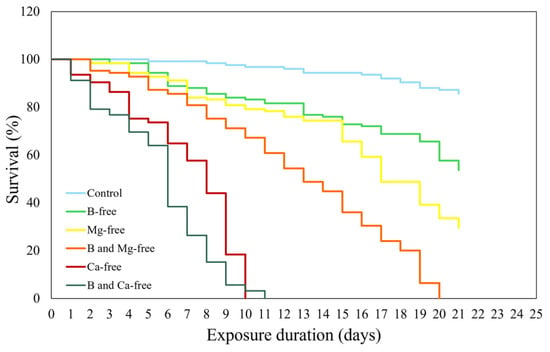
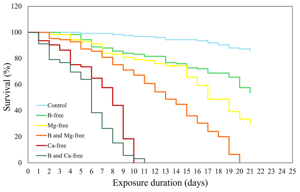
The Kaplan–Meier survival curves showed distinct treatment-dependent survival patterns during the 21-day exposure period (Figure 1). The mean survival percentages (±SD) across the five replicate vessels are provided in Figure S1. Survival remained relatively high in control, whereas more rapid declines in survival were observed under Ca-free and combined B and Ca deficiency conditions. The hierarchy of survival outcomes across treatments was Control > B-free > Mg-free > B and Mg-free > Ca-free > B and Ca-free. Table S1 showed that all treatment groups exhibited significantly different survival patterns (log-rank test, p < 0.05). Table 1 shows a decreased mean lifespan across all treatments. The reduction in survival was relatively moderate under B-free conditions, whereas greater reductions in survival were observed under Mg-free, Ca-free, and combined deficiency conditions.
Figure 1.
Kaplan–Meier survival curves of D. magna exposed to control and different nutrient omission treatments over 21 days. Survival curves were constructed from the original survival records obtained from five replicate vessels per treatment (25 individuals per vessel).
Table 1.
Mean lifespan (days) of D. magna exposed to control and different nutrient omissions over 21 days, calculated from Kaplan–Meier survival curves.
Under B-free conditions, survival was significantly reduced, with a gradual decline throughout the exposure period, remaining >50% by Day 21, suggesting that D. magna retained some capacity to withstand B omission under the conditions examined. Under combined Mg- and Ca-deficient conditions, increased mortality was observed, consistent with the importance of Mg and Ca for body structure, neuromuscular function, and metabolic processes that may contribute to long-term survival [17,36]. By day 10 of the exposure period, 100% mortality was observed in D. magna exposed to Ca-free conditions. Similarly, complete mortality was observed in D. pulex exposed to 0.1 mg Ca L−1 by day 10, whereas all D. galeata individuals died within two days when exposed to 0.5 mg Ca L−1 [16]. The combined omission of B and Ca associated with the most severe effect, with complete mortality observed by Day 11. Similarly, under the combined B and Mg deficiency conditions, mortality increased gradually but reached 100%, by Day 21, reflecting that the combined B and Mg deficiency treatment produced greater adverse effects than B deficiency alone. Therefore, these findings indicate that although survival under B-deficient conditions was relatively high compared with the other nutrient-deficient treatments, greater mortality was observed under the combined B and Mg or B and Ca deficiency conditions, emphasizing the importance of balanced mineral availability for the survival of D. magna under chronic exposure conditions.
3.2.2. Effects of Mineral Deficiencies on Somatic Growth of D. magna
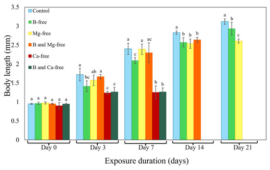
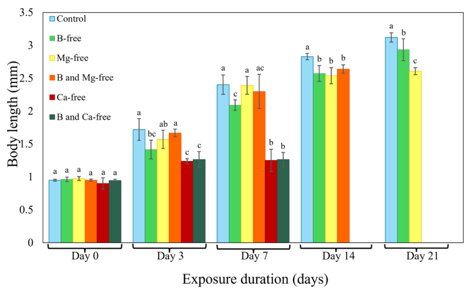
D. magna body length showed distinct temporal patterns across different nutrient-deficient treatments over the 21-day exposure period (Figure 2). On day 0, all groups had similar initial body sizes, indicating uniform starting conditions. Throughout the entire exposure period, body length was significantly lower under B-free conditions than in the control (p < 0.05; one-way ANOVA), suggesting that B availability may be related to growth performance under the conditions examined. B is an essential micronutrient for many organisms, facilitating cellular processes related to growth performance and immune function [7]. Moreover, diets with higher B content can enhance calcification [8], a process critical for normal growth and carapace formation in Daphnia sp. Therefore, reduced B availability may be associated with altered calcification, thereby impairing somatic growth and development. Similarly, Çelik et al. [5] demonstrated that Nile tilapia exhibited reduced growth performance along with negative changes in blood parameters.
Figure 2.
Temporal changes in body length (mm) of D. magna exposed to control and different nutrient omissions for 21 days. Data are presented as the mean ± SD of five replicates (five individuals per replicate; a total of 25 individuals per treatment). Bars with different lowercase letters show significant differences between treatments at each time point (one-way ANOVA, p < 0.05). Body-length data were not available at later exposure periods for treatments in which complete mortality had occurred.
In contrast, Mg deficiency condition did not significantly affect body length until day 14. By day 21, body length was significantly lower than in both the control and B-free treatments. This delayed reduction in body length under Mg-free conditions may be associated with the role of Mg as a cofactor in numerous enzymatic reactions involved in metabolism [37]. Under Mg deficiency, organisms show reduced biomass accumulation [36], altered nutrient absorption, and increased oxidative stress [11]. Similarly, the combined B and Mg deficiency treatment did not result in a significant reduction in body length until day 14, suggesting that the absence of Mg may have modified the growth response to B deficiency under the conditions examined. The delayed growth response observed under the combined deficiency condition may potentially reflect physiological adjustments to nutrient limitation that temporarily mitigate the effects of individual nutrient deficiencies [38].
Under Ca-free and combined B and Ca deficiency conditions, significantly smaller body lengths were observed as early as day 3, with more pronounced growth suppression in Ca-free treatments by day 7 (p < 0.05; one-way ANOVA). No data were collected for Ca-free treatments after day 7, as total mortality was observed by day 10. Ca plays a critical role in D. magna growth and development due to its involvement in exoskeleton calcification and structural integrity [15,17,39]. Growth is particularly sensitive to Ca limitation during the early posthatching period, as neonates undergo more frequent molting than adults, resulting in elevated Ca requirements. In addition, neonate and juvenile stages exhibit higher surface-to-body area ratios, which increase Ca demand for carapace formation [16]. Ca concentrations sufficient to support molting in mature individuals may therefore be inadequate for younger life stages, leading to a critical developmental constraint, often described as a “Ca bottleneck” during early growth [16,40]. The similar growth responses observed in Ca-free and combined B and Ca deficiency treatments suggest that the growth patterns were comparable under these conditions, with no additional reduction in body length apparent under the combined deficiency during the early and mid-exposure periods.
The growth rate analysis was consistent with the body length results on day 7 (Figure S2). Growth rate was significantly lower in the Ca-free and combined B and Ca deficiency treatments than in the control (p < 0.05; one-way ANOVA), while the B-free treatment also showed a significant, but less pronounced, reduction. These findings further support the body length results, with the greatest reduction in growth rate observed under Ca-free conditions, followed by a less pronounced reduction under B-free conditions, while no significant reduction was observed under Mg-free conditions.
3.2.3. Mineral Deficiency-Induced Alterations in Reproductive Performance
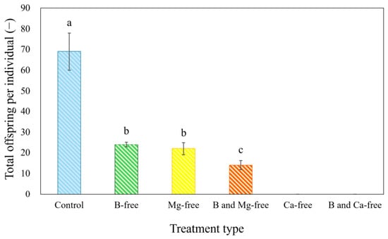
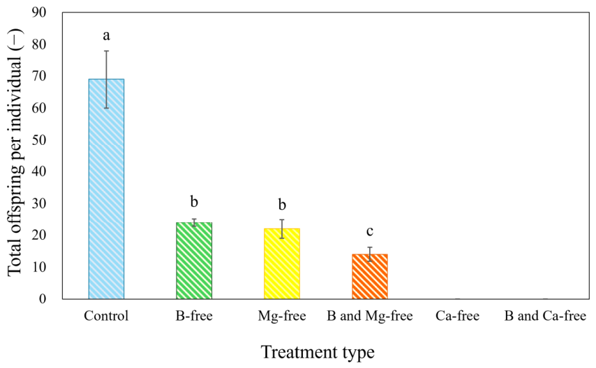
The reproductive output of D. magna over the 21-day exposure period varied substantially among nutrient-free treatments (Figure 3 and Figure S3). Under B-free conditions, reproductive output was significantly lower than the control, with substantially lower total offspring production and offspring per individual than in the control (p < 0.05; one-way ANOVA). However, the reproductive output in this treatment remained higher than that observed in the Mg-free and combined B and Mg deficiency treatments, suggesting a relatively less pronounced reduction in reproductive performance under B-free conditions. B has also been reported to contribute to embryonic cell proliferation and development [41]. Similarly, early embryonic development in zebrafish has been reported to be adversely affected by B deficiency conditions, with alterations in the membrane during the zygote and cleavage periods [6]. In Xenopus laevis, dietary B deficiency has been associated with a substantial increase in egg necrosis and a high incidence of abnormal gastrulation [41].
Figure 3.
Total offspring per individual D. magna during the test period under different treatment conditions. Data are presented as the mean ± SD of five replicates (25 individuals per replicate). Bars with different lowercase letters show significant differences between treatments (one-way ANOVA, p < 0.05). The absence of data for the Ca-free and B and Ca-free treatments indicates complete reproductive failure, with no offspring produced during the test period.
Under Mg-deficient conditions, reproductive output was significantly lower than the control, which may be related to the role of Mg in energy metabolism and its importance for reproductive processes. Inadequate Mg status during pregnancy may negatively affect both maternal and fetal health, leading to impaired implantation, higher incidence of threatened abortion, and premature delivery [42]. Similarly, reduced Mg availability has been shown to impair reproductive function in mice [43]. In aquatic invertebrates, Mg plays an important role in supporting reproductive performance and offspring quality in brood females of Macrobrachium rosenbergii [44]. Under the combined B and Mg deficiency condition, the reproductive output was further reduced, with the lowest offspring among treatments retaining reproductive activity (p < 0.05; one-way ANOVA), indicating that the combined deficiency condition was associated with a greater reduction in reproductive output than B deficiency alone.
Ca-free and combined B and Ca deficiency conditions were associated with complete reproductive failure, with no offspring produced throughout the exposure period. The absence of reproduction under these conditions is consistent with the reported importance of Ca for molting, structural integrity, and embryonic development in cladocerans [15,17]. Previous studies have suggested that Ca availability may influence reproductive performance through its roles in somatic growth and other physiological processes. Similarly, Hessen et al. [17] observed a pronounced decline in age-specific egg production at Ca concentrations < 10 mg Ca L−1, partly attributable to reduced somatic growth and increased energetic costs associated with Ca acquisition under Ca-limited conditions. Therefore, the observed reproductive responses across treatments highlight the importance of adequate mineral availability for reproductive performance in D. magna.
3.3. Biochemical Responses
3.3.1. Nutrient Deficiency-Induced CAT Activity Suppression
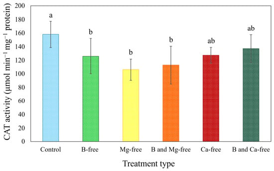
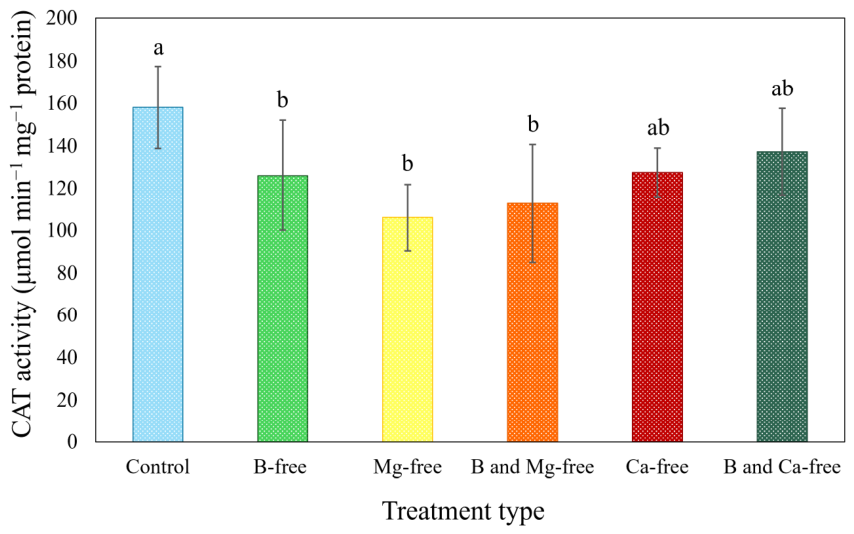
CAT activity in D. magna varied significantly among nutrient-deficient treatments (Figure 4). Under B-free conditions, CAT activity showed a moderate but significant decrease, whereas a greater reduction in CAT activity was observed under Mg-free conditions in D. magna. The combined B and Mg deficiency condition significantly reduced the CAT activity, similar to the Mg-free treatment alone, suggesting that Mg availability may be associated with maintaining CAT activity under these conditions. Finally, the Ca-free treatments showed an intermediate response, with CAT activity not significantly different from that of the other treatment groups.
Figure 4.
CAT activity of D. magna exposed to different nutrient omissions for seven days. Data are presented as the mean ± SD of five replicates (30–40 individuals pooled per replicate). Bars with different lowercase letters show significant differences between treatments (one-way ANOVA, p < 0.05).
CAT is a heme-containing enzyme whose structural integrity, synthesis, and functional efficiency depend on cellular metabolic stability [45] and the availability of essential ions. B influences the metabolism and utilization of essential minerals, including Mg and Ca, acts as a metabolic regulator in several enzymatic systems, and affects overall cellular physiology in animals [7,8]. Therefore, B deficiency may be associated with alterations in antioxidant defense capacity, potentially contributing to the observed decrease in CAT activity.
CAT requires iron as a direct cofactor rather than Mg [45]. Therefore, the changes in CAT activity observed under Mg deficiency may be related to indirect effects on biochemical processes such as energy metabolism, nucleotide synthesis, and oxidative stress [13]. CAT activity can be exhibited by several structurally distinct protein classes [45]. Mg insufficiency has been reported to affect ATP biosynthesis, and may consequently influence ATP-dependent processes, including protein synthesis [13]. These effects may potentially contribute to alterations in the expression or abundance of antioxidant enzymes, such as CAT, thereby affecting antioxidant defense capacity. Furthermore, Mg insufficiency has been reported to disrupt the electron transport chain, which may potentially contribute to enhanced ROS production [13]. While moderate ROS levels can induce antioxidant enzymes, prolonged or severe oxidative stress has been associated with oxidative inactivation of CAT [46]. For instance, increased aortic thiobarbituric acid-reactive substances, which reflect enhanced lipid peroxidation, were reported alongside a significant decrease in CAT activity were observed in Mg-deficient rats [47].
With respect to Ca, Manandhar et al. [48] reported that Ca depletion was associated with reduced CAT activity and increased ROS accumulation, particularly in peroxisomes. This effect has been suggested to be related to alterations in Ca-dependent signaling, as Ca acts as a key intracellular messenger that regulates gene expression and enzyme activity [49]. Prolonged nutrient deprivation may be associated with reduced antioxidant capacity, which could contribute to the observed decrease in CAT activity. Accordingly, CAT activity decreased across treatments, which may reflect alterations in antioxidant capacity under prolonged nutrient-deficient conditions rather than a classical induction response.
3.3.2. Nutrient Deficiency-Induced Alterations in GST Activity
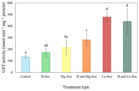
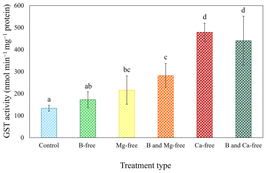
GST activity in D. magna differed significantly among nutrient-deficient treatments (Figure 5). B-free treatment showed a modest, but not significant, elevation in GST activity (p > 0.05; one-way ANOVA), while Mg-free and combined B and Mg deficiency conditions associated with significantly higher GST activity (p < 0.05; one-way ANOVA). The highest GST activity was observed under Ca-free conditions, potentially reflecting cellular stress or activation of detoxification processes. In the combined B and Ca deficiency treatment, GST activity remained elevated, similar to that observed under Ca deficiency alone.
Figure 5.
GST activity of D. magna exposed to different nutrient omissions for seven days. Data are presented as the mean ± SD of five replicates (30–40 individuals pooled per replicate). Bars with different lowercase letters show significant differences between treatments (one-way ANOVA, p < 0.05).
Unlike CAT, GST functions primarily as a phase II detoxification enzyme, responding to the accumulation of secondary oxidative products rather than directly scavenging ROS. Accordingly, GST activity may reflect increased cellular defense, and detoxification demands associated with cellular stress [50]. B affects the regulation of key minerals, such as Mg and Ca, and participates in the regulation of multiple enzyme-mediated metabolic processes [8]. Thus, B deficiency may be associated with cellular stress that could potentially contribute to the observed GST response.
Mg is involved in GSH synthesis, ATP production, and enzyme stability [51]. Hence, Mg deficiency may alter cellular antioxidant and detoxification capacity, potentially contributing to the observed changes in GST activity. In rats, Mg supplementation could restore the reduced liver GSH levels resulting from lead or cadmium exposure [37]. While individual B omission had no significant effect on GST activity, the combined B and Mg deficiency treatment showed a significant increase in GST activity. This suggests that combined B and Mg deficiency treatment may be associated with a greater cellular stress response than either deficiency alone.
Ca plays a critical role in maintaining membrane integrity and regulating intracellular signaling pathways [12]. Its deficiency may affect membrane integrity and potentially contribute to the formation of electrophilic lipid peroxidation products. These compounds have been reported to induce GST activity, as they require conjugation with GSH for detoxification [50]. Moreover, increased ROS and altered Ca levels may be associated with activation of apoptotic pathways [52]. Therefore, GST upregulation under Ca deficiency may reflect an enhanced cellular defense response. Together, these results suggest that B deficiency may influence GST activity in D. magna, potentially as part of a cellular defense response, while the strongest GST response was associated with the Ca deficiency.
3.3.3. Differential Regulation of CAT and GST in Response to Nutrient Deficiencies
The combined analysis of CAT and GST activities in D. magna revealed an opposing and enzyme-specific response to nutrient deficiencies, characterized primarily by CAT inhibition rather than activation. The apparent uncoupled behavior between CAT and GST activities observed in this study may be related to differences in their biochemical roles and regulatory mechanisms. CAT serves as a first-line antioxidant enzyme that rapidly decomposes hydrogen peroxide, but it is highly sensitive to oxidative inactivation and nutrient-dependent structural destabilization [45]. In contrast, GST is a secondary defense enzyme whose induction is typically triggered by the accumulation of oxidized lipids and xenobiotic metabolites rather than by ROS levels alone [50]. As a result, conditions that strongly inhibit CAT activity (e.g., Mg deficiency) may not necessarily result in a proportional increase in GST activity, whereas Ca deficiency, which may affect membrane integrity and intracellular signaling, may enhance GST activity despite relatively modest CAT suppression. Consequently, CAT activity may remain suppressed even with increasing GST activity. The observed CAT inhibition may indicate increased susceptibility to cellular stress in D. magna, while the increased GST activity may be consistent with cellular stress or changes associated with detoxification processes. Assessing multiple antioxidant biomarkers together may therefore provide a more comprehensive indication of nutrient-associated cellular stress responses in aquatic invertebrates.
3.4. Boron Uptake and Internal Distribution in D. magna
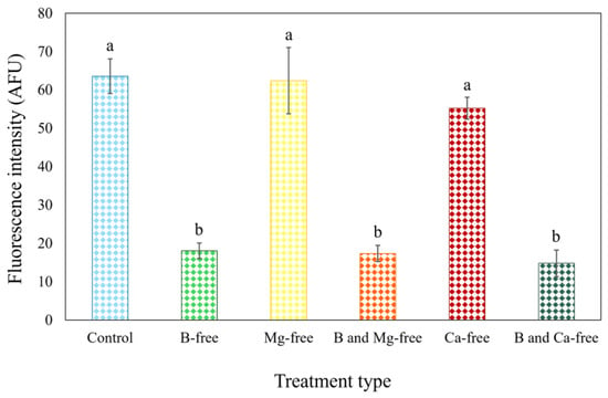
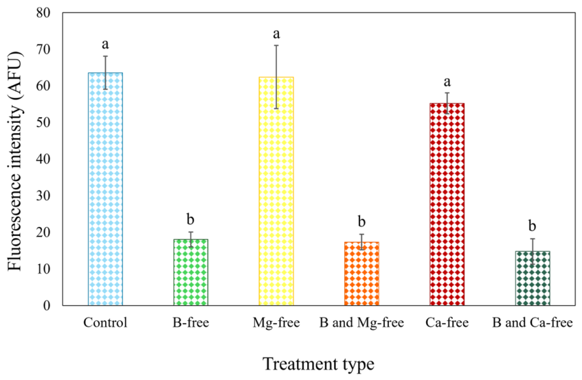
Quantitative fluorescence analysis using the boric acid-responsive DHNBI probe revealed clear treatment-dependent differences in fluorescence intensity associated with B availability in D. magna after seven days of exposure (Figure 6 and Figure S4). Under B-containing conditions, including the control, Mg-free, and Ca-free treatments, D. magna exhibited significantly higher fluorescence intensities in the gut region (p < 0.05; one-way ANOVA), without significant differences among these groups. In contrast, all treatments lacking B (B-free, B and Mg-free, and B and Ca-free) resulted in a pronounced reduction in fluorescence intensity compared with B-containing treatments (p < 0.05; one-way ANOVA).
Figure 6.
Fluorescence intensities obtained from fluorescence images of D. magna after 7 days of exposure to different nutrient-omissions and subsequent incubation with a boric acid-responsive fluorescent probe. Data are presented as the mean ± SD of five replicates (five individuals per replicate; a total of 25 individuals per treatment). Bars with different lowercase letters show significant differences between treatments (one-way ANOVA, p < 0.05).
Fluorescence analysis demonstrated that B associated fluorescence intensity in D. magna varied with external B availability, supporting the potential applicability of the boric acid-responsive fluorescent probe for in vivo assessment of B accumulation. The similarly high fluorescence intensities observed in B-containing treatments suggest that B associated fluorescence was maintained despite Ca or Mg limitation.
Although numerous fluorescent probes have been designed for selective recognition of metal ions, their application for in vivo detection in aquatic organisms remains relatively limited [31]. For example, fluorescent probes have been used to detect Zn in Moina macrocopa and Rheocricotopus larvae, Cd in Moina macrocopa using a Cd-sensitive probe, and Hg in zebrafish using a benzopyran-based fluorescent probe [31,53,54]. For detection of B in living cells, Sarkar et al. [55] observed fluorescence signals in plant roots grown in boric acid-treated water using a Schiff-base ligand as a fluorescent probe.
The pronounced localization of fluorescence signals along the digestive tract suggesting that B associated fluorescence may reflect ingestion-related exposure in D. magna (Figure S4). This pattern is consistent with previous studies showing that trace elements and micronutrients are assimilated in cladocerans via the digestive system following dietary or waterborne exposure [31,53]. Furthermore, DHNBI probes offer distinct advantages over conventional B detection methods, such as ICP-OES and autoradiography, as they allow fluorescence-based visualization of B associated signals without destructive preparation [22,23], and may have potential applications in water-quality assessment by facilitating in vivo visualization of B status in species such as D. magna, which is often used in ecotoxicological studies. Moreover, the probe may provide a useful approach for monitoring internal B dynamics in D. magna and its possible application in the detection of micronutrient stress. Further studies across a broader range of exposure conditions and biological responses are needed to further evaluate its potential application in the assessment of B status and its relevance to micronutrient stress.
3.5. Integrative Relationships Between Nutrient Status, Physiological Stress, and Life History Performance
The present study demonstrates that nutrient deficient conditions were associated with changes in life-history traits, antioxidant responses, and B associated fluorescence in D. magna, suggesting an association between nutrient availability and physiological performance.
The survival hierarchy (Control > B-free > Mg-free > B and Mg-free > Ca-free > B and Ca-free) closely followed the observed growth patterns, particularly at later stages of exposure, indicating an association between these endpoints under nutrient-deficient conditions. Furthermore, reproductive performance, assessed as both total reproductive output and offspring per individual, closely reflected the survival responses across treatments. Nutrient-deficient conditions, particularly under Mg and Ca omission, substantially reduced reproductive output; combined deficiency treatments produced greater reductions in reproductive output than the corresponding single-nutrient deficiency treatments. GST activity upregulation was also closely aligned with survival outcomes; significant increases were observed in the B and Mg-free, Ca-free, and B and Ca-free treatments relative to the control.
A limitation of the present study is that the experimental design included selected, rather than all possible, combinations of B, Mg, and Ca deficiencies. Although these treatments provide insight into the responses to multiple nutrient limitations, the present study could not provide a complete assessment of potential compensatory, inhibitory, or interactive effects among these nutrients. Additional combinations, such as Mg and Ca combined deficiency and simultaneous deficiency of B, Mg, and Ca, would provide further information on the broader interactions among these nutrients. Future studies incorporating these combinations could therefore build upon the present findings to further elucidate nutrient interactions in D. magna. Moreover, the modifications to the OECD 211 protocol, including the use of yeast as a food source and group housing of relatively large number of individuals, may have influenced life-history and biochemical responses; however, their consistent application across all treatments minimized potential confounding effects. Therefore, the absolute life-history and biochemical responses should be interpreted within the context of this modified experimental design. Furthermore, the removal of Ca and Mg from the M4 medium necessarily reduced water hardness and conductivity. Consequently, the observed responses may represent the combined effects of mineral deficiency and the associated changes in water chemistry. Although this study focused on life-history and enzymatic endpoints, future research employing transcriptomic and metabolomic approaches would be valuable for identifying early changes in gene expression and metabolic alterations, thereby offering a more comprehensive understanding of the mechanisms through which D. magna responds to nutrient-deficiency stress.
4. Conclusions
This study shows that nutrient deficiencies, alone and in combination, differently affect D. magna survival, growth, and reproduction. Under B-deficient conditions, a significant but relatively moderate reduction in survival was observed, together with significant decreases in body length, reproductive output, and CAT activity. Under Mg-deficient conditions, moderate adverse responses were observed, including reduced survival, delayed but significant growth suppression, pronounced declines in reproductive performance, and reduced CAT activity. Responses associated with Ca omission had the most severe biological consequences, identified by early mortality, total reproductive collapse, substantial growth impairment, and a rapid increase in GST activity detected within seven days. Under the combined B and Mg- or Ca-deficient conditions, pronounced adverse responses were observed, indicating the critical importance of balanced essential mineral availability under multiple nutrient limitations. The close correspondence between life history endpoints and GST activity suggests that the increased GST activity may reflect cellular stress associated with the observed reductions in organismal performance. Furthermore, the DHNBI probe enabled visualization of treatment-dependent differences in fluorescence-associated B availability and may provide a useful approach for assessing internal B status in D. magna. Together, these findings emphasize the importance of balanced nutrient availability for maintaining physiological stability in aquatic invertebrates and highlight the value of integrating life history, biochemical, and imaging approaches to assess the responses to nutrient deficiency. Future studies should explore the potential for early detection of nutrient stress through biochemical responses, which may facilitate the identification of sublethal effects before impacts on survival or reproduction become evident. Further studies are needed to evaluate biomarker sensitivity and environmental applicability before these biomarkers can be considered for assessing nutrient deficiency and micronutrient conditions in aquatic environments.
Supplementary Materials
The following supporting information can be downloaded at https://www.mdpi.com/article/10.3390/ecologies7030087/s1, Text S1: DHNBI synthesis; Figure S1: Survival curves of D. magna exposed to different nutrient omissions for 21 days. Data are presented as the mean ± SD of five replicates (25 individuals per replicate). * Days correspond to growth measurements, ** Day corresponds to enzyme activity and fluorescence measurements; Figure S2: Growth rate of D. magna during the 7 days of exposure period under different nutrient omissions. Data are presented as the mean ± SD of five replicates (five individuals per replicate; a total of 25 individuals per treatment). Bars with different lowercase letters show significant differences between treatments (one-way ANOVA, p < 0.05); Figure S3: Total reproductive output of D. magna during the exposure period under different nutrient omissions. Data are presented as the mean ± SD of five replicates (25 individuals per replicate). Bars with different lowercase letters show significant differences between treatments (one-way ANOVA, p < 0.05); Figure S4: Brightfield (a), fluorescence (b) and merged (c) images of D. magna exposed to different nutrient omissions for seven days, followed by a 30 min incubation with a 100 µmol L−1 DHNBI boric acid-responsive fluorescent probe; Table S1: Pairwise log-rank test results (χ2 and Bonferroni-adjusted p-values) comparing survival curves of D. magna among all treatments. p < 0.05 indicated statistical significance; Table S2: Water quality parameters of control and nutrient-free treatments during the test period. Data represents mean ± SD for each parameter measured on alternate days throughout the test period; Table S3: Measured B, Ca, and Mg (obtained via ICP-OES) concentrations in different nutrient-free media prepared during the test. Data are presented as mean ± SD for each parameter measured on alternate days throughout the test period.
Author Contributions
Conceptualization, I.M. and T.F.; methodology, I.M., T.F., S.T., M.H. and D.G.K.; formal analysis, D.G.K.; investigation, D.G.K.; resources, S.T., M.H. and T.F.; writing—original draft preparation, D.G.K.; writing—review and editing, D.G.K., I.M., T.F., S.T. and M.H.; supervision, I.M.; funding acquisition, S.T. All authors have read and agreed to the published version of the manuscript.
Funding
This study was partially supported by the Japan Society for the Promotion of Science, Japan (JP24K18844).
Institutional Review Board Statement
Not applicable.
Informed Consent Statement
Not applicable.
Data Availability Statement
Data is contained within the article or Supplementary Materials.
Conflicts of Interest
The authors declare no conflicts of interest.
Abbreviations
The following abbreviations are used in this manuscript:
| ANOVA | Analysis of variance |
| ATP | Adenosine Triphosphate |
| CAT | Catalase |
| CDNB | 1-Chloro-2,4-dinitrobenzene |
| DO | Dissolved oxygen |
| GSH | Reduced glutathione |
| GST | Glutathione S-transferase |
| ICP-OES | Inductively coupled plasma optical emission spectrophotometer |
| OECD | Organization for Economic Cooperation and Development |
| ROS | Reactive oxygen species |
| SPSS | Statistical Package for the Social Sciences |
References
- Bhagyaraj, S.; Al-Ghouti, M.A.; Kasak, P.; Krupa, I. An updated review on boron removal from water through adsorption processes. Emergent Mater. 2021, 4, 1167–1186. [Google Scholar] [CrossRef] [Scilit]
- Liu, X.; Xu, C.; Chen, P.; Li, K.; Zhou, Q.; Ye, M.; Zhang, L.; Lu, Y. Advances in technologies for boron removal from water: A comprehensive review. Int. J. Environ. Res. Public Health 2022, 19, 10671. [Google Scholar] [CrossRef] [Scilit] [PubMed]
- Vera-Maldonado, P.; Aquea, F.; Reyes-Díaz, M.; Cárcamo-Fincheira, P.; Soto-Cerda, B.; Nunes-Nesi, A.; Inostroza-Blancheteau, C. Role of boron and its interaction with other elements in plants. Front. Plant Sci. 2024, 15, 1332459. [Google Scholar] [CrossRef] [Scilit] [PubMed]
- Kohli, S.K.; Kaur, H.; Khanna, K.; Handa, N.; Bhardwaj, R.; Rinklebe, J.; Ahmad, P. Boron in plants: Uptake, deficiency and biological potential. Plant Growth Regul. 2023, 100, 267–282. [Google Scholar] [CrossRef] [Scilit]
- Çelik, M.; Dikel, S.; Öz, M. Investigation of the effect of water and feed sourced boron on the growth performance and blood parameters of Nile tilapia, Oreochromis niloticus. J. World Aquac. Soc. 2024, 55, e13104. [Google Scholar] [CrossRef] [Scilit]
- Eckhert, C.D.; Rowe, R.I. Embryonic dysplasia and adult retinal dystrophy in boron-deficient zebrafish. J. Trace Elem. Exp. Med. 1999, 12, 213–219. [Google Scholar] [CrossRef]
- Abdelnour, S.A.; Abd El-Hack, M.E.; Swelum, A.A.; Perillo, A.; Losacco, C. The vital roles of boron in animal health and production: A comprehensive review. J. Trace Elem. Med. Biol. 2018, 50, 296–304. [Google Scholar] [CrossRef] [Scilit] [PubMed]
- Białek, M.; Czauderna, M.; Krajewska, K.A.; Przybylski, W. Selected physiological effects of boron compounds for animals and humans: A review. J. Anim. Feed Sci. 2019, 28, 307–320. [Google Scholar] [CrossRef] [Scilit]
- Hamid, A.; Bhat, S.U.; Jehangir, A. Local determinants influencing stream water quality. Appl. Water Sci. 2020, 10, 24. [Google Scholar] [CrossRef] [Scilit]
- Hu, Z.; Weynell, M.; Su, N.; Hohl, S.V.; Han, Z.; Wen, Y.; Yang, S. Evidence from Mg isotopes indicating reverse weathering as a significant Mg sink in Tibetan Plateau lakes. Quat. Sci. Rev. 2025, 349, 109138. [Google Scholar] [CrossRef] [Scilit]
- Fiorentini, D.; Cappadone, C.; Farruggia, G.; Prata, C. Magnesium: Biochemistry, nutrition, detection, and social impact of diseases linked to its deficiency. Nutrients 2021, 13, 1136. [Google Scholar] [CrossRef] [Scilit] [PubMed]
- Kovalenko, V.F.; Sova, A.M. Effects of calcium and magnesium ion ratios in natural and drinking water on the vitality of test organisms. J. Water Chem. Technol. 2024, 46, 414–418. [Google Scholar] [CrossRef] [Scilit]
- Ashique, S.; Kumar, S.; Hussain, A.; Mishra, N.; Garg, A.; Gowda, B.H.J.; Farid, A.; Gupta, G.; Dua, K.; Taghizadeh-Hesary, F. A narrative review on the role of magnesium in immune regulation, inflammation, infectious diseases, and cancer. J. Health Popul. Nutr. 2023, 42, 74. [Google Scholar] [CrossRef] [Scilit] [PubMed]
- Alstad, N.E.W.; Skardal, L.; Hessen, D.O. The effect of calcium concentration on the calcification of Daphnia magna. Limnol. Oceanogr. 1999, 44, 2011–2017. [Google Scholar] [CrossRef] [Scilit]
- Ashforth, D.; Yan, N.D. The interactive effects of calcium concentration and temperature on the survival and reproduction of Daphnia pulex at high and low food concentrations. Limnol. Oceanogr. 2008, 53, 420–432. [Google Scholar] [CrossRef] [Scilit]
- Cairns, A.; Yan, N. A review of the influence of low ambient calcium concentrations on freshwater daphniids, gammarids, and crayfish. Environ. Rev. 2009, 17, 67–79. [Google Scholar] [CrossRef] [Scilit]
- Hessen, D.O.; Alstad, N.E.W.; Skardal, L. Calcium limitation in Daphnia magna. J. Plankton Res. 2000, 22, 553–568. [Google Scholar] [CrossRef] [Scilit]
- Bownik, A.; Pawlik-Skowrońska, B. Early indicators of behavioral and physiological disturbances in Daphnia magna (Cladocera) induced by cyanobacterial neurotoxin anatoxin-a. Sci. Total Environ. 2019, 695, 133913. [Google Scholar] [CrossRef] [Scilit] [PubMed]
- Juan, C.A.; Pérez de la Lastra, J.M.; Plou, F.J.; Pérez-Lebeña, E. The chemistry of reactive oxygen species (ROS) revisited: Outlining their role in biological macromolecules (DNA, lipids and proteins) and induced pathologies. Int. J. Mol. Sci. 2021, 22, 4642. [Google Scholar] [CrossRef] [Scilit] [PubMed]
- Jemec, A.; Drobne, D.; Tišler, T.; Sepčić, K. Biochemical biomarkers in environmental studies-lessons learnt from enzymes catalase, glutathione S-transferase and cholinesterase in two crustacean species. Environ. Sci. Pollut. Res. 2010, 17, 571–581. [Google Scholar] [CrossRef] [Scilit] [PubMed]
- Kondo, N.; Takada, S.; Hagimori, M.; Temma, T. Development of a 2-(2-Hydroxyphenyl)-1H-benzimidazole-based fluorescence sensor targeting boronic acids for versatile application in boron neutron capture therapy. Cancers 2023, 15, 1862. [Google Scholar] [CrossRef] [Scilit] [PubMed]
- Kondo, N.; Aoki, E.; Takada, S.; Temma, T. A red-emitting fluorescence sensor for detecting boronic acid-containing agents in cells. Sensors 2022, 22, 7671. [Google Scholar] [CrossRef] [Scilit] [PubMed]
- Takada, S.; Kondo, N.; Hagimori, M.; Temma, T. Development of a switching-type fluorescence sensor for the detection of boronic acid-containing agents. Anal. Sci. 2022, 38, 1289–1296. [Google Scholar] [CrossRef] [Scilit] [PubMed]
- Takada, S.; Du, H.; Kondo, N.; Miyazaki, A.; Hara, F.; Horiyama, S.; Temma, T.; Hagimori, M. A long-wavelength fluorescent probe for efficient dual-color imaging of boronic-acid-containing agents in living cells. Chemosensors 2025, 13, 283. [Google Scholar] [CrossRef] [Scilit]
- Gürkan, S.E.; Gürkan, M.; Sarıtunç, V.; İbiş, E.C.; Güneş, B. Evaluation of possible toxic effects of boric acid in palourde clam (Ruditapes decussatus) through histological changes and oxidative responses. Biol. Trace Elem. Res. 2024, 203, 1151–1161. [Google Scholar] [CrossRef] [Scilit] [PubMed]
- Dağlıoğlu, Y.; Öztürk, B.Y. Comparison of the toxicity of boron nano and microparticles based on the pigmentation, bioaccumulation, and oxidative stress of Chodatodesmus mucronulatus. J. Boron 2018, 3, 157–165. [Google Scholar] [CrossRef] [Scilit]
- OECD. Daphnia magna Reproduction Test, OECD Test Guideline No. 211. Available online: https://www.oecd.org/content/dam/oecd/en/publications/reports/2012/10/test-no-211-daphnia-magna-reproduction-test_g1g24069/9789264185203-en.pdf (accessed on 14 July 2025).
- El-Baz, F.K.; Abdoon, S.M.; El-Sayed, D.A.A.; Mostafa, M.A.; Elsherif, H.M.R.; Safaa, H.M.; Abdon, A.S. Application of defatted Scenedesmus obliquus biomass for broilers’ nutrition. Braz. J. Poult. Sci. 2021, 23, eRBCA-2020. [Google Scholar] [CrossRef] [Scilit]
- Taştan, B.E.; Duygu, E.; Dönmez, G. Boron bioremoval by a newly isolated Chlorella sp. and its stimulation by growth stimulators. Water Res. 2012, 46, 167–175. [Google Scholar] [CrossRef] [Scilit] [PubMed]
- Samarakoon, T.; Fujino, T. Individual and combined effects of humic acid on life-history characteristics of the water flea Moina macrocopa upon whole-lifespan cadmium exposure. Hydrobiologia 2023, 850, 1635–1652. [Google Scholar] [CrossRef] [Scilit]
- Samarakoon, T.; Fujino, T.; Hagimori, M.; Saito, R. Cadmium uptake and oxidative-stress-induced DNA alterations in the freshwater cladoceran Moina macrocopa (Straus 1820) following consecutive short-term exposure assessments. Limnology 2023, 24, 9–23. [Google Scholar] [CrossRef] [Scilit]
- Schindelin, J.; Arganda-Carreras, I.; Frise, E.; Kaynig, V.; Longair, M.; Pietzsch, T.; Preibisch, S.; Rueden, C.; Saalfeld, S.; Schmid, B.; et al. Fiji: An open-source platform for biological-image analysis. Nat. Methods 2012, 9, 676–682. [Google Scholar] [CrossRef] [Scilit] [PubMed]
- Xu, Z.; Li, Y.; Li, M.; Liu, H. Transcriptomic response of Daphnia magna to nitrogen- or phosphorus-limited diet. Ecol. Evol. 2021, 11, 11009–11019. [Google Scholar] [CrossRef] [Scilit] [PubMed]
- Claiborne, A. Catalase activity. In Handbook of Methods for Oxygen Radical Research; Greenwald, R.A., Ed.; CRC Press: Boca Raton, FL, USA, 1985; pp. 283–284. [Google Scholar]
- Habig, W.H.; Pabst, M.J.; Jakoby, W.B. Glutathione S-Transferases: The first enzymatic step in mercapturic acid formation. J. Biol. Chem. 1974, 249, 7130–7139. [Google Scholar] [CrossRef] [Scilit]
- Wei, S.P.; Jiang, W.D.; Wu, P.; Liu, Y.; Zeng, Y.Y.; Jiang, J.; Kuang, S.Y.; Tang, L.; Zhang, Y.A.; Zhou, X.Q.; et al. Dietary magnesium deficiency impaired intestinal structural integrity in grass carp (Ctenopharyngodon idella). Sci. Rep. 2018, 8, 12705. [Google Scholar] [CrossRef] [Scilit] [PubMed]
- Hassan, I.; Abdelfteh, Z.; Ebaid, H.; Al-Tamimi, J.H.; Alhazza, I.M.; Khan, A.; Awad, E.M. Protective effect of magnesium against lead and cadmium-induced liver injury in swiss albino rats. J. King Saud Univ. Sci. 2025, 37, 7052025. [Google Scholar] [CrossRef] [Scilit]
- Rietra, R.P.J.J.; Heinen, M.; Dimkpa, C.O.; Bindraban, P.S. Effects of nutrient antagonism and synergism on yield and fertilizer use efficiency. Commun. Soil Sci. Plant Anal. 2017, 48, 1895–1920. [Google Scholar] [CrossRef] [Scilit]
- Jeziorski, A.; Yan, N.D. Species identity and aqueous calcium concentrations as determinants of calcium concentrations of freshwater crustacean zooplankton. Can. J. Fish. Aquat. Sci. 2006, 63, 1007–1013. [Google Scholar] [CrossRef] [Scilit]
- Rukke, N.A. Effects of low calcium concentrations on two common freshwater crustaceans, Gammarus lacustris and Astacus astacus. Funct. Ecol. 2002, 16, 357–366. [Google Scholar] [CrossRef] [Scilit]
- Fort, D.J.; Stover, E.L.; Strong, P.L.; Murray, F.J.; Keen, C.L. Chronic feeding of a low boron diet adversely affects reproduction and development in Xenopus laevis. J. Nutr. 1999, 129, 2055–2060. [Google Scholar] [CrossRef] [Scilit] [PubMed]
- Skliarov, P.; Fedorenko, S.; Naumenko, S.; Onyshchenko, O.; Pasternak, A.; Roman, L.; Lieshchova, M.; Bilyi, D.; Bobrytska, O. Reviewing effective factors of alimentary deficiency in animals reproductive functions. World’s Vet. J. 2021, 11, 157–169. [Google Scholar] [CrossRef] [Scilit]
- Rosner, J.Y.; Gupta, M.; McGill, M.; Xue, X.; Chatterjee, P.K.; Yoshida-Hay, M.; Robeson, W.; Metz, C.N. Magnesium deficiency during pregnancy in mice impairs placental size and function. Placenta 2016, 39, 87–93. [Google Scholar] [CrossRef] [Scilit] [PubMed]
- Tavabe, K.R.; Rafiee, G.; Shoeiry, M.M.; Houshmandi, S.; Frinsko, M.; Daniels, H. Effects of water hardness and calcium: Magnesium ratios on reproductive performance and offspring quality of Macrobrachium rosenbergii. J. World Aquac. Soc. 2015, 46, 519–530. [Google Scholar] [CrossRef] [Scilit]
- Chelikani, P.; Fita, I.; Loewen, P.C. Diversity of structures and properties among catalases. Cell. Mol. Life Sci. 2004, 61, 192–208. [Google Scholar] [CrossRef] [Scilit] [PubMed]
- Chakravarti, R.; Gupta, K.; Majors, A.; Ruple, L.; Aronica, M.; Stuehr, D.J. Novel insights in mammalian catalase heme maturation: Effect of NO and thioredoxin-1. Free Radic. Biol. Med. 2015, 82, 105–113. [Google Scholar] [CrossRef] [Scilit] [PubMed]
- Zheltova, A.A.; Kharitonova, M.V.; Iezhitsa, I.N.; Spasov, A.A. Magnesium deficiency and oxidative stress: An update. Biomedicine 2016, 6, 8–14. [Google Scholar] [CrossRef] [Scilit] [PubMed]
- Manandhar, L.; Dutta, R.K.; Devkota, P.; Chhetri, A.; Wei, X.; Park, C.; Kwon, H.M.; Park, R. TFEB activation triggers pexophagy for functional adaptation during oxidative stress under calcium deficient-conditions. Cell Commun. Signal. 2024, 22, 142. [Google Scholar] [CrossRef] [Scilit] [PubMed]
- Berridge, M.J.; Lipp, P.; Bootman, M.D. The versatility and universality of calcium signalling. Nat. Rev. Mol. Cell Biol. 2000, 1, 11–21. [Google Scholar] [CrossRef] [Scilit] [PubMed]
- Sharma, R.; Yang, Y.; Sharma, A.; Awasthi, S.; Awasthi, Y.C. Antioxidant role of glutathione S-transferases: Protection against oxidant toxicity and regulation of stress-mediated apoptosis. Antioxid. Redox Signal. 2004, 6, 289–300. [Google Scholar] [CrossRef] [Scilit] [PubMed]
- Chambers, P. Vitamin B6, magnesium, and vitamin D: The triple play. Med. Clin. Res. 2023, 8, 1–7. [Google Scholar] [CrossRef] [Scilit]
- Gao, S.; Wang, Z.; Zhang, C.; Jia, L.; Zhang, Y. Oral exposure to atrazine induces oxidative stress and calcium homeostasis disruption in spleen of mice. Oxidative Med. Cell. Longev. 2016, 2016, 7978219. [Google Scholar] [CrossRef] [Scilit] [PubMed]
- Shrestha, A.K.; Samarakoon, T.; Fujino, T.; Hagimori, M. Detection of Zn2+ ions using a high-affinity low-molecular-weight fluorescence probe in two freshwater organisms. Toxicol. Environ. Health Sci. 2023, 15, 145–155. [Google Scholar] [CrossRef] [Scilit]
- Lv, H.; Yuan, G.; Zhang, G.; Ren, Z.; He, H.; Sun, Q.; Zhang, X.; Wang, S. A novel benzopyran-based colorimetric and near-infrared fluorescent sensor for Hg2+ and its imaging in living cell and zebrafish. Dye. Pigment. 2020, 172, 107658. [Google Scholar] [CrossRef] [Scilit]
- Sarkar, Y.; Das, S.; Datta, R.; Chattopadhyay, S.; Ray, A.; Parui, P.P. Exploitation of a new schiff-base ligand for boric acid fluorescent sensing in aqueous medium with bio-imaging studies in a living plant system. RSC Adv. 2015, 5, 51875–51882. [Google Scholar] [CrossRef] [Scilit]

Disclaimer/Publisher’s Note: The statements, opinions and data contained in all publications are solely those of the individual author(s) and contributor(s) and not of MDPI and/or the editor(s). MDPI and/or the editor(s) disclaim responsibility for any injury to people or property resulting from any ideas, methods, instructions or products referred to in the content. |
© 2026 by the authors. Licensee MDPI, Basel, Switzerland. This article is an open access article distributed under the terms and conditions of the Creative Commons Attribution (CC BY) license.