


Abstract
Musculoskeletal pain is driven by degenerative conditions such as osteoarthritis or intervertebral disk degeneration. This creates a major burden on health systems and patient quality of life. Platelet-rich plasma (PRP) is an orthobiologic that contains platelets and growth factors which promote angiogenesis, modulate inflammation, and support tissue repair. Mechanobiology is central in the healing process, where mechanical loading regulates cellular behavior through mechanotransduction pathways including integrin/FAK and RhoA/ROCK. This use of signaling pathways allows for regeneration along multiple different tissue types. Evidence suggests that PRP and mechanical loading may act together to promote tissue repair, with PRP-derived signals interacting with biomechanical cues to enhance fibroblast activation, remodeling, and regenerative responses. There is variability in PRP preparation, rehabilitation protocols, and tissue-specific responses, which all highlight the need for a more standardized and guided treatment strategy. Understanding the interplay between PRP-derived biologic signaling and mechanobiologic pathways may help inform the development of more standardized, tissue-specific regenerative treatment strategies and could potentially contribute to improved clinical outcomes in patients with musculoskeletal pain.
1. Introduction
Musculoskeletal (MSK) pain affects more than 1.7 billion people worldwide, and it is one of the leading causes of chronic disability. It imposes a substantial personal, societal, and healthcare burden through persistent pain, functional impairment, reduced productivity, and diminished quality of life [1]. Chronic musculoskeletal pain is not only a symptom patients experience, but also a complex process that involves nociceptive signaling, sensitization, and a combination of neural and immune pathways, which all contribute to pain persistence and disease progression [2]. Additionally, degenerative musculoskeletal disorders, including tendinopathy, osteoarthritis, and intervertebral disk degeneration, are all major contributors to chronic pain. This highlights the need for therapies that address tissue pathology causing pain, rather than just controlling symptoms of pain [2,3]. Low back pain is a prevalent musculoskeletal condition around the world and remains a leading cause of disability and healthcare utilization [3,4]. Among its structural contributors, intervertebral disk degeneration is particularly important, as it is strongly associated with chronic pain, functional impairment, and substantial healthcare utilization [3,4]. These conditions have fueled growing interest in regenerative therapies aimed at restoring tissue function and modifying the underlying disease process.
Conventional pain management strategies and procedures performed by interventional pain specialists can provide a level of symptom relief; however, there are limitations in achieving durable tissue restoration and long-term recovery, not just acute symptom relief. These limitations of conventional therapies have stimulated interest in regenerative treatment approaches [3,4]. Recently, regenerative medicine has attracted considerable attention as a possible and promising paradigm in pain medicine by targeting biological mechanisms of tissue degeneration and repair through therapies such as stem cells, growth factors, prolotherapy, and platelet-rich plasma (PRP) [3,5]. Orthobiologics are also utilized in musculoskeletal medicine because of the potential to modulate inflammatory responses, stimulate healing of tissue at the repair site, and to enhance matrix synthesis, while supporting regenerative processes within damaged tissues [3,6]. Among available orthobiologic therapies, PRP has gained significant attention as a autologous blood-derived biologic containing a concentrated source of platelets and multiple other bioactive growth factors [3,6].
Although pre-clinical and mechanistic studies have demonstrated encouraging biologic effects of PRP, clinical outcomes are still inconsistent, and the results are limited. Clinical outcomes are variable depending on differences in PRP composition, preparation techniques, and target tissues [6]. Evidence suggests that the therapeutic effects of PRP are dependent on growth factor delivery and the mechanical environment of the treated tissue, which creates a rationale to examine the interplay between PRP and mechanotransduction pathways [3,6].
PRP, therefore, has become more and more utilized in the treatment of musculoskeletal disorders, and it is seen to augment soft tissue healing and regenerative processes, although clinical outcomes remain variable across the different tissues and conditions [7,8]. The inconsistency of PRP outcomes suggests that biologic therapies do not function in isolation and, rather, different factors within the tissue environment at injury sites influence regenerative responses [8].
Mechanobiology is the study of how physical forces and mechanical properties of the cellular environment regulate behavior, adaptation, and repair processes [9,10]. The different mechanical signals that are generated by the environment of tissue and injury include movement, exercise, deformation, loading, and stiffness. Mechanotransduction, on the other hand, is the process in which cells sense mechanical stimuli and convert those stimuli into signals that alter gene expression, metabolism, and remodeling [9,10]. Several mechanotransduction pathways are identified, which include integrin–FAK signaling, YAP/TAZ mediated transcriptional regulation, and RhoA/ROCK pathways, which coordinate responses to mechanical stress [9]. Mechanical loading is critical in remolding and adaptation and new concepts in regenerative medicine and rehabilitation suggest that appropriately prescribed mechanical loading can enhance the biologic repair process [9].
Given the growing use of PRP in interventional pain medicine, and the importance of mechanotransduction in musculoskeletal healing, understanding the interaction between PRP-derived biologic signals and mechanical loading can help explain treatment variability and optimize regenerative outcomes. The purpose of this review is to examine the mechanobiology of PRP, with a focus on mechanotransduction pathways, tissue loading, and their collective role in regenerative repair within interventional pain medicine.
2. Methodology
This narrative review was conducted using a literature search of the PubMed database to identify different studies that examined mechanobiological effects of PRP in musculoskeletal and pain-related conditions. Articles were identified using relevant keywords and Boolean combinations, which included “platelet-rich plasma” OR “PRP”, “mechanobiology”, “mechanotransduction”, “mechanical loading” and “interventional pain”. The PubMed search was performed throughout the writing process in the months of May and June 2026, with additional targeted searches being conducted for specific conditions including tendinopathy, osteoarthritis, and intervertebral disk degeneration. Additional articles were identified through a manual review of reference lists from relevant publications. No strict date restrictions were applied, which allowed for inclusion of both foundational and contemporary studies. Studies were selected based on their relevance to the biological and mechanical mechanisms underlying PRP-mediated tissue repair, its role in mechanotransduction, and interactions with mechanical loading in musculoskeletal tissue. Eligible publications included preclinical studies, clinical studies, systematic reviews, meta-analyses, narrative reviews, and relevant guidelines. Priority was given to peer reviewed studies, and systematic reviews and higher-level evidence when available. A mind map of the methodology is seen in Figure 1.
Figure 1.
Flow diagram summarizing the methodology of this narrative review. Created with https://BioRender.com (accessed on 16 June 2026).
3. Mechanobiology and Tissue Regeneration
3.1. Mechanotransduction
Mechanotransduction is the changing of mechanical cues to intracellular biochemical signals. This is important for cells and tissues because it allows them to respond adequately to their environment and to maintain their homeostatic regulation. Mechanical cues for repair may include the stiffness of the extracellular matrix, viscosity of the extracellular fluid, hydrostatic pressure, and mechanical loading. Physiologic mechanical cues include cyclic stretch of lung epithelium during breathing, fluid shear stress, and weight-bearing forces on the bone [11].
Mechanosensors are molecular sensors that allow the different cells to detect these mechanical signals. These sensors include integrins, ion channels, cadherins, and G protein-coupled receptors (GPCRs). Integrins are transmembrane receptors that link the extracellular matrix (ECM) to the intracellular actin cytoskeleton. Integrin-mediated adhesion is initiated with conformational changes in the integrin ectodomain, where integrins transition from a low-affinity state to a high-affinity state, allowing for ligand binding [12]. Integrins are important receptors for many different proteins, including collagen, laminin, vitronectin, and fibronectin. The composition of the ECM also alters mechanotransduction through focal adhesions because the ECM’s elastic properties influence force transduction through the integrin complex.
Endocytosis is the process by which integrin trafficking takes place toward and away from the cell membrane. Integrins are linked to the actin cytoskeleton through talin and vinculin, which are F-actin binding proteins. Talin is a mechanosensitive protein with vinculin binding sites that are accessible when talin is stretched. When vinculin binds to talin, it stabilizes focal adhesions by preventing talin from changing out of the active conformation, essentially locking it in place. Vinculin also regulates the binding of talin to actin in the cytoskeleton [11].
Another cytoplasmic protein associated with integrin-mediated mechanotransduction is focal adhesion kinase (FAK) [13]. FAK is a non-receptor tyrosine kinase that is activated via integrin-mediated autophosphorylation of tyrosine 397, which is initially activated by the ECM receiving mechanical signals [13]. FAK then recruits Src and becomes fully active. FAK activates other integrin-associated proteins such as paxillin and talin.
These proteins modulate transcriptional coactivators YAP and TAZ (YAP/TAZ) [14]. First, FAK-Src activates the Rho/GTPase signaling pathway. Rho GTPase rearranges the actin cytoskeleton, thus causing tension. This resulting tension inhibits the Hippo pathway. When the Hippo pathway is inactivated, YAP/TAZ are activated. YAP/TAZ translocate to the nucleus and facilitate gene transcription that promotes cell migration and proliferation. Dysregulated expression of integrins and integrin-associated proteins has been linked to several pathologies, including osteoporosis [15]. The YAP/TAZ mechanotransduction pathway plays a key role in cellular activity, such as differentiation, proliferation, survival, and tissue homeostasis [10].
3.2. Mechanical Loading and Tissue Healing
Mechanical loading refers to the physical forces applied to cells and tissues [16]. At the cellular level, different loading rates influence adhesion complexes including talin, an important and key binding protein that connects integrins to the actin cytoskeleton discussed in the previous section. Increased mechanical loading on talin can induce partial protein unfolding that exposes the vinculin-binding sites (VBS) [12].
At the tissue level, increased mechanical loading can increase tendon stiffness, modulus, and cross-sectional area (CSA). Changes in modulus have a greater impact on tendon stiffness than changes in cross-sectional area. Higher strain resistance training protocols caused larger increases in modulus. Resistance training caused higher modulus increases than other training protocols. This is important to note because tendons are important for transmitting forces from muscles to bones to facilitate movement by storing and releasing elastic energy [16].
Mechanical loading works to regulate tissue healing across all connective and musculoskeletal tissues [17]. It is the principal mechanism behind exercise-based rehabilitation and mechanotherapy. Mechanotherapy uses mechanical load in the therapeutic context to promote remodeling and healing in tendons, cartilage, ligaments, muscles, and bones [18].
Mechanical loading is thought to enhance or contribute to the biologic repair process through growth factor signaling, stem cell activation, ECM remodeling, angiogenesis, and immune modulation [19]. In the context of immune modulation, macrophages are key players because they are especially sensitive to mechanical forces. Macrophages differentiate into different phenotypes and play different roles based on distinctive stimuli from the environment [20]. Mechanical loading also activates the fibroblast FAK-ERK signaling. This stimulates the production of TGF-β, MCP-1, and collagen. Fibroblast activity is integral for the biologic repair process [21].
3.3. Mechanobiology in Musculoskeletal Tissues
Mechanobiology is where the forces as well as the mechanical properties of cells and tissues regulate cell growth and development. While in the bone, integrin–FAK signals and cytoskeletal tension that are RhoA/ROCK-mediated allow osteoblasts to detect the stiffness of the ECM and the mechanical stress [22]. This regulates osteogenic cell adhesion and its differentiation. An important mediator of this mechanism is the Wnt/β-catenin signaling pathway [23].
In cartilage, mechanical loading is important for augmenting the gene expression of chondrocytes. Cartilage maintains and preserves ECM integrity in response to cyclical compressive and shear forces from its environment. These forces increase the type II collagen and proteoglycan synthesis while also suppressing enzymes that normally degrade the ECM, which include A Disintegrin and Metalloproteinase with Thrombospondin Motifs (ADAMTS) and matrix metalloproteinases (MMPs) [24]. However, excessive mechanical stress activates the YAP/TAZ transcriptional coactivator pathway, which suppresses chondrocyte gene expression. Similarly to osteoblasts, the Wnt/β-catenin signaling pathway is an important mediator of this mechanism. In cartilage, mechanotransduction is mediated by integrin–FAK signaling and mechanosensitive calcium channels such as TRPV4 [25].
In skeletal muscle, satellite cells, or myogenic stem cells, are the key players in regeneration. Satellite cells contain Piezo1 on their surface. Piezo1 is a mechanosensitive ion channel and when skeletal muscles contract or go through mechanical strain through movement, the membrane tension causes the opening of Piezo1 channels [26]. When these channels then open, there is a calcium ion influx. Calcium ions serve as critical secondary messengers which function to activate downstream pathways in signaling. Additionally, Piezo1 signaling inhibits senescence caused by p53. Similarly to cartilage and bone, integrin–FAK signaling regulates satellite cell adhesion.
Aging results in a loss of fibronectin, a key component in the ECM that is one of the major binding targets for integrins. The loss of fibronectin leads to weaker FAK signaling and thus impaired muscle repair. The costamere links the sarcolemma to the Z-disk and the myotendinous junction links muscle fibers to tendons. Both structures have an important and functional role in regulating mechanotransduction in skeletal muscle tissue [27]. The mechanosensitive costamere and MTJ help integrate physical force into a biochemical response in skeletal muscle using integrin–FAK signaling, YAP/TAZ signaling, Wnt/β-catenin signaling, and Piezo1 [28,29]. Collectively, these pathways regulate satellite cell behavior, structural adaptation, and muscle regeneration.
In tendons, mechanical forces activate different signaling pathways, which include ILK, integrin–FAK signaling, and mTOR. These pathways together will activate and stimulate collagen synthesis and ECM remodeling. ILK and β1 integrin increase and enhance type I collagen expression and overall matrix protein synthesis [30]. Additionally, mechanical forces increase the expression of transcription factor scleraxis (SCX), which regulates and plays a key role in matrix organization and composition. A lack of sufficient mechanical loading decreases ECM integrity and prevents adequate cell adhesion, overall decreasing repair [9].
In ligaments, Piezo1, Connexin 43 gap junctions, mTOR signaling, and YAP/TAZ signaling in ligamentocytes mediate ligament regeneration. Ligaments are distinct from tendons in that they must adapt to multidirectional loads [31]. As a result, they have different collagen structural organization and ECM composition. Compared to tendons, ligaments have proportionally more type III collagen than type I collagen, increased elastin, and increased proteoglycans [32]. Integrins have increased binding affinity for ECMCMs with a higher proportion of proteoglycans and type III collagen. In both tendons and ligaments, mechanical forces increase the transcription factor scleraxis (SCX) expression, which regulates matrix composition and organization [31].
In intervertebral disks, regeneration is carried out by three types of progenitor cells: annulus fibrosus-derived (AFSPCs), nucleus pulposus-derived (NPSPCs), and cartilage endplate-derived (CEPSPCs). Regeneration of intervertebral disks is a unique challenge for the body due to the complex microenvironment: under constant mechanical load, acidic, and avascular. The key mechanosensors in intervertebral disks are TRPV4, integrins, and N-cadherin [33]. YAP/TAZ signaling has a dual role, and it can be anabolic (healthy) or result in degeneration or fibrosis. Under optimal loading, YAP/TAZ can be translocated to the nucleus and promote the synthesis of ECM components. Under detrimental load, YAP/TAZ are phosphorylated by the Hippo pathway. YAP/TAZ are trapped in the cytoplasm and therefore cannot translocate to the nucleus. This overall affects their ability to promote regeneration [34]. A summary of these findings in knee OA is seen in Figure 2.
Figure 2.
Mechanobiology of PRP-mediated tissue repair in knee osteoarthritis. Integrin/FAK/YAP-TAZ signaling at the site of the injury following intra-articular PRP injection in knee osteoarthritis, promoting chondrocyte activation and extracellular matrix remodeling. Created in https://BioRender.com (accessed on 16 June 2026).
4. Platelet-Rich Plasma: Biological Basis
4.1. PRP Composition
PRP is an autologous blood concentrate consisting of platelets (in concentrations three to five times higher than baseline blood), cytokines, growth factors, and plasma proteins. Alpha granules are integral secretory organelles in platelets that contain most of the bioactive molecules that are responsible for PRP’s tissue healing effects [35]. There are around 50–80 alpha granules per platelet, and together they make up 10% of platelet volume [36]. Cytokines present in PRP include pro-inflammatory cytokines IL-1α, IL-1β, and TGFβ1 [37].
In the alpha granules, PRP contains transforming growth factor-β (TGF-β), insulin-like growth factor-1 (IGF-1), vascular endothelial growth factor (VEGF), epidermal growth factor (EGF), platelet-derived growth factor (PDGF), and fibroblast growth factors (FGFs) [38]. TGF-β suppresses macrophage and lymphocyte proliferation, facilitates angiogenesis, and controls type I collagen production [39]. IGF-1 stimulates fibroblasts to secrete collagen and elastin for ECM formation and stimulates osteoblast proliferation and differentiation for bone production [40]. VEGF stimulates angiogenesis by binding transmembrane receptors such as neuropilin-1 on endothelial cells and increases vascular permeability [41]. EGF increases cytokine production and promotes proliferation and specialization of epithelial cells [42]. In addition, PDGF contributes to amplifying collagen production, fibroblast migration, granular tissue formation, fibroblast recruitment, and activates macrophages [43]. FGFs promote angiogenesis as well [44].
In addition to supporting tissue regeneration, PRP also may contribute to pain relief through anti-inflammatory and immunomodulatory actions, with its analgesic effects influenced by platelet-derived factors that help reduce inflammation and pain [45]. PRP also contains neurotrophic and growth-related mediators, which include PDGF, VEGF, TGF- β, and IGF-1, which have been associated with nerve regeneration, improved sensory and motor recovery, and reduced neuropathic pain [46].
4.2. PRP Classification Systems
PRP can be divided into multiple major categories, which include pure platelet-rich plasma (P-PRP), leukocyte-rich PRP (L-PRP), Leukocyte-poor PRP (LP-PRP), pure platelet-rich fibrin (P-PRF), and leukocyte- and platelet-rich fibrin (L-PRF). PRP preparation starts with a collection of the patients’ blood sample with an anticoagulant, and then centrifugation is done to separate the components [47]. A challenge in PRP research and the clinical application of PRP is that there is an absence of a standardized protocol [48]. Key variables that differ are the centrifugation parameters, the type of anticoagulant that is used and selected, whether an activating agent is added, and which activator is used. Typically, the first centrifugation step uses low centrifugal force, and this process separates the whole blood into distinct layers, with red blood cells being at the bottom due to exhibiting greater density. The layers include a concentrated platelet layer (PRP) is in the middle layer; and a platelet poor plasma (PPP) is in the top layer, or supernatant [49]. The next steps are where variability comes into the picture because there are multiple protocols to discard the top PPP layer and the bottom RBC layer and collect only the middle-concentrated platelet-rich layer (PRP). The PRP layer (technically referred to as the “buffy coat,” or BC layer, at this step) is whitish in color, the RBC layer is red, and the PPP layer is yellow in color [50]. In the end, the PRP is collected with a syringe and applied directly to the surgical site or other sites. Not only is there an elevated platelet concentration, the PRP’s fraction may also contain a large number of leukocytes.
For the preparation of L-PRP, the PPP, BC, and leftover RBCs are transferred to a separate tube using a pipette, and the mixture is subsequently subjected to a second centrifugation step that has a higher force to further concentrate the platelets and leukocytes [51]. Then, the PPP is discarded. The final L-PRP product contains mostly platelets and leukocytes. Variation is introduced at the transfer step because the transfer vehicle is not standardized (pipette or syringe), and eyeballing is the measuring tool most often used. Thus, this key transfer step has a high risk of measurement error. The only difference between P-PRP and L-PRP production is that PPP and the superficial layer of the BC are moved to another tube using a pipette or syringe for hard spin centrifugation. Again, eyeballing is used as the measurement tool. As a result, P-PRP or L-PRP can be produced using this same, vaguely defined method [52]. LP-PRP depends entirely on avoiding the BC layer during collection and collecting the PPP layer at the top. In 2026, there is still no universally standardized protocol [53].
An RCT comparing injectable L-PRP versus LP-PRP for the management of knee osteoarthritis (OA) in 192 patients found that there were no significant differences in clinical scores that included the International Knee Documentation Committee (IKDC) subjective score, Knee injury and Osteoarthritis Outcome Score (KOOS) subscales, the EuroQol-visual analog scale (EQ-VAS), and Tegner score between treatment groups [54].
There is considerable variability in each step of PRP protocols. Different anticoagulants that are used include calcium citrate, acid citrate dextrose, and trisodium citrate acid with citrate dextrose. The first centrifugation step varies from using forces from 160 g to 1500 g, or 1300 rpm to 3500 rpm, for a duration of 3 to 15 min. The second step of centrifugation ranges slightly less, from 2000 rpm to 3600 rpm, for 6 to 15 min. Different activators that are used include calcium chloride, thrombin–calcium citrate, bovine thrombin with 10% calcium chloride solution, and bovine thrombin with calcium chloride [55].
PRP methods also vary substantially, and PRP products should not be characterized just by their preparation technique, since classification also depends on the biologic composition of the final preparation and different PRP families may have distinct biologic signatures and clinical applications [52]. In particular, platelet enrichment relative to blood, leukocyte content, and activation status should be documented when reporting PRP preparations, as these variables are central to PRP classification and improve comparability across studies [56]. A summary of the variables can be seen in Table 1.
Table 1.
Key variables influencing PRP composition and their biological and clinical implications.
4.3. Mechanobiological Effects
PRP and mechanical loading may be used in combination and may interact in the context of tissue repair. One recent review suggested re-incorporating specific types of mechanical load training at different phases of tendon healing after first treating it with PRP injection [57]. There are three phases of tendon healing, with a fourth “phase” of unsuccessful healing that can begin at any point of the healing process [58]. The first phase, the inflammatory phase, comprises the first 7 days after the injury. This is characterized by RBCs, macrophages, tenocytes (tendon cells), and monocytes migrating to the site of injury.
Macrophages remove necrotic debris. During this phase, the rehabilitation method used should be absolute or relative rest. The second phase, the proliferative phase, is weeks 1–4. This phase is characterized by neovascularization, fibroblasts synthesizing type III collagen, and proteolytic degradation. This is the first phase in which mechanical loading (controlled tendon loading) is suggested to help stimulate the regeneration process. The suggested rehabilitation methods include proprioceptive training, strengthening, and stretching. The third phase, the remodeling phase, is weeks 4–6. This is when type I collagen replaces the temporary type III collagen [57]. At this point, the patient should return to normal activity. The suggested rehabilitation method is sport-specific training. Platelets in PRP travel to the site of injury via vascular flow and can detect injuries using endothelial-driven cellular reactions. When they flow and detect the site of injury, they are activated, resulting in degranulation of their alpha granules. When alpha granules are degranulated, they release their contents, including VEGF, PDGF, and TGF-β [57].
PRP also accelerates wound healing by enhancing fibroblast activation, ECM remodeling, and macrophage polarization. Piezo1 is the key mechanosensitive ion channel that drives fibroblast activation as well as aberrant hypertrophic scar formation in some cases. A higher expression of Piezo1 can promote a fibroblast proliferative phenotype. Piezo1 senses tissue stiffness as well [59]. The nuclear translocation of YAP/TAZ promotes fibroblast activation and ECM deposition. PRP may influence fibroblast behavior and ECM remodeling through pathways that involve the Piezo1-YAP/TAZ signaling pathway by activating Piezo1 on the surface of fibroblasts, resulting in fibroblast proliferation [60]. TGF-β1 in PRP controls Piezo1 mRNA transcription and membrane protein expression through the Smad-dependent pathway and the MAPK/YAP pathway [61]. Piezo1 activation increases calcium influx. The calcium influx activates calmodulin-dependent kinase II (CaMKII), which phosphorylates TGF-β1 receptor I (TβRI). When TβRI is phosphorylated, downstream TGF-β1-Smad signaling is activated. In healthy repair, this plays a regulatory role in ECM synthesis and degradation [62]. Finally, PRP affects macrophage polarization by suppressing M1 macrophage polarization and promoting M2 macrophage polarization [63].
4.4. Signaling Pathways Associated with PRP
There are five pathways in signaling that are associated with PRP: integrin signaling, MAPK pathway, PI3K/Akt pathway, TGF-β signaling, and YAP/TAZ signaling. When fibronectin and fibrinogen from PRP bind integrins, focal adhesion kinase (FAK) is recruited and auto phosphorylated at Y397 due to receptor clustering. After FAK is auto phosphorylated, Src can bind [64]. The activated FAK/Src complex acts on tensin, paxillin, and p130cas. The activated FAK/Src complex is an upstream pathway for both the MAPK/ERK and PI3K/Akt cascades [65].
The MAPK/ERK pathway is the canonical Ras-Raf-MEK-ERK cascade that is activated by PRP-derived growth factors PDGF, EGF, and FGF. When these three growth factors bind their individual receptor tyrosine kinases (RTKs), Ras is activated. Ras phosphorylates Raf, Raf phosphorylates MEK1/2, and MEK1/2 phosphorylates ERK1/2. Once ERK1/2 is activated through phosphorylation, it translocates to the nucleus and activates transcription factors that activate cell proliferation and differentiation genes [66].
The PI3K/Akt pathway is important for angiogenesis through PRP-derived VEGF upregulation and is activated by PRP-derived growth factors PDGF, IGF-1, VEGF, and EGF. When these growth factors bind their respective RTKs, they activate PI3K. PI3K generates PIP3, which recruits and subsequently phosphorylates Akt. PI3K/Akt then activate mTOR, which stimulates proliferation, and VEGF, which stimulates angiogenesis [67]. Higher PRP concentrations produce increased phosphorylated PI3K/Akt, suggesting a concentration-dependent relationship [68].
Interestingly, lower PRP concentrations (1%) preferentially activate the MAPK pathway, which stimulates the differentiation of mesenchymal stem cells (MSCs) into osteoblasts to form bone. This is important to note because while both osteoblasts and osteoclasts are respectively building and resorbing bone, osteogenic formation is greater than resorption during the healing phase after a fracture [69]. A higher PRP concentration (3%) preferentially activates the PI3K/Akt pathway, which is important for suppressing apoptosis of MSCs [70].
Higher PRP concentrations not only activate the PI3K/Akt pathway but also the TGF-β pathway. TGF-β binds to type II serine/threonine kinase receptors. The phosphorylate type I serine/threonine kinase receptors then phosphorylate SMAD2/3. SMAD2/3 combines with SMAD4, and the activated SMAD2/3-SMAD4 complex translocates to the nucleus where it activates the transcription of genes that drive ECM remodeling, collagen synthesis, and fibroblast-to-myofibroblast differentiation [68].
The YAP/TAZ pathway is integral to mechanotransduction. In this pathway, the mechanical load activates MST1/2 and MAP4K. MST1/2 binds SAV1 and phosphorylates LATS1/2. LATS1/2 phosphorylates the transcriptional coactivators YAP and TAZ [71]. When the Hippo pathway is inactive, LATS1/2 cannot phosphorylate YAP/TAZ, and then accumulates, translocates to the nucleus, and binds the transcriptional enhanced associated domain (TEAD) family 1–4 of transcription factors [72]. If there is high cytoskeletal tension (based on fibrin from PRP-derived fibrin scaffold), YAP/TAZ stay in the nucleus and drive ECM production. If there is low cytoskeletal tension, the Hippo kinase cascade phosphorylates YAP/TAZ [73]. The phosphorylation of YAP/TAZ traps these coactivators in the cytoplasm where they cannot influence gene expression [74]. PRP plays a role in this signaling pathway because it generates a fibrin matrix with specific properties [75].
5. Clinical Applications in Interventional Pain Medicine
5.1. Tendinopathy
5.1.1. Lateral Epicondylitis
Lateral epicondylitis (LE), commonly known as tennis elbow, is a prevalent tissue disorder affecting the elbow. Conservative treatment options include nonsteroidal anti-inflammatory drugs (NSAIDs), strength exercise training, stretching programs, and injections with corticosteroid [76]. Although corticosteroid injections can provide rapid relief, PRP therapy is proposed as a more effective option for sustained recovery. A meta-analysis comparing corticosteroid injections to PRP injections reported that 730 patients who were enrolled in randomized controlled trials had worse visual analog scale (VAS) pain scores and Disabilities of the Arm, Shoulder and Hand (DASH) scores in the short term, defined as less than two months, compared to the patients with corticosteroid injections. However, despite these early findings, the majority of studies concluded that PRP was more effective in long-term pain relief and function, greater than six months, in patients with LE [77].
In contrast, another meta-analysis that evaluated PRP treatment across LE and the other tendinopathies found no statistical difference in any measurement (VAS pain score and function) [78]. These variations in comparison groups may have contributed to the differing conclusions regarding PRP efficacy.
5.1.2. Achilles Tendinopathy
The Achilles tendon is another site of PRP use because it heals slowly due to its low blood supply. A meta-analysis of PRP injections in Achilles tendinopathy (AT) compared PRP treatment groups to placebos for VISA-A (Victorian Institute of Sports Assessment) scores (an estimate of the severity of Achilles Tendinopathy), changes in Achilles tendon thickness, VAS pain scores, patient satisfaction, and return to sport. PRP treatment groups showed improvements in VISA-A score, Achilles tendon thickness, patient satisfaction, and return to sport (p = 0.27, p = 0.08, p = 0.58, p = 0.40, respectively), but none reached statistical significance. VAS pain scores at the 6- and 24-week follow-ups (p = 0.30, p = 0.11, respectively) did not differ significantly between the groups. However, at the 12-week time point within the same meta-analysis, a statistically significant between-group reduction in VAS pain scores was reported in favor of PRP (p < 0.00001), while this effect was not observed at earlier or later follow-up periods [79]. Similar analyses show a lack of significance for PRP injections in AT using common measurements, VISA-A score (baseline/over time), patient satisfaction, and return to sport (p = 0.93, p = 0.41, p = 0.64, p = 0.54, respectively) over the course of 52 weeks [78]. While isolated time-point improvements have been reported, the overall lack of consistent significance across outcomes in each study only provides low-quality evidence in favor of PRP, while more robust trials are needed.
5.1.3. Patellar Tendinopathy
Research on PRP for patellar tendinopathy (PT) continues to show inconsistent outcomes across studies. In a single-blinded randomized controlled trial, 29 patients with PT were allocated to three treatment groups: PRP (n = 9), Needle tenotomy (NT) (n = 11), and Sham (SH) (n = 9). At 16 weeks, both the PRP and SH treatment groups showed improvements in VAS pain scores, and at 52 weeks, all groups showed improvement, with the PRP group exhibiting the greatest overall effect. Additionally, only the PRP group demonstrated significant improvement in VISA-P scores at 16 and 52 weeks and it was the only group to show significant improvement on the Tegner Activity Scale at 52 weeks [80]. In contrast, a previous study by Masiello K et al. showed no significant evidence in PRP’s favor on any measure [78]. With the continued inconsistency of study designs, the conclusions are also showing variability. A persistent study design of PRP injections would help to distinguish efficacy more clearly.
5.1.4. Rotator Cuff Tendinopathy
Regarding rotator cuff tendinopathies, Masiello et al. reported that most findings favored PRP, but were non-significant overall [78]. However, a significant reduction in VAS pain scores was observed at 3 months and across the overall analysis (p = 0.008, p = 0.006, respectively). Variability in comparator interventions across the studies may contribute to the limited consistency.
In another meta-analysis of five studies, PRP was compared with control treatments for pain reduction and functional outcomes in rotator cuff tendinopathies. No significant difference was found at the early (3–6 weeks) or 12-week follow-up; however, in the long term (24 weeks), a significant reduction in pain with PRP was observed, with an SMD of 0.42 (CI: 0.12–0.72). Functional outcomes also improved in the PRP group, but not significantly [81]. Overall, PRP appears to show more consistent benefits in long-term pain reduction and improvement in function, but evidence strength is moderate. A summary of PRP usage in tendinopathies is included in Table 2.
Table 2.
Summary of evidence for PRP in tendinopathies.
5.1.5. Eccentric Loading and Rehabilitation
Eccentric loading and rehabilitation are commonly combined with other treatment options to enhance tendon stiffness and strength. This approach targets the tendon’s capacity to tolerate load, and its pain response to it [82]. A systematic review found that eccentric exercise programs consistently improved pain and functional outcomes compared to passive interventions. High load and slow tempo exercises were seen to improve the rehabilitation through promoting remodeling and encouraging collagen realignment [82]. The addition of PRP may further improve tendon healing when used alongside eccentric loading. Desouza et al. concluded that PRP with eccentric exercises was superior to eccentric movements alone with placebo injections [83]. This result suggests that PRP may complement the effects of eccentric loading in tendinopathy, although the interaction remains incompletely defined.
5.2. Osteoarthritis
5.2.1. Osteoarthritis Pathophysiology
OA is the most common inflammatory/degenerative joint disease in the geriatric population. Early pathological changes can be seen on the cartilage surface, with fibrillation in regions experiencing increased loads [84]. Chondrocytes proliferate in response to the loss of their matrix. As OA progresses, loss of cartilage results from inflammatory-mediated creation of proteases, leading to progressive release and expression of proinflammatory cytokines by chondrocytes [85]. Further damage occurs with progressive inflammation, leading to chondrocyte apoptosis. In more advanced stages, abnormal bone growth, known as osteophytes and bone cysts, result from increased collagen production, leading to subchondral sclerosis in the affected joint [84].
5.2.2. Cartilage Mechanobiology
Articular cartilage is a specialized load-bearing tissue whose health depends on a balance between mechanical forces, extracellular matrix integrity, and chondrocyte signaling. Mechanical stimulation in healthy cartilage displays enhanced synthesis of extracellular matrix components such as collagens and proteoglycans, and cartilage oligomeric matrix protein [86]. The presence of Transient Receptor Potential Vanilloid 4 (TRPV4) in chondrocytes is the primary signal in this response, amplifying synthesis of the matrix and decreasing catabolic and inflammatory signaling [86]. This, however, is only present with dynamic loading. Increased loading force, static, and low-frequency loading produce opposite effects in extracellular matrix formation, causing deformation of the matrix, blocking critical pathways, and potentially progressing to OA [86].
5.2.3. Synovial Inflammation and Chondrocyte Signaling
Inflammation in OA primarily disrupts cartilage homeostasis through cytokines and chemokines, specifically IL-1, IL-6, and IL-8. In the synovial space, these cytokines induce the synthesis of proinflammatory molecules, which include proteases, nitric oxide, prostaglandins, and leukotrienes. Proteases such as matrix metalloproteinases (MMPs) play an important role in the degradation of articular cartilage [85]. Nitric oxide also plays a role in upregulating MMPs, specifically MMP-1 and MMP-3, via cyclic GMP pathways while simultaneously mediating chondrocyte apoptosis and disrupting mitochondrial function [84]. COX activation also has similar effects of MMP enhancement and chondrocyte apoptosis, and amplifies inflammation through increased production of PGE2 in synovial cavities [84].
5.2.4. Pain and Functional Outcomes
Studies on PRP injections have been conducted to address the symptoms associated with joint inflammation and cartilage degeneration, with there being proposed effects on chondrocyte signaling and cartilage homeostasis. In a study analyzing placebo-controlled RCTs, PRP use was statistically and clinically significant in improving total WOMAC (Western Ontario and McMaster Universities Osteoarthritis Index) scores at 1 month (p = 0.004), 3 months (p < 0.001), 6 months (p = 0.001), and 12 months (p = 0.01). It also showed improvement in individual scores of pain, stiffness, and function. The VAS pain score showed similar results, showing statistical significance at all follow-up points [87]. When compared with corticosteroids in Diaz Haaz and Rizo Castro et al. [88], the multiple studies analyzed showed that the most observed significant effects on patients included pain reduction and long-term functional improvement. In another study, leukocyte-rich and leukocyte-poor PRP injections were studied. While both increased clinical improvement, there was no difference between LR and LP-PRP injections in treating OA, showing that the presence of white blood cells did not have an effect on clinical outcomes in this instance [54]. In comparison to the use of PRP in tendinopathy described above, treatment in OA shows more consistency in its effect on clinical improvement, suggesting that the pathophysiologic differences in OA may contribute to variations in symptomatic response to PRP, although these findings do not necessarily indicate structural cartilage regeneration. Reported improvements in pain and function should be interpreted as symptomatic outcomes, with the current clinical evidence not being consistent.
5.3. Intervertebral Disk Degeneration
5.3.1. Degenerative Disk Disease
Degenerative disk disease is a leading cause of low back pain around the world, arising from an imbalance between anabolic and catabolic activity within the disk [89]. Anabolic processes are supported by notochordal cells in the nucleus pulposus which also suppresses enzymes involved in the degradation of the matrix [90]. However, the cells are lost early in life, which causes early degenerative changes to occur. In addition to aging, factors including genetics, smoking, and obesity can increase the risk of degenerative disk disease [89].
5.3.2. Disk Biomechanics and Annular Stress
The intervertebral disks (IVDs) are composed of structural regions that enable the transmission of load, and the nucleus pulposus (NP) resists compressive loading through its proteoglycan content and high water [91]. Hydrostatic pressure rises in the NP due to stress and generates tension in the annular fibrosus. The annular fibrosus can handle this stress through the strong collagen I fibers in the lamellae. In degenerative disk disease, the disk loses water, which results in decreased hydrostatic pressure that the nucleus pulposus exhibits [91]. As the nucleus loses its ability to distribute load, more stress is transferred to the annulus fibrosus and the endplates of the vertebral body, which promotes annular fissuring, endplate microdamage, and progressive structural failure. These changes also impair segmental biomechanics, causing abnormal motion, altered load sharing, and a cycle of further degeneration which can contribute through a mechanical issue with instability and through the inflammatory system through sensitization [91].
5.3.3. Degenerative–Inflammatory Pathways
In degenerative disk disease, inflammation is believed to arise when the breakdown of the matrix along with annular disruption expose disk tissue to stress, which causes an increase in the production of pro-inflammatory signaling pathways which include IL-1 and TNF-α [92]. These mediators promote further catabolism by amplifying matrix-degrading enzymes such as MMP7, MMP13, and ADAMTS, suppressing extracellular matrix synthesis, and increasing the sensitivity of the nociceptive nerve fibers that exist around the disk itself [89,92]. The result is a self-perpetuating inflammation that speeds up the structural degeneration and degradation and contributes to discogenic pain and radicular symptoms through local nerve irritation and immune activation [92].
5.3.4. Intradiscal PRP Therapy
In an effort to improve symptoms associated with disk degeneration, PRP has been considered for its growth factor content. A meta-analysis of multiple studies of intradiscal PRP found that patients with disk-related back pain had a decrease in pain scores by more than 50% in 1-, 2-, and 6-month follow-ups, which was clinically significant in terms of symptom reduction [93]. In a study with 31 patients, pain scores on the numeric rating scale (NRS) and the short-form health survey (SF-36) decreased from preinjection at 0 weeks (p < 0.001) and at all time points. Physical function also showed significant improvement in SF-36 physical function scores at 48 weeks [94]. Degenerative disks in response to PRP show a similar trend to OA, with consistency in data in most, if not all, time points and measurements. The similar inflammation pathways that the two diseases possess could partially explain the symptomatic improvement observed in clinical studies; however, the evidence remains limited.
5.4. Ligament and Sacroiliac Joint Applications
5.4.1. Ligament Healing Under Tension
Similar to tendons, ligaments may also benefit from load-bearing therapy to promote healing and strengthen the tissue by stimulating fibroblast activity and organizing the extracellular matrix. However, this is dose-dependent, as too little may lead to poorly organized repair, while excessive stress can interfere with the recovery process and even prolong the inflammatory response [95].
5.4.2. Sacroiliac Joint Dysfunction and Mechanical Stabilization
In terms of the sacroiliac joint, mechanical stabilization can be achieved through strengthening of the core stabilizing muscles along with stretching of the piriformis. Dysfunction of the SIJ can be caused by a combination of factors, with most involving the restriction of mobility through muscular imbalance and joint locking. This is difficult to diagnose, however, because of the lack of radiographic pathology, with most diagnoses stemming from physical examination alone [96].
5.4.3. PRP and Rehabilitation
In a case series with four patients with sacroiliac (SI) joint pain and instability, both pain reduction and improvement were significant at both 1-year and 4-year follow-ups. All patients demonstrated improvements in their quality of life, and a return to activity they exhibited before the injury [97]. Similarly, a review of seven studies evaluated PRP injections’ improvements in pain and function [98].
Overall, PRP studies across multiple conditions suggest meaningful clinical improvements in pain and function, with there being potential to reduce inflammation and support tissue healing at damaged sites due to the composition of PRP itself. However, there is an inconsistency in the studies reviewed. While it shows encouraging results, more consistent and reliable studies are needed to bolster confidence in PRP as a biologic therapy.
5.5. Practical Rehabilitation Framework After PRP
Post-PRP rehabilitation follows a structured loading approach, although currently there is a variability in outcomes [99]. Across musculoskeletal conditions, early relative rest is followed by reintroduction of controlled activity slowly, to avoid excessive stress during the tissue repair process, and in OA, exercise supports pain reduction and functional improvement [100]. In tendinopathy, rehabilitation emphasizes increasing mechanical load and avoiding rapid escalation in training intensity or frequency, reflecting the role of overuse in disease development [101]. Mechanical load and avoiding rapid escalation in training intensity or frequency reflect the role of overuse in disease development [101]. Proprioceptive and neuromuscular training further aids recovery by restoring motor control and afferent feedback as well [102]. Overall, post-PRP rehabilitation should be individualized and progressively dosed to optimize tissue adaptation while avoiding a premature overload.
6. Current Challenges and Limitations
Several limitations should be acknowledged regarding PRP in clinical practice. First, there is a lack of PRP standardization among the trials included, and considerable variability in each step of PRP protocols in general. Different anticoagulants, variable centrifugation duration and force, and different activators all contribute to high variability between trials and techniques. For example, one meta-analysis of PRP used for greater trochanteric pain syndrome (GTPS) found that different injection sites were utilized for the same disease among the studies included (for example, gluteal tendons and trochanteric bursa were two of the injection sites) [48]. There is a need for future research to establish standardized PRP preparation protocols and classification. Without more standardization, the comparability and efficacy of PRP between trials are difficult to evaluate [48,103]. A structured summary of the major PRP preparation variables and their potential biological and clinical implications is provided in Table 1, in Section 4.2.
Additionally, there is variability in the platelet concentration. Low PRP concentration is typically 1%, and high PRP concentration is typically 3%. One meta-analysis of 13 RCTs with 791 patients in total showed a direct linear relationship between the platelet concentration factor and patient-reported symptom relief magnitude for lateral epicondylitis (LE) [104]. Similarly, a meta-analysis of 18 RCTs found a statistically significant improvement in the high-platelet PRP group in VAS and WOMAC compared with the placebo. However, the low-platelet PRP group had no significant improvement in VAS score compared with the placebo [87].
In addition, there are mixed clinical trial outcomes regarding PRP use. PRP use may provide pain relief or promote tissue healing in knee OA, LE (lateral epicondylitis), and UCL (ulnar collateral ligament) injury [105,106,107]. However, there is minimal evidence that PRP has significant beneficial effects in ACL repair/reconstruction, meniscal repair, muscle injuries, plantar fasciitis (PF), Achilles tendinopathy, patellar tendinopathy, and rotator cuff (RC) repair [108,109,110,111]. While there is some evidence that shows that PRP is suitable for some pathological conditions, patients should be advised that PRP use is investigational [112].
Furthermore, some evidence suggests that there is not enough proof of statistically significant improvements in certain pathological conditions treated with PRP instead of a standard of care that would justify the high cost and invasiveness of obtaining, making, and incorporating PRP. One systematic review found that while PRP does not have sufficient evidence to support its use in all musculoskeletal pathology, many studies showed positive outcomes with PRP in sports medicine [8]. Various studies show statistically significant improvement in athletes’ recovery times and pain severity. However, more investigation is needed to determine the mechanism behind the improvement and to warrant the use of PRP in sports medicine as a standard of care over traditional operations or procedures [8,113]. Another factor to consider before implementing PRP is the lack of multicenter, large-scale RCTs. Small RCTs are subject to confounding bias, publication bias, attrition bias, and selective reporting bias. Single-center RCTs are vulnerable to selection bias, performance bias, and investigator bias.
The L-PRP versus LP-PRP question is still unresolved and depends on the tissue. Currently, there is not enough evidence to determine whether leukocytes facilitate or impede repair. Leukocytes secrete proteinases that enhance wound healing, but neutrophils release ROS and activate the pro-inflammatory NF-κB pathway [36]. For the treatment of LE, one meta-analysis of 11 RCTs reported that L-PRP groups had lower VAS scores and better success rates at 12 months follow-up compared to LP-PRP [114]. However, in knee OA, one RCT of 192 patients found no significant difference in clinical outcomes between L-PRP and LP-PRP at 12 months follow-up [54]. Most RCTs fail to report leukocyte content, which impedes comparison between studies.
Another major uncontrolled variable is post-PRP injection rehabilitation protocols. One systematic review of 84 studies found considerable variability: return to full activity was reported in only 42% of protocols; postinjection NSAID restriction was reported in 56%; stretching programs were incorporated in 51%; strengthening programs were incorporated in 54%; weight-bearing restrictions were only mentioned in 18%; and pre-injection NSAID restriction was only reported in 20% of the studies [115]. The lack of homogeneity hinders interstudy comparison [115].
One clinical practice consideration is insurance and reimbursement limitations. PRP is not covered by most insurance plans in the United States. A single treatment is usually $500–$2500, and many patients must have additional treatments. The median cost is around $800. Since PRP is not FDA-approved, insurance companies lack the evidence they need to justify covering the cost of injections [13].
One regulatory consideration is the fact that PRP is FDA-cleared, not FDA-approved [116]. FDA-cleared (FDA’s 510(k) premarketing pathway) means drugs, biologics, and other devices have been proven to have the same use and similar properties as an already market-available device [117]. Clinical trials are not required for FDA clearance, and FDA clearance does not mean a device is safe or effective. FDA-approved means that a device has undergone extensive, rigorous clinical trials that prove safety and significant clinical efficacy. PRP is regulated as a class II (moderate-risk) device [13].
A massive gap in PRP research is the paucity of clinical trials designed to evaluate the underlying mechanobiology: how does PRP influence the mechanical environment? PRP trials tend to focus on the biologic properties of PRP itself, while limited evaluation is given to its potential interaction with mechanical loading. Currently, no RCTs have changed post-injection loading parameters (including frequency, type, magnitude, and timing) to use them as independent variables. Explanations for the effects of PRP are described based on in vitro studies [118].
7. Future Directions
7.1. PRP Combined with Mechanotherapy
Going forward, advancements in PRP therapy will likely depend on its combination with novel regenerative medicine techniques and therapies. One encouraging area of investigation is the combination of PRP with mechanotherapy. Instead of viewing PRP injections alone, treatments in the future could combine biologic therapies with rehabilitation. Mechanotherapy utilizes controlled mechanical stress to stimulate mechanotransduction pathways involved in tissue repair, potentially influencing or augmenting the regenerative effects of PRP [9]. So, future studies should determine ideal rehabilitation timelines, loading intensities, and exercise progressions following PRP administration to maximize tissue healing and functional recovery [9].
7.2. Rehabilitation-Guided Biologic Protocols
Building on this integral approach, another major direction involves the development of rehabilitation-guided biologic protocols that combine biologic treatments with individualized rehabilitation programs. This approach recognizes that successful tissue regeneration depends not only on growth factor delivery but also on appropriate mechanical stimulation during recovery [119]. Standardized rehabilitation pathways following PRP injections may improve outcomes in tendon, ligament, muscle, and cartilage injuries [9].
7.3. Personalized Regenerative Medicine
Expanding upon individualized treatments, there is a growing emphasis on individualized therapies in regenerative medicine. Future treatment models may incorporate patient-specific factors such as age, genetics, inflammatory status, injury characteristics, and biomarker profiles to guide PRP preparation and administration [120]. Patient-specific approaches could help identify patients most likely to benefit from treatment while improving dosage regimens and treatment timing [120].
7.4. Wearable Biomechanics Monitoring
In addition to these clinical strategies, technological innovations may also contribute to future PRP applications through wearable biomechanics monitoring systems. Wearable sensors capable of measuring movement patterns, loading forces, and rehabilitation compliance could offer real-time feedback [9]. Integrating biomechanical data with PRP treatment protocols may allow clinicians to make data-driven adjustments to rehabilitation programs and improve long-term outcomes [9].
7.5. PRP-Derived Exosomes
New research has also revealed the role PRP-derived exosomes can play as next-generation regenerative therapeutics. Exosomes contain signaling molecules that can influence cellular communication, tissue repair, and inflammation while potentially avoiding some of the limitations associated with whole-cell therapies [121]. Future studies may investigate whether PRP-derived exosomes can provide more targeted regenerative effects than conventional PRP preparations [121].
7.6. Combination Orthobiological Therapies
Combination orthobiologic therapies represent another encouraging area of study. Researchers are exploring whether PRP can be combined with other biologic treatments, including mesenchymal stem cells, exosomes, and growth factor-based therapies, to achieve synergistic regenerative effects [120]. Such multimodal approaches may provide excellent tissue repair and functional recovery [120].
7.7. Biomaterials and Scaffold Integration
Advances in biomaterials and scaffold integration may additionally enhance PRP delivery and effectiveness. Biomaterial scaffolds can provide structural support while enabling sustained release of growth factors, creating an environment that helps cellular proliferation, angiogenesis, and tissue remodeling [122]. Future scaffold technologies may increase PRP retention at injury sites and increase the duration of therapeutic activity in chronic wounds and musculoskeletal applications [122].
7.8. PRP Combined with Stem Cell Therapies
Similarly, PRP combined with stem cell therapy has demonstrated potential throughout regenerative medicine. PRP may improve stem cell survival, proliferation, and differentiation while providing an environment that supports tissue regeneration [120]. Future investigations should consider the safety and effectiveness [121].
7.9. Standardized Preparation Protocols and Long-Term Mechanobiology-Based Clinical Trials
To fully realize the potential of these approaches, future progress in the field will require uniform standards for preparation protocols and long-term mechanobiology-based clinical trials. Variability in platelet concentration, leukocyte content, activation methods, and injection techniques currently limits the comparability of PRP studies. Standardizing protocols will be necessary to improve repeatability and clinical translation [6]. Additionally, long-term clinical trials investigating the interaction between biologic therapies and mechanobiological principles will be necessary to establish optimal treatment strategies and fully understand the regenerative potential of PRP in different patient populations [62].
8. Conclusions
Musculoskeletal pain is a global health burden affecting 1.7 billion people worldwide. Degenerative conditions such as osteoarthritis, tendinopathy, and intervertebral disk degeneration remain leading causes of pain, disability, and healthcare utilization. PRP is an active biological compound that contains platelets and growth factors that help modulate the inflammatory response around the site, enhance angiogenesis, and stimulate tissue repair. The therapeutic effects of PRP are also influenced by the mechanobiology, where the mechanical loading and extracellular matrix regulate the cell behavior through different pathways that include integrin/FAK, YAP/TAZ, and RhoA/ROCK. Mechanotransduction plays a critical role in tissue healing, with mechanical loading serving as a key regulator of stem cell activation, collagen synthesis, and inflammatory responses. Together, these processes influence tissue remodeling, repair, and regeneration across multiple tissue types.
PRP and mechanical loading may interact, wherein PRP provides the biologic signals such as growth factors, and mechanotherapy provides the physical cues necessary to activate and enhance regenerative signaling pathways. However, while this interaction is biologically plausible and supported by preclinical and mechanistic evidence, it remains incompletely validated in high-quality clinical trials. Clinical evidence supporting the use of PRP has been seen in osteoarthritis and intervertebral disk degeneration cases, where meaningful improvements in pain and function have been observed, although findings remain heterogeneous and do not consistently demonstrate structural tissue regeneration. Future advances in PRP therapy will likely depend on greater standardization of preparation protocols, integration with mechanotherapy-based rehabilitation strategies, and the development of personalized treatment approaches tailored to individual patient and tissue characteristics. Overall, PRP–mechanotherapy remains a promising but not yet fully established therapeutic concept in clinical practice.
Author Contributions
A.I.A., J.M.F., J.B.D., C.L.R., J.H. and A.D.K. all contributed equally to the writing of this manuscript. All authors have read and agreed to the published version of the manuscript.
Funding
This research received no external funding.
Data Availability Statement
No new data are presented or generated in this review article.
Acknowledgments
During the preparation of this manuscript, the authors used OpenAI (GPT-5) for the purpose of idea generation and grammar clarity. The authors have reviewed and edited the output and take full responsibility for the content of this publication.
Conflicts of Interest
The authors declare no conflicts of interest.
References
- Gupta, A.; Frey-Law, L.A. Insight in musculoskeletal pain—2023. Front. Pain Res. 2024, 5, 1411879. [Google Scholar] [CrossRef] [Scilit]
- Bonanni, R.; Cariati, I.; Tancredi, V.; Iundusi, R.; Gasbarra, E.; Tarantino, U. Chronic pain in musculoskeletal diseases: Do you know your enemy? J. Clin. Med. 2022, 11, 2609. [Google Scholar] [CrossRef] [Scilit] [PubMed]
- Wang, F.; Cheung, C.W.; Wong, S.S.C. Regenerative medicine for the treatment of chronic low back pain: A narrative review. J. Int. Med. Res. 2023, 51, 03000605231155777. [Google Scholar] [CrossRef] [Scilit] [PubMed]
- Sayed, D.; Grider, J.; Strand, N.; Hagedorn, J.M.; Falowski, S.; Lam, C.M.; Tieppo Francio, V.; Beall, D.P.; Tomycz, N.D.; Davanzo, J.R. The American Society of Pain and Neuroscience (ASPN) evidence-based clinical guideline of interventional treatments for low back pain. J. Pain Res. 2022, 15, 3729–3832. [Google Scholar] [CrossRef] [Scilit] [PubMed]
- Anwar, A.I.; Abd-Elsayed, A.; Kaye, A.D. Regenerative Medicine Strategies for Spinal Cord Injury: Advances in Stem Cell Therapy. Brain Sci. 2026, 16, 461. [Google Scholar] [CrossRef] [Scilit] [PubMed]
- Rothrauff, B.B.; Featherall, J.T.; Uppstrom, T.J.; Gohring, G.; Nelson, A.-L.; Evans, T.A.; Millett, P.J.; Provencher, M.T.; Philippon, M.J.; Huard, J. Platelet-Rich Plasma in the Treatment of Musculoskeletal Disease in 2025 and Beyond. Am. J. Sports Med. 2026, 54, 03635465251395284. [Google Scholar] [CrossRef] [Scilit]
- Mlynarek, R.A.; Kuhn, A.W.; Bedi, A. Platelet-rich plasma (PRP) in orthopedic sports medicine. Am. J. Orthop. 2016, 45, 290–326. [Google Scholar] [PubMed]
- O’Dowd, A. Update on the use of platelet-rich plasma injections in the management of musculoskeletal injuries: A systematic review of studies from 2014 to 2021. Orthop. J. Sports Med. 2022, 10, 23259671221140888. [Google Scholar] [CrossRef] [Scilit] [PubMed]
- Jin, G.-Z. Exercise-Based Mechanotherapy: From Biomechanical Principles and Mechanotransduction to Precision Regenerative Rehabilitation. Int. J. Mol. Sci. 2026, 27, 694. [Google Scholar] [CrossRef] [Scilit] [PubMed]
- Panciera, T.; Azzolin, L.; Cordenonsi, M.; Piccolo, S. Mechanobiology of YAP and TAZ in physiology and disease. Nat. Rev. Mol. Cell Biol. 2017, 18, 758–770. [Google Scholar] [CrossRef] [Scilit] [PubMed]
- Uray, I.P.; Uray, K. Mechanotransduction at the Plasma Membrane-Cytoskeleton Interface. Int. J. Mol. Sci. 2021, 22, 11566. [Google Scholar] [CrossRef] [Scilit] [PubMed]
- Sun, Z.; Guo, S.S.; Fässler, R. Integrin-mediated mechanotransduction. J. Cell Biol. 2016, 215, 445–456. [Google Scholar] [CrossRef] [Scilit] [PubMed]
- Jones, I.A.; Togashi, R.C.; Thomas Vangsness, C., Jr. The Economics and Regulation of PRP in the Evolving Field of Orthopedic Biologics. Curr. Rev. Musculoskelet. Med. 2018, 11, 558–565. [Google Scholar] [CrossRef] [Scilit] [PubMed]
- Katoh, K. Integrin and Its Associated Proteins as a Mediator for Mechano-Signal Transduction. Biomolecules 2025, 15, 166. [Google Scholar] [CrossRef] [Scilit] [PubMed]
- Klein-Nulend, J.; Bacabac, R.G.; Veldhuijzen, J.P.; Van Loon, J.J. Microgravity and bone cell mechanosensitivity. Adv. Space Res. 2003, 32, 1551–1559. [Google Scholar] [CrossRef] [Scilit] [PubMed]
- Lazarczuk, S.L.; Maniar, N.; Opar, D.A.; Duhig, S.J.; Shield, A.; Barrett, R.S.; Bourne, M.N. Mechanical, Material and Morphological Adaptations of Healthy Lower Limb Tendons to Mechanical Loading: A Systematic Review and Meta-Analysis. Sports Med. 2022, 52, 2405–2429. [Google Scholar] [CrossRef] [Scilit] [PubMed]
- Maganaris, C.N.; Chatzistergos, P.; Reeves, N.D.; Narici, M.V. Quantification of Internal Stress-Strain Fields in Human Tendon: Unraveling the Mechanisms that Underlie Regional Tendon Adaptations and Mal-Adaptations to Mechanical Loading and the Effectiveness of Therapeutic Eccentric Exercise. Front. Physiol. 2017, 8, 91. [Google Scholar] [CrossRef] [Scilit] [PubMed]
- Khan, K.M.; Scott, A. Mechanotherapy: How physical therapists’ prescription of exercise promotes tissue repair. Br. J. Sports Med. 2009, 43, 247–252. [Google Scholar] [CrossRef] [Scilit] [PubMed]
- Szczesny, S.E.; Lee, C.S.; Soslowsky, L.J. Remodeling and repair of orthopedic tissue: Role of mechanical loading and biologics. Am. J. Orthop. (Belle Mead NJ) 2010, 39, 525–530. [Google Scholar] [PubMed]
- Yu, D.; Xu, C.; Jiang, J.; Shen, W.; Zhu, H. How do mechanical forces impact macrophages in the processes of mechanosensing and mechanotransduction? J. Zhejiang Univ. Sci. B 2025, 27, 129–148. [Google Scholar] [CrossRef] [Scilit] [PubMed]
- Harn, H.I.; Ogawa, R.; Hsu, C.K.; Hughes, M.W.; Tang, M.J.; Chuong, C.M. The tension biology of wound healing. Exp. Dermatol. 2019, 28, 464–471. [Google Scholar] [CrossRef] [Scilit] [PubMed]
- Khatiwala, C.B.; Kim, P.D.; Peyton, S.R.; Putnam, A.J. ECM compliance regulates osteogenesis by influencing MAPK signaling downstream of RhoA and ROCK. J. Bone Miner. Res. 2009, 24, 886–898. [Google Scholar] [CrossRef] [Scilit] [PubMed]
- Thompson, W.R.; Rubin, C.T.; Rubin, J. Mechanical regulation of signaling pathways in bone. Gene 2012, 503, 179–193. [Google Scholar] [CrossRef] [Scilit] [PubMed]
- Vincent, T.L.; McClurg, O.; Troeberg, L. The Extracellular Matrix of Articular Cartilage Controls the Bioavailability of Pericellular Matrix-Bound Growth Factors to Drive Tissue Homeostasis and Repair. Int. J. Mol. Sci. 2022, 23, 6003. [Google Scholar] [CrossRef] [Scilit] [PubMed]
- Du, J.; Sun, X.; Ao, L.; Zhou, X.; Shi, H.; Yong, Q.; Zhang, X.; Guan, T. Impact of Abnormal Mechanical Stress on Chondrocyte Death in Osteoarthritis. Med. Sci. Monit. 2025, 31, e948290. [Google Scholar] [CrossRef] [Scilit] [PubMed]
- Dienes, B.; Bazsó, T.; Szabó, L.; Csernoch, L. The Role of the Piezo1 Mechanosensitive Channel in the Musculoskeletal System. Int. J. Mol. Sci. 2023, 24, 6513. [Google Scholar] [CrossRef] [Scilit] [PubMed]
- Peter, A.K.; Cheng, H.; Ross, R.S.; Knowlton, K.U.; Chen, J. The costamere bridges sarcomeres to the sarcolemma in striated muscle. Prog. Pediatr. Cardiol. 2011, 31, 83–88. [Google Scholar] [CrossRef] [Scilit] [PubMed]
- Wang, H.V.; Chang, L.W.; Brixius, K.; Wickström, S.A.; Montanez, E.; Thievessen, I.; Schwander, M.; Müller, U.; Bloch, W.; Mayer, U.; et al. Integrin-linked kinase stabilizes myotendinous junctions and protects muscle from stress-induced damage. J. Cell Biol. 2008, 180, 1037–1049. [Google Scholar] [CrossRef] [Scilit] [PubMed]
- Heng, B.C.; Zhang, X.; Aubel, D.; Bai, Y.; Li, X.; Wei, Y.; Fussenegger, M.; Deng, X. An overview of signaling pathways regulating YAP/TAZ activity. Cell Mol. Life Sci. 2021, 78, 497–512. [Google Scholar] [CrossRef] [Scilit] [PubMed]
- Mousavizadeh, R.; Hojabrpour, P.; Eltit, F.; McDonald, P.C.; Dedhar, S.; McCormack, R.G.; Duronio, V.; Jafarnejad, S.M.; Scott, A. β1 integrin, ILK and mTOR regulate collagen synthesis in mechanically loaded tendon cells. Sci. Rep. 2020, 10, 12644. [Google Scholar] [CrossRef] [Scilit] [PubMed]
- Stańczak, M.; Biały, M.; Hagner-Derengowska, M. Ligament Cell Biology: Effect of Mechanical Loading. Cell Physiol. Biochem. 2025, 59, 252–295. [Google Scholar] [CrossRef] [Scilit] [PubMed]
- Birch, H.L.; Thorpe, C.T.; Rumian, A.P. Specialisation of extracellular matrix for function in tendons and ligaments. Muscles Ligaments Tendons J. 2013, 3, 12–22. [Google Scholar] [CrossRef] [Scilit] [PubMed]
- Clouet, J.; Fusellier, M.; Camus, A.; Le Visage, C.; Guicheux, J. Intervertebral disc regeneration: From cell therapy to the development of novel bioinspired endogenous repair strategies. Adv. Drug Deliv. Rev. 2019, 146, 306–324. [Google Scholar] [CrossRef] [Scilit] [PubMed]
- Shi, Z.W.; Tuo, Y.; Feng, C.S.; Zhu, L.; Song, Z.R.; Liu, T.J.; Hao, D.J. Roles of Hippo-YAP/TAZ signalling in intervertebral disc degeneration. Biomed. Pharmacother. 2023, 159, 114099. [Google Scholar] [CrossRef] [Scilit] [PubMed]
- He, J.; Wang, D. Integrating platelet-rich plasma therapy into nursing practice: A review of biological mechanisms and clinical applications. Front. Bioeng. Biotechnol. 2026, 14, 1711058. [Google Scholar] [CrossRef] [Scilit] [PubMed]
- Mercader-Ruiz, J.; Beitia, M.; Delgado, D.; Sánchez, P.; Porras, B.; Gimeno, I.; González, S.; Benito-Lopez, F.; Basabe-Desmonts, L.; Sánchez, M. Current Challenges in the Development of Platelet-Rich Plasma-Based Therapies. Biomed. Res. Int. 2024, 2024, 6444120. [Google Scholar] [CrossRef] [Scilit] [PubMed]
- Ponomarev, E.D. Fresh Evidence for Platelets as Neuronal and Innate Immune Cells: Their Role in the Activation, Differentiation, and Deactivation of Th1, Th17, and Tregs during Tissue Inflammation. Front. Immunol. 2018, 9, 406. [Google Scholar] [CrossRef] [Scilit] [PubMed]
- Pavlovic, V.; Ciric, M.; Jovanovic, V.; Stojanovic, P. Platelet Rich Plasma: A short overview of certain bioactive components. Open Med. 2016, 11, 242–247. [Google Scholar] [CrossRef] [Scilit] [PubMed]
- Viñals, F.; Pouysségur, J. Transforming growth factor beta1 (TGF-beta1) promotes endothelial cell survival during in vitro angiogenesis via an autocrine mechanism implicating TGF-alpha signaling. Mol. Cell Biol. 2001, 21, 7218–7230. [Google Scholar] [CrossRef] [Scilit] [PubMed]
- Bikle, D.D.; Tahimic, C.; Chang, W.; Wang, Y.; Philippou, A.; Barton, E.R. Role of IGF-I signaling in muscle bone interactions. Bone 2015, 80, 79–88. [Google Scholar] [CrossRef] [Scilit] [PubMed]
- Herzog, B.; Pellet-Many, C.; Britton, G.; Hartzoulakis, B.; Zachary, I.C. VEGF binding to NRP1 is essential for VEGF stimulation of endothelial cell migration, complex formation between NRP1 and VEGFR2, and signaling via FAK Tyr407 phosphorylation. Mol. Biol. Cell 2011, 22, 2766–2776. [Google Scholar] [CrossRef] [Scilit] [PubMed]
- Berlanga-Acosta, J.; Gavilondo-Cowley, J.; López-Saura, P.; González-López, T.; Castro-Santana, M.D.; López-Mola, E.; Guillén-Nieto, G.; Herrera-Martinez, L. Epidermal growth factor in clinical practice—A review of its biological actions, clinical indications and safety implications. Int. Wound J. 2009, 6, 331–346. [Google Scholar] [CrossRef] [Scilit] [PubMed]
- Tanzadehpanah, H.; Nobari, S.; Hoseini, A.J.; Ghotbani, F.; Mehrabzadeh, M.; Jalili Shahri, J.; Alipour, A.; Sheykhhasan, M.; Manoochehri, H.; Darroudi, S.; et al. Effect of platelet-rich plasma on angiogenic and regenerative properties in patients with critical limb ischemia. Regen. Ther. 2025, 28, 517–526. [Google Scholar] [CrossRef] [Scilit] [PubMed]
- Ahmed, A.B.; Thatcher, S.; Khorsandi, J.; Ahmed, Z.; Lee, M.; Jaouhari, A.; Bond, B.; Merchant, A. Platelet-Rich Plasma (PRP) and Recombinant Growth Factor Therapies in Cutaneous Wound Healing: Mechanisms, Clinical Applications, and Future Directions. J. Clin. Med. 2025, 14, 8583. [Google Scholar] [CrossRef] [Scilit] [PubMed]
- Kuffler, D.P. Variables affecting the potential efficacy of PRP in providing chronic pain relief. J. Pain Res. 2019, 12, 109–116. [Google Scholar] [CrossRef] [Scilit] [PubMed]
- Shang, K.; Liu, Y.; Qadeer, A. Platelet-rich plasma in peripheral nerve injury repair: A comprehensive review of mechanisms, clinical applications, and therapeutic potential. Exp. Biol. Med. 2025, 250, 10746. [Google Scholar] [CrossRef] [Scilit] [PubMed]
- Dhurat, R.; Sukesh, M. Principles and Methods of Preparation of Platelet-Rich Plasma: A Review and Author’s Perspective. J. Cutan. Aesthet. Surg. 2014, 7, 189–197. [Google Scholar] [CrossRef] [Scilit] [PubMed]
- Mahajan, U.; Papaleontiou, A.; Imam, M.A.; Mahmood, A. Assessing Standardisation in Platelet-Rich Plasma (PRP) Injections for the Management of Greater Trochanteric Pain Syndrome (GTPS): A Systematic Review of the Available Literature. Cureus 2024, 16, e74690. [Google Scholar] [CrossRef] [Scilit] [PubMed]
- Perez, A.G.; Lana, J.F.; Rodrigues, A.A.; Luzo, A.C.; Belangero, W.D.; Santana, M.H. Relevant aspects of centrifugation step in the preparation of platelet-rich plasma. ISRN Hematol. 2014, 2014, 176060. [Google Scholar] [CrossRef] [Scilit] [PubMed]
- Chou, T.M.; Chang, H.P.; Wang, J.C. Autologous platelet concentrates in maxillofacial regenerative therapy. Kaohsiung J. Med. Sci. 2020, 36, 305–310. [Google Scholar] [CrossRef] [Scilit] [PubMed]
- Dashore, S.; Chouhan, K.; Nanda, S.; Sharma, A. Preparation of Platelet-Rich Plasma: National IADVL PRP Taskforce Recommendations. Indian. Dermatol. Online J. 2021, 12, S12–S23. [Google Scholar] [CrossRef] [Scilit] [PubMed]
- Dohan Ehrenfest, D.M.; Rasmusson, L.; Albrektsson, T. Classification of platelet concentrates: From pure platelet-rich plasma (P-PRP) to leucocyte- and platelet-rich fibrin (L-PRF). Trends Biotechnol. 2009, 27, 158–167. [Google Scholar] [CrossRef] [Scilit] [PubMed]
- Yadav, S. Discussing the Debate: Leukocyte-Rich Platelet-Rich Plasma Versus Leukocyte-Poor Platelet-Rich Plasma. Cureus 2024, 16, e58381. [Google Scholar] [CrossRef] [Scilit] [PubMed]
- Di Martino, A.; Boffa, A.; Andriolo, L.; Romandini, I.; Altamura, S.A.; Cenacchi, A.; Roverini, V.; Zaffagnini, S.; Filardo, G. Leukocyte-Rich versus Leukocyte-Poor Platelet-Rich Plasma for the Treatment of Knee Osteoarthritis: A Double-Blind Randomized Trial. Am. J. Sports Med. 2022, 50, 609–617. [Google Scholar] [CrossRef] [Scilit] [PubMed]
- Calciolari, E.; Dourou, M.; Akcali, A.; Donos, N. Differences between first- and second-generation autologous platelet concentrates. Periodontol 2000 2025, 97, 52–73. [Google Scholar] [CrossRef] [Scilit] [PubMed]
- Ragni, E.; Taiana, M.M.; Čengić, T.; de Girolamo, L.; Ostojić, M. PRP or not PRP: Is the debate surrounding platelets-based blood-derived products evolving? Knee Surg. Sports Traumatol. Arthrosc. 2025, 33, 1920–1924. [Google Scholar] [CrossRef] [Scilit] [PubMed]
- Neph, A.; Schroeder, A.; Enseki, K.R.; Everts, P.A.; Wang, J.H.; Onishi, K. Role of Mechanical Loading for Platelet-Rich Plasma-Treated Achilles Tendinopathy. Curr. Sports Med. Rep. 2020, 19, 209–216. [Google Scholar] [CrossRef] [Scilit] [PubMed]
- Chartier, C.; ElHawary, H.; Baradaran, A.; Vorstenbosch, J.; Xu, L.; Efanov, J.I. Tendon: Principles of Healing and Repair. Semin. Plast. Surg. 2021, 35, 211–215. [Google Scholar] [CrossRef] [Scilit] [PubMed]
- Lin, Y.; Wen, D.; Chen, K.; Hu, Z.; Ho, C.; Liu, Y.; Zhou, Z.; Gao, Y.; Li, Q.; Zhang, Y. Piezo1 and tissue fibrosis: Insights into its role and potential for modulation. Burns Trauma 2025, 13, tkaf054. [Google Scholar] [CrossRef] [Scilit] [PubMed]
- Braidotti, N.; Chen, S.N.; Long, C.S.; Cojoc, D.; Sbaizero, O. Piezo1 Channel as a Potential Target for Hindering Cardiac Fibrotic Remodeling. Int. J. Mol. Sci. 2022, 23, 8065. [Google Scholar] [CrossRef] [Scilit] [PubMed]
- Chen, F.; Lyu, L.; Xing, C.; Chen, Y.; Hu, S.; Wang, M.; Ai, Z. The pivotal role of TGF-β/Smad pathway in fibrosis pathogenesis and treatment. Front. Oncol. 2025, 15, 1649179. [Google Scholar] [CrossRef] [Scilit] [PubMed]
- Tian, J.; Yang, J.; Fu, W.; Cheng, B. Platelet-Rich Plasma (PRP) bidirectionally modulates scar formation via the Piezo1-YAP/TAZ axis: A novel mechanotransduction hypothesis. Front. Cell Dev. Biol. 2026, 14, 1734266. [Google Scholar] [CrossRef] [Scilit] [PubMed]
- Uchiyama, R.; Toyoda, E.; Maehara, M.; Wasai, S.; Omura, H.; Watanabe, M.; Sato, M. Effect of Platelet-Rich Plasma on M1/M2 Macrophage Polarization. Int. J. Mol. Sci. 2021, 22, 2336. [Google Scholar] [CrossRef] [Scilit] [PubMed]
- Zhao, X.; Guan, J.L. Focal adhesion kinase and its signaling pathways in cell migration and angiogenesis. Adv. Drug Deliv. Rev. 2011, 63, 610–615. [Google Scholar] [CrossRef] [Scilit] [PubMed]
- Guan, J.L. Role of focal adhesion kinase in integrin signaling. Int. J. Biochem. Cell Biol. 1997, 29, 1085–1096. [Google Scholar] [CrossRef] [Scilit] [PubMed]
- Zhang, J.; Wang, X.; Vikash, V.; Ye, Q.; Wu, D.; Liu, Y.; Dong, W. ROS and ROS-Mediated Cellular Signaling. Oxid. Med. Cell Longev. 2016, 2016, 4350965. [Google Scholar] [CrossRef] [Scilit] [PubMed]
- Chen, K.; Li, Y.; Zhang, X.; Ullah, R.; Tong, J.; Shen, Y. The role of the PI3K/AKT signalling pathway in the corneal epithelium: Recent updates. Cell Death Dis. 2022, 13, 513. [Google Scholar] [CrossRef] [Scilit] [PubMed]
- Liu, H.; Huang, W.; Jiang, S.; Yan, B.; Kong, C.; Zhu, W.; Xie, Q.; Cui, C. Mechanism of Platelet-Rich Plasma in Promoting Diabetic Wound Healing via the PI3K/AKT Signaling Pathway to Regulate Collagen Synthesis and Angiogenesis. J. Diabetes Res. 2026, 2026, e1254597. [Google Scholar] [CrossRef] [Scilit] [PubMed]
- ElHawary, H.; Baradaran, A.; Abi-Rafeh, J.; Vorstenbosch, J.; Xu, L.; Efanov, J.I. Bone Healing and Inflammation: Principles of Fracture and Repair. Semin. Plast. Surg. 2021, 35, 198–203. [Google Scholar] [CrossRef] [Scilit] [PubMed]
- Wang, S.; Guo, D.; Li, S.; Tang, R.; Jiang, H.; Li, X.; Li, Y.; Yang, J.; Li, L.; Fei, J. Platelet-rich plasma concentrations regulate MSCs osteogenesis via MAPK/PI3K-AKT pathways to mitigate inflammatory bone loss. Platelets 2026, 37, 2642053. [Google Scholar] [CrossRef] [Scilit] [PubMed]
- Wei, Y.; Hui, V.L.Z.; Chen, Y.; Han, R.; Han, X.; Guo, Y. YAP/TAZ: Molecular pathway and disease therapy. MedComm 2023, 4, e340. [Google Scholar] [CrossRef] [Scilit] [PubMed]
- Fu, M.; Hu, Y.; Lan, T.; Guan, K.L.; Luo, T.; Luo, M. The Hippo signalling pathway and its implications in human health and diseases. Signal Transduct. Target. Ther. 2022, 7, 376, Correction in Signal Transduct. Target. Ther. 2024, 9, 5. https://doi.org/10.1038/s41392-023-01682-3. [Google Scholar] [CrossRef] [Scilit] [PubMed]
- Dobrokhotov, O.; Samsonov, M.; Sokabe, M.; Hirata, H. Mechanoregulation and pathology of YAP/TAZ via Hippo and non-Hippo mechanisms. Clin. Transl. Med. 2018, 7, 23. [Google Scholar] [CrossRef] [Scilit] [PubMed]
- Hong, W.; Guan, K.L. The YAP and TAZ transcription co-activators: Key downstream effectors of the mammalian Hippo pathway. Semin. Cell Dev. Biol. 2012, 23, 785–793. [Google Scholar] [CrossRef] [Scilit] [PubMed]
- Wang, H.; Yu, H.; Huang, T.; Wang, B.; Xiang, L. Hippo-YAP/TAZ signaling in osteogenesis and macrophage polarization: Therapeutic implications in bone defect repair. Genes. Dis. 2023, 10, 2528–2539. [Google Scholar] [CrossRef] [Scilit] [PubMed]
- Tosti, R.; Jennings, J.; Sewards, J.M. Lateral epicondylitis of the elbow. Am. J. Med. 2013, 126, e351–e356. [Google Scholar] [CrossRef] [Scilit] [PubMed]
- Xu, Y.; Li, T.; Wang, L.; Yao, L.; Li, J.; Tang, X. Platelet-Rich Plasma Has Better Results for Long-term Functional Improvement and Pain Relief for Lateral Epicondylitis: A Systematic Review and Meta-analysis of Randomized Controlled Trials. Am. J. Sports Med. 2024, 52, 2646–2656. [Google Scholar] [CrossRef] [Scilit] [PubMed]
- Masiello, F.; Pati, I.; Veropalumbo, E.; Pupella, S.; Cruciani, M.; De Angelis, V. Ultrasound-guided injection of platelet-rich plasma for tendinopathies: A systematic review and meta-analysis. Blood Transfus. 2022, 21, 119. [Google Scholar] [PubMed]
- Arthur Vithran, D.T.; Xie, W.; Opoku, M.; Essien, A.E.; He, M.; Li, Y. The efficacy of platelet-rich plasma injection therapy in the treatment of patients with achilles tendinopathy: A systematic review and meta-analysis. J. Clin. Med. 2023, 12, 995. [Google Scholar] [CrossRef] [Scilit] [PubMed]
- van der Heijden, R.A.; Stewart, Z.; Moskwa, R.; Liu, F.; Wilson, J.; Hetzel, S.J.; Thelen, D.; Heiderscheit, B.; Kijowski, R.; Lee, K. Platelet-rich plasma for patellar tendinopathy: A randomized controlled trial correlating clinical outcomes and quantitative imaging. Radiol. Adv. 2024, 1, umae017. [Google Scholar] [CrossRef] [Scilit] [PubMed]
- Lin, M.-T.; Wei, K.-C.; Wu, C.-H. Effectiveness of platelet-rich plasma injection in rotator cuff tendinopathy: A systematic review and meta-analysis of randomized controlled trials. Diagnostics 2020, 10, 189. [Google Scholar] [CrossRef] [Scilit] [PubMed]
- Sharif, F.; Ahmad, A.; Gilani, S.A.; Mahmood, D. A systematic review: Impact of dry needling, isometric, and eccentric exercises on pain and function in individuals with patellar tendinopathy. Front. Rehabil. Sci. 2024, 5, 1263295. [Google Scholar] [CrossRef] [Scilit] [PubMed]
- Desouza, C.; Dubey, R.; Shetty, V. Platelet-rich plasma in chronic Achilles tendinopathy. Eur. J. Orthop. Surg. Traumatol. 2023, 33, 3255–3265. [Google Scholar] [CrossRef] [Scilit] [PubMed]
- Yunus, M.H.M.; Nordin, A.; Kamal, H. Pathophysiological Perspective of Osteoarthritis. Medicina 2020, 56, 614. [Google Scholar] [CrossRef] [Scilit] [PubMed]
- Sanchez-Lopez, E.; Coras, R.; Torres, A.; Lane, N.E.; Guma, M. Synovial inflammation in osteoarthritis progression. Nat. Rev. Rheumatol. 2022, 18, 258–275. [Google Scholar] [CrossRef] [Scilit] [PubMed]
- Sanchez-Adams, J.; Leddy, H.A.; McNulty, A.L.; O’Conor, C.J.; Guilak, F. The mechanobiology of articular cartilage: Bearing the burden of osteoarthritis. Curr. Rheumatol. Rep. 2014, 16, 451. [Google Scholar] [CrossRef] [Scilit] [PubMed]
- Bensa, A.; Previtali, D.; Sangiorgio, A.; Boffa, A.; Salerno, M.; Filardo, G. PRP Injections for the Treatment of Knee Osteoarthritis: The Improvement Is Clinically Significant and Influenced by Platelet Concentration: A Meta-analysis of Randomized Controlled Trials. Am. J. Sports Med. 2025, 53, 745–754. [Google Scholar] [CrossRef] [Scilit] [PubMed]
- Diaz Haaz, D.I.; Rizo Castro, O. Efficacy and Safety of Intra-articular Platelet-Rich Plasma (PRP) Versus Corticosteroid Injections in the Treatment of Knee Osteoarthritis: A Systematic Review of Randomized Clinical Trials. Cureus 2025, 17, e80948. [Google Scholar] [CrossRef] [Scilit] [PubMed]
- Kirnaz, S.; Capadona, C.; Wong, T.; Goldberg, J.L.; Medary, B.; Sommer, F.; McGrath, L.B., Jr.; Härtl, R. Fundamentals of Intervertebral Disc Degeneration. World Neurosurg. 2022, 157, 264–273. [Google Scholar] [CrossRef] [Scilit] [PubMed]
- Erwin, W.M.; Islam, D.; Inman, R.D.; Fehlings, M.G.; Tsui, F.W. Notochordal cells protect nucleus pulposus cells from degradation and apoptosis: Implications for the mechanisms of intervertebral disc degeneration. Arthritis Res. Ther. 2011, 13, R215. [Google Scholar] [CrossRef] [Scilit] [PubMed]
- Kirnaz, S.; Capadona, C.; Lintz, M.; Kim, B.; Yerden, R.; Goldberg, J.L.; Medary, B.; Sommer, F.; McGrath, L.B., Jr.; Bonassar, L.J.; et al. Pathomechanism and Biomechanics of Degenerative Disc Disease: Features of Healthy and Degenerated Discs. Int. J. Spine Surg. 2021, 15, 10–25. [Google Scholar] [CrossRef] [Scilit] [PubMed]
- Molinos, M.; Almeida, C.R.; Caldeira, J.; Cunha, C.; Gonçalves, R.M.; Barbosa, M.A. Inflammation in intervertebral disc degeneration and regeneration. J. R. Soc. Interface 2015, 12, 20141191, Correction in J. R. Soc. Interface 2015, 12, 20150429. https://doi.org/10.1098/rsif.2015.0429. [Google Scholar] [CrossRef] [Scilit] [PubMed]
- Peng, B.; Xu, B.; Wu, W.; Du, L.; Zhang, T.; Zhang, J. Efficacy of intradiscal injection of platelet-rich plasma in the treatment of discogenic low back pain: A single-arm meta-analysis. Medicine 2023, 102, e33112. [Google Scholar] [CrossRef] [Scilit] [PubMed]
- Zhang, J.; Liu, D.; Gong, Q.; Chen, J.; Wan, L. Intradiscal Autologous Platelet-Rich Plasma Injection for Discogenic Low Back Pain: A Clinical Trial. Biomed. Res. Int. 2022, 2022, 9563693. [Google Scholar] [CrossRef] [Scilit] [PubMed]
- Leong, N.L.; Kator, J.L.; Clemens, T.L.; James, A.; Enamoto-Iwamoto, M.; Jiang, J. Tendon and Ligament Healing and Current Approaches to Tendon and Ligament Regeneration. J. Orthop. Res. 2020, 38, 7–12. [Google Scholar] [CrossRef] [Scilit] [PubMed]
- Clavel, A.L. Sacroiliac joint dysfunction: From a simple pain in the butt to integrated care for complex low back pain. Tech. Reg. Anesth. Pain Manag. 2011, 15, 40–50. [Google Scholar] [CrossRef] [Scilit]
- Ko, G.D.; Mindra, S.; Lawson, G.E.; Whitmore, S.; Arseneau, L. Case series of ultrasound-guided platelet-rich plasma injections for sacroiliac joint dysfunction. J. Back. Musculoskelet. Rehabil. 2017, 30, 363–370. [Google Scholar] [CrossRef] [Scilit] [PubMed]
- Rothenberg, J.B.; Godha, K.; Civitarese, D.M.; Malanga, G.; Singh, J.R.; Panero, A.; Everts, P.; Dididze, M.; Jayaram, P. Pain and functional outcomes of the sacroiliac joint after platelet-rich plasma injection: A descriptive review. Regen. Med. 2021, 16, 87–100. [Google Scholar] [CrossRef] [Scilit] [PubMed]
- Kruse, R.C.; Rossmiller, K.D.; Fleagle, T.R. Postprocedure protocols after intraarticular orthobiologic injections-A scoping review. PM R 2025, 17, 463–468. [Google Scholar] [CrossRef] [Scilit] [PubMed]
- Lambova, S. Exercise Programmes for Osteoarthritis with Different Localization. Curr. Rheumatol. Rev. 2018, 14, 123–130. [Google Scholar] [CrossRef] [Scilit] [PubMed]
- Rutland, M.; O’Connell, D.; Brismée, J.M.; Sizer, P.; Apte, G.; O’Connell, J. Evidence-supported rehabilitation of patellar tendinopathy. N. Am. J. Sports Phys. Ther. 2010, 5, 166–178. [Google Scholar] [PubMed]
- Lephart, S.M.; Pincivero, D.M.; Giraldo, J.L.; Fu, F.H. The role of proprioception in the management and rehabilitation of athletic injuries. Am. J. Sports Med. 1997, 25, 130–137. [Google Scholar] [CrossRef] [Scilit] [PubMed]
- Zhou, H.; Huang, Q.; Chen, Y.; Wang, J.; Jiang, H. Biological Mechanisms and Clinical Challenges of Platelet-Rich Plasma in Chronic Musculoskeletal Pain: From Standardized Preparation to Multi-Omics-Guided Precision Therapy. J. Pain Res. 2025, 18, 5931–5939. [Google Scholar] [CrossRef] [Scilit] [PubMed]
- Oeding, J.F.; Varady, N.H.; Messer, C.J.; Dines, J.S.; Williams, R.J.; Rodeo, S.A. Platelet Concentration Explains Variability in Outcomes of Platelet-Rich Plasma for Lateral Epicondylitis: A High Dose Is Critical for a Positive Response: A Systematic Review and Meta-analysis With Meta-regression. Am. J. Sports Med. 2025, 53, 2489–2496. [Google Scholar] [CrossRef] [Scilit] [PubMed]
- Nawaz, H.M.T.; Jawwad, M.A.; Khan, M.U.; Qadir, M.J.; Farooq, M.O.; Nawaz, M.H. Efficacy of Platelet-Rich Plasma Injections in Knee Osteoarthritis: A Systematic Review and Meta-Analysis. Cureus 2025, 17, e94288. [Google Scholar] [CrossRef] [Scilit] [PubMed]
- Kemp, J.A.; Olson, M.A.; Tao, M.A.; Burcal, C.J. Platelet-Rich Plasma versus Corticosteroid Injection for the Treatment of Lateral Epicondylitis: A Systematic Review of Systematic Reviews. Int. J. Sports Phys. Ther. 2021, 16, 597–605. [Google Scholar] [CrossRef] [Scilit] [PubMed]
- Ifarraguerri, A.M.; Berk, A.N.; Rao, A.J.; Trofa, D.P.; Ahmad, C.S.; Martin, A.; Fleischli, J.E.; Saltzman, B.M. A systematic review of the outcomes of partial ulnar collateral ligament tears of the elbow in athletes treated non-operatively with platelet-rich plasma injection. Shoulder Elb. 2024, 16, 413–428. [Google Scholar] [CrossRef] [Scilit] [PubMed]
- Zhu, T.; Zhou, J.; Hwang, J.; Xu, X. Effects of Platelet-Rich Plasma on Clinical Outcomes After Anterior Cruciate Ligament Reconstruction: A Systematic Review and Meta-analysis. Orthop. J. Sports Med. 2022, 10, 23259671211061535. [Google Scholar] [CrossRef] [Scilit] [PubMed]
- Li, Z.; Weng, X. Platelet-rich plasma use in meniscus repair treatment: A systematic review and meta-analysis of clinical studies. J. Orthop. Surg. Res. 2022, 17, 446. [Google Scholar] [CrossRef] [Scilit] [PubMed]
- Madhi, M.I.; Yausep, O.E.; Khamdan, K.; Trigkilidas, D. The use of PRP in treatment of Achilles Tendinopathy: A systematic review of literature. Study design: Systematic review of literature. Ann. Med. Surg. 2020, 55, 320–326. [Google Scholar] [CrossRef] [Scilit] [PubMed]
- Barman, A.; Sinha, M.K.; Sahoo, J.; Jena, D.; Patel, V.; Patel, S.; Bhattacharjee, S.; Baral, D. Platelet-rich plasma injection in the treatment of patellar tendinopathy: A systematic review and meta-analysis. Knee Surg. Relat. Res. 2022, 34, 22. [Google Scholar] [CrossRef] [Scilit] [PubMed]
- Milano, G.; Sánchez, M.; Jo, C.H.; Saccomanno, M.F.; Thampatty, B.P.; Wang, J.H.C. Platelet-rich plasma in orthopaedic sports medicine: State of the art. J. ISAKOS 2019, 4, 188–195. [Google Scholar] [CrossRef] [Scilit]
- Gholami, M.; Ravaghi, H.; Salehi, M.; Yekta, A.A.; Doaee, S.; Jaafaripooyan, E. A systematic review and meta-analysis of the application of platelet rich plasma in sports medicine. Electron. Phys. 2016, 8, 2325–2332. [Google Scholar] [CrossRef] [Scilit] [PubMed]
- Shim, J.W.; Lee, J.S.; Park, Y.B.; Cho, H.C.; Jung, H.S. The effect of leucocyte concentration of platelet-rich plasma on outcomes in patients with lateral epicondylitis: A systematic review and meta-analysis. J. Shoulder Elb. Surg. 2022, 31, 634–645. [Google Scholar] [CrossRef] [Scilit] [PubMed]
- Townsend, C.; Von Rickenbach, K.J.; Bailowitz, Z.; Gellhorn, A.C. Post-Procedure Protocols Following Platelet-Rich Plasma Injections for Tendinopathy: A Systematic Review. PM R 2020, 12, 904–915. [Google Scholar] [CrossRef] [Scilit] [PubMed]
- Beitzel, K.; Allen, D.; Apostolakos, J.; Russell, R.P.; McCarthy, M.B.; Gallo, G.J.; Cote, M.P.; Mazzocca, A.D. US definitions, current use, and FDA stance on use of platelet-rich plasma in sports medicine. J. Knee Surg. 2015, 28, 29–34. [Google Scholar] [CrossRef] [Scilit] [PubMed]
- Van Norman, G.A. Drugs, Devices, and the FDA: Part 2: An Overview of Approval Processes: FDA Approval of Medical Devices. JACC Basic. Transl. Sci. 2016, 1, 277–287. [Google Scholar] [CrossRef] [Scilit] [PubMed]
- Gruber, R. How to explain the beneficial effects of platelet-rich plasma. Periodontology 2000 2025, 97, 95–103. [Google Scholar] [CrossRef] [Scilit] [PubMed]
- Honbo, E.S.; Mattfeld, R.; Khadavi, M.; Podesta, L. Clinical Rationale and Rehabilitation Guidelines for Post Biologic Therapy. Phys. Med. Rehabil. Clin. N. Am. 2023, 34, 239–263. [Google Scholar] [CrossRef] [Scilit] [PubMed]
- Islam, M.T.; Bulut, D.; Sharabidze, Z. Regenerative medicine in orthopaedic surgery: Pioneering advances and their applications. Innovations 2025, 9, 82–94. [Google Scholar]
- Chu, C.H.; Lee, R.P.; Wu, W.T.; Chen, I.H.; Yeh, K.T.; Wang, C.C. Advancing Osteoarthritis Treatment: The Therapeutic Potential of Mesenchymal Stem Cell-Derived Exosomes and Biomaterial Integration. Biomedicines 2024, 12, 2478. [Google Scholar] [CrossRef] [Scilit] [PubMed]
- Altalbawy, F.M.A.; Mukhlif, B.A.M.; Hussen, A.; Mohammed, J.S.; Jyothi, S.J.; Singh, A.; Mishra, S.B.; Chauhan, A.S.; Astaneh, M.E.; Fereydouni, N. Regenerative potential of PRP-based scaffolds in chronic wound healing: Mechanisms, advances, and therapeutic insights. Regen. Ther. 2025, 30, 278–298. [Google Scholar] [CrossRef] [Scilit] [PubMed]
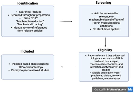
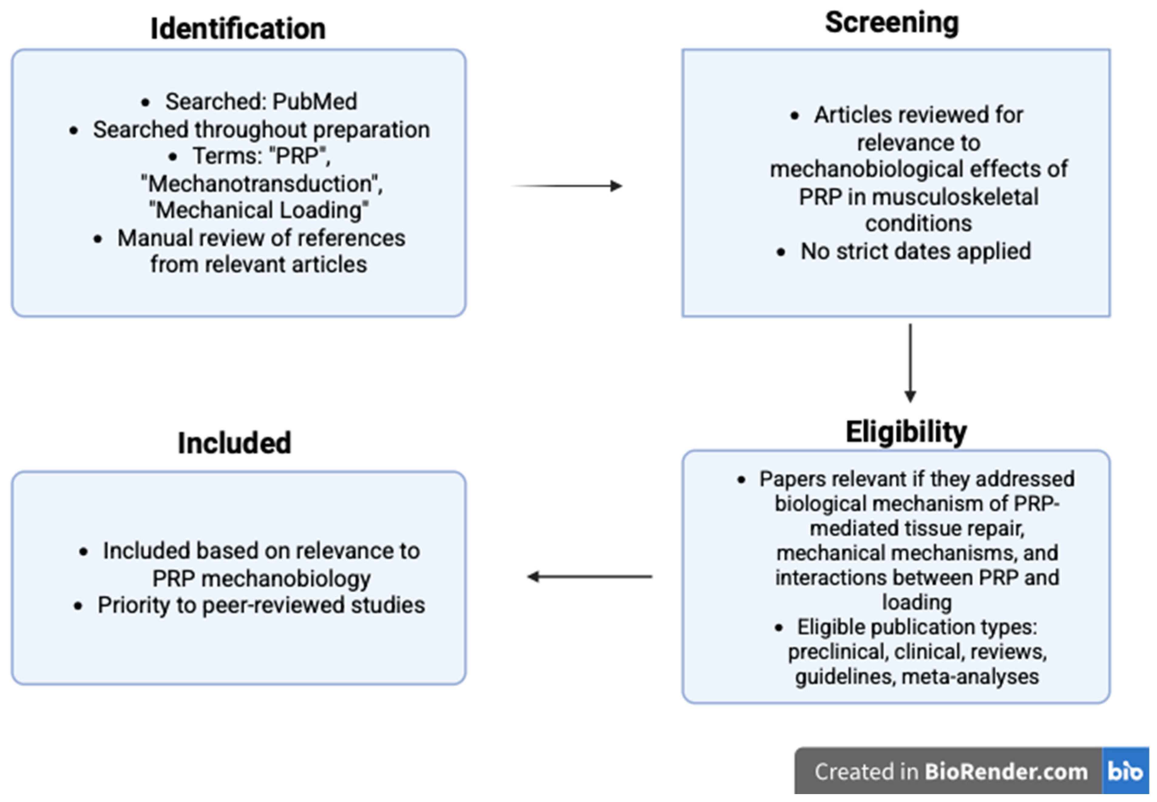
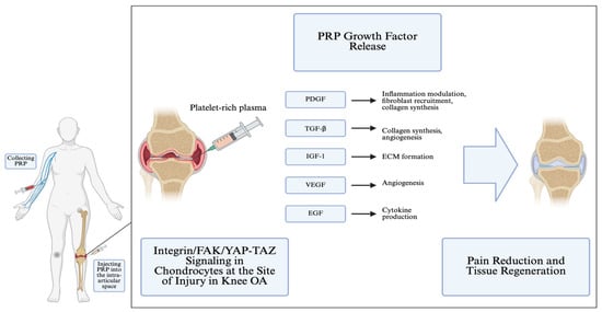
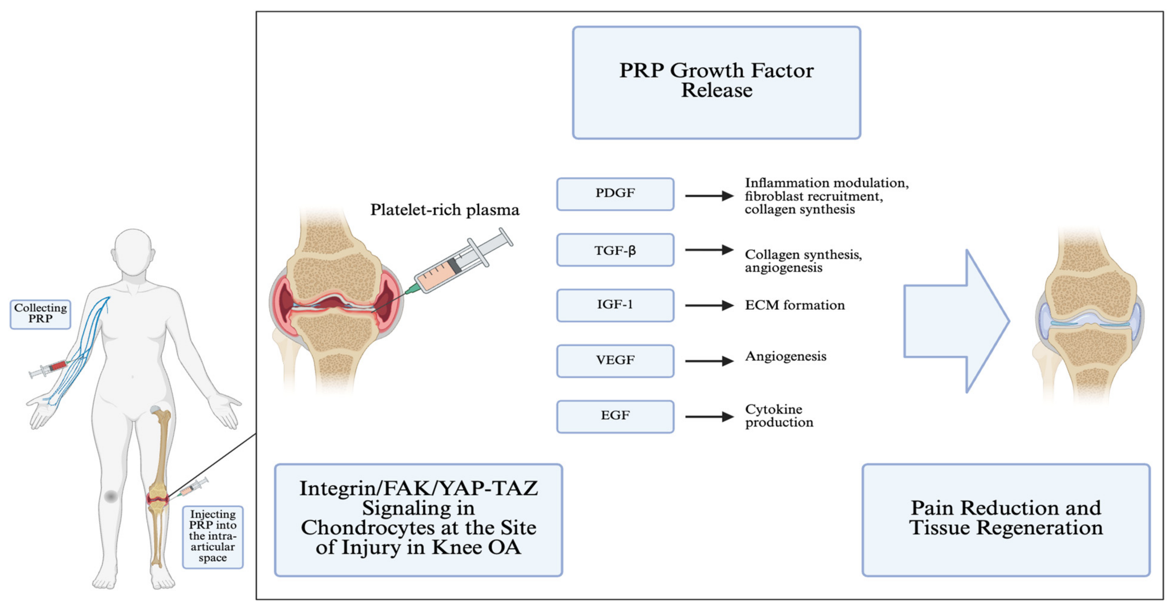

Disclaimer/Publisher’s Note: The statements, opinions and data contained in all publications are solely those of the individual author(s) and contributor(s) and not of MDPI and/or the editor(s). MDPI and/or the editor(s) disclaim responsibility for any injury to people or property resulting from any ideas, methods, instructions or products referred to in the content. |
© 2026 by the authors. Licensee MDPI, Basel, Switzerland. This article is an open access article distributed under the terms and conditions of the Creative Commons Attribution (CC BY) license.