


Abstract
Metal additive manufacturing (AM) facilitates the production of geometrically complex components, yet its broader industrial use remains limited by the risk of defect formation and uncertainties in their detection, originating from the highly dynamic and high-temperature process environment. To make additive manufacturing more reliable and establish high-quality parts, it is important to understand how these defects form and how their characteristics appear during the process. This review explains the main causes of common defects, such as cracking, porosity, lack of fusion, and inclusions in metal AM processes, including Powder Bed Fusion and Directed Energy Deposition. It also connects main defect formation mechanisms to the optical, thermal, acoustic, and spectroscopic signals that can be measured during the process. Moreover, it is described how commonly used in situ monitoring systems work and how their signals correspond to melt pool dynamics, vapor plume, particle movement, and the solidification process for each kind of defect. An overview is provided of how data from these systems are analyzed, including the extraction of features from images, the evaluation of temperature fields, and the use of time and frequency domain techniques for various signals. By linking the physics of defect formation to measurable process signals, the interpretation of sensor data is enabled, and potential strategies for monitoring specific problems are outlined. Finally, recent developments are examined, including the integration of multiple sensors, advanced feature-representation approaches, and real-time data interpretation coupled with adaptive control. Together, these directions represent promising advances towards more intelligent and reliable monitoring systems for the future of metal AM.
1. Introduction
Additive Manufacturing (AM) has advanced the fabrication of metallic components by allowing the production of intricate geometries with high precision and reduced material waste (e.g., [1]). Nevertheless, maintaining process stability and achieving components without defects continues to be a significant challenge, limiting industrial adoption of metal AM technologies [2,3,4,5]. Metal AM processes can be broadly categorized into two major groups: Powder Bed Fusion (PBF) and Directed Energy Deposition (DED). In the PBF process, a thin layer of metal powder is first spread across the build platform and then selectively melted using a focused energy source, typically a laser in Laser Powder Bed Fusion (PBF-LB/M) or an electron beam in Electron Beam Powder Bed Fusion (PBF-EB/M) to form each part layer by layer [1,6,7]. In contrast, DED processes feed metallic material directly into a melt pool generated by a concentrated heat source. Depending on the configuration, the feedstock can be powder or wire, while the energy input is commonly provided by a laser (DED-LB/M) or an electric arc (DED-arc/M) [8,9,10]. Despite these differences, the processes show similar defect formation mechanisms related to various physical phenomena, including melt pool instability, thermal fluctuations, rapid solidification, and interactions between the molten material and the surrounding atmosphere [11,12,13]. The layer-by-layer nature and rapid thermal cycling in these processes cause strong microstructural and morphological instabilities, which often cause the formation of porosity [14], lack of fusion (LoF) [15], cracking [16], and inclusions [17]. These defects influence mechanical strength, fatigue resistance, and corrosion performance, often preventing the qualification of AM parts for industries [18]. Therefore, the ability to detect and quantify these defects is essential for understanding their origin and developing strategies to mitigate them. Traditional ex situ inspection techniques, including X-ray computed tomography (CT), optical microscopy (OM), and ultrasonic testing, offer detailed post-process characterization of defects but are time-consuming, costly, and incapable of real-time monitoring of the process [19,20,21].
To achieve in situ defect detection and enable closed-loop process control, researchers have developed in situ monitoring systems capable of recording transient process signatures during fabrication (e.g., [22]). Various sensors have been used, including optical emission spectroscopy (OES), to monitor plasma and vapor plume characteristics [23,24], infrared (IR) thermography and pyrometry for melt pool temperature mapping [25,26], high-speed and coaxial vision systems for geometric monitoring [27,28], and acoustic emission sensors for elastic wave detection related to cracking or keyhole formation [29,30]. The signals of various sensors can be used to extract measurable features such as melt pool geometry, spectral intensity, temperature gradients, and acoustic frequency that can be correlated with the physical mechanisms of defect formation in different metal AM processes [31]. Moreover, integrating in situ sensor data with machine learning (ML) techniques has created new opportunities to interpret the extensive datasets generated during metal AM. By identifying complex relationships across various signals, ML models identify relationships between signals and defect mechanisms, classify defect types, and help to develop closed-loop process control strategies [32,33].
Several researchers have addressed in situ monitoring techniques in metal AM. For instance, Montazeri et al. [34] developed an in situ porosity monitoring method for PBF-LB/M of Inconel 718 using a multispectral optical emission sensor combined with graph theoretic signal analysis. Optical emission data were continuously recorded during fabrication and correlated with porosity levels measured by X-ray CT. By applying the graph Fourier transform to the spectral signals, they found that high-frequency graph Fourier transform coefficients and localized intensity fluctuations increased systematically in layers with porosity, whereas stable layers showed stronger correlations across emission channels. These findings demonstrate that specific graph-spectral features of the emission signal are directly linked to pore formation.
Similarly, in the DED process, Herzog et al. [35] developed a multi-axis IR monitoring system equipped with three synchronized cameras to track melt pool geometry alongside layer-wise vertical displacement during deposition. By combining data from complementary viewing angles, their image processing framework detected surface irregularities such as depressions that preceded the formation of internal defects at thin wall intersections. These depressions were shown to originate from reduced laser spot overlap caused by automatic fileting in the tool path, and micro-CT confirmed that they were the direct precursor to porosity formation. Through experiments on an intersecting wall test geometry, they linked porosity observed at narrow angles (approximately 30°) to reduced laser spot overlap resulting from automatic toolpath fileting. Micro-CT validation confirmed the defect locations, and subsequent removal of the filets eliminated the pores, consequently establishing a direct relationship between toolpath design, local irradiance distribution, and defect formation. Additionally, Kim et al. [36] investigated acoustic emission (AE) monitoring for detecting microcrack formation during DED-LB/M of SiC-reinforced stainless steel 316L composites. They recorded and analyzed sound signals in the time, frequency, and time–frequency domains, showing that in-process cracking produces a unique audible “ping” between 12 and 16 kHz. Using signal processing techniques such as high-pass filtering and Harmonic Percussive Source Separation (HPSS), they successfully isolated both post-process and hidden in-process cracking signatures. They also demonstrated that acoustic monitoring provides a simple and effective in situ method for identifying crack formation during rapid cooling, improving in situ quality control during DED-LB/M process.
While in situ monitoring and data analysis methods for metal AM have advanced rapidly, most studies still address either sensor development or data modeling separately, without clearly linking the measured signals to the physical processes that cause defect formation. As a result, there is still no unified understanding that connects defect mechanisms, their physical origins, signal features, and sensor performance among different metal AM techniques. Thus, this review paper addresses the gap by examining in situ monitoring techniques in DED and PBF. Then, it analyzes common defect types, including cracking, porosity, LoF, and inclusions, with the physical reasons that lead to their formation and the corresponding optical, thermal, and acoustic responses measured by different sensors. In doing so, the review links various defect formation physics to signal interpretation, introducing a basis for selecting suitable monitoring methods for each defect type in metal AM.
2. Defects in Metal Additive Manufacturing
Defect formation is a key challenge in metal AM, as it reduces the structural integrity, mechanical performance, and overall reliability of fabricated components. Each AM process presents distinct defect types, influenced by differences in energy source, feedstock delivery method, processing atmosphere, and feedstock characteristics. Among these defect types, their dominance depends on the specific metal AM process. In PBF, defects such as porosity and inclusions may result from excessive vaporization, as well as from powder-dependent factors, including irregular or non-spherical particle morphology, broad particle-size distributions, poor flowability or inadequate packing density, and surface contamination or oxidation [37,38]. In contrast, DED processes are more likely to form LoF and cracking due to non-uniform heat input, high thermal gradients, and the development of residual stresses [39].
To mitigate these dominant defects, process parameter optimization plays a crucial role. In PBF, adjusting laser power, scan speed, hatch spacing, and improving powder quality can significantly reduce porosity formation [40]. In DED, controlling heat input, implementing interlayer temperature control, substrate preheating, and reducing thermal gradients are effective strategies to minimize LoF and cracking [41,42].
Despite differences, the main mechanisms, for instance, uneven energy distribution, melt pool instability, and complex solidification dynamics, are common in metal AM processes [43]. These conditions often lead to several issues during processing, such as defects and geometrical imperfections [44]. Defects in metal AM can be categorized into four principal groups: cracking, porosity, LoF, and inclusions. Cracking occurs due to high thermal gradients, solidification shrinkage, or accumulation of residual stresses that exceed the local ductility of the alloy, leading to fracture paths [45]. Process-induced porosity is caused by entrapped gas, unstable keyhole collapse, or inadequate wetting, resulting in voids that reduce density and fatigue life [46,47]. LoF defects occur when insufficient energy input or improper overlap between melt tracks prevents complete bonding between adjacent layers, often leaving unmolten regions or planar voids at layer interfaces [15]. Inclusions form when oxides, unmelted particles, or contaminants become trapped in the melt pool, creating localized chemical or mechanical discontinuities that can be identified as stress concentrators [17]. Each of these defect types originates from distinct physical mechanisms, demonstrates characteristic morphologies, and can potentially generate measurable process signatures that can be collected using appropriate sensing technologies.
On the other hand, geometrical imperfections also play an important role in determining the overall quality of metal AM components. Features such as surface waviness, structure warping, high surface roughness, and edge defects represent geometric inaccuracies relative to the nominal shape that can originate from melt pool instability [39], uneven heat distribution, residual stress accumulation [39], or non-uniform melt pool behavior, and deposition parameters leading to poor surface finish and porosity [39]. Although these imperfections occur at the macroscopic scale, they are often linked to process irregularities and can act as visible indicators of subsurface defects. However, their measurement alone does not improve dimensional accuracy, and characterization remains important for understanding the relationship between surface morphology and process stability [48,49].
Since most defects, such as cracks and porosity, originate from complex interactions between thermal and mechanical phenomena during fabrication, online monitoring techniques have become fundamental tools for monitoring these processes. Since conventional post-process methods like microscopy and CT are time-consuming and unsuitable for real-time use, in situ monitoring has become increasingly prominent due to its ability to detect the physical responses that emerge as defects develop during the process. Among the various sensing approaches, acoustic and thermal monitoring methods have proven particularly effective for identifying and predicting defect formation, as they can record physical signatures during the process [50,51,52]. The following sections examine the main defect types in metal AM, including cracking, LoF, porosity, and inclusions. Each subsection describes the physical mechanisms that lead to their formation, identifies the process signals that can be measured during the process, and reviews the sensing techniques most suitable for their detection. As shown in Figure 1, the framework is shown as a structured chain linking defects, signals, and sensors. The physical origin of a defect (input) generates measurable process signatures such as thermal, acoustic, or optical signals. These signals are captured by suitable in situ sensors and transformed into extractable features for defect detection (output). The relationship between defect types, their physical causes, and the corresponding monitoring sensors forms a layered framework linking process physics to in situ monitoring. Although both defects and geometrical imperfections are considered, the present review focuses specifically on defects, which are analyzed in detail to clarify their origin and detection from sensor data. By investigating these relationships in metal AM processes, this study establishes an integrated comparative perspective on defect formation, recognition through sensor feedback, and the use of suitable sensors for specific defect types in metal AM.
Figure 1.
Layered framework connecting defects and geometrical imperfections in metal AM with their physical causes and suitable monitoring sensors, representing the link between process physics and in situ monitoring [53].
3. Signal Processing and Feature Extraction
In situ monitoring links the physics of defect formation to measurable process signals that can be interpreted in real time. In metal AM, in situ sensing is typically based on vision-based measurements that monitor melt pool geometry, thermal emission, and spatter dynamics [54]; and signals such as acoustic emission and ultrasound that record elastic or airborne waves generated by rapid process transients [55]. Among different sensing methods, a consistent data processing workflow can be identified. Firstly, denoising and segmenting the raw signals, secondly, extracting a representative set of engineered features, and finally, analyzing features to detect defects or classify process states. In vision-based monitoring, the extracted features most often describe melt pool morphology, including its length, width, area, and aspect ratio, together with intensity or temperature-related statistics when radiometric cameras or pyrometers are employed. Moreover, morphological descriptors alone provide valuable information on process stability; however, combining geometric attributes with intensity or temperature fields better differentiates process variations and potential defect formation. In PBF processes, melt pool instability plays a key role in defect formation, including porosity and LoF. Spatter dynamics, plume oscillations, temperature variations, and gas flow patterns provide measurable features that reflect these instabilities [56,57,58]. As an example, Bidare et al. [58] combined high-speed imaging, Schlieren visualization, and multi-physics modeling to analyze the interactions between the laser, powder, and surrounding gas, revealing how particle motion, denudation, and agglomeration are influenced by these phenomena. Figure 2a shows high-speed side and top images of single-track scans, highlighting spatter trajectories, plume oscillations, and particle ejection. Figure 2b presents calculated temperature fields, revealing localized gradients that indicate transient melt pool instability. Figure 2c illustrates Schlieren visualization of heated Argon gas flow, showing convection-driven particle motion and drag forces that affect powder redistribution. From these observations, high spatter counts and larger particles correlate with increased porosity risk, plume oscillations and emission fluctuations reflect transient melt pool instability, and gas flow-induced particle motion is associated with potential LoF regions.
Figure 2.
Melt pool behavior during single track scans at 50 W/0.1 m/s, 100 W/0.5 m/s, and 200 W/1 m/s. (a) High-speed images showing melt pool shape, plume direction, and spatter trajectories, (b) Calculated temperature fields highlighting localized gradients and melt pool instability, (c) Schlieren visualization of heated Argon gas illustrating convection-driven particle motion and drag forces [58].
On the other hand, time-series sensing has seen rapid growth, with piezoelectric or optical microphones placed on the build or in the chamber to record transient phenomena that resulted in LoF, or crack initiation. For time-series data, features can be organized by representation domain (see Table 1). Time domain statistics (e.g., mean, Root Mean Square (RMS), variance, rise time, and counts) are computationally suitable for online defect detection. Frequency domain data (e.g., spectral centroid, Spectral Skewness) measure characteristics based on various mechanisms. Time–frequency analysis methods, such as the Short Time Fourier Transform (STFT) and Continuous Wavelet Transform (CWT), capture how the spectral content of a signal evolves, making them effective for identifying rapid events or overlapping events that arise during AM processing. In parallel, cepstral representations, notably Mel Frequency Cepstral Coefficients (MFCCs), give a compact characterization of the signal’s spectral shape and are frequently used as stable inputs for ML models. Therefore, sensor-based feature extraction in metal AM can be broadly categorized according to the analysis domain. Time domain features are widely applied for real-time detection of amplitude-based anomalies such as spatter bursts or crack initiation, owing to their simplicity and low computational cost [59,60]. Frequency domain features, often obtained through Fast Fourier Transform (FFT) or power-spectral-density analysis, enable defect differentiation by capturing frequency-dependent signatures associated with keyhole collapse, porosity, or process instabilities [26,43,61]. Meanwhile, time–frequency domain methods such as STFT or CWT preserve both temporal and spectral information, allowing detection of transient or overlapping events, for instance, simultaneous spatter and crack propagation [62,63].
Table 1.
AE signal analysis domains in metal AM.
While the general signal processing workflow remains consistent across sensing technologies, the selection of analysis methods depends strongly on the defect type. For instance, crack detection often relies on time domain amplitude thresholds and time–frequency analysis to capture rapid acoustic transients associated with crack initiation and propagation [69]. In contrast, porosity and LoF are more frequently identified through frequency domain signatures, melt pool geometry fluctuations, and intensity-based image features that reflect instability in heat transfer and melt dynamics [69]. Compositional anomalies, such as inclusions, are typically analyzed using spectral peak ratios, emissivity variations, or multivariate statistical approaches applied to optical or hyperspectral data [70].
Thus, to extract relevant information from signals, the measured temporal, spectral, or geometric features must be interpreted with respect to the physical mechanisms that control defect formation. Accordingly, the following sections review each major defect type. For each defect, the underlying physical drivers are outlined and related to the characteristic process signals they generate, such as optical, thermal, acoustic, or spectral data. Sensing approaches and feature indicators identified in the literature as most effective for detection are summarized. By linking signal interpretation to defect formation physics, this structure provides a coherent foundation for comparing monitoring strategies across different metal AM processes.
4. Cracking
Cracking in metal AM originates from thermal gradients, alloy solidification behavior, local strain accommodation, and residual stress, and it limits industrial use and reduces structural integrity [1]. Crack occurrence strongly depends on the material being deposited [71]. Metals and alloys that are prone to cracking during fusion welding typically display similar susceptibility in AM, due to the similar thermal gradients and solidification stresses [72]. These cracks can form both during and after deposition, either on the surface or within internal layers of the fabricated structure. In general, cracks are classified into two main categories: hot cracking and cold cracking.
4.1. Hot Cracking Mechanisms
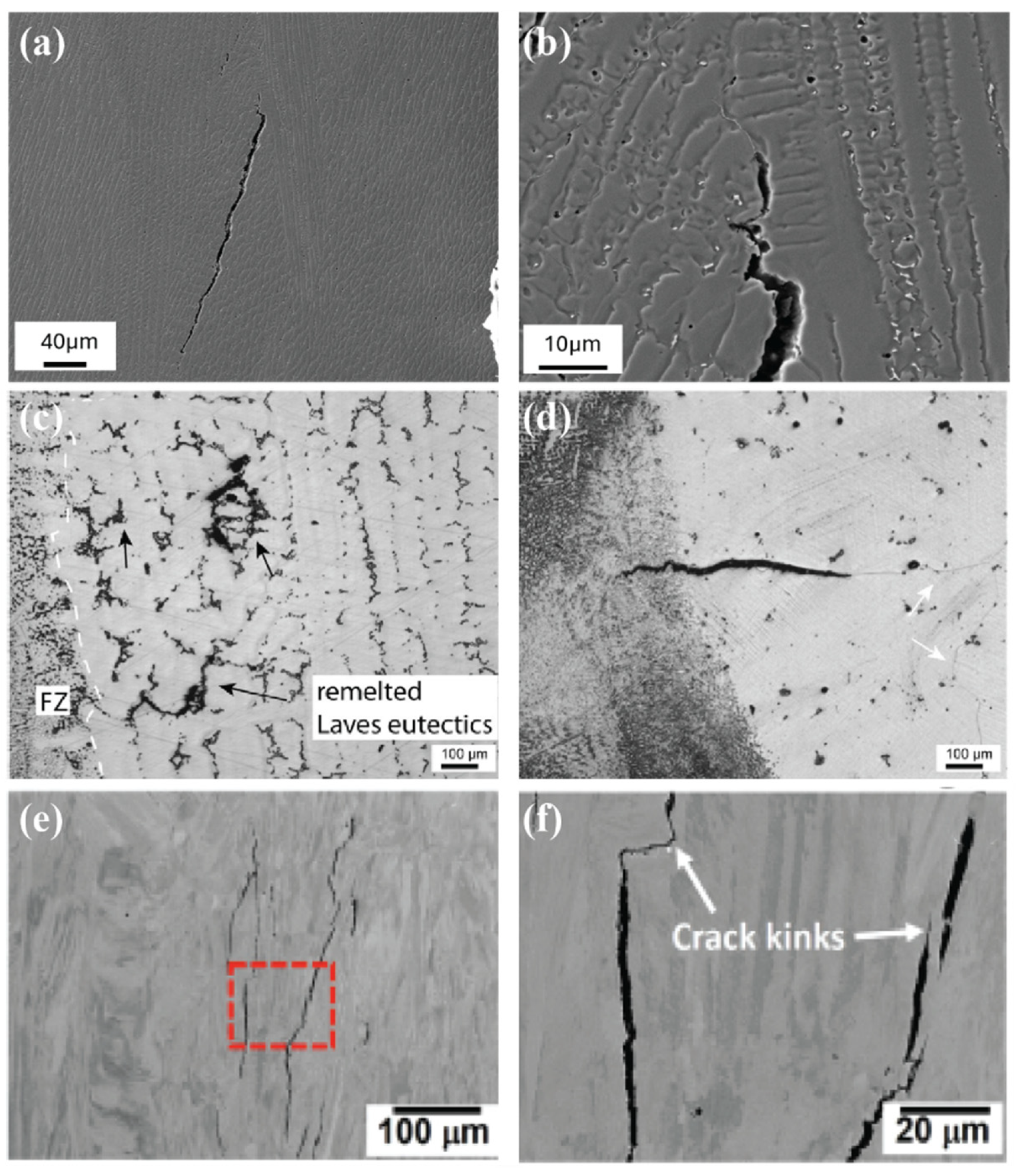
In metal AM, hot cracking occurs during solidification and includes both solidification cracking and liquation cracking [73,74,75]. Solidification cracks develop in the fusion zone of the deposit during the final stages of solidification [11], as illustrated in Figure 3a,b. These cracks typically propagate along grain boundaries and are often linked to low-melting-point constituents that form liquid films between dendrites [74,75]. Solidification cracking is controlled by the intricate interaction between thermal fields and material properties within the highly sensitive mushy zone. The operative factors integrate into four coupled thermal-metallurgical mechanisms. First, the brittle temperature range, which indicates the duration and extent of material brittleness, is fundamentally controlled by the metallurgical phenomenon of impurity segregation, which depresses the zero-ductility temperature. Second, the permeability of the interdendritic mushy zone addresses the critical role of the residual liquid in effectively feeding the thermally induced shrinkage gaps. This backfeeding is hindered by rapid solidification and restricted by the complexity of the developing dendritic region. Third, the solidification microstructure morphology, influenced by the thermal gradient, directly affects the cracking path. Columnar growth creates continuous, weak liquid films along grain boundaries, while a refined, equiaxed structure enhances crack resistance. Finally, the interfacial energy balance indicates the stability of these molten layers. Collectively, these four interactions define whether the ductility of the mushy zone can accommodate the applied contraction strains during the final stages of solidification [76].
Liquation cracking occurs in the heat-affected zone (HAZ) or the partially melted zone (PMZ) of previously deposited layers or the base material (Figure 3c,d). It forms when low-melting eutectic constituents or grain boundary precipitates undergo partial melting during rapid heating, while the surrounding material remains below the liquidus temperature, often as a result of segregation effects [73,74,77]. The combination of this localized melting with tensile stresses induced by solidification and thermal shrinkage during cooling promotes crack formation. Once initiated, these liquation cracks remain trapped in the HAZ and may propagate as subsequent layers are deposited [45,78]. Alloys with a wide solidus-liquidus temperature range (e.g., Ni-based superalloys) [45,79], high solidification shrinkage (e.g., Ti–6Al–4V) [80], or significant thermal shrinkage (e.g., Al-based alloys) [81] are particularly susceptible to liquation cracking.
4.2. Cold Cracking Mechanism
Cold cracking, depicted in Figure 3e,f, develops as the material cools toward room temperature and is mainly shaped by residual stresses generated during the rapid thermal cycles inherent to the AM process [77,82]. These stresses arise from repeated heating and cooling, which induce thermal expansion and contraction, leading to the accumulation of internal stress within the deposited layers. If these stresses are not adequately relieved during cooling, they can persist in the final structure, creating conditions favorable for crack formation [45,82]. Additionally, cold cracks typically initiate at points of stress concentration, such as microstructural inhomogeneities or pre-existing defects, geometrical features, and propagate through regions with microstructures, such as coarse grains or brittle phases [83]. The presence of cracks, whether hot or cold, significantly diminishes the wear, corrosion resistance, and fatigue strength of AM fabricated structures, therefore negatively affecting their overall performance [84,85].
Figure 3.
Representative cracking mechanisms in Ni-based superalloys. (a,b) Solidification cracking in DED LB/M Hastelloy X along solidification grain boundaries with terminal solidification products, (c,d) Liquation cracking in the base metal HAZ after homogenization, propagating along interdendritic regions and former solidification grain boundaries with remelted Laves eutectics [78], (e,f) Solid-state (cold) cracking in CM247LC and IN939, with interlayer cracks crossing multiple melt pool layers [86].
4.3. Other Crack Formation Mechanisms
In addition to hot and cold cracking in metal AM, the weld metal’s HAZ may also experience other cracking mechanisms, such as ductility-dip cracking (DDC), post-weld heat treatment cracking (PWHT), strain-age cracking (SAC), lamellar cracking, and copper-contamination cracking (CCC) [75,79,87,88]. DDC is a solid-state, intergranular cracking mechanism that occurs in some face-centered cubic (FCC) alloys at elevated temperatures [89]. It occurs in alloys such as Ni-, Ti-, Cu-, Al-, and austenitic stainless steels due to a temporary reduction in ductility between Ts and ~0.5Ts. It propagates intergranularly along thermally modified high-angle grain boundaries and is promoted by boundary precipitates, alloy composition, and strain conditions [90]. PWHT cracking or stress relief treatments, especially in low-alloy steels containing strong carbide formers (Cr, Mo, V). Proper control of composition, residual stress, and thermal treatment can mitigate this issue [91]. SAC develops in the HAZ of precipitation-strengthened Ni-based alloys when aging reactions occur simultaneously with local straining. Age-hardening kinetics play a major role in this type of cracking, with alloys containing a high γ′ volume fraction being more susceptible due to rapid precipitation-induced loss of ductility in the heat-affected zone. In contrast, Inconel 718 exhibits lower cracking susceptibility owing to its slower γ′ precipitation kinetics [90].
4.4. Emissions from Crack Formation and Propagation
Detecting and understanding process-induced defects in metal AM is critical for the structural integrity and performance of fabricated components. Acoustic sensing detects the elastic wave emissions characteristic of crack initiation and propagation [84], whereas thermal sensing records the temperature fluctuations and gradients that occur before or during crack formation [92]. In combination, these sensors provide an integrated perspective on the mechanical and thermal mechanisms that drive cracking [69,93].
AE sensing relies on the detection of transient elastic waves generated when a material undergoes sudden stress release, such as during crack initiation, delamination, or spatter impact [94]. The rapid thermal cycles and localized solidification shrinkage generate residual stresses. When these stresses exceed the local material strength, crack initiation or interlayer separation occurs, releasing stored elastic energy as high-frequency stress waves [84]. These waves propagate through the substrate or deposited layers and are detected by piezoelectric AE sensors, which convert mechanical vibrations into electrical signals. The recorded waveforms contain information about the defect type, intensity, and spatial location, enabling non-contact, real-time monitoring of process stability [95,96]. Different defect mechanisms produce distinct acoustic signatures depending on the amount of energy released. Crack-related signals generally exhibit short durations, sharp amplitude peaks, and high energy values, while other process noises, such as melt pool turbulence or particle impingement, tend to be more continuous and of lower amplitude [36,66,97].
4.5. Acoustic Sensors for Crack Detection
Various types of acoustic sensors have been used for crack detection, including optical microphones [24], ICP® (piezoelectric) microphones [26], directional microphones [66], and AE sensors [30]. With detection capabilities spanning from tens of kilohertz to nearly 1 MHz, AE sensors can capture a wide spectrum of crack signals. Rapid crack initiation is generally marked by high-frequency bursts (300–600 kHz and above) [24,30], whereas lower-frequency components tend to arise from slower crack growth or process instabilities [36,66,98]. These frequencies are consistent with the ranges reported for different defect mechanisms, as summarized in Table 2. On the other hand, Signal processing techniques such as STFT, CWT, and time domain analysis allow the identification of characteristic peaks in acoustic energy that correspond to cracking [36,66].
Table 2.
Characteristic acoustic emission frequency ranges and peak frequencies for various cracking mechanisms.
4.5.1. ML-Supported Signal Analysis
Recent advancements integrate ML to enhance crack detection accuracy. Time, frequency, and time–frequency features extracted from raw AE signals can be used to train algorithms such as K-means clustering, Support Vector Machines (SVM), Random Forests (RF), and neural networks [29,30,107]. These models can distinguish between cracks and other defect types like porosity, achieving detection accuracies up to 94% [107]. Combining ML with advanced denoising techniques, such as HPSS and bandpass filtering, more effectively distinguishes crack-generated emissions from process-induced noise [67,68]. Thus, acoustic sensors are used as a tool for monitoring crack formation in AM. When their signals are collected across different frequency ranges and processed with established analysis and classification methods, it becomes possible to detect cracks as they develop, distinguish them from other process characteristics, and identify their location in real time. This supports timely control of defects and strengthens the reliability of the final part [89,98,100].
4.5.2. Acoustic Crack Characterization Examples
As an example, Kim et al. [36] developed an acoustic monitoring approach to identify micro-crack formation during DED-LB/M of SiC particle reinforced stainless steel 316L composites. By analyzing the acoustic signals recorded during the process, they characterized cracking in the time, frequency, and time–frequency domains. The study revealed that cracking produces a distinct audible “ping” sound typically occurring within the 12–16 kHz frequency range, corresponding to micro-crack formation during the rapid cooling. Using high-pass filtering and HPSS, they successfully removed background process noise and revealed hidden in-process cracking signals. OM images also confirmed that samples with cracks showed voids and surface defects, while crack-free samples showed smooth surfaces. The spectrograms for cracked samples displayed clear post DED-LB/M cracking signals. These findings indicate that in-process and post-process cracking signals share similar frequency characteristics, allowing crack detection even when the signals are partially hidden.
Moreover, García de la Yedra et al. [24] developed an online defect detection method for laser-based AM using AE and thermal monitoring. Their study used three in-process measurement systems, including an optical microphone, a laser vibrometer, and a thermal camera to monitor cracking and delamination during the process. By adjusting process parameters and substrate conditions, they intentionally activated two distinct cracking mechanisms, including layer delamination and HAZ cracking. They correlated AE data sets with liquid-penetrant and microscopic analyses for different samples, including small HAZ cracking and crack-free specimens. Weber et al. [66] investigated preheated low-carbon substrates, which produced high-acoustic-energy peaks corresponding to layer cracking, whereas preheated medium-carbon alloys showed single, lower-energy peaks corresponding to HAZ cracking. Samples without cracking showed a background level AE energy of almost 0.03. These acoustic results matched the results of conventional inspections, confirming the reliability of AE monitoring for real-time defect detection. They also compared two signal processing approaches, which are STFT and CWT, to analyze AE data collected during deposition. While CWT provided higher resolution and more detailed information, STFT identified all critical transient events corresponding to delamination with less computational effort. The spectrograms of two experiments indicate AE signals analyzed by CWT and STFT, and both approaches successfully detected eight transient events per experiment, demonstrating that STFT can be effectively used for real-time monitoring with lower computational cost. In another study, Gaja et al. [30] developed a real-time defect detection and classification system using AE monitoring combined with unsupervised K-means clustering and Principal Component Analysis (PCA). AE signals were collected during the process in an oxidized environment to stimulate different defect mechanisms. The analysis successfully distinguished two main defect types, including cracking, which are characterized by short duration, high amplitude, and high signal energy, and porosities, with shorter decay time, lower amplitude, and moderate energy. Signal energy was identified as the most important feature for classification, while frequency had little effect. Validation with OM confirmed a strong correlation between AE clusters and actual defects. They demonstrated that unsupervised AE analysis can reliably detect and classify defects in real-time, providing an effective tool for monitoring AM.
4.6. Thermal Monitoring for Crack Detection
Acoustic monitoring captures the elastic waves generated as cracks form, while thermal sensing provides additional context by observing the temperature evolution and gradients that create crack-prone conditions. Thermal sensing records melt pool temperature fields and their evolution during the process. Variations in melt pool size, shape, and cooling rate reflect how heat is distributed, helping to identify thermal gradients and indicate where residual stresses are likely to develop and potentially trigger crack initiation and growth. High thermal gradients, rapid cooling, or localized overheating often correspond to regions where cracking occurs [59,94]. As an example for thermal sensing, Mazzarisi et al. [92] conducted a study that examined the full thermal field during multitrack DED-LB/M. Their analysis revealed that cracking predominantly occurred in regions with elevated temperatures and high thermal gradients, highlighting that excessive thermal fluctuations play a significant role in promoting crack initiation. Similarly, Wu et al. [25] observed distinct changes in peak temperature when the laser beam passed over simulated cracking in DED-LB/M. These temperature variations proved to be reliable indicators of cracking, showing that shifts in the thermal field can be linked to discontinuities within the material.
4.7. Sensors for Crack Detection
Overall, the studies discussed above show that acoustic and thermal sensing each capture aspects of crack development in metal AM. Their effectiveness, however, depends not only on the physical quantity being measured but also on factors such as sensor bandwidth, sampling rate, and the specific features extracted from the recorded signal. Recent research has therefore moved toward using multiple sensing modes together and applying more refined analysis techniques, including time–frequency signal representations, statistical feature sets, and data-driven classifiers, to strengthen the reliability of crack detection. To show how these approaches are being implemented across different metal AM processes, Table 3 provides an overview of the sensor types, sampling rates, extracted features, and analysis methods reported in the literature for detecting cracks in metal AM.
Table 3.
Sensor types and analysis methods for crack detection in metal AM.
5. Porosity
5.1. Origin and Appearance of Process Pores
Defects such as porosity also play a critical role in determining the final quality of AM parts. Unlike cracks, which exhibit strong acoustic and thermal characteristics, porosity often forms gradually through irregular melting or gas entrapment. Therefore, its detection requires different sensing strategies, often involving combinations of thermal imaging, optical sensing, or AE analysis calibrated to detect this type of defect [22]. Porosity is among the most detrimental defects in AM-fabricated materials that reduce density, mechanical strength, and overall structural reliability. It can appear in various forms, including gas porosity, keyhole collapse, and LoF [111,112], which can be observed across various alloy systems such as titanium [113,114,115], nickel-based superalloys [116,117], and aluminum alloys [118]. LoF can also lead to pore formation [119]. This section focuses specifically on gas porosity, while LoF defects are discussed separately in Section 6. Moreover, pore size, shape, and distribution are primarily controlled by the amount of gas present in the melt pool and by the melt flow dynamics that entrap this gas. Local thermal conditions and specific process parameters indirectly shape these mechanisms by altering melt pool stability [120]. Such voids act as stress concentrators, promoting crack initiation and propagation, and ultimately decreasing fatigue life, ductility, and corrosion resistance. The formation and severity of porosity are highly sensitive to process parameters such as laser power, scan speed, and powder feed rate, which collectively determine the heat input and melt pool dynamics [121,122,123,124].
During deposition, gas porosity forms when gas becomes trapped in the melt pool, most commonly as a result of insufficient melting, shielding gas entrapment, or instabilities that occur during solidification. Possible sources include residual gases in the powder feedstock, entrapped shielding gas during powder delivery [118], and gaseous species such as hydrogen, which exhibit limited solubility in solid metal compared to the liquid phase [125]. In addition to gas pores, metal AM can also form metallurgical or shrinkage pores, which originate from inadequate feeding of the solidifying melt and result from a different mechanism than gas entrapment [126,127]. As the melt pool solidifies, these gases may become trapped within the material, forming spherical or irregular voids depending on cooling rate and melt flow conditions. The melt pool hydrodynamics, particularly the convection flow and Marangoni-driven circulation, play a crucial role in determining whether the bubbles escape, coalesce, or remain entrapped within the solidifying metal [115,116,128,129]. Moreover, differences in gas solubility, particularly hydrogen, between the liquid and solid phases during solidification promote the entrapment of gas pores. Gas porosity is commonly observed in PBF and DED components and can significantly decrease mechanical integrity and performance [119,123,130,131]. For instance, Park et al. [132] explored the mechanisms determining pore formation and enlargement during DED, highlighting the role of residual stress and alloy composition. They found that when the buffer layer contained cracks, the associated stress relief prevented the development of large pores in the surface layer. However, when the buffer layer was crack-free, tensile residual stresses accumulated and were transmitted into the surface layer, increasing the internal pressure of trapped gas and causing pores to expand. Their results indicate that the evolution of residual stress across layers strongly influences both the formation and subsequent growth of gas pores during DED.
Although keyhole instability is a major pore-forming mechanism in PBF, it is less dominant in DED, which typically operates in conduction mode, characterized by a larger melt pool and lower energy density [133,134,135]. These defects usually appear as spherical voids concentrated near the bottom of the melt pool. Although both gas and keyhole pores exhibit roughly spherical morphologies, gas pores tend to have smoother surfaces, whereas keyhole pores display irregular, uneven contours [136].
5.2. Detection Possibilities of Process Pores
Since porosity formation is closely linked to melt pool stability, thermal gradients, and gas behavior, effective in situ detection relies on sensors that can resolve process instabilities. Irregular melting, keyhole oscillations, and gas entrapment modify the melt pool shape and apparent brightness; such changes in melt pool width, depth, and luminous intensity have been recorded by high-speed imaging and shown to match the pores in AM [137]. Moreover, high thermal gradients control melt pool depth, keyhole stability, and solidification time, which can control whether gas bubbles escape or become trapped. IR cameras and pyrometers can detect surface temperature fields, thermal gradients, and cooling rate fluctuations [138,139]. In addition, acoustic process monitoring can sense pore formation. Keyhole and melt pool instabilities that lead to keyhole pores generate characteristic changes in the acoustic spectrum; by analyzing features such as the power spectral density of airborne or structure-borne sound, recent work has uncovered acoustic signatures that can be used to predict the location of keyhole pores with high accuracy during PBF-LB/M [61,140]. For example, Kouprianoff et al. [141] demonstrated that low-frequency, air-borne AE from 2 to20 kHz can reliably indicate balling and single track instabilities, linking characteristic amplitudes to the formation of porosity. On the other hand, Hamidi Nasab et al. [142] showed that AE features in the 10–40 kHz band show clear amplitude changes when the melt pool transitions between conduction, stable keyhole, and unstable keyhole regimes. Their operando synchronization of X-ray imaging and AE measurements confirms that AE can resolve melt pool dynamic shifts with a time resolution of 100 µs, enabling real-time detection of defect formation. A visual summary of the AE frequency ranges and various pore behaviors reported in these two studies is presented in Figure 4. It also illustrates the thermal implications of these defects, including localized overheating and reduced heat conductivity due to pore accumulation and melt pool instability.
Figure 4.
Combined representation of low-frequency AE reported in [141,142], together with the associated thermal effects such as reduced heat conductivity resulting from melt pool instabilities.
5.3. Sensors for Porosity Detection
Multi-physical responses form the foundation for modern sensor-based porosity detection strategies, enabling real-time monitoring and data-driven prediction of defect formation in metal AM processes. The key sensors and analysis methods used for porosity monitoring are summarized in Table 4. Several in situ monitoring systems have been implemented to identify the early indicators of gas and keyhole pore formation.
Table 4.
Various sensor types and analysis methods for porosity detection in metal AM.
5.4. Signal Processing for Porosity Detection
In optical and thermal porosity monitoring methods, most data-driven approaches follow a similar methodology. First, an in situ sensor, typically a coaxial or off-axis camera, pyrometer, or combined optical and thermal module, is integrated into the deposition system to record melt pool behavior in real time. Second, porosity data is obtained from ex situ characterization, such as metallography or X-ray CT, and image processing algorithms are used to extract quantitative pore metrics. Finally, these in situ signals and ex situ pore characteristics are linked to train ML models capable of mapping melt pool signatures to internal defect states. For instance, Zhang et al. [143] proposed an advanced deep learning-based approach for real-time porosity monitoring in AM. In their study, a coaxial high-speed camera was integrated with the laser processing head to continuously record melt pool images during direct laser deposition of sponge titanium powders. Before constructing the supervised deep learning model, each melt-pool image was paired with corresponding porosity attributes extracted from metallographic cross sections. To achieve this, a MATLAB-based image processing framework was developed to automatically quantify pore features from raw microscopic images. The algorithm included a sequence of thresholding, noise removal, and binary segmentation steps that isolated pore regions from the metallic matrix, thereby enabling reliable calculation of porosity parameters. These processed datasets, combined with the melt pool images, formed the input-output pairs used to train the predictive model. Using these comprehensive datasets, the authors developed a compact CNN capable of learning complex correlations between melt pool dynamics and internal pore formation. The model demonstrated strong predictive performance, achieving an accuracy of approximately 91% for porosity-occurrence detection and an RMS error near 1.3% for local volume-porosity estimation. Although its sensitivity decreased for sub 30 µm pores that produced minimal optical signatures, the results confirmed that coupling coaxial optical sensing with CNN-based data interpretation provides an effective route toward automated, non-destructive quality assurance in laser-based additive manufacturing.
Moreover, Taylor et al. [57] developed a high-speed, coaxial melt pool imaging system for PBF-LB/M that captures feature-rich data at high spatial resolution, enabling voxel-wise classification of local porosity levels using ML models trained on realistic porosity distributions (Figure 5a,b). Their approach achieved porosity detection sensitivity by correlating melt pool image features with statistically defined porosity thresholds. In contrast, Ren et al. [7] employed plasma emission spectroscopy for in situ porosity monitoring during DED (Figure 5c), coupling time and position-synchronized spectral features with CT-derived porosity labels to train a random forest classifier. Their results demonstrated that specific spectral intensities and statistical descriptors are strongly correlated with porosity formation, highlighting the potential of spectroscopic sensing for real-time quality assessment in laser-based AM. For keyhole detection, Wang et al. [164] investigated melt pool and keyhole dynamics PBF-LB/M by integrating synchrotron X-ray imaging with high-speed and high spatial resolution IR thermography. Using Ti-6Al-4V and 410 stainless steel spot melts, they quantitatively characterized melt pool geometry, keyhole behavior, vapor plume formation, and thermal evolution. By correlating IR-derived thermal features with X-ray-observed melt pool boundaries, as shown in Figure 5d, they demonstrated that IR imaging can predict keyhole and melt pool characteristics traditionally accessible only through synchrotron X-ray imaging, enabling practical pathways for real-time monitoring and control in AM.
Figure 5.
Examples of in situ sensing and data-driven porosity detection in metal AM. (a) Raw coaxial melt pool image and corresponding segmented image, with a 4 mm × 4 mm field of view, (b) Distributions and correlations between pore size and voxel-level porosity derived from melt pool imaging data for two PBF-LB/M builds, illustrating data characteristics used for model training [57], (c) Schematic of a DED system equipped with spectral optical monitoring, with comparison between overlaid binary X-ray CT porosity maps and plasma emission spectral feature contours for porosity identification [7], (d) High speed IR and synchrotron X-ray observations of PBF-LB/M melt pools immediately before laser shut off under different processing conditions [164].
Additionally, Shin et al. [26] developed a data-driven multi-defect diagnosis framework to enhance in situ quality monitoring in DED. Their experimental platform integrated a CCD camera and a pyrometer to simultaneously capture the melt pool’s geometric and thermal responses during deposition. A dedicated OpenCV-based image pre-processing algorithm was designed to isolate pore regions by eliminating bright noise zones, applying binarization, and performing sequential morphological operations to calculate the pore-area ratio from cross-sectional images. From the refined melt pool data, statistically significant geometric and thermal features were extracted and subsequently used to train several machine-learning classifiers, including KNN, SVM, and ANN. These individual binary classifiers were integrated into a unified multi-defect diagnosis framework capable of distinguishing normal deposition, melting-ball formation, low-porosity, and high-porosity conditions.
Overall, porosity formation in AM results from a combination of gas entrapment, incomplete fusion, and unstable melt pool behavior. The interplay between laser energy input, powder dynamics, and solidification kinetics determines the ultimate pore morphology, size, and distribution. Therefore, optimizing thermal and material process parameters is critical for minimizing porosity and enhancing the integrity of fabricated components.
6. Lack of Fusion
6.1. Lack of Fusion Formation
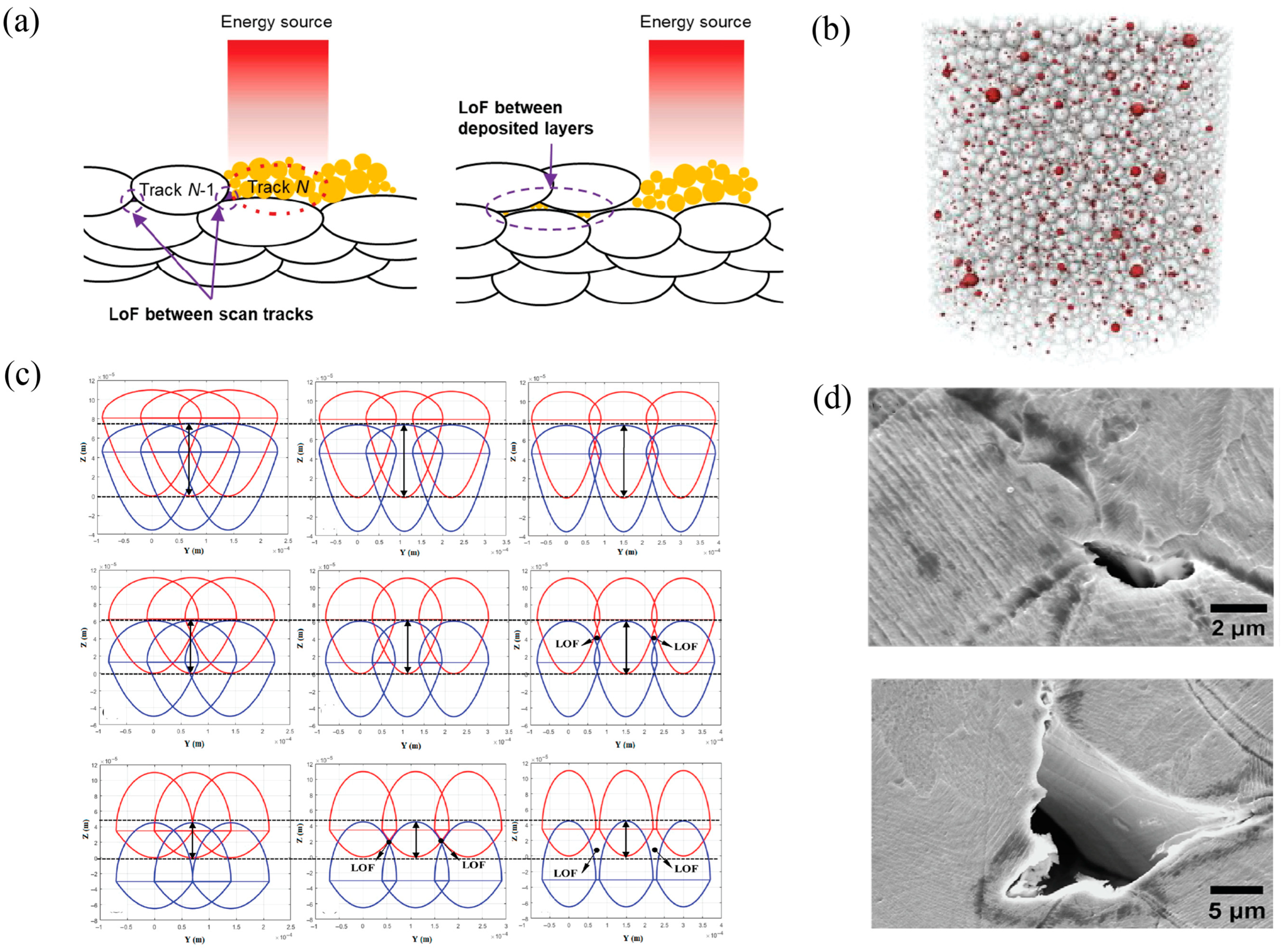
LoF, also referred to as incomplete fusion, is a discontinuity that arises when melting fails to occur either between the substrate and the deposited material or between adjacent deposited layers [165]. This defect is caused by insufficient energy input, typically due to low laser power or high scanning speed, which causes poor bonding between the melt pool and the substrate or previous layer or track [166] and an irregular interlayer morphology [167]. It can be formed based on two main reasons. The first reason is insufficient overlap between consecutive scan tracks, leaving regions of powder partially or completely unmelted (Figure 6a left). The second one occurs due to incomplete melting or bonding between successive layers in powder-based processes (Figure 6a right), resulting in weak interlayer adhesion and entrapped unmelted particles [168]. Characteristic indicators of LoF include unmelted powder particles embedded within the build, which contribute to a reduction in part density. Unmelted regions and LoF pores are often visible within fracture zones and are recognized as a key factor leading to part failure. LoF defects decrease interlayer integrity, promote structural instability, and act as stress concentrators, thereby diminishing the fatigue life of the additively manufactured component [122,169].
As an example, Siddique et al. [47] investigated that LoF is often associated with non-melted powder particles, which hinder proper bonding between layers. Similarly, Moalei et al. [170] attributed this defect to the incomplete melting of powder particles. Tan et al. [46], who processed AISI 316L stainless steel via DED, observed that incomplete fusion and gas entrapment due to moisture evaporation were the main origins of defects. The principal origins of defect formation, summarized in Figure 6b–d, reflect three dominant process factors. First, the initial gas content within the powder feedstock is a well-documented contributor to porosity in metal AM [171]. Second, variations in-process parameters strongly influence the melting regime, thereby altering both the type and the density of defects that develop within the part [7,172,173]. Third, geometric process parameters such as layer thickness and hatch spacing affect melt pool overlap and local heat accumulation, giving rise to distinct defect morphologies. Additionally, in Figure 6d SEM analysis of specimens reveals irregular LoF pores with sharp, non-spherical morphologies, indicative of insufficient local melting and incomplete consolidation during processing [78].
Figure 6.
Representative mechanisms contributing to LoF formation in metal AM, including: (a) LoF defects caused by insufficient track overlap, incomplete interlayer melting and weak metallurgical bonding [168]. (b) This defect originates from the initial gas content of the powder feedstock [171]. (c) Cross sections of two-layer, three-track melt pools produced at 250 W and 0.3 m/s for varying hatch distance and layer thickness [174]. (d) SEM microstructural analysis LoF, revealing irregular pore morphology associated with insufficient local melting and incomplete consolidation of powder particles [78].
6.2. Lack of Fusion Detection
Detection and monitoring of LoF can be effectively achieved using optical imaging, AE sensing, thermal imaging, and electron-based signal imaging techniques [22]. The formation of LoF defects during AM generates identifiable physical indicators that can be captured by in situ sensors monitoring the process environment. When the energy input is too low, unmelted or partially melted regions remain cooler than the surrounding material, which appears in IR thermography as abrupt temperature drops. Conversely, LoF also disrupts heat conduction to the underlying layers; the reduced heat flow path causes heat to accumulate in adjacent molten regions, creating locally elevated surface temperatures. These abrupt temperature variations represent the local discontinuities detectable in thermal maps. Optical sensors can reveal LoF by capturing melt pool indicators such as irregular bead width, discontinuous melt pool shapes, and a shallow or unstable melt pool boundary, all of which originate from insufficient energy absorption. Thermal imaging detects temperature signatures associated with LoF, including cooler unmolten regions where the energy input is too low to produce full melting, and, in some cases, locally hotter regions where missing fusion interrupts the heat flow path, causing heat to accumulate. These abrupt temperature variations represent the local discontinuities characteristic of incomplete fusion. Moreover, Inadequate bonding between layers also alters the way mechanical energy is released, generating lower intensity AE events that differ from the sharper transients linked to keyhole instability or cracking [175]. AE analysis can distinguish LoF by observing systematic reductions in signal amplitude together with a redistribution of spectral energy toward broader, lower frequency components. These changes reflect the weaker bonding and irregular melt pool dynamics that formed when fusion is incomplete [141]. Additionally, geometric sensors such as laser profilometers or coaxial height monitors can detect the subtle variations in track height or surface waviness that often appear alongside under-deposition and insufficient layer wetting [176,177]. Optical, thermal, acoustic, and geometric sensors can each indicate LoF defects, and combining their measurements and signals further enhances the reliability of real-time identification of insufficient fusion in the metal AM process. To make the link between these sensing behaviors and practical detection strategies clearer, Table 5 summarizes the main sensor modalities and analysis tools used for LoF identification in metal AMs. The table highlights how each technique captures different aspects of the process signals described in this section [178,179,180].
Table 5.
Various sensor types and analysis methods for LoF detection in metal AM.
6.3. Sensors for Lack of Fusion Detection
Multiple recent studies have examined how optical emission, acoustic sensing, and data-driven analysis can be used to classify melting behavior and detect LoF defects in DED and PBF. Their results highlight the distinct temporal, spectral, and energetic signatures that occur as the melt pool shifts from conduction mode into LoF in metal AM.
As an example, Wasmer et al. [23] integrated AE and OES with ML algorithms to monitor and classify process regimes in DED. The study explored four different chemical compositions under two sets of process parameters, identifying multiple processing modes, including conduction mode and regimes associated with varying levels of LoF defects. For feature extraction, raw AE signals were analyzed in both the time and frequency domains, while OES data were processed using spectral intensity peaks corresponding to characteristic emission lines. Seven ML models were implemented to classify both chemical composition and process regime. The results demonstrated that OES achieved markedly higher classification accuracies (78.25–96.38%) compared to AE (57.79–78.89%). The relatively lower AE accuracy was attributed to substantial noise within the raw acoustic signals, underscoring the importance of advanced signal-processing techniques to enhance AE-based defect classification in the complex DED-LB/M environment. Bottom of Form Moreover, Drissi-Daoudi et al. [185] conducted a comprehensive AE study to monitor defect formation during DED of three alloys: stainless steel 316L, bronze (CuSn8), and Inconel 718, with focus on LoF defects. Representative cross-sectional micrographs illustrated the distinct melt pool morphologies associated with LoF pores, conduction mode, and keyhole pores across the three materials. The raw AE signals showed a systematic increase in amplitude from conduction mode toward keyhole conditions, and LoF cases consistently occupied the lower end of this amplitude range due to their reduced melt-pool energy. Time domain feature analysis showed clear statistical separations between the conduction and keyhole regimes, with RMS amplitude distributions providing the strongest discriminative behavior. Within this range, LoF for stainless steel and Inconel clustered at moderate RMS levels, reflecting their lower melt pool energy compared to fully stable conduction or keyhole conditions. Bronze exhibited even lower RMS values, consistent with its lower near-IR absorptivity, which reduces the effective energy transferred into the melt pool under identical laser settings. Frequency domain analysis revealed dominant spectral peaks around 10 kHz and 40 kHz for all three alloys, corresponding to the resonant sensitivity range of the AE sensor and the elastic wave emissions generated by rapid thermal fluctuations, melt pool instabilities, and solidification. Time–frequency wavelet analysis further confirmed concentrated energy in these bands, with LoF in stainless steel and Inconel showing stronger activity near 10 kHz, while bronze displayed more pronounced responses near 40 kHz. These consistent temporal and spectral patterns show that LoF conditions produce characteristic AE signatures shaped by melt pool energy and material properties, supporting the use of AE sensing for real-time defect identification in metal AM.
7. Inclusions
7.1. Inclusions Appearance and Impact
Inclusions are non-metallic constituents distributed within the metallic matrix, typically showing a smooth or spherical morphology. The formation of these defects is primarily associated with compositional microsegregation involving deoxidizing and elements enhancing fluidity, such as Si, Al, and Mg, present in the feedstock material [186]. During processing, the highly localized and intense energy sources produce a high-temperature melt pool, creating conditions that favor the oxidation of these reactive alloying elements [187]. In AM processes, oxygen originating from the feedstock surface or the surrounding atmosphere can readily react with these elements, forming stable oxide particles that become entrapped within the solidifying matrix [188]. For example, at melt pool temperatures approaching 2000 K [189] stainless steel oxidizes readily even under low oxygen partial pressures. During large-scale fabrication or repair, maintaining such low oxygen levels is technically demanding, which increases the probability of oxidation under these thermal conditions. In addition, gas-atomized metal powders commonly used in AM already possess oxygen concentrations of at least 200 ppm [190], indicating that oxidation may begin during the material preparation stage. Therefore, oxygen dissolution and accumulation throughout the AM process are common and can significantly influence the final microstructure and properties of the fabricated part [191,192]. As an example, Figure 7 shows the TEM microstructure of the heat-treated AM samples, revealing a high number density of nanoscale particles with sizes ranging from approximately 20 to 100 nm. STEM–EDS analysis indicates that these inclusions are enriched in oxygen, manganese, and chromium, consistent with oxide phases. Their formation is attributed to oxygen pickup during the additive manufacturing process and subsequent thermal exposure, leading to the precipitation of stable nanoscale oxides within the matrix [78].
Figure 7.
TEM images of nanoscale inclusions in heat-treated AM samples, (a) 25,000× magnification, (b) 100,000× magnification showing oxide particles [78].
Oxide and silicate inclusions, often enriched with elements such as Si, Mn, and O, have been reported in sizes ranging from approximately 0.05 to 15 µm [192]. As an example, Barkia et al. [192] investigated the microstructure of 316L austenitic stainless steel produced by DED, revealing a heterogeneous cellular substructure containing elevated levels of oxide and transition metal-rich precipitates distributed along cell boundaries. These fine, in situ-formed oxides contributed to enhanced strength by impeding dislocation motion while maintaining high ductility, demonstrating that controlled inclusion formation can locally strengthen the matrix without embrittlement. Moreover, Smith et al. [132] performed a detailed quantitative analysis of oxide inclusions in DED stainless steel, focusing on their role as dislocation barriers.
7.2. Real-Time Inclusions Detection
Smith et al. also quantified location-dependent inclusion diameters across the build height, emphasizing the influence of thermal history and cooling rate on inclusion coarsening. Additionally, Jun et al. [193] introduced an innovative vision-based monitoring system capable of identifying inclusions during the process. Using passive optical sensing and image processing algorithms, the system detected bright, high gray value regions linked to oxide inclusions in the melt pool and along the solid–liquid boundary. This contrast forms because oxide particles have higher reflectivity and lower emissivity compared to the surrounding molten metal, causing them to scatter more incident light and appear brighter in visible-range imaging. Moreover, the passive optical approaches enable real-time identification of inclusion-induced surface features when they appear within the visible melt pool region. However, the detection remains limited to features that are optically exposed at the melt surface or solidification boundary, consistent with the sensing configuration used in these studies.
Together, these studies reveal that inclusion formation is not only controlled by compositional and thermal factors but also detectable through in situ optical and imaging-based sensing. Oxide layers and inclusion-rich regions scatter and reflect incident light more strongly than the surrounding molten metal, appearing as bright, high-intensity features in passive optical recordings. By combining microstructural characterization with real-time monitoring, these works collectively advance our understanding of inclusion evolution and open new routes toward sensor-integrated quality assurance in AM processes [194,195].
7.3. Sensors for Inclusions Detection
7.3.1. Principle Detection Possibilities
The formation of inclusions in metal AM generates characteristic process signals that can be identified through in situ monitoring. Because inclusions often result from localized oxidation, reactive alloying element and gas reactions, and the entrapment of particles within the molten pool, their development slightly alters the optical, thermal, and plasma behavior of the process zone. When reactive elements such as Si, Al, or Mg oxidize, the intensity and spectral balance of the plasma plume shift, producing measurable variations that OES or coaxial photodiodes can detect. A detailed overview of the sensor types capable of resolving these changes is provided in Table 6, which highlights how optical, thermal, and spectroscopic modalities capture different physical signatures associated with inclusion formation. Oxide particles suspended in the melt pool can locally modify both emissivity and heat transfer. Since metal oxides generally show lower emissivity than the surrounding molten metal, they radiate less thermal energy at the same temperature, producing local intensity depressions in IR thermography or pyrometry [196]. In addition, oxide particles may absorb energy during formation and alter local convection patterns, which can generate slight thermal non-uniformities in the recorded melt pool images. Particle entrapment can also disturb melt pool flow, creating small optical fluctuations that high-speed imaging can resolve. Although these signatures are typically weaker than those associated with porosity or cracking, combining thermal, optical, and X-ray information greatly enhances the detection of oxidation and inclusions during the AM process. Thus, it is reasonable to assume that oxide particles in the melt pool change local emissivity and scattering characteristics, potentially producing thermal or optical contrasts recorded by IR thermography or high-speed imaging as reported in Table 6 below.
Table 6.
Various sensor types and analysis methods for inclusions detection in metal AM.
7.3.2. X-Ray Inclusions Detection
For instance, Parab et al. [182] developed a vacuum-integrated PBF-LB/M setup, shown in Figure 8a, that enabled oxygen-free processing and parallel in situ X-ray imaging. This configuration allowed real-time visualization of both surface and subsurface melt pool behavior under practical processing conditions. The X-ray system recorded rapid melt pool dynamics such as spatter, keyhole fluctuations, and particle motion. Figure 8b illustrates the timing scheme used to synchronize the X-ray pulses with the laser process, enabling ultrafast recording of the dynamic process. The controlled low-oxygen atmosphere minimized oxidation artifacts, allowing the intrinsic behavior of inclusions, including nucleation, transport, and engulfment, to be observed directly in the melt pool. As such, this setup provides one of the clearest demonstrations of how ultrafast X-ray monitoring can reveal mechanisms of inclusion formation in PBF-LB/M. This precise temporal control was critical for identifying the true kinetic signature of inclusion particles. The system acted as a stroboscopic microscope capable of visualizing discrete inclusions due to their higher X-ray absorption contrast relative to the surrounding melt, which mainly results from differences in density and atomic number. This contrast allowed the particles to be tracked as they formed, moved, and were trapped during solidification. The dual-mode timing architecture further revealed correlations between vapor depression collapse and inclusion, providing one of the first quantitative links between melt pool instability and inclusion entrapment in PBF-LB/M. Typical nonmetallic inclusions (e.g., Al2O3, TiO2 or SiO2) have densities markedly different from common PBF-LB/M base alloys such as Ti-6Al-4V, Inconel 718 or 316L (Table 7), which converts into measurable X-ray absorption contrast and enables particle-scale visualization.
Figure 8.
(a) Stainless steel chamber layout showing optical access ports optimized for coincident laser irradiation and X-ray transmission, (b) Temporal synchronization scheme of laser and X-ray pulses under the 24 bunch and hybrid modes of the APS synchrotron, enabling imaging frequencies from tens of kHz to 10 MHz, and Timing-gated acquisition sequence ensuring phase-locked laser, detector, and X-ray exposure for sub-microsecond resolution of inclusion motion and vapor-depression collapse [203], (c) Experimental configuration of single spot OES setup used to monitor the vapor plume and melt pool chemistry during PBF-LB/M of Nd-Fe-B alloys [202], (d) Comparison of Fe and Nd contents obtained by in situ OES and ex situ XRF/ICP-OES analyses, confirming quantitative agreement within the ex situ confidence intervals [202], (e) Geometry of ELO detector in PBF-EB/M, showing the BSE collection path through the heat shield aperture, (f) Representative inclusion detection results from γ-TiAl builds from A to C micrographs revealing Ta-rich inclusions and associated β-phase segregation, (g) right-hand plot correlating measured detector voltage with alloy effective atomic number (Zeff), demonstrating quantitative material contrast calibration [201].
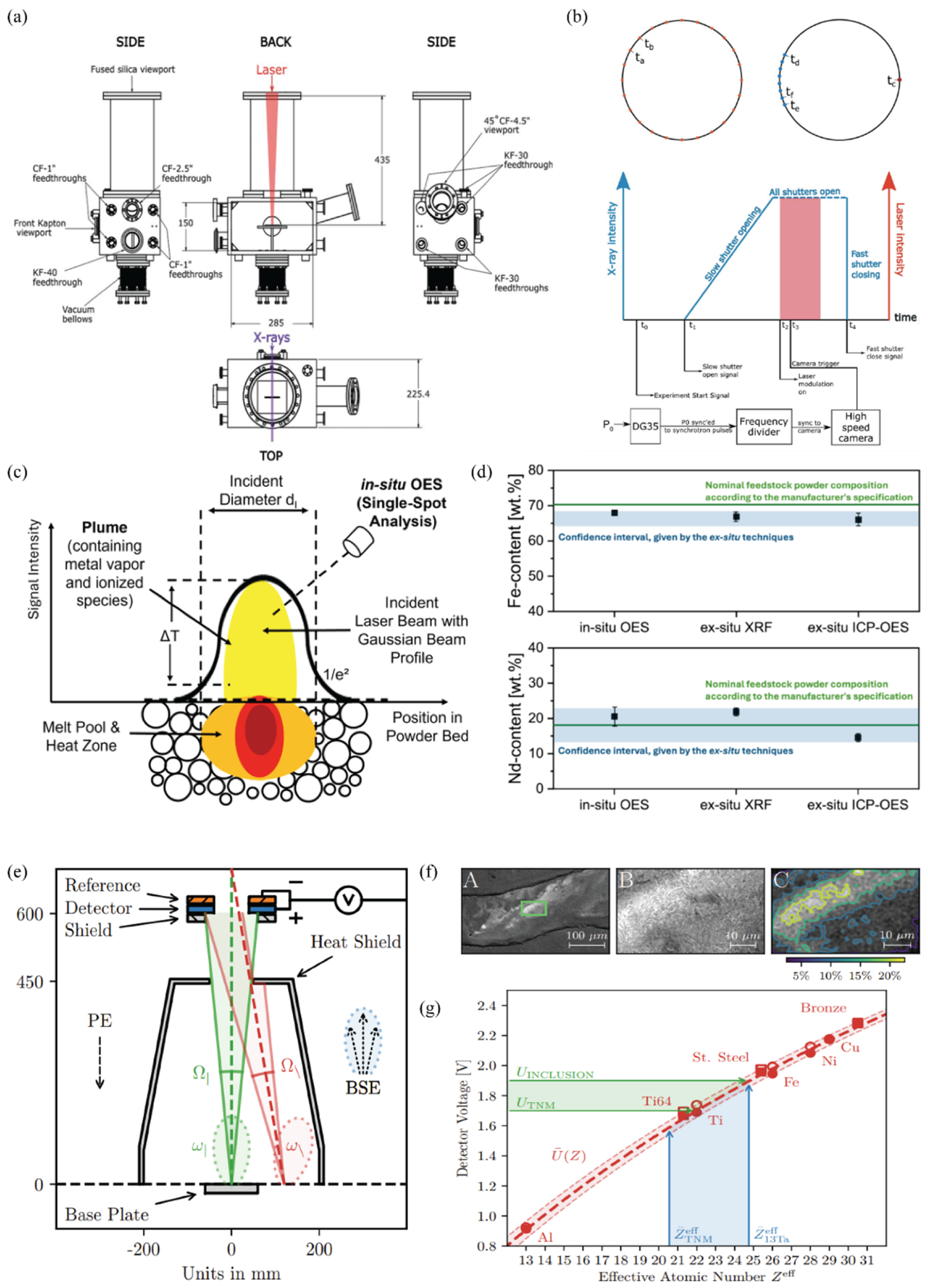
Table 7.
Typical densities of common non-metallic inclusions and base alloys in PBF-LB/M.
7.3.3. Element Loss Detection
Although inclusions originate from the incorporation of oxygen or other foreign species, their formation is frequently accompanied by the selective depletion of reactive alloying elements. Local oxidation or vaporization processes can consume or remove elements such as Nd, making their loss a useful indirect indicator of inclusion-forming reactions. As an example, Ziefuß et al. [202] developed a pioneering in situ OES framework to quantitatively monitor the elemental composition of metallic alloys during PBF-LB/M. Their work targeted the Nd-Fe-B system as a composition-sensitive model material, enabling the investigation of elemental losses and redistribution during laser melting. The thermal and temporal design of their experiment is schematically represented in Figure 8c, which illustrates a static laser and powder interaction at the initial stage. At this initial stage, before starting the scanning motion, the bell-shaped energy distribution of the laser imposes an uneven thermal profile across the powder surface, generating a dense vapor plume rich in excited Nd and Fe atoms. These excited species emit discrete optical lines that are recorded by the OES system in real time. By applying Boltzmann corrections to the measured line intensities, they established a relationship between the plume emissivity and the local temperature and elemental concentration within the small observation volume sampled by the OES system. This sensitivity makes the method effective for detecting compositional deviations such as elemental vaporization, and for identifying characteristics that may indirectly indicate segregation or inclusion formation reactions manifested, for example, as local depletion of reactive species in the plume. While OES probes only the vapor phase, anomalies in the emitted spectra can reveal melt pool chemistry changes when vaporization is coupled with oxidation or other reactions. It should also be noted that elemental vaporization is material dependent; alloys containing highly volatile species, such as Nd in Nd-Fe-B, generate strong plume signals, whereas more stable compositions may not. Moreover, Ziefuß et al. [202] also complemented their measurements with ex situ chemical analysis to validate the in situ results, the comparison of which is presented in Figure 8d. It shows the Fe and Nd contents measured by in situ OES alongside data from X-ray fluorescence (XRF) and inductively coupled plasma OES (ICP-OES), with the manufacturer’s nominal powder composition indicated by a green reference line. The blue shaded region demonstrates the confidence interval derived from the spread between both ex situ methods. Although XRF probes only the surface and ICP-OES represents a bulk-averaged analysis, the in situ OES data fell squarely within this confidence range, confirming the method’s quantitative reliability.
7.3.4. Electron Optical Imaging
On the other hand, as for Electron Optical Imaging (ELO), Gardfjell et al. [201] investigated the potential of ELO as an in situ monitoring method for detecting inclusions and compositional variations during PBF-EB. They demonstrated that BSE signals in a PBF-EB system can be used for quantitative material contrast and analyzed how detector geometry affects the measured intensity. As visualized in Figure 8e, by processing the BSE signal within the detector’s solid angle, they corrected the geometry-dependent intensity variations (STSA effect), allowing the measured contrast to reflect true material differences rather than viewing angle artifacts. The strength of this approach was demonstrated through an inclusion detection, where five metallic inclusions were successfully observed on as-built γ-TiAl components. As illustrated in Figure 8f, the STSA corrected ELO signal displayed a clear local intensity increase at positions that were later confirmed by SEM to contain regions enriched in tantalum. Moreover, the corresponding microstructure in Figure 8g further confirms the presence of these heavy element inclusions. Together, these observations demonstrate that inclusions containing elements with high atomic numbers, such as tantalum, produce a distinct BSE contrast and can be reliably identified using this approach. The ability to visualize these microstructural features directly on the as-built surface highlights ELO’s low noise topographical performance and its material contrast sensitivity sufficient to discriminate inclusions smaller than 100 µm, equivalent to only a few pixels in the detector image. Beyond qualitative detection, Gardfjell et al. [201]. introduced a quantitative material contrast framework linking the ELO signal intensity to the effective atomic number (Zeff) of the imaged alloy. By calibrating the system across twelve reference materials, they established that Zeff could be predicted to within one atomic number unit from the measured detector voltage. The validation results, shown in Figure 8g, compare the measured voltages of pure elements (circles) and selected alloys (squares) with theoretical predictions (red curve (U−(Z)). Data for the Ti-Al-Nb-Mo base alloy (TNM) and the Ta-enriched variant (TNM-13Ta) aligned remarkably well with the calibration, confirming that in situ material identification and contamination mapping are achievable during an actual build. Minor discrepancies between predicted and measured signals were attributed to factors such as temperature-induced thermionic emission, surface curvature, or beam-current drift; nevertheless, the correlation between voltage and atomic composition remained strong.
8. Future Perspective
8.1. Advancements in Sensor Technologies
Multi-modal sensing is one of the keyways; instead of relying on a single data source, next-generation platforms will integrate multiple sensor modalities (e.g., combining optical cameras, IR thermography, AE, and coaxial photodiodes) into unified systems. Such multi-modal setups can provide complementary views of the process (surface appearance, thermal fields, acoustic events, etc.), improving defect detectability for both PBF and DED. Research suggests that future “smart” sensors may even incorporate several modalities in one device (for example, cameras that capture both IR and visible spectra) to reduce system complexity. Equally important is making sensors robust to harsh AM environments. In laser-based PBF and DED, sensors must face intense thermal radiation and spatter, while in electron beam processes, they must tolerate high vacuum and X-ray radiation. Overall, the trajectory is toward higher-fidelity, multi-modal, and ruggedized sensors that can be seamlessly integrated into AM machines to record a higher amount of data from the build process [210].
8.2. Integration of Machine Learning and AI
Another critical future direction is the deep integration of ML and artificial intelligence (AI) with in situ monitoring. Rather than treating sensing and ML as separate efforts, research is converging on adaptive, data-centric frameworks that tightly couple real-time sensor data with AI-driven analysis [168]. In both PBF and DED, the large volume of monitoring data (images, videos, spectra, etc.) calls for automated, intelligent processing. Future systems will employ real-time inference algorithms running on edge devices or high-performance computing infrastructure to identify defect characteristics. To improve the efficiency of algorithms and robustness, new approaches like transfer learning and federated learning are being explored, which allow models to generalize across machines and conditions with minimal new training data [168].
8.3. Closed-Loop Control
Future PBF and DED machines will increasingly incorporate feedback mechanisms that adjust process parameters in real time to avoid or fix defects as they form. The ultimate vision is an autonomous system where the printing process can “heal” itself for example, by pausing and re-melting a problematic region, modifying the laser power or scan speed on the fly, or changing the powder feed rate in DED if a flaw is detected. Initial steps toward this vision are already evident in research. For instance, controllers that use thermal sensor feedback to maintain a stable melt pool size, thereby preventing overheating or LoF. Another example is the use of AE or plasma spectroscopy signals to detect a developing keyhole or crack and then immediately modulate the energy input to mitigate it. However, implementing true defect correction in real time faces challenges, including the need for ultra-fast data processing and actuation as well as strategies to distinguish false alarms from true defect indications. DED processes, with their flexible toolpaths, offer a testbed for such adaptive control (e.g., steering a robotic deposition head to fill a void or adjusting wire feed to eliminate a pore), whereas in PBF the high scan speeds and multi-laser coordination make intervention more complex. Ongoing research is addressing these issues, and it is anticipated that future machines will routinely include a closed-loop control layer. The ability to respond to dynamic changes in the melt pool or build environment and automatically tune parameters can revolutionize AM by drastically reducing defect.
8.4. Scalability and Standardization for Industrial Deployment
For in situ monitoring to transition from research labs to widespread industrial use, scalability and standardization are paramount. One aspect is the scalability of data handling and computation. Future monitoring systems must manage with massive data throughput from high-resolution, multi-sensor setups, especially in modern AM machines. This is driving the development of scalable software architectures (such as cloud or edge computing pipelines) that can process and analyze sensor data in parallel and in real time. There is also movement toward standardized metrics for defect detection performance and part quality indicators to consistently evaluate monitoring system effectiveness. Industrial and regulatory bodies (e.g., ASTM, ISO) are beginning to discuss norms for digital quality records derived from in situ monitoring, which could eventually feed into certification of critical AM parts. Achieving this would allow results from different sites to be compared and combined, accelerating learning. In summary, the future will see in situ monitoring systems that are scalable, interoperable, and standardized, enabling reliable defect detection and control across the diverse landscape of industrial metal AM. To illustrate how these technological directions converge, Figure 9 presents a conceptual framework for next-generation in situ monitoring, showing how multi-modal sensing, real-time data processing, machine learning, and automated feedback can be combined into a unified architecture for both PBF and DED systems.
Figure 9.
Integrated multi-modal sensing and AI-assisted closed-loop control framework for future metal AM.
9. Conclusions
The four principal defect types investigated in Figure 10 originate from various physical mechanisms yet share common process dependencies such as heat input, fluid dynamics, and alloy chemistry. Each defect mechanism (input) produces characteristic physical signals, which are detected by appropriate sensors and translated into engineered features for monitoring and classification (output). This representation clarifies how physical relevance guides sensor selection and data analysis. By linking the underlying physics of defect formation with the corresponding sensor responses and extracted signal features, this conceptual structure highlights how the physical mechanisms of key defects are directly reflected in their measurable sensor signatures. In summary, each defect mechanism produces a characteristic pattern in the sensor data, which makes it possible to distinguish various defect types. The selection of a monitoring method should be done based on the physical origin of the targeted defect, the required detection sensitivity, and the level of process integration. Acoustic emission sensing is suitable for detecting crack initiation due to its sensitivity to elastic wave generation. Optical and thermal imaging systems are more effective for melt pool related defects such as porosity and LoF. Spectroscopic methods are advantageous for identifying compositional anomalies such as non-metallic inclusions.
Figure 10.
Summary framework linking the four major defect categories in metal AM cracking, porosity, LoF, and inclusions to their underlying physical mechanisms, suitable in situ sensors, and key extracted process features.
- Cracking caused by thermal gradients, shrinkage strains, and grain boundary liquation produces elastic waves and local heat spikes during crack initiation and propagation. These physical events generate high-frequency acoustic signals, short STFT and CWT bursts, and localized thermal rate fluctuations that can be captured with broadband AE sensors and high-speed thermal imaging.
- Process porosity caused by gas entrapment, insufficient melting, or melt pool instability changes the thermal and geometric behavior of the melt pool. As pores form or approach the surface, they disturb heat flow and reduce local melt pool absorption, which appears as fluctuations in melt pool area, aspect ratio, and surface brightness. These surface-level changes can be observed with coaxial imaging, photodiodes, plume monitoring, and low-amplitude acoustic sensors that accompany bubble growth or collapse.
- LoF, caused by insufficient energy input or insufficient track overlap, disrupts heat conduction and melt flow. The resulting irregular thermal distribution produces unstable melt pool geometry, fluctuations in emission intensity, local shifts in radiance, and characteristic surface features such as incomplete hatch filling. These characteristics can be observed using vision-based imaging, thermal sensing, and acoustic signals linked to melt flow instability.
- Non-metallic inclusions originating from oxidation, powder contamination, or vapor phase reactions modify the local chemistry and optical properties of the melt pool. These changes influence the emitted light through altered emissivity, modified elemental line ratios, and localized bright spot contrasts. As a result, inclusions can be detected as distinct intensity variations in hyperspectral or multichannel optical data.
Across the defect categories, observable raw signals support the extraction of engineered features in the time domain (RMS, variance, kurtosis), frequency domain (dominant bands, power spectral density (PSD) peaks), and time–frequency domain (wavelet or STFT transients), forming a unified basis for data-driven defect detection. Figure 10 summarizes these relationships by mapping each defect mechanism to its corresponding sensing modality and feature space.
Author Contributions
Conceptualization, S.T.N., F.H., Y.M. and J.V.; methodology, S.T.N., F.H. and Y.M.; validation, S.T.N., F.H. and Y.M.; formal analysis, S.T.N.; data curation, S.T.N., F.H. and Y.M.; writing—original draft preparation, S.T.N.; writing—review and editing, F.H., Y.M. and J.V.; supervision, S.T.N., F.H., Y.M. and J.V.; project administration, S.T.N.; funding acquisition, J.V. All authors have read and agreed to the published version of the manuscript.
Funding
The authors acknowledge funding from the Knowledge Foundation (KK stiftelsen), Sweden, for the projects “RAMP—Robust Additive Manufacturing in production and material re-use” (202100-4052) and “DEDICATE—Directed Energy Deposition for Industrial Competitiveness in Additive Manufacturing Technologies”.
Institutional Review Board Statement
Not applicable.
Informed Consent Statement
Not applicable.
Data Availability Statement
The original contributions presented in the study are included in the article; further inquiries can be directed to the corresponding author.
Conflicts of Interest
The authors declare no conflicts of interest.
Abbreviations
The following abbreviations are used in this manuscript:
| AM | Additive Manufacturing |
| AE | Acoustic Emission |
| AI | Artificial Intelligence |
| ANN | Artificial Neural Network |
| APS | Advanced Photon Source |
| BSE | Backscattered Electrons |
| CGAN | Conditional Generative Adversarial Network |
| CNN | Convolutional Neural Network |
| CT | Computed Tomography |
| CWT | Continuous Wavelet Transform |
| DDC | Ductility-Dip Cracking |
| DED | Directed Energy Deposition |
| DED-arc/M | Directed Energy Deposition -arc /Metal |
| DED-LB/M | Directed Energy Deposition -Laser Beam /Metal |
| DT | Decision Tree |
| ELO | Electron Optical Imaging |
| EWMA | Exponentially Weighted Moving Average |
| FCC | Face Centered Cubic |
| FFT | Fast Fourier Transform |
| HAZ | Heat Affected Zone |
| HPSS | Harmonic Percussive Source Separation |
| ICP | inductively coupled plasma |
| IR | Infrared |
| LDA | Linear Discriminant Analysis |
| LIBS | Laser-Induced Breakdown Spectroscopy |
| LoF | Lack of Fusion |
| LR | Linear Regression |
| LSTM | Long Short-Term Memory |
| MA | Moving Average |
| MAE | Mean Absolute Error |
| MFCCs | Mel Frequency Cepstral Coefficients |
| MGL | mean gray level |
| ML | Machine Learning |
| MLP | Multi-Layer Perceptron |
| OES | Optical Emission Spectroscopy |
| OM | Optical Microscopy |
| PBF | Powder Bed Fusion |
| PBF-EB/M | Electron Beam Powder Bed Fusion |
| PBF-LB/M | Laser Powder Bed Fusion |
| PWHT | Post-Weld Heat Treatment Cracking |
| QDA | Quadratic Discriminant Analysis |
| RF | Random Forests |
| RMS | Root Mean Square |
| SAC | Strain-Age Cracking |
| SAXS | small-angle X-ray scattering |
| SOM | Self-Organizing Map |
| SPC | Statistical Process Control |
| STFT | Short Time Fourier Transform |
| STSA | Surface Tilt and Solid Angle |
| SVM | Support Vector Machines |
| ViT | Vision Transformer |
| VQVAE | Vector Quantized Variational Autoencoder |
| XRF | X-ray fluorescence |
References
- DebRoy, T.; Wei, H.L.; Zuback, J.S.; Mukherjee, T.; Elmer, J.W.; Milewski, J.O.; Beese, A.M.; Wilson-Heid, A.; De, A.; Zhang, W. Additive manufacturing of metallic components—Process, structure and properties. Prog. Mater. Sci. 2018, 92, 112–224. [Google Scholar] [CrossRef] [Scilit]
- Babu, A.; Ebrahimi, A.; Wu, K.-H.; Richardson, I.M.; Hermans, M.J.M. Local control of microstructure and mechanical properties of high-strength steel in electric arc-based additive manufacturing. J. Mater. Res. Technol. 2023, 26, 1508–1526. [Google Scholar] [CrossRef] [Scilit]
- Raghavendra Pai, K.; Vijayan, V.; Narayan Prabhu, K. Recent challenges and advances in metal additive manufacturing: A review. Mater. Today Proc. 2024, 102, 246–254. [Google Scholar] [CrossRef] [Scilit]
- Chen, H.; He, Y.; Dash, S.S.; Zou, Y. Additive manufacturing of metals and alloys to achieve heterogeneous microstructures for exceptional mechanical properties. Mater. Res. Lett. 2024, 12, 149–171. [Google Scholar] [CrossRef] [Scilit]
- Moradi, A.; Tajalli, S.; Mosallanejad, M.H.; Saboori, A. Intelligent laser-based metal additive manufacturing: A review on machine learning for process optimization and property prediction. Int. J. Adv. Manuf. Technol. 2025, 136, 527–560. [Google Scholar] [CrossRef] [Scilit]
- Babuska, T.F.; Krick, B.A.; Susan, D.F.; Kustas, A.B. Comparison of powder bed fusion and directed energy deposition for tailoring mechanical properties of traditionally brittle alloys. Manuf. Lett. 2021, 28, 30–34. [Google Scholar] [CrossRef] [Scilit]
- Ren, W.; Mazumder, J. In-situ porosity recognition for laser additive manufacturing of 7075-Al alloy using plasma emission spectroscopy. Sci. Rep. 2020, 10, 19493. [Google Scholar] [CrossRef] [Scilit]
- Chowdhury, S.; Yadaiah, N.; Prakash, C.; Ramakrishna, S.; Dixit, S.; Gupta, L.R.; Buddhi, D. Laser powder bed fusion: A state-of-the-art review of the technology, materials, properties & defects, and numerical modelling. J. Mater. Res. Technol. 2022, 20, 2109–2172. [Google Scholar] [CrossRef] [Scilit]
- Mohanty, S.; Gokuldoss Prashanth, K. Metallic Coatings through Additive Manufacturing: A Review. Materials 2023, 16, 2325. [Google Scholar] [CrossRef] [Scilit]
- Piscopo, G.; Iuliano, L. Current research and industrial application of laser powder directed energy deposition. Int. J. Adv. Manuf. Technol. 2022, 119, 6893–6917. [Google Scholar] [CrossRef] [Scilit]
- Armstrong, M.; Mehrabi, H.; Naveed, N. An overview of modern metal additive manufacturing technology. J. Manuf. Process. 2022, 84, 1001–1029. [Google Scholar] [CrossRef] [Scilit]
- Osipovich, K.; Kalashnikov, K.; Chumaevskii, A.; Gurianov, D.; Kalashnikova, T.; Vorontsov, A.; Zykova, A.; Utyaganova, V.; Panfilov, A.; Nikolaeva, A.; et al. Wire-Feed Electron Beam Additive Manufacturing: A Review. Metals 2023, 13, 279. [Google Scholar] [CrossRef] [Scilit]
- Dekis, M.; Tawfik, M.; Egiza, M.; Dewidar, M. Challenges and developments in wire arc additive manufacturing of steel: A review. Results Eng. 2025, 26, 104657. [Google Scholar] [CrossRef] [Scilit]
- Kan, W.H.; Chiu, L.N.S.; Lim, C.V.S.; Zhu, Y.; Tian, Y.; Jiang, D.; Huang, A. A critical review on the effects of process-induced porosity on the mechanical properties of alloys fabricated by laser powder bed fusion. J. Mater. Sci. 2022, 57, 9818–9865. [Google Scholar] [CrossRef] [Scilit]
- Bustillos, J.; Kim, J.; Moridi, A. Exploiting lack of fusion defects for microstructural engineering in additive manufacturing. Addit. Manuf. 2021, 48, 102399. [Google Scholar] [CrossRef] [Scilit]
- Sun, K.; Mohamed, A.E.-M.A.; Jeong, M.; Head, J.; Lewis, E.R.; Ibrahim, P.; Brooks, O.P.; Sheridan, R.S.; Attallah, M.M. On the origin of cracking in laser powder bed fusion processed LaCe(Fe,Mn,Si)13, and the impact of post-processing. J. Alloys Compd. 2023, 968, 172017. [Google Scholar] [CrossRef] [Scilit]
- Yeganeh, M.; Shahryari, Z.; Talib Khanjar, A.; Hajizadeh, Z.; Shabani, F. Inclusions and Segregations in the Selective Laser-Melted Alloys: A Review. Coatings 2023, 13, 1295. [Google Scholar] [CrossRef] [Scilit]
- Haribaskar, R.; Kumar, T.S. Defects in Metal Additive Manufacturing: Formation, Process Parameters, Postprocessing, Challenges, Economic Aspects, and Future Research Directions. 3D Print. Addit. Manuf. 2024, 11, e1629–e1655. [Google Scholar] [CrossRef] [Scilit]
- D’Hondt, L.; Cavaro, M.; Payan, C.; Mensah, S. Acoustical characterisation and monitoring of microbubble clouds. Ultrasonics 2019, 96, 10–17. [Google Scholar] [CrossRef] [Scilit] [PubMed]
- Moradi, A.; Tajalli, S.; Behjat, A.; Saboori, A.; Iuliano, L. Surface Engineering of EB-PBF Ti6Al4V via Anodization: Multifunctional Improvements Through TiO2 Nanotube Arrays. Coatings 2025, 15, 993. [Google Scholar] [CrossRef] [Scilit]
- Sun, X.; Huang, L.; Xiao, B.G.; Zhang, Q.; Li, J.Q.; Ding, Y.H.; Fang, Q.H.; He, W.; Xie, H.M. X-ray computed tomography in metal additive manufacturing: A review on prevention, diagnostic, and prediction of failure. Thin-Walled Struct. 2025, 207, 112736. [Google Scholar] [CrossRef] [Scilit]
- Peng, X.; Kong, L.; An, H.; Dong, G. A Review of In Situ Defect Detection and Monitoring Technologies in Selective Laser Melting. 3D Print. Addit. Manuf. 2023, 10, 438–466. [Google Scholar] [CrossRef] [Scilit] [PubMed]
- Wasmer, K.; Wüst, M.; Cui, D.; Masinelli, G.; Pandiyan, V.; Shevchik, S. Monitoring of functionally graded material during laser directed energy deposition by acoustic emission and optical emission spectroscopy using artificial intelligence. Virtual Phys. Prototyp. 2023, 18, e2189599. [Google Scholar] [CrossRef] [Scilit]
- García de la Yedra, A.; Pfleger, M.; Aramendi, B.; Cabeza, M.; Zubiri, F.; Mitter, T.; Reitinger, B.; Scherleitner, E. Online cracking detection by means of optical techniques in laser-cladding process. Struct. Control Health Monit. 2019, 26, e2291. [Google Scholar] [CrossRef] [Scilit]
- Wu, Y.; Cui, B.; Xiao, Y. Crack Detection during Laser Metal Deposition by Infrared Monochrome Pyrometer. Materials 2020, 13, 5643. [Google Scholar] [CrossRef] [Scilit]
- Shin, H.; Lee, J.; Choi, S.-K.; Lee, S.W. Development of multi-defect diagnosis algorithm for the directed energy deposition (DED) process with in situ melt-pool monitoring. Int. J. Adv. Manuf. Technol. 2023, 125, 357–368. [Google Scholar] [CrossRef] [Scilit]
- Goossens, L.; Hooreweder, B. A virtual sensing approach for monitoring melt-pool dimensions using high speed coaxial imaging during laser powder bed fusion of metals. Addit. Manuf. 2021, 40, 101923. [Google Scholar] [CrossRef] [Scilit]
- Mazzoleni, L.; Demir, A.G.; Caprio, L.; Pacher, M.; Previtali, B. Real-Time Observation of Melt Pool in Selective Laser Melting: Spatial, Temporal and Wavelength Resolution Criteria. IEEE Trans. Instrum. Meas. 2019, 69, 1179–1190. [Google Scholar] [CrossRef] [Scilit]
- Taheri, H.; Koester, L.W.; Bigelow, T.A.; Faierson, E.J.; Bond, L.J. In Situ Additive Manufacturing Process Monitoring with an Acoustic Technique: Clustering Performance Evaluation Using K-Means Algorithm. J. Manuf. Sci. Eng. 2019, 141, 041011. [Google Scholar] [CrossRef] [Scilit]
- Gaja, H.; Liou, F. Defects monitoring of laser metal deposition using acoustic emission sensor. Int. J. Adv. Manuf. Technol. 2017, 90, 561–574. [Google Scholar] [CrossRef] [Scilit]
- Arnold, C.; Breuning, C.; Körner, C. Electron-Optical In Situ Imaging for the Assessment of Accuracy in Electron Beam Powder Bed Fusion. Materials 2021, 14, 7240. [Google Scholar] [CrossRef] [Scilit]
- Herzog, T.; Brandt, M.; Trinchi, A.; Sola, A.; Molotnikov, A. Process monitoring and machine learning for defect detection in laser-based metal additive manufacturing. J. Intell. Manuf. 2024, 35, 1407–1437. [Google Scholar] [CrossRef] [Scilit]
- Petrich, J.; Snow, Z.; Corbin, D.; Reutzel, E.W. Multi-modal sensor fusion with machine learning for data-driven process monitoring for additive manufacturing. Addit. Manuf. 2021, 48, 102364. [Google Scholar] [CrossRef] [Scilit]
- Montazeri, M.; Nassar, A.R.; Dunbar, A.J.; Rao, P. In-process monitoring of porosity in additive manufacturing using optical emission spectroscopy. IISE Trans. 2020, 52, 500–515. [Google Scholar] [CrossRef] [Scilit]
- Herzog, T.; Brandt, M.; Trinchi, A.; Sola, A.; Hagenlocher, C.; Molotnikov, A. Defect detection by multi-axis infrared process monitoring of laser beam directed energy deposition. Sci. Rep. 2024, 14, 3861. [Google Scholar] [CrossRef] [Scilit]
- Kim, H.-S.; Park, S.-H. Acoustic signal monitoring using audible cracking sounds for efficient in-situ crack detection in laser directed energy deposition of hard surfaces. Addit. Manuf. Lett. 2024, 9, 100210. [Google Scholar] [CrossRef] [Scilit]
- Vock, S.; Kloeden, B.; Kirchner, A.; Weissgaerber, T.; Kieback, B. Powders for powder bed fusion: A review. Prog. Addit. Manuf. 2019, 4, 383–397. [Google Scholar] [CrossRef] [Scilit]
- Lewis, G. Aspects of the Powder in Metal Additive Manufacturing: A Review. World J. Eng. Technol. 2022, 10, 363–409. [Google Scholar] [CrossRef]
- Herzog, D.; Seyda, V.; Wycisk, E.; Emmelmann, C. Additive manufacturing of metals. Acta Mater. 2016, 117, 371–392. [Google Scholar] [CrossRef] [Scilit]
- Moradi, A.; Tajalli, S.; Taghian, M.; Behjat, A.; Saboori, A.; Iuliano, L. Enhancing additive manufacturing quality and productivity using machine learning: A study on laser powder bed fusion of AISI 316L-2.5%Cu. Prog. Addit. Manuf. 2026, 11, 1969–1987. [Google Scholar] [CrossRef] [Scilit]
- Koopmann, E.T.; Jäger, T.; Kaminsky, C.; Zeidler, H. Development of heating strategies to reduce crack formation in the manufacturing process of tool components using directed energy deposition. Prog. Addit. Manuf. 2025, 10, 1745–1753. [Google Scholar] [CrossRef] [Scilit]
- Sahraeidolatkhaneh, A.; Ariaseta, A.; Aydin, G.; Nilsen, M.; Sikström, F. Influence of Substrate Preheating on Processing Dynamics and Microstructure of Alloy 718 Produced by Directed Energy Deposition Using a Laser Beam and Wire. Metals 2025, 15, 1184. [Google Scholar] [CrossRef] [Scilit]
- Svetlizky, D.; Das, M.; Zheng, B.; Vyatskikh, A.L.; Bose, S.; Bandyopadhyay, A.; Schoenung, J.M.; Lavernia, E.J.; Eliaz, N. Directed energy deposition (DED) additive manufacturing: Physical characteristics, defects, challenges and applications. Mater. Today 2021, 49, 271–295. [Google Scholar] [CrossRef] [Scilit]
- du Plessis, A.; Yadroitsava, I.; Yadroitsev, I. Effects of defects on mechanical properties in metal additive manufacturing: A review focusing on X-ray tomography insights. Mater. Des. 2020, 187, 108385. [Google Scholar] [CrossRef] [Scilit]
- Wang, J.; Li, L.; Tao, W. Crack initiation and propagation behavior of WC particles reinforced Fe-based metal matrix composite produced by laser melting deposition. Opt. Laser Technol. 2016, 82, 170–182. [Google Scholar] [CrossRef] [Scilit]
- Tan, Z.E.; Pang, J.H.L.; Kaminski, J.; Pepin, H. Characterisation of porosity, density, and microstructure of directed energy deposited stainless steel AISI 316L. Addit. Manuf. 2019, 25, 286–296. [Google Scholar] [CrossRef] [Scilit]
- Siddique, S.; Imran, M.; Rauer, M.; Kaloudis, M.; Wycisk, E.; Emmelmann, C.; Walther, F. Computed tomography for characterization of fatigue performance of selective laser melted parts. Mater. Des. 2015, 83, 661–669. [Google Scholar] [CrossRef] [Scilit]
- Qin, L.; Wang, K.; Li, X.; Zhou, S.; Yang, G. Review of the Formation Mechanisms and Control Methods of Geometrical Defects in Laser Deposition Manufacturing. Chinese J. Mech. Eng. Addit. Manuf. Front. 2022, 1, 100052. [Google Scholar] [CrossRef] [Scilit]
- Chahal, V.; Taylor, R.M. A review of geometric sensitivities in laser metal 3D printing. Virtual Phys. Prototyp. 2020, 15, 227–241. [Google Scholar] [CrossRef] [Scilit]
- Chen, L.; Yao, X.; Feng, W.; Chew, Y.; Moon, S.K. Multimodal Sensor Fusion for Real-Time Location-Dependent Defect Detection in Laser-Directed Energy Deposition. 2023. Available online: https://doi.org/10.1115/DETC2023-110284 (accessed on 12 February 2026).
- Karthikeyan, A.; Balhara, H.; Hanchate, A.; Lianos, A.K.; Bukkapatnam, S.T.S. In-situ surface porosity prediction in hybrid-directed energy deposition (DED) process using explainable multimodal sensor fusion. J. Manuf. Process. 2025, 156, 904–919. [Google Scholar] [CrossRef] [Scilit]
- Khanafer, K.; Cao, J.; Kokash, H. Condition Monitoring in Additive Manufacturing: A Critical Review of Different Approaches. J. Manuf. Mater. Process. 2024, 8, 95. [Google Scholar] [CrossRef] [Scilit]
- Brandau, B.; Hemschik, R.; Sousa, J.P.; Brueckner, F.; Kaplan, A.F.H. Enhancing laser cladding stability: Defects and schlieren-based analytics during L-DED. Addit. Manuf. 2025, 103, 104758. [Google Scholar] [CrossRef] [Scilit]
- Repossini, G.; Laguzza, V.; Grasso, M.; Colosimo, B.M. On the use of spatter signature for in-situ monitoring of Laser Powder Bed Fusion. Addit. Manuf. 2017, 16, 35–48. [Google Scholar] [CrossRef] [Scilit]
- Pandiyan, V.; Wróbel, R.; Richter, R.A.; Leparoux, M.; Leinenbach, C.; Shevchik, S. Monitoring of Laser Powder Bed Fusion process by bridging dissimilar process maps using deep learning-based domain adaptation on acoustic emissions. Addit. Manuf. 2024, 80, 103974. [Google Scholar] [CrossRef] [Scilit]
- Guo, Q.; Zhao, C.; Escano, L.I.; Young, Z.; Xiong, L.; Fezzaa, K.; Everhart, W.; Brown, B.; Sun, T.; Chen, L. Transient dynamics of powder spattering in laser powder bed fusion additive manufacturing process revealed by in-situ high-speed high-energy x-ray imaging. Acta Mater. 2018, 151, 169–180. [Google Scholar] [CrossRef] [Scilit]
- Taylor, P.L.; Williams, R.J.; de Winton, H.C.; Fernandez, V.; Larsen, S.; Hooper, P.A. Digital 3D defect maps: Detecting localised porosity with high-speed melt pool imaging data in LPBF. Addit. Manuf. 2025, 111, 104982. [Google Scholar] [CrossRef] [Scilit]
- Bidare, P.; Bitharas, I.; Ward, R.M.; Attallah, M.M.; Moore, A.J. Fluid and particle dynamics in laser powder bed fusion. Acta Mater. 2018, 142, 107–120. [Google Scholar] [CrossRef] [Scilit]
- Seleznev, M.; Gustmann, T.; Friebel, J.M.; Peuker, U.A.; Kühn, U.; Hufenbach, J.K.; Biermann, H.; Weidner, A. In situ detection of cracks during laser powder bed fusion using acoustic emission monitoring. Addit. Manuf. Lett. 2022, 3, 100099. [Google Scholar] [CrossRef] [Scilit]
- Pandiyan, V.; Drissi-Daoudi, R.; Shevchik, S.; Masinelli, G.; Le-Quang, T.; Logé, R.; Wasmer, K. Semi-supervised Monitoring of Laser powder bed fusion process based on acoustic emissions. Virtual Phys. Prototyp. 2021, 16, 481–497. [Google Scholar] [CrossRef] [Scilit]
- Tempelman, J.R.; Wachtor, A.J.; Flynn, E.B.; Depond, P.J.; Forien, J.-B.; Guss, G.M.; Calta, N.P.; Matthews, M.J. Detection of keyhole pore formations in laser powder-bed fusion using acoustic process monitoring measurements. Addit. Manuf. 2022, 55, 102735. [Google Scholar] [CrossRef] [Scilit]
- Giulio, M.; Joseph, P.; Luigi, N. A Time-Frequency Domain Feature Extraction Approach Enhanced by Computer Vision for Wire Arc Additive Manufacturing Monitoring Using Fourier and Wavelet Transform. J. Adv. Manuf. Syst. 2024, 23, 741–762. [Google Scholar] [CrossRef] [Scilit]
- Zhang, H.; Wu, Q.; Tang, W.; Yang, J. Acoustic Signal-Based Defect Identification for Directed Energy Deposition-Arc Using Wavelet Time–Frequency Diagrams. Sensors 2024, 24, 4397. [Google Scholar] [CrossRef] [Scilit]
- Li, S.; Chen, B.; Tan, C.; Song, X. In situ identification of laser directed energy deposition condition based on acoustic emission. Opt. Laser Technol. 2024, 169, 110152. [Google Scholar] [CrossRef] [Scilit]
- Gaja, H.; Liou, F. Defect classification of laser metal deposition using logistic regression and artificial neural networks for pattern recognition. Int. J. Adv. Manuf. Technol. 2018, 94, 315–326. [Google Scholar] [CrossRef] [Scilit]
- Weber, J.U.; Knabe, M.; Sayilgan, V.; Emmelmann, C. Signal processing of airborne acoustic emissions from laser metal deposited structures. Procedia CIRP 2022, 111, 359–362. [Google Scholar] [CrossRef] [Scilit]
- Chen, L.; Bi, G.; Yao, X.; Tan, C.; Su, J.; Ng, N.P.H.; Chew, Y.; Liu, K.; Moon, S.K. Multisensor fusion-based digital twin for localized quality prediction in robotic laser-directed energy deposition. Robot. Comput. Integr. Manuf. 2023, 84, 102581. [Google Scholar] [CrossRef] [Scilit]
- Chen, L.; Yao, X.; Tan, C.; He, W.; Su, J.; Weng, F.; Chew, Y.; Ng, N.P.H.; Moon, S.K. In-situ crack and keyhole pore detection in laser directed energy deposition through acoustic signal and deep learning. Addit. Manuf. 2023, 69, 103547. [Google Scholar] [CrossRef] [Scilit]
- Song, J.Y.; Dass, A.; Moridi, A.; McLaskey, G.C. Detection of defects during laser-powder interaction by acoustic emission sensors and signal characteristics. Addit. Manuf. 2024, 82, 104035. [Google Scholar] [CrossRef] [Scilit]
- Lough, C.S.; Escano, L.I.; Qu, M.; Smith, C.C.; Landers, R.G.; Bristow, D.A.; Chen, L.; Kinzel, E.C. In-situ optical emission spectroscopy of selective laser melting. J. Manuf. Process. 2020, 53, 336–341. [Google Scholar] [CrossRef] [Scilit]
- Clare, A.T.; Woizeschke, P.; Rankouhi, B.; Pfefferkorn, F.E.; Bartels, D.; Schmidt, M.; Wits, W.W. Metal multi-material additive manufacturing: Overcoming barriers to implementation. CIRP Ann. 2025, 74, 869–893. [Google Scholar] [CrossRef] [Scilit]
- Wei, H.L.; Mukherjee, T.; Zhang, W.; Zuback, J.S.; Knapp, G.L.; De, A.; DebRoy, T. Mechanistic models for additive manufacturing of metallic components. Prog. Mater. Sci. 2021, 116, 100703. [Google Scholar] [CrossRef] [Scilit]
- Karmiol, Z.; Chidambaram, D. Comparison of Performance and Oxidation of Nitronic-50 and Stainless Steel 316 in Subcritical and Supercritical Water Environments. Metall. Mater. Trans. A 2016, 47, 2498–2508. [Google Scholar] [CrossRef] [Scilit]
- Tumer, M.; Karahan, T.; Mert, T. Evaluation of microstructural and mechanical properties of dissimilar Inconel 625 nickel alloy–UNS S32205 duplex stainless steel weldment using MIG welding. Weld. World 2020, 64, 21–35. [Google Scholar] [CrossRef] [Scilit]
- Nelson, D.E.; Baeslack, V.V.A.; Lippold, J.C. An Investigation of Weld Hot Cracking in Duplex Stainless Steels. 2013. Available online: https://api.semanticscholar.org/CorpusID:212682900 (accessed on 12 February 2026).
- Brennan, M.C.; Keist, J.S.; Palmer, T.A. Defects in Metal Additive Manufacturing Processes. J. Mater. Eng. Perform. 2021, 30, 4808–4818. [Google Scholar] [CrossRef] [Scilit]
- Müller, M.; Gerdt, L.; Schrüfer, S.; Riede, M.; López, E.; Brueckner, F.; Leyens, C. Laser-based directed energy deposition and characterisation of cBN-reinforced NiAl-based coatings. Int. J. Adv. Manuf. Technol. 2024, 134, 657–675. [Google Scholar] [CrossRef] [Scilit]
- Hanning, F.; Khan, A.K.; Andersson, J.; Ojo, O. Advanced microstructural characterisation of cast ATI 718Plus®—Effect of homogenisation heat treatments on secondary phases and repair welding behaviour. Weld. World 2020, 64, 523–533. [Google Scholar] [CrossRef] [Scilit]
- Shankar, V.; Gill, T.P.S.; Mannan, S.L.; Sundaresan, S. Solidification cracking in austenitic stainless steel welds. Sadhana 2003, 28, 359–382. [Google Scholar] [CrossRef] [Scilit]
- Kobryn, P.A.; Semiatin, S.L. Microstructure and texture evolution during solidification processing of Ti–6Al–4V. J. Mater. Process. Technol. 2003, 135, 330–339. [Google Scholar]
- Huang, C.; Cao, G.; Kou, S. Liquation cracking in partial penetration aluminium welds: Assessing tendencies to liquate, crack and backfill. Sci. Technol. Weld. Join. 2004, 9, 149–157. [Google Scholar] [CrossRef] [Scilit]
- Mo, B.; Li, T.; Shi, F.; Deng, L.; Liu, W. Crack initiation and propagation within nickel-based high-temperature alloys during laser-based directed energy deposition: A review. Opt. Laser Technol. 2024, 179, 111327. [Google Scholar] [CrossRef] [Scilit]
- Park, G.-W.; Shin, S.; Kim, J.-Y.; Koo, Y.-M.; Lee, W.; Lee, K.-A.; Park, S.S.; Jeon, J.B. Analysis of solidification microstructure and cracking mechanism of a matrix high-speed steel deposited using directed-energy deposition. J. Alloys Compd. 2022, 907, 164523. [Google Scholar] [CrossRef] [Scilit]
- Butt, M.M.; Laieghi, H.; Kvvssn, V.; Uddin, Z.; Shah, M.; Ansari, P.; Salamci, M.; Patterson, A.; Kizil, H. Fatigue performance in additively manufactured metal alloys. Prog. Addit. Manuf. 2024, 10, 1809–1841. [Google Scholar] [CrossRef] [Scilit]
- Song, J.; Li, Y.; Deng, Q.; Cheng, Z.; Chin, B. Cracking Mechanism of Laser Cladding Rapid Manufacturing 316L Stainless Steel. Key Eng. Mater. 2009, 419–420, 413–416. [Google Scholar] [CrossRef] [Scilit]
- Tang, Y.T.; Panwisawas, C.; Ghoussoub, J.N.; Gong, Y.; Clark, J.W.G.; Németh, A.A.N.; McCartney, D.G.; Reed, R.C. Alloys-by-design: Application to new superalloys for additive manufacturing. Acta Mater. 2021, 202, 417–436. [Google Scholar] [CrossRef] [Scilit]
- Böllinghaus, T.; Herold, H.; Cross, C.E.; Lippold, J.C. Hot Cracking Phenomena in Welds II; Springer Science & Business Media: Berlin/Heidelberg, Germany, 2008. [Google Scholar]
- Verma, J.; Taiwade, R.V. Effect of welding processes and conditions on the microstructure, mechanical properties and corrosion resistance of duplex stainless steel weldments—A review. J. Manuf. Process. 2017, 25, 134–152. [Google Scholar] [CrossRef] [Scilit]
- Caruso, M.; Frame, L. Ductility dip cracking mechanisms and characterization: A review. J. Mater. Sci. 2024, 59, 16789–16814. [Google Scholar] [CrossRef] [Scilit]
- Solid-State Cracking. In Welding Metallurgy and Weldability; Wiley: Hoboken, NJ, USA, 2015; pp. 130–212. [CrossRef] [Scilit]
- Kromm, A.; Lausch, T.; Schroepfer, D.; Rhode, M.; Kannengiesser, T. Influence of welding stresses on relief cracking during heat treatment of a creep-resistant 13CrMoV steel Part II: Mechanisms of stress relief cracking during post weld heat treatment. Weld. World 2020, 64, 819–829. [Google Scholar] [CrossRef] [Scilit]
- Mazzarisi, M.; Angelastro, A.; Latte, M.; Colucci, T.; Palano, F.; Campanelli, S.L. Thermal monitoring of laser metal deposition strategies using infrared thermography. J. Manuf. Process. 2023, 85, 594–611. [Google Scholar] [CrossRef] [Scilit]
- Nie, X.; Peng, F.; Hu, Z.; Qi, Y.; Zhu, H.; Zhang, H. The Effect of Thermal Cycle on Hot Cracking Evolution and Formation Mechanism in Thin Wall, Single Layer, and Cubic Samples of High-Strength Al-Cu-Mg-Mn Alloys Fabricated by Laser Powder Bed Fusion. 3D Print. Addit. Manuf. 2023, 11, 1996–2013. [Google Scholar] [CrossRef] [Scilit]
- Muktadir, M.; Hasan, M.N.; Alam, M. Additive manufacturing and acoustic emission: A brief review. J. Addit. Manuf. Technol. 2023, 2, 632. [Google Scholar] [CrossRef]
- Volkovas, V.; Dulevicius, J. Acoustic emission used for detection of crack generation in propellers of turbine-pump units. Russ. J. Nondestruct. Test. 2006, 42, 248–254. [Google Scholar] [CrossRef] [Scilit]
- Kabir, M.; Kazari, H.; Ozevin, D. Piezoelectric MEMS acoustic emission sensors. Sens. Actuators A Phys. 2018, 279, 53–64. [Google Scholar] [CrossRef] [Scilit]
- Wu, C.-Y.; Lu, M.-C.; Yang, W.-C.; Chi, N.-C. Experimental study of balling defect generation and audible sound analysis in directed energy deposition metal additive manufacturing. Int. J. Adv. Manuf. Technol. 2024, 134, 205–221. [Google Scholar] [CrossRef] [Scilit]
- Hauser, T.; Reisch, R.T.; Kamps, T.; Kaplan, A.F.H.; Volpp, J. Acoustic emissions in directed energy deposition processes. Int. J. Adv. Manuf. Technol. 2022, 119, 3517–3532. [Google Scholar] [CrossRef] [Scilit]
- Meng, X. Development of an Acoustic Emission Monitoring System for Crack Detection During Arc Welding. Ph.D. Thesis, University of Birmingham, Birmingham, UK, 2019. [Google Scholar]
- Ansari, M.J.; Roccisano, A.; Arcondoulis, E.J.G.; Schulz, C.; Schläfer, T.; Hall, C. Relationship between associated acoustic emission and crack position during directed energy deposition of a metal matrix composite. J. Manuf. Process. 2025, 147, 177–190. [Google Scholar] [CrossRef] [Scilit]
- Charunetratsamee, S.; Poopat, B.; Jirarungsatian, C. Feasibility Study of Acoustic Emission Monitoring of Hot Cracking in GTAW Weld. Key Eng. Mater. 2013, 545, 236–240. [Google Scholar] [CrossRef] [Scilit]
- Griffin, J.M.; Jones, S.; Perumal, B.; Perrin, C. Investigating the Detection Capability of Acoustic Emission Monitoring to Identify Imperfections Produced by the Metal Active Gas (MAG) Welding Process. Acoustics 2023, 5, 714–745. [Google Scholar] [CrossRef] [Scilit]
- Shiraiwa, T.; Kawate, M.; Briffod, F.; Kasuya, T.; Enoki, M. Evaluation of hydrogen-induced cracking in high-strength steel welded joints by acoustic emission technique. Mater. Des. 2020, 190, 108573. [Google Scholar] [CrossRef] [Scilit]
- Raeker, E.B.; Tulshibagwale, N.; Mullin, K.M.; Lamb, J.D.; Pollock, T.M. Detection and Classification of Cracking Via Acoustic Emission During Laser-Melting Screening for Additive Manufacturing. JOM 2025, 77, 7274–7286. [Google Scholar] [CrossRef] [Scilit]
- Guo, C.; Li, G.; Li, S.; Hu, X.; Lu, H.; Li, X.; Xu, Z.; Chen, Y.; Li, Q.; Lu, J.; et al. Additive manufacturing of Ni-based superalloys: Residual stress, mechanisms of crack formation and strategies for crack inhibition. Nano Mater. Sci. 2023, 5, 53–77. [Google Scholar] [CrossRef] [Scilit]
- Fardan, A.; Fazi, A.; Peng, R.L.; Mishurova, T.; Thuvander, M.; Bruno, G.; Brodin, H.; Hryha, E. Fine-Tuning Melt Pools and Microstructures: Taming Cracks in Powder Bed Fusion—Laser Beam of a non-weldable Ni-base Superalloy. Materialia 2024, 34, 102059. [Google Scholar] [CrossRef] [Scilit]
- Li, K.; Li, T.; Ma, M.; Wang, D.; Deng, W.; Lu, H. Laser cladding state recognition and crack defect diagnosis by acoustic emission signal and neural network. Opt. Laser Technol. 2021, 142, 107161. [Google Scholar] [CrossRef] [Scilit]
- Prieto, C.; Fernandez, R.; Gonzalez, C.; Diez, M.; Arias, J.; Sommerhuber, R.; Lücking, F. In situ process monitoring by optical microphone for crack detection in Laser Metal Deposition applications. In 11th CIRP Conference on Photonic Technologies [LANE 2020]; Elsevier: Amsterdam, The Netherlands, 2020. [Google Scholar]
- Koester, L.; Taheri, H.; Bond, L.; Faierson, E. Acoustic monitoring of additive manufacturing for damage and process condition determination. AIP Conf. Proc. 2019, 2102, 20005. [Google Scholar] [CrossRef] [Scilit]
- Rey, P.; Prieto, C.; González, C.; Tzimanis, K.; Souflas, T.; Stavropoulos, P.; Rathore, J.S.; Bergeaud, V.; Vienne, C.; Bredif, P. Data analysis to assess part quality in DED-LB/M based on in-situ process monitoring. Procedia CIRP 2022, 111, 345–350. [Google Scholar] [CrossRef] [Scilit]
- Wu, B.; Pan, Z.; Ding, D.; Cuiuri, D.; Li, H.; Xu, J.; Norrish, J. A review of the wire arc additive manufacturing of metals: Properties, defects and quality improvement. J. Manuf. Process. 2018, 35, 127–139. [Google Scholar] [CrossRef] [Scilit]
- Wang, S.; Ning, J.; Zhu, L.; Yang, Z.; Yan, W.; Dun, Y.; Xue, P.; Xu, P.; Bose, S.; Bandyopadhyay, A. Role of porosity defects in metal 3D printing: Formation mechanisms, impacts on properties and mitigation strategies. Mater. Today 2022, 59, 133–160. [Google Scholar] [CrossRef] [Scilit]
- Sinclair, L.; Clark, S.J.; Chen, Y.; Marussi, S.; Shah, S.; Magdysyuk, O.V.; Atwood, R.C.; Baxter, G.J.; Jones, M.; McCartney, D.G.; et al. Sinter formation during directed energy deposition of titanium alloy powders. Int. J. Mach. Tools Manuf. 2022, 176, 103887. [Google Scholar] [CrossRef] [Scilit]
- Iantaffi, C.; Leung, C.L.A.; Chen, Y.; Guan, S.; Atwood, R.C.; Lertthanasarn, J.; Pham, M.-S.; Meisnar, M.; Rohr, T.; Lee, P.D. Oxidation induced mechanisms during directed energy deposition additive manufactured titanium alloy builds. Addit. Manuf. Lett. 2021, 1, 100022. [Google Scholar] [CrossRef] [Scilit]
- Chen, Y.; Clark, S.J.; Sinclair, L.; Leung, C.L.A.; Marussi, S.; Connolley, T.; Atwood, R.C.; Baxter, G.J.; Jones, M.A.; Todd, I. Synchrotron X-ray imaging of directed energy deposition additive manufacturing of titanium alloy Ti-6242. Addit. Manuf. 2021, 41, 101969. [Google Scholar] [CrossRef] [Scilit]
- Chen, Y.; Clark, S.J.; Collins, D.M.; Marussi, S.; Hunt, S.A.; Fenech, D.M.; Connolley, T.; Atwood, R.C.; Magdysyuk, O.V.; Baxter, G.J.; et al. Correlative Synchrotron X-ray Imaging and Diffraction of Directed Energy Deposition Additive Manufacturing. Acta Mater. 2021, 209, 116777. [Google Scholar] [CrossRef] [Scilit]
- Bidare, P.; Mehmeti, A.; Jiménez, A.; Li, S.; Garman, C.; Dimov, S.; Essa, K. High-density direct laser deposition (DLD) of CM247LC alloy: Microstructure, porosity and cracks. Int. J. Adv. Manuf. Technol. 2022, 120, 8063–8074. [Google Scholar]
- Kim, H.J.; Shim, D.S. Characterization of the deposit-foaming of pure aluminum and Al-Mg-0.7 Si alloys using directed energy deposition based on their metallurgical characteristics and compressive behaviors. Addit. Manuf. 2022, 59, 103119. [Google Scholar] [CrossRef] [Scilit]
- Xie, D.; Lv, F.; Shen, L.; Tian, Z.; Jiang, R.; Zhao, J. Effect of higher laser power remelting on porosity and mechanical performance of part built by laser powder bed fusion. Int. J. Adv. Manuf. Technol. 2023, 125, 4779–4791. [Google Scholar] [CrossRef] [Scilit]
- Dilip, J.J.S.; Zhang, S.; Teng, C.; Zeng, K.; Robinson, C.; Pal, D.; Stucker, B. Influence of processing parameters on the evolution of melt pool, porosity, and microstructures in Ti-6Al-4V alloy parts fabricated by selective laser melting. Prog. Addit. Manuf. 2017, 2, 157–167. [Google Scholar]
- Xia, M.; Gu, D.; Yu, G.; Dai, D.; Chen, H.; Shi, Q. Porosity evolution and its thermodynamic mechanism of randomly packed powder-bed during selective laser melting of Inconel 718 alloy. Int. J. Mach. Tools Manuf. 2017, 116, 96–106. [Google Scholar] [CrossRef] [Scilit]
- Sohn, H.; Liu, P.; Yoon, H.; Yi, K.; Yang, L.; Kim, S. Real-time porosity reduction during metal directed energy deposition using a pulse laser. J. Mater. Sci. Technol. 2022, 116, 214–223. [Google Scholar] [CrossRef] [Scilit]
- Zhang, K.; Chen, Y.; Marussi, S.; Fan, X.; Fitzpatrick, M.; Bhagavath, S.; Majkut, M.; Lukic, B.; Jakata, K.; Rack, A.; et al. Pore evolution mechanisms during directed energy deposition additive manufacturing. Nat. Commun. 2024, 15, 1715. [Google Scholar] [CrossRef] [Scilit]
- Sinha, S.; Mukherjee, T. Mitigation of Gas Porosity in Additive Manufacturing Using Experimental Data Analysis and Mechanistic Modeling. Materials 2024, 17, 1569. [Google Scholar] [CrossRef] [Scilit]
- Xi, J.; Hu, Y.; Xing, H.; Han, Y.; Zhang, H.; Jiang, J.; Nikbin, K. The low-cycle fatigue behavior, failure mechanism and prediction of SLM Ti-6Al-4V alloy with different heat treatment methods. Materials 2021, 14, 6276. [Google Scholar] [CrossRef] [Scilit]
- Fredriksson, H.; Svensson, I. On the mechanism of pore formation in metals. Metall. Trans. B 1976, 7, 599–606. [Google Scholar] [CrossRef] [Scilit]
- Frieden Templeton, W.; Hinnebusch, S.; Strayer, S.; To, A.; Pistorius, C.; Narra, S. A mechanistic explanation of shrinkage porosity in laser powder bed fusion additive manufacturing. Acta Mater. 2023, 266, 119632. [Google Scholar] [CrossRef] [Scilit]
- Wang, H.; Gould, B.; Moorehead, M.; Haddad, M.; Couet, A.; Wolff, S.J. In situ X-ray and thermal imaging of refractory high entropy alloying during laser directed deposition. J. Mater. Process. Technol. 2022, 299, 117363. [Google Scholar] [CrossRef] [Scilit]
- Wolff, S.J.; Wang, H.; Gould, B.; Parab, N.; Wu, Z.; Zhao, C.; Greco, A.; Sun, T. In situ X-ray imaging of pore formation mechanisms and dynamics in laser powder-blown directed energy deposition additive manufacturing. Int. J. Mach. Tools Manuf. 2021, 166, 103743. [Google Scholar] [CrossRef] [Scilit]
- Malekipour, E.; El-Mounayri, H. Common defects and contributing parameters in powder bed fusion AM process and their classification for online monitoring and control: A review. Int. J. Adv. Manuf. Technol. 2018, 95, 527–550. [Google Scholar]
- Ekubaru, Y.; Gokcekaya, O.; Ishimoto, T.; Sato, K.; Manabe, K.; Wang, P.; Nakano, T. Excellent strength–ductility balance of Sc-Zr-modified Al–Mg alloy by tuning bimodal microstructure via hatch spacing in laser powder bed fusion. Mater. Des. 2022, 221, 110976. [Google Scholar] [CrossRef] [Scilit]
- Park, G.-W.; Song, S.; Park, M.; Shin, S.; Kim, D.-J.; Koo, Y.-M.; Kim, S.; Lee, K.-A.; Kim, B.J.; Park, S.S.; et al. Effect of residual stress on pore formation in multi-materials deposited via directed energy deposition. Addit. Manuf. 2024, 81, 104016. [Google Scholar] [CrossRef] [Scilit]
- Jeon, I.; Liu, Y.; Ryu, K. Online Melt Pool Depth Estimation During Directed Energy Deposition Using Coaxial Infrared Camera, Laser Line Scanner, and Artificial Neural Network. Addit. Manuf. 2021, 47, 102295. [Google Scholar] [CrossRef] [Scilit]
- Liao, S.; Webster, S.; Huang, D.; Council, R.; Ehmann, K.; Cao, J. Simulation-guided variable laser power design for melt pool depth control in directed energy deposition. Addit. Manuf. 2022, 56, 102912. [Google Scholar] [CrossRef] [Scilit]
- Naiel, M.A.; Ertay, D.S.; Vlasea, M.; Fieguth, P. Adaptive vision-based detection of laser-material interaction for directed energy deposition. Addit. Manuf. 2020, 36, 101468. [Google Scholar] [CrossRef] [Scilit]
- Tan, Q.; Liu, Y.; Fan, Z.; Zhang, J.; Yin, Y.; Zhang, M.-X. Effect of processing parameters on the densification of an additively manufactured 2024 Al alloy. J. Mater. Sci. Technol. 2020, 58, 34–45. [Google Scholar] [CrossRef] [Scilit]
- Khairallah, S.A.; Anderson, A.T.; Rubenchik, A.; King, W.E. Laser powder-bed fusion additive manufacturing: Physics of complex melt flow and formation mechanisms of pores, spatter, and denudation zones. Acta Mater. 2016, 108, 36–45. [Google Scholar] [CrossRef] [Scilit]
- Bostan, B.; Hinnebusch, S.; Anderson, D.; To, A.C. Accurate detection of local porosity in laser powder bed fusion through deep learning of physics-based in-situ infrared camera signatures. Addit. Manuf. 2025, 101, 104701. [Google Scholar] [CrossRef] [Scilit]
- Hooper, P.A. Melt pool temperature and cooling rates in laser powder bed fusion. Addit. Manuf. 2018, 22, 548–559. [Google Scholar] [CrossRef] [Scilit]
- Tempelman, J.R.; Mudunuru, M.K.; Karra, S.; Wachtor, A.J.; Ahmmed, B.; Flynn, E.B.; Forien, J.-B.; Guss, G.M.; Calta, N.P.; DePond, P.J.; et al. Uncovering acoustic signatures of pore formation in laser powder bed fusion. Int. J. Adv. Manuf. Technol. 2024, 130, 3103–3114. [Google Scholar] [CrossRef] [Scilit]
- Kouprianoff, D.; Yadroitsava, I.; du Plessis, A.; Luwes, N.; Yadroitsev, I. Monitoring of Laser Powder Bed Fusion by Acoustic Emission: Investigation of Single Tracks and Layers. Front. Mech. Eng. 2021, 7, 678076. [Google Scholar] [CrossRef] [Scilit]
- Hamidi Nasab, M.; Masinelli, G.; de Formanoir, C.; Schlenger, L.; Van Petegem, S.; Esmaeilzadeh, R.; Wasmer, K.; Ganvir, A.; Salminen, A.; Aymanns, F.; et al. Harmonizing sound and light: X-ray imaging unveils acoustic signatures of stochastic inter-regime instabilities during laser melting. Nat. Commun. 2023, 14, 8008. [Google Scholar] [CrossRef] [Scilit]
- Zhang, B.; Liu, S.; Shin, Y.C. In-Process monitoring of porosity during laser additive manufacturing process. Addit. Manuf. 2019, 28, 497–505. [Google Scholar] [CrossRef] [Scilit]
- Zhang, Y.; Shen, S.; Li, H.; Hu, Y. Review of in situ and real-time monitoring of metal additive manufacturing based on image processing. Int. J. Adv. Manuf. Technol. 2022, 123, 1–20. [Google Scholar] [CrossRef] [Scilit]
- Ramalho, A.; Assad, A.; Bevans, B.; Deschamps, F.; Santos, T.G.; Oliveira, J.P.; Rao, P. Understanding and detection of process instabilities in wire arc directed energy deposition additive manufacturing using meltpool imaging and machine learning. Mater. Des. 2025, 258, 114598. [Google Scholar] [CrossRef] [Scilit]
- Pandiyan, V.; Cui, D.; Richter, R.A.; Parrilli, A.; Leparoux, M. Real-time monitoring and quality assurance for laser-based directed energy deposition: Integrating co-axial imaging and self-supervised deep learning framework. J. Intell. Manuf. 2025, 36, 909–933. [Google Scholar] [CrossRef] [Scilit]
- Haley, J.C.; Schoenung, J.M.; Lavernia, E.J. Observations of particle-melt pool impact events in directed energy deposition. Addit. Manuf. 2018, 22, 368–374. [Google Scholar] [CrossRef] [Scilit]
- Khanzadeh, M.; Chowdhury, S.; Marufuzzaman, M.; Tschopp, M.A.; Bian, L. Porosity prediction: Supervised-learning of thermal history for direct laser deposition. J. Manuf. Syst. 2018, 47, 69–82. [Google Scholar] [CrossRef] [Scilit]
- Gaikwad, A.; Yavari, R.; Montazeri, M.; Cole, K.; Bian, L.; Rao, P. Toward the digital twin of additive manufacturing: Integrating thermal simulations, sensing, and analytics to detect process faults. IISE Trans. 2020, 52, 1204–1217. [Google Scholar] [CrossRef] [Scilit]
- Guo, W.; Tian, Q.; Guo, S.; Guo, Y. A physics-driven deep learning model for process-porosity causal relationship and porosity prediction with interpretability in laser metal deposition. CIRP Ann. 2020, 69, 205–208. [Google Scholar] [CrossRef] [Scilit]
- Bappy, M.M.; Liu, C.; Bian, L.; Tian, W. Morphological Dynamics-Based Anomaly Detection Towards In Situ Layer-Wise Certification for Directed Energy Deposition Processes. J. Manuf. Sci. Eng. 2022, 144, 111007. [Google Scholar] [CrossRef] [Scilit]
- Montazeri, M.; Nassar, A.R.; Stutzman, C.B.; Rao, P. Heterogeneous sensor-based condition monitoring in directed energy deposition. Addit. Manuf. 2019, 30, 100916. [Google Scholar] [CrossRef] [Scilit]
- Era, I.Z.; Zhou, F.; Raihan, A.S.; Ahmed, I.; Abul-Haj, A.; Craig, J.; Das, S.; Liu, Z. In-situ melt pool characterization via thermal imaging for defect detection in Directed Energy Deposition using Vision Transformers. J. Manuf. Process. 2025, 145, 11–21. [Google Scholar] [CrossRef] [Scilit]
- Ren, W.; Wen, G.; Zhang, Z.; Mazumder, J. Quality monitoring in additive manufacturing using emission spectroscopy and unsupervised deep learning. Mater. Manuf. Process. 2021, 37, 1339–1346. [Google Scholar] [CrossRef] [Scilit]
- He, F.; Yuan, L.; Mu, H.; Yang, J.; Ding, D.; Li, H.; Pan, Z. A recurrent neural network-based monitoring system using time-sequential molten pool images in wire arc directed energy deposition. Mech. Syst. Signal Process. 2025, 232, 112733. [Google Scholar] [CrossRef] [Scilit]
- Le, T.-N.; Lee, M.-H.; Lin, Z.-H.; Tran, H.-C.; Lo, Y.-L. Vision-based in-situ monitoring system for melt-pool detection in laser powder bed fusion process. J. Manuf. Process. 2021, 68, 1735–1745. [Google Scholar] [CrossRef] [Scilit]
- Yang, L.; Lo, L.; Ding, S.; Özel, T. Monitoring and detection of meltpool and spatter regions in laser powder bed fusion of super alloy Inconel 625. Prog. Addit. Manuf. 2020, 5, 367–378. [Google Scholar] [CrossRef] [Scilit]
- Gould, B.; Wolff, S.; Parab, N.; Zhao, C.; Lorenzo-Martin, M.C.; Fezzaa, K.; Greco, A.; Sun, T. In Situ Analysis of Laser Powder Bed Fusion Using Simultaneous High-Speed Infrared and X-ray Imaging. JOM 2021, 73, 201–211. [Google Scholar] [CrossRef] [Scilit]
- Baumgartl, H.; Tomas, J.; Buettner, R.; Merkel, M. A deep learning-based model for defect detection in laser-powder bed fusion using in-situ thermographic monitoring. Prog. Addit. Manuf. 2020, 5, 277–285. [Google Scholar] [CrossRef] [Scilit]
- Mitchell, J.A.; Ivanoff, T.A.; Dagel, D.; Madison, J.D.; Jared, B. Linking pyrometry to porosity in additively manufactured metals. Addit. Manuf. 2020, 31, 100946. [Google Scholar] [CrossRef] [Scilit]
- Chen, B.; Yao, Y.; Tan, C.; Huang, Y.; Feng, J. A study on spectral characterization and quality detection of direct metal deposition process based on spectral diagnosis. Int. J. Adv. Manuf. Technol. 2018, 96, 4231–4241. [Google Scholar] [CrossRef] [Scilit]
- Smith, R.J.; Hirsch, M.; Patel, R.; Li, W.; Clare, A.T.; Sharples, S.D. Spatially resolved acoustic spectroscopy for selective laser melting. J. Mater. Process. Technol. 2016, 236, 93–102. [Google Scholar] [CrossRef] [Scilit]
- Shevchik, S.A.; Masinelli, G.; Kenel, C.; Leinenbach, C.; Wasmer, K. Deep Learning for In Situ and Real-Time Quality Monitoring in Additive Manufacturing Using Acoustic Emission. IEEE Trans. Ind. Inform. 2019, 15, 5194–5203. [Google Scholar] [CrossRef] [Scilit]
- Wang, R.; Garcia, D.; Kamath, R.R.; Dou, C.; Ma, X.; Shen, B.; Choo, H.; Fezzaa, K.; Yu, H.Z.; Kong, Z. In situ melt pool measurements for laser powder bed fusion using multi sensing and correlation analysis. Sci. Rep. 2022, 12, 13716. [Google Scholar] [CrossRef] [Scilit]
- AWS. Standard Welding Terms and Definitions. Sl sn. 2001. Available online: https://pubs.aws.org/Download_PDFS/AWS_A3.0M_A3.0_2025_PV.pdf (accessed on 12 February 2026).
- Cao, S.; Gu, D. Laser metal deposition additive manufacturing of TiC/Inconel 625 nanocomposites: Relation of densification, microstructures and performance. J. Mater. Res. 2015, 30, 3616–3628. [Google Scholar] [CrossRef] [Scilit]
- Kobryn, P.A.; Moore, E.H.; Semiatin, S.L. The effect of laser power and traverse speed on microstructure, porosity, and build height in laser-deposited Ti-6Al-4V. Scr. Mater. 2000, 43, 299–305. [Google Scholar] [CrossRef] [Scilit]
- Wang, P.; Wu, Z.; Ten, J.J.S.; Huang, J.; Nai, M.L.S. Towards intelligent defect detection in metal powder bed fusion: A review of in situ monitoring, data pre-processing, and machine learning. Mater. Sci. Eng. R Rep. 2026, 167, 101112. [Google Scholar] [CrossRef] [Scilit]
- Everton, S.K.; Hirsch, M.; Stravroulakis, P.; Leach, R.K.; Clare, A.T. Review of in-situ process monitoring and in-situ metrology for metal additive manufacturing. Mater. Des. 2016, 95, 431–445. [Google Scholar] [CrossRef] [Scilit]
- Molaei, R.; Fatemi, A.; Phan, N. Significance of hot isostatic pressing (HIP) on multiaxial deformation and fatigue behaviors of additive manufactured Ti-6Al-4V including build orientation and surface roughness effects. Int. J. Fatigue 2018, 117, 352–370. [Google Scholar] [CrossRef] [Scilit]
- Cunningham, R.; Nicolas, A.; Madsen, J.; Fodran, E.; Anagnostou, E.; Sangid, M.D.; Rollett, A.D. Analyzing the effects of powder and post-processing on porosity and properties of electron beam melted Ti-6Al-4V. Mater. Res. Lett. 2017, 5, 516–525. [Google Scholar] [CrossRef] [Scilit]
- Kasperovich, G.; Haubrich, J.; Gussone, J.; Requena, G. Correlation between porosity and processing parameters in TiAl6V4 produced by selective laser melting. Mater. Des. 2016, 105, 160–170. [Google Scholar] [CrossRef] [Scilit]
- Cunningham, R.; Narra, S.P.; Montgomery, C.; Beuth, J.; Rollett, A.D. Synchrotron-Based X-ray Microtomography Characterization of the Effect of Processing Variables on Porosity Formation in Laser Power-Bed Additive Manufacturing of Ti-6Al-4V. JOM 2017, 69, 479–484. [Google Scholar] [CrossRef] [Scilit]
- Stwora, A.; Teimouri, R.; Habel, J. Role of Laser Powder Bed Fusion Process Factors in Determining the Porosity Formation in 3D Printing of Stainless Steel 316L: Theoretical Modeling and Experimental Verification. Materials 2025, 18, 5490. [Google Scholar] [CrossRef] [Scilit]
- Drissi-Daoudi, R.; Masinelli, G.; de Formanoir, C.; Wasmer, K.; Jhabvala, J.; Logé, R.E. Acoustic emission for the prediction of processing regimes in Laser Powder Bed Fusion, and the generation of processing maps. Addit. Manuf. 2023, 67, 103484. [Google Scholar] [CrossRef] [Scilit]
- Williams, R.J.; Davies, C.M.; Hooper, P.A. In situ monitoring of the layer height in laser powder bed fusion. Mater. Des. Process. Commun. 2021, 3, e173. [Google Scholar] [CrossRef] [Scilit]
- Lucà, F.; Chiariotti, P.; Bugatti, M.; Grasso, M.; Rigone, S.; Zappa, E.; Colosimo, B.M. Toward in situ monitoring of 3D surface topography in laser powder bed fusion additive manufacturing via polarized imaging. Prog. Addit. Manuf. 2025, 10, 9971–9987. [Google Scholar] [CrossRef] [Scilit]
- Suresh, V.; Balasubramaniam, B.; Yeh, L.-H.; Li, B. Recent Advances in In Situ 3D Surface Topographical Monitoring for Additive Manufacturing Processes. J. Manuf. Mater. Process. 2025, 9, 133. [Google Scholar] [CrossRef] [Scilit]
- Errico, V.; Palano, F.; Campanelli, S.L. Advancing powder bed fusion-laser beam technology: In-situ layerwise thermal monitoring solutions for thin-wall fabrication. Prog. Addit. Manuf. 2025, 10, 3361–3375. [Google Scholar] [CrossRef] [Scilit]
- Schwerz, C.; Nyborg, L. Linking In Situ Melt Pool Monitoring to Melt Pool Size Distributions and Internal Flaws in Laser Powder Bed Fusion. Metals 2021, 11, 1856. [Google Scholar] [CrossRef] [Scilit]
- Gibson, B.T.; Richardson, B.S.; Sundermann, T.W.; Love, L.J. Beyond the Toolpath: Site-Specific Melt Pool Size Control Enables Printing of Extra-Toolpath Geometry in Laser Wire-Based Directed Energy Deposition. Appl. Sci. 2019, 9, 4355. [Google Scholar] [CrossRef] [Scilit]
- Silvestri, A.; Hassanin, A.; de Alteriis, G.; Viola, E.; Schiano Lo Moriello, R.; Squillace, A. Advanced multi-sensor monitoring system in wire arc additive manufacturing for enhanced process and parts production. Mater. Res. Proc. 2025, 54, 264–273. [Google Scholar] [CrossRef] [Scilit]
- Assad, A.; Bevans, B.D.; Potter, W.; Rao, P.; Cormier, D.; Deschamps, F.; Hamilton, J.D.; Rivero, I. V Process mapping and anomaly detection in laser wire directed energy deposition additive manufacturing using in-situ imaging and process-aware machine learning. Mater. Des. 2024, 245, 113281. [Google Scholar] [CrossRef] [Scilit]
- Luo, Q.; Shimanek, J.D.; Simpson, T.W.; Beese, A.M. An Image-Based Transfer Learning Approach for Using In Situ Processing Data to Predict Laser Powder Bed Fusion Additively Manufactured Ti-6Al-4V Mechanical Properties. 3D Print. Addit. Manuf. 2025, 12, 48–60. [Google Scholar] [CrossRef] [Scilit]
- Drissi-Daoudi, R.; Pandiyan, V.; Logé, R.; Shevchik, S.; Masinelli, G.; Ghasemi-Tabasi, H.; Parrilli, A.; Wasmer, K. Differentiation of materials and laser powder bed fusion processing regimes from airborne acoustic emission combined with machine learning. Virtual Phys. Prototyp. 2022, 17, 181–204. [Google Scholar] [CrossRef] [Scilit]
- Smith, T.R.; Sugar, J.D.; San Marchi, C.; Schoenung, J.M. Microstructural development in DED stainless steels: Applying welding models to elucidate the impact of processing and alloy composition. J. Mater. Sci. 2021, 56, 762–780. [Google Scholar] [CrossRef] [Scilit]
- Chen, L.; Liu, W.; Song, L. A multiscale investigation of deformation heterogeneity in additively manufactured 316L stainless steel. Mater. Sci. Eng. A 2021, 820, 141493. [Google Scholar] [CrossRef] [Scilit]
- Marya, M.; Singh, V.; Marya, S.; Hascoet, J.Y. Microstructural Development and Technical Challenges in Laser Additive Manufacturing: Case Study with a 316L Industrial Part. Metall. Mater. Trans. B 2015, 46, 1654–1665. [Google Scholar] [CrossRef] [Scilit]
- Devesse, W.; De Baere, D.; Guillaume, P. High Resolution Temperature Measurement of Liquid Stainless Steel Using Hyperspectral Imaging. Sensors 2017, 17, 91. [Google Scholar] [CrossRef] [Scilit]
- Eo, D.-R.; Park, S.-H.; Cho, J.-W. Controlling inclusion evolution behavior by adjusting flow rate of shielding gas during direct energy deposition of AISI 316 L. Addit. Manuf. 2020, 33, 101119. [Google Scholar] [CrossRef] [Scilit]
- Klar, E.; Samal, P.K. Powder Metallurgy Stainless Steels: Processing, Microstructures, and Properties; ASM International: Materials Park, OH, USA, 2007. [Google Scholar]
- Barkia, B.; Aubry, P.; Haghi-Ashtiani, P.; Auger, T.; Gosmain, L.; Schuster, F.; Maskrot, H. On the origin of the high tensile strength and ductility of additively manufactured 316L stainless steel: Multiscale investigation. J. Mater. Sci. Technol. 2020, 41, 209–218. [Google Scholar] [CrossRef] [Scilit]
- Xiong, J.; Pi, Y.; Chen, H. Deposition height detection and feature point extraction in robotic GTA-based additive manufacturing using passive vision sensing. Robot. Comput. Integr. Manuf. 2019, 59, 326–334. [Google Scholar] [CrossRef] [Scilit]
- Kisielewicz, A.; Sadeghi, E.; Sikström, F.; Christiansson, A.-K.; Palumbo, G.; Ancona, A. In-process spectroscopic detection of chromium loss during Directed Energy Deposition of alloy 718. Mater. Des. 2020, 186, 108317. [Google Scholar] [CrossRef] [Scilit]
- Ancona, A.; Sikström, F.; Christiansson, A.-K.; Nilsen, M.; Mi, Y.; Kisielewicz, A. Monitoring and control of directed energy deposition using a laser beam. In Additive Manufacturing of High-Performance Metallic Materials; Pederson, R., Andersson, J., Joshi, S., Eds.; Elsevier: Amsterdam, The Netherlands, 2023; pp. 612–638. Available online: https://hv.diva-portal.org/smash/record.jsf?pid=diva2%3A1819526&dswid=-6660 (accessed on 12 February 2026).
- Ohtsuki, T.; Pistorius, P.C. Oxide Behavior During Laser Surface Melting. Metals 2025, 15, 627. [Google Scholar] [CrossRef] [Scilit]
- Haddad, M.; Cohen, M.; Bafahm Alamdari, A.; Welk, B.; Fezzaa, K.; Wolff, S. Process-structure relationships in laser directed energy deposition of molybdenum powder within a Ti–6Al–4V matrix. Addit. Manuf. 2025, 105, 104780. [Google Scholar] [CrossRef] [Scilit]
- Lednev, V.; Tretyakov, R.; Sdvizhenskii, P.; Grishin, M.; Asyutin, R.; Stavertiy, A.; Pershin, S. Laser induced breakdown spectroscopy for in situ multielemental analysis during additive manufacturing process. J. Phys. Conf. Ser. 2018, 1109, 012050. [Google Scholar] [CrossRef] [Scilit]
- Spencer, J.; Squires, B.; McWilliams, B.; Cho, K.; Dahotre, N.B.; Voevodin, A.A. Laser induced breakdown spectroscopy for composition monitoring of graded AlCu alloy surface. Surf. Coat. Technol. 2024, 494, 131375. [Google Scholar] [CrossRef] [Scilit]
- Ioannidou, C.; König, H.-H.; Semjatov, N.; Ackelid, U.; Staron, P.; Körner, C.; Hedström, P.; Lindwall, G. In-situ synchrotron X-ray analysis of metal Additive Manufacturing: Current state, opportunities and challenges. Mater. Des. 2022, 219, 110790. [Google Scholar] [CrossRef] [Scilit]
- Gardfjell, M.; Reith, M.; Franke, M.; Körner, C. In Situ Inclusion Detection and Material Characterization in an Electron Beam Powder Bed Fusion Process Using Electron Optical Imaging. Materials 2023, 16, 4220. [Google Scholar] [CrossRef] [Scilit] [PubMed]
- Ziefuss, A.R.; Gabriel, P.; Streubel, R.; Nachev, M.; Sures, B.; Eibl, F.; Barcikowski, S. In-situ composition analysis during laser powder bed fusion of Nd-Fe-based feedstock using machine-integrated optical emission spectroscopy. Mater. Des. 2024, 244, 113211. [Google Scholar] [CrossRef] [Scilit]
- Parab, N.D.; Zhao, C.; Cunningham, R.; Escano, L.I.; Fezzaa, K.; Everhart, W.; Rollett, A.D.; Chen, L.; Sun, T. Ultrafast X-ray imaging of laser-metal additive manufacturing processes. J. Synchrotron Radiat. 2018, 25, 1467–1477. [Google Scholar] [CrossRef] [Scilit]
- Alumina Al2O3—Properties of Aluminum Oxide. Available online: https://www.azom.com/article.aspx?ArticleID=3734 (accessed on 1 December 2025).
- Titanium Dioxide. Available online: https://en.wikipedia.org/wiki/Titanium_dioxide?utm (accessed on 1 December 2025).
- Silica—Silicon Dioxide (SiO2). Available online: https://www.azom.com/article.aspx?ArticleID=1114&utm (accessed on 1 December 2025).
- Ti64. Available online: https://www.upmet.com/sites/default/files/datasheets/ti-6al-4v.pdf?utm (accessed on 1 December 2025).
- ASTM Material Data Sheet. Available online: https://asm.matweb.com/search/specificmaterial.asp?bassnum=ninc34&utm (accessed on 1 December 2025).
- EN 1.4404/AISI 316L. Available online: https://www.alfalaval.se/produkter/flodeshantering/en-1-4404-aisi-316l/?utm (accessed on 1 December 2025).
- Ansari, M.J.; Roccisano, A.; Arcondoulis, E.J.G.; Schulz, C.; Schläfer, T.; Hall, C. Advancements in In-Situ Monitoring Technologies for Detecting Process-Induced Defects in the Directed Energy Deposition Process: A Comprehensive Review. Materials 2025, 18, 4304. [Google Scholar] [CrossRef] [Scilit]
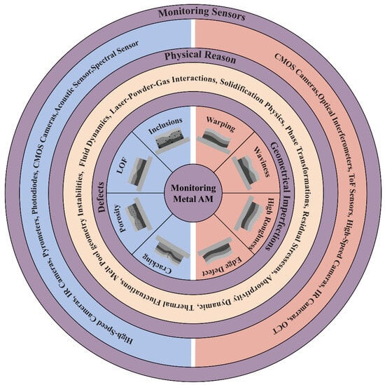
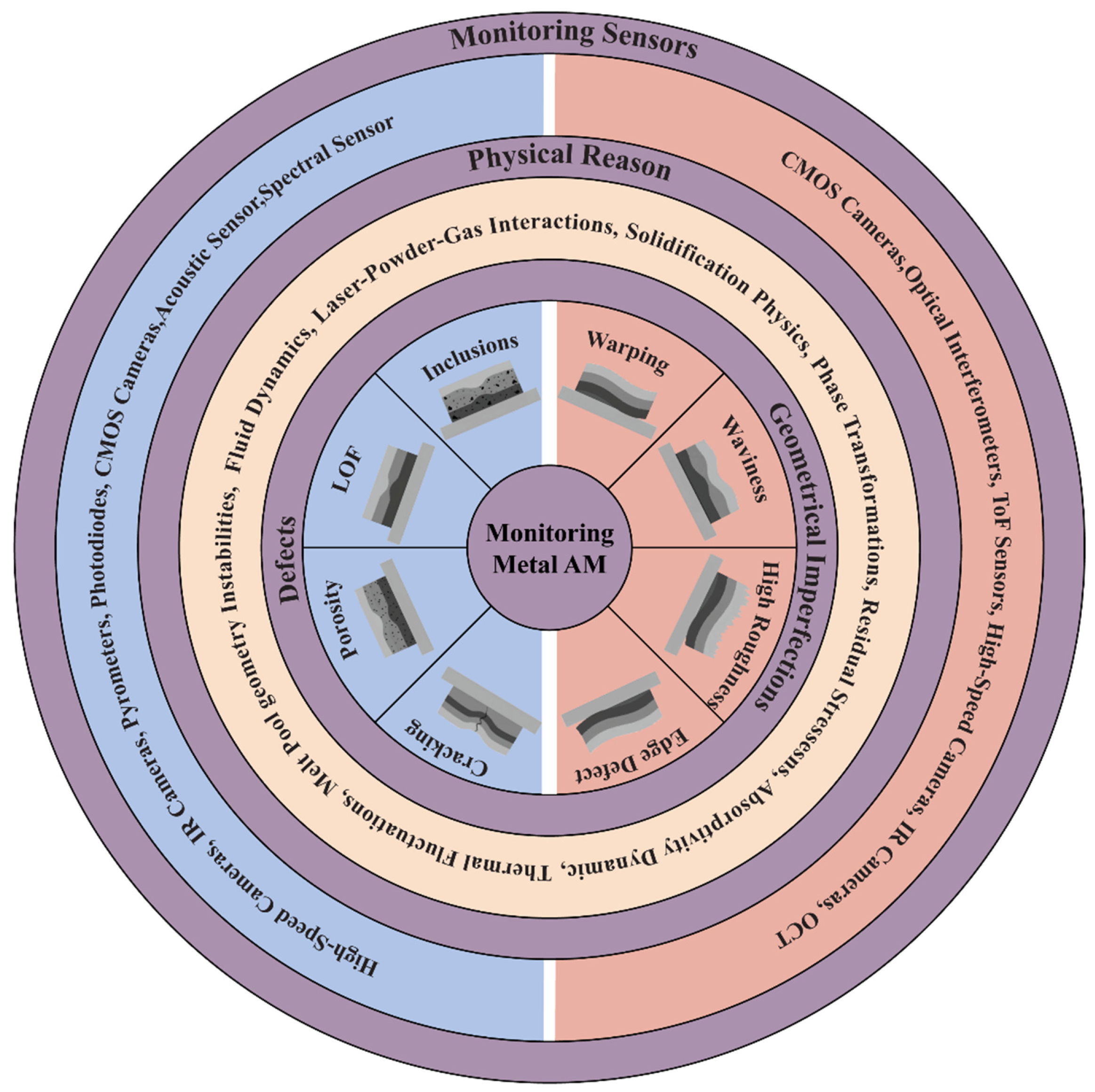
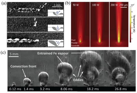
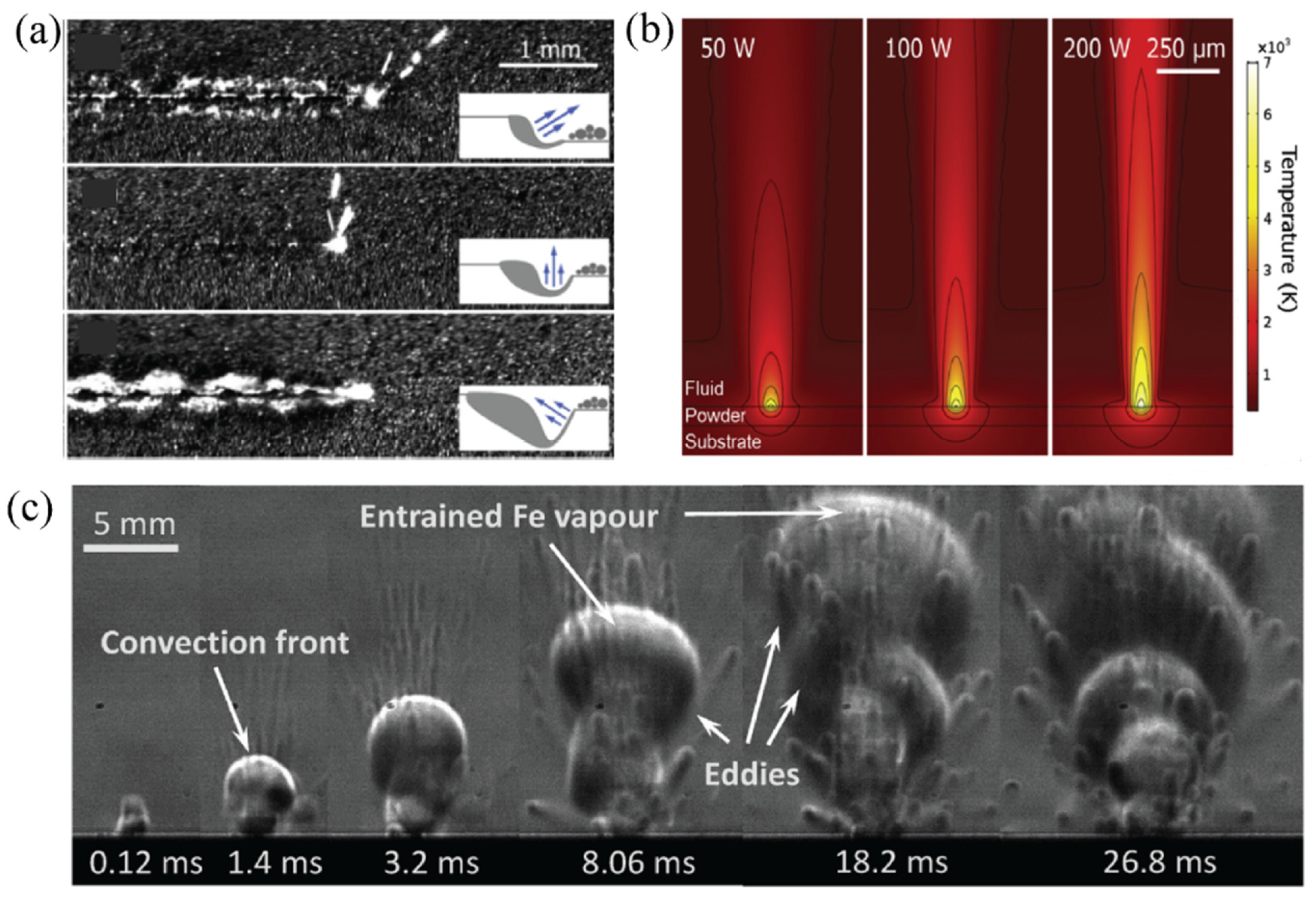
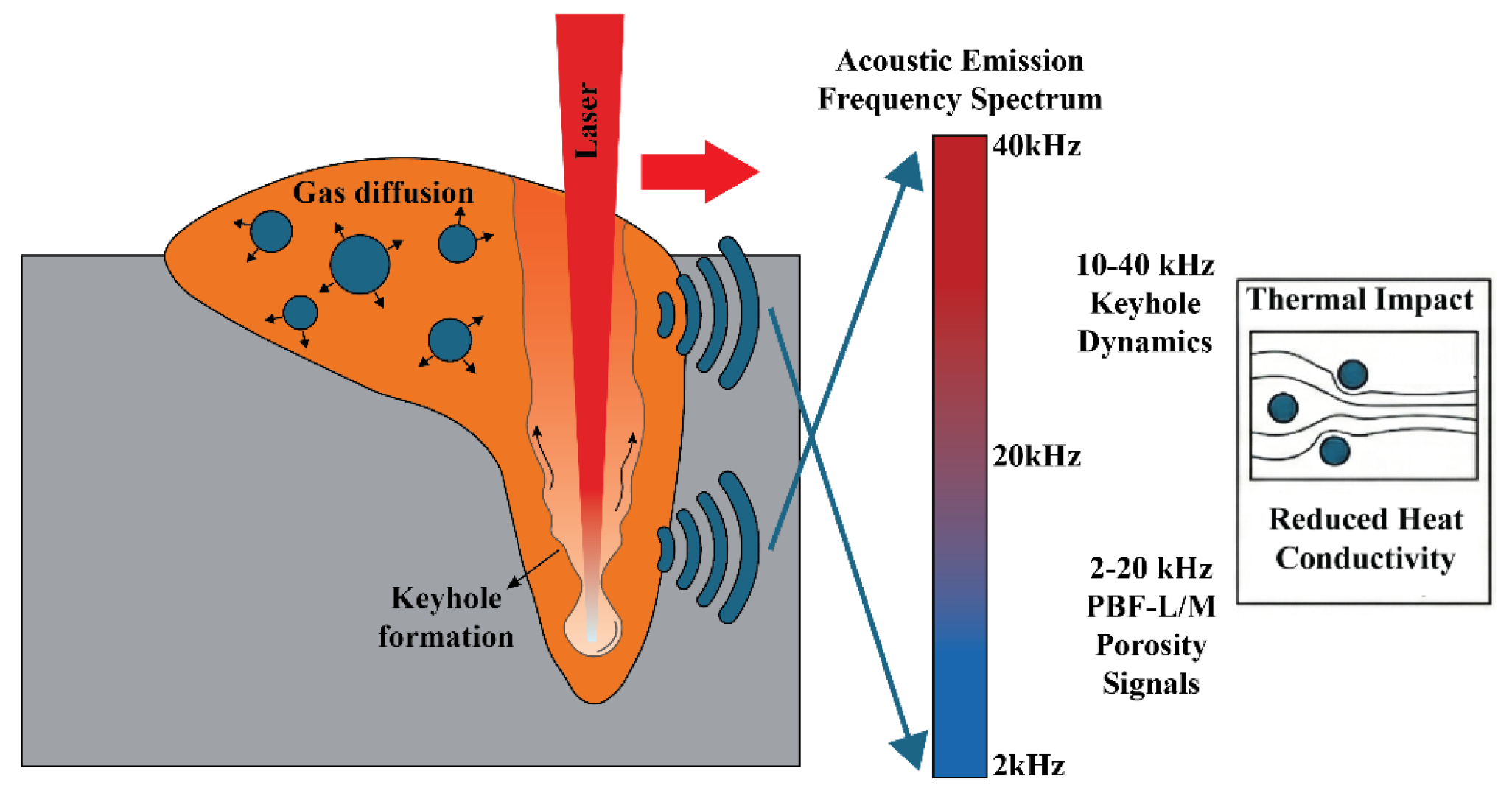
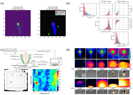
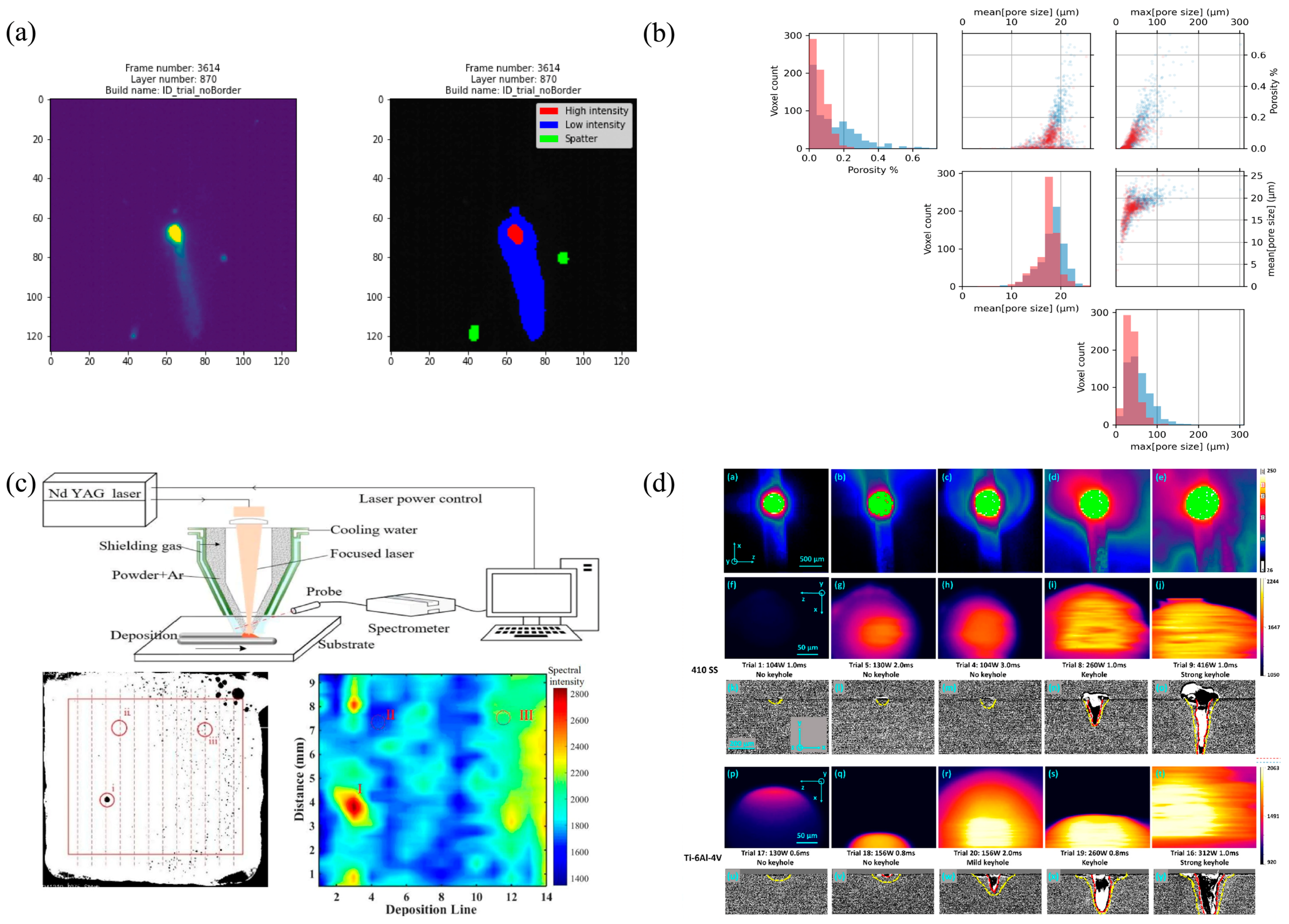
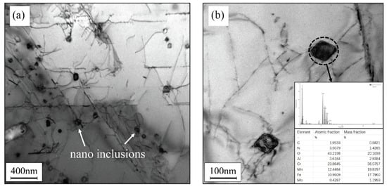
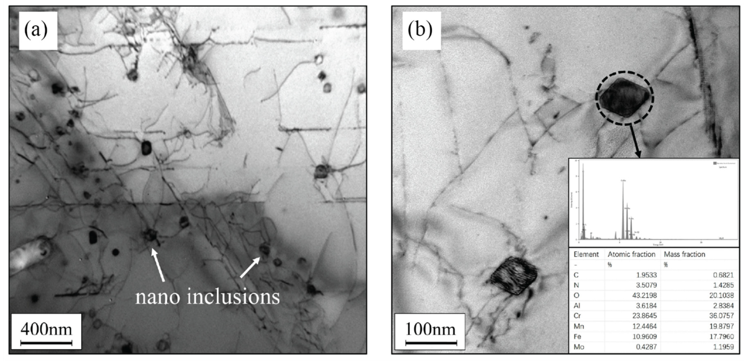
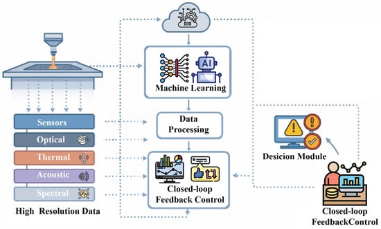
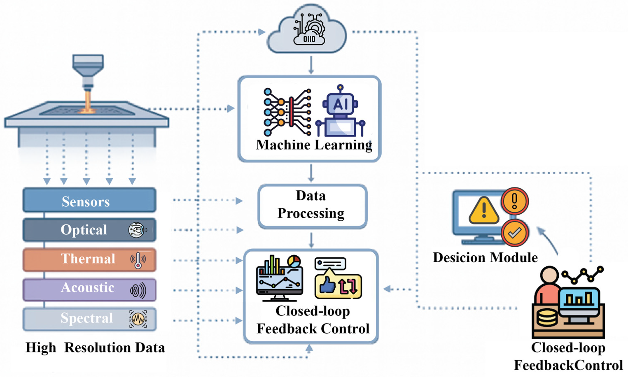
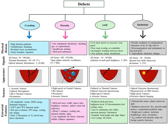
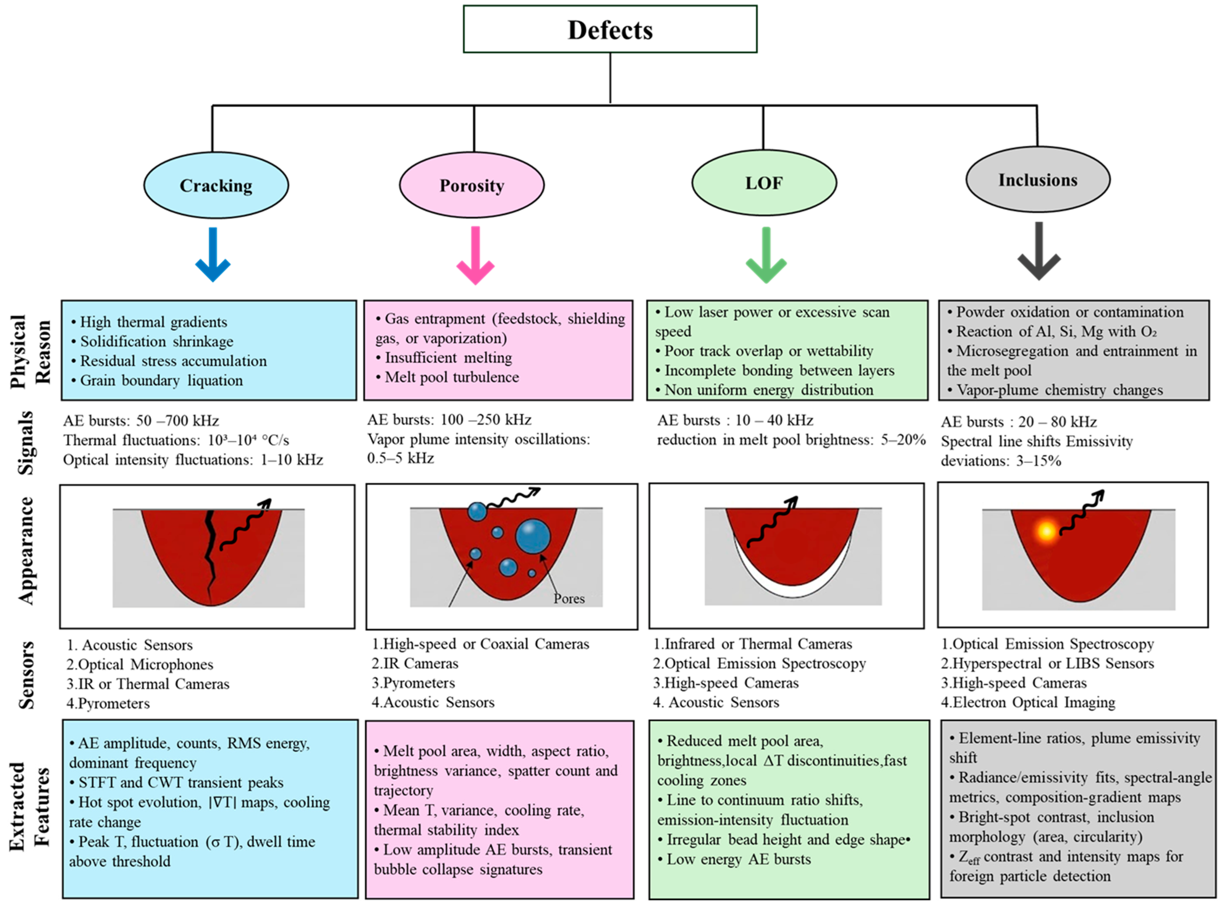

Disclaimer/Publisher’s Note: The statements, opinions and data contained in all publications are solely those of the individual author(s) and contributor(s) and not of MDPI and/or the editor(s). MDPI and/or the editor(s) disclaim responsibility for any injury to people or property resulting from any ideas, methods, instructions or products referred to in the content. |
© 2026 by the authors. Licensee MDPI, Basel, Switzerland. This article is an open access article distributed under the terms and conditions of the Creative Commons Attribution (CC BY) license.