


Abstract
Background/Objectives: To evaluate the clinical effects of alveolar ridge preservation using a hyaluronic acid-based product combined with a resorbable collagen cone compared with spontaneous healing. Methods: In this prospective pilot comparative clinical study, extraction sockets were treated either with hyaluronic acid and a collagen cone (test group) or left to heal spontaneously (control group). Mesial and distal ridge height changes (ΔHm, ΔHd), ridge width reduction (ΔW), buccal mucosal thickness changes (ΔTmb), and occlusal mucosal thickness at 3 months (TmoT3) were assessed. Early wound healing was appreciated at 14 days follow-up, and postoperative complications after 7 and 14 days. Results: Complete 3-month follow-up data were available for 18 test and 14 control patients. Mesial bone loss was significantly lower in the test group than in controls [test ΔHm = 1.0 (1.0–1.0) vs. control ΔHm = 2.0 (1.0–2.0); p < 0.001]. Horizontal ridge width reduction was significantly lower in the test group [ΔW = 1.0 (1.0–1.0) vs. 2.0 (1.0–2.0); p = 0.021]. Buccal mucosal thickness was better maintained in the test group [ΔTmb = 0.0 (0.0–0.5) vs. 1.0 (0.5–1.0); p < 0.001], while TmoT3 was greater in the test group, in an exploratory analysis (p = 0.049). A statistically significant difference in early wound-healing scores favoring the test group was observed [4.0 (4.0–5.0) vs. 4.0 (3.0–4.0); p = 0.032]. No statistically detectable difference in postoperative complications was observed between groups. Conclusions: Alveolar ridge preservation was associated with reduced mesial vertical and horizontal dimensional changes and more favorable soft tissue outcomes compared with spontaneous healing. These preliminary findings should be considered hypothesis-generating.
1. Introduction
Tooth extraction remains unavoidable in certain non-restorable clinical situations, including extensive carious destruction, traumatic injuries, and severe periodontal bone loss [1]. However, extraction is invariably followed by volumetric alterations of both hard and soft tissues [2], which may complicate future implant placement, adversely affect the esthetic outcomes of surgical and prosthetic rehabilitation [3], and interfere with orthodontic space closure [4]. Post-extraction ridge remodelling is considerable, with horizontal bone loss of 2.6–4.5 mm and vertical loss of 0.4–3.9 mm reported in healed sockets [5]. Approximately 50% of ridge width reduction occurs during the first year, most of it within the first 3 months after extraction [6]. In addition to hard tissue remodelling, substantial soft tissue collapse is expected to occur following tooth extraction closely following the underlying post-extraction bone resorption process [7].
The extent of post-extraction ridge resorption varies considerably both within and between individuals in terms of rate, duration, and magnitude of tissue loss [2,8]. Post-extraction dimensional changes are influenced by systemic factors [9,10,11,12], as well as local factors including extraction trauma, pre-existing bone loss, chronic infections, and periodontal phenotype [13,14,15].
Alveolar ridge preservation has been proposed to attenuate the physiological post-extraction resorption process and improve the predictability of subsequent implant and oral rehabilitation procedures [16,17], although it cannot completely prevent ridge remodelling [18,19]. Compared with spontaneous healing, alveolar ridge preservation techniques have been associated with reduced vertical buccal bone loss and improved preservation of ridge width, with reported dimensional benefits ranging from 0.16–1.72 mm vertically and 1.61–1.99 mm horizontally [19,20].
Various biomaterials have been investigated for alveolar ridge preservation, including bone substitutes, barrier membranes, and biological agents [21,22,23,24,25]. The choice of ridge preservation procedures is influenced by the feasibility of future implant placement, the quality and quantity of soft tissues, residual buccal bone dimensions, and the expected success of implant rehabilitation [26]. However, the available evidence remains heterogeneous and does not currently support clear recommendations regarding the most effective biomaterials or treatment protocols [27,28].
Although soft tissue quality following post-extraction healing is essential for the long-term biological stability of dental implants and the prognosis of prosthetic restorations [29,30,31,32,33,34], soft tissue changes in extraction sockets have received considerably less attention than post-extraction bone remodelling, and the available evidence remains limited [35,36,37]. Nevertheless, socket preservation procedures have been associated with reduced soft tissue volume loss after six months of healing, with values of 62.66 ± 17.50 mm3 at the buccal aspect in sites treated with a xenograft and membrane compared with 106.41 ± 24.00 mm3 in spontaneously healed sites [19].
The use of hyaluronic acid in alveolar ridge preservation has emerged as a logical therapeutic approach given its role as a natural component of the extracellular matrix actively involved in tissue healing. Hyaluronic acid exhibits osteogenic, anti-inflammatory, and antimicrobial properties, while presenting a favorable safety profile compared with other biologically active molecules [38]. It may be applied either alone or in combination with other biomaterials [38,39]. However, the evidence regarding the effectiveness of hyaluronic acid in alveolar ridge preservation remains heterogeneous, particularly with respect to quantitative and qualitative changes in hard and soft tissues reported in both clinical and preclinical studies [40]. Consequently, the available evidence is still insufficient to support routine clinical use of hyaluronic acid in socket preservation procedures [40], and further clinical investigations are warranted to clarify its therapeutic potential [28] and quantify its benefit in terms of new bone formation [39].
Based on this background, the present study aimed to clinically evaluate post-extraction socket healing following the application of a commercially available hyaluronic acid-based product combined with a collagen cone to stabilize the blood clot, enhance hemostasis, and protect the extraction socket during the early healing phase, compared with spontaneous healing. Healing outcomes were assessed through clinical measurements of vertical and horizontal ridge dimensional changes between baseline (T0) and 3 months (T3), appreciation of early healing of ridge mucosa after 14 days (T2), and the occurrence of postoperative complications at 7 (T1) and 14 days (T2). The findings are expected to contribute to a better understanding of the role of hyaluronic acid-based biomaterials in alveolar ridge preservation and to support evidence-based clinical decisions.
2. Materials and Methods
2.1. Study Design and Population
This prospective pilot comparative clinical study evaluated post-extraction socket healing following the application of hyaluronic acid (hyaDENT BG®, Regedent AG, Zurich, Switzerland) in combination with a resorbable collagen cone (Collacone®, Botiss Biomaterials GmbH, Zossen, Germany) (test group), compared with spontaneous healing (control group).
Patients were recruited between March 2022 and February 2025 from the Periodontology Outpatient Clinic and the Integrated Outpatient Unit of the First Department of Oral and Maxillofacial Surgery, Emergency County Clinical Hospital Cluj-Napoca, Romania. The study protocol was approved by the Ethics Committees of the Emergency County Clinical Hospital Cluj-Napoca (Approval No. 1304/4 May 2022) and the Iuliu Hațieganu University of Medicine and Pharmacy, Cluj-Napoca (Approval No. 62/3 January 2022). Written informed consent was obtained from all participants prior to enrolment. The study was conducted in accordance with the principles of the Declaration of Helsinki for research involving human subjects. Reporting was aligned with the TREND recommendations whenever applicable to the study design [41]. The study was not prospectively registered in a clinical trial registry.
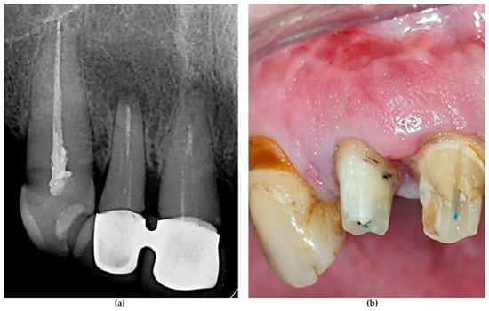
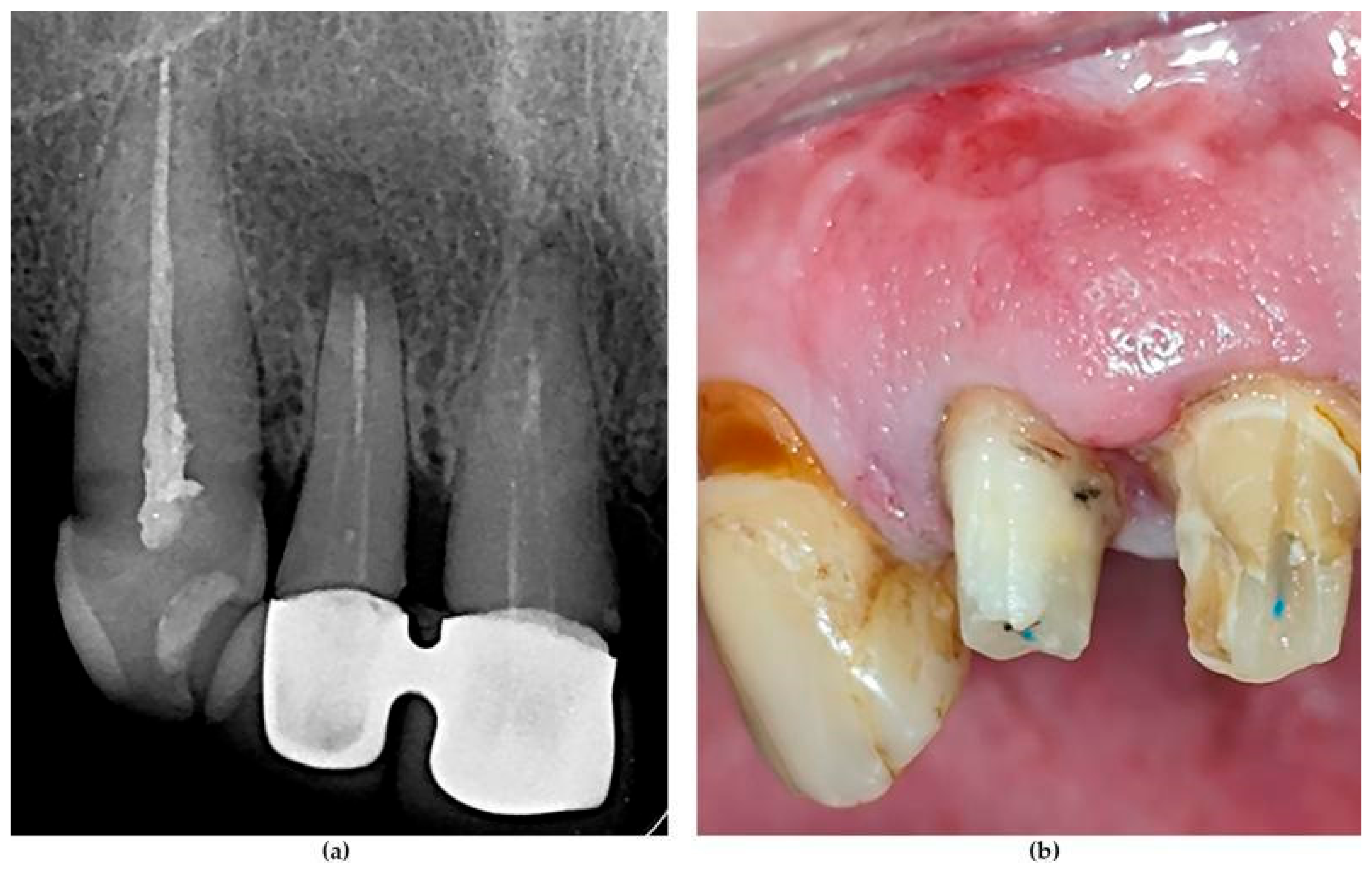
Eligible participants were between 18 and 70 years of age and presented with at least one tooth indicated for extraction (Figure 1), excluding third molars [42]. Additional inclusion criteria comprised a post-extraction site considered suitable for future dental implant placement, presence of both mesial and distal teeth neighbouring the extraction site, and the presence of at least three intact socket walls at the time of extraction.
Figure 1.
Inclusion criterion represented by a symptomatic recurrent periapical lesion following a second periapical surgery at tooth #12 in a 54-year-old female patient. (a) Radiographic appearance of the lesion. (b) Clinical intraoral view. (Photos from the personal archive of the authors P.-A.N. and A.R.).
Exclusion criteria included general contraindications to oral surgery, long-term nonsteroidal anti-inflammatory drug therapy, current treatment with bisphosphonates or antibiotics, a history of radiotherapy in the head and neck region within the previous five years, uncontrolled metabolic disorders, uncontrolled periodontitis [43], pregnancy or lactation, acute infection at the extraction site (e.g., abscess), heavy smoking (>20 cigarettes/day), and inadequate oral hygiene (Oral Hygiene Index > 20%) [44].
Patients were enrolled consecutively during the study period. Allocation to the test or control group was not randomized. The test group was recruited first and included patients who accepted alveolar ridge preservation with hyaluronic acid and a collagen cone. Subsequently, a control group consisting of patients undergoing spontaneous socket healing was enrolled using the same eligibility criteria.
Due to the nature of the intervention, participants, the operator administering the treatment, and the examiner assessing outcomes were aware of group allocation. The statistician was blinded to group allocation.
The evaluation of dimensional changes was performed at the time of extraction (T0) and three months after treatment (T3). Soft tissue healing was assessed at 7 days (T1) and 14 days (T2), and immediate postoperative complications were recorded in the first 14 days.
2.2. Surgical Protocol and Postoperative Care
One week before surgery, all patients received professional supragingival oral hygiene cleaning and oral hygiene instructions. Patients were instructed to rinse twice daily with 0.2% chlorhexidine mouthwash (Corsodyl®, GlaxoSmithKline Consumer Healthcare, Brentford, UK).
All surgical procedures were standardized and performed by the same operator (P.-A.N.). Tooth extraction and alveolar ridge preservation procedures were carried out as atraumatically as possible following conventional surgical principles. Briefly, local anaesthesia was achieved using 4% articaine with 1:100,000 epinephrine (Ubistezin™ Forte, 3M Deutschland GmbH, Neuss, Germany). An intrasulcular incision was made around the tooth to be extracted, followed by careful luxation and extraction using periotomes (Periotome®, Directa AB, Upplands Väsby, Sweden), elevators (LM-Dental™, LM-Instruments Oy, Parainen, Finland), and extraction forceps (MegaGen Implant Co., Ltd., Daegu, Republic of Korea). All extraction sockets were thoroughly curetted and irrigated with sterile saline solution.
In the test group, approximately two thirds of the hyaluronic acid gel contained in one syringe (hyaDENT BG®, Regedent AG) was applied within the extraction socket. Subsequently, a resorbable collagen cone (Collacone®, Botiss Biomaterials) was impregnated with the remaining one third of the hyaluronic acid gel and inserted into the extraction socket. The collagen material was trimmed and adapted to the individual socket dimensions to achieve complete coverage and sealing of the coronal aspect of the extraction socket. The material was stabilized using a crossed mattress suture placed over the socket with a 5-0 non-resorbable polyamide suture (Supramid®, SMI, St. Vith, Belgium). The same suturing technique was applied in the control group.
Postoperative care was standardized for all patients and included rinsing twice daily with 0.2% chlorhexidine mouthwash for 4 weeks (Corsodyl®). Ibuprofen or paracetamol was prescribed as needed. Sutures were removed 14 days after surgery.
2.3. Patient Characteristics and Outcome Assessment
Baseline demographic, medical, and behavioral characteristics, including alcohol and tobacco consumption, were collected from medical records and patient interviews. According to the WHO definition, patients were considered drinkers if they had consumed alcohol within the previous 12 months and non-drinkers if they had not consumed alcohol during this period. Non-drinkers included both lifetime abstainers and former drinkers [45].
Smoking status was categorized according to the threshold values used in the 2018 EFP/AAP periodontal classification for grade determination (<10 or ≥10 cigarettes/day) [35], while heavy smokers were excluded from the study.
To reduce potential bias, all clinical evaluations were performed by the same examiner (P.-A.N.). Clinical measurements were obtained using a UNC-15 periodontal probe (Hu-Friedy, Chicago, IL, USA) with 1 mm markings. All measurements were rounded to the nearest millimeter.
The following parameters were recorded:
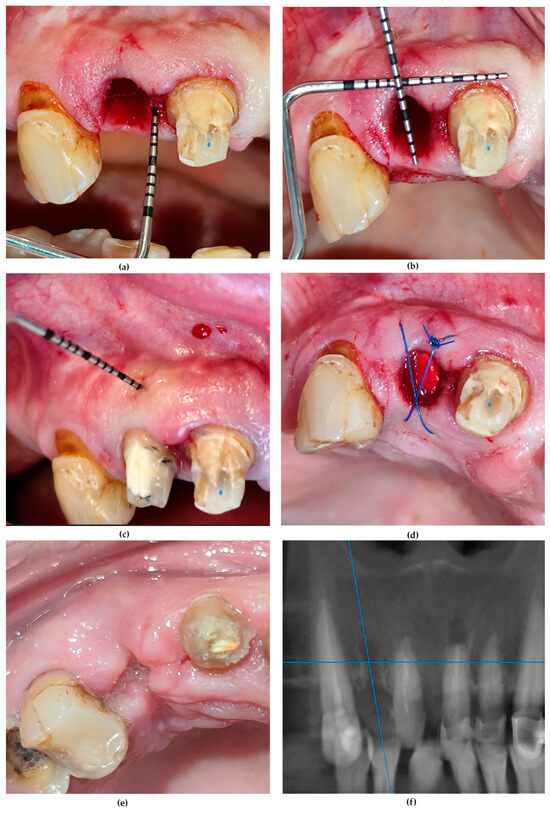
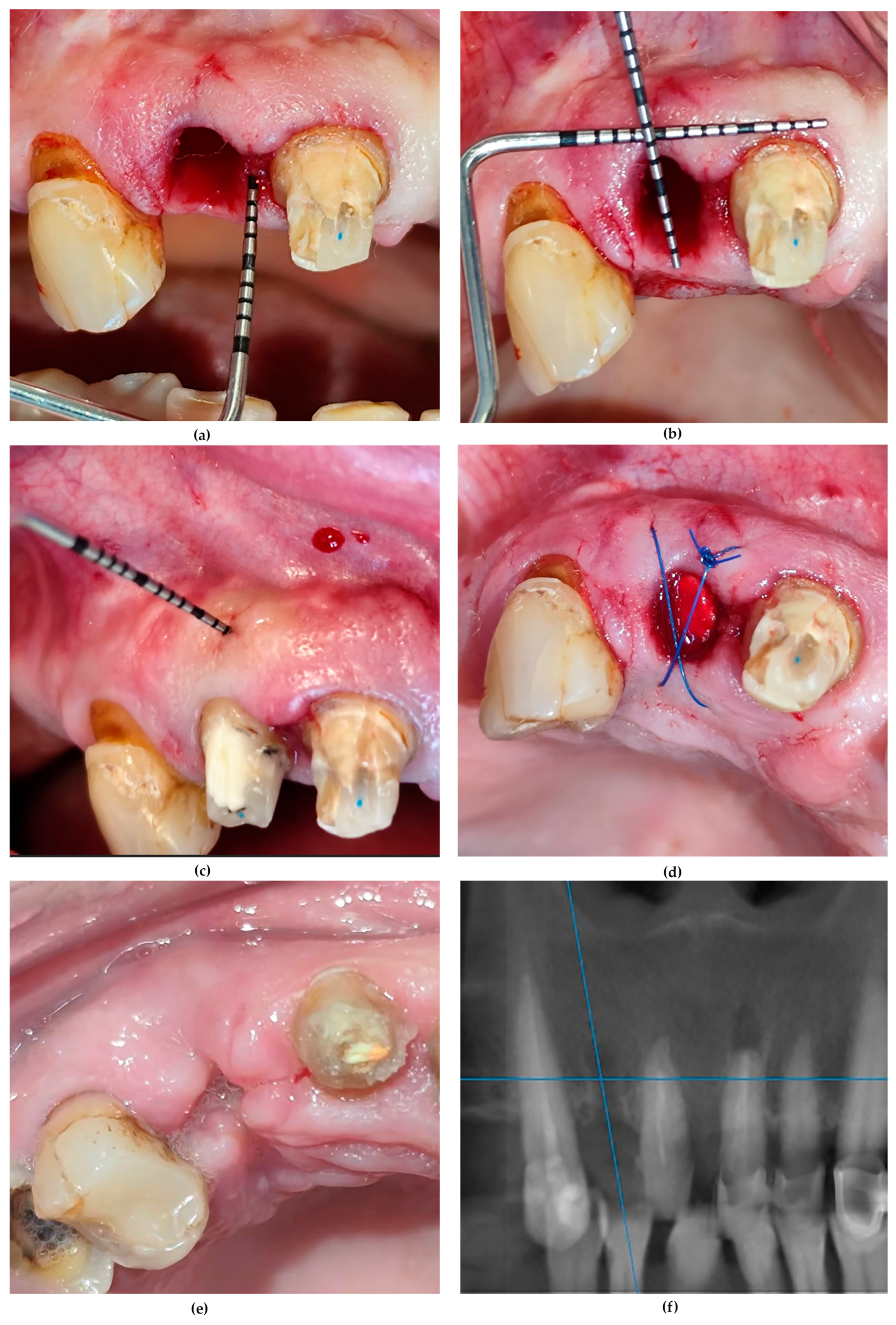
- Mesial and distal ridge bone height (Hm and Hd), measured relative to the cemento-enamel junction or crown preparation of the adjacent teeth immediately after extraction (baseline) (T0) (Figure 2a) and after 3 months (T3) through transmucosal probe penetration.
- Ridge width (W), defined as the horizontal dimension of the extraction site (hard and soft tissues combined) measured at the central aspect of the ridge at T0 and T3 (Figure 2b). At T0, ridge width was measured at the midpoint of the mesiodistal distance between the two neighboring teeth, and this reference was recorded in the patient chart and used to reproduce the measurement location at T3.
- Mucosal thickness of the ridge, assessed at the buccal aspect (Tmb) at T0 before socket suture (Figure 2c,d) and T3 and occlusal aspect (Tmo) at T3 follow-up.
Figure 2.
Outcome measurements and alveolar ridge preservation at tooth #12 in the clinical case presented in Figure 1. (a) Measurement of mesial ridge height (Hm = 4 mm) from the crestal bone to the cervical margin of the adjacent prepared tooth at baseline (T0). (b) Measurement of the central ridge width (W = 7 mm) at T0. (c) Measurement of buccal mucosal thickness (Tmb = 2 mm) at T0. (d) Socket sealed with a collagen cone impregnated with hyaluronic acid and stabilized with cross-mattress sutures. (e) Early wound healing score of 4 according to the modified Landry Healing Index at the post-extraction site. (f) CBCT image three months after alveolar ridge preservation (Photos from the personal archive of the authors P.-A.N. and A.R.).
At the 3-month follow-up, transgingival bone sounding under local anesthesia was performed at the midpoint of the ridge, both mesiodistally and buccolingually, to assess alveolar bone dimensions and mucosal thickness.
Early wound healing at 14 days after surgery was assessed using a modified 5-point ordinal scale adapted from Landry et al. [46] ranging from score 1 (very poor healing) to score 5 (excellent healing) (Figure 2e) (Supplementary Material, Table S1).
At T3, intra-alveolar bone healing was qualitatively assessed on periapical radiographs or CBCT captures (Figure 2f).
Postoperative complications were recorded at 7 days (T1) and 14 days (T2), including bleeding, progressive pain, swelling, suppuration, and alveolar osteitis.
The primary outcome was vertical ridge (socket) wall loss at the mesial and distal aspects of the extraction sites, determined by subtracting the measurements obtained at baseline from those recorded at T3 (ΔHm = HmT3 − HmT0; ΔHd = HdT3 − HdT0). Secondary outcomes comprised changes in horizontal socket width (ΔW = WT0 − WT3), changes in buccal mucosal thickness from T0 to T3 (ΔTmb = TmbT0 − TmbT3), occlusal mucosal thickness at T3 (TmoT3), early healing scores and presence of postoperative complications. The assessment schedule and the evaluated outcome variables are presented in Supplementary Material, Table S2.
2.4. Sample Size Calculation and Data Analysis
No formal a priori sample size calculation was performed. The sample size was based on patient availability and on numbers reported in previous alveolar ridge preservation studies [47,48].
Before study initiation, the principal investigator (P.-A.N.) received written instructions regarding the study design, surgical procedures, clinical measurements, and data collection protocols, and participated in two training and measurement-standardization sessions supervised by two experienced periodontists (A.R. and A.S.).
For intra-examiner calibration, ridge height, ridge width, and mucosal thickness evaluated at the 3-month follow-up (T3) were measured twice, 48 h apart, in four subjects not included in the study. Intra-examiner reproducibility was assessed using a pooled intraclass correlation coefficient (ICC), which demonstrated excellent agreement (ICC = 0.94).
All included records contained complete data.
Statistical analysis was performed using MedCalc® Statistical Software version 23.5.9 (MedCalc Software Ltd., Ostend, Belgium). The distribution of continuous variables was assessed using the Shapiro–Wilk test. Continuous variables were expressed as mean and standard deviation or median and interquartile range (IQR), while categorical variables were presented as absolute frequencies and percentages. Baseline comparisons between groups were performed using the Mann–Whitney U test for continuous variables and the chi-square test/Fisher’s exact test, as appropriate, for categorical variables. For the principal longitudinal outcomes, effect sizes for between-group differences in change scores were additionally expressed as rank-biserial correlations (r_rb) with 95% confidence intervals (CI).
Repeated-measures analyses were performed for Hm, Hd, W, and Tmb measured at T0 and T3 after logarithmic transformation prior to analysis. However, change scores (Δ) were not log-transformed. Therefore, although a number of the change scores contained zero values, they did not influence the process of logarithmic transformation. Two-way repeated-measures ANOVA, with time as the within-subject factor and group as the between-subject factor, was used to determine differences over time as well as differences between groups. The group × time interaction was employed to evaluate whether the changes in variables across time differed between the groups. The distribution of model residuals was also assessed using the Shapiro–Wilk test. Repeated-measures ANOVA was selected because each outcome was measured at two fixed time points (T0 and T3), and the main longitudinal comparison of interest was the group × time interaction.
The group × time interaction analyses for the longitudinal outcomes Hm, Hd, W and Tmb were viewed as a group of related tests; therefore, they were adjusted using the Holm–Bonferroni method. The analyses for TmoT3, early wound healing, postoperative complications, and relationships between healing scores and patient characteristics were exploratory, so they were not adjusted for multiplicity, and nominal p-values are reported for these tests.
An analysis considering the patient as the unit of analysis was conducted to overcome the limitation of within-patient clustering, since multiple extraction sockets from the same patient may introduce data dependency. Hence, only one data point from each patient was selected, generating 32 independent data points (18 for the test group; 14 for the control group). The Mann–Whitney U test was used to conduct between-group comparisons of changes in the primary outcome measurements.
3. Results
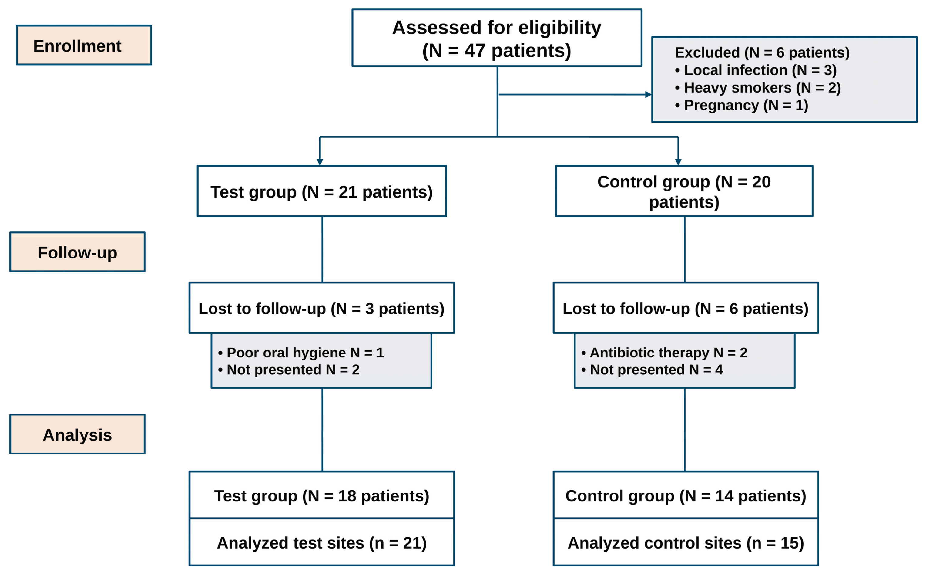
A total of 21 patients were enrolled in the test group and 20 in the control group. Complete 3-month follow-up data were obtained from 18 and 14 patients, respectively (Figure 3). Complete data was analysed from 21 preserved post-extraction sites (test) and 15 naturally healed sites (control).
Figure 3.
Flow chart of the patients. Abbreviations: N, n, number.
The two groups were comparable at baseline, with no statistically significant differences in demographic or behavioural characteristics (Table 1).
Table 1.
Patient characteristics and type of extracted teeth (%, percentage).
Quantitative outcome measures are presented in Table 2.
Table 2.
Primary and secondary outcomes.
Regarding vertical ridge dimensions, statistically significant differences between groups were observed at the 3-month evaluation for the mesial aspect (HmT3, p = 0.002). Mesial bone height (Hm) changed significantly over time in both control [2.0 (1.0–2.0)] and test [1.0 (1.0–1.0)] groups, although the magnitude of bone loss (ΔHm) was significantly greater in the control group than in the test group (p < 0.001).
The values of distal ridge height differed significantly between the groups at T0 (HdT0, p = 0.033) and at T3 (HdT3, p = 0.039). However, the group × time interaction was not significant (p = 0.571), indicating no evidence of a differential change in distal ridge height between the groups over time. Therefore, the difference observed at T3 between groups most likely reflects the pre-existing baseline imbalance rather than an effect of the intervention.
With respect to horizontal ridge dimensions, ridge width at 3 months (WT3) was significantly greater in the test group than in the control group (p = 0.004). Ridge width decreased significantly over the observation period in both the control [ΔW = 2.0 (1.0–2.0)] and test [ΔW = 1.0 (1.0–1.0)] groups, with a significantly greater magnitude of horizontal ridge loss in the control group (p = 0.021).
At the level of buccal mucosa, Tmb significantly decreased in control group in the observation period (p < 0.001). Also, mucosal thickness (Tm) decreased significantly over time in control group [ΔTmb = 1.0 (0.5–1.0)] as compared with test group [ΔTmb = 0.0 (0.0–0.5)] (p < 0.001).
In addition, occlusal mucosal thickness at T3 (TmoT3) was significantly greater in the test group (p = 0.049), in an exploratory analysis.
At 14 days postoperatively, no significant correlation was found between age and healing scores (Spearman’s rho = −0.095, p = 0.580), and no differences were observed between males and females [median 4 (4–5) vs. 4 (4–5), respectively; p = 0.892]. Similarly, alcohol consumption was not associated with healing outcomes [median 4 (4–5) in non-drinkers vs. 4 (3.5–5) in drinkers; p = 0.385].
In an exploratory analysis, a statistically significant difference in Landry wound-healing scores favoring the test group was observed (median 4 [4–5] vs. 4 [3–4], respectively; p = 0.032), although the median score was 4 in both groups and the distributions overlapped. In addition, non-smokers exhibited significantly better healing than smokers, with median scores of 5 (4–5) and 4 (4–4), respectively (p = 0.015).
In this pilot cohort, no statistically detectable difference in the incidence of postoperative complications was observed between groups at 7 days (T1) or 14 days (T2) (p = 0.210 and p = 0.167, respectively). In the test group, two preserved sockets were associated with patient-reported moderate pain at T1, although no clinically relevant local alterations were observed. In the control group, complications were recorded in four extraction sockets at T1, including two cases of postoperative bleeding and two cases of alveolar osteitis accompanied by pain. The sites diagnosed with alveolar osteitis remained symptomatic at the 14-day follow-up (T2).
Effect-size estimates for the between-group differences in change were r_rb = 0.69 (95% CI 0.45–0.93) for Hm, r_rb = 0.08 (95% CI −0.27–0.43) for Hd, r_rb = 0.42 (95% CI 0.07–0.77) for W, and r_rb = 0.63 (95% CI 0.33–0.94) for Tmb.
A patient-level sensitivity analysis in which only one extraction socket per participant was selected (n = 32) produced quite similar findings to the main socket-level analysis. Group differences were statistically significant for changes in mesial ridge height (ΔHm) (p < 0.001) and buccal mucosal thickness (ΔTmb) (p = 0.001), whilst no group difference was found for changes in distal ridge height (ΔHm) (p = 0.479). The change in horizontal ridge width (ΔW) showed the same directional trend, but was non-significant in the patient-level analysis (p = 0.058).
4. Discussion
The present study evaluated the clinical effects of alveolar ridge preservation using hyaluronic acid combined with a collagen cone on post-extraction dimensional ridge changes and selected soft tissue parameters (mucosal thickness and early wound healing), compared with spontaneous healing over a 3-month follow-up period. As the study was non-randomized and the two groups were recruited consecutively, the findings should be interpreted with caution because of the potential for selection and temporal bias. Postoperative complications occurring at 7 and 14 days were also assessed.
Although significant mesial bone resorption (∆Hm) occurs in both groups, alveolar ridge preservation with hyaluronic acid and a collagen cone was associated with significantly better maintenance of mesial vertical ridge dimensions compared with spontaneous healing (p < 0.001). In contrast, ridge preservation was not associated with superior preservation of distal bone height (∆Hd) compared with natural socket healing (p = 0.571). Nevertheless, any finding related to the mesial–distal discrepancy should be interpreted with caution, and no specific biological factor can be inferred from the current data, as heterogeneity between extraction sites, the baseline difference in distal ridge height, measurement error, and the limited sample size may have influenced the result.
The baseline difference in distal ridge height between the groups limits the interpretation of cross-sectional comparisons at T3; accordingly, interpretation of this outcome was based primarily on the longitudinal group × time comparison, which was not associated with significant between-group difference in change. Comparable vertical buccal ridge resorption values of 1 mm and 0.45 mm were reported for ridge preservation performed with a xenograft and collagen matrix with and without adjunctive local application of a hyaluronic acid-based gel, respectively, after 4 months (p = 0.351) [49]. However, comparison with the present results should be made with caution because both the use of grafting material and the route of hyaluronic acid administration differed from those used in our study [49]. The adjunctive use of hyaluronic acid in combination with an allograft and advanced platelet-rich fibrin was associated with significantly lower bone height loss compared with the same protocol performed without hyaluronic acid (0.19 ± 0.51 mm vs. 0.82 ± 0.95 mm, respectively; p = 0.011) [50]. Abaza et al. (2024) reported significantly lower crestal bone resorption following ridge preservation with a hyaluronic acid-based gel compared with untreated control sites (0.33 ± 0.15 mm vs. 0.98 ± 0.18 mm, respectively; p < 0.001) [51]. Moreover, the adjunctive use of hyaluronic acid in combination with a xenograft and a free gingival graft resulted in significantly improved vertical ridge preservation compared with the same protocol performed without hyaluronic acid, yielding mean bone height changes of 0.73 ± 0.52 mm and 1.42 ± 0.16 mm, respectively (p = 0.018) [52].
Other authors observed relatively stable proximal ridge dimensions following flapless tooth extraction after 8 weeks of healing [13]. Overall, the effects of ridge preservation on vertical dimensional changes appear more heterogeneous than those observed horizontally and are strongly influenced by site-specific characteristics, including buccal bone wall integrity, periodontal phenotype, and socket morphology [13,18].
Superior maintenance of horizontal ridge dimensions in test sites (p = 0.021) is consistent with previous systematic reviews reporting that alveolar ridge preservation procedures primarily reduce post-extraction dimensional changes in the horizontal dimension [18,20]. Although the approximately 1 mm between-group difference may be clinically relevant in situations where ridge width is critical for subsequent implant placement, its magnitude should be interpreted cautiously considering the 1 mm resolution of the measurement method.
The present study showed that ridge preservation was associated with more stable buccal mucosal thickness (Tmb) over the healing period (p < 0.001) and greater occlusal mucosal thickness at 3 months (TmoT3) compared with spontaneously healed sockets (p = 0.049). Before extraction, both our groups exhibited a relatively thick buccal soft tissue phenotype, with a median thickness (Tmb) of 2 mm, a condition generally associated with greater extracellular matrix content, collagen density, vascularity, and more favorable healing outcomes [13]. Nevertheless, a reduction in buccal mucosal thickness was observed in non-preserved sites. In contrast, Chappuis et al. (2015) reported spontaneous thickening of the buccal soft tissues following extraction, occurring concomitantly with post-extraction bone resorption during an 8-week healing period [53].
In the present study, the median occlusal mucosal thickness was 2 mm in preserved ridges and 1 mm in non-preserved sockets, whereas Thoma et al. (2020) reported corresponding values of approximately 3 mm and 1.5 mm, respectively [54]. Other clinical studies reported increased soft tissue volume and thickness in preserved sites compared with spontaneous healing [54,55], as well as fewer soft tissue invaginations in sockets covered with collagen matrices compared with sites treated with bone substitutes or left untreated [54]. These findings support our decision to use a collagen cone to seal and protect the socket entrance.
Soft tissue healing plays a critical role in post-extraction ridge repair [56], which justified the use of the modified Landry Wound Healing Index during the early postoperative period. Overall, early wound healing was favorable in both study groups; however, a statistically significant difference in early wound-healing scores favoring the test group was observed (p = 0.032), although the median score was similar between groups. Other studies reported comparable post-operative wound healing outcomes for hyaluronic acid-preserved and natural healed ridges [49].
The assessment of early healing was particularly relevant in the present study because hyaluronic acid has been associated with stimulation of fibroblast activity [57], osteogenic differentiation of mesenchymal stromal cells [58] angiogenesis and soft tissue repair [59], as well as anti-inflammatory and antibacterial effects [28,60]. The product used in the present study contains both cross-linked and non-cross-linked hyaluronic acid. The available evidence regarding the biological effects of these two formulations on local cell responses remains inconsistent [59,61,62]. However, cross-linked molecules enhance the persistence in the surgical sites, which could ameliorate bone healing [39]. The observed bone healing response may be related to the prolonged local persistence of cross-linked hyaluronic acid molecules, together with the presence of the superficially positioned collagen cone [39].
Consistent with the findings of the present study, recent narrative reviews [28] and systematic reviews [39] evaluating alveolar ridge preservation with hyaluronic acid, either alone or in combination with other biomaterials, have reported generally favorable effects on soft tissue healing as well as bone remodelling mostly by stimulating new bone formation [39]. One of the three clinical studies included in a recent systematic review [39] reported significantly greater ridge width and lower radiographic crestal bone resorption in sites treated with hyaluronic acid. However, the outcome assessment was based exclusively on radiographic evaluation and did not include clinical measurements [51]. Moreover, the adjunctive use of hyaluronic acid in combination with a xenograft and a free gingival graft resulted in significantly improved radiographic vertical ridge preservation compared with the same protocol performed without hyaluronic acid [52]. Despite several studies reporting reduced radiographic vertical bone loss following ridge preservation procedures incorporating hyaluronic acid compared with protocols without hyaluronic acid, the overall evidence regarding its effect on vertical ridge preservation remains heterogeneous and inconclusive [39]. Nevertheless, improved soft tissue closure of the socket entrance has been reported in hyaluronic acid-preserved sites when post-extraction ridge protection was achieved through additional sealing procedures during healing [39]. These findings could be clinically relevant, as favorable soft tissue conditions and an adequate width of keratinized mucosa following post-extraction ridge healing have been associated with improved biological stability and long-term outcomes of implant-supported rehabilitations [29,30,31,32], supporting the rationale for the use of a collagen cone in the present study. This concept may extend beyond biomaterials, as preservation of the external contour may also depend on adequate mechanical support and sealing of the coronal/transmucosal aspect of the healing site. In immediate implant therapy, management of the implant–socket gap and transmucosal contour may similarly involve strategies aimed at stabilizing the healing compartment and supporting peri-implant hard- and soft-tissue architecture.
However, the available evidence remains heterogeneous because of differences in study protocols, biomaterial formulations, outcome measures, and patient populations, limiting the formulation of definitive clinical recommendations [39]. No long-term results on ridge preservation with hyaluronic acid -based products are available.
Within this context, the present findings may provide additional clinical evidence regarding the potential influence of hyaluronic acid in association with a collagen cone on post-extraction dimensional changes and soft tissue outcomes after 3 months of healing.
The biomaterials selected in the present study allowed the application of a minimally invasive flapless ridge preservation approach, as elevation of a mucoperiosteal flap may exacerbate post-extraction vertical bone resorption [63,64]. However, the influence of flap elevation on post-extraction dimensional changes remains also controversial [65], and the potential advantages of minimally invasive ridge preservation procedures remain incompletely understood, warranting further investigation [66].
Several limitations should be acknowledged. First, the osseous phenotype of the extraction sites was not characterized, despite evidence indicating that thick buccal bone wall phenotypes (>1 mm) undergo substantially less post-extraction resorption than thin-wall phenotypes [13]. Furthermore, the relatively limited sample size, unequal group distribution, and the absence of standardized radiographic imaging allowing more accurate assessment of hard tissue changes may have influenced the interpretation of the results. Because some patients contributed more than one extraction site, the observations may not have been fully independent. Moreover, site-specific heterogeneity related to tooth morphology or the presence of three- versus four-wall sockets was reported descriptively but could not be reliably assessed due to the small sample size, despite its potential influence on the outcomes. Furthermore, the 1 mm resolution of the periodontal probe and rounding of measurements to the nearest millimeter limited the ability to detect dimensional changes below 1 mm and may have contributed to the narrow distribution of some outcome measures and the lack of examiner blinding may have introduced detection bias particularly for manual measurements and assessment of the Landry healing score.
The non-randomized design should also be considered when interpreting the findings. Future long-term studies using larger and more homogeneous cohorts, together with standardized three-dimensional imaging protocols, including serial intraoral scanning and digital superimposition, are warranted to further clarify the role of hyaluronic acid in alveolar ridge preservation. As four participants contributed with two extraction sockets each, there were some observations included within the main socket-level analyses that were not fully independent. Despite this, a sensitivity analysis conducted using only one extraction socket per participant showed that the results in relation to vertical ridge height and buccal mucosal thickness remained consistent, although the horizontal ridge width result ceased to be statistically significant; this must be considered carefully. As this pilot study was not formally powered to establish clinical efficacy or safety, the findings should be interpreted as preliminary estimates requiring confirmation in adequately powered studies.
5. Conclusions
Within the limitations of this prospective comparative clinical study, alveolar ridge preservation using hyaluronic acid combined with a collagen cone was associated with reduced mesial vertical bone loss and improved preservation of horizontal ridge dimensions compared with spontaneous healing after tooth extraction.
The ridge preservation approach was also associated with more favorable soft tissue outcomes, including better maintenance of buccal mucosal thickness and increased occlusal mucosal thickness after 3 months of healing.
The observed findings should be considered preliminary and hypothesis-generating, as the study was not powered to establish definitive clinical efficacy or safety.
Supplementary Materials
The following supporting information can be downloaded at: https://www.mdpi.com/article/10.3390/surgeries7030104/s1, Table S1: Modified Landry Healing Index used for early wound healing assessment; Table S2: Primary and secondary outcomes at baseline (T0, extraction), 7 days (T1), 14 days (T2), and 3 months (T3) after surgery.
Author Contributions
Conceptualization, P.-A.N., A.R. and S.A.; Methodology, P.-A.N., A.R., P.Ș. and A.S.; Validation, A.R., P.Ș. and S.A.; Formal Analysis, Ș.V. and I.C.M.; Investigation, P.-A.N., A.C. and I.C.M.; Resources, P.-A.N.; Data Curation, Ș.V.; Writing—Original Draft Preparation, P.-A.N., A.R., A.B. and A.S.; Writing—Review & Editing, P.-A.N., A.C., A.R., Ș.V., P.Ș., S.A., I.C.M., A.B., V.T. and A.S.; Visualization, A.C., P.Ș., S.A., V.T. and A.S.; Supervision, A.R. and P.Ș.; Project Administration, P.-A.N. and A.S.; Funding Acquisition, P.-A.N. All authors have read and agreed to the published version of the manuscript.
Funding
The present study was supported partially from an internal research project funded by the Iuliu Hațieganu University of Medicine and Pharmacy Cluj-Napoca, România (Grant no. 881/36/1 December 2022 and Grant no. 772/29/1 November 2023).
Institutional Review Board Statement
The study was conducted in accordance with the Declaration of Helsinki, and approved by the Ethics Committee of the Emergency County Clinical Hospital Cluj-Napoca (Approval No. 1304/4 May 2022) and the Iuliu Hațieganu University of Medicine and Pharmacy, Cluj-Napoca (Approval No. 62/3 January 2022).
Informed Consent Statement
Informed consent was obtained from all subjects involved in the study.
Data Availability Statement
The data presented in this study are available on request from the corresponding author. The data are not publicly available due to patient privacy and ethical restrictions.
Acknowledgments
This work is part of a project of the Ministry of Research, Innovation and Digitization (Romania), through CCCDI–UEFISCDI, project number PN-IV-P7-7.1-PED-2024-1383 (contract no. 120PED/5 May 2025), within PNCDI IV. The authors would like to express their sincere gratitude to the Iuliu Hațieganu University of Medicine and Pharmacy, Cluj-Napoca, Romania for its support in conducting this research (Grant no. 881/36/1 December 2022 and Grant no. 772/29/1 November 2023).
Conflicts of Interest
The authors declare no conflicts of interest.
Abbreviations
The following abbreviations are used in this manuscript:
| T0 | BASELINE |
| T1 | AFTER 7 DAYS |
| T2 | AFTER 14 DAYS |
| T3 | AFTER 3 MONTHS |
References
- Chapple, I.L. Time to Take Periodontitis Seriously. BMJ 2014, 348, g2645. [Google Scholar] [CrossRef] [Scilit] [PubMed]
- Leblebicioglu, B.; Salas, M.; Ort, Y.; Johnson, A.; Yildiz, V.O.; Kim, D.G.; Agarwal, S.; Tatakis, D.N. Determinants of Alveolar Ridge Preservation Differ by Anatomic Location. J. Clin. Periodontol. 2013, 40, 387–395. [Google Scholar] [CrossRef] [Scilit] [PubMed]
- Lambert, F.; Montero, E.; Laleman, I.; de Albornoz, A.C.; Yousfi, H.; Sanz-Sánchez, I. Esthetic and Patient-Reported Outcomes in Immediate Implants with Adjunctive Surgical Procedures to Increase Soft Tissue Thickness/Height: A Systematic Review. Periodontology 2000 2025, 99, 61–80. [Google Scholar] [CrossRef] [Scilit] [PubMed]
- Reichert, C.; Götz, W.; Smeets, R.; Wenghöfer, M.; Jäger, A. The Impact of Nonautogenous Bone Graft on Orthodontic Treatment. Quintessence Int. 2010, 41, 665–672. [Google Scholar] [PubMed]
- Ten Heggeler, J.M.; Slot, D.E.; van der Weijden, G.A. Effect of Socket Preservation Therapies Following Tooth Extraction in Non-Molar Regions in Humans: A Systematic Review. Clin. Oral Implant. Res. 2011, 22, 779–788. [Google Scholar] [CrossRef] [Scilit] [PubMed]
- Schropp, L.; Wenzel, A.; Kostopoulos, L.; Karring, T. Bone Healing and Soft Tissue Contour Changes Following Single-Tooth Extraction: A Clinical and Radiographic 12-Month Prospective Study. Int. J. Periodontics Restor. Dent. 2003, 23, 313–323. [Google Scholar]
- Clementini, M.; Castelluzzo, W.; Ciaravino, V.; Agostinelli, A.; Vignoletti, F.; Ambrosi, A.; De Sanctis, M. The Effect of Immediate Implant Placement on Alveolar Ridge Preservation Compared to Spontaneous Healing after Tooth Extraction: Soft Tissue Findings from a Randomized Controlled Clinical Trial. J. Clin. Periodontol. 2020, 47, 1536–1546. [Google Scholar] [CrossRef] [Scilit] [PubMed]
- Kingsmill, V.J. Post-Extraction Remodeling of the Adult Mandible. Crit. Rev. Oral Biol. Med. 1999, 10, 384–404. [Google Scholar] [CrossRef] [Scilit] [PubMed]
- Yang, S.; Li, Y.; Liu, C.; Wu, Y.; Wan, Z.; Shen, D. Pathogenesis and Treatment of Wound Healing in Patients with Diabetes after Tooth Extraction. Front. Endocrinol. 2022, 13, 949535. [Google Scholar] [CrossRef] [Scilit] [PubMed]
- Jahangiri, L.; Devlin, H.; Ting, K.; Nishimura, I. Current Perspectives in Residual Ridge Remodeling and Its Clinical Implications: A Review. J. Prosthet. Dent. 1998, 80, 224–237. [Google Scholar] [CrossRef] [Scilit] [PubMed]
- Bollen, A.-M.; Taguchi, A.; Hujoel, P.P.; Hollender, L.G. Number of Teeth and Residual Alveolar Ridge Height in Subjects with a History of Self-Reported Osteoporotic Fractures. Osteoporos. Int. 2004, 15, 970–974. [Google Scholar] [CrossRef] [Scilit] [PubMed]
- Saldanha, J.B.; Casati, M.Z.; Neto, F.H.; Sallum, E.A.; Nociti, F.H., Jr. Smoking May Affect the Alveolar Process Dimensions and Radiographic Bone Density in Maxillary Extraction Sites: A Prospective Study in Humans. J. Oral Maxillofac. Surg. 2006, 64, 1359–1365. [Google Scholar] [CrossRef] [Scilit] [PubMed]
- Chappuis, V.; Araujo, M.G.; Buser, D. Clinical Relevance of Dimensional Bone and Soft Tissue Alterations Post-Extraction in Esthetic Sites. Periodontology 2000 2017, 73, 73–83. [Google Scholar] [CrossRef] [Scilit] [PubMed]
- Kim, T.; Kim, S.; Song, M.; Lee, C.; Yagita, H.; Williams, D.W.; Sung, E.C.; Hong, C.; Shin, K.-H.; Kang, M.K.; et al. Removal of Pre-Existing Periodontal Inflammatory Condition before Tooth Extraction Ameliorates Medication-Related Osteonecrosis of the Jaw-Like Lesion in Mice. Am. J. Pathol. 2018, 188, 2318–2327. [Google Scholar] [CrossRef] [Scilit] [PubMed]
- Tsigarida, A.; Toscano, J.; de Brito Bezerra, B.; Geminiani, A.; Barmak, A.B.; Caton, J.; Papaspyridakos, P.; Chochlidakis, K. Buccal Bone Thickness of Maxillary Anterior Teeth: A Systematic Review and Meta-Analysis. J. Clin. Periodontol. 2020, 47, 1326–1343. [Google Scholar] [CrossRef] [Scilit] [PubMed]
- Avila-Ortiz, G.; Gubler, M.; Romero-Bustillos, M.; Nicholas, C.; Zimmerman, M.; Barwacz, C. Efficacy of Alveolar Ridge Preservation: A Randomized Controlled Trial. J. Dent. Res. 2020, 99, 402–409. [Google Scholar] [CrossRef] [Scilit] [PubMed]
- De Angelis, P.; Cavalcanti, C.; Manicone, P.F.; Liguori, M.G.; Rella, E.; De Rosa, G.; Palmieri, A.; D’Addona, A. A Comparison of Guided Bone Regeneration vs. the Shell Technique Using Xenogeneic Bone Blocks in Horizontal Bone Defects: A Randomized Clinical Trial. Dent. J. 2024, 12, 137. [Google Scholar] [CrossRef] [Scilit] [PubMed]
- Avila-Ortiz, G.; Chambrone, L.; Vignoletti, F. Effect of Alveolar Ridge Preservation Interventions Following Tooth Extraction: A Systematic Review and Meta-Analysis. J. Clin. Periodontol. 2019, 46, 195–223. [Google Scholar] [CrossRef] [Scilit] [PubMed]
- De Angelis, P.; De Rosa, G.; Manicone, P.F.; De Giorgi, A.; Cavalcanti, C.; Speranza, A.; Grassi, R.; D’Addona, A. Hard and Soft Tissue Evaluation of Alveolar Ridge Preservation Compared to Spontaneous Healing: A Retrospective Clinical and Volumetric Analysis. Int. J. Implant Dent. 2022, 8, 62. [Google Scholar] [CrossRef] [Scilit] [PubMed]
- MacBeth, N.; Trullenque-Eriksson, A.; Donos, N.; Mardas, N. Hard and Soft Tissue Changes Following Alveolar Ridge Preservation: A Systematic Review. Clin. Oral Implant. Res. 2017, 28, 982–1004. [Google Scholar] [CrossRef] [Scilit] [PubMed]
- Jambhekar, S.; Kernen, F.; Bidra, A.S. Clinical and Histologic Outcomes of Socket Grafting after Flapless Tooth Extraction: A Systematic Review of Randomized Controlled Clinical Trials. J. Prosthet. Dent. 2015, 113, 371–382. [Google Scholar] [CrossRef] [Scilit] [PubMed]
- Parashis, A.O.; Hawley, C.E.; Stark, P.C.; Ganguly, R.; Hanley, J.B.; Steffensen, B. Prospective Clinical and Radiographic Study of Alveolar Ridge Preservation Combining Freeze-Dried Bone Allograft with Two Xenogeneic Collagen Matrices. J. Periodontol. 2016, 87, 416–425. [Google Scholar] [CrossRef] [Scilit] [PubMed]
- Suárez-López Del Amo, F.; Monje, A. Efficacy of Biologics for Alveolar Ridge Preservation/Reconstruction and Implant Site Development: An American Academy of Periodontology Best Evidence Systematic Review. J. Periodontol. 2022, 93, 1827–1847. [Google Scholar] [CrossRef] [Scilit] [PubMed]
- Mardas, N.; Macbeth, N.; Donos, N.; Jung, R.E.; Zuercher, A.N. Is Alveolar Ridge Preservation an Overtreatment? Periodontology 2000 2023, 93, 289–308. [Google Scholar] [CrossRef] [Scilit] [PubMed]
- Alenazi, A.; Alotaibi, A.; Aljaeidi, Y.; Alqhtani, N. The Need for Socket Preservation: A Systematic Review. J. Med. Life 2022, 15, 309–312. [Google Scholar] [CrossRef] [Scilit] [PubMed]
- Jung, R.E.; Ioannidis, A.; Hämmerle, C.H.F.; Thoma, D.S. Alveolar Ridge Preservation in the Esthetic Zone. Periodontology 2000 2018, 77, 165–175. [Google Scholar] [CrossRef] [Scilit] [PubMed]
- Al Yafi, F.; Alchawaf, B.; Nelson, K. What Is the Optimum for Alveolar Ridge Preservation? Dent. Clin. N. Am. 2019, 63, 399–418. [Google Scholar] [CrossRef] [Scilit] [PubMed]
- Nistor, P.A.; Cândea, A.; Micu, I.C.; Soancă, A.; Caloian, C.S.; Bârdea, A.; Roman, A. Advancements in Hyaluronic Acid Effect in Alveolar Ridge Preservation: A Narrative Review. Diagnostics 2025, 15, 137. [Google Scholar] [CrossRef] [Scilit] [PubMed]
- Thoma, D.S.; Naenni, N.; Figuero, E.; Hämmerle, C.H.F.; Schwarz, F.; Jung, R.E.; Sanz-Sánchez, I. Effects of Soft Tissue Augmentation Procedures on Peri-Implant Health or Disease: A Systematic Review and Meta-Analysis. Clin. Oral Implant. Res. 2018, 29, 32–49. [Google Scholar] [CrossRef] [Scilit] [PubMed]
- Ramanauskaite, A.; Schwarz, F.; Sader, R. Influence of Width of Keratinized Tissue on the Prevalence of Peri-Implant Diseases: A Systematic Review and Meta-Analysis. Clin. Oral Implant. Res. 2022, 33, 8–31. [Google Scholar] [CrossRef] [Scilit] [PubMed]
- Mahardawi, B.; Jiaranuchart, S.; Tompkins, K.A.; Pimkhaokham, A. Efficacy of the Autogenous Dentin Graft for Implant Placement: A Systematic Review and Meta-Analysis of Randomized Controlled Trials. Int. J. Oral Maxillofac. Surg. 2023, 52, 604–612. [Google Scholar] [CrossRef] [Scilit] [PubMed]
- Jankowski, T.; Jankowska, A.; Kazimierczak, N.; Kazimierczak, W.; Janiszewska-Olszowska, J. The Significance of Keratinized Mucosa in Implant Therapy: Narrative Literature Review and Case Report Presentation. J. Clin. Med. 2024, 13, 3501. [Google Scholar] [CrossRef] [Scilit] [PubMed]
- Zhang, S.; Sheng, R.; Fan, Z.; Wang, F.; Di, P.; Shi, J.; Zou, D.; Li, D.; Zhang, Y.; Chen, Z.; et al. Expert Consensus on Peri-Implant Keratinized Mucosa Augmentation at Second-Stage Surgery. Int. J. Oral Sci. 2025, 17, 51. [Google Scholar] [CrossRef] [Scilit] [PubMed]
- da Costa Deller, F.A.; Perussolo, J.; de Souza, A.B.; de Oliveira, R.P.; Matarazzo, F.; Araújo, M.G. The Role of the Keratinized Mucosa in Peri-Implant Diseases Onset and Brushing Discomfort: A 10-Year Follow-Up. Clin. Oral Implant. Res. 2026, 37, 785–795. [Google Scholar] [CrossRef] [Scilit] [PubMed]
- Tonetti, M.S.; Sanz, M. Implementation of the New Classification of Periodontal Diseases: Decision-Making Algorithms for Clinical Practice and Education. J. Clin. Periodontol. 2019, 46, 398–405. [Google Scholar] [CrossRef] [Scilit] [PubMed]
- Atieh, M.A.; Shah, M.; Hakam, A.; AlAli, F.; Aboushakra, I.; Alsabeeha, N.H.M. Alveolar Ridge Preservation versus Early Implant Placement in Single Non-Molar Sites: A Systematic Review and Meta-Analysis. Clin. Oral Implant. Res. 2024, 35, 1055–1071. [Google Scholar] [CrossRef] [Scilit] [PubMed]
- Strauss, F.J.; Fukuba, S.; Naenni, N.; Jung, R.; Jonker, B.; Wolvius, E.; Pijpe, J. Alveolar Ridge Changes 1-Year after Early Implant Placement, with or without Alveolar Ridge Preservation at Single-Implant Sites in the Aesthetic Region: A Secondary Analysis of Radiographic and Profilometric Outcomes from a Randomized Controlled Trial. Clin. Implant Dent. Relat. Res. 2024, 26, 356–368. [Google Scholar] [CrossRef] [Scilit] [PubMed]
- James, A.W.; LaChaud, G.; Shen, J.; Asatrian, G.; Nguyen, V.; Zhang, X.; Ting, K.; Soo, C. A Review of the Clinical Side Effects of Bone Morphogenetic Protein-2. Tissue Eng. Part B Rev. 2016, 22, 284–297. [Google Scholar] [CrossRef] [Scilit] [PubMed]
- Bayyigit, L.; Domic, D.; Ulm, C.; Bertl, K.; Stavropoulos, A. Does Hyaluronic Acid Affect Hard Tissue Healing in Alveolar Bone Augmentation Procedures? A Systematic Review of Controlled Clinical Trials. Clin. Oral Investig. 2026, 30, 93. [Google Scholar] [CrossRef] [Scilit] [PubMed]
- Domic, D.; Bertl, K.; Lang, T.; Pandis, N.; Ulm, C.; Stavropoulos, A. Hyaluronic Acid in Tooth Extraction: A Systematic Review and Meta-Analysis of Preclinical and Clinical Trials. Clin. Oral Investig. 2023, 27, 7209–7229. [Google Scholar] [CrossRef] [Scilit] [PubMed]
- Des Jarlais, D.C.; Lyles, C.; Crepaz, N.; Trend Group. Improving the Reporting Quality of Nonrandomized Evaluations of Behavioral and Public Health Interventions: The TREND Statement. Am. J. Public Health 2004, 94, 361–366. [Google Scholar] [CrossRef] [Scilit] [PubMed]
- Broers, D.L.M.; Dubois, L.; de Lange, J.; Su, N.; de Jongh, A. Reasons for Tooth Removal in Adults: A Systematic Review. Int. Dent. J. 2022, 72, 52–57. [Google Scholar] [CrossRef] [Scilit] [PubMed]
- Costea, C.A.; Christodorescu, R.; Soancă, A.; Roman, A.; Micu, I.C.; Stratul, Ș.I.; Rusu, D.; Popescu, D.M.; Popa-Wagner, A.; Bulboacă, A.E. Periodontitis in Ischemic Stroke Patients: Case Definition Challenges of the New Classification Scheme (2018). J. Clin. Med. 2022, 11, 520. [Google Scholar] [CrossRef] [Scilit] [PubMed]
- O’Leary, T.J.; Drake, R.B.; Naylor, J.E. The Plaque Control Record. J. Periodontol. 1972, 43, 38. [Google Scholar] [CrossRef] [Scilit] [PubMed]
- World Health Organization (WHO). Global Health Observatory (GHO) Data: Abstainers (12 Months) and Current Drinkers (12 Months); World Health Organization: Geneva, Switzerland, 2026. Available online: https://www.who.int/data/gho/indicator-metadata-registry/imr-details/456 (accessed on 10 March 2026).
- Landry, R.G.; Turnbull, R.S.; Howley, T. Effectiveness of Benzydamine HCl in the Treatment of Periodontal Post-Surgical Patients. Res. Clin. Forums 1988, 10, 105–118. [Google Scholar]
- Matumoto, E.K.; Corrêa, M.G.; Couso-Queiruga, E.; Monteiro, M.F.; Graham, Z.; Braz, S.H.G.; Ribeiro, F.V.; Pimentel, S.P.; Cirano, F.R.; Casati, M.Z. Influence of Partially Exposed Nonabsorbable Membrane for Alveolar Ridge Preservation: A Randomized Controlled Trial. Clin. Implant Dent. Relat. Res. 2023, 25, 447–457. [Google Scholar] [CrossRef] [Scilit] [PubMed]
- MacBeth, N.D.; Donos, N.; Mardas, N. Alveolar Ridge Preservation with Guided Bone Regeneration or Socket Seal Technique: A Randomized, Single-Blind Controlled Clinical Trial. Clin. Oral Implant. Res. 2022, 33, 681–699. [Google Scholar] [CrossRef] [Scilit] [PubMed]
- Eeckhout, C.; Ackerman, J.; Glibert, M.; Cosyn, J. A Randomized Controlled Trial Evaluating Hyaluronic Acid Gel as Wound Healing Agent in Alveolar Ridge Preservation. J. Clin. Periodontol. 2022, 49, 280–291. [Google Scholar] [CrossRef] [Scilit] [PubMed]
- Kloss, F.R.; Kau, T.; Heimes, D.; Kämmerer, P.W.; Kloss-Brandstätter, A. Enhanced Alveolar Ridge Preservation with Hyaluronic Acid-Enriched Allografts: A Comparative Study of Granular Allografts with and without Hyaluronic Acid Addition. Int. J. Implant Dent. 2024, 10, 42. [Google Scholar] [CrossRef] [Scilit] [PubMed]
- Abaza, G.; Abdel Gaber, H.K.; Afifi, N.S.; Adel-Khattab, D. Injectable Platelet-Rich Fibrin versus Hyaluronic Acid with Bovine-Derived Xenograft for Alveolar Ridge Preservation: A Randomized Controlled Clinical Trial with Histomorphometric Analysis. Clin. Implant Dent. Relat. Res. 2024, 26, 88–102. [Google Scholar] [CrossRef] [Scilit] [PubMed]
- Husseini, B.; Friedmann, A.; Wak, R.; Ghosn, N.; Khoury, G.; El Ghoul, T.; Abboud, C.K.; Younes, R. Clinical and Radiographic Assessment of Cross-Linked Hyaluronic Acid Addition in Demineralized Bovine Bone-Based Alveolar Ridge Preservation: A Human Randomized Split-Mouth Pilot Study. J. Stomatol. Oral Maxillofac. Surg. 2023, 124, 101426. [Google Scholar] [CrossRef] [Scilit] [PubMed]
- Chappuis, V.; Engel, O.; Shahim, K.; Reyes, M.; Katsaros, C.; Buser, D. Soft Tissue Alterations in Esthetic Postextraction Sites: A Three-Dimensional Analysis. J. Dent. Res. 2015, 94, 187S–193S. [Google Scholar] [CrossRef] [Scilit] [PubMed]
- Thoma, D.S.; Bienz, S.P.; Lim, H.C.; Lee, W.Z.; Hämmerle, C.H.; Jung, R.E. Explorative Randomized Controlled Study Comparing Soft Tissue Thickness, Contour Changes, and Soft Tissue Handling of Two Ridge Preservation Techniques and Spontaneous Healing Two Months after Tooth Extraction. Clin. Oral Implant. Res. 2020, 31, 565–574. [Google Scholar] [CrossRef] [Scilit] [PubMed]
- Eeckhout, C.; Seyssens, L.; Glibert, M.; Keppens, L.; Nollet, B.; Lambert, M.; Cosyn, J. A Randomized Controlled Trial Comparing Collagen Matrix to Hemostatic Gelatin Sponge as Socket Seal in Alveolar Ridge Preservation. J. Clin. Med. 2024, 13, 2293. [Google Scholar] [CrossRef] [Scilit] [PubMed]
- Cardaropoli, G.; Araújo, M.; Lindhe, J. Dynamics of Bone Tissue Formation in Tooth Extraction Sites: An Experimental Study in Dogs. J. Clin. Periodontol. 2003, 30, 809–818. [Google Scholar] [CrossRef] [Scilit] [PubMed]
- Schmidt, J.; Pavlík, V.; Suchánek, J.; Nešporová, K.; Soukup, T.; Kapitán, M.; Pilbauerová, N. Low-, Medium-, and High-Molecular-Weight Hyaluronic Acid Effects on Human Dental Pulp Stem Cells in Vitro. Int. J. Biol. Macromol. 2023, 253, 127220. [Google Scholar] [CrossRef] [Scilit] [PubMed]
- Nugraheni, V.I.; Asrianti Bagio, D.; Margono, A.; Julianto, I. The Effect of Hydrogel Hyaluronic Acid on Dentine Sialophosphoprotein Expression of Human Dental Pulp Stem Cells. Eur. Endod. J. 2023, 8, 280–285. [Google Scholar] [CrossRef] [Scilit] [PubMed]
- Fujioka-Kobayashi, M.; Müller, H.D.; Mueller, A.; Lussi, A.; Sculean, A.; Schmidlin, P.R.; Miron, R.J. In Vitro Effects of Hyaluronic Acid on Human Periodontal Ligament Cells. BMC Oral Health 2017, 17, 44. [Google Scholar] [CrossRef] [Scilit] [PubMed]
- Bertl, K.; Bruckmann, C.; Isberg, P.E.; Klinge, B.; Gotfredsen, K.; Stavropoulos, A. Hyaluronan in Non-Surgical and Surgical Periodontal Therapy: A Systematic Review. J. Clin. Periodontol. 2015, 42, 236–246. [Google Scholar] [CrossRef] [Scilit] [PubMed]
- Yu, K.H.; Hou, J.; Jin, Z.; Wu, K.; Xu, S.; Yang, N.; Shen, Y.; Tang, T.; Guo, S. A Cochlear Implant Loaded with Dexamethasone and Coated with Hyaluronic Acid to Inhibit Fibroblast Adhesion and Proliferation. J. Drug Deliv. Sci. Technol. 2018, 46, 173–181. [Google Scholar] [CrossRef] [Scilit]
- Asparuhova, M.B.; Kiryak, D.; Eliezer, M.; Mihov, D.; Sculean, A. Activity of Two Hyaluronan Preparations on Primary Human Oral Fibroblasts. J. Periodontal Res. 2019, 54, 33–45. [Google Scholar] [CrossRef] [Scilit] [PubMed]
- Guruprasad, Y.; Dhurubatha, J.; Kumar, S.; Sultana, R.; Bakshi, H.T.; Desai, D.T. A Comparative Study of the Flap and Flapless Techniques of Ridge Preservation: A Clinical Double-Blinded Study. J. Pharm. Bioallied Sci. 2023, 15, S1065–S1068. [Google Scholar] [CrossRef] [Scilit] [PubMed]
- Lee, J.B.; Chu, S.; Amara, H.B.; Song, H.; Son, M.; Lee, J.; Kim, H.; Koo, K.; Rhyu, I. The Effects of Hyaluronic Acid and Deproteinized Bovine Bone Mineral with 10% Collagen for Ridge Preservation in Compromised Extraction Sockets. J. Periodontol. 2021, 92, 1564–1575. [Google Scholar] [CrossRef] [Scilit] [PubMed]
- Siu, T.L.; Dukka, H.; Saleh, M.H.A.; Tattan, M.; Dib, Z.; Ravidà, A.; Greenwell, H.; Wang, H.L.; Araujo, M.G. Flap versus Flapless Alveolar Ridge Preservation: A Clinical and Histological Single-Blinded, Randomized Controlled Trial. J. Periodontol. 2023, 94, 184–192. [Google Scholar] [CrossRef] [Scilit] [PubMed]
- Araújo, M.G.; Hürzeler, M.B.; Dias, D.R.; Matarazzo, F. Minimal invasiveness in the alveolar ridge preservation, with or without concomitant implant placement. Periodontology 2000 2023, 91, 65–88. [Google Scholar] [CrossRef] [Scilit] [PubMed]

Disclaimer/Publisher’s Note: The statements, opinions and data contained in all publications are solely those of the individual author(s) and contributor(s) and not of MDPI and/or the editor(s). MDPI and/or the editor(s) disclaim responsibility for any injury to people or property resulting from any ideas, methods, instructions or products referred to in the content. |
© 2026 by the authors. Licensee MDPI, Basel, Switzerland. This article is an open access article distributed under the terms and conditions of the Creative Commons Attribution (CC BY) license.