


Abstract
Background: In head and neck reconstructive surgery, flap loss remains a major complication and continues to represent a significant challenge in perioperative management. Although free tissue transfer is widely used, unsatisfactory outcomes are still reported across different flap procedures. Anaesthetic management plays an important role in influencing flap perfusion through its effects on systemic haemodynamics, regional blood flow, and microcirculation. However, there is currently no consensus on universally acceptable haemodynamic targets, and the impact of intraoperative strategies appears to be highly application-specific. Materials and Methods: This narrative review was conducted in accordance with the 2019 SANRA guidelines. PubMed® was used as the primary database for literature selection. Relevant studies addressing anaesthetic management in head and neck free flap surgery were reviewed, with a particular focus on intraoperative haemodynamic control, ischemia–reperfusion injury, fluid and transfusion management, vasoactive agents, and advanced monitoring techniques. Results: Ischemia–reperfusion injury represents a major mechanism of vascular compromise in free flap surgery and has a significant impact on microcirculatory perfusion. The literature suggests that several anaesthetic strategies—including goal-directed fluid therapy, cautious use of vasopressors, and advanced haemodynamic monitoring—may support intraoperative haemodynamic stability and improve flap perfusion. Nevertheless, the magnitude of haemodynamic improvement achievable with these strategies and their effect on graft survival vary according to patient characteristics, surgical factors, and flap type. Conclusions: Current evidence indicates that anaesthetic management has the potential to contribute to improved intraoperative haemodynamic control in head and neck free flap reconstruction, thereby supporting graft viability. However, haemodynamic targets and management strategies cannot be generalised and should be interpreted within specific clinical contexts. Rather than aiming for optimisation, future research should focus on defining acceptable clinical outcomes for individual applications and on evaluating whether achievable haemodynamic improvements are sufficient to reduce flap-related complications to clinically acceptable levels.
1. Introduction
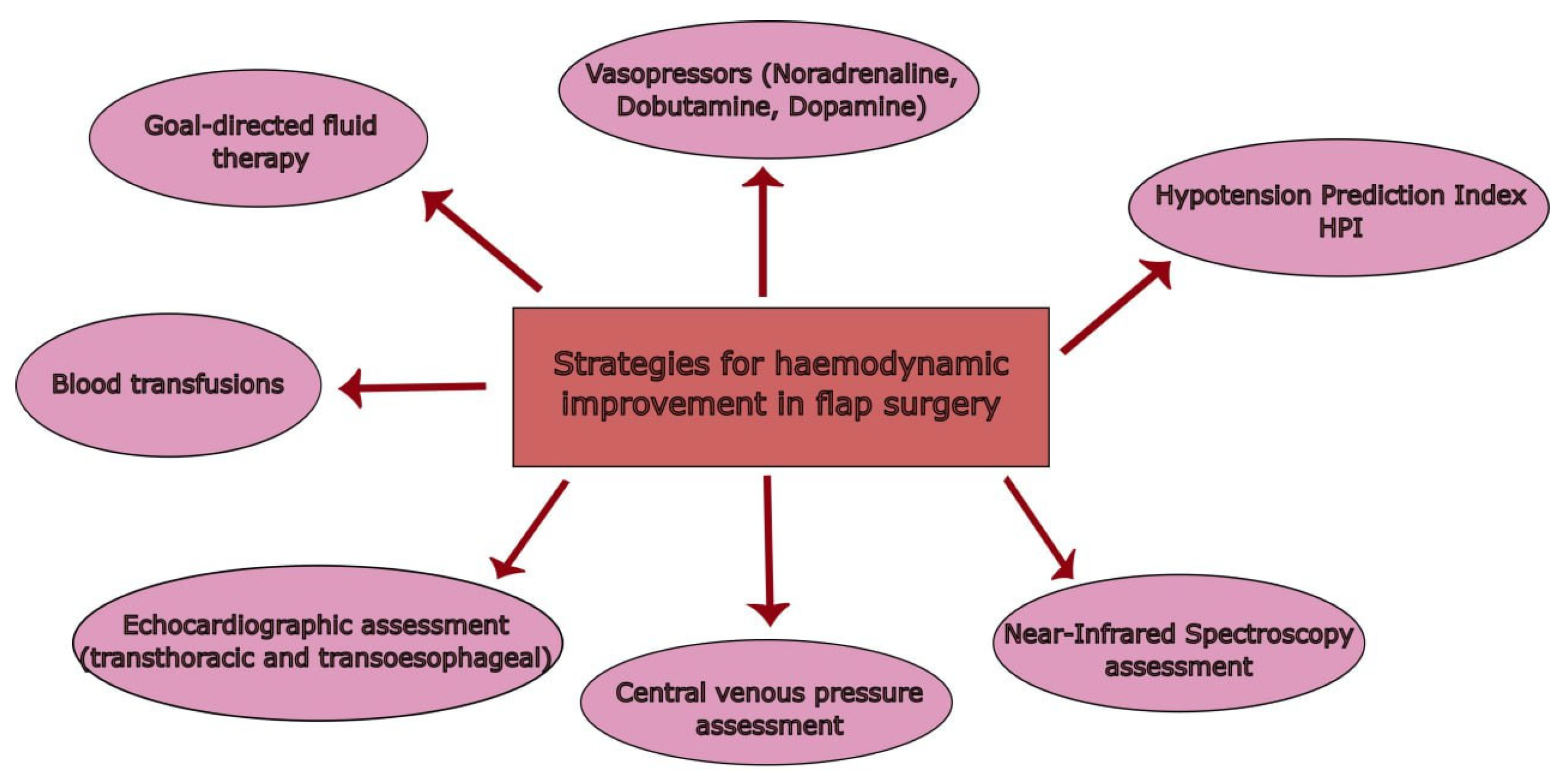
In maxillofacial clinical practice, a wide range of conditions—including predominantly malignant lesions—may involve the jaws and surrounding tissues and require complex surgical management. These conditions often necessitate partial or total resection of affected structures, resulting in defects that compromise both function and aesthetics. Reconstruction is therefore a critical component of treatment, aimed at restoring oral function, facial symmetry, and quality of life. Several reconstructive techniques are available, including local, regional (pedicled), and free tissue flaps [1,2,3]. In technical terms, a flap is a unit of tissue that is transferred from one site (the donor site) to another one (the receiving site) while maintaining its original blood supply. Depending on the location of the donor and recipient sites, flaps can be classified as local or distant (regional) flaps [4]. Although free flaps are widely adopted in head and neck reconstruction because of their versatility and reliability, flap-based procedures as a whole remain associated with a non-negligible rate of unsatisfactory outcomes, including partial or total flap loss. Importantly, it is not always clear whether these complications are primarily related to free flaps alone or are shared across different flap types, as definitions and reporting vary across studies [5]. Despite substantial advances in microsurgical techniques and postoperative monitoring, flap loss continues to represent one of the most significant adverse events in head and neck reconstruction, posing an ongoing challenge for both surgeons and anaesthetists. From an anaesthesiological perspective, perioperative management plays a central role in influencing flap perfusion through its effects on systemic haemodynamics, regional blood flow, fluid balance, temperature control, and the use of vasoactive agents. Haemodynamic instability, excessive fluid administration, or inappropriate vasopressor use may adversely affect microcirculatory flow within the flap and contribute to vascular compromise. However, although numerous strategies have been proposed to support flap perfusion, there is currently no consensus on universally acceptable haemodynamic targets during free flap surgery. Haemodynamic requirements are highly application-specific and depend on patient characteristics, surgical factors, and flap type. As a result, it remains unclear which intraoperative approaches can reliably translate into clinically meaningful improvements in graft survival and complication rates [6,7]. The aim of this narrative review is therefore not to define optimal or ideal haemodynamic conditions, but rather to examine current anaesthetic management strategies that have the potential to improve intraoperative haemodynamic control during head and neck free flap reconstruction. By synthesising the available evidence on fluid management, vasoactive drugs, ischemia–reperfusion injury, and advanced monitoring techniques, this review seeks to clarify what is currently known, highlight existing knowledge gaps, and discuss how anaesthetic management may contribute to reducing flap-related complications to clinically acceptable levels. Figure 1 provides a schematic overview of all the strategies for haemodynamic optimisation in flap surgery that are discussed throughout this article.
Figure 1.
A schematic overview of the strategies for perioperative haemodynamic management in flap surgery discussed throughout this article.
2. Materials and Methods
This narrative review was developed following the various points of the SANRA guidelines, taking as a reference the most recently published version, specifically that of 2019. It is a useful tool as it improves the methodological standards of narrative reviews, providing a useful guide that includes several points to follow, such as explaining the relevance of the article and defining its objectives, the methodology of data searched in the literature, and the underlying scientific reasoning. Our goal in applying these criteria is to achieve greater clarity and completeness for the readers of the article itself [8]. PubMed® was used as the main database for literature selection. The main reason for this choice was the wide availability of peer-reviewed scientific publications. The search was based on the following keywords: “maxillofacial surgery”, “head and neck reconstruction”, “free flaps”, “anaesthesiological management”, “haemodynamic control”, “vasopressors”, “vasoconstrictors”, “ischaemia-reperfusion”, “microvascular free tissue transfer”, “intraoperative echocardiography”, “fluid management”, “transfusion management”, “central venous pressure”, “Near-Infrared Spectroscopy”, “Hypotension prediction index”. Boolean operators (and, or, not) were also used in the search, with the aim of linking keywords together and thus improving quality and sensitivity. These terms were chosen to meet the objectives of this review, to evaluate the best haemodynamic management in cases of maxillofacial surgery with free flap apposition, with the aim of avoiding the adverse consequences associated with poor management. No date restrictions were applied, but abstracts were then manually evaluated and selected to make choices that were as consistent as possible with the objectives of our narrative review.
3. Results
The literature reports a relatively high success rate for head and neck free flap surgery, ranging from 90% to 98% [9,10,11]. Several factors can influence this rate, including patient-related characteristics (such as comorbidities and baseline vascular status) and the experience of the surgical team. Factors that primarily affect flap vascularisation include previous radiotherapy to the operative field, which is associated with increased vascular congestion, and systemic diseases that impair vascular function, such as diabetes mellitus or renal failure [12,13]. With regard to surgical technique, the literature indicates that the success rate is mainly related to the experience of the surgical team with a specific technique than to the type of flap used. In fact, no significant differences in success have been demonstrated between musculocutaneous, fasciocutaneous, and perforator-based flaps [14,15]. Another factor that undeniably affects surgical outcome is the degree of malignancy of the lesion, probably because more aggressive tumours often require more extensive neck dissections and therefore cause greater vascular damage [16]. Careful monitoring of flap vascularisation is also a key determinant of surgical success, allowing early detection of any abnormalities and timely intraoperative haemodynamic optimisation [17]. Despite the multiple possible causes of flap failure, ischemia–reperfusion (I/R) injury remains the main cause of vascular compromise in free flap surgery and the leading cause of flap loss in the postoperative period [18,19]. This is particularly critical in this patient population, as even brief periods of ischemia can be extremely harmful. Ischemia–reperfusion injury leads to the generation of reactive oxygen species (ROS), which are highly damaging to tissues. The ischemic phase is followed by reperfusion, during which restoration of blood flow and oxygen delivery to the ischemic tissue coincides with an influx of ROS. This process triggers cellular, inflammatory, and metabolic alterations, causing structural and functional damage and contributing to tissue necrosis [20]. The combination of inflammation and increased ROS production in the early stages of reperfusion has a marked impact on the microcirculation of the entire flap [21]. In the most severe cases, this process progresses to complete flap necrosis, most commonly due to pedicle thrombosis leading to vascular insufficiency. In such circumstances, immediate revision of the anastomosis is essential to restore adequate blood flow. However, while this intervention is crucial to salvage the flap, it can aggravate I/R injury through several mechanisms, including intravascular haemoconcentration, endothelial swelling, increased interstitial oedema, and amplification of inflammatory responses. After a critical threshold is reached, these changes may also culminate in the no-reflow phenomenon, representing another mechanism that can result in total flap loss [22,23,24]. In clinical practice, several strategies are available to protect tissues from oxidative damage. These include treatment with inhaled nitric oxide, which exerts protective effects through its antioxidant and anti-inflammatory properties [25]. Various studies also support the use of anti-leukocyte therapies, particularly for their ability to limit the release of pro-inflammatory mediators by leukocytes [26]. Finally, the administration of antioxidants before surgery may play a preventive role, replenishing antioxidant reserves depleted by ROS and thereby helping to reduce the risk of I/R injury [27]. From an anaesthesiological standpoint, it is therefore crucial to implement perioperative strategies aimed at supporting and maintaining adequate haemodynamic control during surgery. Below, we summarise the main approaches described in the literature.
3.1. Vasopressors
The intraoperative use of vasopressors can help maintain haemodynamic stability, which is crucial as it may limit the overall volume of fluids administered and, consequently, reduce fluid-related postoperative complications [28,29]. Overall, the use of vasopressors appears to be safe in free flap transfer procedures for head and neck reconstruction. In particular, the use of agents such as phenylephrine, ephedrine or calcium chloride administered as intravenous boluses does not appear to be associated with increased rates of flap failure or other postoperative morbidity in this setting [30]. In general, the literature supports the judicious use of vasopressors to maintain hemodynamic stability, guided by well-defined algorithms based on intraoperative target blood pressure values. It also emphasises the importance of developing clear protocols in order to tailor vasopressor therapy to the specific needs of each individual patient [31]. A recent study by Ooms et al. demonstrated a direct correlation between microvascular free flap perfusion and intraoperative noradrenaline dose, suggesting that the dosage of this vasopressor can be used to modulate flap perfusion and thereby support flap viability and survival [32]. The role of other agents, particularly dobutamine, has also been investigated in this type of surgery. Compared to norepinephrine, both drugs have been shown to increase blood flow to the free flap. However, the main limitation of noradrenaline is its vasoconstrictive effect on the flap itself, an effect that is not observed with dobutamine; for this reason, dobutamine may be preferable to noradrenaline in this context [33]. The dose recommended in the literature to improve flap blood flow while minimising systemic cardiovascular effects is 4–5 µg/kg/min [34]. Another very promising drug in this regard is dopamine, a vasopressor that, at low doses (2–10 μg/kg/min), has a predominant effect on beta receptors rather than alpha receptors. The use of this agent has been shown to increase blood flow to the free flap, also helping to maintain adequate blood pressure and reducing the degree of peripheral vasoconstriction typically associated with noradrenaline. However, this drug should be used with caution because of its potential cardiovascular side effects, such as arrhythmias and tachycardia [35]. The vasopressors discussed above and their main positive and negative effects on free flaps are summarised in Table 1.
Table 1.
Vasopressor agents and their positive and negative effects on free flaps.
3.2. Fluid Management
Goal-directed fluid therapy is a valuable tool for intraoperative haemodynamic management. When guided by minimally invasive cardiac output monitoring, it can help optimise circulatory status and, importantly, avoid fluid overload [36,37]. Optimal haemodynamic conditions for free flaps include a hyperdynamic circulation characterised by high cardiac output, peripheral vasodilation, and a wide pulse pressure [38]. In this setting, minimally invasive monitoring systems can be used to derive key parameters such as cardiac output, cardiac index (CI) and stroke volume index (SVI) from the arterial pulse waveform [39]. Of particular interest is the study by Al Saied et al., which compared free flap survival in patients managed predominantly with fluid therapy versus vasopressors. The authors demonstrated a clear advantage, with higher flap survival and fewer adverse effects, in the group managed with goal-directed fluid therapy compared with vasopressor-based management [40].
3.3. Haemoglobin and Red Cell Blood Transfusions
Transfusion management is a key anaesthesiological challenge in major surgery, including head and neck procedures involving free flaps. These operations can cause significant intraoperative blood loss, often necessitating transfusions. A target haematocrit between 30% and 35% appears to provide the best compromise between blood viscosity and flow, while optimising oxygen-carrying capacity [41]. A postoperative haematocrit threshold of 25% has been proposed as an indication for transfusion. Adoption of this threshold has been associated with a reduction in both transfusion rates and free flap failure [42].
3.4. Transthoracic and Transesophageal Echocardiography
In recent years, new strategies have been developed to enable more accurate and precise intraoperative monitoring, thereby supporting optimal haemodynamic management. Among these, echocardiography, in both its transthoracic and transoesophageal forms, plays a key role. This technique was initially introduced in cardiac surgery to support more comprehensive anaesthesiological management and to facilitate the identification of haemodynamic instability during general anaesthesia [43]. Subsequently, its use has become established in non-cardiac surgery as well, where it represents a valuable tool for accurately diagnosing intraoperative problems such as hypovolaemia or right or left ventricular failure [44]. Transesophageal echocardiography, in particular, has recently gained widespread use in clinical practice as a useful modality for detecting severe intraoperative haemodynamic instability. It represents a valid alternative to pulmonary artery catheterisation, as it is less invasive and capable of providing detailed information on atrial and ventricular function [45]. In addition, it allows the identification of valvular abnormalities and offers a more accurate assessment of left ventricular end-diastolic volume than that obtained from pulmonary capillary wedge pressure [46]. Among the echocardiographic parameters useful for assessing intraoperative volume status, one of the most important is the diameter of the inferior vena cava and its respiratory variation. A reduced caval diameter with marked collapsibility suggests inadequate intravascular volume [47,48]. Furthermore, Gouveia et al. have proposed a protocol that incorporates several echocardiographic variables indicative of the patient’s haemodynamic profile. In addition to vena cava assessment, this includes calculation of left ventricular cardiac output (CO) using the velocity–time integral (VTI), derived from the time–velocity curve of blood flow. Stroke volume is obtained by multiplying the VTI by the cross-sectional area of the left ventricular outflow tract (LVOT). Left ventricular systolic function can then be evaluated using MAPSE (Mitral Annular Plane Systolic Excursion), which measures the systolic displacement of the mitral annulus towards the apex in the apical four-chamber view and reflects longitudinal contractile function [49]. Right ventricular dysfunction is a recognised predictor of increased intraoperative mortality, and echocardiography can therefore also be used to evaluate right ventricular performance. In this regard, the most used parameter is TAPSE (Tricuspid Annular Plane Systolic Excursion), which quantifies the systolic movement of the tricuspid annulus [50]. Although no specific data are currently available on the use of echocardiography for haemodynamic assessment in head and neck free flap surgery, its high accuracy and minimally invasive nature suggest that it could be a valuable adjunct in this setting, where meticulous monitoring is essential to ensure flap viability.
3.5. CVP and Central Venous Catheterisation
Regarding other haemodynamic parameters, central venous pressure (CVP) is less accurate than cardiac output (CO) for assessing a patient’s volume status and is therefore not considered a useful parameter for haemodynamic monitoring [51]. The placement of a central venous catheter may facilitate more effective fluid management; however, the high rate of malposition and displacement of catheters inserted in the neck region, which is directly involved in the surgical field, must be taken into account. Current evidence suggests that peripherally inserted central catheters (PICCs) or midline catheters are more suitable options in this context [52,53].
3.6. Near-Infrared Spectroscopy
In the field of intraoperative monitoring, the use of near-infrared spectroscopy (NIRS) has become increasingly widespread in recent years. This technique provides information on tissue oxygenation and metabolism [54]. A relevant application is its use in assessing flap oxygenation, providing information on the effectiveness of its perfusion. In particular, it has been shown that low levels of tissue saturation identified using this method are correlated with the risk of flap ischemia and necrosis, thereby impacting the overall success rate of the surgical procedure [55,56]. This technique has also demonstrated a significant ability to predict alterations in free flap blood flow well before the onset of clinical signs [57]. The potential role of cerebral NIRS has so far been investigated only in head and neck surgery involving carotid artery lesions, where it is used to assess the adequacy of cerebral blood flow during these high-risk procedures [58,59].
3.7. Hypotension Prediction Index (HPI)
A recently introduced and useful intraoperative monitoring tool is represented by the Hypotension Prediction Index (HPI). This algorithm uses machine learning to analyse the arterial pressure waveform and detect the development of hypotension early and with high accuracy [60]. This method has been shown to be superior to standard haemodynamic monitoring in reducing the incidence and severity of intraoperative hypotensive episodes during non-cardiac surgery [61]. Focusing specifically on its use in head and neck free flap surgery, the relevance of this technology in this patient population is evident. It allows for more accurate and precise calibration of fluid therapy and vasopressors, with the aim of avoiding sudden pressure fluctuations and thereby ensuring adequate flap perfusion [60,62]. Moreover, intraoperative use of HPI-guided haemodynamic management has been shown to improve flap oxygenation and perfusion up to 24 h after surgery [63].
3.8. Complications and Prevention
Free flap surgery is associated with several complications, most of which are related to flap loss. Clinical studies indicate that the primary cause of flap loss is damage to the flap’s vascular system, particularly thrombotic events affecting the flap itself [64]. This complication is most frequently observed in cancer patients, who have an underlying tendency toward hypercoagulability [65]. Insufficient blood flow, due to reduced perfusion pressure at the arteriolar level, has been identified as the main cause of these ischemic complications [66]. Considering these factors, intraoperative haemodynamic management appears is of fundamental importance. As previously described, a key strategy for preventing complications, particularly ischemic flap loss, is maintaining adequate intravascular volume with fluid therapy, which may be combined, when necessary, with vasopressor support in order to maintain haemodynamic stability [40,67].
4. Conclusions
Free flap reconstruction in head and neck surgery represents a complex clinical scenario in which surgical success depends on the delicate interaction between microvascular technique and perioperative physiological management. From an anaesthesiological standpoint, intraoperative haemodynamic management plays a crucial role not only in maintaining overall patient stability, but also in supporting adequate flap perfusion and reducing the risk of vascular compromise. The available literature suggests that several anaesthetic strategies—including goal-directed fluid therapy, judicious use of vasoactive agents, and advanced haemodynamic monitoring—can contribute to improved control of intraoperative blood pressure and cardiac output. However, the evidence does not support the definition of universally optimal haemodynamic targets. Instead, the magnitude and clinical relevance of haemodynamic improvements appear to be highly dependent on the specific surgical application, patient characteristics, and flap type. Rather than aiming for optimisation or best possible outcomes, future research should focus on defining what constitutes an acceptable clinical outcome for specific reconstructive scenarios. A top-down approach would begin by identifying procedure-specific benchmarks for flap survival and complication rates, then determining the haemodynamic conditions required to achieve these benchmarks, and finally evaluating which anaesthetic strategies are capable of meeting these requirements. Conversely, a bottom-up approach would assess the degree of haemodynamic improvement achievable with individual interventions—such as vasopressors, fluid strategies, or monitoring technologies—and examine whether these improvements translate into meaningful gains in flap viability for defined clinical contexts. In this perspective, advanced monitoring tools such as echocardiography, near-infrared spectroscopy, and machine learning-based algorithms like the Hypotension Prediction Index may represent valuable adjuncts, not as universal solutions, but as components of a tailored, application-specific approach to haemodynamic management. Clarifying how these tools can be integrated into structured perioperative pathways remains an important area for future investigation. In conclusion, anaesthetic management has the potential to meaningfully support graft survival in head and neck free flap surgery, but its role should be framed in terms of achievable improvements and clinical relevance rather than optimisation. A clearer definition of acceptable outcomes and a structured evaluation of haemodynamic strategies will be essential to translate physiological control into tangible benefits for patients.
Author Contributions
Conceptualisation, M.G. and B.A.; methodology, M.G. and G.F.; validation, M.F. and C.G.; formal analysis, S.L.; investigation, A.F.; data curation, A.F. and S.L.; writing—original draft preparation, A.F. and S.L.; writing—review and editing, A.F. and S.L.; visualisation, M.G., B.E. and M.F.; supervision, B.E., G.F., C.G. and B.A.; project administration, M.G. All authors have read and agreed to the published version of the manuscript.
Funding
This research received no external funding.
Institutional Review Board Statement
Not applicable.
Data Availability Statement
The original data presented in the study are openly available in PubMed at https://pubmed.ncbi.nlm.nih.gov.
Acknowledgments
The authors have reviewed and edited the output and take full responsibility for the content of this publication.
Conflicts of Interest
The authors declare no conflicts of interest.
Abbreviations
The following abbreviations are used in this manuscript:
| DVT | deep vein thrombosis |
| ICU | intensive care unit |
| I/R | ischemia–reperfusion |
| ROS | reactive oxygen species |
| CI | cardiac index |
| SVI | stroke volume index |
| CO | cardiac output |
| VTI | Velocity Time Integral |
| LVOT | left ventricular outflow tract |
| MAPSE | Mitral Annular Plane Systolic Excursion |
| TAPSE | Tricuspid Annular Plane Systolic Excursion |
| CVP | central venous pressure |
| PICC | peripherally inserted central venous catheters |
| NIRS | near-infrared spectroscopy |
| HPI | Hypotension Prediction Index |
References
- Thapliyal, G.K. Peterson’s Principles of Oral & Maxillofacial Surgery. Med. J. Armed Forces India 2006, 62, 89. [Google Scholar] [CrossRef] [Scilit] [PubMed Central]
- Valentini, V.; Gennaro, P.; Torroni, A.; Longo, G.; Aboh, I.V.; Cassoni, A.; Battisti, A.; Anelli, A. Scapula free flap for complex maxillofacial reconstruction. J. Craniofac. Surg. 2009, 20, 1125–1131. [Google Scholar] [CrossRef] [Scilit] [PubMed]
- Achal, K.S.; Farrell, C.; Smith, A.B.; Mücke, T.; Mitchell, D.A.; Kanatas, A.N. Anterolateral thigh skinfold thickness and the European head and neck cancer patient: A prospective study. J. Craniomaxillofac. Surg. 2011, 39, 111–112. [Google Scholar] [CrossRef] [Scilit] [PubMed]
- Eckardt, A.M.; Kokemüller, H.; Tavassol, F.; Gellrich, N.C. Reconstruction of oral mucosal defects using the nasolabial flap: Clinical experience with 22 patients. Head Neck Oncol. 2011, 3, 28. [Google Scholar] [CrossRef] [Scilit] [PubMed] [PubMed Central]
- Ojuva, A.M.; Rocans, R.P.; Zarins, J.; Bine, E.; Mahauri, I.; Donina, S.; Mamaja, B.; Vanags, I. Novel Challenges and Opportunities for Anesthesia and Perioperative Care in Microvascular Flap Surgery: A Narrative Review. Clin. Pract. 2024, 14, 2187–2201. [Google Scholar] [CrossRef] [Scilit] [PubMed] [PubMed Central]
- Hagau, N.; Longrois, D. Anesthesia for free vascularized tissue transfer. Microsurgery 2009, 29, 161–167. [Google Scholar] [CrossRef] [Scilit] [PubMed]
- Lauretta, M.; Caporali, L.; Manera, S.; Prucher, G.M.; Melotti, R. Anaesthetic Challenging in Microsurgical Flap Reconstruction: A Systematic Review. J. Anesth. Clin. Res. 2018, 9, 2. [Google Scholar] [CrossRef]
- Baethge, C.; Goldbeck-Wood, S.; Mertens, S. SANRA-a scale for the quality assessment of narrative review articles. Res. Integr. Peer Rev. 2019, 4, 5. [Google Scholar] [CrossRef] [Scilit] [PubMed] [PubMed Central]
- Urken, M.L.; Weinberg, H.; Buchbinder, D.; Moscoso, J.F.; Lawson, W.; Catalano, P.J.; Biller, H.F. Microvascular free flaps in head and neck reconstruction. Report of 200 cases and review of complications. Arch. Otolaryngol. Head Neck Surg. 1994, 120, 633–640. [Google Scholar] [CrossRef] [Scilit] [PubMed]
- Haughey, B.H.; Wilson, E.; Kluwe, L.; Piccirillo, J.; Fredrickson, J.; Sessions, D.; Spector, G. Free flap reconstruction of the head and neck: Analysis of 241 cases. Otolaryngol. Head Neck Surg. 2001, 125, 10–17. [Google Scholar] [CrossRef] [Scilit] [PubMed]
- Hanasono, M.M.; Corbitt, C.A.; Yu, P.; Skoracki, R.J. Success of sequential free flaps in head and neck reconstruction. J. Plast. Reconstr. Aesthet. Surg. 2014, 67, 1186–1193. [Google Scholar] [CrossRef] [Scilit] [PubMed]
- Choi, J.W.; Kim, Y.C.; Jeon, D.N.; Jeong, W.S.; Koh, K.S.; Oh, T.S.; Eom, J.S.; Kim, E.K.; Hong, J.P.; Suh, H.P. Impact of Recipient Vein Selection on Venous Patency and Free Flap Survival in 652 Head and Neck Reconstructions. J. Reconstr. Microsurg. 2020, 36, 73–81. [Google Scholar] [CrossRef] [Scilit] [PubMed]
- Ishimaru, M.; Ono, S.; Suzuki, S.; Matsui, H.; Fushimi, K.; Yasunaga, H. Risk Factors for Free Flap Failure in 2846 Patients with Head and Neck Cancer: A National Database Study in Japan. J. Oral Maxillofac. Surg. 2016, 74, 1265–1270. [Google Scholar] [CrossRef] [Scilit] [PubMed]
- Spoerl, S.; Schoedel, S.; Spanier, G.; Mueller, K.; Meier, J.K.; Reichert, T.E.; Ettl, T. A decade of reconstructive surgery: Outcome and perspectives of free tissue transfer in the head and neck. Experience of a single center institution. Oral Maxillofac. Surg. 2020, 24, 173–179. [Google Scholar] [CrossRef] [Scilit] [PubMed] [PubMed Central]
- Lin, Y.; He, J.F.; Zhang, X.; Wang, H.M. Intraoperative factors associated with free flap failure in the head and neck region: A four-year retrospective study of 216 patients and review of the literature. Int. J. Oral Maxillofac. Surg. 2019, 48, 447–451. [Google Scholar] [CrossRef] [Scilit] [PubMed]
- Üstün, G.G.; Kaplan, G.O.; Sert, G.; Uzun, H. Flap loss in head and neck reconstruction: Is there a singular cause for failure? J. Plast. Reconstr. Aesthet. Surg. 2024, 91, 353–359. [Google Scholar] [CrossRef] [Scilit] [PubMed]
- Chang, E.I.; Zhang, H.; Liu, J.; Yu, P.; Skoracki, R.J.; Hanasono, M.M. Analysis of risk factors for flap loss and salvage in free flap head and neck reconstruction. Head Neck 2016, 38, E771–E775. [Google Scholar] [CrossRef] [Scilit] [PubMed]
- Wang, W.Z.; Baynosa, R.C.; Zamboni, W.A. Update on ischemia-reperfusion injury for the plastic surgeon: 2011. Plast. Reconstr. Surg. 2011, 128, 685e–692e. [Google Scholar] [CrossRef] [Scilit] [PubMed]
- van den Heuvel, M.G.; Buurman, W.A.; Bast, A.; van der Hulst, R.R. Review: Ischaemia-reperfusion injury in flap surgery. J. Plast. Reconstr. Aesthet. Surg. 2009, 62, 721–726. [Google Scholar] [CrossRef] [Scilit] [PubMed]
- Camargo, C.P.; Margarido, N.F.; Guandelini, E.; Vieira, G.A.; Jacomo, A.L.; Gemperli, R. Description of a new experimental model skin flap for studying skin viability in rats. Acta Cir. Bras. 2014, 29, 166–170. [Google Scholar] [CrossRef] [Scilit] [PubMed]
- Im, M.J.; Manson, P.N.; Bulkley, G.B.; Hoopes, J.E. Effects of superoxide dismutase and allopurinol on the survival of acute island skin flaps. Ann. Surg. 1985, 201, 357–359. [Google Scholar] [CrossRef] [Scilit] [PubMed] [PubMed Central]
- Harder, Y.; Amon, M.; Laschke, M.W.; Schramm, R.; Rücker, M.; Wettstein, R.; Bastiaanse, J.; Frick, A.; Machens, H.G.; Küntscher, M.; et al. An old dream revitalised: Preconditioning strategies to protect surgical flaps from critical ischaemia and ischaemia-reperfusion injury. J. Plast. Reconstr. Aesthet. Surg. 2008, 61, 503–511. [Google Scholar] [CrossRef] [Scilit] [PubMed]
- Menger, M.D.; Laschke, M.W.; Amon, M.; Schramm, R.; Thorlacius, H.; Rücker, M.; Vollmar, B. Experimental models to study microcirculatory dysfunction in muscle ischemia-reperfusion and osteomyocutaneous flap transfer. Langenbecks Arch. Surg. 2003, 388, 281–290. [Google Scholar] [CrossRef] [Scilit] [PubMed]
- Menger, M.D.; Rücker, M.; Vollmar, B. Capillary dysfunction in striated muscle ischemia/reperfusion: On the mechanisms of capillary “no-reflow”. Shock 1997, 8, 2–7. [Google Scholar] [CrossRef] [Scilit] [PubMed]
- Phillips, L.; Toledo, A.H.; Lopez-Neblina, F.; Anaya-Prado, R.; Toledo-Pereyra, L.H. Nitric oxide mechanism of protection in ischemia and reperfusion injury. J. Investig. Surg. 2009, 22, 46–55. [Google Scholar] [CrossRef] [Scilit] [PubMed]
- Panés, J.; Perry, M.; Granger, D.N. Leukocyte-endothelial cell adhesion: Avenues for therapeutic intervention. Br. J. Pharmacol. 1999, 126, 537–550. [Google Scholar] [CrossRef] [Scilit] [PubMed] [PubMed Central]
- Pisoschi, A.M.; Pop, A. The role of antioxidants in the chemistry of oxidative stress: A review. Eur. J. Med. Chem. 2015, 97, 55–74. [Google Scholar] [CrossRef] [Scilit] [PubMed]
- Naik, A.N.; Freeman, T.; Li, M.M.; Marshall, S.; Tamaki, A.; Ozer, E.; Agrawal, A.; Kang, S.Y.; Old, M.O.; Seim, N.B. The Use of Vasopressor Agents in Free Tissue Transfer for Head and Neck Reconstruction: Current Trends and Review of the Literature. Front. Pharmacol. 2020, 11, 1248. [Google Scholar] [CrossRef] [Scilit] [PubMed] [PubMed Central]
- Gardner, J.R.; Gau, V.; Page, P.; Dunlap, Q.; King, D.; Crabtree, D.; Sunde, J.; Vural, E.; Moreno, M.A. Association of Continuous Intraoperative Vasopressor Use with Reoperation Rates in Head and Neck Free-Flap Reconstruction. JAMA Otolaryngol. Head Neck Surg. 2021, 147, 1059–1064. [Google Scholar] [CrossRef] [Scilit] [PubMed] [PubMed Central]
- Fang, L.; Liu, J.; Yu, C.; Hanasono, M.M.; Zheng, G.; Yu, P. Intraoperative Use of Vasopressors Does Not Increase the Risk of Free Flap Compromise and Failure in Cancer Patients. Ann. Surg. 2018, 268, 379–384. [Google Scholar] [CrossRef] [Scilit] [PubMed]
- Taylor, R.J.; Patel, R.; Wolf, B.J.; Stoll, W.D.; Hornig, J.D.; Skoner, J.M.; Hand, W.R.; Day, T.A. Intraoperative vasopressors in head and neck free flap reconstruction. Microsurgery 2021, 41, 5–13. [Google Scholar] [CrossRef] [Scilit] [PubMed] [PubMed Central]
- Ooms, M.; Winnand, P.; Heitzer, M.; Vohl, N.; Katz, M.; Bickenbach, J.; Hölzle, F.; Modabber, A. Impact of Vasopressors on Microvascular Free Flap Perfusion in Head and Neck Reconstruction. Microsurgery 2025, 45, e70095. [Google Scholar] [CrossRef] [Scilit] [PubMed] [PubMed Central]
- Eley, K.A.; Young, J.D.; Watt-Smith, S.R. Epinephrine, norepinephrine, dobutamine, and dopexamine effects on free flap skin blood flow. Plast. Reconstr. Surg. 2012, 130, 564–570. [Google Scholar] [CrossRef] [Scilit] [PubMed]
- Kim, S.H.; Moon, Y.J.; Kim, J.W.; Hong, J.Y.; Kim, W.J.; Hwang, J.H. Effective dose of dobutamine in augmenting free flap blood flow during reconstructive surgery of the lower extremity. Medicine 2019, 98, e15358. [Google Scholar] [CrossRef] [Scilit] [PubMed] [PubMed Central]
- Raittinen, L.; Kääriäinen, M.T.; Lopez, J.F.; Pukander, J.; Laranne, J. The Effect of Norepinephrine and Dopamine on Radial Forearm Flap Partial Tissue Oxygen Pressure and Microdialysate Metabolite Measurements: A Randomized Controlled Trial. Plast. Reconstr. Surg. 2016, 137, 1016e–1023e. [Google Scholar] [CrossRef] [Scilit] [PubMed]
- Goswami, U.; Jain, A. Anaesthetic implications of free-flap microvascular surgery for head and neck malignancies—A relook. J. Anaesthesiol. Clin. Pharmacol. 2021, 37, 499–504. [Google Scholar] [CrossRef] [Scilit] [PubMed] [PubMed Central]
- McCauley, P.; Moore, M.; Duggan, E. Anaesthesia for reconstructive free flap surgery for head and neck cancer. Br. J. Hosp. Med. 2022, 83, 1–9. [Google Scholar] [CrossRef] [Scilit] [PubMed]
- Pearse, R.M.; Ackland, G.L. Perioperative fluid therapy. BMJ 2012, 344, e2865. [Google Scholar] [CrossRef] [Scilit] [PubMed]
- Funk, D.; Bohn, J.; Mutch, W.; Hayakawa, T.; Buchel, E.W. Goal-directed fluid therapy for microvascular free flap reconstruction following mastectomy: A pilot study. Plast. Surg. 2015, 23, 231–234. [Google Scholar] [CrossRef] [Scilit] [PubMed] [PubMed Central]
- Al Saied, G.; Almutairi, H.M.; Alharbi, Y.; Almohanna, M.; Almutairi, A. Comparison Between the Impact of Vasopressors and Goal-Directed Fluid Therapy on the Management of Free Flap Reconstruction of Head and Neck and Monitoring in ICU. Cureus 2020, 12, e12108. [Google Scholar] [CrossRef] [Scilit] [PubMed] [PubMed Central]
- Mak, Q.; Chan, H.; Irwin, M. Anaesthesia for plastic and reconstructive surgery. Anaesth. Intensive Care Med. 2023, 24, 800–805. [Google Scholar] [CrossRef] [Scilit]
- Rossmiller, S.R.; Cannady, S.B.; Ghanem, T.A.; Wax, M.K. Transfusion criteria in free flap surgery. Otolaryngol. Head Neck Surg. 2010, 142, 359–364. [Google Scholar] [CrossRef] [Scilit] [PubMed]
- Shanewise, J.S.; Cheung, A.T.; Aronson, S.; Stewart, W.J.; Weiss, R.L.; Mark, J.B.; Savage, R.M.; Sears-Rogan, P.; Mathew, J.P.; Quiñones, M.A.; et al. ASE/SCA guidelines for performing a comprehensive intraoperative multiplane transesophageal echocardiography examination: Recommendations of the American Society of Echocardiography Council for Intraoperative Echocardiography and the Society of Cardiovascular Anesthesiologists Task Force for Certification in Perioperative Transesophageal Echocardiography. Anesth. Analg. 1999, 89, 870–884. [Google Scholar] [CrossRef] [Scilit] [PubMed]
- Jasudavisius, A.; Arellano, R.; Martin, J.; McConnell, B.; Bainbridge, D. A systematic review of transthoracic and transesophageal echocardiography in non-cardiac surgery: Implications for point-of-care ultrasound education in the operating room. Can. J. Anaesth. 2016, 63, 480–487. [Google Scholar] [CrossRef] [Scilit] [PubMed]
- American Society of Anesthesiologists and Society of Cardiovascular Anesthesiologists Task Force on Transesophageal Echocardiography. Practice guidelines for perioperative transesophageal echocardiography. A report by the American Society of Anesthesiologists and the Society of Cardiovascular Anesthesiologists Task Force on Transesophageal Echocardiography. Anesthesiology 1996, 84, 986–1006. [Google Scholar] [CrossRef] [Scilit] [PubMed]
- Task Force for Preoperative Cardiac Risk Assessment and Perioperative Cardiac Management in Non-cardiac Surgery; European Society of Cardiology (ESC); Poldermans, D.; Bax, J.J.; Boersma, E.; De Hert, S.; Eeckhout, E.; Fowkes, G.; Gorenek, B.; Hennerici, M.G.; et al. Guidelines for pre-operative cardiac risk assessment and perioperative cardiac management in non-cardiac surgery. Eur. Heart J. 2009, 30, 2769–2812. [Google Scholar] [CrossRef] [Scilit] [PubMed]
- Barbier, C.; Loubières, Y.; Schmit, C.; Hayon, J.; Ricôme, J.L.; Jardin, F.; Vieillard-Baron, A. Respiratory changes in inferior vena cava diameter are helpful in predicting fluid responsiveness in ventilated septic patients. Intensive Care Med. 2004, 30, 1740–1746. [Google Scholar] [CrossRef] [Scilit] [PubMed]
- Yanagawa, Y.; Sakamoto, T.; Okada, Y. Hypovolemic shock evaluated by sonographic measurement of the inferior vena cava during resuscitation in trauma patients. J. Trauma 2007, 63, 1245–1248. [Google Scholar] [CrossRef] [Scilit] [PubMed]
- Gouveia, V.; Marcelino, P.; Reuter, D.A. The role of transesophageal echocardiography in the intraoperative period. Curr. Cardiol. Rev. 2011, 7, 184–196. [Google Scholar] [CrossRef] [Scilit] [PubMed] [PubMed Central]
- Rong, L.Q.; Yum, B.; Abouzeid, C.; Palumbo, M.C.; Brouwer, L.R.; Devereux, R.B.; Girardi, L.N.; Weinsaft, J.W.; Gaudino, M.; Kim, J. Echocardiographic predictors of intraoperative right ventricular dysfunction: A 2D and speckle tracking echocardiography study. Cardiovasc. Ultrasound 2019, 17, 11. [Google Scholar] [CrossRef] [Scilit] [PubMed] [PubMed Central]
- Chalmers, A.; Turner, M.W.; Anand, R.; Puxeddu, R.; Brennan, P.A. Cardiac output monitoring to guide fluid replacement in head and neck microvascular free flap surgery-what is current practice in the UK? Br. J. Oral Maxillofac. Surg. 2012, 50, 500–503. [Google Scholar] [CrossRef] [Scilit] [PubMed]
- Minkovich, L.; Djaiani, G.; McCluskey, S.A.; Mitsakakis, N.; Gilbert, R.W.; Beattie, W.S. Frequent malpositions of peripherally inserted central venous catheters in patients undergoing head and neck surgery. Can. J. Anaesth. 2011, 58, 709–713. [Google Scholar] [CrossRef] [Scilit] [PubMed]
- Locatello, L.G.; Saitta, T.; Maggiore, G.; Signorini, P.; Pinelli, F.; Adembri, C. A 5-year experience with midline catheters in the management of major head and neck surgery patients. J. Vasc. Access 2023, 24, 1412–1420. [Google Scholar] [CrossRef] [Scilit] [PubMed]
- Ali, J.; Cody, J.; Maldonado, Y.; Ramakrishna, H. Near-Infrared Spectroscopy (NIRS) for Cerebral and Tissue Oximetry: Analysis of Evolving Applications. J. Cardiothorac. Vasc. Anesth. 2022, 36, 2758–2766. [Google Scholar] [CrossRef] [Scilit] [PubMed]
- Hill, W.F.; Webb, C.; Monument, M.; McKinnon, G.; Hayward, V.; Temple-Oberle, C. Intraoperative Near-infrared Spectroscopy Correlates with Skin Flap Necrosis: A Prospective Cohort Study. Plast. Reconstr. Surg. Glob. Open 2020, 8, e2742. [Google Scholar] [CrossRef] [Scilit] [PubMed] [PubMed Central]
- Moubayed, S.P.; Mourad, M.; Urken, M.L. What Are the Optimal Monitoring Techniques in Head and Neck Microvascular Reconstruction? ORL J. Otorhinolaryngol. Relat. Spec. 2016, 78, 241–244. [Google Scholar] [CrossRef] [Scilit] [PubMed]
- Newton, E.; Butskiy, O.; Shadgan, B.; Prisman, E.; Anderson, D.W. Outcomes of free flap reconstructions with near-infrared spectroscopy (NIRS) monitoring: A systematic review. Microsurgery 2020, 40, 268–275. [Google Scholar] [CrossRef] [Scilit] [PubMed]
- Nicholson, O.A.; Holdaway, D.; Nicholson, R. Near-infrared spectroscopy in head and neck surgery. ANZ J. Surg. 2021, 91, 455. [Google Scholar] [CrossRef] [Scilit] [PubMed]
- Ramadurai, R.; Ravichandrane, B.; Elangobaalan, S.; Palanisamy, T.A. Cerebral Near-Infrared Spectroscopy: A Viable Tool in an Anesthesiologist’s Armamentarium for Pediatric Head and Neck Surgeries. Cureus 2024, 16, e53592. [Google Scholar] [CrossRef] [Scilit] [PubMed] [PubMed Central]
- Hatib, F.; Jian, Z.; Buddi, S.; Lee, C.; Settels, J.; Sibert, K.; Rinehart, J.; Cannesson, M. Machine-learning Algorithm to Predict Hypotension Based on High-fidelity Arterial Pressure Waveform Analysis. Anesthesiology 2018, 129, 663–674. [Google Scholar] [CrossRef] [Scilit] [PubMed]
- Li, W.; Hu, Z.; Yuan, Y.; Liu, J.; Li, K. Effect of hypotension prediction index in the prevention of intraoperative hypotension during noncardiac surgery: A systematic review. J. Clin. Anesth. 2022, 83, 110981. [Google Scholar] [CrossRef] [Scilit] [PubMed]
- Szrama, J.; Gradys, A.; Woźniak, A.; Nowak, Z.; Bartkowiak, T.; Lohani, A.; Zwoliński, K.; Koszel, T.; Kusza, K. The Hypotension Prediction Index in Free Flap Transplant in Head and Neck Surgery: Protocol of a Prospective Randomized Controlled Trial. Life 2025, 15, 400. [Google Scholar] [CrossRef] [Scilit] [PubMed] [PubMed Central]
- Pražetina, M.; Šribar, A.; Sokolović Jurinjak, I.; Matošević, J.; Peršec, J. Effect of machine learning-guided haemodynamic optimization on postoperative free flap perfusion in reconstructive maxillofacial surgery: A study protocol. Br. J. Clin. Pharmacol. 2024, 90, 684–690. [Google Scholar] [CrossRef] [Scilit] [PubMed]
- Salama, A.R.; McClure, S.A.; Ord, R.A.; Pazoki, A.E. Free-flap failures and complications in an American oral and maxillofacial surgery unit. Int. J. Oral Maxillofac. Surg. 2009, 38, 1048–1051. [Google Scholar] [CrossRef] [Scilit] [PubMed]
- Breidung, D.; Delavari, S.; Grimme, S.; Habild, G.; Billner, M.; Kraus, D.; Reichert, B.; Megas, I.F. Postoperative Complications of Flap Procedures in Chest Wall Defect Reconstruction: A Two-Center Experience. Medicina 2024, 60, 834. [Google Scholar] [CrossRef] [Scilit] [PubMed] [PubMed Central]
- Woodard, C.R. Complications in facial flap surgery. Facial Plast. Surg. Clin. N. Am. 2013, 21, 599–604. [Google Scholar] [CrossRef] [Scilit] [PubMed]
- Mastrolonardo, E.V.; Lu, J.S.; Elliott, Z.; Knops, A.; Philips, R.; Urdang, Z.; Mady, L.J.; Curry, J.M. Evaluating the impact of hemodynamic support measures on head and neck free tissue transfer outcomes: A population-based analysis. Oral Oncol. 2023, 143, 106461. [Google Scholar] [CrossRef] [Scilit] [PubMed]

Disclaimer/Publisher’s Note: The statements, opinions and data contained in all publications are solely those of the individual author(s) and contributor(s) and not of MDPI and/or the editor(s). MDPI and/or the editor(s) disclaim responsibility for any injury to people or property resulting from any ideas, methods, instructions or products referred to in the content. |
© 2026 by the authors. Licensee MDPI, Basel, Switzerland. This article is an open access article distributed under the terms and conditions of the Creative Commons Attribution (CC BY) license.