


Abstract
Aloe-emodin is an anthraquinone with a wide range of medicinal applications, including anti-angiogenic, anticancer, antimicrobial, antiviral, anti-inflammatory, and antioxidant activities. In this review, the functionalization of aloe-emodin using various synthetic methods, including alkylation, condensation, esterification, the Finkelstein reaction, and the Kabachnik–Fields reaction was reported. The biological activity of the synthesized aloe-emodin derivatives is discussed, with a focus on their potential future applications as anticancer agents, enzyme inhibitors, anti-inflammatory agents, and antimicrobial agents. This review also discusses the structure–activity relationship (SAR) and the mechanism of action (e.g., molecular docking studies, cell membrane-disrupting capacity, and apoptosis studies). This review highlights the many contributions made towards the design and development of novel, biologically active aloe-emodin derivatives.
1. Introduction
Aloe-emodin is a biologically active anthraquinone found in Cassia occidentalis, Polygonum multiflorum Thunb, Rheum palmatum, and Aloe vera plants [1]. Its anthracenedione core structure consists of two benzene rings linearly fused to a 1,4-benzoquinone, with two hydroxyl groups at the C1 and C8 position, two carbonyl groups at the C9 and C10 position and a benzylic hydroxyl on the C3 position attached (Figure 1, also including the numbering system). Several drugs that contain the anthraquinone core have been approved by the U.S. Food and Drug Administration (FDA) for market use (Figure 2). For instance, doxorubicin was developed for the chemotherapeutic treatment of breast cancer [2], daunorubicin was approved for use against acute myeloid leukemia [3] and epirubicin is used as chemotherapy for patients with early breast cancer [4]. Mitoxantrone has been approved for the use against acute myeloid leukemia, prostate cancer and for multiple sclerosis [5,6] and pixantrone was conditionally approved for use by patients with multiply relapsed or refractory aggressive B cell non-Hodgkin’s lymphoma [7]. The natural product aloe-emodin itself has gained popularity due to its diverse pharmacological applications including anticancer [8,9], anti-inflammation [10], antimicrobial [11], antioxidant [12] and photodynamic therapy [13,14].
Figure 1.
Anthraquinone and aloe-emodin.
Figure 2.
FDA-approved marketed anthraquinone drugs.
Since 2003, several researchers have dedicated efforts to structurally modify aloe-emodin to improve its biological properties. For example, Yang et al. [15] designed and synthesized an aloe-emodin derivative containing an α-chloroacetamide moiety showing promising cytotoxic activities against ovarian cancer and leukemia cells. Since this discovery several other studies have focused on understanding the mechanism of action and SAR correlated to its cytotoxic activity [16,17,18,19]. In another study, Kumar et al. [20] modified the aloe-emodin structure to include a furan-3,4-dicarboxylate moiety and afforded derivatives that showed promising cytotoxic and anti-inflammatory activity. Other studies on anti-inflammatory activity revealed the novel ethyl succinate-containing aloe-emodin as a promising candidate. The studies showed that this novel derivative could target inflammation through autophagy promotion, pro-inflammatory mediators and various signaling pathways [21,22,23,24]. All these efforts show the versatility of aloe-emodin derivatives and their multiple medicinal applications.
There are many ways to structurally modify aloe-emodin for improved medicinal potency. SAR analysis is therefore necessary to identify key functional groups associated with improved biological activity. Molecular docking studies can further reveal the importance of certain functional groups for improved biological activity by elucidating the mode of interaction between a drug and their specific target at the molecular level [25]. For example, several studies have indicated the importance of the hydroxyl groups at the C1 and C8 position for the biological activity of aloe-emodin derivatives due to their interaction with amino acid residues [26,27,28,29,30].
In this review the synthesis, structure–activity relationship analysis, mechanism of action and biological activities associated with aloe-emodin derivatives will be covered. This text aims to summarize the main findings of aloe-emodin derivatives as promising anticancer, enzyme-inhibiting, anti-inflammatory, and antimicrobial compounds. This review will highlight the importance of aloe-emodin as a scaffold for the further design and synthesis of novel derivatives for biological applications, and the text is organized by anticancer agents, enzyme inhibitors, anti-inflammatory agents and antimicrobial agents. Some of the original biological values and standard errors were rounded to provide data consistency across the review.
2. Aloe-Emodin Derivatives as Anticancer Agents
2.1. Nitrogen-Containing Derivatives
Kumar et al. [31] synthesized pyrazole-containing derivatives of aloe-emodin 6 for analysis as potential anticancer agents against cancer cell lines from breast (MDA-MB-231 and MCF-7), liver (HEPG2) and skin melanoma (B16F10), and against normal cells (HEK-293). The pyrazole-containing derivatives 6 were synthesized in a multistep process (Scheme 1), whereby the benzylic alcohol group of aloe-emodin 1 was oxidized with pyridinium chlorochromate (PCC) to produce carbaldehyde 2 that undergoes condensation with phenyl hydrazine derivatives 3 in the presence of a catalytic amount of acetic acid to synthesize hydrazone derivatives 4. This was followed by what the authors call a [3 + 2] cycloaddition between the hydrazone derivatives 4 and dimethyl but-2-ynedioate 5 to yield pyrazole derivatives 6. Most probably, a pyrazoline is formed first, which under the reaction conditions is aromatized in the presence of ambient oxygen. Most of the pyrazole-containing derivatives 6 tested by Kumar et al. [31] showed improved cytotoxicity against the cancer cell lines compared to aloe-emodin 1. The compounds 6a–6d were among the pyrazole-containing derivatives that showed the highest potency against the cancer cells in comparison to aloe-emodin 1 and doxorubicin (Figure 3). Derivatives 6a and 6b showed the highest potency, with IC50 values of 0.90 and 0.99 µM against breast cancer cells (MDA-MB-231), respectively (Table 1). The structure–activity analysis (SAR) indicates that ethyl groups on the ester and methyl groups on the pyrazole-linked phenyl group contributed to the higher anticancer activities against the cancer cells. Despite the promising anticancer activity, the derivatives 6a and 6b showed poor selectivity towards cancer cells. These derivatives exhibited high cytotoxicity against normal HEK-293 cells, with IC50 values of 1.03 and 1.12 µM, respectively. Substituting the ethyl group 6a–b on the ester with a methyl group 6c–d did lead to slightly lower anticancer activity against the cancer cell lines. However, the cytotoxicity of 6c and 6d against the normal HEK-293 cells improved, with IC50 values of 30 and 21 µM, respectively. The pyrazole-containing derivatives 6c and 6d showed higher selectivity for cancer cell lines in comparison to the 6a and 6b. For this reason, these derivatives were chosen to further investigate their mechanistic process against MDA-MB-231 cells. It was revealed that 6b and 6c were able to cause membrane damage, apoptosis, and G2/M cell cycle arrest and activate caspase enzymes in MDA-MB-231 cells in a dose-dependent manner. These biological analyses reveal the pyrazole-containing derivatives 6c and 6d as promising anticancer agents against breast cancer cells.
Scheme 1.
Synthesis of pyrazole-containing derivatives 6.
Figure 3.
Structure of promising anticancer pyrazole-containing compounds 6.
Table 1.
In vitro anticancer activity of pyrazole-containing derivatives.
Kumar et al. [20] synthesized furan-3,4-dicarboxylate aloe-emodin derivatives 8 to investigate potential anticancer agents against tongue (CAL27), oral (SCC09 and HSC), breast (MDA-MB-453) and colon (HCT15) cell lines, and as promising anti-inflammatory agents. The furan-3,4-dicarboxylate derivatives 8 were synthesized via a two-step reaction (Scheme 2) involving the oxidation of aloe-emodin 1, followed by multicomponent condensation of the carbaldehyde 2 with dialkyl acetylene dicarboxylates 7 and isocyanides. Among all the furan-3,4-dicarboxylate derivatives 8 investigated, derivatives 8a–f showed the highest potency (<12.5 µM) against cancer cell lines in comparison to aloe-emodin 1 (Figure 4). Analysis of the structures showed that isocyanide derivatives contributed to the enhanced potency against cancer cells and not the addition of 3,4-diethyl or 3,4-dimethyl carboxylates. Specifically, the isobutyl (8a) and 2,4,4-trimethylpentan-2-yl (8d) substituents resulting from the corresponding isocyanides led to high potency against oral (SSC09 and HSC) and breast (MDA-MB-453) cancer cell types, compared to the furan-3,4-dicarboxylate derivatives 8b–c and 8e–h investigated (Table 2). Mechanism studies on 8a–b and 8d were conducted and they revealed that these furan-3,4-dicarboxylate derivatives were able to impede cell proliferation in CAL27 and SSC09 cancer cells through cell cycle arrest at the Go/G1 phase in a dose-dependent manner. Further in silico studies on 8a–b against SCC09 cancer cells demonstrated that these derivatives could bind to multiple CLK kinases, e.g., CLK1, CLK2, CLK3 and CLK4. The molecular docking studies revealed that the hydroxy groups of 8e interact with the CLK kinases via hydrogen and covalent bonds (π-π) to the amino acid residues. These results indicate that the 8a–b derivatives possibly exhibit anticancer activity by bonding with CLK kinases. Kumar et al. [20] further investigated the anti-inflammatory effects of the furan-3,4-dicarboxylate derivatives 8 through interleukin-6 (IL-6) inhibition in lipopolysaccharide (LPS)-stimulated RAW 264.7 cells. Most of the derivatives showed an increase in RAW 264.7 cell death at dosages as low as 12.5 µM, apart from 8e to 8h derivatives. These derivatives (2.5–10 µM) showed an increase in inhibition activity against IL-6 in a dose-dependent manner in comparison to aloe-emodin 1. Overall, these results indicate that furan-3,4-dicarboxylate derivatives 8 are promising anticancer and anti-inflammatory agents.
Scheme 2.
Synthesis of furan-3,4-dicarboxylate derivatives 8.
Figure 4.
Structure of promising anticancer furan-3,4-dicarboxylate compounds 8.
Table 2.
In vitro anticancer activity of furan-3,4-dicarboxylate derivatives.
Long et al. [32] reported the synthesis of the bisHCl salt of a N-hydroxyethyl piperazine-containing derivative 12 to investigate its ability to induce apoptosis in oral cancer (CAL27) cells through reactive oxygen species (ROS) production, DNA damage and mitochondrial pathways. Synthesis occurred via a three-step reaction (Scheme 3) where aloe-emodin 1 is brominated with phosphorus tribromide (PBr3) in carbon tetrachloride (CCl4) to produce the alkyl halide 9. Nucleophilic substitution of the alkyl halide 9 with the N-hydroxyethyl piperazine 10 was followed by treatment with hydrochloric acid in ethanol to yield the N-hydroxyethyl piperazine-containing salt derivative 12. The salt 12 showed higher cell proliferation inhibition (IC50 = 14.4 µM) against CAL27 cancer cells compared to aloe-emodin (IC50 = 89.1 µM) and carboplatin (IC50 = 44.7 µM). The mechanistic analysis suggests that derivative 12 was able to induce apoptosis through intracellular ROS production in CAL27 cancer cells, DNA intercalation and mitochondrial damage.
Scheme 3.
Synthesis of N-hydroxyethyl piperazine-containing salt derivative 12.
Zhang et al. [33] synthesized and investigated α-amino phosphonate derivatives 13 as potential anticancer agents against the lung (A549), breast (MDA-MB-231) and liver (HepG2) cancer cells. The oxidation of aloe-emodin 1 was catalyzed by 4-hydroxy-TEMPO with trichloroisocyanuric acid (TCCA) as the oxidant and sodium carbonate as the base to yield the carbaldehyde 2, providing a more sustainable way to produce the latter in comparison with the earlier procedure (Scheme 2) using the toxic chromium (VI). Imine condensation of the primary amine 13 with the carbaldehyde 2 in toluene, followed by the addition of diethyl phosphite in methylbenzene, afforded the α-amino phosphonate derivatives 14 (Scheme 4). Most of the derivatives showed anticancer activities against the cancer cells tested, but only α-amino phosphate derivatives 14a–f showed higher potency in comparison to aloe-emodin 1 (Figure 5). SAR studies showed that substituents on the para position of the phenyl ring are essential for improved anticancer activity (Table 3). The type of halogen also did not lead to significant improvements in the anticancer activities when comparing the potency of derivatives 14c–e. The most potent α-amino phosphate derivatives were 14a and 14b. Derivative 14a exhibited the highest activity against MDA-MBA-231 (IC50 = 14.5 µM) and HepG2 (IC50 = 6.5 µM) cancer cells, and 14b showing highest activity against A549 (IC50 = 7.5 µM) cancer cells. Mechanistic studies suggest that derivative 14a undergoes electrostatic interactions with the DNA backbone via the groove binding mode; this interaction was not observed for 14b. Further investigation using spectroscopy, viscosity and molecular docking results revealed that 14a and 14b are intercalating with DNA. DNA damage was further investigated via single-cell gel electrophoresis (SCGE), also known as a COMET assay, and the analysis revealed that 14b was able to cause DNA breakage that would hinder DNA replication and cause cancer cell death.
Scheme 4.
Synthesis of α-amino phosphonate derivatives 14.
Figure 5.
Structure of promising anticancer α-amino phosphonate compounds 14.
Table 3.
In vitro anticancer activity of α-amino phosphonate derivatives.
Cui et al. [34] synthesized a variety of 1,8-di-O-alkyl derivatives containing amino, thiocyano and selenocyano substituents to investigate them as potential anticancer agents against human colon (HCT 116) and human liver (HepG2) cancer cells. The 3-[(alkylamino)methyl] derivatives 18a–e were synthesized by a two-step reaction starting from the bromination of aloe-emodin 1 via the Appel reaction to yield the alkyl halide 9 that undergoes nucleophilic substitution with amine (Scheme 5). The 3-[(alkylamino)methyl] derivatives 18f–h were formed by tosyl chloride reacting with the alcohol group of O-dihexylated compound 15 to yield the alkyl sulfonate 16. The alkyl sulfonate 16 undergoes a Finkelstein reaction with sodium iodide (NaI) to yield the alkyl iodide 17, followed by another nucleophilic substitution with amines to yield the 1,8-di-O-hexyl derivatives 18f–h (Scheme 5). Most of the 3-[(alkylamino)methyl] derivatives 18 showed higher anticancer activity in comparison to the aloe-emodin 1 (IC50 = 8.7 µm) (Figure 6). The SAR studies revealed that aloe-emodin containing diethylamino 18a, pyrrolidinyl 18b, piperidinyl 18c, 1-methylpiperazinyl 18d and imidazolyl 18e substituents contributed to enhanced anticancer activity against HCT 116 and HepG2 cancer cells compared to aloe-emodin 1 (Table 4). Meanwhile, only 1,8-di-O-hexyl aloe-emodin derivatives that contained diethylamino 18f, 1-methylpiperazinyl 18g and imidazolyl 18h substituents contributed to improved potency against the cancer cells tested.
Scheme 5.
Synthesis of nitrogen-containing derivatives 18.
Figure 6.
Structures of promising anticancer compounds 18.
Table 4.
In vitro anticancer activity of 1,8-di-O-alkyl derivatives.
Cui et al. [34] further synthesized thiocyano and selenocyano derivatives 21 via a two-step reaction which starts with bromination following the Appel reaction to yield the alkyl halide derivative 20, followed by a nucleophilic substitution with a thiocyanate (KSCN) or selenocyanate (KSeCN) (Scheme 6). All the thiocyano and selenocyano derivatives 21 (Figure 7) showed higher anticancer activity in comparison to the aloe-emodin 1 (Table 4). SAR analysis revealed that the addition of thiocyano and selenocyano substituents 21c–d to 1,8-di-O-hexyl aloe-emodin contributed to the highest potency against HCT 116 cancer cells amongst all synthesized derivatives with IC50 of 1.9 µM and 0.2 µM, respectively. Furthermore, the selenocyano substituent 21c also contributed to the highest potency against HepG2 cancer cells with an IC50 value of 0.7 µM. It is known that the HepG2 cancer cells overexpresses the multidrug resistance gene 1 (MDR1) that encodes the P-glycoprotein. The P-glycoprotein works as an efflux pump that removes anticancer drugs, reducing their efficacy and ensuring ongoing cancer cell survival [35]. It is possible that the addition of selenocyano substituents makes the derivative 21c less susceptible to cell expulsion by P-glycoproteins, thereby accumulating in the HepG2 cancer cell and causing cell death. However, further studies must be carried out to corroborate this potential mechanistic mode.
Scheme 6.
Synthesis of thiocyano- and selenocyano-containing derivatives 21.
Figure 7.
Structures of promising anticancer compounds 21.
Structures possessing nucleic acid bases are known for their use as clinical chemotherapy agents, e.g., tegafur, 5-fluoruracil and azathioprine [36,37,38]. Cui et al. [39] synthesized several nucleic acid-containing derivatives 22 and 23 to analyze their potential anticancer activity against human colon (HCT 116) and human liver (HepG2) cancer cells. The derivatives 22 were synthesized using the alkyl halide 9 through nucleophilic substitution with the nucleobases that were deprotonated with sodium hydride (NaH) (Scheme 7). The nucleobase containing O-alkylated derivatives 23 were synthesized in the same way starting from the alkyl sulfonate 16 (Scheme 8). Most of these derivatives 22 and 23 showed improved potency against HCT 116 and HepG2 cancer cells compared to 5-fluoruracil 24 (Figure 8, Table 5). SAR studies indicated that the addition of adenine (22a), uracil (22b) and 5-fluoruracil (22c) substituents to aloe-emodin was essential for increased potency against HCT 116 and HepG2 cancer cells. Increased potency was only observed for 1,8-di-O-hexylaloe-emodin derivatives when adenine (23a) or thymine (23b) substituents were added to the structure.
Scheme 7.
Synthesis of nucleic acid derivatives 22.
Scheme 8.
Synthesis of nucleic acid derivatives 23.
Figure 8.
Structures of promising anticancer nucleic acid compounds 22–23 and 5-fluoruracil 24.
Table 5.
In vitro anticancer activity of 1,8-di-O-alkyl derivatives.
Thimmegowda et al. [40] synthesized the salts of amino derivatives 26 to analyze their anticancer activity against liver cancer (HepG2), lung cancer (NCI-H460), prostate cancer (PC3) and cervical cancer (HeLa) cells. The synthesis occurred via a three-step reaction (Scheme 9) starting from the bromination of aloe-emodin 1 with hydrogen bromide (HBr) to yield the alkyl bromide 14. Nucleophilic substitution of primary amines to the alkyl halide 9 in dimethylacetamide (DMA) as a polar aprotic solvent, cesium carbonate (Cs2CO3) as the base and potassium iodide (KI) as the catalyst afforded the secondary amine derivative 25. The salt derivatives 26 were produced following a reaction with concentrated hydrochloric acid. The ammonium salts 26a–d were the only compounds that showed any activity against HepG2, NCI-H460, PC3 and HeLa cancer cells (Figure 9). The derivatives 26a, 26b and 26d exhibited greater potency against HepG2 cancer cells in comparison to aloe-emodin 1 (IC50 = 26.2 µM), while all four derivatives 26a–d showed improved activity against NCl-H460 in comparison to aloe-emodin 1 (IC50 = 33.7 µM). The derivatives showed comparable-to-worsened activity against PC3 and HeLa compared to aloe-emodin 1 (Table 6). These results indicate that the addition of L-serine methyl (26a), β-alanine ethyl ester (26b), 2-(2-aminoethyl)pyridine (26c) and 3-(2-aminoethyl) pyridine (26d) substituents to aloe-emodin contributed to the enhanced activity against prostate, cervical, liver and lung cancer cells.
Scheme 9.
Synthesis of nitrogen-containing salt derivatives 26.
Figure 9.
Structures of promising anticancer nitrogen-containing salt compounds 26.
Table 6.
In vitro anticancer activity of nitrogen-containing salt derivatives.
Chen et al. [41] synthesized carboxamide derivatives 30 to analyze their potency against breast (MDA-MB-231), lung (A549) and cervical (HeLa) cancer cells. The synthesis begins with the methylation of aloe-emodin 1 to produce 1,8-di-O-methylaloe-emodin 27, followed by oxidation with the strong but toxic oxidizing agent chromium trioxide (CrO3) to yield the alkylated rhein derivative 28 (Scheme 10). Thionyl chloride (SOCl2) activates carboxylic acid derivative 28 to produce the acid chloride 29, which reacts with amines to produce carboxamide derivatives 30. Out of all the carboxamide derivatives 30 (Figure 10), the compounds 30a, 30c and 30d improved the inhibition rates against MDA-MB-231 cancer cells compared to aloe-emodin 1 at a dosage of 3 µM (Table 7). The inhibition rates against A549 cancer cells were improved compared to aloe-emodin 1 by the compounds 30a, 30b, 30d and 30e, also at a dosage of 3 µM. None of the carboxamide derivatives synthesized by Chen et al. [41] showed an improved inhibition rate against HeLa cancer cells, nor were there improvements observed against the cancer cells tested when the dosage was increased to 30 µM.
Scheme 10.
Synthesis of carboxamide derivatives 30.
Figure 10.
Structures of promising anticancer carboxamide compounds 30.
Table 7.
In vitro anticancer activity of carboxamide derivatives.
Yang et al. [15] synthesized amide 35 and heterocycle-containing 38 derivatives starting from the alkylated rhein derivative 28 to improve potency against cervical cancer HeLa and leukemia MOLT4 cells for future anticancer application. The reaction described in Scheme 11 shows the formation of acyl azide 31 from the alkylated rhein derivative 28 and diphenylphosphoryl azide (DPPA) with triethylamine (Et3N) as the base. The resulting acyl azide 31 undergoes a Curtius rearrangement to produce an isocyanate derivative, followed by hydrolysis with diluted NaOH to yield the aryl amine 32. The aryl amine 32 is demethylated with phenylthiol (PhSH) and base, and 33 is N-acetylated to produce α-chloroacetamide derivative 34, which undergoes a nucleophilic substitution with secondary amines to produce the amide derivatives 35. The synthesis of the heterocycle-containing derivatives 38 is described in Scheme 12 [15]. The aryl amine reacts in the presence of isopentyl nitrite and diiodomethane to yield the aryl iodide 36. The resulting aryl iodide 36 goes through Suzuki coupling with boronic acid derivatives (RB(OH)2), palladium complex (Pd(PPh3)4) and the potassium carbonate (K2CO3) as the base to yield the intermediate derivatives 37 that is demethylated to produce the heterocycle-containing derivatives 38. The aryl iodide 36 was also subjected to demethylation, but instead a C-S bond formation through the coupling of aryl iodide 36 with thiol occurred to produce the thiophenol derivative 38c (Figure 11). The potency of all amides 35 and heterocycle-containing 38 derivatives were compared with rhein 40 and doxorubicin. The derivatives 35 and 38 (Figure 11) showed improved potency against both HeLa and MOLT4 cancer cells compared to rhein 39, but none were more active than doxorubicin (Table 8). Interestingly, the α-chloroacetamide derivative 34 was the most active against HeLa and MOLT4 cells with IC50 values of 2.7 and 0.6 µM, respectively. The addition of amide 35 or heterocycle 38 substituents did not further enhance their potency against the HeLa and MOLT4 cancer cells in comparison to the α-chloroacetamide derivative 34.
Scheme 11.
Synthesis of amide derivatives 35.
Scheme 12.
Synthesis of heterocycle-containing derivatives 38.
Figure 11.
Structures of promising anticancer compounds 35 and 38.
Table 8.
In vitro anticancer activity of amide and heterocycle-containing derivatives.
Gu et al. [16] carried out a study of α-chloroacetamide derivative 34 to investigate the mechanism in which this compound causes cancer cell death in acute lymphoblastic leukemia (ALL) cell lines. The results indicate that α-chloroacetamide derivative 34 was able to activate p53 and cause cancer cell death through MDM2 degradation, whereby the dimerization of MDM2-MDM4 is blocked. Blocking the dimerization leads to the degradation and self-ubiquitination of the MDM2 protein. The mechanistic study revealed that 34 activates p53 differently in comparison to doxorubicin and nutlin-3 which activates p53 through DNA damage. Interestingly, α-chloroacetamide derivative 34 can only target cancer cells with expressed levels of MDM2. Normal cells with low levels of MDM2 expression were minimally impacted by the α-chloroacetamide derivative 34, with a survival rate of 90%. Further investigation into the potency of compound 34 revealed cytotoxicity against acute lymphoblastic leukemia cells obtained from the bone marrow from pediatric patients with an IC50 value of 0.3–2.4 µmol/L, which was correlated with the expression levels of MDM2. All these results reveal the α-chloroacetamide derivative 34 as a promising candidate for the induction of MDM2 degradation.
Draganov et al. [17] further functionalized the α-chloroacetamide derivative 34 to investigate the SAR and its potency against cervical cancer (HeLa), glioblastoma (T98G) and leukemia (K562, MOLT4 and EU-1) cell lines. The synthesis of the α-chloroacetamide derivatives 44 is described in Scheme 13. Rhein 39 reacts with halogenated alkylating agents, sodium iodide (NaI) and potassium carbonate (K2CO3) to yield the alkylated rhein derivative 40, followed by deprotection of the benzylic alcohol group with sodium hydroxide to yield the alkylated rhein derivative 41. A reaction between the alkylated rhein derivative 41, diphenylphosphoryl azide (DPPA) and triethylamine (Et3N) as the base forms the acyl azide 42. The aryl amine 43 is formed through a Curtius rearrangement of the acyl azide 42 to an isocyanate derivative followed by hydrolysis with water. The aryl amine 43 is N-acetylated with an acyl chloride to yield the acetamide derivatives 44. Most of the synthesized derivatives exhibited potent activity against the MOLT4 and EU-1 cell lines tested but did not show improvements compared to doxorubicin. The derivatives 44 indicated in Figure 12 were the most potent among all the synthesized acetamide derivatives (Table 9). The SAR analysis of the most potent derivatives revealed that ethyl 44a and azido 44b substituents for α-chloroacetamide derivatives was necessary for improved potency against MOLT4 and EU-1 cells with IC50 values ranging from 0.08 to 0.28 µM. Replacing the chlorine with other halides on the acetamide chain did not lead to improved potency. The highest potency against MOLT4 (IC50 = 0.03 µM) and EU-1 (IC50 = 0.8 µM) cells could be achieved when an α-iodoacetamide replacement was combined with a benzyl substituent 44c on the hydroxyl groups. Interestingly, only derivative 44c showed higher activity against MOLT4 and EU-1 cells compared to doxorubicin. Further studies revealed that the mechanism of action of 44a was through downregulation of MDM2 and activation of p53 in a dose-dependent manner as was similarly reported previously by Gu et al. [16] for α-chloroacetamide derivative 34.
Scheme 13.
Synthesis of acetamide derivatives 44.
Figure 12.
Structures of promising anticancer compounds 44.
Table 9.
In vitro anticancer activity of acetamide derivatives.
An issue with α-chloroacetamide derivatives is poor water solubility, which makes their future application as anticancer agents challenging. Anifowose et al. [18] aimed to improve the water solubility of the α-chloroacetamide derivatives by developing amino acid-based prodrugs for future application as anticancer agents. The synthesis of the amino acid-based prodrugs 52 is indicated in Scheme 14. Rhein 39 undergoes O-alkylation with iodomethane (CH3I) and sodium carbonate (NaHCO3) to produce the alkylated rhein derivative 45. A tert-butyl(dimethyl)silyl (TBS) protection using potassium iodide (KI) as a catalyst produces the TBS-protected rhein derivative 46, which is further methyl-deprotected with lithium hydroxide (LiOH) to afford the compound 47. The carboxylic acid of 47 is converted into the acyl azide 48 by a reaction with diphenylphosphoryl azide (DPPA) and triethylamine (Et3N). The acyl azide 48 undergoes a Curtius rearrangement and hydrolysis to yield the aryl amine 49. Following a N-acetylation the α-chloroacetamide derivative 50 is produced and further TBS-deprotected with tetra-n-butylammonium fluoride (TBAF) to produce the compound 51. The amino acid prodrugs 52 were prepared using N-Boc-protected amino acids with DMAP and 1-ethyl-3-(3-dimethylaminopropyl) carbodiimide (EDC) in acetonitrile (ACN). The amino acid prodrugs 53 were synthesized in quantitative yields by N-Boc deprotection of 52 with HCl in diethyl ether. All the amino acid prodrugs 53 (Figure 13) were analyzed for stability, solubility and cytotoxic activity. The drug release study was done to test chemical hydrolysis stability and enzyme-specific release of the amino acid prodrug 53 under physiological conditions (PBS, pH 7.4 and 37 °C). The amino acid prodrugs 53 showed a slow hydrolysis rate with t1/2 value ranging from 31.5 to 268.6 min (Table 10). The prodrug containing the glycine (53a) moiety showed the fastest hydrolysis rate compared to the other derivatives, with a t1/2 value of 31.5 min. The addition of the enzymes porcine liver esterase (PLE) and porcine pancreas kallikrein (PPK) enhanced the hydrolysis rate of the amino acid prodrugs 53, with a t1/2 value ranging from 1.0 to 117.9 and 0.2–126.3 min, respectively. The amino acid derivatives with the phenyl alanine (53f) and tyrosine (53g) moiety showed the lowest hydrolysis rate in the presence of PLE compared to other prodrugs, while the addition of PPK led to the prodrug with the arginine (53i) moiety showing the lowest hydrolysis t1/2 of 0.2 min. These show the preference of PLE for hydrophobic amino acids and PPK for basic amino acids. Solubility tests revealed that all the amino acid derivatives 53 showed better solubility in PBS (pH 7.4) compared to compound 34 and 51. Anifowose et al. [18] described that prodrugs containing the phenylalanine (Phe) moiety often show low solubilities compared to other amino acid moieties. However, the solubility studies revealed that the prodrug containing the phenylalanine (53f) moiety had an 8.2- and 11.6-thousand-fold increase in solubility compared to the compounds 34 and 51, respectively. This indicates that solubility improvement can be achieved through amino acid protection of compound 51. Further cytotoxic analysis revealed that the addition of amino acid moieties did not improve cytotoxic activity compared to 34; their activities against HeLa and EU-1 cells were maintained in the IC50 range of 2.8–13.0 µM and 1.1–3.2 µM, respectively.
Scheme 14.
Synthesis of amino acid prodrugs 53.
Figure 13.
Amino acid prodrugs 53.
Table 10.
The half-life, solubility and cytotoxic activity of amino acid derivatives 53.
Anifowose et al. [19] further modified the α-chloroacetamide derivative 34 to explore the importance of the anthraquinone core and the α-chloroacetamide group for cytotoxic activity, specifically against EU-1 cell lines. Herein we focus on the importance of the modification of the α-chloroacetamide group for cytotoxic activity. Synthesis of compounds 57 further elucidated the SAR importance of the α-chloroacetamide for cytotoxic activity. Compound 57 was synthesized by replacing the NH group with a methylene group as indicated in Scheme 15. The aryl iodide 36 is produced through a Sandmeier-type reaction between aryl amine 31, nitric acid (HNO3), sulfuric acid (H2SO4) and potassium iodide (KI). Thereafter, the aryl iodide 36 undergoes a Pd/Cu-catalyzed Sonogashira coupling reaction with propargyl alcohol, palladium complex (Pd(PPh3)Cl2), copper iodide (CuI) and triethylamine (Et3N) to afford the alcohol 54. The alcohol 54 undergoes a thiolation through a reaction with propanethiol to produce the vinyl sulfide 55, which is hydrolyzed without further purification to afford α-hydroxyl ketone derivative 56. An iodination reaction using the N-chlorosuccinimide (NCS) and triphenylphosphine (PPh3) yielded compound 57. In compound 57 a chloroketone with a methylene group instead of an NH group is present with the purpose of combating the hydrolysis issues faced with the α-chloroacetamide derivative 34. Compound 57 showed a promising cytotoxic activity against the EU-1 cell line with an IC50 value of 1.90 µM, which was comparable with the α-chloroacetamide derivative 34 (IC50 = 1.37 µM). However, Anifowose et al. [19] reported that the mechanism of action was different to 34, whereby compound 57 induced p53 upregulation with no downregulation in the MDM2 protein observed.
Scheme 15.
Synthesis of anthraquinone derivative 57.
2.2. O-Aminoglycosides
Breiner-Goldstein et al. [42] synthesized novel aloe-emodin O-aminoglycoside derivatives that showed promising activity against cancer, by testing cytotoxicity against leukemia (MOLT4), breast (MCF-7) and ovarian (OVCAR3, SKOV-3 and NAR) cancer cells. The aloe-emodin O-glycoside derivatives were prepared via a Lewis acid-catalyzed activation reaction of acosamine glycosyl donor 58 with aloe-emodin 1 and trimethylsilyl trifluoromethanesulfonate (TMSOTf), a Lewis acid catalyst, to yield a mixture of α-anomer 59 and its β-anomer (Scheme 16). The α-anomer 59 undergoes acetate deprotection with K2CO3 to afford compound 60. Thereafter, aloe-emodin O-aminoglycoside derivative 61 is synthesized via a palladium-catalyzed hydrogenation of the azide moiety on 60. Among the aloe-emodin O-glycoside derivatives 61–64 studied (Figure 14), compound 61 showed the highest activity against all the cancer cells with IC50 values ranging from 5.2 to 8.6 µM compared to aloe-emodin 1 and doxorubicin (Table 11). The SAR studies revealed that an α-glycosidic linkage and an amine (NH2) group at the equatorial position in the O-glycoside contributed to increased activity against cancer cells while improving solubility. The mechanistic study showed that 61 does not induce cytotoxicity via cell cycle arrest or DNA intercalation but instead it was observed that the compound was able to permeate the plasma membrane and accumulate within the ovarian (NAR) cancer cell. NAR cells overexpress the P-glycoprotein that is responsible for transporting drugs out of the cancer cells, thereby guaranteeing their continuing survival. It is probable that the aloe-emodin O-glycoside derivative 61 can resist the P-glycoprotein efflux expressed by the NAR cells. The downside of aloe-emodin derivative 61 is that it is not selective for cancer cells as observed via its cytotoxicity against normal human lymphocytes with IC50 of 3 µM [42].
Scheme 16.
Synthesis of aloe-emodin O-glycosides derivative 61.
Figure 14.
Aloe-emodin O-glycosides derivatives 62–64.
Table 11.
In vitro anticancer activity of aloe-emodin O-aminoglycoside derivatives.
2.3. Hybrid Derivatives
Zonta et al. [43] synthesized a novel L-isoleucine methyl ester derivative as a promising protein kinase A (PKA) inhibitor for cancer treatment. The L-isoleucine methyl ester derivative 66 was synthesized via a two-step reaction (Scheme 17) that starts with the esterification of aloe-emodin 1 with N-tert-butoxycarbonyl-L-isoleucine succinimide ester 65 (Boc-Ile-OSu), using triethylamine (Et3N) as a base for the regeneration of the (4-dimethylaminopyridine) DMAP catalyst. This is followed by Boc-deprotection using trifluoroacetic acid (TFA). The in vitro PKA assay revealed that compound 66 exhibited the highest inhibition potential against PKA with an IC50 value of 27 µM compared to other derivatives synthesized. The molecular docking study revealed that compound 66 was inducing inhibition likely through hydrogen bonding, hydrophobic and salt bridge interactions with amino acid residues of the PKA.
Scheme 17.
Synthesis of amino acid hybrid derivative 66.
Yuan et al. [44] synthesized an anthraquinone hybrid 67 to improve on the cytotoxicity activity of aloe-emodin 1 and rhein 39 against liver (HepG2), nasopharyngeal (CNE), lung (NCI-H460), ovarian (SKOV-3) and cervical (HeLa) cancer cells. The anthraquinone hybrid 67 was synthesized via a Steglich esterification reaction which uses N,N′-dicyclohexylcarbodiimide (DCC) as the coupling agent and the 4-dimethylaminopyridine (DMAP) as the acyl transfer agent in dimethylformamide (DMF) (Scheme 18). The rhein–aloe-emodin hybrid 67 showed improved activity against all the cancer cells tested in comparison to aloe-emodin 1 and rhein 39 on their own (Table 12). Further studies would have to be done to determine the mechanism in which the anthraquinone hybrid induces improved cytotoxicity against liver, nasopharyngeal, lung, ovarian and cervical cancer cells.
Scheme 18.
Synthesis of anthraquinone hybrid 67.
Table 12.
In vitro anticancer activity of the anthraquinone hybrid.
Gecibesler et al. [45] synthesized the NαFmoc-L-Lysine-containing aloe-emodin derivatives 71 to investigate potential activity against prostate (PC3), cervical (HeLa) and colon (HT-29) cancer cells. The NαFmoc-L-Lysine-containing derivative 71 was synthesized via a two-step reaction involving O-propargylation and a click chemistry copper-catalyzed azide-alkyne cycloaddition (Scheme 19). The O-propargylation of aloe-emodin 1 with propargyl bromide 68 in potassium carbonate (K2CO3) as the base yielded the alkyne-ether derivative 69. This was followed by a CuAAC between the alkyne-ester derivative 69 and NαFmoc-L-Lys-(NεN3) 70 using copper (I) as the catalyst and N,N-diisopropylethylamine (DIPEA) as the base to produce the NαFmoc-L-Lysine-containing aloe-emodin derivative 71. The SAR studies revealed that the addition of the NαFmoc-L-Lysine moiety contributed to the increased activity of 71 against HeLa, HT-29 and PC-3 cancer cells compared to aloe-emodin 1, alkyne-ester derivative 69 and 5-fluoruracil 24 (Table 13). Further investigations into the antioxidant activity of these compounds revealed that although 71 had a higher activity compared to aloe-emodin 1 and alkyne-ester derivative 69, it did not exceed the activities demonstrated by the reference antioxidants, e.g., BHA, BHT, vitamin E or EDTA. Binding capacity studies using spectroscopic techniques revealed that aloe-emodin 1, and the derivatives 69 and 71, did not interact with human serum albumin (HSA), which could be due to solubility challenges with these compounds in water. Further in silico molecular docking studies revealed that 69 can interact with the Sudlow’s site I, while 71 can interact with the HEME site of the HSA structure, through hydrogen, van der Waals and hydrophobic interactions with the amino acid residues. Binding studies into HSA are of importance in pharmacodynamics, because they reveal the effective in vivo dosage for the targeted cancer cell [46].
Scheme 19.
Synthesis of NαFmoc-L-Lysine hybrid derivative 71.
Table 13.
In vitro anticancer activity of anthraquinone hybrid.
Stringaro et al. [47] synthesized a novel peptide hybrid derivative 74 with anticancer activity against breast (SKBR3) and lung (A549) cancer cells. The peptide moiety comprised the cell-penetrating peptide (CPP, e.g., PKKKRKV) and LTV (LTVSPWY). The peptide LTV was chosen for its ability to specifically target breast cancer cells (e.g., SKBR3) and enter the cell membrane [48], while the CPP was chosen due to its nuclear targeting function. The derivative 74 was synthesized starting from a Fischer esterification reaction that occurred by a p-toluenesulfonic acid (PTSA)-catalyzed acylation of aloe-emodin 1 with glutaric anhydride 72 to yield the ester derivative 73 (Scheme 20). The peptide (CPP-LTV) moiety was coupled to the ester derivative 73 via an amidation mechanism with N,N′-diisopropylcarbodiimide (DIC) and ethyl cyanohydroxyiminoacetate (oxyma) as the coupling agents, facilitating the formation of the peptide hybrid derivative 74. The peptide hybrid derivative 74 was able to cause a greater reduction in cell viability (>70%) against SKBR3 and A549 cancer cells at dosages of 20 and 50 µM compared to aloe-emodin 1. Intracellular distribution studies revealed that hybrid 74 undergoes an endocytosis uptake mechanism within 15 min of treatment. After 48 h treatment, it is noticed that hybrid 74 is concentrated in the nucleus of SKBR3 and in the plasmic membrane of A549 cancer cells. Noticeably, hybrid 74 was more concentrated in SKBR3, which is indicative of higher diffusion or uptake due to the higher expression of HER2 receptors in this cancer cell type. The role of aloe-emodin 1, 73 and 74 in natural killer cells (NK92) mediating chronic myeloid leukemia (K562) cell death were studied. The results indicated that aloe-emodin 1 and 73 reduced the cytotoxic effect of NK92 on the K562 cells, while this effect was not shown upon treatment with 74.
Scheme 20.
Synthesis of peptide hybrid derivative 74.
Shang et al. [49] synthesized coumarin–triazole hybrid derivatives to evaluate the activity against lung (A549), stomach (SGC-7901), liver (HepG2), breast (MCF-7) and colon (HCT-8) cancer cells and against normal (HK-2) cells. The synthesis of these coumarin–triazole hybrid derivatives started via the bromination of aloe-emodin 1 (Appel reaction), followed by a nucleophilic substitution with sodium azide (NaN3) to afford alkyl azide derivative 75. Click reaction of the alkyl azide derivatives 75, O-acylated or O-alkylated derivatives 76 with coumarin alkyne derivatives and the copper(I) thiophene-2-dicarboxylate afforded derivatives 77 (Scheme 21). The coumarin–triazole derivatives 77 given in Figure 15 were among the most active against the cancer cells, with component 77b being the most active against all the cancer cells tested compared to aloe-emodin 1 and etoposide (Table 14). The SAR studies on the most potent derivatives revealed that the triazole linkage position (77a), chloride addition (77b), position of methyl group (77c), and the addition of a methoxy group (77d) were of importance for improved cytotoxicity against all cancer cells compared to aloe-emodin 1. Less bulky functional groups on the hydroxyl of aloe-emodin such as methyl (77h) and acetal (77i) lead to higher activity compared to bulkier substituents such as benzyl, benzaldehyde and tosyl. Despite the promising cytotoxicity shown by the hybrids 77a–d and 77i, they showed poor selectivity towards cancer cells. These hybrids exhibited high cytotoxicity against normal HK-2 cells, with IC50 values in the range of <1–10 µM.
Scheme 21.
Synthesis of coumarin–triazole hybrid derivatives 77.
Figure 15.
Structures of promising anticancer compounds.
Table 14.
In vitro anticancer activity of coumarin–triazole derivatives.
3. Enzyme Inhibitors
3.1. Acetylcholinesterase Inhibitors
Acetylcholinesterase is an enzyme that catalyzes the hydrolysis of the neurotransmitter acetylcholine into choline and acetate. The decrease in acetylcholine in the brain is associated with dementia and Alzheimer’s disease [50]. The current strategy for the treatment of Alzheimer’s disease is to design acetylcholinesterase inhibitors that would reduce the breakdown of acetylcholine in the brain. Several promising acetylcholinesterase inhibitors such as tacrine, donepezil, rivastigmine and galantamine have been developed throughout the years. These are important structures to further design and synthesize derivatives with improved potency and selectivity towards the acetylcholinesterase.
Shi et al. [26] designed and synthesized a variety of aloe-emodin derivatives as promising acetylcholinesterase inhibitors. The aloe-emodin derivatives 80 and 81 were synthesized, starting with the chlorination of compounds 1 and 27 with thionyl chloride (SOCl2) to produce the alkyl chlorides 78 and 79, followed by a nucleophilic substitution with a tertiary amine or pyridine (Scheme 22). The SAR studies on the most potent compounds revealed that derivatives with quaternary ammonium moieties 80 and 81 showed improved inhibition percentages against acetylcholinesterase compared to starting compound 1 and 27 or their alkyl chlorides 78 and 79 (Figure 16, Table 15). The SAR studies also revealed that the hydroxyl groups of aloe-emodin 1 were important for the inhibition of acetylcholinesterase. Protection of these groups with methoxy, acetyl, benzoyl or tosyl did not lead to improvements in the acetylcholinesterase inhibition compared to aloe-emodin 1. Among all synthesized derivatives, compound 80a showed the highest inhibition percentage of acetylcholinesterase and an IC50 value of 0.09 µM that was more potent than tacrine (IC50 = 0.29). Molecular docking studies revealed that 80a can bond with the catalytic active site via one hydrogen bonding between the 8-hydroxyl and histidine-440 residue, which further demonstrates the importance of the hydroxyl groups. In addition, 80a can also bond to the peripheral anionic site via a π-π interaction between the pyridine moiety with the tryptophan-279 and tyrosine-334 residues.
Scheme 22.
Synthesis of aloe-emodin derivatives 80–81.
Figure 16.
Structures of promising acetylcholinesterase inhibitors.
Table 15.
In vitro acetylcholinesterase inhibition by aloe-emodin derivatives.
3.2. Xanthine Oxidase Inhibitors
Xanthine oxidase is an enzyme that catalyzes the transformation of purine nucleotides into xanthine and uric acid [51]. The uric acid is usually excreted from the human body via the kidneys. However, high production of uric acid can lead to hyperuricemia which is associated with oxidative stress that can cause diseases such as gout, cancer, inflammation and stroke. A strategy to combat hyperuricemia is by designing and synthesizing promising xanthine oxidase inhibitors. Currently, allopurinol and febuxostat are the xanthine oxidase inhibitors that are most used clinically [52]. However, both inhibitors have shown extreme side-effects [53]. Therefore, it is essential to further develop novel derivatives with improved potency, selectivity and reduced side-effects.
Shi et al. [54] designed and synthesized novel aloe-emodin derivatives to investigate their potential applications as xanthine oxidase inhibitors. The aloe-emodin imine derivatives 82 were synthesized via the oxidation of aloe-emodin 1 to yield the carbaldehyde 2, followed by condensation with an amine (Scheme 23). Among all the aloe-emodin derivatives, compounds 2 and 82a–b (Figure 17) showed a higher inhibitory percentage and IC50 values (2.8, 3.9 and 8.4 µM) compared to aloe-emodin 1 and allopurinol (IC50 = 11.2 µM), as stated in Table 16. The pyridine, trimethylamine and N,N-dimethylaniline of the derivatives 80 and 81 were also tested and revealed no inhibitory activity with the exception of 80b, which showed an inhibition percentage and IC50 value of 68.9% and 18.7 µM, respectively. The carbaldehyde 2 exhibited the highest inhibitory percentage and IC50 value of 93.3% and 2.8 µM. Further molecular docking studies revealed that 2 can bind to the catalytic site of xanthine dehydrogenase through several hydrogen bonds with glutamine-802, serine-876, arginine-880 and threonine-1010. This emphasizes the importance of hydroxyl groups on aloe-emodin derivatives.
Scheme 23.
Synthesis of aloe-emodin derivatives 82.
Figure 17.
Structures of promising xanthine oxidase inhibitors.
Table 16.
In vitro xanthine oxidase inhibition by aloe-emodin derivatives.
3.3. Tyrosinase Inhibitors
Tyrosinase is an enzyme containing copper ions that can catalyze hydroxylation of monophenols to o-diphenols and further oxidation to o-quinones, while simultaneously reducing molecular oxygen to water. Tyrosinase is involved in the melanin pigment biosynthesis pathway, which is important for protection of the skin against the sun. High melanin pigment synthesis is correlated with various dermatological disorders such as age spots, freckles (lentigines), melasma, hyperpigmentation and a higher chance of developing skin cancer [55]. Currently, the tyrosinase inhibitors considered for clinical use exhibit heat degradation, low activity, toxicity or are being banned due to the risk of cancer [56].
Liu et al. [57] synthesized twenty aloe-emodin derivatives, and among them the thiosemicarbazide derivatives were the most promising tyrosinase inhibitors. The thiosemicarbazone derivatives 83 and 85 were synthesized starting from the oxidation of compounds 1 and 27, followed by condensation of aldehydes 2 and 84 with thiosemicarbazide (Scheme 24). The thiosemicarbazone 83 and 85 exhibited higher inhibition and IC50 (24.5 and 28.0 µM) values compared to aloe-emodin 1 (IC50 = 32.8 µM) but was not improved compared to kojic acid (IC50 = 23.6 µM), as stated in Table 17. The tyrosinase inhibition improvements can be rationalized based on the potential chelation of the copper ions in the active site by the sulfur atom of the thiosemicarbazone derivatives, thereby inhibiting the enzyme catalysis. Further analysis into the enzyme kinetics with tyrosinase revealed that aloe-emodin 1 works via a competitive inhibition mechanism, while 83 works in an irreversible inhibition mode.
Scheme 24.
Synthesis of thiosemicarbazone derivatives 83 and 85.
Table 17.
In vitro tyrosinase inhibition by aloe-emodin derivatives.
4. Aloe-Emodin Derivatives as Anti-Inflammatory Agents
Inflammation is an immune response that is caused by damaged tissue, pathogen infection or toxic compounds. The inflammation can trigger chemical release via signaling pathways such as the NF-κB, Akt, MAPK and NLRP3 signaling pathways. Persistent inflammation can lead to chronic inflammation-related diseases such as rheumatoid arthritis, atherosclerosis, cancer, cardiovascular diseases and type 2 diabetes [58,59]. Various scientists have designed and synthesized aloe-emodin derivatives to analyze their potential anti-inflammatory activities and mechanism of action.
Qiu et al. [60] synthesized aloe-emodin derivatives containing N-heterocyclic substituents and analyzed their anti-inflammatory agents on LPS-induced nitric oxide production in RAW264.7 murine macrophages. The synthesis of these derivatives is achieved via a two-step reaction, starting with the chlorination of 1 or 27 to yield the alkyl chloride derivatives 78–79, followed by nucleophilic substitution with piperazines 86 to yield aloe-emodin derivatives 87 (Scheme 25). Derivatives containing azetidines, pyrrolidines, morpholines and piperidine substituents were also synthesized, but exhibited a nitric oxide production inhibition of less than 50%. The aloe-emodin derivatives 87 (Figure 18, Table 18) showed an inhibition in the range of 57–106.9%. The SAR studies conducted on O-dimethylated derivatives 87a–e revealed that the highest inhibition of nitric oxide production was achieved by compounds with 2-hydroxyethyl (87a), 3-hydroxypropyl (87b), 4-benzhydryl (87c), (3-chlorophenyl)(phenyl) (87d) and bis(4-fluorophenyl) (87e) moieties. The bis(4-fluorophenyl) (87e) moiety exhibited the most potent inhibition (78.2%) and IC50 (5.7 µM) value against nitric acid production compared to aloe-emodin 1 (IC50 = 33.10 µM). Further investigation into unprotected hydroxyl group derivatives 87f–g revealed that 2-hydroxyethyl (87f), 3-hydroxypropyl (87g) and 2-hydroxyethoxy (87h) substituents showed improved inhibition (87–106.9%) and IC50 (5.8–7.4 µM) values against nitric oxide production by LPS-induced RAW264.7 macrophages compared to aloe-emodin 1 (IC50 = 33.10 µM). Compounds 87e–g were further chosen for in vivo metabolic studies, due to the solubility issues exhibited by 87h. Metabolic studies on the human liver microsome revealed that compound 87e had the longest half-life (292.2 min) and the lowest clearance time (5 mL min−1g−1 protein) compared to 87f–g. Further studies demonstrated that 87e could inhibit the LPS-induced pro-inflammatory mediators IL-1β, TNF-α, PGE2 iNOS and COX-2, and the inhibition of NF-κB signaling pathway activation. An in vivo study on dextran sodium sulfate (DSS)-induced ulcerative colitis in mice treated with 87e led to weight loss recovery, reduced disease activity index, colon tissue shortening prevention, improved colitis lesions, and inflammatory cell reduction, which was not observed when the mice were treated with aloe-emodin 1. Overall, the in vitro and in vivo results indicate that compound 87e is a promising anti-inflammatory agent for further investigation.
Scheme 25.
Synthesis of aloe-emodin derivatives 87.
Figure 18.
Structures of promising anti-inflammatory compounds 87.
Table 18.
In vitro nitric oxide production inhibition in LPS-induced RAW264.7 macrophages.
Shang et al. [61] synthesized novel tertiary amines 89, investigating their anti-inflammatory activity against LPS-induced nitric oxide production in RAW264.7 murine macrophages. The tertiary amines 89 were synthesized by O-chloroacetylation of aloe-emodin 1 to yield a methyl chloroacetate derivative 88, followed by nucleophilic substitution with secondary amines (Scheme 26). Most of the tertiary amines 89 (Figure 19) showed comparable-to-higher inhibition compared to dexamethasone and aloe-emodin 1, with the derivatives 89i showing the highest inhibition and IC50 (3.15 µM) value against nitric oxide production. SAR studies on the most potent anti-inflammatory compounds revealed no clear difference in inhibition between the aliphatic (89a–c) and heterocyclic (89d–i) derivatives. Acetyl, isobutyryl, benzoyl and benzyl protected derivatives with piperazines attached were also synthesized, but none were more potent than 89i. Further studies on 89i revealed that this compound was not cytotoxic even at dosages up to 25 µM and is not a nitric oxide free radical scavenger. Compound 89i can significantly inhibit LPS-induced pro-inflammatory mediators TNF-α, IL-1β, IL-6 and PGE2. Moreover, it was reported that compound 89i could significantly reduce the mRNA expression of LPS-induced IL-1β, IL-6 and COX-2 and significantly suppress the protein expression of iNOS and COX-2. The results indicate that the suppression of LPS-induced pro-inflammatory mediators by 89i might be due to the inhibition of the NF-κB, Akt and JNK signaling pathways.
Scheme 26.
Synthesis of nitrogen-containing derivatives 89.
Figure 19.
Structures of promising anti-inflammatory compounds 89.
Tang et al. [21] synthesized a novel ethyl succinate derivative 91 as a promising anti-inflammatory agent against atherosclerosis, but the reaction mechanism was not described therein. However, there is a patent assigned to the synthesis of ethyl succinate derivative 91 [62]. The patent describes a Fischer esterification between aloe-emodin 1 and 4-ethoxy-4-oxobutanoic acid 90 with H2SO4 as the acid catalyst to afford the ethyl succinate derivative 91 as shown in Scheme 27. Tang et al. [21] did an in vivo study on high-fat diet (HFD)-induced atherosclerosis in mice and revealed that aloe-emodin 1 and 91 were able to significantly reduce the lesion size, lipid content, CD68-positive macrophage aggregates and collagen content in the aorta. Furthermore, the mice treated with aloe-emodin 1 or 91 exhibited lower levels of triglycerides, total cholesterol and low-density lipoprotein cholesterol commonly found in atherosclerosis, while the high-density lipoprotein cholesterol that prevents inflammation was increased. Mechanistic studies on compound 91 were further investigated by looking into the autophagic effect, which acts as a protective mechanism by preventing atherosclerosis lesion formation [63]. The results indicate that the treatment of human aortic endothelial cells with 91 led to autophagy, autophagic vacuoles, autophagosome and autolysosome accumulation which was found to be regulated via AMBRA1-mediated autophagy.
Scheme 27.
Synthesis of ethyl succinate derivative 91.
Feng et al. [22] further investigated the inflammatory response of LPS-induced human leukemia mononuclear cell line (THP-1) macrophages upon treatment with compound 91. The in vitro studies done with LPS-induced THP-1 macrophages showed that treatment with 91 led to a reduction in the mRNA expression of the inflammatory factor IL-6, migration of THP-1 macrophages and oxidative stress. In vivo studies revealed that compound 91 had a protective physiological response, reduced the cytokine IL-6 levels, and improved the symptoms of lung and liver tissue injury in mice with LPS-induced inflammation. Compound 91 was shown to inhibit inflammation through the NLRP3 signaling pathway by reducing the expression of NLRP3, IL-1β and caspase-1 protein.
Yu et al. [23] investigated HFD-induced cardiac inflammatory response when treated with compound 91. The in vivo study done on rats with HFD-induced inflammation revealed a reduction in triglycerides, total cholesterol, and low-density lipoprotein cholesterol. However, there was no significant improvement in the high-density lipoprotein cholesterol levels observed in rats. Treatment with 91 also mitigated lipid aggregation, the cardiac systolic and diastolic dysfunction, the LPS-induced pro-inflammatory mediators TNF-α and IL-6, and cardiac infiltrative inflammation in rats. In vitro studies revealed that compound 91 was able to reduce structural damage in cardiomyocytes with palmitic acid-induced inflammation. Additionally, the results indicated that compound 91 could reduce inflammation through a reduction in mRNA expression of pro-inflammatory factors TNF-α and IL-6, and through regulation of CD36 protein expression.
Hu et al. [24] in 2023 investigated the impact and mechanism of compound 91 for the treatment of diabetic cardiomyopathy. The investigations were done using Sprague-Dawley (SD) rats with HFD-induced diabetic cardiomyopathy. The in vivo studies indicated that the cardiac function and structural changes were improved for the SD rats treated with aloe-emodin 1 and 91. Further in vivo studies on the heart tissue of SD rats revealed that treatment with 91 reduced protein expression of NLRP3 and GSDMD-N and caspase-1, and mRNA expression of IL-18 and IL-1β associated with the proptosis pathway. In vivo studies were done to determine whether a high glucose diet (HGD) could induce proptosis in cardiac H9C2 cells. The results revealed that glucose induces an increase in gene and protein expression that are involved in the proptosis pathway, such as NLRP3, GSDMD-N and caspase-1. Through the in vitro and in vivo studies, it was revealed that 91 was able to reduce the inflammation through the proptosis pathway by reducing the expression of NLRP3 in HGD-induced H9C2 cells.
5. Aloe-Emodin Derivatives as Antimicrobial Agents
Multidrug resistance is a global health issue that reduces the effectiveness of drugs against microbes such as bacteria and fungi that were otherwise not impacted. Multidrug resistance has consequences for clinical applications whereby antimicrobial treatment effectiveness and infection prevention are reduced, thereby increasing mortality, morbidity, hospitalization time and the use of antibiotics [64]. Microbes such as Acinetobacter baumannii, Enterococcus faecalis, Escherichia coli, Klebsiella pneumoniae, Pseudomonas aeruginosa and Staphylococcus aureus strains are a global concern due to their multidrug resistance. Clinical drugs such as antibiotics, antifungals and antiparasitics that are used to treat diseases and infections are impacted by the global rise in microbial resistance. It is essential to focus on the development, synthesis and biological evaluation of novel compounds that can overcome multidrug resistance in microbes. Researchers have modified aloe-emodin with azole [27], sulfonyl hydrazone [28], azolyl acyl hydrazonyl [29] and α-amino phosphate [30], benzimidazolic [65], lactone [66] and phenyl [67] substituents to discover antimicrobial drugs with improved antimicrobial activity, more selectivity, less drug resistance and more effective biofilm disruption.
Liang et al. [27] synthesized the azole derivatives 92 via a two-step reaction involving the chlorination of the aloe-emodin 1 to yield the alkyl halide 78 which undergoes nucleophilic substitution with azoles and potassium carbonate (K2CO3) as the base (Scheme 28). The azole derivatives 92 were among the most active antimicrobials compared to the alkyl halide 78 (Figure 20), but only against the Gram-positive bacteria MRSA, Enterococcus faecalis, and Staphylococcus aureus (Table 19). None of the azole derivatives showed increased activity against any of the Gram-negative bacteria tested compared to norfloxacin. Among the derivatives, the 2-methyl-5-nitroimidazolyl 92a and 2-mercaptobenzimidazolyl 92g derivatives were the most active against Staphylococcus aureus with a minimum inhibitory value (MIC) of 4 µg/mL, which was two times higher than norfloxacin. Compared to norfloxacin, the tetrazole derivative 92b showed the highest activity against MRSA and Enterococcus faecalis with MICs of 2 and 4 µg/mL, respectively, while the addition of a methyl moiety 92c to the tetrazole derivative led to reduced antibacterial activity against MRSA and Enterococcus faecalis. Benzimidazolyl derivatives 92d–f were two times more active against Enterococcus faecalis than norfloxacin. All the azole derivatives were analyzed for antifungal activity and showed potent activity against Aspergillus fumigatus compared to fluconazole. Derivatives 92h and 92i that contained the 2-mercaptotriazole and benzotriazolyl moiety were the most potent against Aspergillus fumigatus with an MIC value of 4 µg/mL. The derivative 92b was further used to investigate the mechanism whereby it inhibits Gram-positive bacteria, specifically MRSA. The results revealed that MRSA treated with 92b led to no bacterial resistance, a reduction in oxidative stress through a reduction in intracellular ROS production and the disruption of its cell membrane. Further molecular docking studies revealed that 92b can interact with the MRSA DNA isomerase via hydrogen bonding of 8-hydroxyl with DC10 and DG11 residues, and of 4-nitrogen on the tetrazolyl moiety with the serine-1084 residue. Spectroscopic methods further revealed that the 92b was able to undergo DNA intercalation, which would likely lead to an inhibition in DNA replication followed by bacterial inhibition.
Scheme 28.
Synthesis of triazole-containing derivatives 92.
Figure 20.
Structures of promising antibacterial agents 92.
Table 19.
The in vitro bacterial and fungal inhibition by azole derivatives 78.
Deng et al. [28] synthesized the sulfonyl hydrazones 94 via a two-step reaction where aloe-emodin 1 is oxidized to produce carbaldehyde 2, followed by a condensation reaction with benzenesulfonyl hydrazide 93 and acetic acid as the catalyst (Scheme 29). The derivatives 2 and 94a–c (Figure 21) showed good activity against the Gram-positive and negative bacteria compared to norfloxacin (Table 20). None of the sulfonyl hydrazone derivatives synthesized showed any antibacterial activity against MRSA N315, Enterococcus faecalis, Staphylococcus aureus 29231, Escherichia coli or Acinetobacter baumannii. The SAR study revealed that the introduction of a protection group of the 1- and 8-hydroxyl groups, such as methyl or ethyl groups, did not contribute to improved activity against Gram-positive or negative bacteria, which reveals their importance in antibacterial activity. In addition, replacing the benzene 94a moiety on the sulfonyl hydrazone derivatives with the benzyl, benzofuran and naphthalene group led to decreased antibacterial activity, apart from the addition of the thienyl 94c group, which showed four-fold improvement against Staphylococcus aureus compared to norfloxacin. Derivative 94a showed good activity against various bacteria, showing the best activity against Staphylococcus aureus ATCC 25923 with an MIC value of 0.5 µg/mL that was eight times more potent than norfloxacin. All the sulfonyl hydrazone derivatives 94 showed low blood toxicity in comparison to aloe-emodin 1, especially derivative 94a, which showed the lowest blood toxicity with an RBC value of 373. The membrane selectivity index (SI) of the derivatives was studied to determine their selectivity for Staphylococcus aureus 25923 and safety in mammals. Hydrazone 94a showed the highest SI value of 746, which means that this compound is selective for Staphylococcus aureus 25923. Mechanism studies revealed that Staphylococcus aureus 25923 treated with 94a showed improvements in growth inhibition and biofilm reduction with increased dosage, and there was no significant bacterial resistance. Studies into the bacterial membrane impact revealed that 94a caused disruption in the cell membrane potential (depolarization), membrane damage through permeation and release of intracellular nucleic acid that was dependent on the dosage. The compound 94a could further induce Staphylococcus aureus 25923 bacterial death through the intracellular production of ROS and the inhibition of its cell respiration by reducing the lactate dehydrogenase activity. Additionally, 94a can interact with the lactate dehydrogenase enzyme through hydrogen bonding of the 1-hydroxyl and 8-hydroxyl with asparagine-137 and threonine-94 residues, respectively. The carbonyl group interacts with the arginine-98 residue via hydrogen bonding, while the two oxygen atoms on the sulfonyl groups interact with asparagine-137, histidine-192 and threonine-247 residues. Further in vitro molecular docking studies revealed that 94a was able to interact with DNA via the intercalative binding method.
Scheme 29.
Synthesis of sulfonyl hydrazone derivatives 94.
Figure 21.
Structures of promising antibacterial compounds 94.
Table 20.
The in vitro antibacterial inhibition by sulfonyl hydrazone derivatives 94.
Wang et al. [29] synthesized the azolyl acylhydrazonyl derivatives 95 through condensation of O-alkylated carbaldehyde derivatives 2 or 84 with hydrazides in the presence of acetic acid functioning as the catalyst (Scheme 30). Hydrazones 95 (Figure 22) showed the highest potency against various Gram-positive and negative bacteria that was 2–64 times more active compared to norfloxacin (Table 21). None of the derivatives showed more activity against MRSA compared to aloe-emodin or norfloxacin. The SAR study of the most potent derivatives showed that the addition of a nitro-group (95c and 95f), 1- and 8-hydroxyl groups or insertion of thioether (95h–i) was of importance for improved antibacterial activity. Substitution of the five-membered azolyl 95a–g with a benzene-fused azolyl 95j–k group did not enhance their antibacterial activity. Derivative 95i was the most active against all bacterial strains with MIC values as low as 0.25 µg/mL but did now show any inhibitory impact against the Escherichia coli strain. Further in vitro studies with 95i showed minimal hemolysis, normal cells (LO2 and HUVEC) retained 72% viability and dose-dependent bacterial growth inhibition. Further studies on the membrane revealed that 95i could disrupt membrane depolarization of the bacterial strains Staphylococcus aureus, Staphylococcus aureus 29213 and Staphylococcus aureus 25923. The membrane of these bacterial strains could also be disrupted by the permeation of 95i and release of intracellular nucleic acid, and induce metabolic inactivation. In vitro molecular docking studies using the spectroscopic technique showed that 95i interacts with DNA via the intercalation method. There is hydrogen bonding amongst the carbonyl group on the aloe-emodin with DT-27 and 5IU-26 residues, while the carbonyl on the acylhydrazonyl interacts with DA-23, DT-27 and 5IU-26 residues of DNA. Further molecular studies using computational analysis revealed that there is noncovalent bonding between the 95i-DNA gyrase complex. The 1- and 8-hydroxyl groups interact with the tyrosine-1322 residue, while the carbonyl on the acylhydrazonyl interacts with the arginine-1271 residue.
Scheme 30.
Synthesis of azolyl acylhydrazonyl derivatives 95.
Figure 22.
Structures of promising antimicrobial compounds 95.
Table 21.
The in vitro antibacterial inhibition by azolyl acylhydrazonyl derivatives 95.
Deng et al. [30] synthesized α-amino phosphate derivatives 97 via a two-step reaction (Scheme 31) by first oxidizing aloe-emodin 1 with trichloroisocyanuric acid (TCCA) using TEMPO as the catalyst to yield carbaldehyde 2. This further reacts via the Kabachnik–Fields reaction using sulfanilamides 96 and phosphites with boron trifluoride etherate (BF3 · Et2O) as the catalyst to yield the amino phosphonate derivatives 97. Most of the amino phosphonate derivatives synthesized showed improved activity against the bacterial strains compared to aloe-emodin 1, reference sulfonamides and norfloxacin. Derivatives 97 (Figure 23) showed the highest inhibitory activity that was 2–32 times more potent than norfloxacin (Table 22). None of the synthesized derivatives showed improved inhibition against the P. aeruginosa strain compared to norfloxacin. The SAR study revealed that the addition of sulfanilamide, sulfamethoxazole, sulfisoxazole, sulfathiazole and sulfadiazine moieties was important for improved bacterial inhibition. The most potent inhibitory activity was shown by 97j with MIC values from 0.25 to 2 µg/mL against Enterococcus faecalis, Staphylococcus aureus ATCC 25923, Klebsiella pneumoniae, Escherichia coli ATCC 25922 and Pseudomonas aeruginosa ATCC 27853. Derivative 97j showed a rapid killing effect and no significant development of multidrug resistance against Enterococcus faecalis and Escherichia coli ATCC 25922. In addition, 97j showed no effect on the viability and structure of the red blood cells, which proves its selectivity for bacterial cells. Further studies on the membrane of Enterococcus faecalis revealed that 97j could cause membrane disruption and depolarization that led to nucleic acid leakage and ROS production. Spectroscopic analysis for in vitro molecule docking studies revealed that 97j interacts with DNA via intercalation through multiple hydrogen bonds. The amino acid residues of the DNA can interact via hydrogen bonding with the 1- and 8-hydroxyl, the oxygen and hydrogen of sulfonamide and the nitrogen of the pyrimidine group of derivative 97j.
Scheme 31.
Synthesis of amino phosphonate derivatives 97.
Figure 23.
Structures of promising amino phosphonate antibacterial compounds 97.
Table 22.
The in vitro antibacterial inhibition by amino phosphonate derivatives.
Hernandez-Molina et al. [65] synthesized benzimidazole derivatives 99 via the oxidation of derivatives 1 and 27 with manganese dioxide (MnO2), followed by a cyclization through aerobic condensation of o-phenylenediamine derivatives 98 with potassium iodide (KI) as the catalyst (Scheme 32). Most of the synthesized benzimidazoles did not show any activity against E. coli or the yeast Candida albicans and mold Aspergillus fumigatus; only the benzimidazole derivatives 99 shown in Figure 24 were not completely inactive. Carbaldehyde 2 was the derivative that showed the most activity against S. aureus with an IC50 value of 2.0 µM but it was less active than the antibiotic drug doxycycline (Table 23). Among all derivatives, the carbaldehyde 2, benzimidazole 99a–b and methoxy-benzimidazole 99c showed the highest antiprotozoal activity against either Leishmania infantum, Trypanosoma cruzi, Trypanosoma brucei or Trypanosoma brucei rhodesiense, but they were not more active than miltefosine, benznidazole and suramine. Compound 99c showed the best antiprotozoal activity with IC50 values of 0.5–3.41 µM and exhibited the lowest cytotoxicity against normal human MRC5-cells with an IC50 value of less than 0.5 µM.
Scheme 32.
Synthesis of benzimidazole derivatives 99.
Figure 24.
Structures of promising antimicrobial compounds 99.
Table 23.
In vitro antimicrobial inhibition by benzimidazole derivatives.
Li et al. [66] reported an interesting biosynthetic method to convert aloe-emodin 1 into lactone-containing derivatives 100 through fungal fermentation using Fusarium citricola HPU-L64 (Scheme 33). Derivatives 100c–d and 100f were inactive (MIC > 100 µM) against all bacterial strains, while 100a–b and 100e showed antibacterial activity only against Bacillus subtilis and Staphylococcus aureus (Table 24). Lactone-containing derivative 100e showed the highest improvement in antibacterial activity against Bacillus subtilis of 2.6 µM compared to aloe-emodin 1 (14.8 µM) and ciprofloxacin (6.0 µM).
Scheme 33.
Synthesis of lactone-containing derivatives 100.
Table 24.
In vitro antibacterial inhibition by lactone-containing derivatives.
Bringmann et al. [67] synthesized phenylanthraquinone derivatives via a multistep reaction for analysis of their antiparasitic activity against Plasmodium falciparum, Trypanosoma cruzi, Trypanosoma brucei rhodes, Leishmania donovani and cytotoxicity against mammalian cells (L6). The synthesis starts with alkylation of the phenol groups of aloe-emodin 1, whereby 1- and 8-hydroxyl groups were first alkylated with isopropyl iodide (iPrI) with Cs2CO3 as the base followed by O-acetylation of the benzylic alcohol to yield compound 101 (Scheme 34). This compound was brominated with bromine (Br2) in the presence of sodium acetate (NaOAc) to yield di-brominated derivative 102, which underwent saponification and two-step oxidation to produce the carboxylic acid derivative 103. The acid 103 underwent Steglich esterification with 3,5-dimethoxyphenol 104 in the presence of the coupling agent N,N′-dicyclohexylcarbodiimide (DCC) and catalyst 4-dimethylaminopyridine (DMAP) to yield the dibromo ester derivative 105, which was subjected to a palladium-catalyzed intramolecular cyclization to produce the biaryl lactone 106. Subsequently, this lactone 106 reacted with borane and oxazaborolidine S-enantiomer 107 functioning as the catalyst to perform a reductive ring cleavage and produce the (M)- and (P)-phenylanthraquinone isomers 108 and 109, respectively. The (P)-isomer 109 reacted with 1,2-dibromotetrachloroethane ((CBrCl2)2) and polymer-bound triphenylphosphine (PPh3) to yield (M)-110 which was reduced hydrogenolytically with palladium catalysis in the presence of sodium acetate as a buffer to give (P)-111 (Scheme 35). Derivatives 108, 109 and 111 showed improved antiparasitic activity against Plasmodium falciparum and Trypanosoma brucei rhodes compared to aloe-emodin 1, but were not more active than the reference antiparasitic drugs chloroquine and melarsoprol (Table 25). None of the derivatives showed improved activity against Trypanosoma cruzi and Leishmania donovani compared to aloe-emodin 1 or reference antiparasitic drugs. The phenylanthraquinone derivative designed based on aloe-emodin did not result in improved antiparasitic activity compared to the parent molecule.
Scheme 34.
Synthesis of phenylanthraquinone derivatives starting from aloe-emodin.
Scheme 35.
Synthesis of phenylanthraquinone derivatives.
Table 25.
In vitro antiparasitic inhibition by phenylanthraquinone derivatives.
Dai et al. [68] synthesized aromatic carboxylic acid-containing derivatives to investigate their potential antiplasmodial activity against the Dd2 chloroquine-resistant strain Plasmodium falciparum. The derivatives 112 were synthesized via the Steglich esterification of aloe-emodin and substituted cinnamic acids using 4-dimethylaminopyridine (DMAP) and the 1-ethyl-3-(3-dimethylaminopropyl)carbodiimide (EDC) as the coupling agent in dry dichloromethane (Scheme 36). The derivatives 112 given in Figure 25 showed the most potency against Plasmodium falciparum. The highest antiplasmodial activity was shown by 112b with an IC50 value of 1.3 µM which was improved compared to the extracted forms of aloe-emodin 1 and compound 113 (Table 26). The SAR studies of these potent derivatives revealed that two methoxy moieties at the para and meta position on the cinnamate phenyl were important for improved antiplasmodial activity.
Scheme 36.
Synthesis of cinnamic acid-containing derivatives 97.
Figure 25.
Structures of promising antiplasmodial compounds.
Table 26.
In vitro antiplasmodial activity of natural and synthetic derivatives.
6. Perspectives and Conclusions
This review highlights the many contributions to the design and development of novel, biologically active aloe-emodin derivatives. The aloe-emodin derivatives show promising application as anticancer agents, enzyme inhibitors, anti-inflammatory agents, and antimicrobial agents, and remain an interesting starting point for further chemistry and medicinal research. In future work, it may be of interest to put more emphasis on investigating the mechanism of action, to implement machine learning, and to apply green chemistry and/or different methods for aromatic carbon functionalization in the aloe-emodin core.
- Research on the mechanism of action of derivatives is essential to understanding drug metabolism, toxicity, and selectivity. The library of novel aloe-emodin derivatives with promising biological activities continues to grow, while studies on their target-specific mechanisms remain lacking. Their target-specific mechanism can provide new insights important for pharmacological applications. In the future, it is of interest to focus on bridging the gap by conducting target-focused in vitro and in vivo mechanistic research to ensure safety and efficacy. When the target is known, the next step may be to obtain structural data, which might serve as the basis for modeling or docking studies, leading to more rational computer-assisted drug design.
- Due to the growing aloe-emodin derivative library, it is worth considering machine learning models. Machine learning can be used to predict molecular properties using absorption, distribution, metabolism, excretion, and toxicity (ADMET) models, which could help address the lipophilicity challenge often faced by aloe-emodin. Biological activity can be predicted by quantitative structure–activity relationship (QSAR) modeling, thereby finding promising aloe-emodin derivatives and accelerating the development and design of the drug discovery process.
- It is also of interest for sustainability practices to focus on implementing green chemistry when designing and developing novel derivatives. This can be achieved by developing new methods to use one-pot synthesis, reduce hazardous chemicals, reduce waste accumulation, improve atom economy, and be energy efficient in large-scale processes.
- This review discusses the functionalization of aloe-emodin through the phenolic hydroxyls and benzylic alcohol groups. This approach presents future prospects for enhancing biological activity via the aromatic carbon functionalization of aloe-emodin, a modification not yet documented. The selectivity of these functionalization reactions remains an unresolved research issue. Different sites of functionalization offer potential for new aloe-emodin derivatives, as demonstrated with analogous ring-fused anthraquinone derivatives (Figure 26). These derivatives, none of which are derived from aloe-emodin, have exhibited significant anticancer activity against various cancer cell lines, including gastric adenocarcinoma (AGS), bladder carcinoma, cervical adenocarcinoma, lung cancer, neuroblastoma, and leukemia cells. Exploring alternative strategies for the functionalization of aloe-emodin will expand the existing compound library and provide new insights into synthetic methodologies and pharmacological applications.
Figure 26.
Anthraquinone derivatives [69,70,71,72,73].
Author Contributions
Conceptualization, J.S. and W.D.; writing—original draft preparation, J.S.; writing—review and editing, J.S., N.J., P.F. and W.D. All authors have read and agreed to the published version of the manuscript.
Funding
This review was funded by the European Union (FED/2019/406-549).
Data Availability Statement
No new data were created or analyzed in this study.
Acknowledgments
The authors would like to thank the SISSTEM team at the University of Aruba and KU Leuven.
Conflicts of Interest
The authors declare no conflicts of interest.
References
- Dong, X.; Zeng, Y.; Liu, Y.; You, L.; Yin, X.; Fu, J.; Ni, J. Aloe-Emodin: A Review of Its Pharmacology, Toxicity, and Pharmacokinetics. Phytother. Res. 2020, 34, 270–281. [Google Scholar] [CrossRef] [Scilit]
- Bisht, A.; Avinash, D.; Sahu, K.K.; Patel, P.; Das Gupta, G.; Kurmi, B. Das A Comprehensive Review on Doxorubicin: Mechanisms, Toxicity, Clinical Trials, Combination Therapies and Nanoformulations in Breast Cancer. Drug Deliv. Transl. Res. 2025, 15, 102–133. [Google Scholar] [CrossRef] [Scilit] [PubMed]
- Alzahrani, A.M.; Alnuhait, M.A.; Alqahtani, T. The Clinical Safety and Efficacy of Cytarabine and Daunorubicin Liposome (CPX-351) in Acute Myeloid Leukemia Patients: A Systematic Review. Cancer Rep. 2025, 8, e70199. [Google Scholar] [CrossRef] [Scilit] [PubMed]
- Khasraw, M.; Bell, R.; Dang, C. Epirubicin: Is It like Doxorubicin in Breast Cancer? A Clinical Review. Breast 2012, 21, 142–149. [Google Scholar] [CrossRef] [Scilit]
- Thomas, X.; Archimbaud, E. Mitoxantrone in the Treatment of Acute Myelogenous Leukemia: A Review. Hematol. Cell Ther. 1997, 39, 163–174. [Google Scholar] [CrossRef] [Scilit]
- Jain, K.K. Evaluation of Mitoxantrone for the Treatment of Multiple Sclerosis. Expert Opin. Investig. Drugs 2000, 9, 1139–1149. [Google Scholar] [CrossRef] [Scilit] [PubMed]
- Barrenetxea Lekue, C.; Grasso Cicala, S.; Leppä, S.; Stauffer Larsen, T.; Herráez Rodríguez, S.; Alonso Caballero, C.; Jørgensen, J.M.; Toldbod, H.; Leal Martínez, I.; D’Amore, F. Pixantrone beyond Monotherapy: A Review. Ann. Hematol. 2019, 98, 2025–2033. [Google Scholar] [CrossRef] [Scilit]
- Sanders, B.; Ray, A.M.; Goldberg, S.; Clark, T.; McDaniel, H.R.; Atlas, S.E.; Farooqi, A.; Konefal, J.; Lages, L.C.; Lopez, J.; et al. Anti-Cancer Effects of Aloe-Emodin: A Systematic Review. J. Clin. Transl. Res. 2017, 3, 283–296. [Google Scholar] [CrossRef] [Scilit]
- Şeker Karatoprak, G.; Küpeli Akkol, E.; Yücel, Ç.; Bahadir Acikara, Ö.; Sobarzo-Sánchez, E. Advances in Understanding the Role of Aloe Emodin and Targeted Drug Delivery Systems in Cancer. Oxidative Med. Cell. Longev. 2022, 2022, 7928200. [Google Scholar] [CrossRef] [Scilit]
- Park, M.Y.; Kwon, H.J.; Sung, M.K. Evaluation of Aloin and Aloe-Emodin as Anti-Inflammatory Agents in Aloe by Using Murine Macrophages. Biosci. Biotechnol. Biochem. 2009, 73, 828–832. [Google Scholar] [CrossRef] [Scilit]
- Qun, T.; Zhou, T.; Hao, J.; Wang, C.; Zhang, K.; Xu, J.; Wang, X.; Zhou, W. Antibacterial Activities of Anthraquinones: Structure-Activity Relationships and Action Mechanisms. RSC Med. Chem. 2023, 14, 1446–1471. [Google Scholar] [CrossRef] [Scilit] [PubMed]
- Li, Z.; Yao, R.; Guo, H.; Jing, W.; Guo, X.; Liu, X.; Pan, Y.; Cao, P.; Zhang, L.; Yang, J.; et al. Research Progress on Chemical Compositions, Pharmacological Activities, and Toxicities of Quinone Compounds in Traditional Chinese Medicines. Toxics 2025, 13, 559. [Google Scholar] [CrossRef] [Scilit]
- Nowak-Perlak, M.; Bromke, M.A.; Ziółkowski, P.; Woźniak, M. The Comparison of the Efficiency of Emodin and Aloe-Emodin in Photodynamic Therapy. Int. J. Mol. Sci. 2022, 23, 6276. [Google Scholar] [CrossRef] [Scilit]
- Spiegel, M. On the Photosensitizing Properties of Aloe-Emodin in Photodynamic Therapy: Insights from the Molecular Modeling. J. Phys. Chem. B 2025, 129, 5683–5697. [Google Scholar] [CrossRef] [Scilit]
- Yang, X.; Sun, G.; Yang, C.; Wang, B. Novel Rhein Analogues as Potential Anticancer Agents. ChemMedChem 2011, 6, 2294–2301. [Google Scholar] [CrossRef] [Scilit]
- Gu, L.; Zhang, H.; Liu, T.; Draganov, A.; Yi, S.; Wang, B.; Zhou, M. Inhibition of MDM2 by a Rhein-Derived Compound AQ-101 Suppresses Cancer Development in SCID Mice. Mol. Cancer Ther. 2018, 17, 497–507. [Google Scholar] [CrossRef] [Scilit]
- Draganov, A.B.; Yang, X.; Anifowose, A.; De La Cruz, L.K.C.; Dai, C.; Ni, N.; Chen, W.; De Los Santos, Z.; Gu, L.; Zhou, M.; et al. Upregulation of P53 through Induction of MDM2 Degradation: Anthraquinone Analogs. Bioorg. Med. Chem. 2019, 27, 3860–3865. [Google Scholar] [CrossRef] [Scilit]
- Anifowose, A.; Yuan, Z.; Yang, X.; Pan, Z.; Zheng, Y.; Zhang, Z.; Wang, B. Upregulation of P53 through Induction of MDM2 Degradation: Amino Acid Prodrugs of Anthraquinone Analogs. Bioorg. Med. Chem. Lett. 2020, 30, 126786. [Google Scholar] [CrossRef] [Scilit]
- Anifowose, A.; Agbowuro, A.A.; Tripathi, R.; Lu, W.; Tan, C.; Yang, X.; Wang, B. Inducing Apoptosis through Upregulation of P53: Structure–Activity Exploration of Anthraquinone Analogs. Med. Chem. Res. 2020, 29, 1199–1210. [Google Scholar] [CrossRef] [Scilit] [PubMed]
- Dileep Kumar, G.; Siva, B.; Ashwini, K.; Vinod Kumar, J.; Ramalingam, V.; Sai Balaji, A.; Suresh Babu, K. Design, Synthesis, Cytotoxic, and Anti-Inflammatory Activities of Some Novel Analogues of Aloe-Emodin Isolated from the Rhizomes of Rheum Emodi. Nat. Prod. Res. 2023, 37, 1511–1517. [Google Scholar] [CrossRef] [Scilit] [PubMed]
- Tang, X.; Zhang, Y.; Liu, X.; Li, X.; Zhao, H.; Cui, H.; Shi, Y.; Chen, Y.; Xu, H.; Meng, Z.; et al. Aloe-Emodin Derivative Produces Anti-Atherosclerosis Effect by Reinforcing AMBRA1-Mediated Endothelial Autophagy. Eur. J. Pharmacol. 2022, 916, 174641. [Google Scholar] [CrossRef] [Scilit]
- Feng, B.; Zhao, X.; Zhao, W.; Jiang, H.; Ren, Z.; Chen, Y.; Yuan, Y.; Du, Z. Ethyl 2-Succinate-Anthraquinone Attenuates Inflammatory Response and Oxidative Stress via Regulating NLRP3 Signaling Pathway. Front. Pharmacol. 2021, 12, 719822. [Google Scholar] [CrossRef] [Scilit]
- Yu, Y.; Pan, Y.; Fan, Z.; Xu, S.; Gao, Z.; Ren, Z.; Yu, J.; Li, W.; Liu, F.; Gu, J.; et al. LuHui Derivative, A Novel Compound That Inhibits the Fat Mass and Obesity-Associated (FTO), Alleviates the Inflammatory Response and Injury in Hyperlipidemia-Induced Cardiomyopathy. Front. Cell Dev. Biol. 2021, 9, 731365. [Google Scholar] [CrossRef] [Scilit] [PubMed]
- Hu, Y.; Zhang, S.; Lou, H.; Mikaye, M.S.; Xu, R.; Meng, Z.; Du, M.; Tang, P.; Chen, Z.; Chen, Y.; et al. Aloe-Emodin Derivative, an Anthraquinone Compound, Attenuates Pyroptosis by Targeting NLRP3 Inflammasome in Diabetic Cardiomyopathy. Pharmaceuticals 2023, 16, 1275. [Google Scholar] [CrossRef] [Scilit] [PubMed]
- Meng, X.-Y.; Zhang, H.-X.; Mezei, M.; Cui, M. Molecular Docking: A Powerful Approach for Structure-Based Drug Discovery. Curr. Comput. Aided-Drug Des. 2012, 7, 146–157. [Google Scholar] [CrossRef] [Scilit]
- Shi, D.H.; Huang, W.; Li, C.; Wang, L.T.; Wang, S.F. Synthesis, Biological Evaluation and Molecular Modeling of Aloe-Emodin Derivatives as New Acetylcholinesterase Inhibitors. Bioorg. Med. Chem. 2013, 21, 1064–1073. [Google Scholar] [CrossRef] [Scilit]
- Liang, X.Y.; Battini, N.; Sui, Y.F.; Ansari, M.F.; Gan, L.L.; Zhou, C.H. Aloe-Emodin Derived Azoles as a New Structural Type of Potential Antibacterial Agents: Design, Synthesis, and Evaluation of the Action on Membrane, DNA, and MRSA DNA Isomerase. RSC Med. Chem. 2021, 12, 602–608. [Google Scholar] [CrossRef] [Scilit]
- Deng, Z.; Bheemanaboina, R.R.Y.; Luo, Y.; Zhou, C.H. Aloe Emodin-Conjugated Sulfonyl Hydrazones as Novel Type of Antibacterial Modulators against S. Aureus 25923 through Multifaceted Synergistic Effects. Bioorg. Chem. 2022, 127, 106035. [Google Scholar] [CrossRef] [Scilit]
- Wang, Y.X.; Deng, Z.; Bibi, A.; Fang, B.; Zhou, C.H. Unique Azolyl Acylhydrazonyl Hybridization of Aloe Emodins to Access Potential Antibacterial Agents. Chin. J. Chem. 2024, 42, 1741–1758. [Google Scholar] [CrossRef] [Scilit]
- Deng, Z.; Sun, H.; Bheemanaboina, R.R.Y.; Luo, Y.; Zhou, C.H. Natural Aloe Emodin-Hybridized Sulfonamide Aminophosphates as Novel Potential Membrane-Perturbing and DNA-Intercalating Agents against Enterococcus Faecalis. Bioorg. Med. Chem. Lett. 2022, 64, 128695. [Google Scholar] [CrossRef] [Scilit]
- Kumar, G.D.; Siva, B.; Vadlamudi, S.; Bathula, S.R.; Dutta, H.; Suresh Babu, K. Design, Synthesis, and Biological Evaluation of Pyrazole-Linked Aloe Emodin Derivatives as Potential Anticancer Agents. RSC Med. Chem. 2021, 12, 791–796. [Google Scholar] [CrossRef] [Scilit] [PubMed]
- Long, X.; Yang, P.; Chen, L.; Zhong, W.; Chen, S.; Li, Y.; Lin, S.; Tian, W. Novel Aloe Emodin–Hydroxyethyl Piperazine Hybrid Dihydrochloride Induces Oral Cancer CAL-27 Cells Apoptosis through ROS Production, DNA Damage and Mitochondrial Pathways. Med. Chem. Res. 2023, 32, 2549–2561. [Google Scholar] [CrossRef] [Scilit]
- Zhang, Q.; Wang, J.; Lan, F.; Zhai, H.; Li, F.; Ma, T.; Li, D.; Hou, H. Synthesis and DNA Interaction of Aloe-Emodin α-Amino Phosphate Derivatives. J. Mol. Struct. 2023, 1279, 134950. [Google Scholar] [CrossRef] [Scilit]
- Cui, X.R.; Takahashi, K.; Shimamura, T.; Koyanagi, J.; Komada, F.; Saito, S. Preparation of 1,8-Di-O-Alkylaloe-Emodins and 15-Amino-, 15-Thiocyano-, and 15-Selenocyanochrysophanol Derivatives from Aloe-Emodin and Studying Their Cytotoxic Effects. Chem. Pharm. Bull. 2008, 56, 497–503. [Google Scholar] [CrossRef] [Scilit]
- Patel, D.; Sethi, N.; Patel, P.; Shah, S.; Patel, K. Exploring the Potential of P-Glycoprotein Inhibitors in the Targeted Delivery of Anti-Cancer Drugs: A Comprehensive Review. Eur. J. Pharm. Biopharm. 2024, 198, 114267. [Google Scholar] [CrossRef] [Scilit]
- Longley, D.B.; Harkin, D.P.; Johnston, P.G. 5-Fluorouracil: Mechanisms of Action and Clinical Strategies. Nat. Rev. Cancer 2003, 3, 330–338. [Google Scholar] [CrossRef] [Scilit] [PubMed]
- Thompson, J.T.; Wood, D.M.; Dargan, P.I. Review of the Fluoropyrimidine Antidote Uridine Triacetate. Br. J. Clin. Pharmacol. 2025, 91, 615–627. [Google Scholar] [CrossRef] [Scilit]
- Ruwali, M. Pharmacogenetics and Cancer Treatment: Progress and Prospects. In Molecular Medicine; Nalbantoglu, S., Amri, H., Eds.; IntechOpen: London, UK, 2019; Volume 15, pp. 1–14. [Google Scholar]
- Cui, X.-R.; Saito, R.; Kubo, T.; Kon, D.; Hirano, Y.; Saito, S. Preparations of Anthraquinone and Naphthoquinone Derivatives and Their Cytotoxic Effects. Chem. Pharm. Bull 2011, 59, 302–314. [Google Scholar] [CrossRef] [Scilit]
- Thimmegowda, N.R.; Park, C.; Shwetha, B.; Sakchaisri, K.; Liu, K.; Hwang, J.; Lee, S.; Jeong, S.J.; Soung, N.K.; Jang, J.H.; et al. Synthesis and Antitumor Activity of Natural Compound Aloe Emodin Derivatives. Chem. Biol. Drug Des. 2015, 85, 638–644. [Google Scholar] [CrossRef] [Scilit]
- Chen, C.; Cao, T.; Li, Y.; Hu, Y.; Yang, H.; Yin, S. Synthesized Derivatives of Aloe-Emodin as Proliferation Inhibitors for Human Breast Adenocarcinoma, Human Nonsmall Cell Lung Carcinoma, and Human Cervix Carcinoma. Chem. Nat. Compd. 2020, 56, 30–33. [Google Scholar] [CrossRef] [Scilit]
- Breiner-Goldstein, E.; Evron, Z.; Frenkel, M.; Cohen, K.; Meiron, K.N.; Peer, D.; Roichman, Y.; Fridman, M.; Wise, G.S. Targeting Anthracycline-Resistant Tumor Cells with Synthetic Aloe-Emodin Glycosides. ACS Med. Chem. Lett. 2011, 2, 528–531. [Google Scholar] [CrossRef] [Scilit] [PubMed]
- Zonta, N.; Cozza, G.; Gianoncelli, A.; Korb, O.; Exner, T.E.; Meggio, F.; Zagotto, G.; Moro, S. Scouting Novel Protein Kinase A (PKA) Inhibitors by Using a Consensus Docking-Based Virtual Screening Approach. Lett. Drug Des. Discov. 2009, 6, 327–336. [Google Scholar] [CrossRef] [Scilit]
- Yuan, Y.-F.; Hu, X.-Y.; He, Y.; Deng, J.-G. Synthesis and Anti-Tumor Activity Evaluation of Rhein-Aloe Emodin Hybrid Molecule. Nat. Prod. Commun. 2012, 7, 207–210. [Google Scholar] [CrossRef] [Scilit]
- Gecibesler, I.H.; Disli, F.; Bayindir, S.; Toprak, M.; Tufekci, A.R.; Sahin Yaglıoglu, A.; Altun, M.; Kocak, A.; Demirtas, I.; Adem, S. The Isolation of Secondary Metabolites from Rheum Ribes L. and the Synthesis of New Semi-Synthetic Anthraquinones: Isolation, Synthesis and Biological Activity. Food Chem. 2021, 342, 128378. [Google Scholar] [CrossRef] [Scilit] [PubMed]
- Thomas, C.; Byer-Alcorace, A.; Wang, T. Human Serum Albumin Immobilized On Magnetizable Beads: A Rapid Method for Compound HSA Binding Study. J. Pharm. Sci. 2024, 113, 1359–1367. [Google Scholar] [CrossRef] [Scilit] [PubMed]
- Stringaro, A.; Serra, S.; Gori, A.; Calcabrini, A.; Colone, M.; Dupuis, M.L.; Spadaro, F.; Cecchetti, S.; Vitali, A. Peptide-Mediated Targeted Delivery of Aloe-Emodin as Anticancer Drug. Molecules 2022, 27, 4615. [Google Scholar] [CrossRef] [Scilit]
- Shadidi, M.; Sioud, M. Identification of Novel Carrier Peptides for the Specific Delivery of Therapeutics into Cancer Cells. FASEB J. 2003, 17, 256–258. [Google Scholar] [CrossRef] [Scilit]
- Shang, H.; Hu, Y.; Li, J.; Li, L.; Tian, Y.; Li, X.; Wu, Q.; Zou, Z. The Synthesis and Biological Evaluation of Aloe-Emodin-Coumarin Hybrids as Potential Antitumor Agents. Molecules 2022, 27, 6153. [Google Scholar] [CrossRef] [Scilit]
- Marucci, G.; Buccioni, M.; Ben, D.D.; Lambertucci, C.; Volpini, R.; Amenta, F. Efficacy of Acetylcholinesterase Inhibitors in Alzheimer’s Disease. Neuropharmacology 2021, 190, 108352. [Google Scholar] [CrossRef] [Scilit]
- Kostić, D.A.; Dimitrijević, D.S.; Stojanović, G.S.; Palić, I.R.; Dordević, A.S.; Ickovski, J.D. Xanthine Oxidase: Isolation, Assays of Activity, and Inhibition. J. Chem. 2015, 2015, 294858. [Google Scholar] [CrossRef] [Scilit]
- Liu, Y.; Lu, H.; Fang, Z.; Lu, S. Hesperetin Acts as a Potent Xanthine Oxidase Inhibitor: New Evidence from Its Reactive Oxygen Suppression and Enzyme Binding. Int. J. Biol. Macromol. 2025, 306, 141429. [Google Scholar] [CrossRef] [Scilit] [PubMed]
- White, W.B.; Saag, K.G.; Becker, M.A.; Borer, J.S.; Gorelick, P.B.; Whelton, A.; Hunt, B.; Castillo, M.; Gunawardhana, L. Cardiovascular Safety of Febuxostat or Allopurinol in Patients with Gout. N. Engl. J. Med. 2018, 378, 1200–1210. [Google Scholar] [CrossRef] [Scilit] [PubMed]
- Shi, D.H.; Huang, W.; Li, C.; Liu, Y.W.; Wang, S.F. Design, Synthesis and Molecular Modeling of Aloe-Emodin Derivatives as Potent Xanthine Oxidase Inhibitors. Eur. J. Med. Chem. 2014, 75, 289–296. [Google Scholar] [CrossRef] [Scilit]
- Zolghadri, S.; Bahrami, A.; Hassan Khan, M.T.; Munoz-Munoz, J.; Garcia-Molina, F.; Garcia-Canovas, F.; Saboury, A.A. A Comprehensive Review on Tyrosinase Inhibitors. J. Enzyme Inhib. Med. Chem. 2019, 34, 279–309. [Google Scholar] [CrossRef] [Scilit]
- Zilles, J.C.; dos Santos, F.L.; Kulkamp-Guerreiro, I.C.; Contri, R.V. Biological Activities and Safety Data of Kojic Acid and Its Derivatives: A Review. Exp. Dermatol. 2022, 31, 1500–2521. [Google Scholar] [CrossRef] [Scilit]
- Liu, J.; Wu, F.; Chen, C. Design and Synthesis of Aloe-Emodin Derivatives as Potent Anti-Tyrosinase, Antibacterial and Anti-Inflammatory Agents. Bioorg. Med. Chem. Lett. 2015, 25, 5142–5146. [Google Scholar] [CrossRef] [Scilit]
- Sugimoto, M.A.; Sousa, L.P.; Pinho, V.; Perretti, M.; Teixeira, M.M. Resolution of Inflammation: What Controls Its Onset? Front. Immunol. 2016, 7, 160. [Google Scholar] [CrossRef] [Scilit]
- Chen, L.; Deng, H.; Cui, H.; Fang, J.; Zuo, Z.; Deng, J.; Li, Y.; Wang, X.; Zhao, L. Inflammatory Responses and Inflammation-Associated Diseases in Organs. Oncotarget 2018, 9, 7204. [Google Scholar] [CrossRef] [Scilit]
- Qiu, X.; Pei, H.; Ni, H.; Su, Z.; Li, Y.; Yang, Z.; Dou, C.; Chen, L.; Wan, L. Design, Synthesis and Anti-Inflammatory Study of Novel N-Heterocyclic Substituted Aloe-Emodin Derivatives. Chem. Biol. Drug Des. 2021, 97, 358–371. [Google Scholar] [CrossRef] [Scilit] [PubMed]
- Shang, H.; Guo, J.; Wang, P.; Li, L.; Tian, Y.; Li, X.; Zou, Z. Design, Synthesis and Anti-Inflammatory Evaluation of Aloe-Emodin Derivatives as Potential Modulators of Akt, NF-ΚB and JNK Signaling Pathways. Eur. J. Med. Chem. 2022, 238, 114511. [Google Scholar] [CrossRef] [Scilit]
- Du, Z.; Yang, B.; Wang, J.; Yuan, Y. Preparation of Aloe Emodin Succinate Compound as Antiinflammatory Drug. CN2019-10802609, 28 August 2019. [Google Scholar]
- Shao, B.Z.; Han, B.Z.; Zeng, Y.X.; Su, D.F.; Liu, C. The Roles of Macrophage Autophagy in Atherosclerosis. Acta Pharmacol. Sin. 2016, 37, 150–156. [Google Scholar] [CrossRef] [Scilit]
- Catalano, A.; Iacopetta, D.; Ceramella, J.; Scumaci, D.; Giuzio, F.; Saturnino, C.; Aquaro, S.; Rosano, C.; Sinicropi, M.S. Multidrug Resistance (MDR): A Widespread Phenomenon in Pharmacological Therapies. Molecules 2022, 27, 616. [Google Scholar] [CrossRef] [Scilit]
- Hernandez-Molina, Y.; Verniest, G.; Casals-Hung, M.; Acevedo-Martanez, J.; Cos, P. Synthesis and Biological Evaluation of Benzimidazolyl Substituted Aloe-Emodin Derivatives. Arkivoc 2019, 2019, 152–162. [Google Scholar] [CrossRef] [Scilit]
- Li, W.; Chai, M.; Zhang, L.; Yang, M.; Zhang, Z.; Wang, Z.; He, X. Anthractones A-F, Antibacterial Anthraquinone Lactones from the Biotransformation of Aloe-Emodin by Fusarium Citricola. Phytochemistry 2026, 243, 114726. [Google Scholar] [CrossRef] [Scilit]
- Bringmann, G.; Menche, D.; Kraus, J.; Mühlbacher, J.; Peters, K.; Peters, E.M.; Brun, R.; Bezabih, M.; Abegaz, B.M. Atropo-Enantioselective Total Synthesis of Knipholone and Related Antiplasmodial Phenylanthraquinones. J. Org. Chem. 2002, 67, 5595–5610. [Google Scholar] [CrossRef] [Scilit]
- Dai, Y.; Harinantenaina, L.; Bowman, J.D.; Da Fonseca, I.O.; Brodie, P.J.; Goetz, M.; Cassera, M.B.; Kingston, D.G.I. Isolation of Antiplasmodial Anthraquinones from Kniphofia Ensifolia, and Synthesis and Structure-Activity Relationships of Related Compounds. Bioorg. Med. Chem. 2014, 22, 269–276. [Google Scholar] [CrossRef] [Scilit]
- Castro-Castillo, V.; Suárez-Rozas, C.; Castro-Loiza, N.; Theoduloz, C.; Cassels, B.K. Annulation of Substituted Anthracene-9,10-Diones Yields Promising Selectively Antiproliferative Compounds. Eur. J. Med. Chem. 2013, 62, 688–692. [Google Scholar] [CrossRef] [Scilit]
- Malik, M.S.; Alsantali, R.I.; Jassas, R.S.; Alsimaree, A.A.; Syed, R.; Alsharif, M.A.; Kalpana, K.; Morad, M.; Althagafi, I.I.; Ahmed, S.A. Journey of Anthraquinones as Anticancer Agents-a Systematic Review of Recent Literature. RSC Adv. 2021, 11, 35806–35827. [Google Scholar] [CrossRef] [Scilit]
- Rixson, J.E.; Abraham, J.R.; Egoshi, Y.; Skelton, B.W.; Young, K.; Gilbert, J.; Sakoff, J.A.; Gericke, K.M.; McCluskey, A.; Stewart, S.G. The Synthesis and Biological Activity of Novel Anthracenone-Pyranones and Anthracenone-Furans. Bioorg. Med. Chem. 2015, 23, 3552–3565. [Google Scholar] [CrossRef] [Scilit] [PubMed]
- Shchekotikhin, A.E.; Glazunova, V.A.; Dezhenkova, L.G.; Luzikov, Y.N.; Buyanov, V.N.; Treshalina, H.M.; Lesnaya, N.A.; Romanenko, V.I.; Kaluzhny, D.N.; Balzarini, J.; et al. Synthesis and Evaluation of New Antitumor 3-Aminomethyl-4,11-Dihydroxynaphtho [2,3-f]Indole-5,10-Diones. Eur. J. Med. Chem. 2014, 86, 797–805. [Google Scholar] [CrossRef] [Scilit] [PubMed]
- Shchekotikhin, A.E.; Dezhenkova, L.G.; Tsvetkov, V.B.; Luzikov, Y.N.; Volodina, Y.L.; Tatarskiy, V.V.; Kalinina, A.A.; Treshalin, M.I.; Treshalina, H.M.; Romanenko, V.I.; et al. Discovery of Antitumor Anthra [2,3-b]Furan-3-Carboxamides: Optimization of Synthesis and Evaluation of Antitumor Properties. Eur. J. Med. Chem. 2016, 112, 114–129. [Google Scholar] [CrossRef] [Scilit] [PubMed]
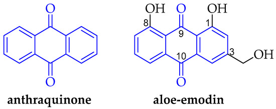
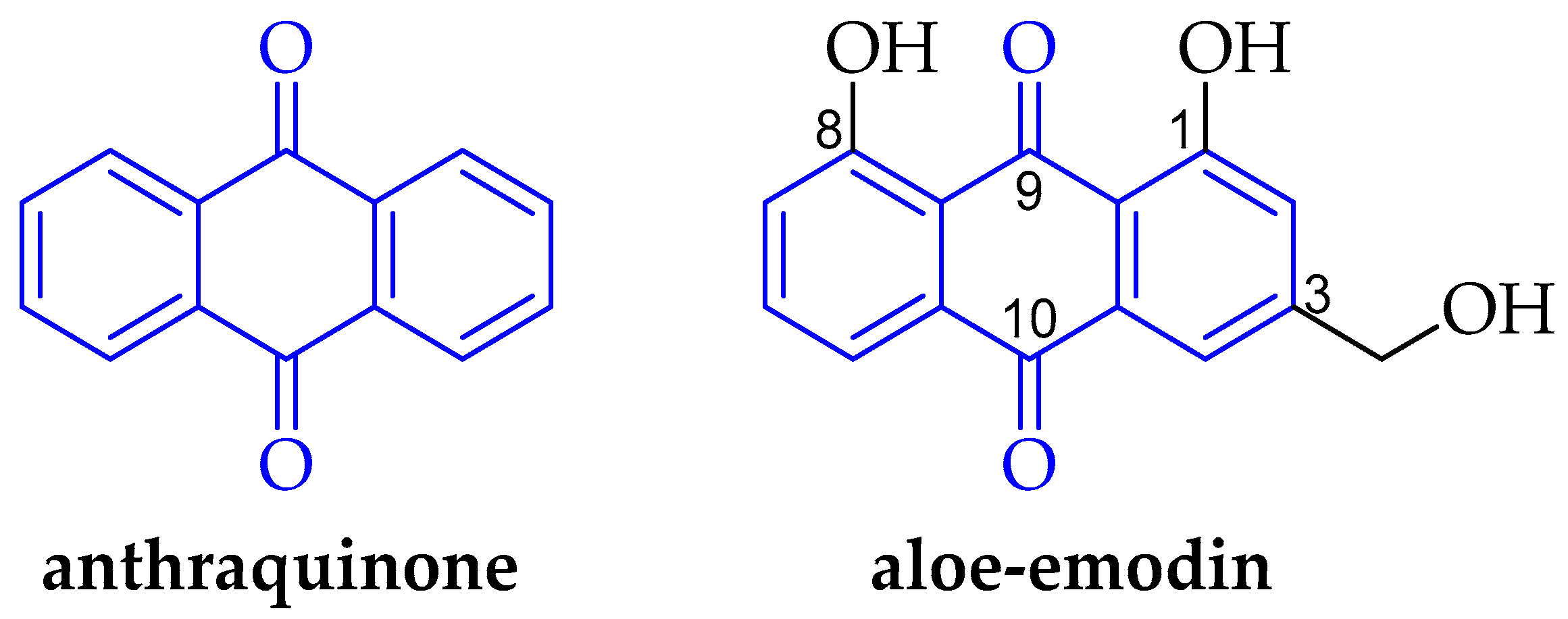
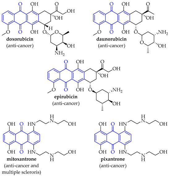
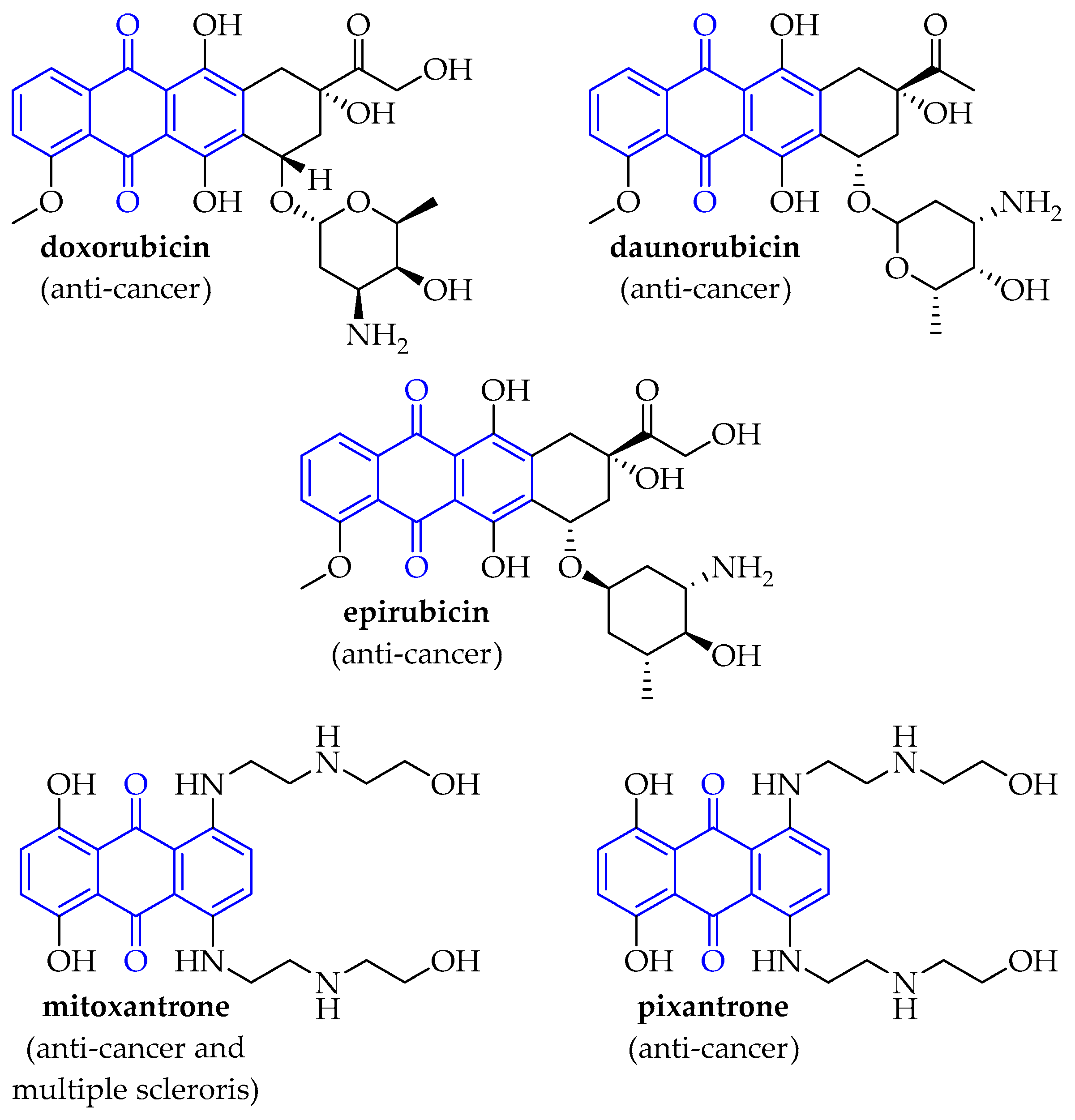
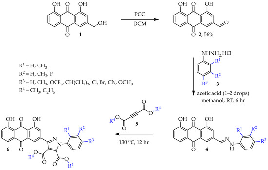
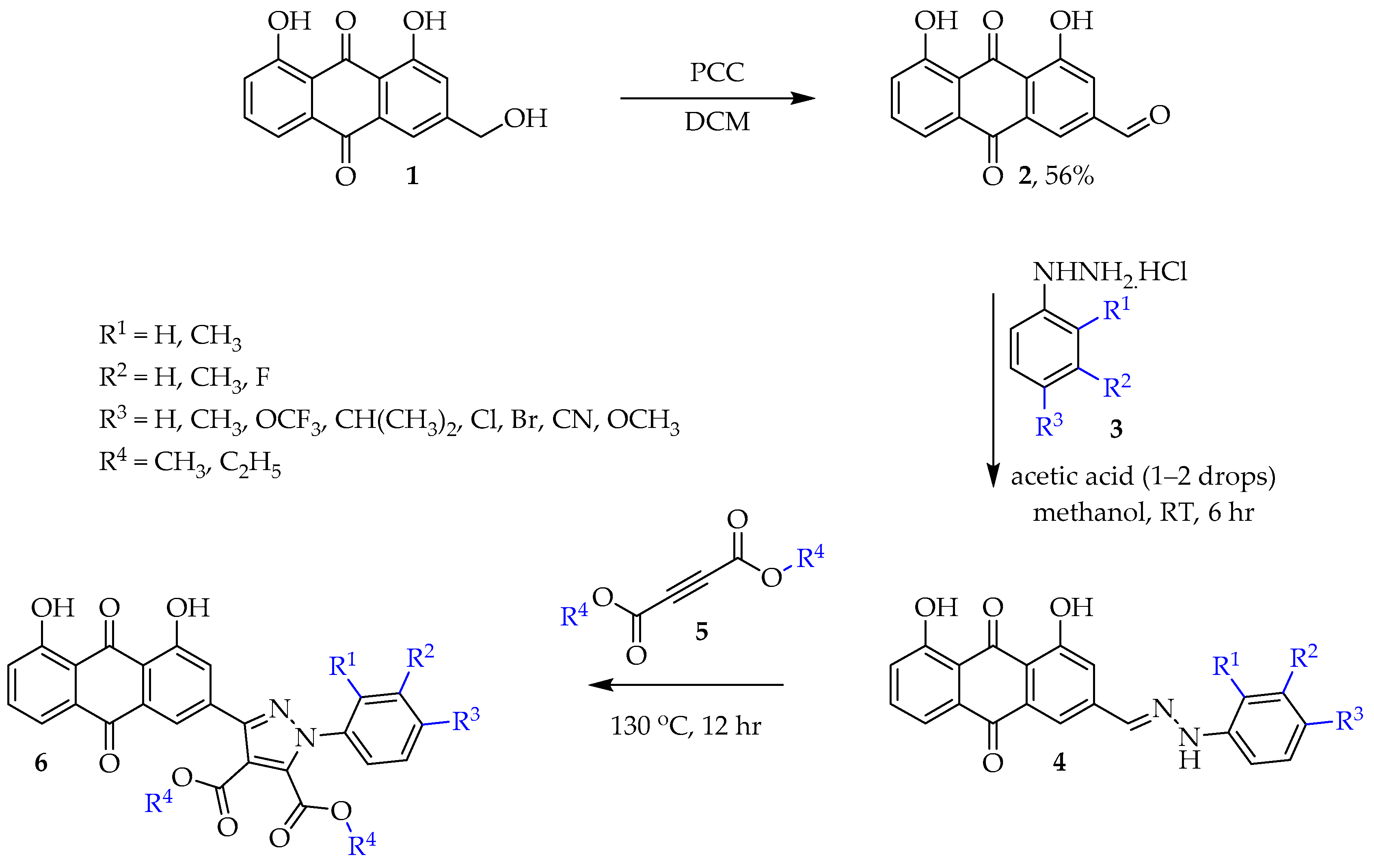
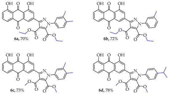
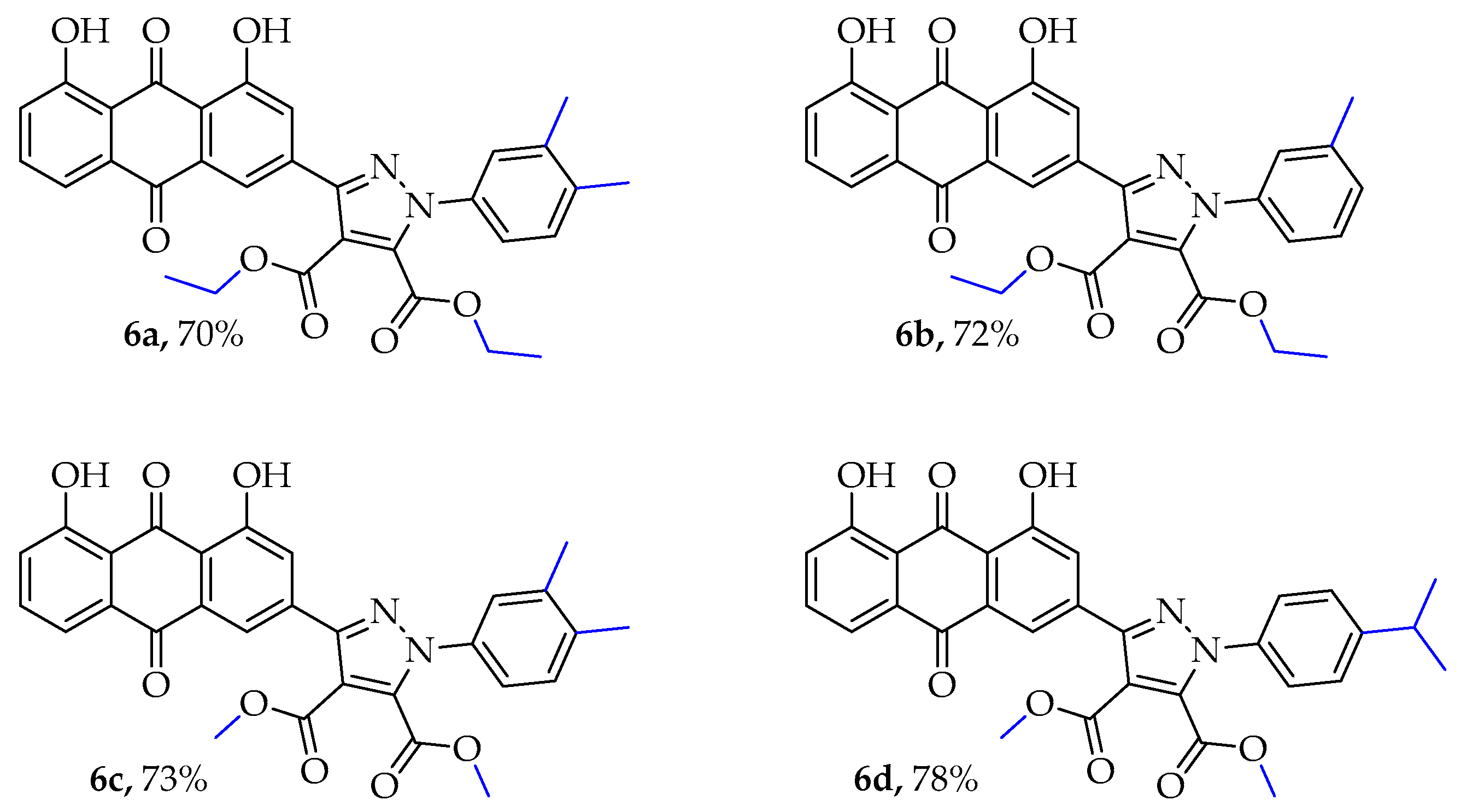
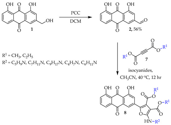
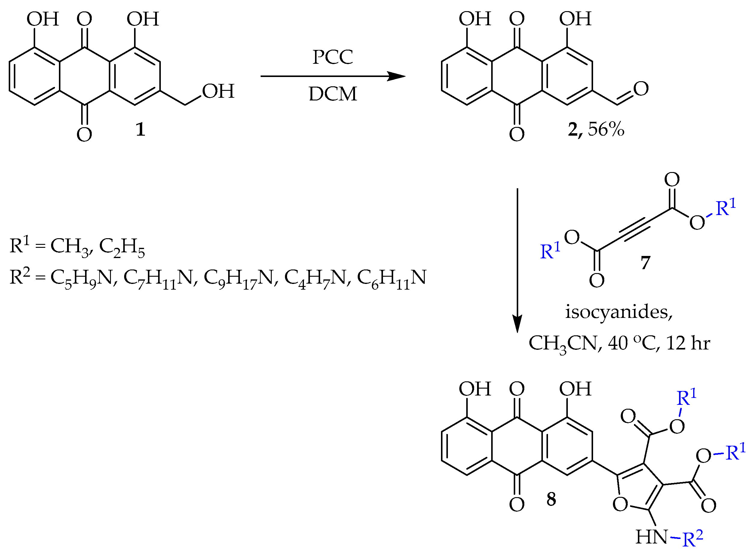
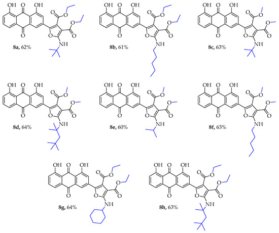
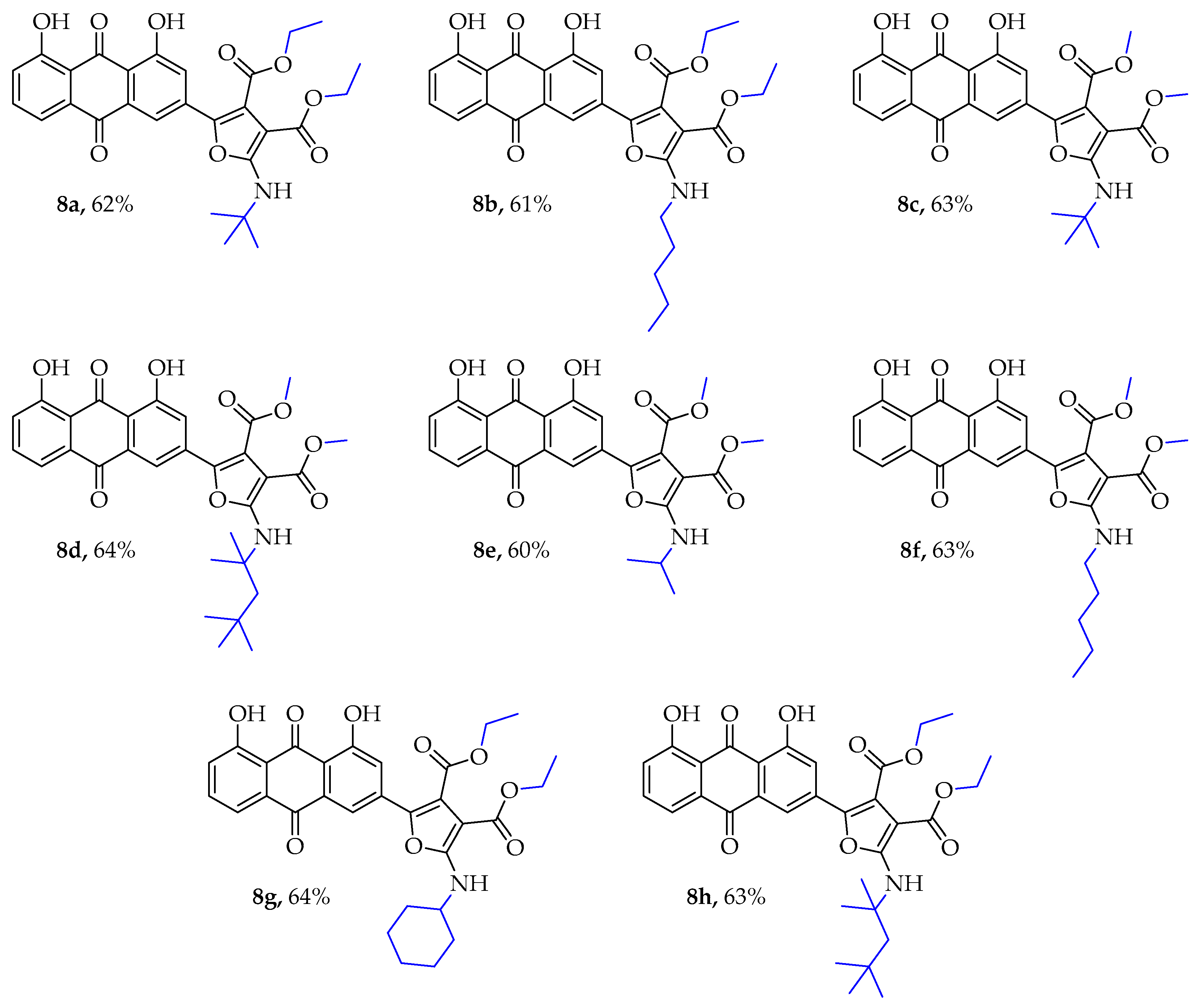
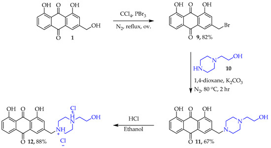
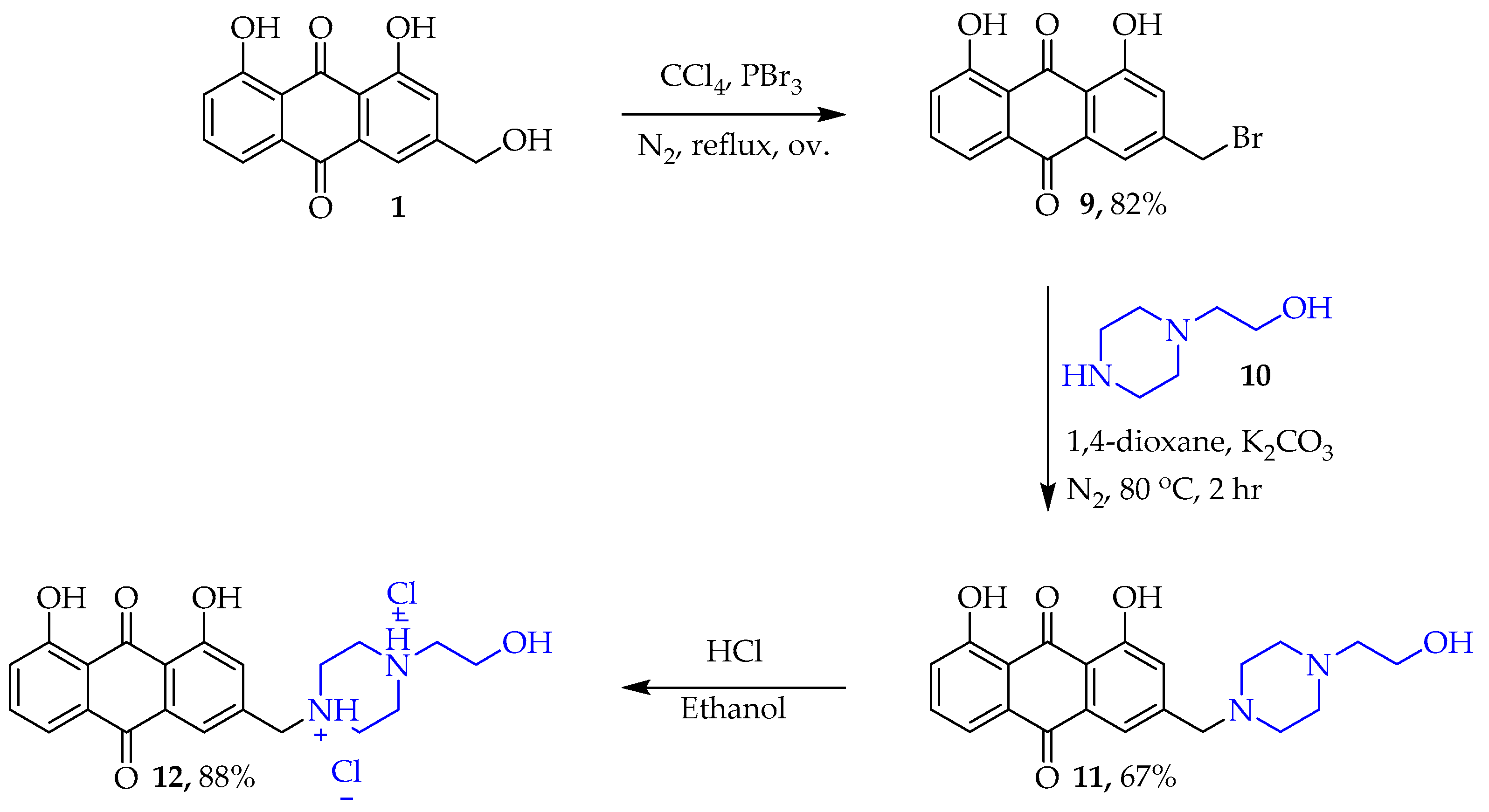
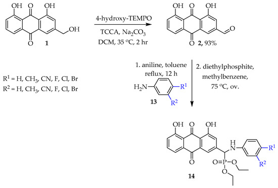
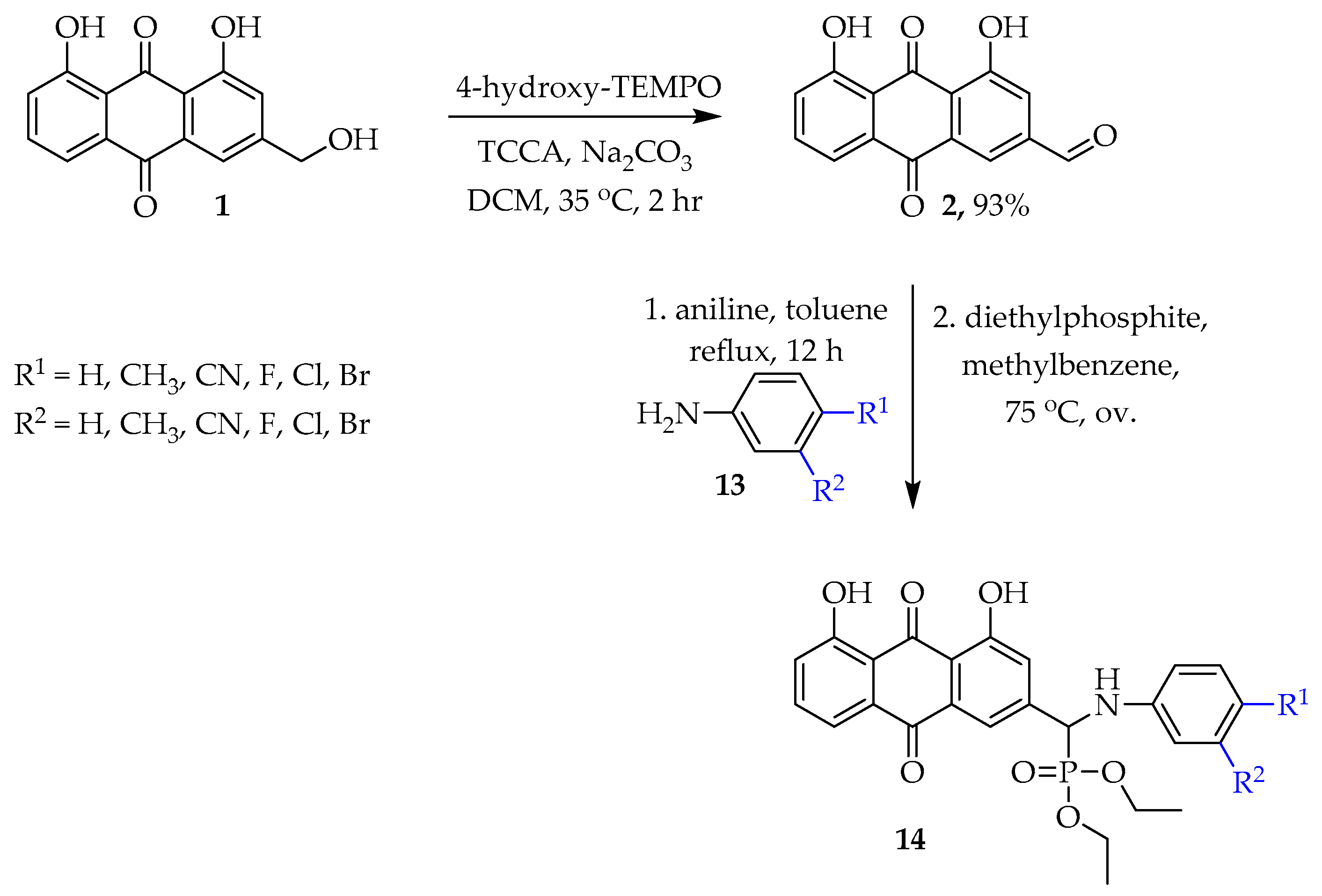
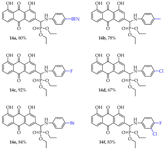
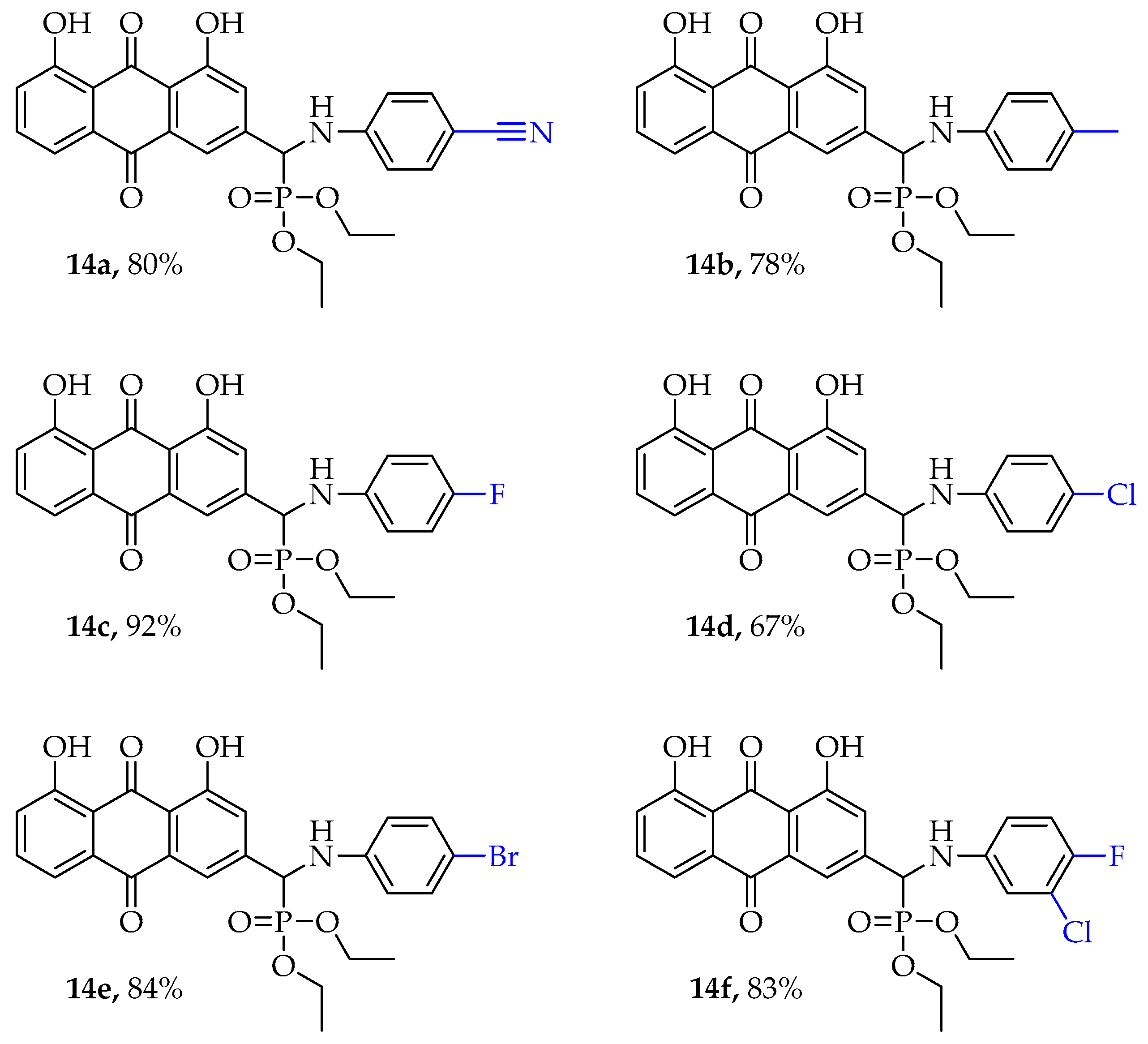
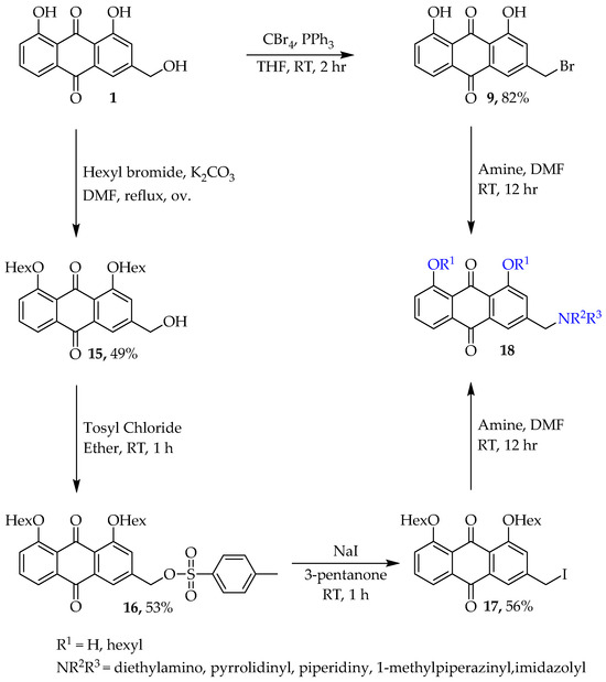
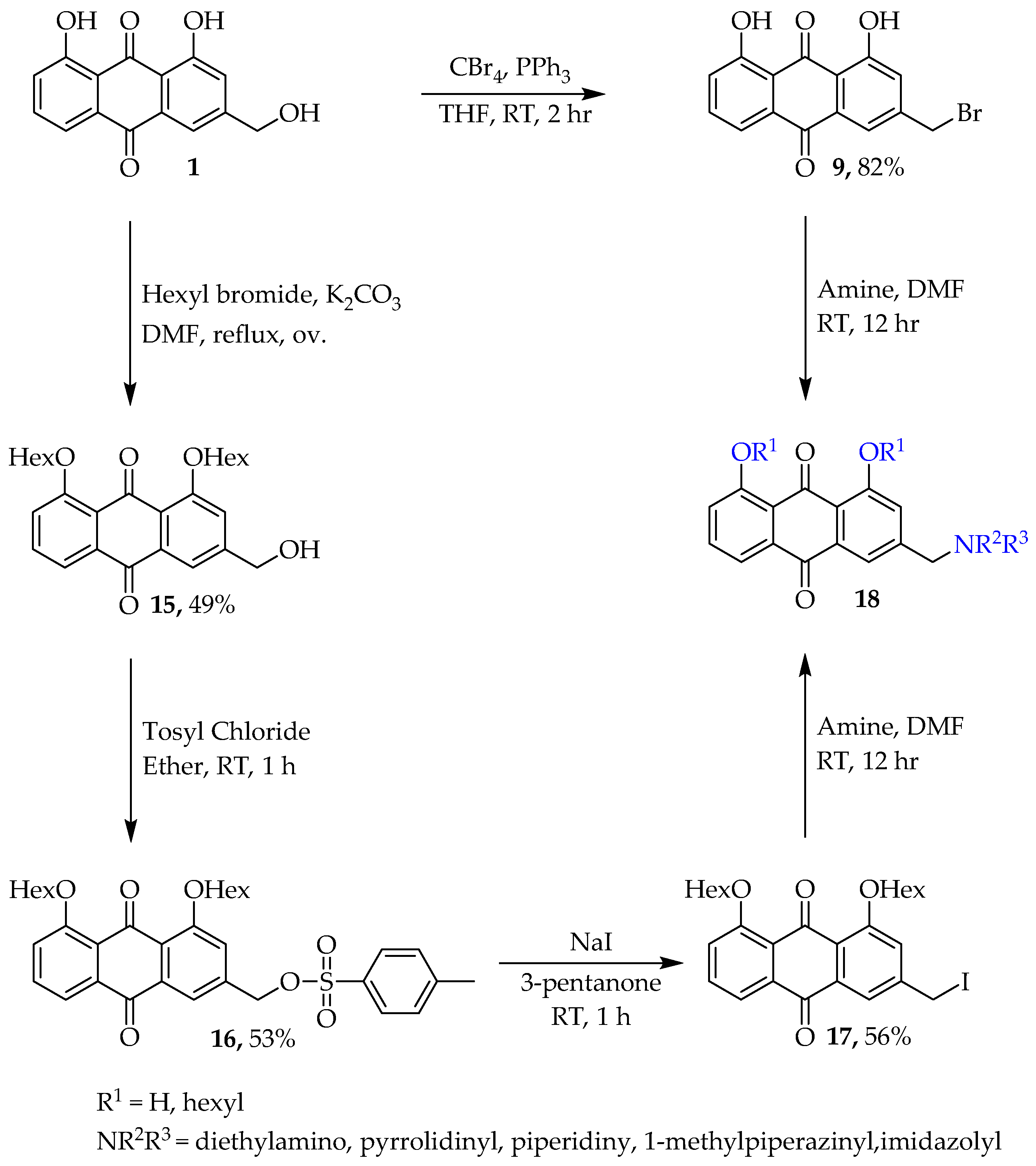
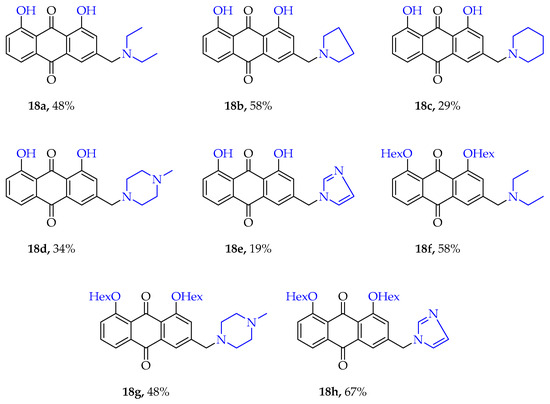
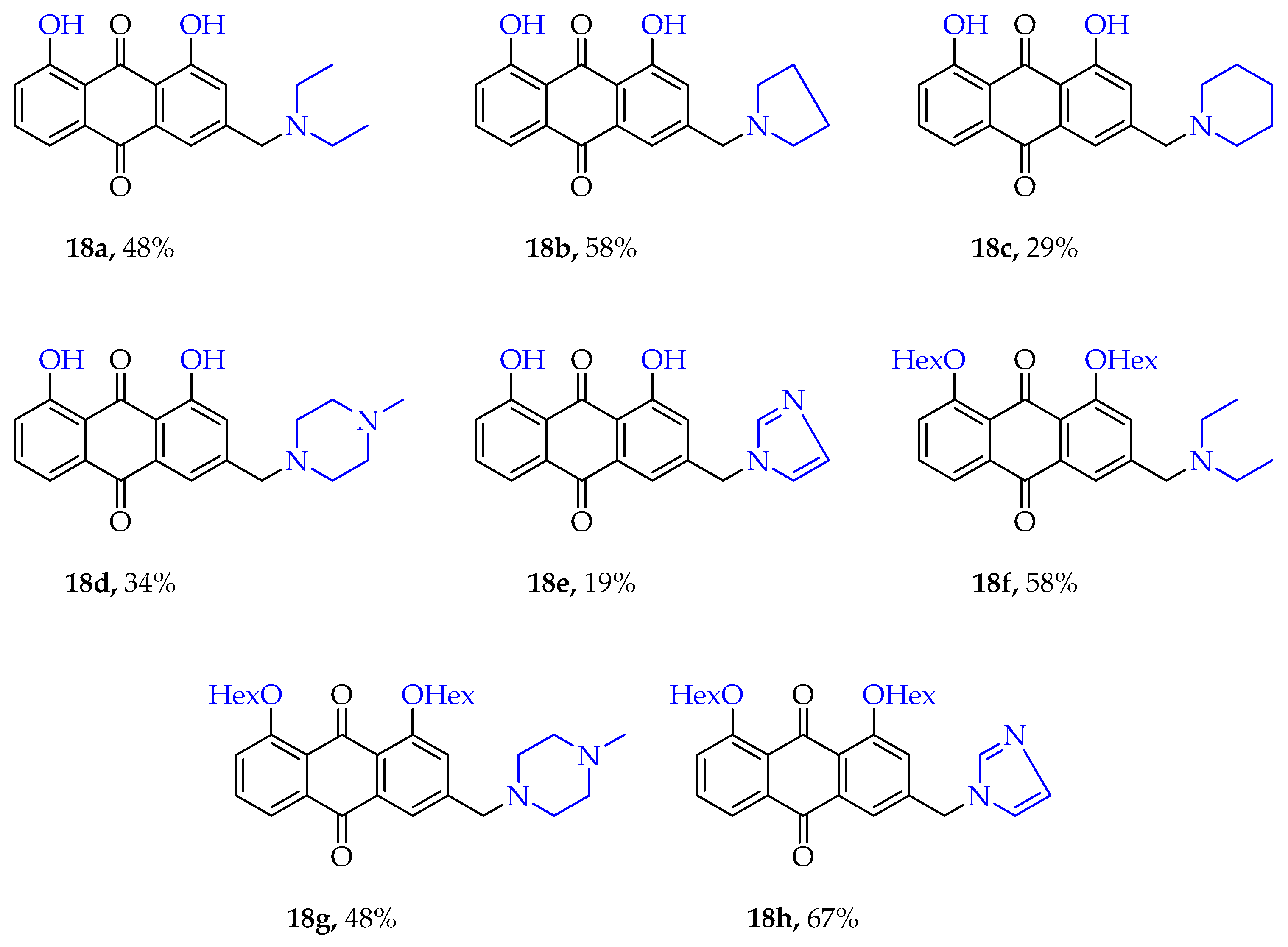
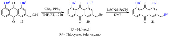
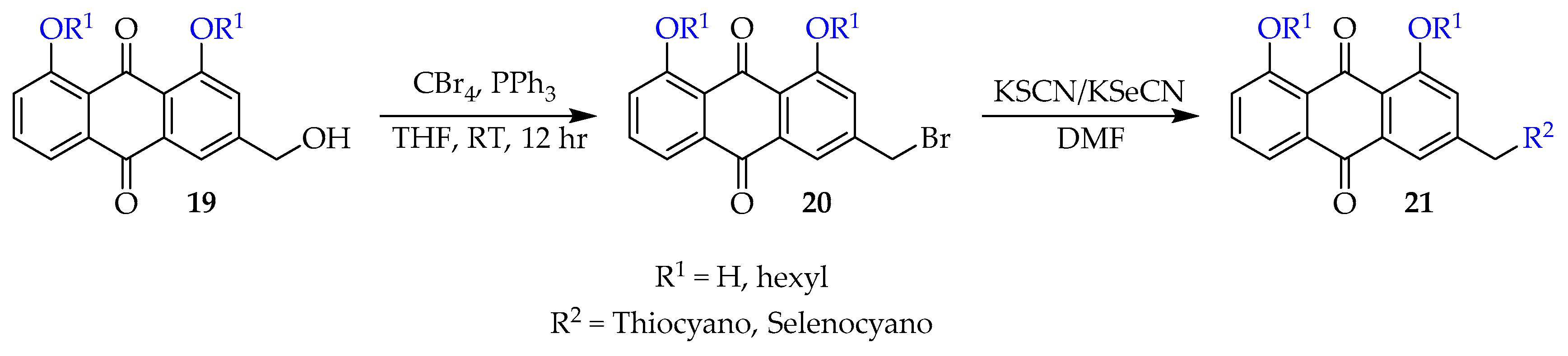
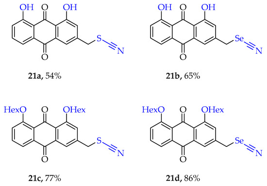
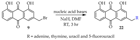
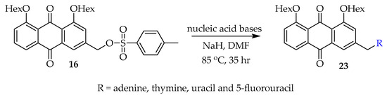
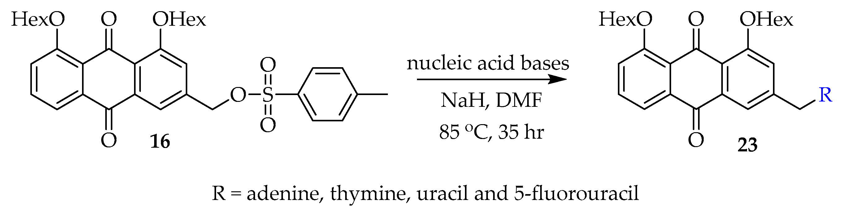
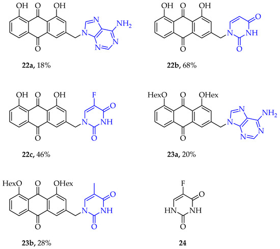
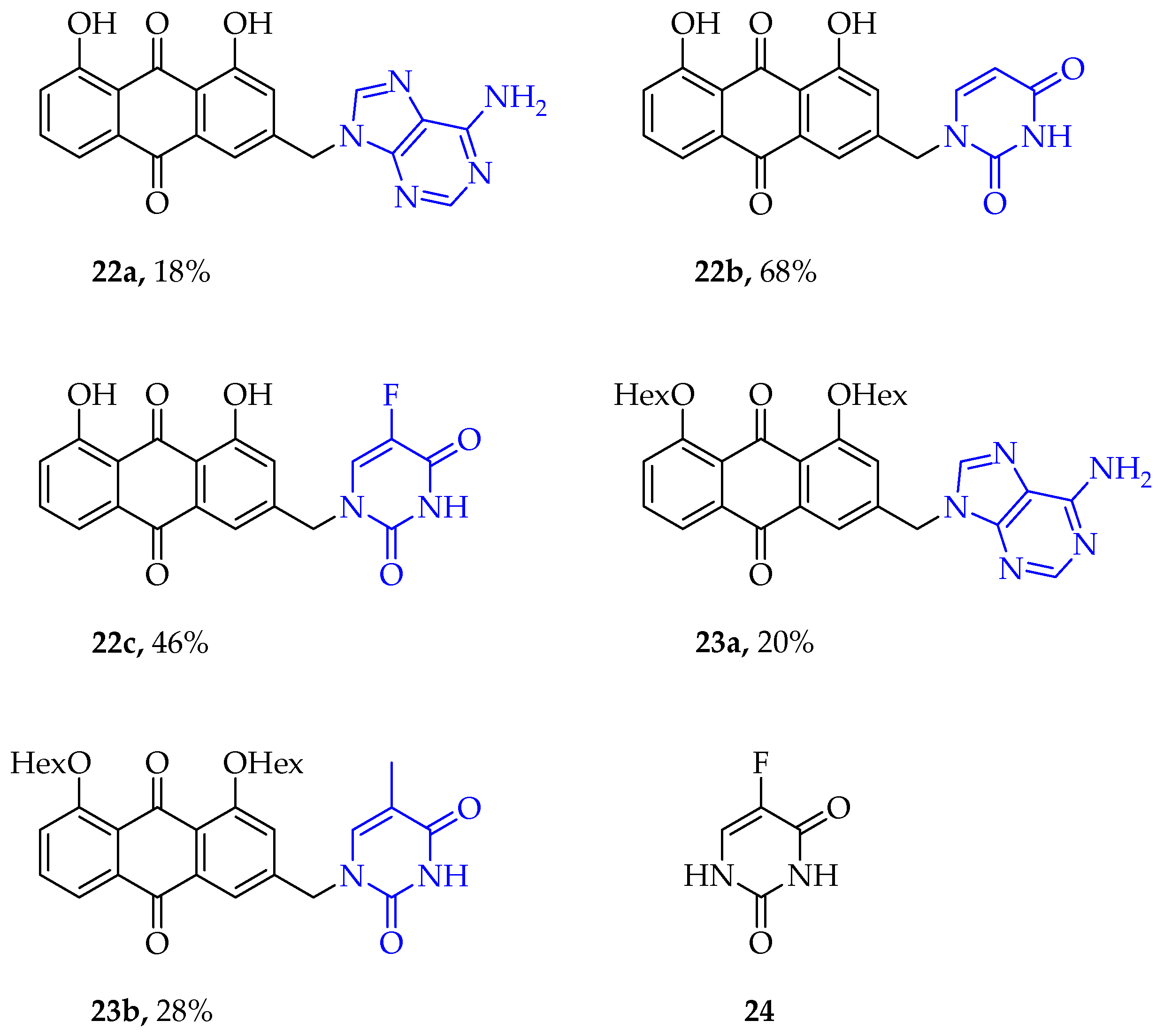
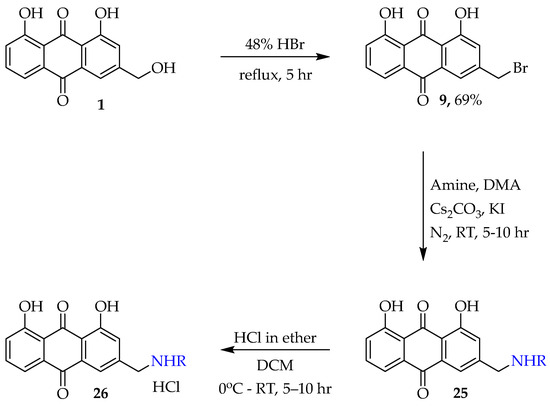
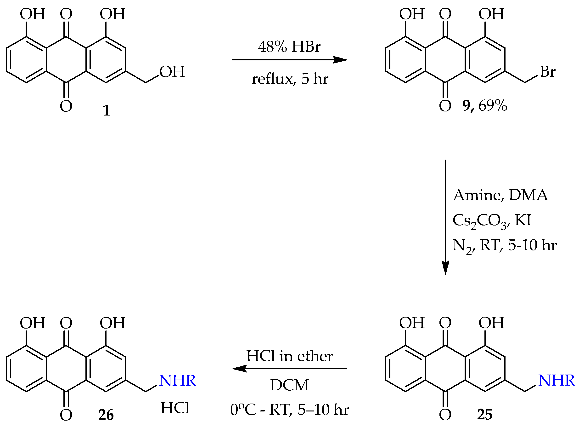
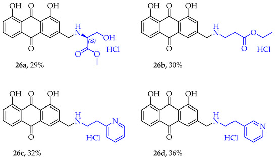
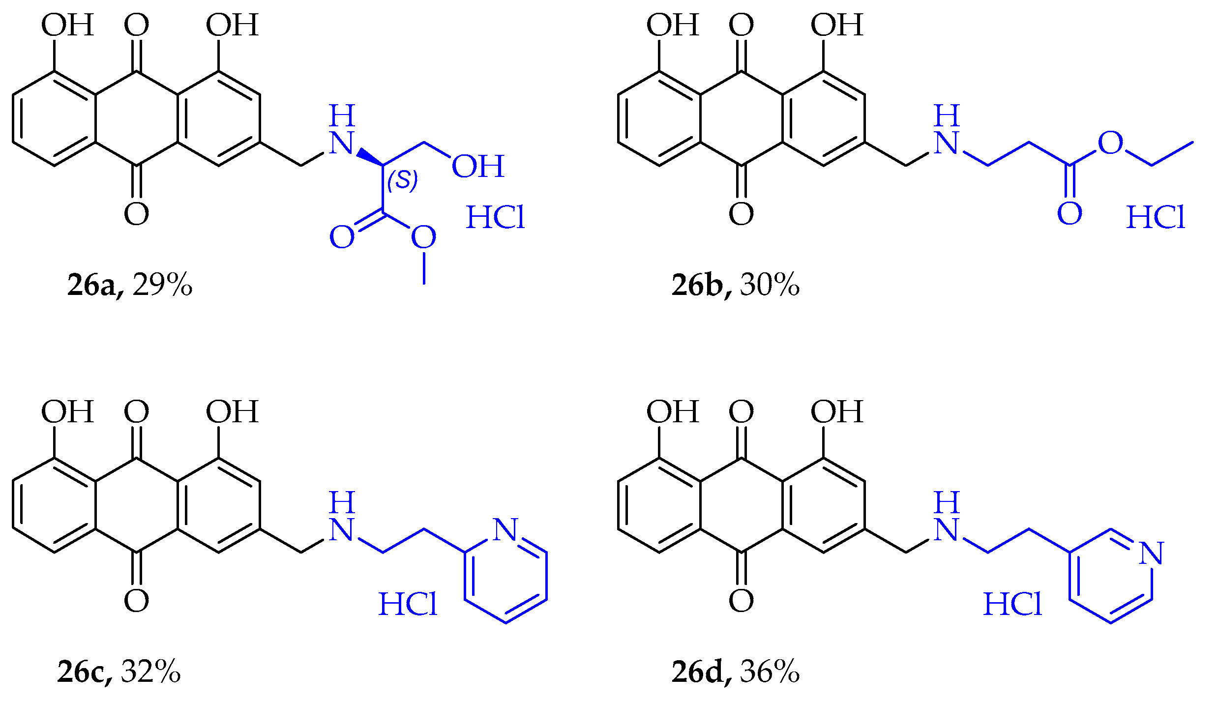
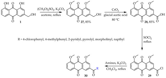
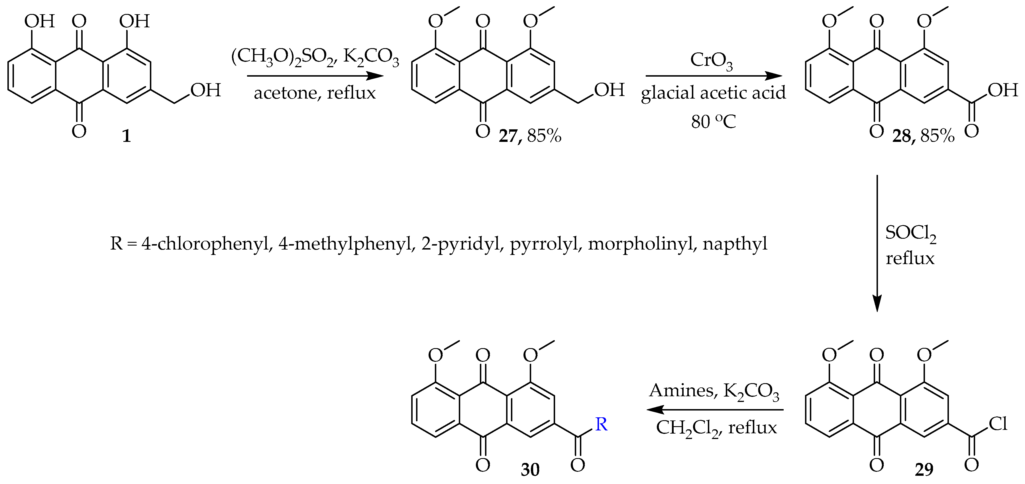
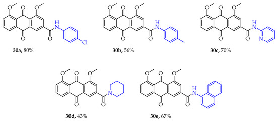
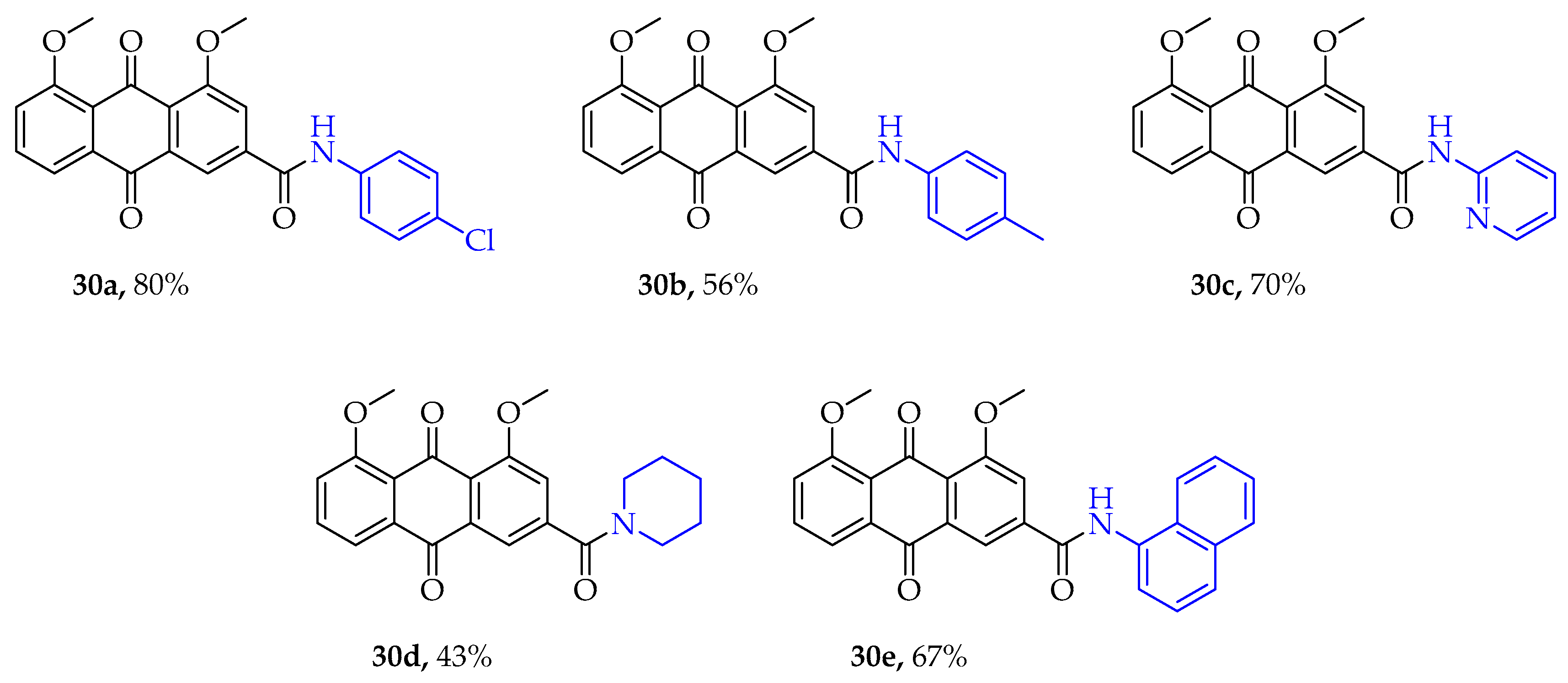
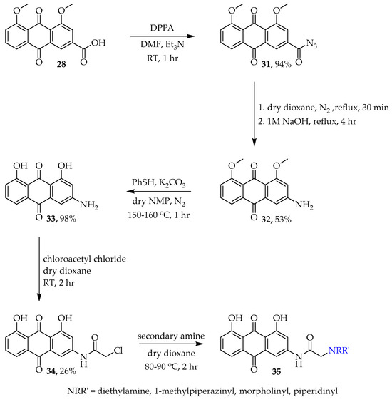
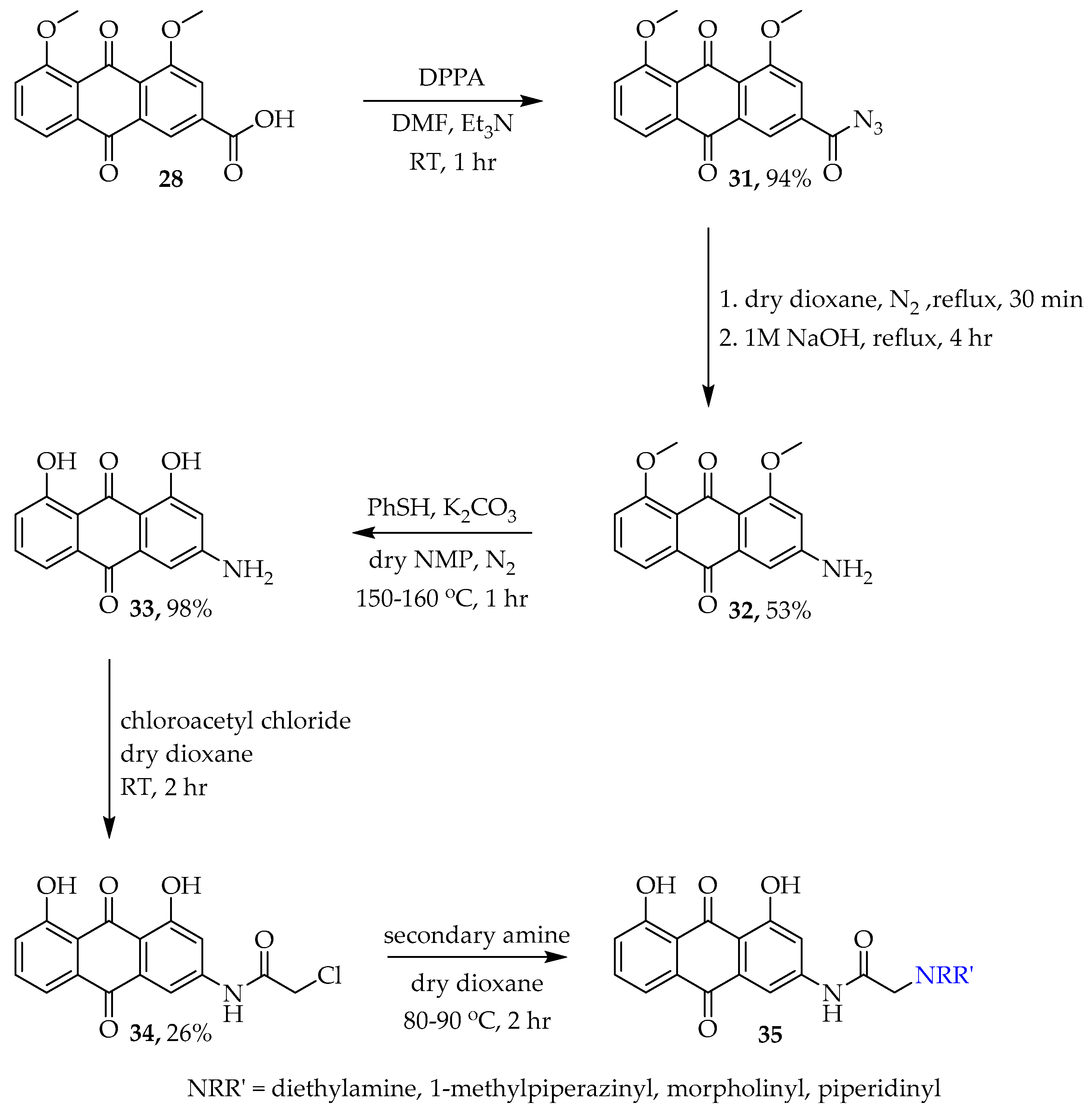
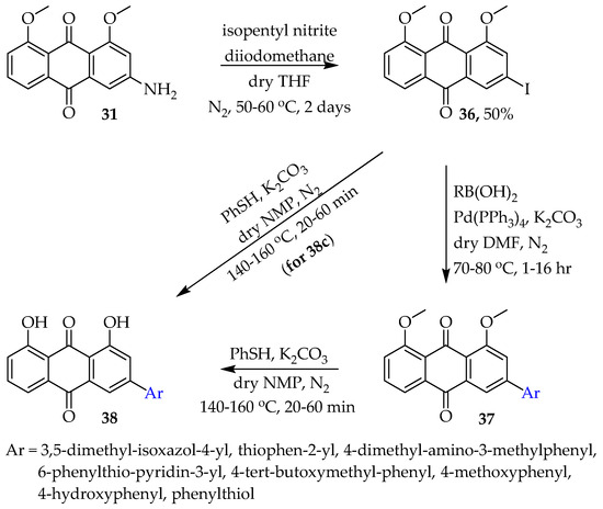
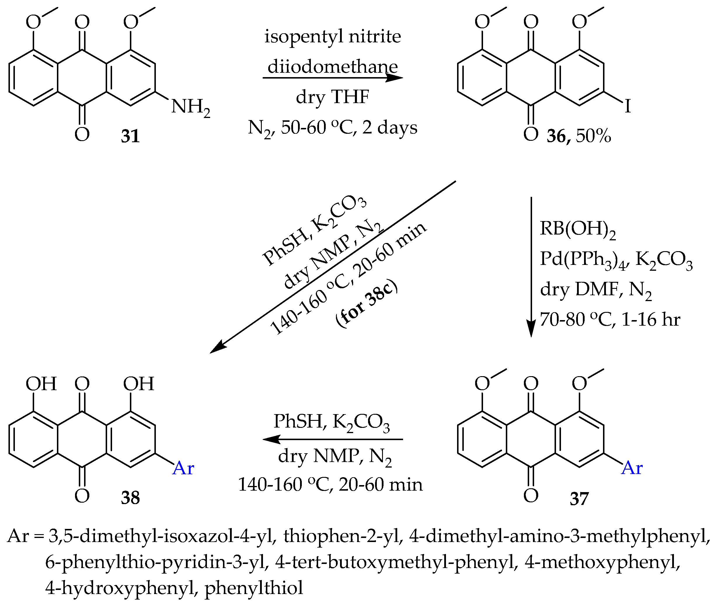
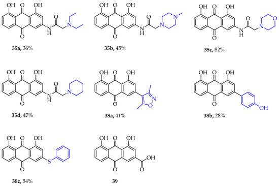
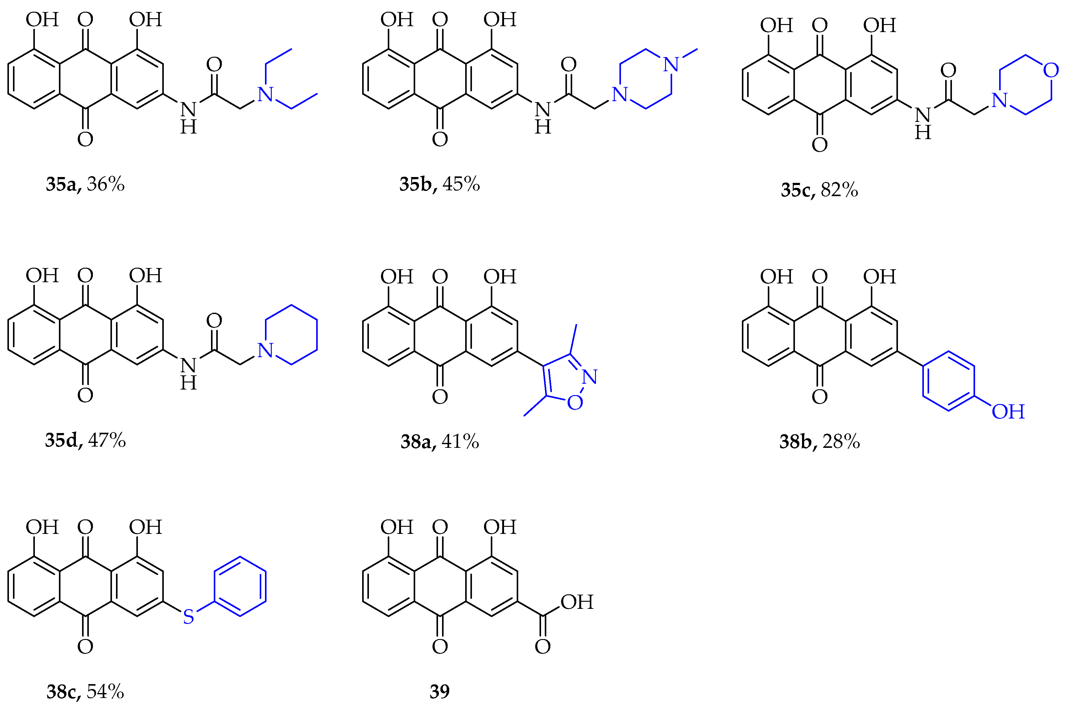
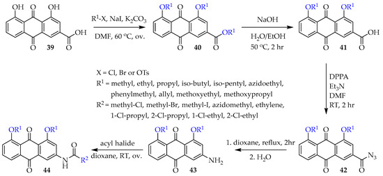
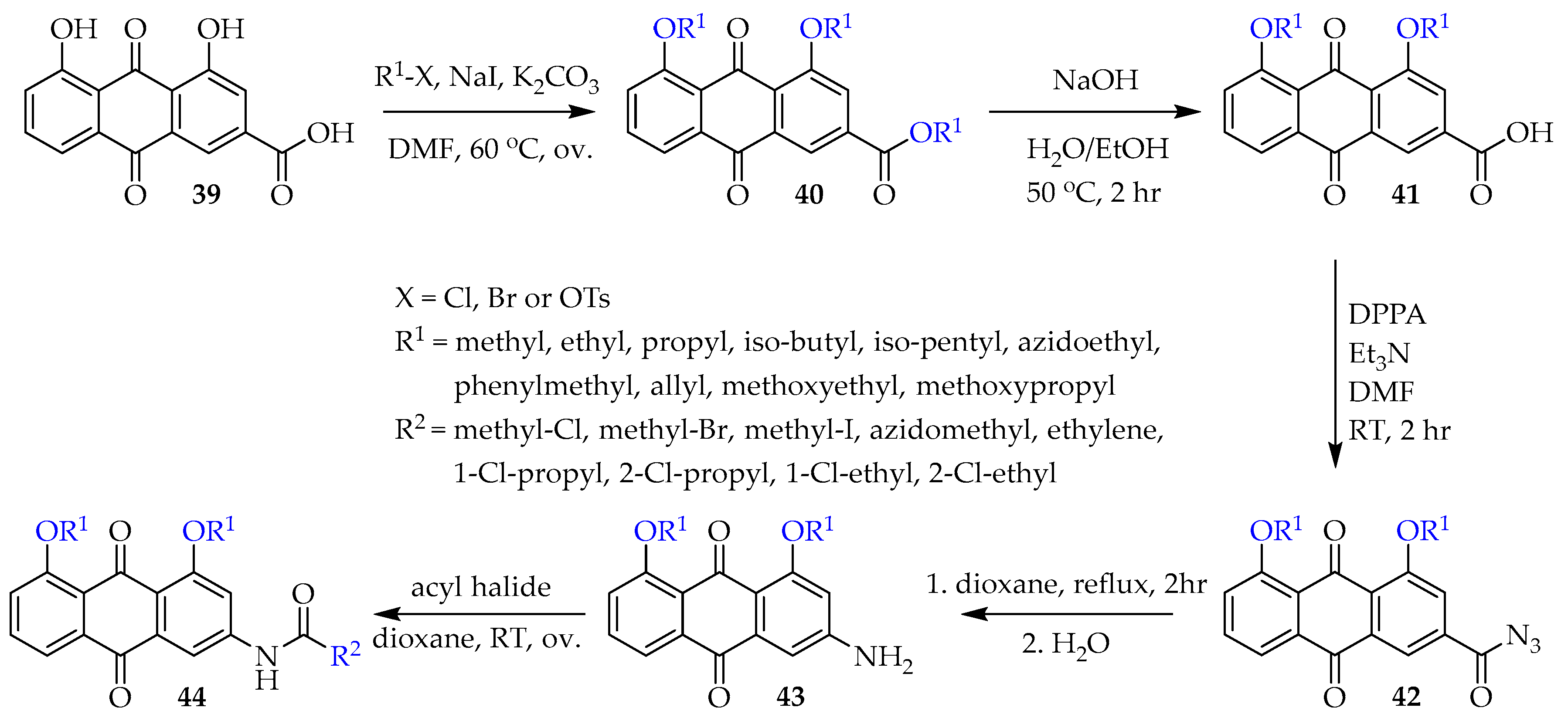
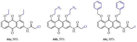
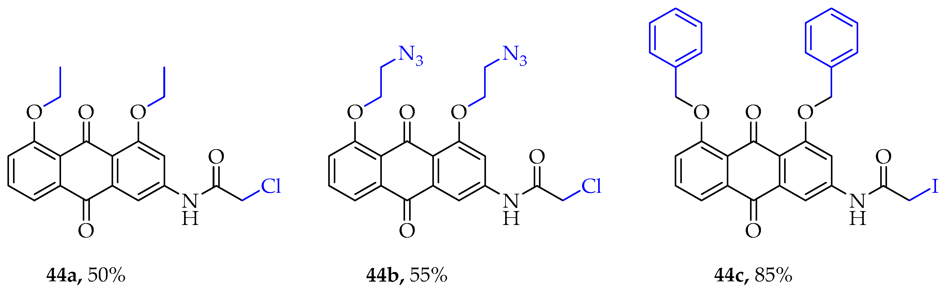
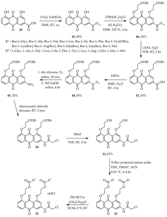
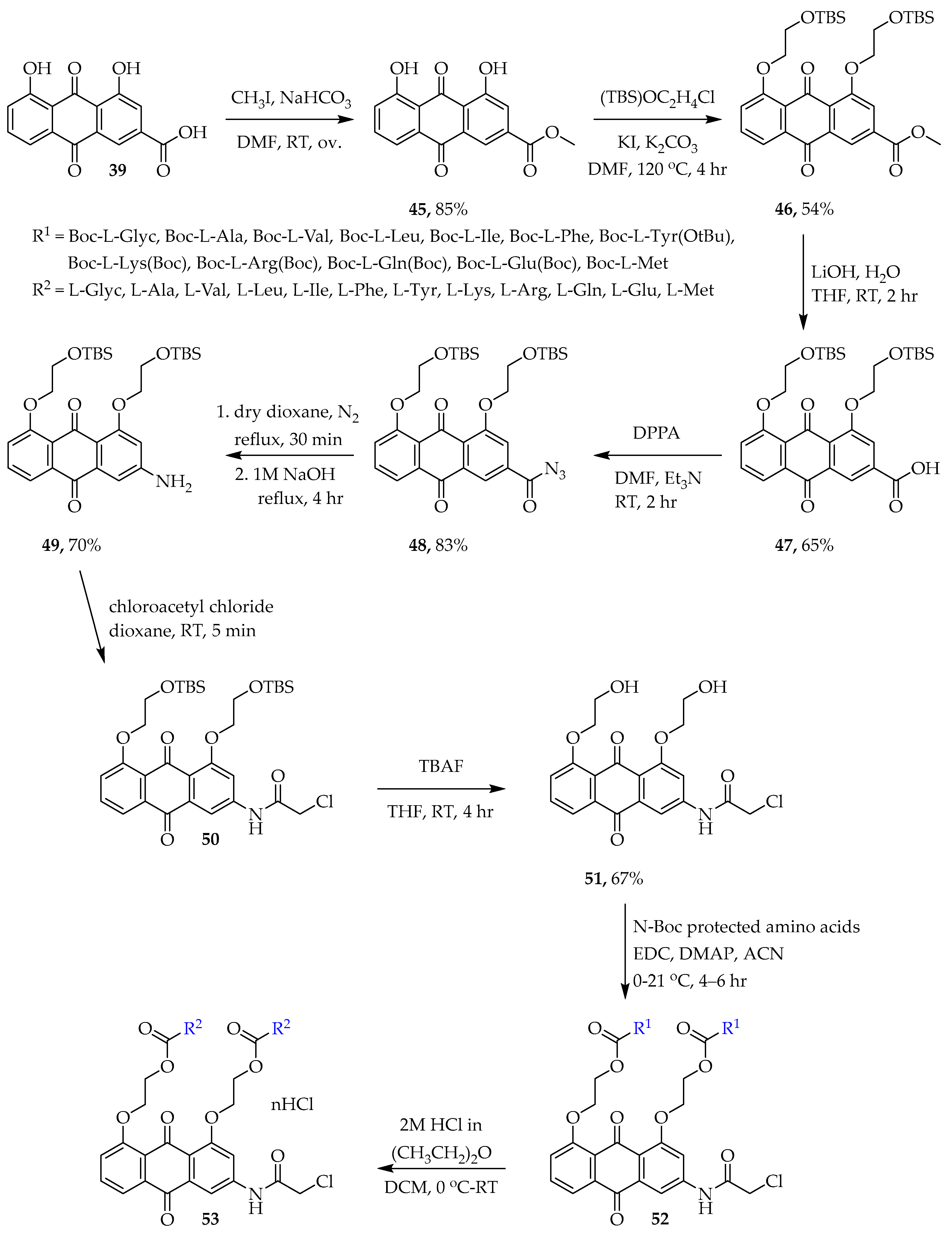
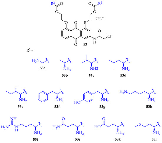
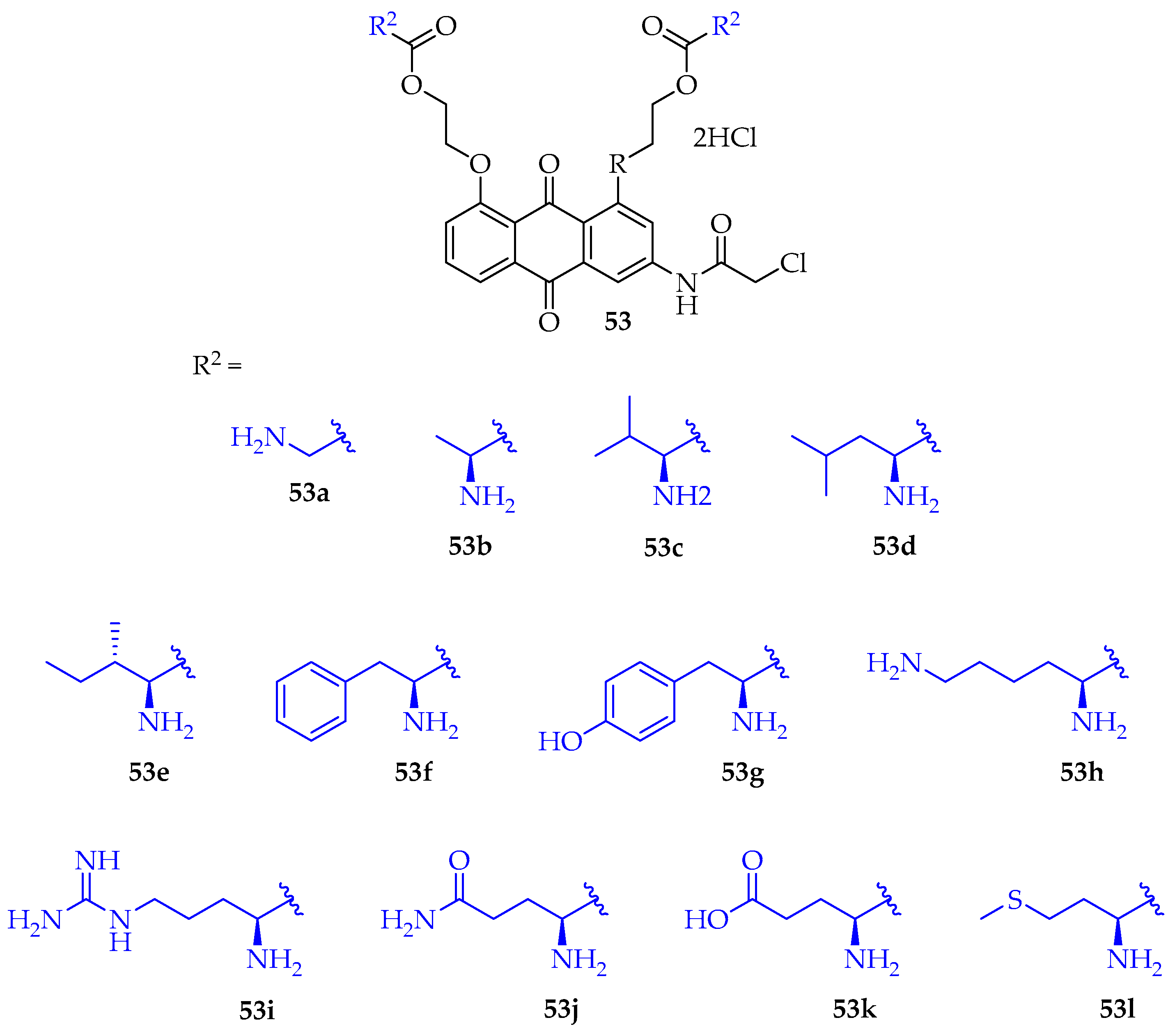
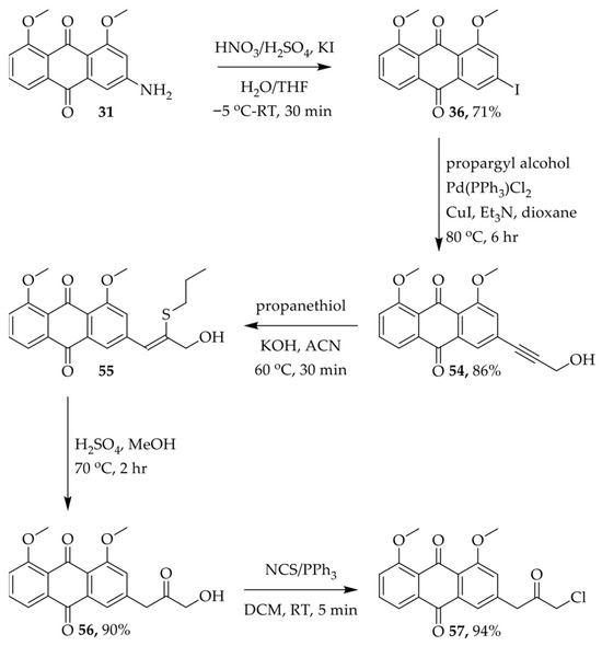
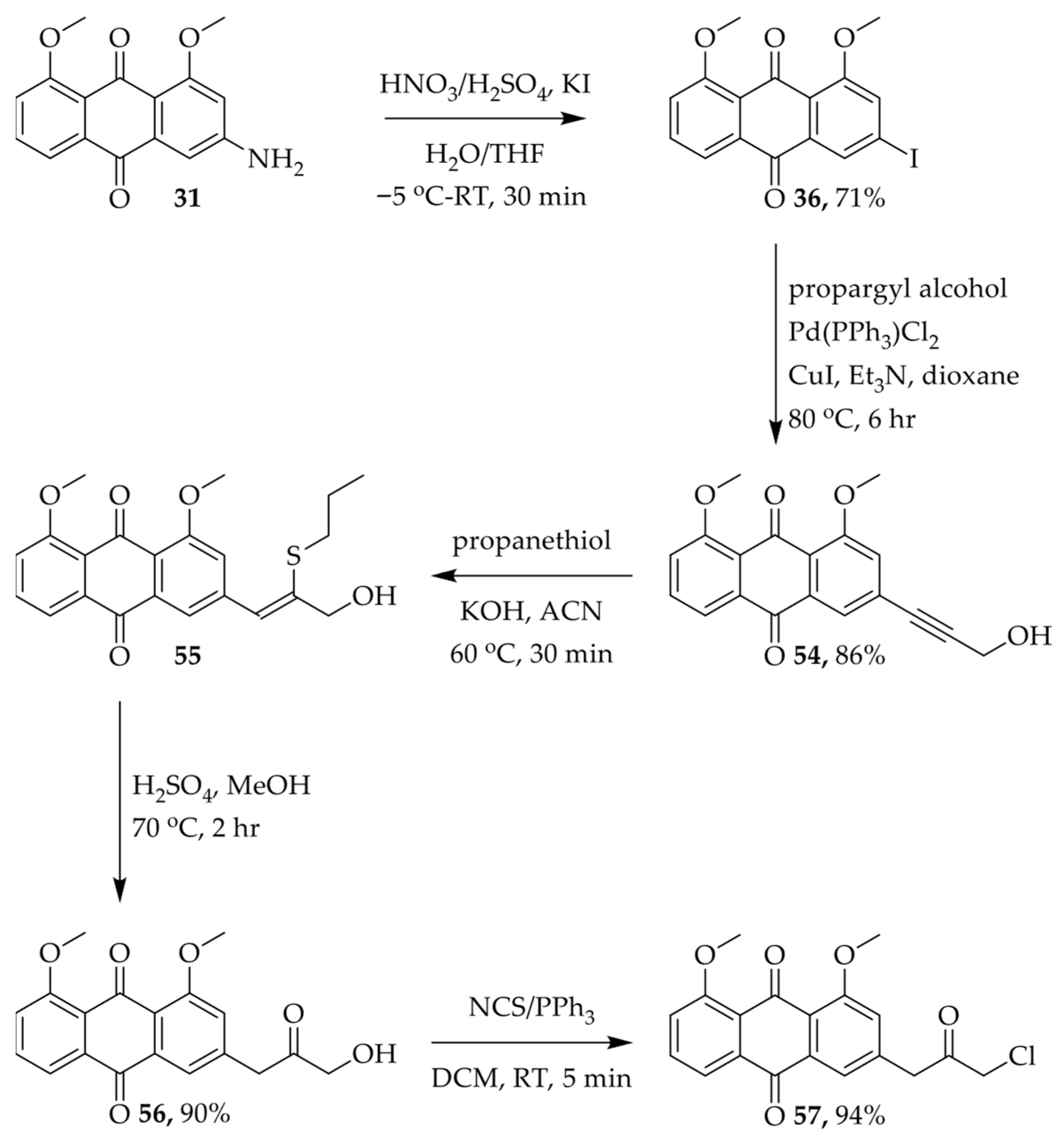
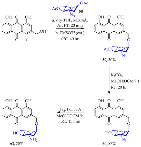
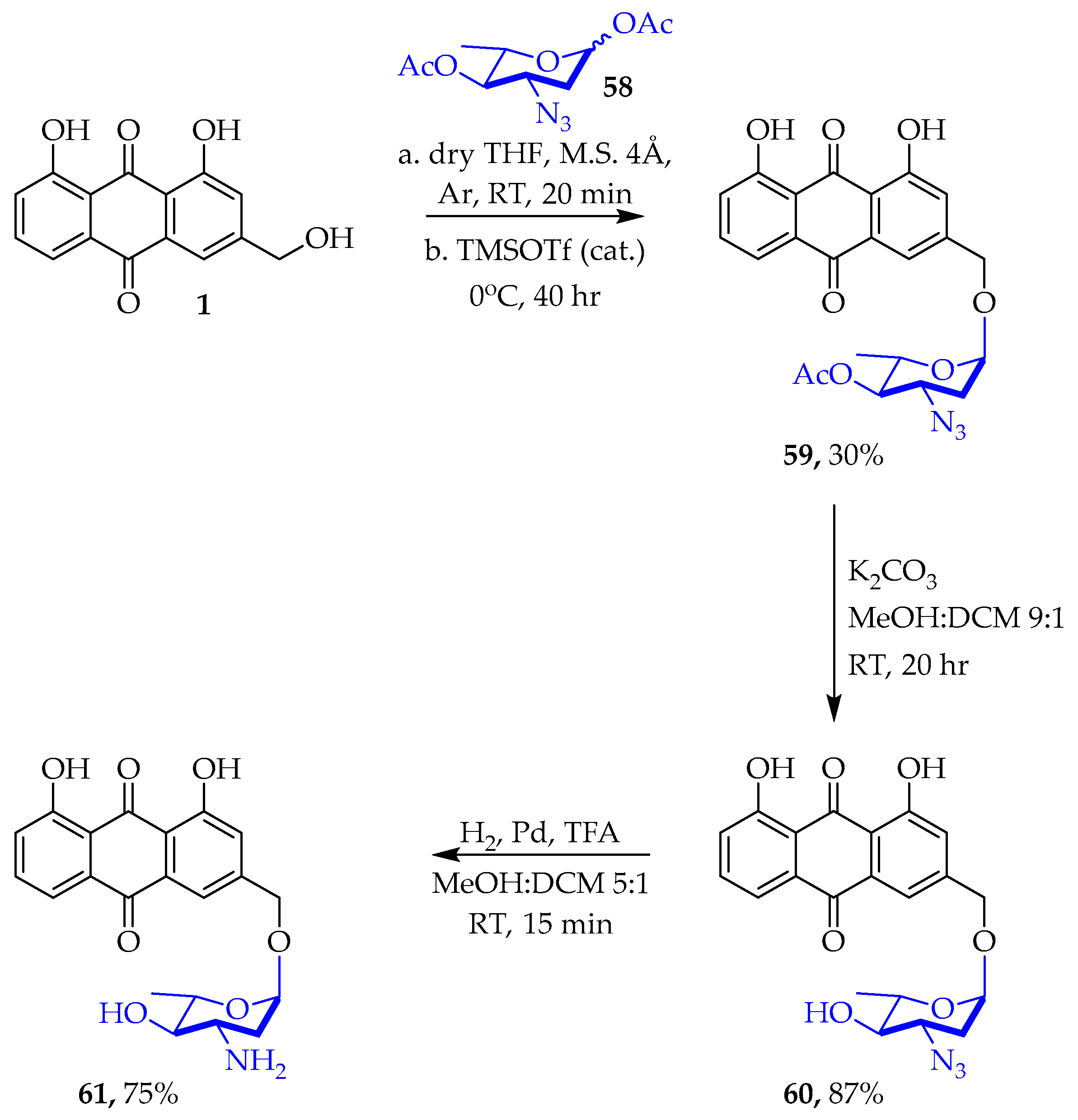
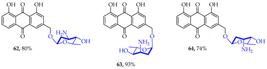
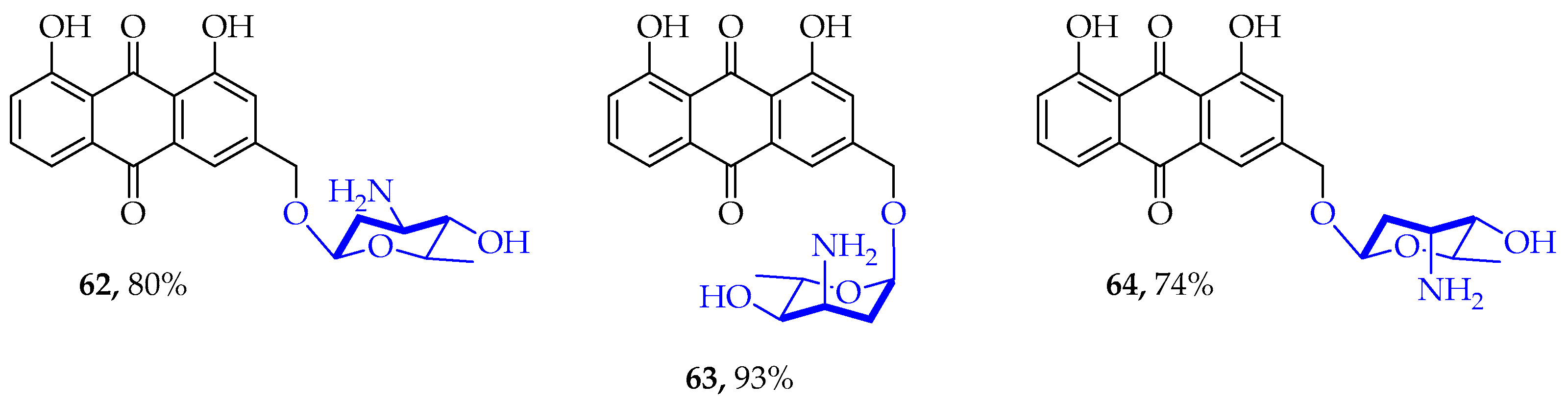




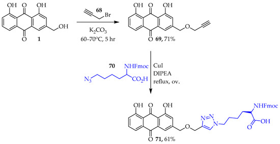
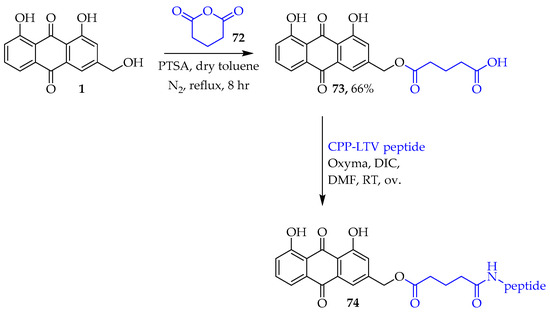
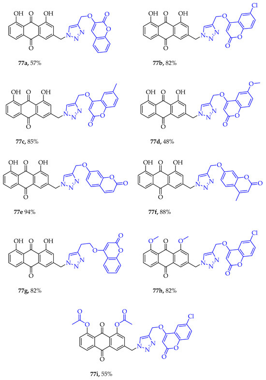
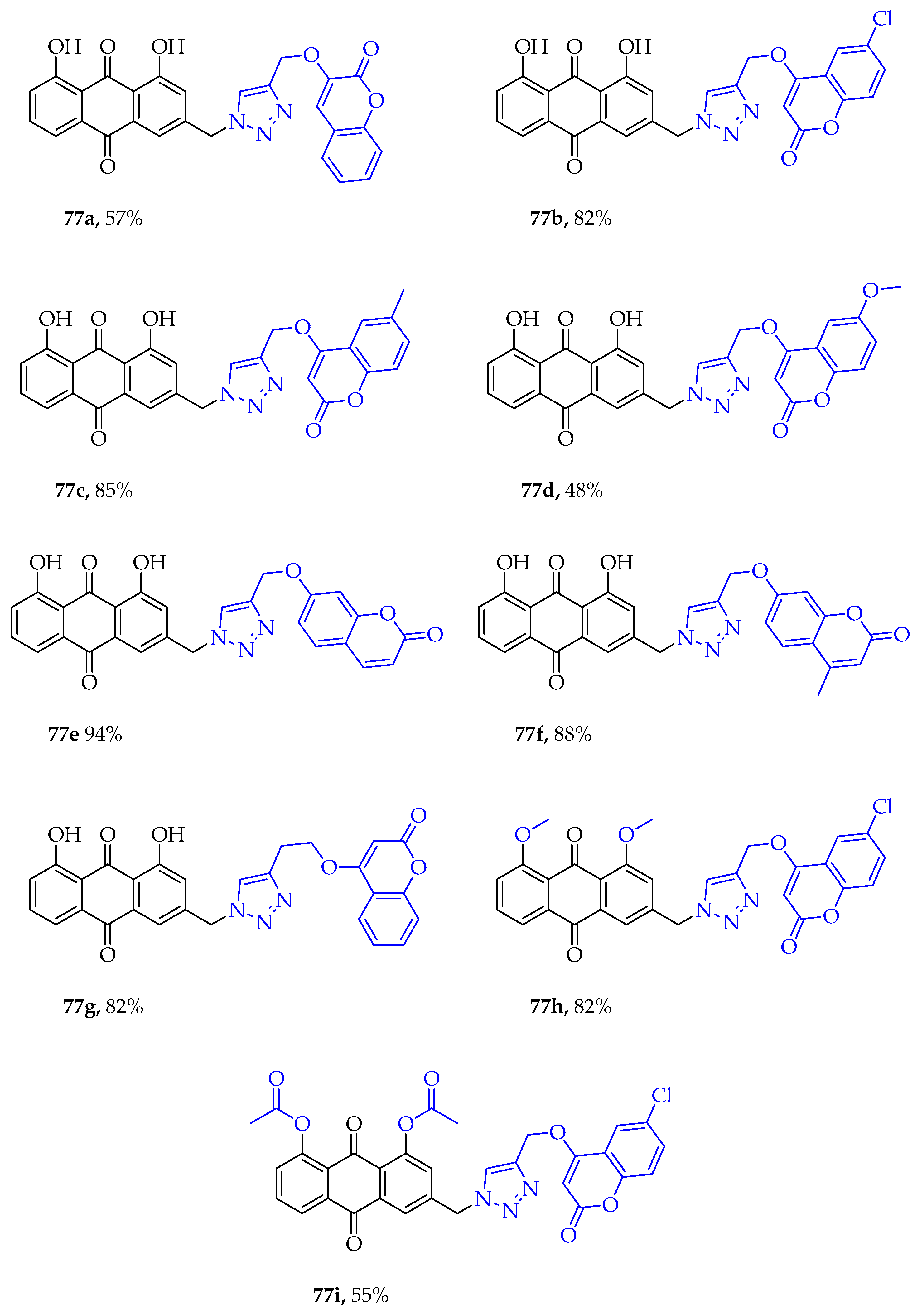
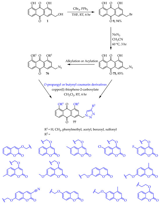
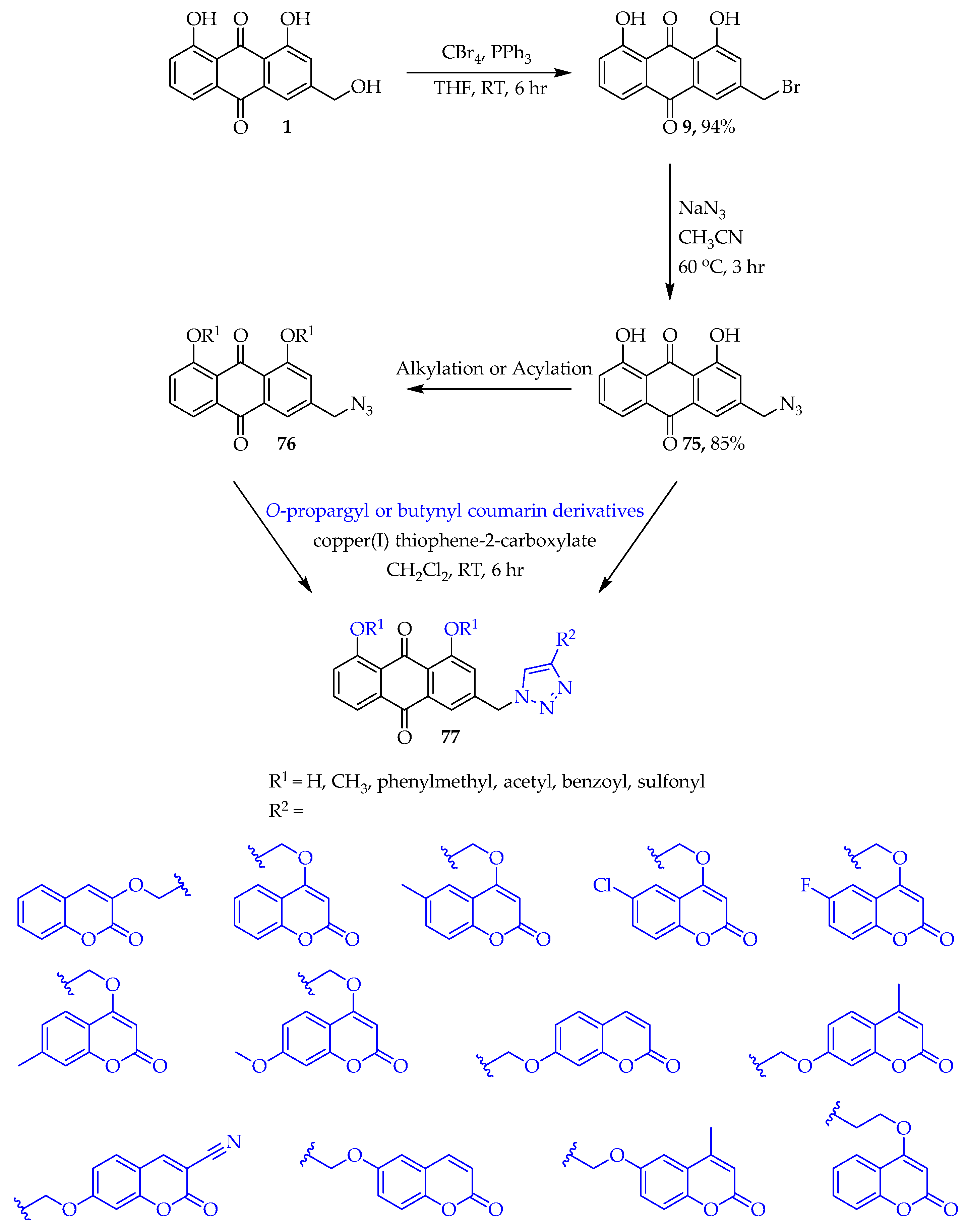
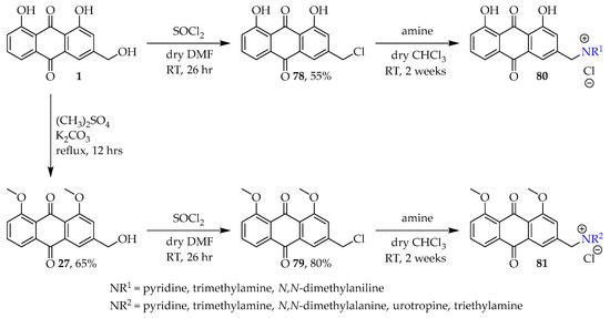
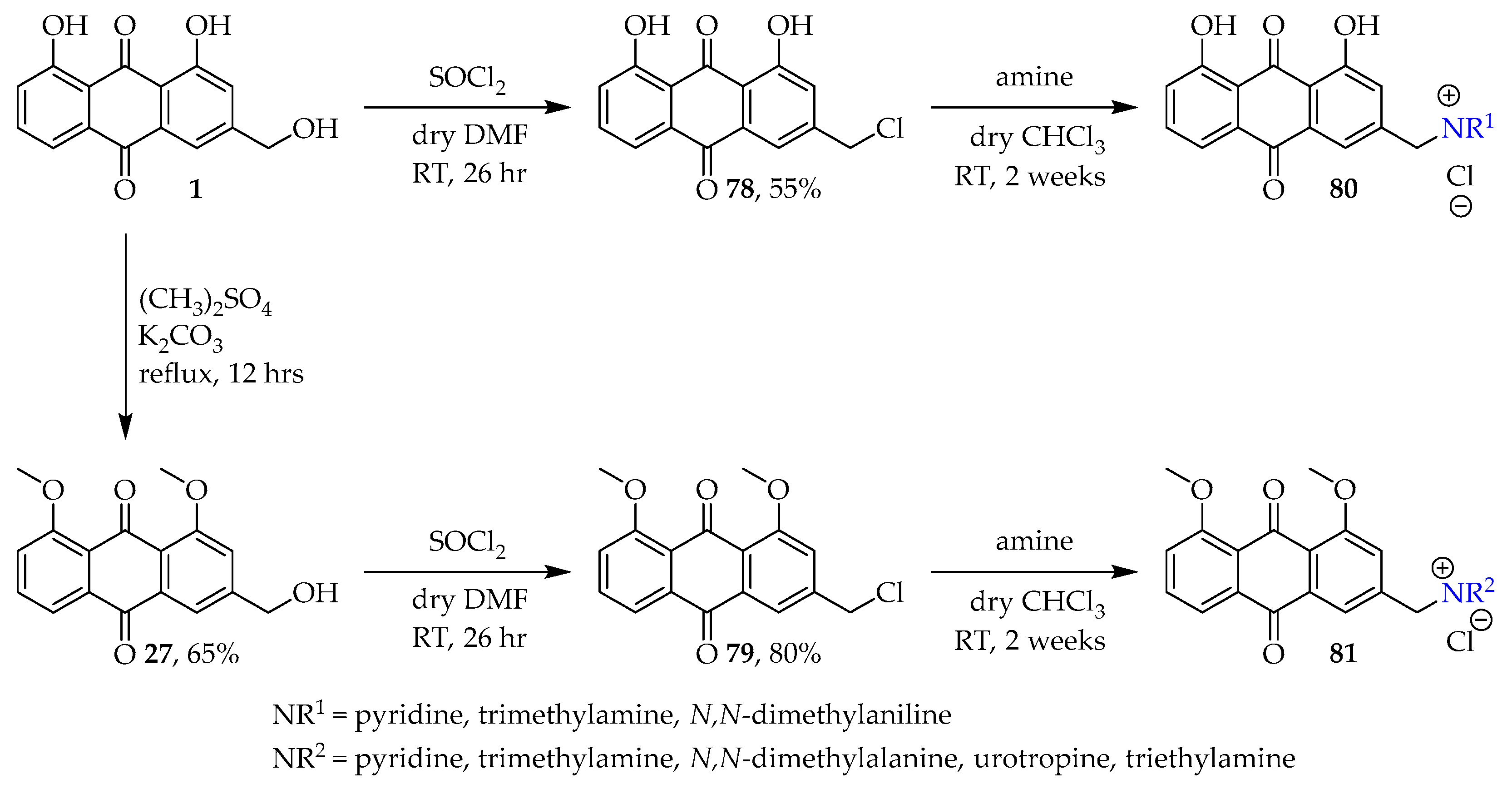
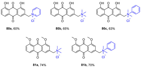
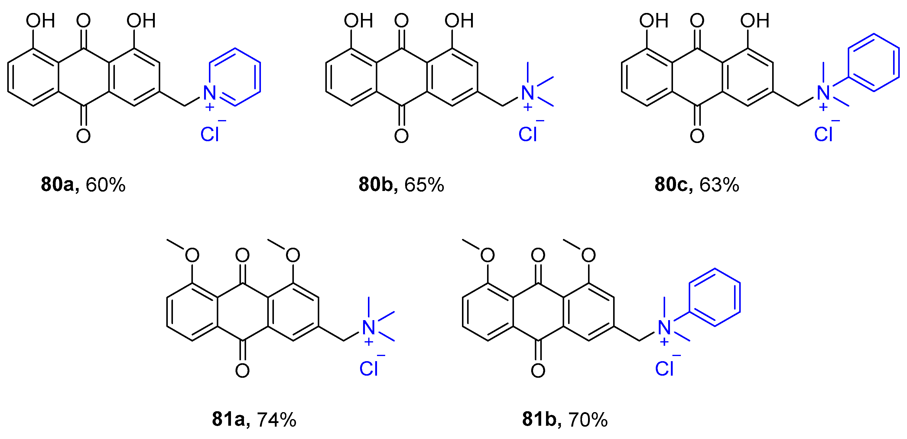


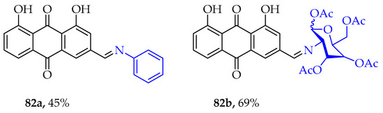
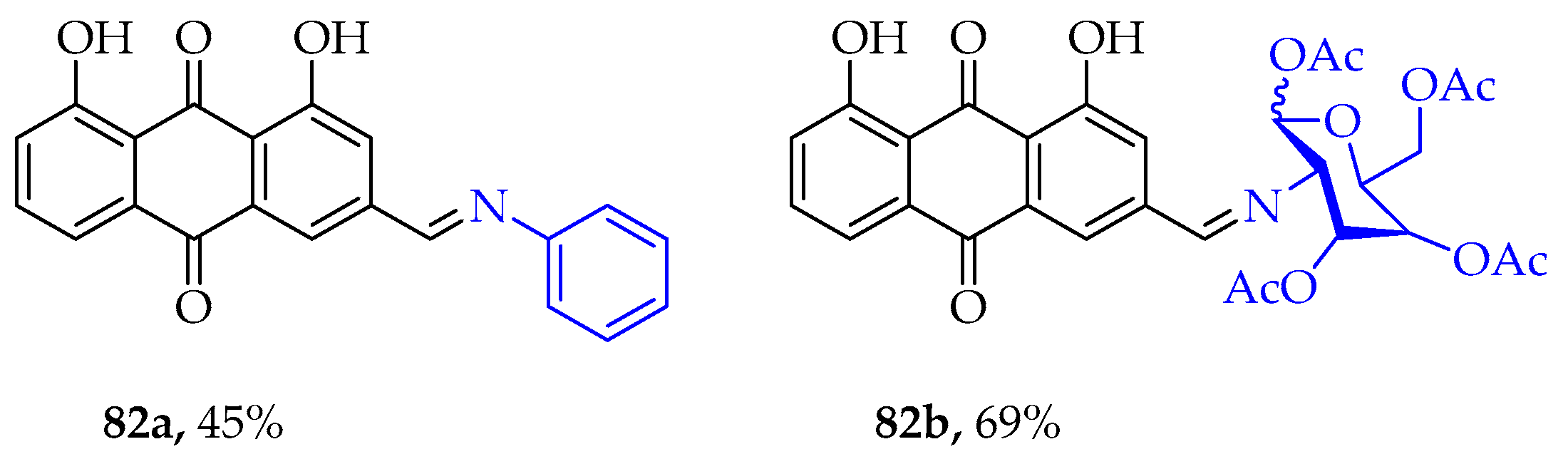
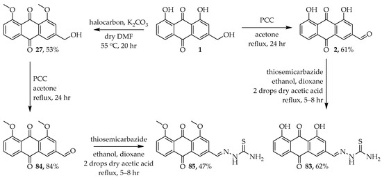
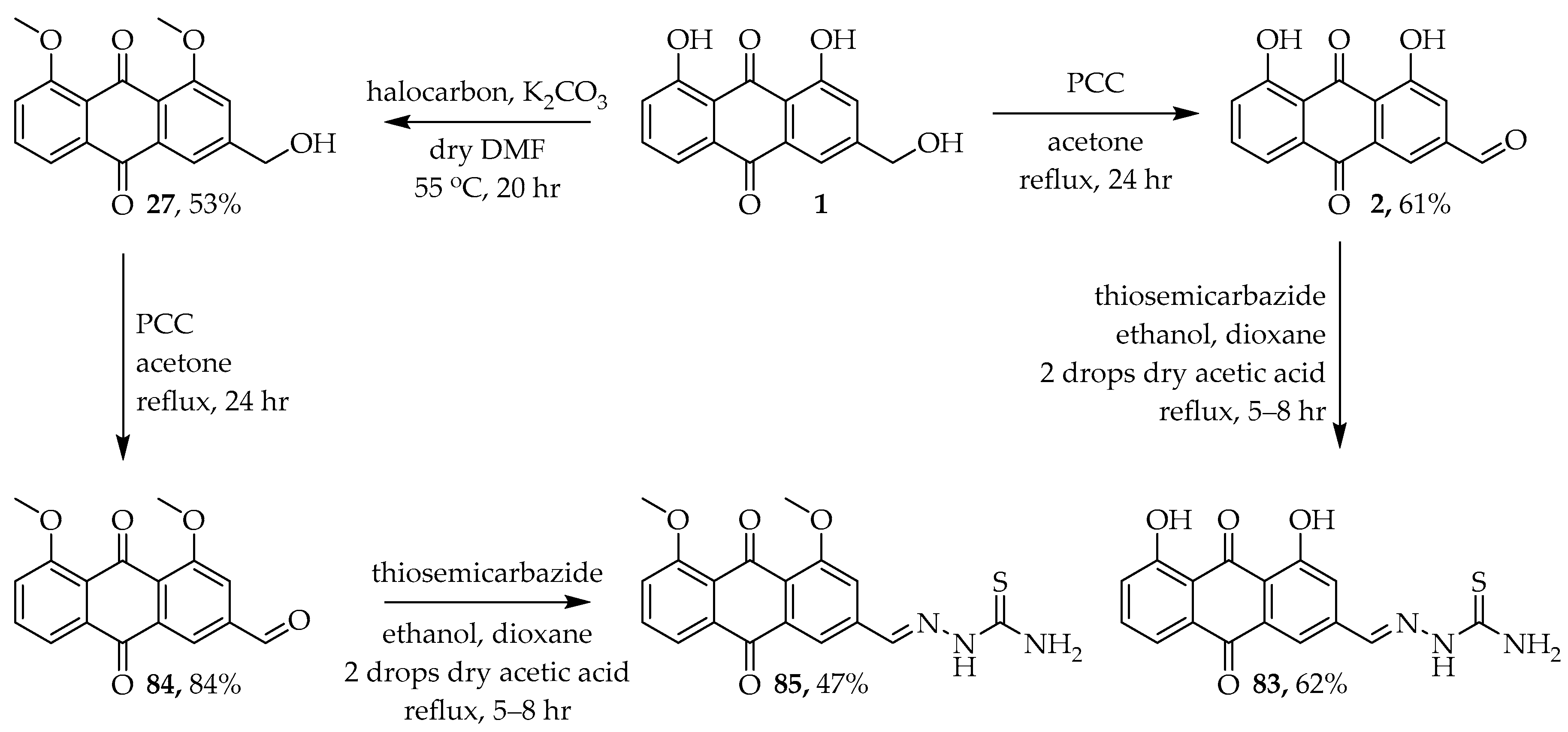
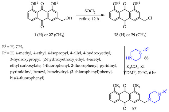
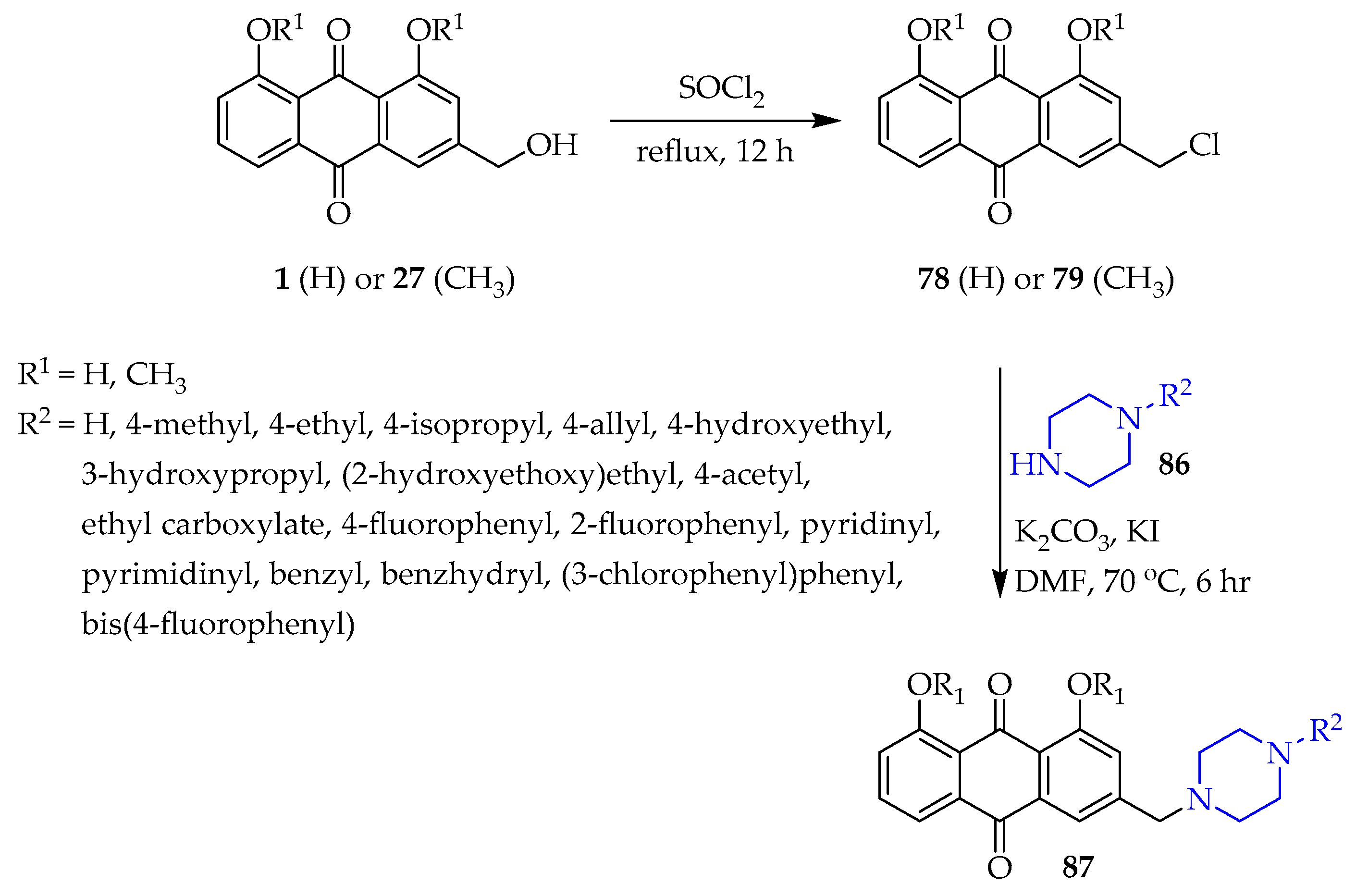
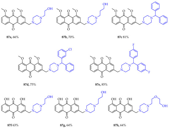
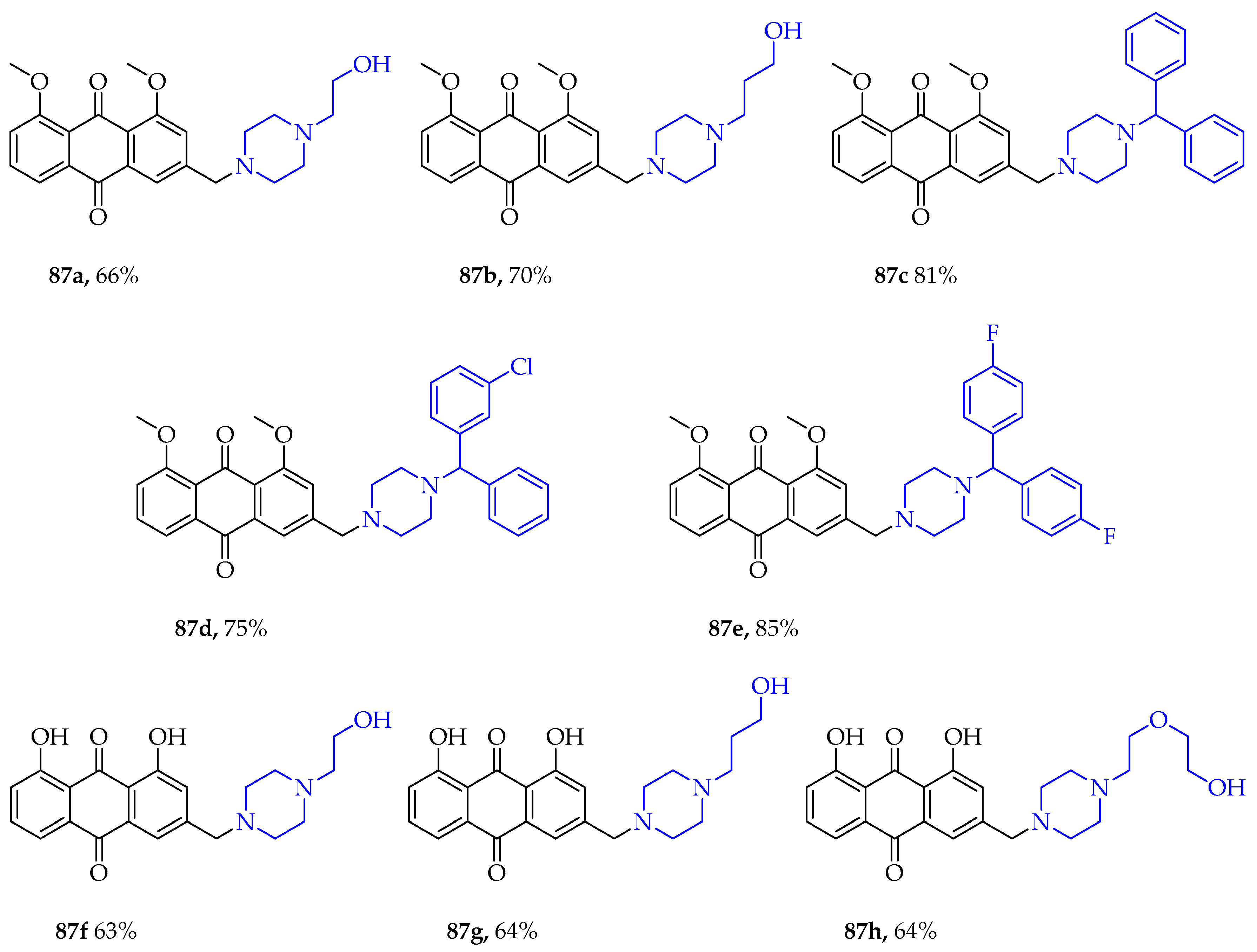
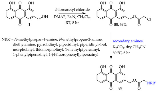
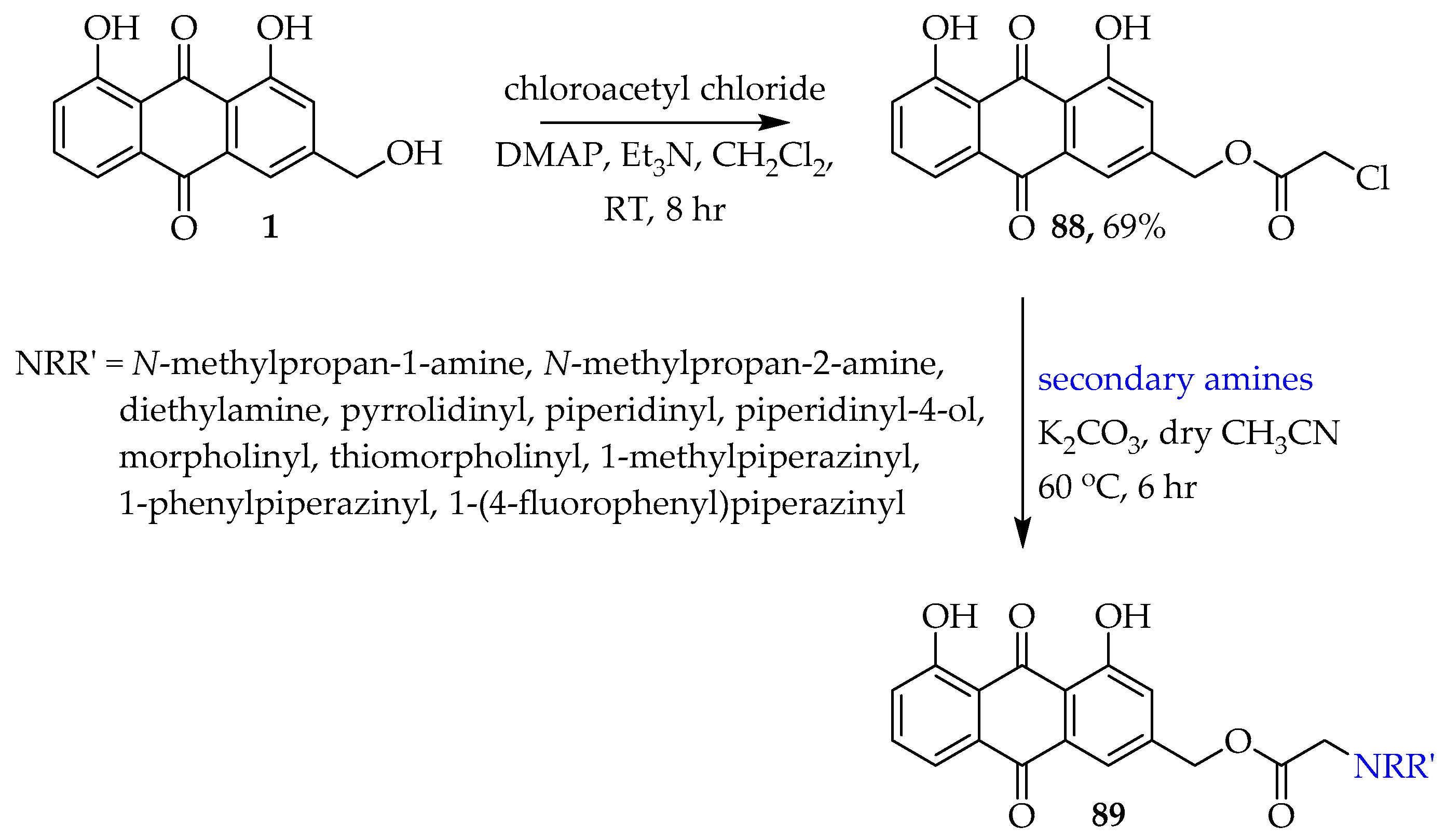
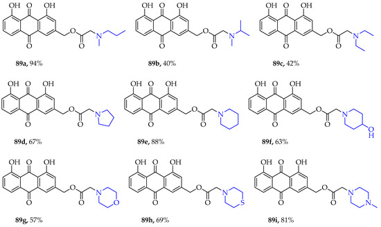
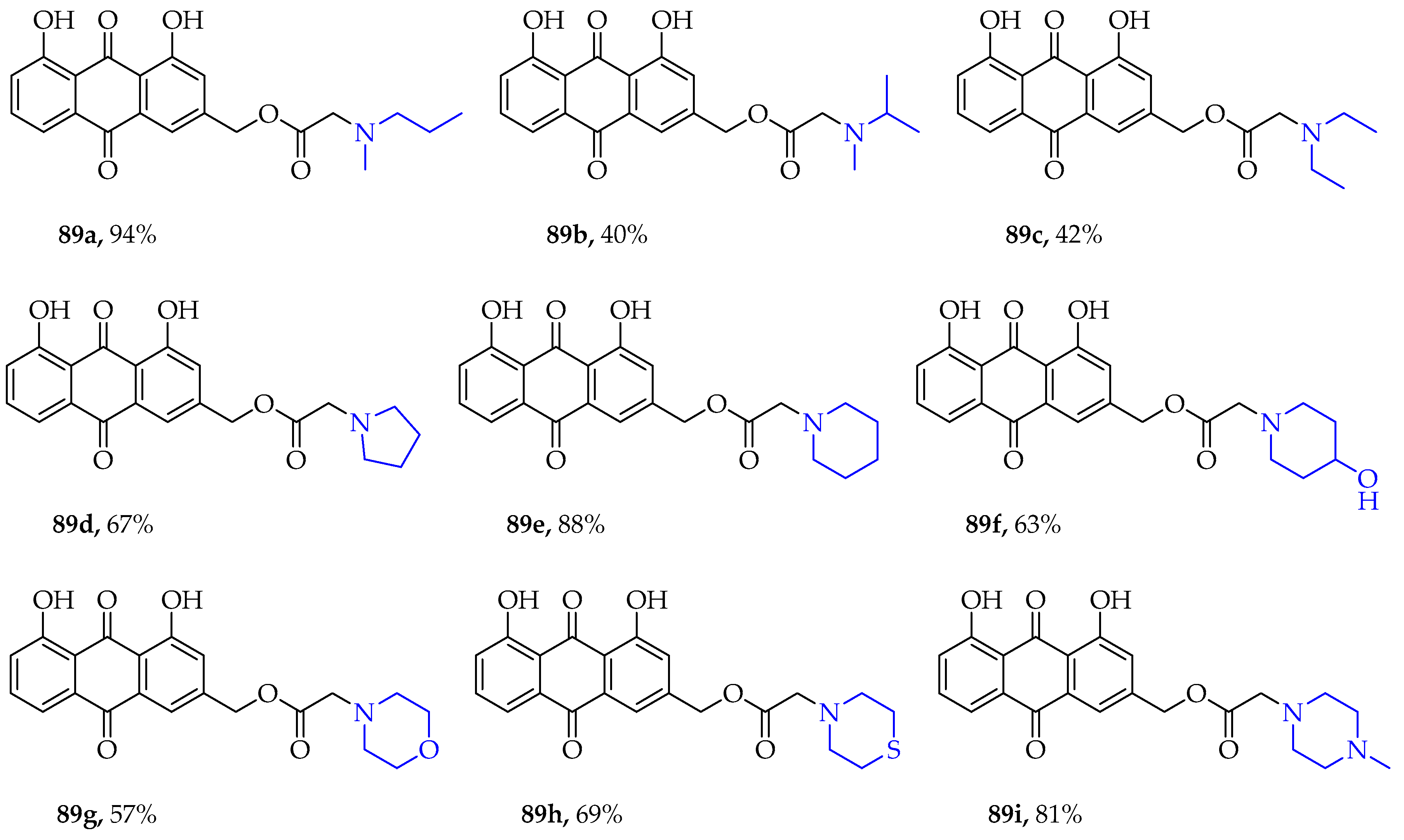




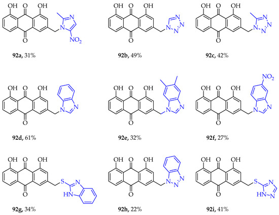
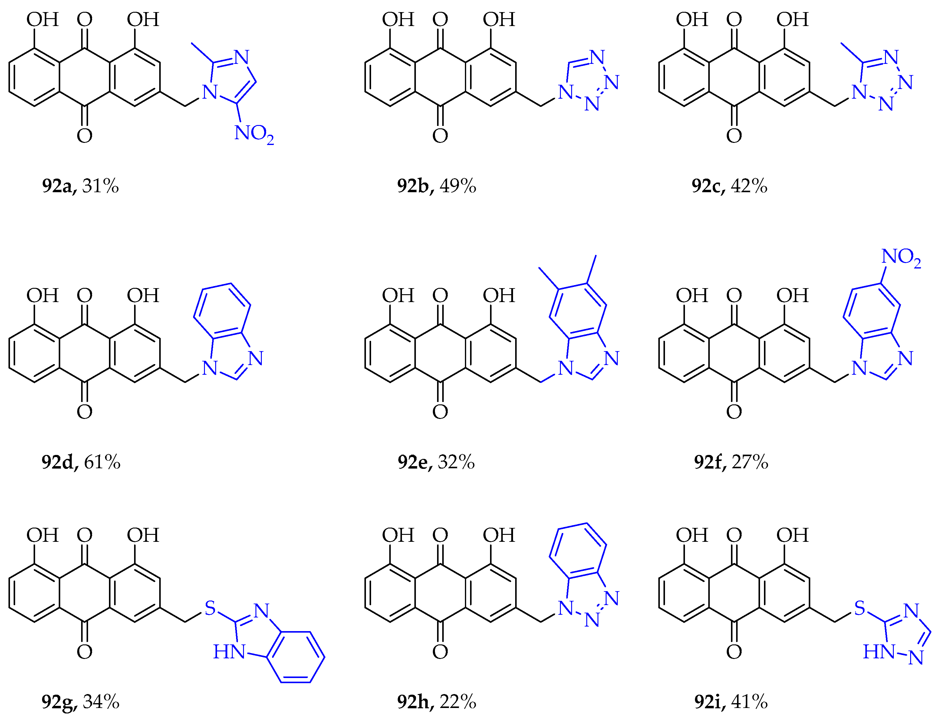
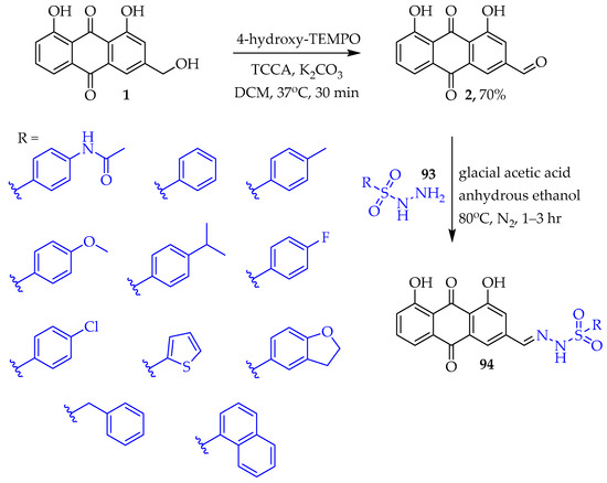
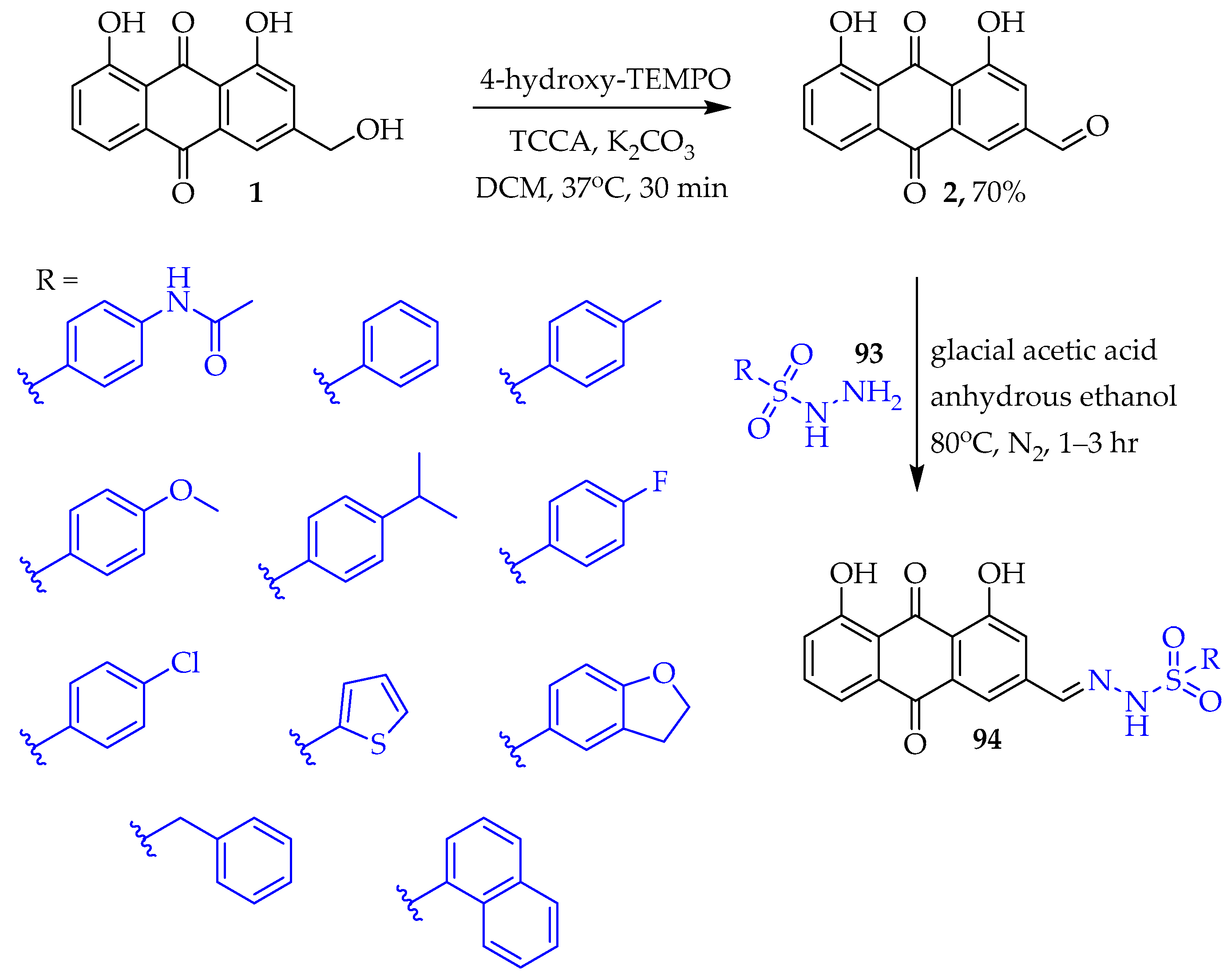
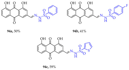
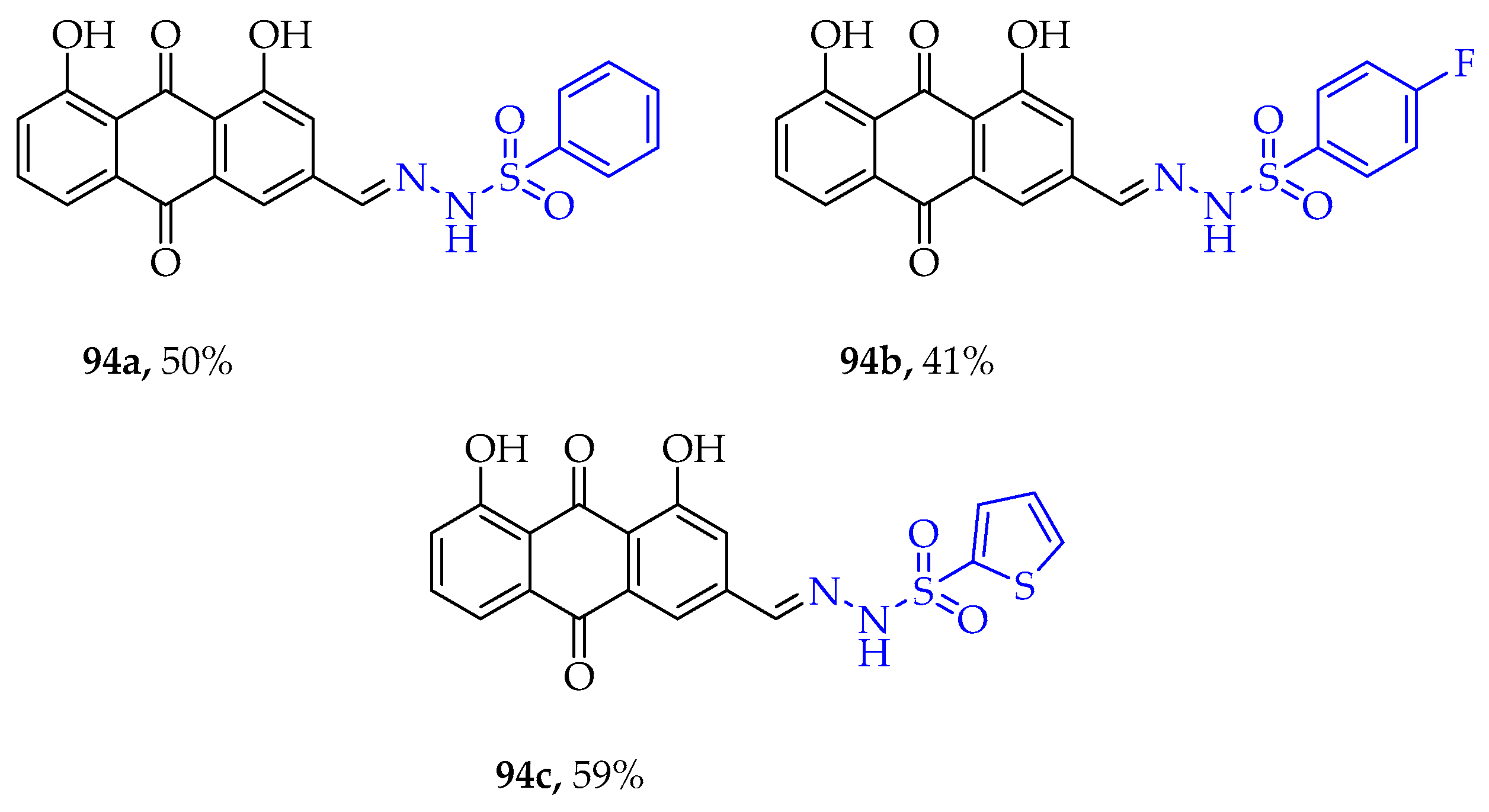
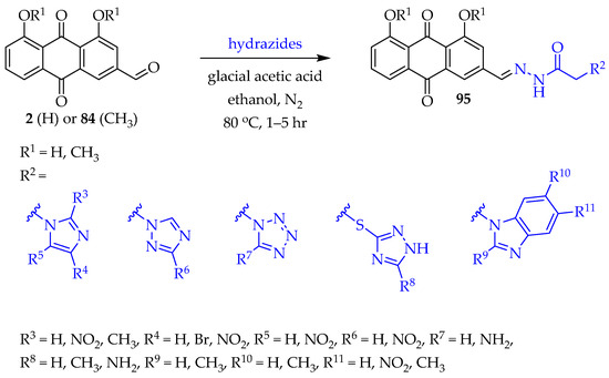
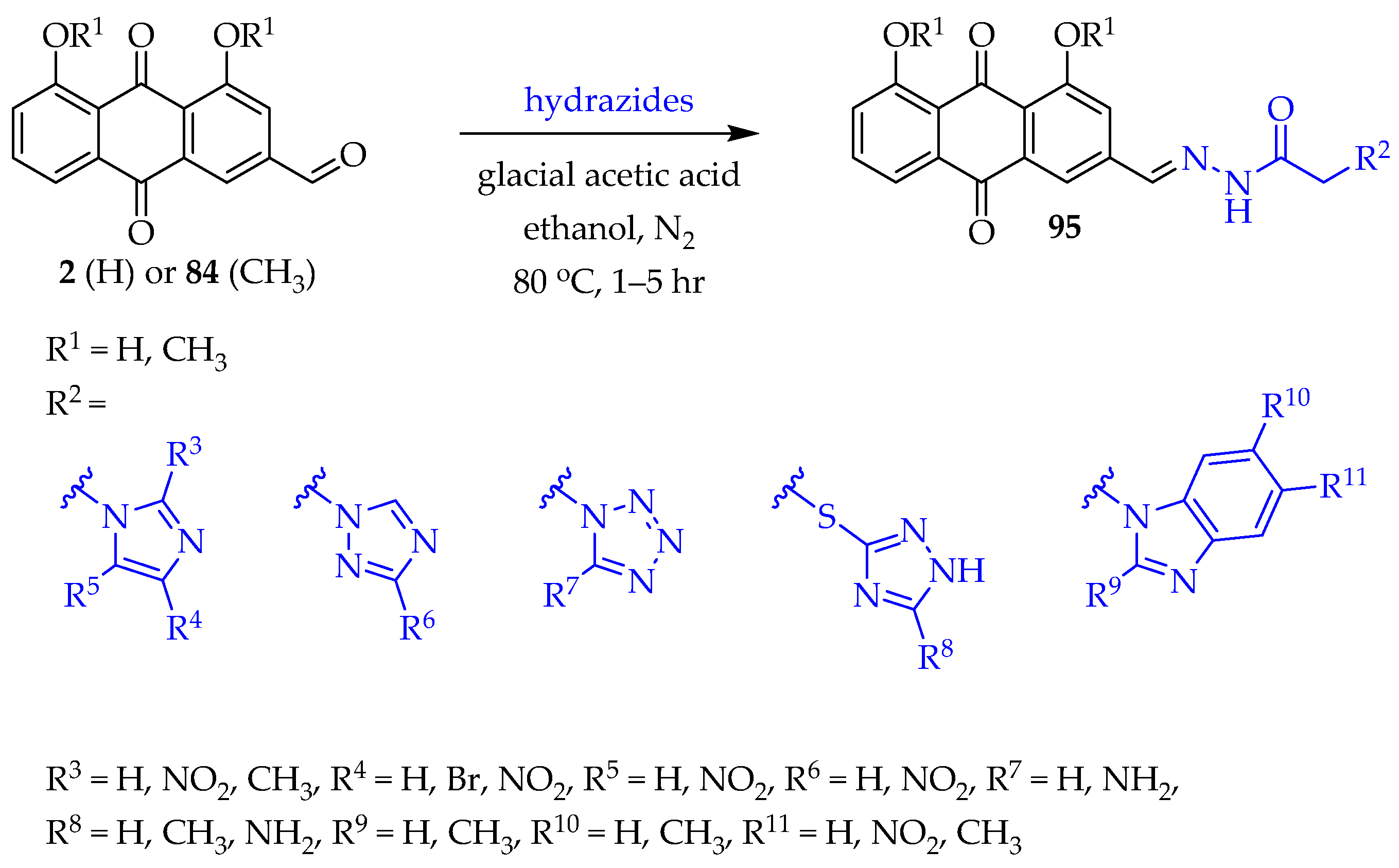
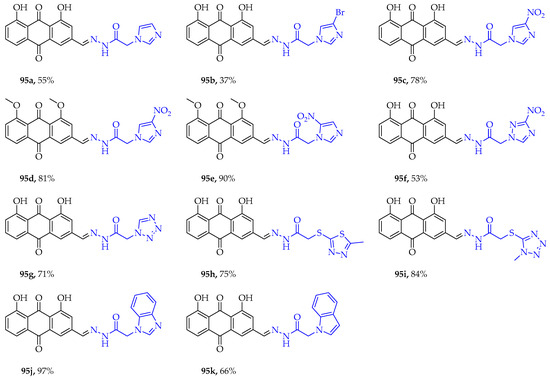
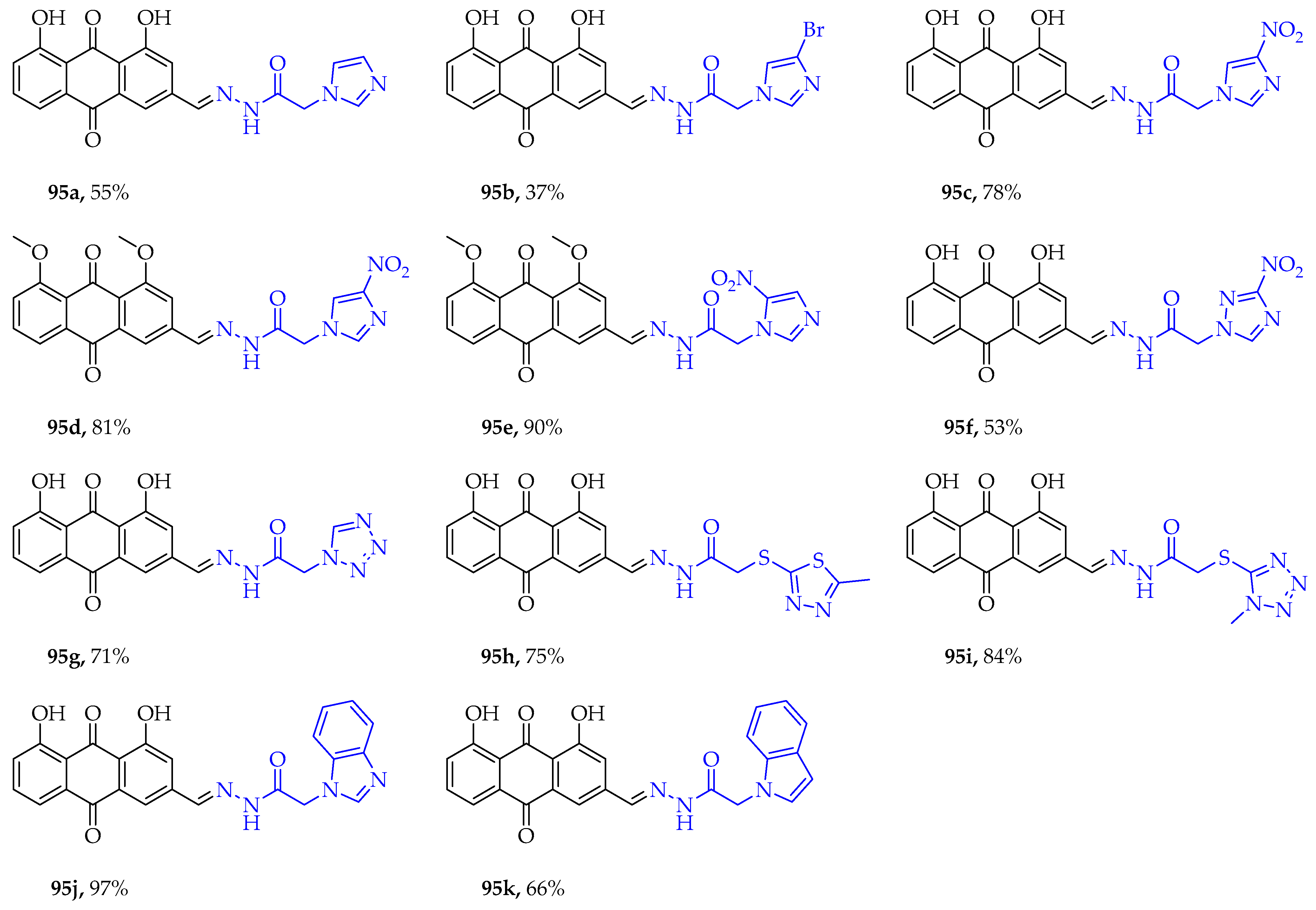
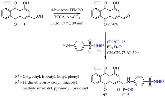
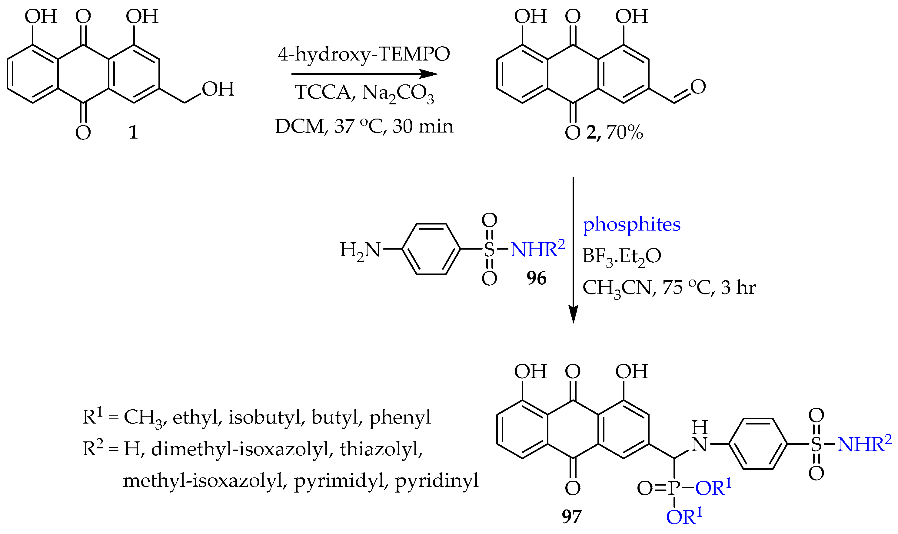
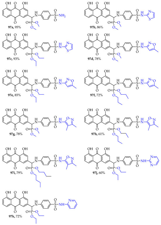
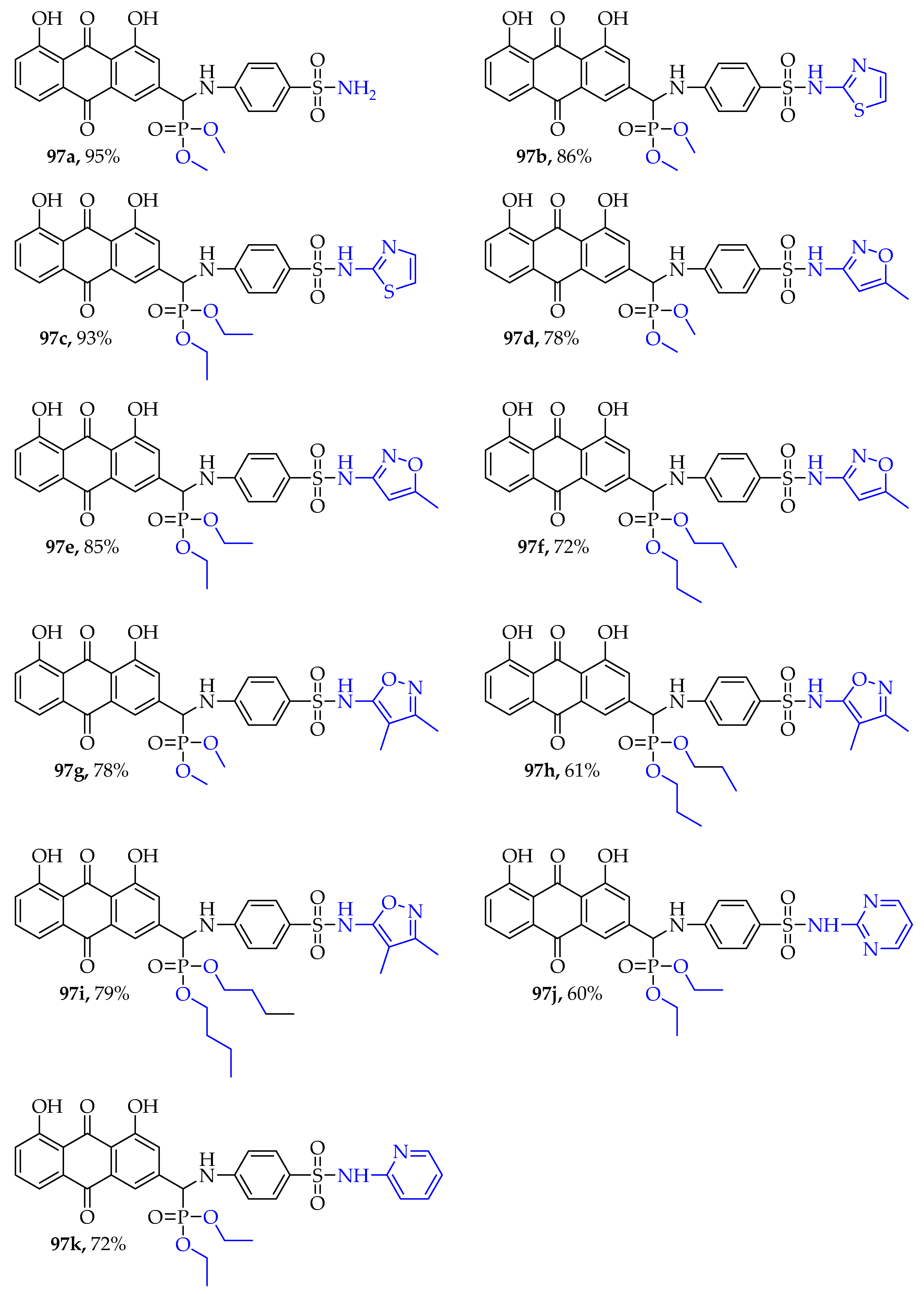
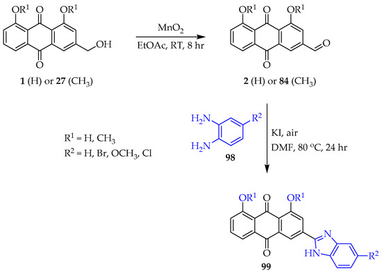
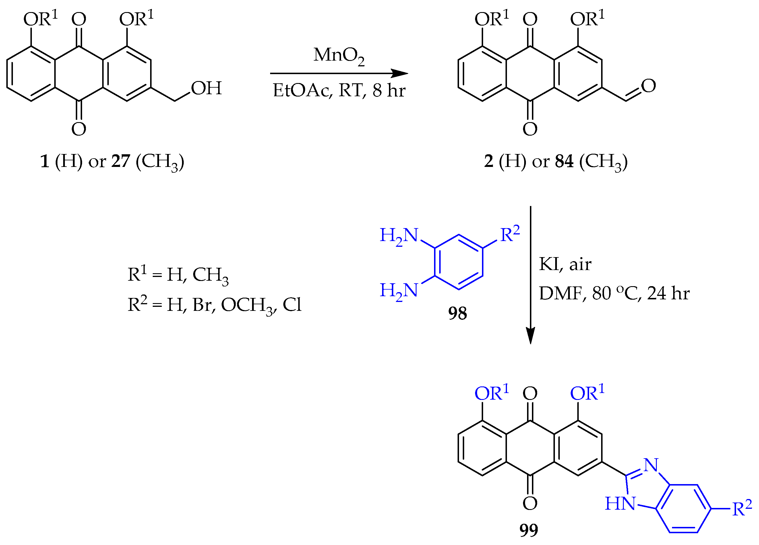
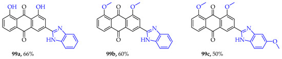
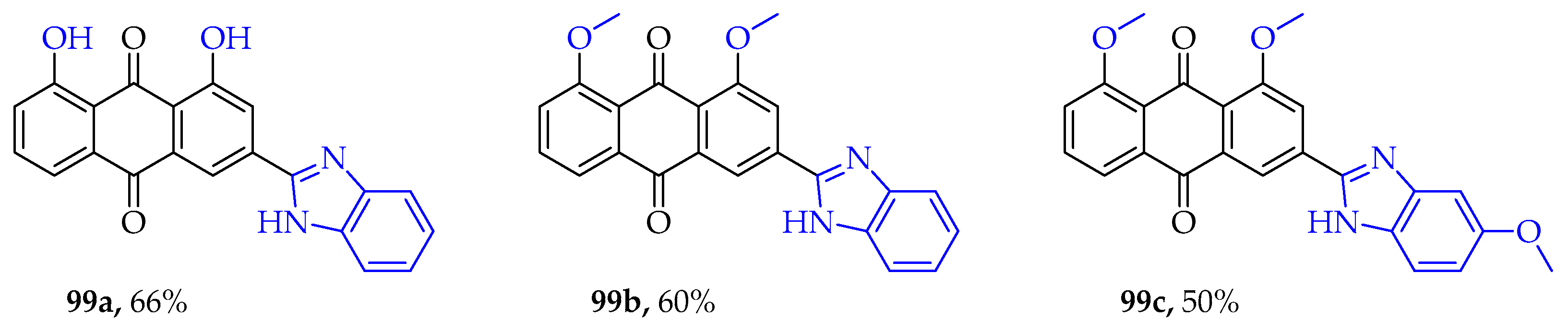
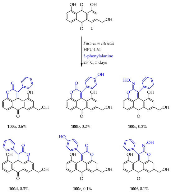
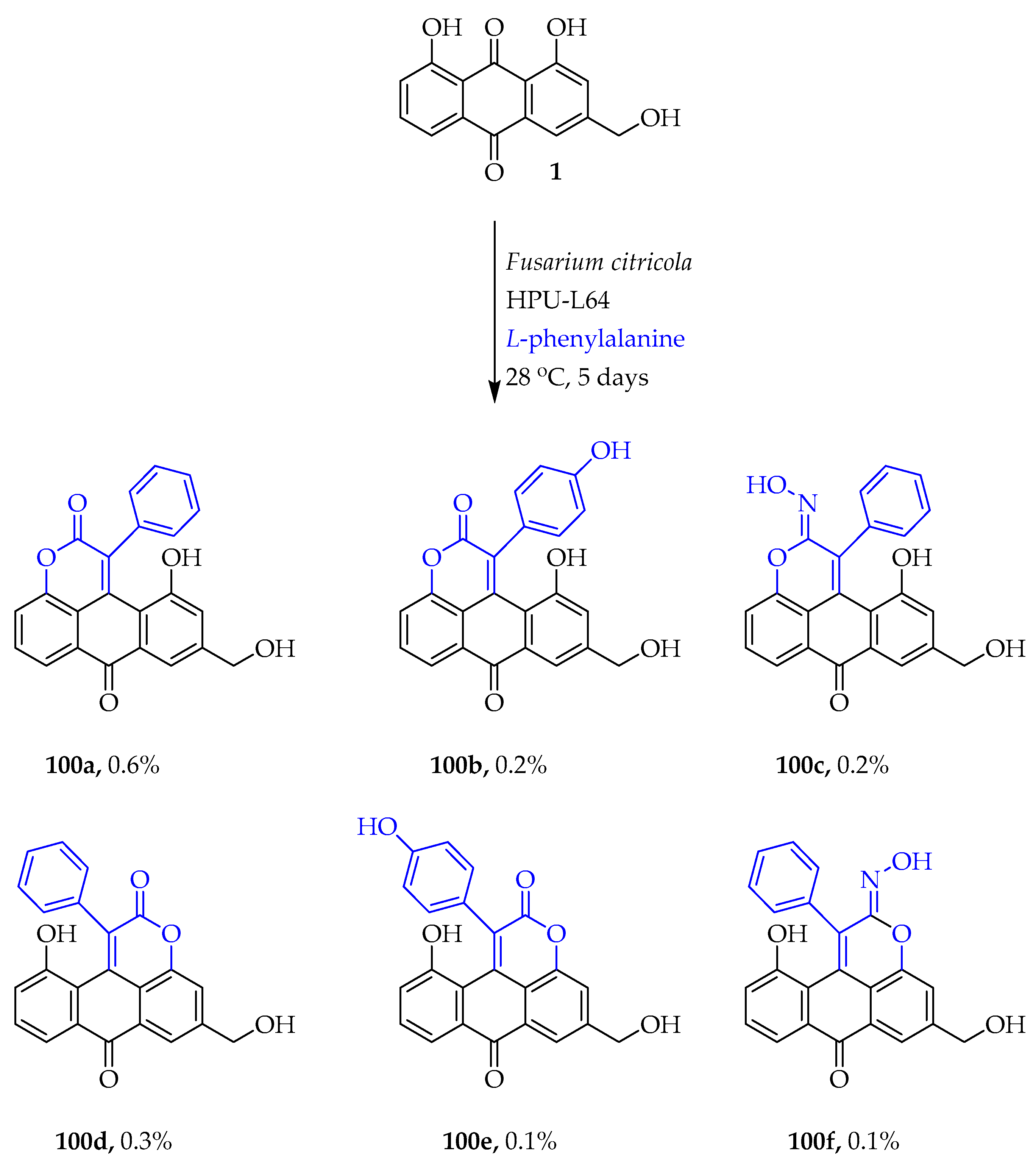
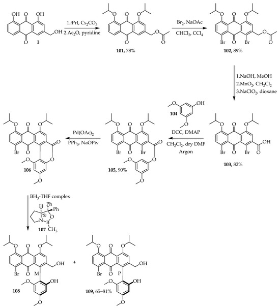
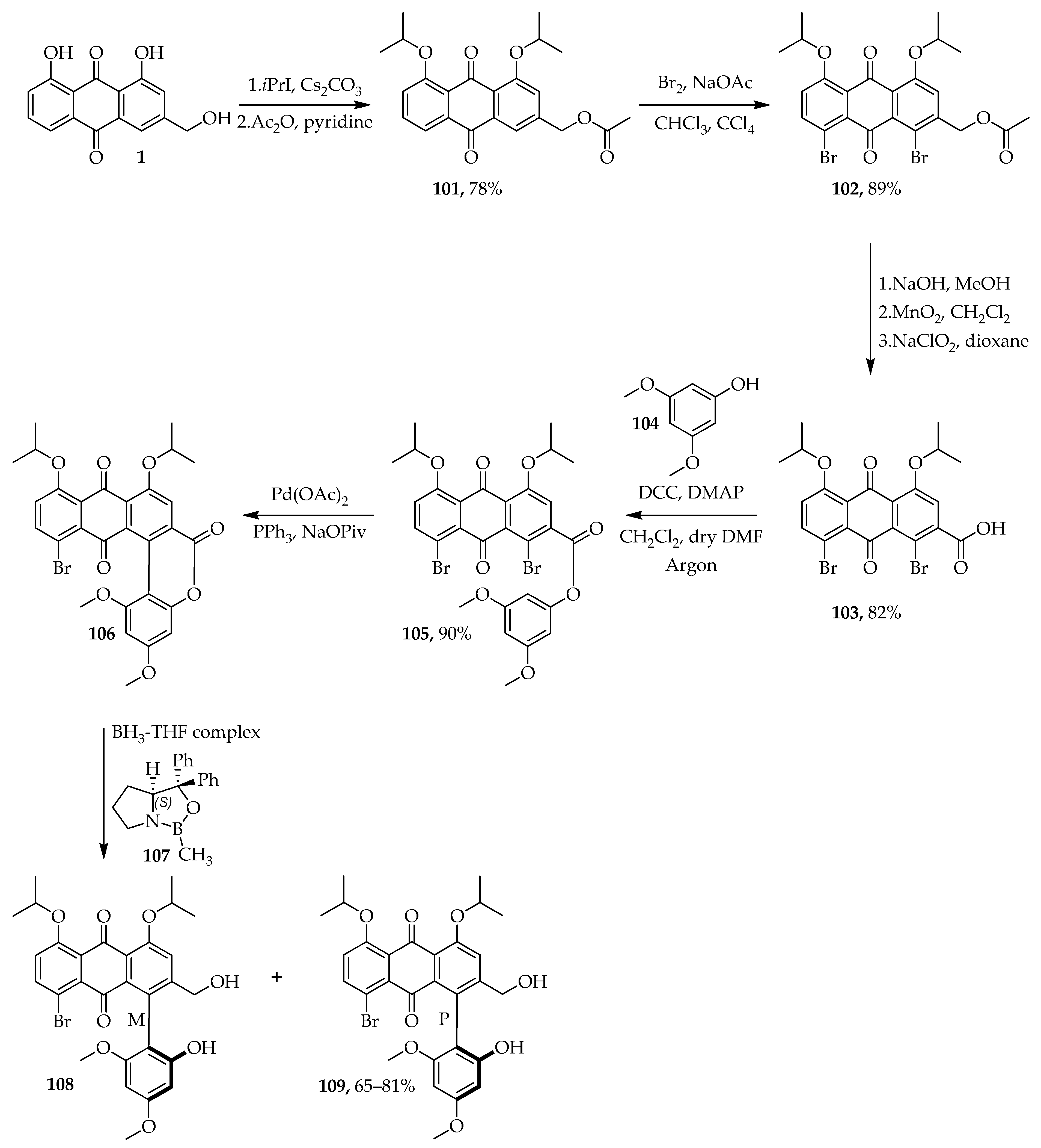
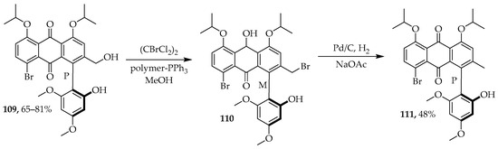
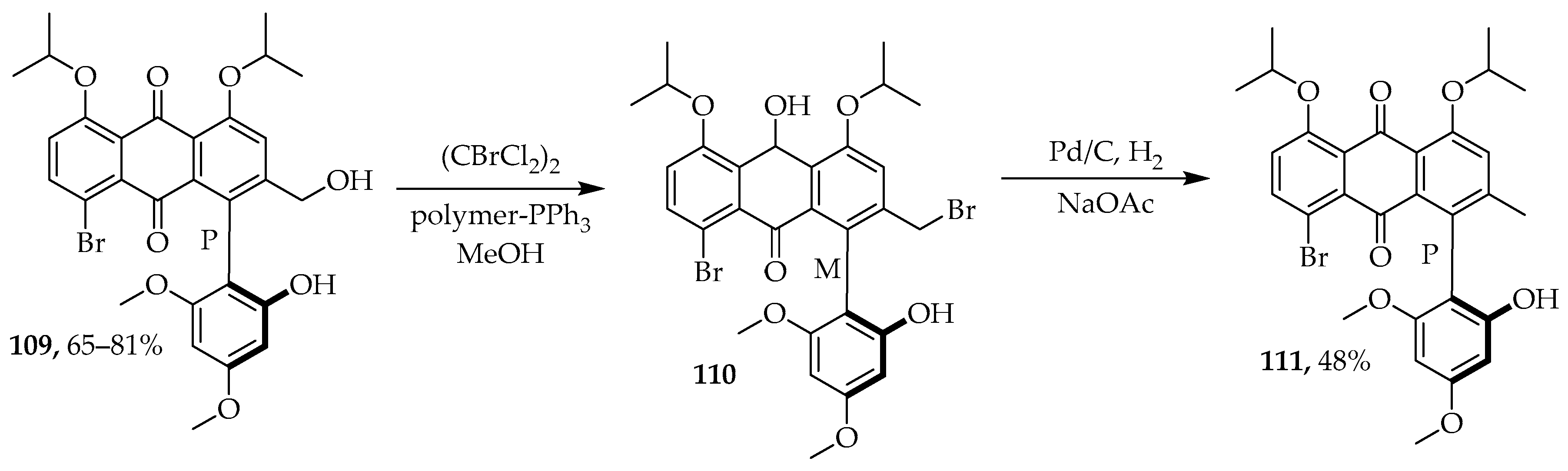
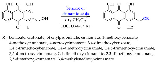
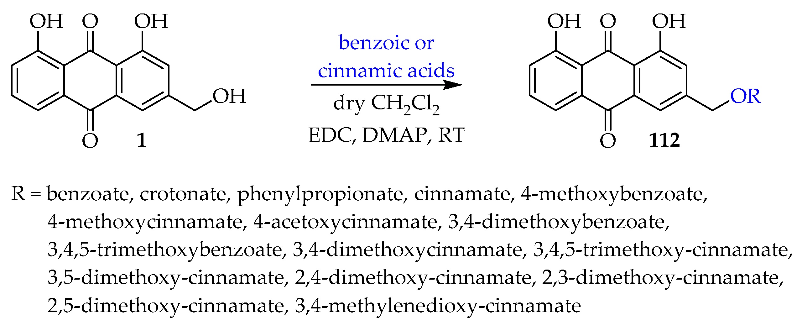
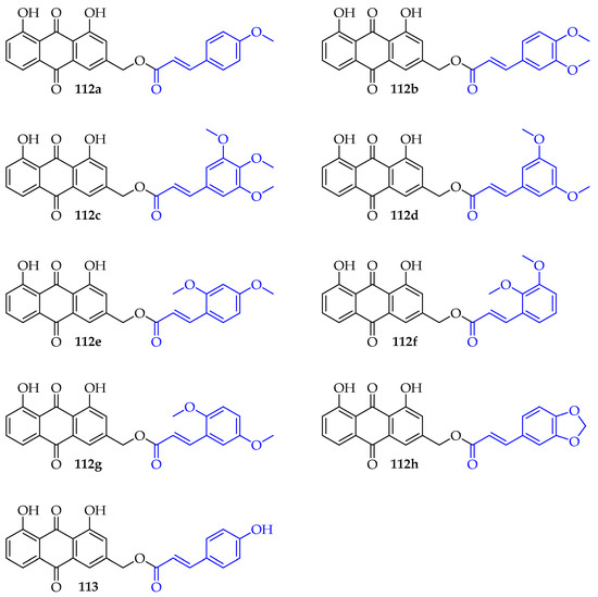
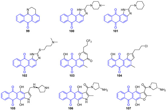
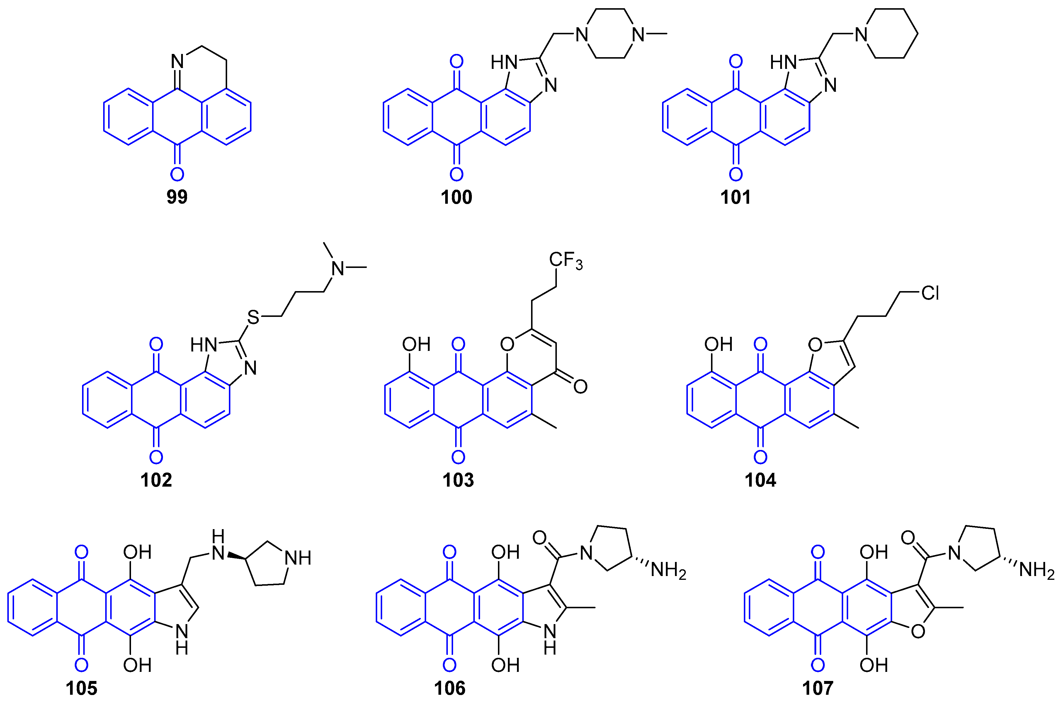

Disclaimer/Publisher’s Note: The statements, opinions and data contained in all publications are solely those of the individual author(s) and contributor(s) and not of MDPI and/or the editor(s). MDPI and/or the editor(s) disclaim responsibility for any injury to people or property resulting from any ideas, methods, instructions or products referred to in the content. |
© 2026 by the authors. Licensee MDPI, Basel, Switzerland. This article is an open access article distributed under the terms and conditions of the Creative Commons Attribution (CC BY) license.