


Abstract
Background: Well-differentiated pancreatic neuroendocrine tumors (PanNETs) develop in ~80% of patients with multiple endocrine neoplasia type 1 (MEN1) and remain the leading cause of MEN1-related mortality. Whether MEN1-associated PanNETs display distinct clinicopathologic or prognostic characteristics compared with non-MEN1 PanNETs remains incompletely defined. Methods: We retrospectively reviewed clinical, pathological, and outcome data from 817 patients who underwent surgical resection for well-differentiated PanNETs, including 39 MEN1-associated and 778 non-MEN1 PanNETs, with a total of 7752 person-years of follow-up. Tumor characteristics, stage, and survival outcomes were compared between two groups. Results: MEN1-associated PanNETs represented 4.8% of the cohort and included both functional and non-functional tumors. Patients with MEN1 more frequently had functional tumors, with insulinomas and gastrinomas predominating. Patients with MEN1 were significantly younger at surgery. Tumor size and grade distribution were similar between the two groups, although tumors >2 cm were more prevalent in patients with MEN1. MEN1-associated tumors demonstrated significantly lower rates of lymphovascular and perineural invasion, but similar rates of lymph node metastasis and stage III disease when only lymph node metastases attributable to a pancreatic primary were included. Long-term overall survival, disease-specific survival, and cumulative incidence of relapses did not differ significantly between MEN1 and non-MEN1 patients. Conclusions: In this retrospective cohort of surgically resected, well-differentiated PanNETs, MEN1-associated tumors exhibited similar histologic grade and tumor size distributions to non-MEN1 PanNETs. MEN1 status was associated with a higher prevalence of functional tumors and lymph node metastases, but lower rates of vascular and perineural invasion. Despite these features, no statistically significant differences in long-term overall survival, disease-specific survival, or cumulative incidence of relapse were observed between surgically resected patients with and without MEN1, though the study was underpowered to detect modest differences.
1. Introduction
Multiple endocrine neoplasia type 1 (MEN1) syndrome (MIM#131100), also known as Wermer syndrome, is an autosomal dominant hereditary tumor syndrome caused by inactivating germline variants in the MEN1 tumor suppressor gene [1,2]. The syndrome is characterized by the development of multiple endocrine tumors of the parathyroid glands, pituitary gland, and foregut neuroendocrine system [3,4]. The diagnosis of MEN1 can be made based on clinical, family history, and/or genetic criteria as defined by clinical practice guidelines [5].
Well-differentiated pancreatic neuroendocrine tumors (PanNETs) have been reported to occur in 30–80% of patients with MEN1 over their lifetime and are the leading cause of MEN1-related disease-specific mortality [6,7]. The clinical behavior of MEN1-associated PanNETs presents unique management challenges. MEN1-associated PanNETs, in contrast to non-MEN1 PanNETs, typically arise at a younger age, are more frequently multifocal, and may secrete functional hormones causing distinct clinical syndromes [8,9]. Due to the multifocality and heterogeneity of MEN1-associated PanNETs, there remains no clear consensus regarding the optimal timing, extent, and type of surgical intervention [10,11,12]. The recently published 2025 international consensus guidelines and clinical practice recommendations provide updated frameworks for MEN1 diagnosis, surveillance, and management [5,13]. Current recommendations advocate for active surveillance of asymptomatic non-functional PanNETs smaller than 2 cm, with surgical intervention reserved for tumors exceeding 2 cm, tumors demonstrating significant growth on serial imaging, or tumors causing clinically significant hormone-related symptoms in functional tumors [5,13]. These recommendations are based on the premise that smaller, non-functional tumors are likely to follow an indolent clinical course, whereas larger tumors carry increased risk of metastasis [5,11,13,14]. The evidence supporting these guidelines derives predominantly from series with relatively smaller cohort size, variable follow-up durations, and heterogeneous patient populations [15,16].
A fundamental question that remains incompletely resolved is whether MEN1-associated PanNETs are a biologically distinct entity compared with non-MEN1 PanNETs. Prior studies comparing MEN1-associated and non-MEN1 PanNETs have yielded conflicting results regarding tumor size, grade, metastatic potential, and survival outcomes. Two recent multi-institutional large cohort studies suggested that survival for patients with MEN1-associated PanNETs is comparable to those with sporadic tumors [17,18]. However, both studies had relatively short-term follow-up (both 5 years), and were multi-institutional, introducing significant inter-institutional heterogeneity in pathologic assessment and lack of uniformly applied standard surgical criteria. Furthermore, direct pathologic comparison of tumor characteristics including histologic grade, patterns of invasion, and staging parameters between MEN1 and non-MEN1 tumors in large surgical series remains sparse.
Therefore, this study aims to comprehensively characterize the clinicopathologic features and long-term prognostic outcomes of surgically resected well-differentiated PanNETs in patients with MEN1compared with a large cohort of non-MEN1 tumors, with extended follow-up from a single institution. We aimed to define whether MEN1 syndrome status independently influences tumor biology, pathologic characteristics, or long-term survival outcomes in patients undergoing surgical resection for PanNETs retrospectively. Our goal is to improve the understanding of MEN1-associated PanNETs with the hope of guiding patient management.
2. Materials and Methods
This study was approved by the Johns Hopkins Institutional Review Board. Patients who underwent surgical resection for well-differentiated pancreatic neuroendocrine tumors between 1990 and 2025 at the Johns Hopkins Hospital were retrospectively identified. A total of 817 patients were included, comprising 39 patients with MEN1 syndrome and 778 patients with non-MEN1 PanNETs. MEN1 diagnosis was established based on clinical criteria, genetic testing, or both, in accordance with established guidelines [5,13,19]. Of the 39 patients classified as having MEN1 syndrome, 13 (33.3%) were confirmed by identification of a pathogenic germline MEN1 variant. Three patients (7.7%) harbored variants of uncertain significance (VUS); however, these individuals also met clinical diagnostic criteria by having involvement of at least two MEN1-associated endocrine organs and/or a significant family history of MEN1 and were therefore included in the MEN1 cohort. The remaining 23 patients (59.0%) were classified based on clinical criteria, defined as the presence of tumors in at least two of the three principal MEN1-associated organs (parathyroid, pituitary, and pancreatic/duodenal neuroendocrine tumors) and/or a first-degree relative with confirmed MEN1 syndrome. Genetic testing was performed using next-generation sequencing panels or Sanger sequencing of the MEN1 gene, depending on the era of diagnosis. Patients with a MEN1 clinical phenotype but confirmed negative germline MEN1 testing (phenocopies) were not identified in our cohort [20]. Detailed clinical and genetic information for each MEN1 patient is provided in Supplementary Table S1. None of the 39 MEN1 patients had concurrent von Hippel-Lindau (VHL) syndrome, neurofibromatosis type 1 (NF1), MEN4, or tuberous sclerosis complex (TSC). The non-MEN1 group includes a small number of patients with genetically confirmed VHL, NF1, or TSC; therefore, this comparator group is designated as ‘non-MEN1 PanNETs’ rather than ‘sporadic PanNETs’. Patients with poorly differentiated neuroendocrine carcinomas (NECs) or mixed neuroendocrine–non-neuroendocrine neoplasms were excluded to maintain a homogeneous well-defined cohort. The electronic medical record was retrospectively reviewed for each patient to collect clinical data including demographics, MEN1 status, tumor functionality, type of surgery, and duration of follow-up. Functional tumors were defined by clinical syndromes and biochemical evidence of hormone hypersecretion [16].
Classification of gastrinomas required careful chart review of each case due to the anatomic complexity of gastrin-producing tumors in the foregut. Gastrinomas were classified based on the site of the identifiable primary tumor as determined by imaging, endoscopy, operative findings, and pathologic examination. In MEN1 patients, gastrinomas were categorized as: (1) duodenal primary with peripancreatic lymph node metastasis, when a duodenal tumor was identified as the source; (2) lymph node metastasis without identifiable primary, when metastatic gastrinoma was present in peripancreatic lymph nodes but no primary tumor could be localized despite thorough evaluation (recognizing that many of these are likely small duodenal primaries); or (3) pancreatic primary, when the gastrinoma clearly originated within the pancreatic parenchyma with no other site of tumor identified. This classification was essential for accurate interpretation of lymph node metastasis rates, as duodenal gastrinomas characteristically metastasize to peripancreatic lymph node basins and may be encountered incidentally during pancreatic resection for concurrent PanNETs.
The well-differentiated PanNETs were reviewed and assessed for the following histopathologic features: tumor size, histologic variant, tumor grade (Ki-67 proliferation index), lymphovascular invasion, perineural invasion, lymph node status, and pathologic stage [21]. Tumors were graded according to the 2026 World Health Organization (WHO) classification criteria using mitotic activity and Ki-67 index [22]. Ki-67 proliferative index was determined by immunohistochemistry in areas of highest labeling (hot spots), with counting of at least 500 tumor cells. Grade 2 tumors were further subdivided into G2a (Ki-67 3–10%) and G2b (Ki-67 11–20%) based on evidence that higher Ki-67 values within the G2 range confer increased risk of recurrence and metastasis [23]. When discordance occurred between Ki-67 index and mitotic count, the higher grade was assigned, consistent with WHO recommendations [22]. Tumor staging followed the American Joint Committee on Cancer (AJCC) staging system and TNM staging for neuroendocrine tumor was applied [24]. For patients with multiple PanNETs, particularly those with MEN1 syndrome, the largest or most clinically significant tumor was used for T-staging, grading, and pathologic staging purposes. Functional status was assigned based on the presence of a clinical hormone hypersecretion syndrome; no patients had multiple concurrent functional syndromes.
Demographic and clinical variables were compared between patients with and without MEN1 diagnosis using Fisher exact tests for categorical measures and Student t test or Wilcoxon rank sum tests for continuous measures. The primary analytic cohort for clinicopathologic comparisons included all 817 patients with M0 disease at surgery (778 non-MEN1, 39 MEN1). Three pre-specified time-to-event analyses were performed: (1) Overall survival (OS) was defined as the time from surgical resection to death from any cause or last follow-up as of 07/01/2025. Patients who died or were lost to follow-up within 30 days of surgery (n = 20, all non-MEN1) were excluded from OS analysis to focus on long-term outcomes and minimize the influence of perioperative mortality unrelated to tumor biology, yielding an analytic sample of 797 patients (758 non-MEN1, 39 MEN1). Survival was estimated using the Kaplan–Meier method and compared between patients with and without MEN1 diagnosis with Cox proportional hazards models. (2) Disease-specific survival (DSS) was defined as the time from surgical resection to death or last follow-up due to PanNET. Patients who died without relapse were censored at the date of death. Patients lacking complete data on cause of death or relapse status were excluded, yielding an analytic sample of 752 patients (718 non-MEN1, 34 MEN1). DSS was estimated using the Kaplan–Meier method and compared using Cox proportional hazards models. (3) Cumulative incidence of relapse (CIR) was defined as the interval from surgical resection to disease progression or last follow-up, with death before progression treated as a competing event. The same 752 patients with complete relapse ascertainment were included. The cumulative incidence of relapse was estimated using Fine and Gray’s method [25] and compared between patients with and without MEN1 diagnosis using a subdistribution hazards model. All time-to-event models adjust for age and sex. All hazard ratios are reported with 95% confidence intervals. Two-sided p values < 0.05 were considered statistically significant. Analyses were completed using R software version 4.5.1 [26].
3. Results
3.1. Demographic and Clinical Characteristics
MEN1-associated PanNETs comprised 4.8% of the cohort (39/817). The female-to-male ratio was similar between MEN1 and non-MEN1 groups. MEN1 patients were significantly younger at the time of surgery, with a mean age of 41.8 years compared to 57.4 years of non-MEN1 patients, and 89.7% of the patients with MEN1 were younger than 60 years (p < 0.001). Most MEN1-associated PanNETs (64.1%) were located in the distal pancreas compared to 50.9% in non-MEN1-associated tumors (p < 0.001). Functional PanNETs were more common in MEN1 patients (46.2% in MEN1 vs. 13.5% in non-MEN1, p < 0.001). Insulinomas and gastrinomas predominated, while pancreatic polypeptide–, glucagon-, ACTH-, and VIP-secreting tumors were rare. The MEN1-associated gastrinoma cohort could be divided into three categories: (1) Eight of the 11 (72.7%) had lymph node metastatic gastrinomas with a known duodenal primary; (2) two of the 11 (18.2%) had lymph node metastatic gastrinomas without an identifiable primary; and (3) one (9.1%) had a gastrinoma originating from the pancreas. Excluding gastrinomas from duodenal origin and metastatic to a peri-pancreatic lymph node, functional tumors were still more frequent in MEN1 patients (25.6% vs. 13.5%, p = 0.04). Importantly from a surgical standpoint, R0 resection rates were similar between the two groups (92.4% in MEN1 versus 86.8% in non-MEN1, p = 0.42) (Table 1).
Table 1.
Demographics, clinicopathologic characteristics, and outcomes, separately according to MEN1 status.
3.2. Pathological Features and Outcomes
Histological variants, including cystic and sclerosing patterns, were observed in both groups without statistically significant differences. T-stage distribution except T4, as reflected by tumor size, was virtually identical between the two groups with a mean size of 2.9 cm in MEN1 and 3.1 cm in non-MEN1 (p = 0.42); however, tumors larger than 2 cm were more prevalent in patients with MEN1. Grade distribution based on Ki-67 labeling indices was essentially superimposable between MEN1 and non-MEN1 tumors (p > 0.99). The majority of MEN1-associated PanNETs were low-grade (grade 1 or 2). MEN1-associated PanNETs demonstrated significantly lower rates of lymphovascular and perineural invasion compared with non-MEN1 tumors (2.6% vs. 21.6%, p = 0.002, and 10.3% vs. 29.2%, p = 0.006, respectively). After excluding the peripancreatic lymph node metastasis from duodenal primary gastrinomas, the rate of lymph node metastasis and stage III disease are similar between the two groups. These findings align with previous observations that MEN1-associated duodenal gastrinomas may preferentially spread to lymph nodes despite limited local invasiveness [27] (Table 1).
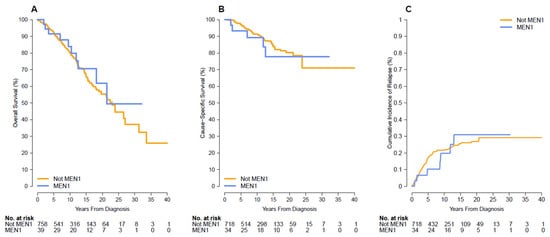
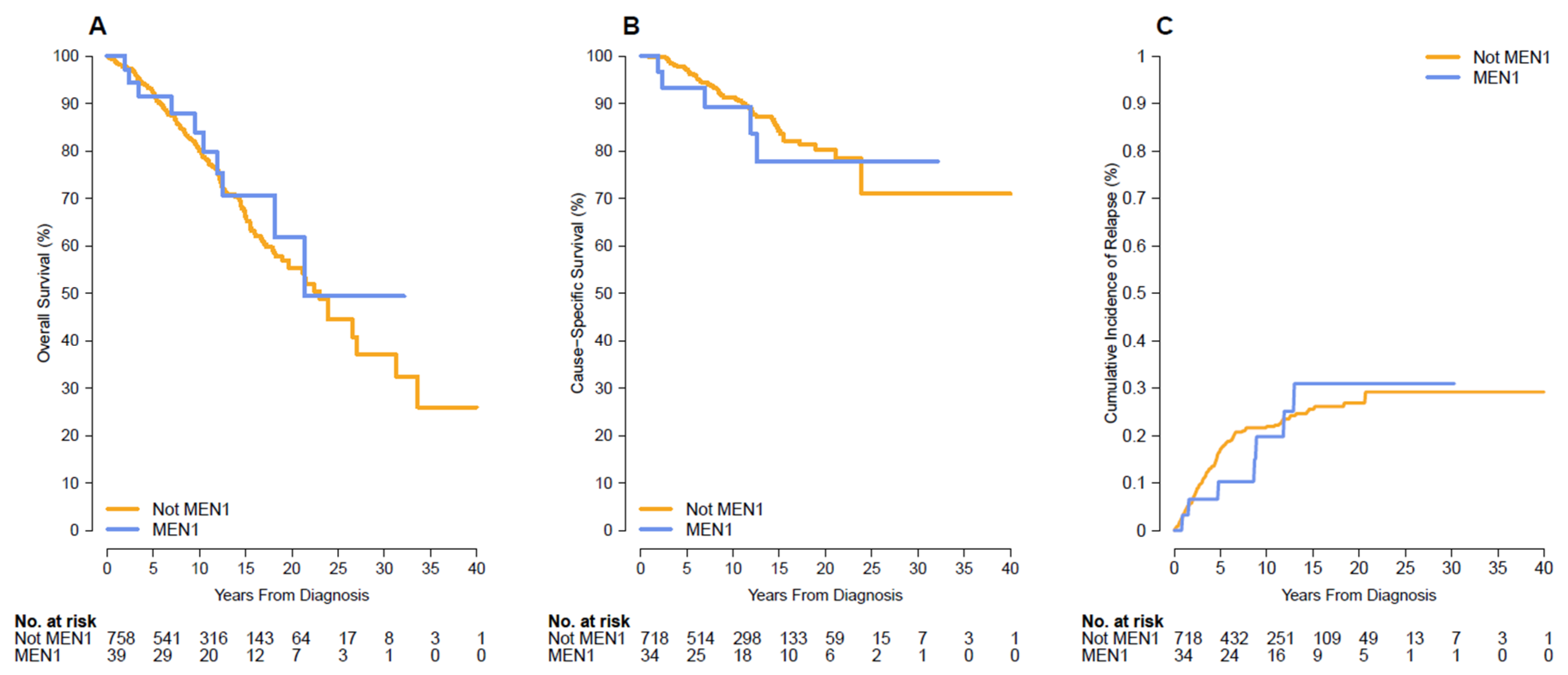
Sample sizes for survival analyses differed from the baseline cohort due to pre-specified exclusion criteria. For overall survival analysis, 20 patients (all non-MEN1) were excluded due to perioperative death or loss to follow-up within 30 days of surgery, yielding 797 patients (758 non-MEN1, 39 MEN1). For disease-specific survival and cumulative incidence of relapse analyses, an additional 45 patients (40 non-MEN1, 5 MEN1) were excluded due to incomplete data on relapse status or cause of death, yielding 752 patients (718 non-MEN1, 34 MEN1) for these analyses. With a total follow-up of 7752 person-years, overall survival (OS) was similar between patients with and without MEN1 (p = 0.17). Ten-year overall survival rates from the time of surgery were 83.9% for patients with MEN1 [95% CI:71.8, 98.1] and 79.9% for patients without MEN1 [95% CI: 76.6, 83.4] with adjusted Hazard Ratio (aHR) of 1.59. (Table 2 and Figure 1A). Five-year overall survival from the time of surgery was 91.4% in MEN1 and 92.3% in non-MEN1, consistent with prior 5-years follow up outcome studies [16,17]. Disease-specific survival at 10 years was 77.7% for patients with MEN1 [95% CI: 61.6, 98.0] and 80.1% for patients without MEN1 [95% CI: 75.0, 85.6] (p = 0.60) (Table 3 and Figure 1B). The cumulative incidence of relapses (CSS) was 0.20 for patients with MEN1 [95% CI: 0.03, 0.36] and 0.22 for patients without MEN1 [95% CI: 0.19, 0.25] at 10-years (p = 0.88) (Table 4 and Figure 1C). The OS and CSS with PSW are similar, with no significant statistical difference with OS of 1.59 (regression adjustment) vs. 1.52 (PSW), and CSS of 1.62 (regression adjustment) vs. 1.24 (PSW).
Table 2.
Overall survival according to MEN1 status *.
Figure 1.
Survival outcomes and relapses in PanNET patients with and without MEN1. (A) Kaplan–Meier curve for overall survival (OS) comparing patients with MEN1 (n = 39) and patients without MEN1 (Not MEN1; n = 758), measured from the time of surgery. (B) Kaplan–Meier curve for cause-specific survival (CSS) comparing MEN1 (n = 34) and Not MEN1 (n = 718) patients from time of the surgery to death. (C) Cumulative incidence of relapse (CIR) comparing MEN1 (n = 34) and Not MEN1 (n = 718) patients from the time of surgery to death. The number of patients at risk at each 5-year interval is displayed below each panel. Follow-up extended up to 40 years from the time of surgery.
Table 3.
Disease-specific survival according to MEN1 status *.
Table 4.
Cumulative incidence of relapses according to MEN1 status *.
4. Discussion
This study is one of the largest single institution comparative analyses of surgically resected MEN1-associated and non-MEN1 PanNETs with extended long-term follow-up (average 10 years), providing robust data to characterize the clinicopathologic features and prognostic outcomes in these two patient populations. In this retrospective surgical cohort, MEN1-associated PanNETs exhibited certain distinctive clinical and pathologic characteristics, including younger age at presentation, higher prevalence of functional tumors, larger tumor size, and higher nodal disease burden from duodenal primaries, yet no statistically significant difference in long-term overall survival was observed compared with non-MEN1 tumors.
An important consideration when interpreting our findings is that the enrollment period (1990–2025) spans substantial evolution in MEN1 management paradigms. Earlier in the study period, surgical approaches for MEN1-associated PanNETs were often more aggressive, with lower thresholds for intervention. In contrast, contemporary practice, as reflected in the 2025 international consensus guidelines and clinical practice recommendations [5,13], emphasizes structured surveillance programs with more selective surgical criteria based on tumor size (>2 cm threshold), growth kinetics, and functional status. This evolution in practice patterns may have influenced the composition of our surgical cohort, with earlier cases potentially including smaller or more indolent tumors that would be managed non-surgically under current guidelines. Despite this heterogeneity, our long-term outcome data provide valuable insights into the natural history of surgically resected MEN1-associated PanNETs across different management eras and support the favorable prognosis associated with surgical resection when indicated.
The observation that patients with MEN1 underwent surgery at a significantly younger age than non-MEN1 patients is consistent with the natural history of hereditary tumor syndromes, in which germline deleterious variants predispose to earlier tumor development and surveillance programs facilitate earlier detection [28,29]. The predominance of functional tumors, particularly insulinomas and gastrinomas, among MEN1 patients in our cohort is consistent with the well-documented MEN1 tumor biology [3,4]. These functional tumors often prompt surgical intervention independent of size criteria due to refractory symptoms or incorrect medical management, potentially contributing to the surgical case mix observed in our series.
Although the average size was similar between patients with and without MEN1, a notable finding in our study is the higher proportion of tumors exceeding 2 cm among patients with MEN1 compared with non-MEN1 cases. The higher prevalence of larger tumors in our surgical cohort may reflect selection bias inherent to guideline-concordant practice, consistent with the 2025 consensus recommendations that advocate surveillance for tumors < 2 cm and surgery only for larger lesions. Under these contemporary criteria, smaller MEN1-associated tumors are managed non-surgically while larger tumors are preferentially referred for resection [5,13]. Alternatively, this finding could indicate that a subset of MEN1-associated PanNETs demonstrate more accelerated growth kinetics requiring intervention. A third possibility could be that MEN1-associated PanNETs begin to grow earlier in life than non-MEN1 PanNETs and thus have a growth advantage over the non-MEN1 tumors that start to grow later. Distinguishing between these possibilities requires prospective studies incorporating comprehensive surveillance data and serial imaging assessments or an extensive morphological analysis of PanNETs in large resection specimens from MEN1 patients of various ages compared to matched non-MEN1 patients.
Despite the higher proportion of larger tumors in MEN1 patients, long-term outcomes remained comparable to non-MEN1 patients. Several factors may explain this apparent paradox. First, MEN1 patients undergo routine surveillance, potentially enabling detection at earlier biological stages regardless of tumor size. Second, the higher proportion of functional tumors in MEN1 patients leads to earlier symptomatic presentation and surgical intervention before the development of aggressive features. Third, and perhaps most importantly, the significantly lower rates of lymphovascular invasion (2.6% vs. 22.1%) and perineural invasion (10.3% vs. 29.7%) in MEN1-associated tumors suggest inherently less aggressive biological behavior that may offset the expected negative prognostic impact of larger tumor size.
While we observed only one primary pancreatic gastrinoma, peripancreatic lymph node metastasis of gastrinomas were much more frequent in patients with MEN1. The majority of gastrinomas in MEN1 patients arise not within the pancreatic parenchyma but rather in the duodenum, particularly within the so-called “gastrinoma triangle” [30,31,32]. These duodenal gastrinomas, which are characteristically small and often multifocal, demonstrate a well-documented propensity for early lymph node metastasis to peripancreatic and periduodenal lymph node basins, frequently disproportionate to their diminutive primary tumor size [28,29,33]. When MEN1 patients undergo pancreatic resection for concurrent PanNETs, the regional lymphadenectomy specimen may harbor metastasis originating from synchronous duodenal neuroendocrine tumors rather than from the resected pancreatic primary. The higher rate of nodal involvement may therefore reflect, at least in part, the inclusion of cases with concurrent duodenal gastrinoma-derived nodal metastasis rather than indicating a higher metastatic potential of the PanNETs themselves. Second, it is also possible that different tumor populations within the MEN1 enteropancreatic axis may be contributing to the observed nodal disease burden. MEN1 often involves multiple synchronous micro- and macro-PanNETs; the “dominant” resected lesion may not always be the biologic driver of nodal spread, and nodal metastasis could reflect dissemination from one of the patient’s multiple separate primaries. The former mechanism appears to be the primary driver of the higher lymph node metastatic rate observed in our study, as the metastatic rates became comparable between the two groups once we excluded positive nodes derived from known duodenal primaries as well as involved peripancreatic lymph nodes for which no primary tumor could be identified. Regardless of mechanism, the finding is clinically relevant because nodal positivity did not translate into inferior long-term survival outcomes in this cohort. From a practical standpoint, this finding underscores the importance of comprehensive preoperative evaluation in patients with MEN1 to identify synchronous duodenal tumors. Additionally, careful gross examination of specimens in MEN1 patients may help clarify the origin of nodal metastases.
In this retrospective cohort of surgically resected, well-differentiated PanNETs, MEN1-associated tumors shared key histopathologic features with non-MEN1 PanNETs, including similar tumor grade and T-stage distributions. Despite being diagnosed at a younger age, and frequently presenting with larger tumors, patients with MEN1-associated PanNETs do not exhibit inferior long-term overall survival, disease-specific survival, and cumulative incidence of relapses. These data argue against an assumption that MEN1 status alone confers worse post-resection prognosis. Instead, they reinforce that tumor grade and stage remain central anchors of risk stratification across both MEN1 and non-MEN1 settings, at least among retrospective, surgically treated, well-differentiated tumor cohorts. The results also support current strategies emphasizing individualized surgical decision-making and careful surveillance, particularly for small, low-grade lesions in patients with MEN1.
Our survival findings are concordant with recent data from other MEN1 cohorts. Modica et al. reported on 100 patients with MEN1 and found no statistically significant sex-based differences in the occurrence of MEN1-associated tumors or in survival outcomes among patients with duodenopancreatic NETs, and no significant difference between male and female patients [34]. Similarly, in our cohort, the sex distribution did not differ significantly between MEN1 and non-MEN1 groups (53.8% vs. 48.3% female, p = 0.52). Our decision to adjust survival analyses for both age and sex was made a priori given the established prognostic relevance of these factors in PanNET cohorts overall [35]. The concordance between our findings and those of Modica et al. provides reassurance that sex is unlikely to be a major confounder in MEN1-associated PanNET survival analyses, though larger studies would be required to definitively exclude modest sex-based prognostic differences.
MEN1 is a multisystem syndrome in which competing morbidities from concurrent endocrine manifestations may influence overall survival independently of PanNET behavior. Interpretation of survival outcomes in MEN1 patients requires consideration of the broader MEN1 clinical phenotype beyond pancreatic neuroendocrine disease. Recent evidence has highlighted the importance of comprehensive clinical characterization of MEN1 patients, including evaluation of previously underappreciated disease manifestations. Modica et al. recently reported that mild autonomous cortisol secretion (MACS) was present in 68.2% of MEN1 patients with adrenal lesions who underwent systematic hormonal evaluation, a prevalence substantially higher than typically observed in sporadic adrenal incidentalomas [36]. MACS has been associated with increased cardiometabolic morbidity, including hypertension, diabetes mellitus, and dyslipidemia, which may contribute to non-cancer-related mortality. These findings underscore that overall survival in MEN1 patients reflects the cumulative burden of multiorgan endocrine disease rather than PanNET behavior alone. Accordingly, the absence of a significant overall survival difference between MEN1 and non-MEN1 PanNET patients in our cohort may partly reflect competing risks from other MEN1 manifestations not captured in our disease-specific survival analysis. Comprehensive endocrine evaluation of MEN1 patients, including systematic assessment of adrenal function, may identify modifiable risk factors that influence long-term outcomes.
A major strength of this study is the large cohort and extended time horizon within a single institution, which reduces heterogeneity in pathology review and surgical approach compared with multi-institution studies. Several limitations of our study should be acknowledged. First, the retrospective design introduces potential selection biases, as our cohort comprises only surgically resected tumors and excludes non-surgically managed cases that may follow different clinical trajectories. Second, while our MEN1 cohort of 39 patients represents a substantial series for this rare syndrome, the sample size limits statistical power for comparing outcomes. The wide 95% confidence intervals for hazard ratios (OS: aHR 1.59, 95% CI 0.82–3.08; DSS: HR 1.62, 95% CI 0.63–4.17) indicate that we cannot exclude the possibility of a small increased risk of death in MEN1 patients, nor can we exclude a modest protective effect. Accordingly, our finding of “no statistically significant difference” should not be interpreted as evidence that survival is equivalent between groups, but rather that the present study does not detect a difference. Third, treatment heterogeneity including adjuvant therapies, management of recurrent disease, and systemic therapy for metastatic progression could not be fully standardized across the study period. Fourth, molecular alterations beyond MEN1 status were not assessed and may provide additional prognostic insight. Fifth, the non-MEN1 comparator group is not synonymous with sporadic tumors, as it includes a small number of patients with other hereditary syndromes associated with PanNETs, including von Hippel-Lindau syndrome, neurofibromatosis type 1, and tuberous sclerosis complex. Although these cases represent a minority of the non-MEN1 cohort, their inclusion may introduce minor heterogeneity in the comparator group.
5. Conclusions
In conclusion, in this retrospective cohort of surgically resected, well-differentiated PanNETs with extended follow-up, MEN1-associated tumors exhibited distinctive clinicopathologic characteristics including younger patient age, higher prevalence of functional tumors, larger tumor size, lower rates of lymphovascular and perineural invasion, and similar rates of lymph node metastasis compared with non-MEN1 tumors. Despite these differences, no statistically significant difference in long-term overall survival was observed between patients with and without MEN1, though the study was underpowered to detect modest survival differences, and larger collaborative studies are needed to definitively characterize the prognostic impact of MEN1 status. These findings underscore the importance of tumor grade and stage over hereditary status in prognostic assessment and support current surgical management approaches for MEN1-associated PanNETs. Future studies should incorporate molecular profiling to determine whether specific genomic features beyond MEN1 mutation status stratify prognosis within the MEN1-associated PanNET population. Additionally, prospective registries capturing comprehensive surveillance data, growth kinetics, and outcomes for conservatively managed tumors would complement surgical series in defining the natural history of MEN1-associated pancreatic neuroendocrine disease.
Supplementary Materials
The following supporting information can be downloaded at: https://www.mdpi.com/article/10.3390/endocrines7040070/s1, Table S1: Genetic testing and clinical aspects of MEN1 patients.
Author Contributions
Conceptualization, T.C. and R.H.H.; methodology, software, formal analysis and data curation, A.L.B.; investigation, T.C. and A.L.B.; writing—original draft preparation, review and editing, T.C.; review, editing and supervision, R.H.H. and P.A.; slides review and diagnosis, R.H.H., P.A., T.C., A.K. and G.K.; slides digitalization, A.L.K.; surgery and clinical aspects, J.H., R.A.B., W.R.B., J.L.C., K.L. and C.S. All authors have read and agreed to the published version of the manuscript.
Funding
This work was supported by the Stringer Foundation, and Doug and Julie Ostrover (Gift money, no funding number available).
Institutional Review Board Statement
The study was approved by the Institutional Review Board of Johns Hopkins University (IRB 00017139 and IRB00383371).
Data Availability Statement
The data that support the findings of this study are available from Johns Hopkins, but restrictions apply to the availability of these data, which were used under ethical approval for the current study and are not publicly available. De-identified data may be available from the corresponding author upon reasonable request and with permission from the institutional review board.
Conflicts of Interest
The authors declare no conflicts of interest.
Abbreviations
The following abbreviations are used in this manuscript:
| PanNET | Pancreatic Neuroendocrine Tumor |
| WHO | World Health Organization |
| AJCC | American Joint Committee on Cancer |
| MEN1 | Multiple Endocrine Neoplasia Type 1 |
| OS | Overall Survival |
| DSS | Disease Specific Survival |
| CIR | Cumulative Incidence Rate |
| aHR | Adjusted Hazard Ratio |
| PY | Person-Years |
| VHL | Von Hippel-Lindau |
| NF1 | Neurofibromatosis Type 1 |
| TSC | Tuberous Sclerosis Complex |
| NEC | Neuroendocrine Carcinoma |
| MACS | Mild Autonomous Cortisol Secretion |
References
- Chandrasekharappa, S.C.; Guru, S.C.; Manickam, P.; Olufemi, S.E.; Collins, F.S.; Emmert-Buck, M.R.; Debelenko, L.V.; Zhuang, Z.; Lubensky, I.A.; Liotta, L.A.; et al. Positional cloning of the gene for multiple endocrine neoplasia-type 1. Science 1997, 276, 404–407. [Google Scholar] [CrossRef] [Scilit] [PubMed]
- Lemmens, I.; Van de Ven, W.J.; Kas, K.; Zhang, C.X.; Giraud, S.; Wautot, V.; Buisson, N.; De Witte, K.; Salandre, J.; Lenoir, G.; et al. Identification of the multiple endocrine neoplasia type 1 (MEN1) gene. The European Consortium on MEN1. Hum. Mol. Genet. 1997, 6, 1177–1183. [Google Scholar] [CrossRef] [Scilit] [PubMed]
- Marini, F.; Falchetti, A.; Del Monte, F.; Carbonell Sala, S.; Gozzini, A.; Luzi, E.; Brandi, M.L. Multiple endocrine neoplasia type 1. Orphanet J. Rare Dis. 2006, 1, 38. [Google Scholar] [CrossRef] [Scilit] [PubMed]
- Marx, S.; Spiegel, A.M.; Skarulis, M.C.; Doppman, J.L.; Collins, F.S.; Liotta, L.A. Multiple endocrine neoplasia type 1: Clinical and genetic topics. Ann. Intern. Med. 1998, 129, 484–494. [Google Scholar] [CrossRef] [Scilit] [PubMed]
- Brandi, M.L.; Pieterman, C.R.C.; English, K.A.; Lines, K.E.; Shariq, O.A.; Marini, F.; Cuny, T.; Lewis, M.A.; Stratakis, C.A.; Perrier, N.D.; et al. Multiple endocrine neoplasia type 1 (MEN1): Recommendations and guidelines for best practice. Lancet Diabetes Endocrinol. 2025, 13, 699–721, Correction in Lancet Diabetes Endocrinol. 2025, 13, E14. [Google Scholar] [CrossRef] [Scilit] [PubMed]
- Ito, T.; Igarashi, H.; Uehara, H.; Berna, M.J.; Jensen, R.T. Causes of death and prognostic factors in multiple endocrine neoplasia type 1: A prospective study: Comparison of 106 MEN1/Zollinger-Ellison syndrome patients with 1613 literature MEN1 patients with or without pancreatic endocrine tumors. Medicine 2013, 92, 135–181. [Google Scholar] [CrossRef] [Scilit] [PubMed]
- Goudet, P.; Murat, A.; Binquet, C.; Cardot-Bauters, C.; Costa, A.; Ruszniewski, P.; Niccoli, P.; Ménégaux, F.; Chabrier, G.; Borson-Chazot, F.; et al. Risk factors and causes of death in MEN1 disease. A GTE (Groupe d’Etude des Tumeurs Endocrines) cohort study among 758 patients. World J. Surg. 2010, 34, 249–255. [Google Scholar] [CrossRef] [Scilit] [PubMed]
- Triponez, F.; Dosseh, D.; Goudet, P.; Cougard, P.; Bauters, C.; Murat, A.; Cadiot, G.; Niccoli-Sire, P.; Chayvialle, J.A.; Calender, A.; et al. Epidemiology data on 108 MEN 1 patients from the GTE with isolated nonfunctioning tumors of the pancreas. Ann. Surg. 2006, 243, 265–272. [Google Scholar] [CrossRef] [Scilit] [PubMed]
- Triponez, F.; Sadowski, S.M.; Pattou, F.; Cardot-Bauters, C.; Mirallié, E.; Le Bras, M.; Sebag, F.; Niccoli, P.; Deguelte, S.; Cadiot, G.; et al. Long-term Follow-up of MEN1 Patients Who Do Not Have Initial Surgery for Small ≤2 cm Nonfunctioning Pancreatic Neuroendocrine Tumors, an AFCE and GTE Study: Association Francophone de Chirurgie Endocrinienne & Groupe d’Etude des Tumeurs Endocrines. Ann. Surg. 2018, 268, 158–164. [Google Scholar] [CrossRef] [Scilit] [PubMed]
- Yates, C.J.; Newey, P.J.; Thakker, R.V. Challenges and controversies in management of pancreatic neuroendocrine tumours in patients with MEN1. Lancet Diabetes Endocrinol. 2015, 3, 895–905. [Google Scholar] [CrossRef] [Scilit] [PubMed]
- Akerström, G.; Hessman, O.; Skogseid, B. Timing and extent of surgery in symptomatic and asymptomatic neuroendocrine tumors of the pancreas in MEN 1. Langenbecks Arch. Surg. 2002, 386, 558–569. [Google Scholar] [CrossRef] [Scilit] [PubMed]
- Falconi, M.; Eriksson, B.; Kaltsas, G.; Bartsch, D.K.; Capdevila, J.; Caplin, M.; Kos-Kudla, B.; Kwekkeboom, D.; Rindi, G.; Klöppel, G.; et al. Vienna Consensus Conference participants. ENETS Consensus Guidelines Update for the Management of Patients with Functional Pancreatic Neuroendocrine Tumors and Non-Functional Pancreatic Neuroendocrine Tumors. Neuroendocrinology 2016, 103, 153–171. [Google Scholar] [CrossRef] [Scilit] [PubMed]
- Del Rivero, J.; Gangi, A.; Annes, J.P.; Jasim, S.; Keller, J.; Lundholm, M.D.; Silverstein, J.M.; Vaghaiwalla, T.M.; Wermers, R.A. American Association of Clinical Endocrinology Consensus Statement on Management of Multiple Endocrine Neoplasia Type 1. Endocr. Pract. 2025, 31, e403–e418. [Google Scholar] [CrossRef] [Scilit] [PubMed]
- Niederle, B.; Selberherr, A.; Bartsch, D.K.; Brandi, M.L.; Doherty, G.M.; Falconi, M.; Goudet, P.; Halfdanarson, T.R.; Ito, T.; Jensen, R.T.; et al. Multiple Endocrine Neoplasia Type 1 and the Pancreas: Diagnosis and Treatment of Functioning and Non-Functioning Pancreatic and Duodenal Neuroendocrine Neoplasia within the MEN1 Syndrome—An International Consensus Statement. Neuroendocrinology 2021, 111, 609–630. [Google Scholar] [CrossRef] [Scilit] [PubMed]
- Ekeblad, S.; Skogseid, B.; Dunder, K.; Oberg, K.; Eriksson, B. Prognostic factors and survival in 324 patients with pancreatic endocrine tumor treated at a single institution. Clin. Cancer Res. 2008, 14, 7798–7803. [Google Scholar] [CrossRef] [Scilit] [PubMed]
- Scarpa, A.; Mantovani, W.; Capelli, P.; Beghelli, S.; Boninsegna, L.; Bettini, R.; Panzuto, F.; Pederzoli, P.; Delle Fave, G.; Falconi, M. Pancreatic endocrine tumors: Improved TNM staging and histopathological grading permit a clinically efficient prognostic stratification of patients. Mod. Pathol. 2010, 23, 824–833. [Google Scholar] [CrossRef] [Scilit] [PubMed]
- Zhang, L.; Ventin, M.; Arya, S.; Gangi, A.; Hendifar, A.E.; Nissen, N.N.; Wei, A.C.; Zureikat, A.H.; He, J.; Ferrone, C.R. PAncreatic Neuroendocrine Disease Alliance (PANDA). Surgical and Oncologic Outcomes After Pancreatectomy for Pancreatic Neuroendocrine Tumor in Multiple Endocrine Neoplasia Type 1 and von Hippel-Lindau Syndrome: A Large, Multi-Institutional, Cohort Study. J. Am. Coll. Surg. 2026, 242, 1602–1612. [Google Scholar] [CrossRef] [Scilit] [PubMed]
- Bergquist, J.R.; Shariq, O.A.; Li, A.Y.; Worth, P.J.; Chatzizacharias, N.; Soonawalla, Z.; Athanasopoulos, P.; Toumpanakis, C.; Hansen, P.; Parks, R.W.; et al. International Neuroendocrine Tumor Study Group. Clinical features and postoperative survival in patients with sporadic versus multiple endocrine neoplasia type 1-related pancreatic neuroendocrine tumors: An international cohort study. Surgery 2022, 172, 723–728. [Google Scholar] [CrossRef] [Scilit] [PubMed]
- Brandi, M.L.; Gagel, R.F.; Angeli, A.; Bilezikian, J.P.; Beck-Peccoz, P.; Bordi, C.; Conte-Devolx, B.; Falchetti, A.; Gheri, R.G.; Libroia, A.; et al. Guidelines for diagnosis and therapy of MEN type 1 and type 2. J. Clin. Endocrinol. Metab. 2001, 86, 5658–5671. [Google Scholar] [CrossRef] [PubMed]
- Ruggeri, R.M.; Benevento, E.; Hasballa, I.; Grossrubatscher, E.M.; Modica, R.; Albertelli, M.; Golisano, B.; Guarnieri, V.; Pugliese, F.; Guarnotta, V.; et al. The clinical challenge of MEN1 phenocopies: Insights from a multicentric national retrospective study. J. Endocrinol. Investig. 2026, 49, 573–584. [Google Scholar] [CrossRef] [Scilit] [PubMed]
- Klimstra, D.S.; Hruban, R.H.; Sigel, C.S.; Klöppel, G. Tumors of the Pancreas. In ARP Atlas of Tumor and Non-Tumor Pathology; Series 5, Fascicle 15; American Registry of Pathology: Arlington, VA, USA, 2023; p. 256. [Google Scholar]
- WHO Classification of Tumours Editorial Board. Digestive system tumours. In WHO Classification of Tumours Series, 6th ed.; Intenational Agency for Research on Cancer: Lyon, France, 2026; Volume 1. [Google Scholar]
- Kiemen, A.L.; Young, E.D.; Blackford, A.L.; Wu, P.; Burkhart, R.A.; Burns, W.R.; Cameron, J.L.; Lafaro, K.; Shubert, C.; Gaillard, Z.; et al. Prognostic Features in Surgically Resected Well-Differentiated Pancreatic Neuroendocrine Tumors: An Analysis of 904 Patients with 7882 Person-Years of Follow-Up. Endocr. Pathol. 2025, 36, 24. [Google Scholar] [CrossRef] [Scilit] [PubMed]
- Amin, M.B.; Edge, S.B.; Greene, F.L.; Byrd, D.R.; Brookland, R.K.; Washington, M.K.; Gershenwald, J.E.; Compton, C.C.; Hess, K.R.; Sullivan, D.C.; et al. AJCC Cancer Staging Manual, 8th ed.; Springer: Berlin/Heidelberg, Germany, 2017. [Google Scholar]
- Fine, J.P.; Gray, R.J. A proportional hazards model for the subdistribution of a competing risk. J. Am. Stat. Assoc. 1999, 94, 496–509. [Google Scholar] [CrossRef] [Scilit] [PubMed]
- R Core Team. R: A Language and Environment for Statistical Computing; R Foundation for Statistical Computing: Vienna, Austria, 2025; Available online: https://www.R-project.org (accessed on 7 June 2026).
- Al-Salameh, A.; Cadiot, G.; Calender, A.; Goudet, P.; Chanson, P. Clinical aspects of multiple endocrine neoplasia type 1. Nat. Rev. Endocrinol. 2021, 17, 207–224. [Google Scholar] [CrossRef] [Scilit] [PubMed]
- Kamilaris, C.D.C.; Stratakis, C.A. Multiple Endocrine Neoplasia Type 1 (MEN1): An Update and the Significance of Early Genetic and Clinical Diagnosis. Front. Endocrinol. 2019, 10, 339. [Google Scholar] [CrossRef] [Scilit] [PubMed]
- Marini, F.; Giusti, F.; Tonelli, F.; Brandi, M.L. Pancreatic Neuroendocrine Neoplasms in Multiple Endocrine Neoplasia Type 1. Int. J. Mol. Sci. 2021, 22, 4041. [Google Scholar] [CrossRef] [Scilit] [PubMed]
- Anlauf, M.; Garbrecht, N.; Henopp, T.; Schmitt, A.; Schlenger, R.; Raffel, A.; Krausch, M.; Gimm, O.; Eisenberger, C.F.; Knoefel, W.T.; et al. Sporadic versus hereditary gastrinomas of the duodenum and pancreas: Distinct clinico-pathological and epidemiological features. World J. Gastroenterol. 2006, 12, 5440–5446. [Google Scholar] [CrossRef] [Scilit] [PubMed]
- Anlauf, M.; Perren, A.; Meyer, C.L.; Schmid, S.; Saremaslani, P.; Kruse, M.L.; Weihe, E.; Komminoth, P.; Heitz, P.U.; Klöppel, G. Precursor lesions in patients with multiple endocrine neoplasia type 1-associated duodenal gastrinomas. Gastroenterology 2005, 128, 1187–1198. [Google Scholar] [CrossRef] [Scilit] [PubMed]
- Pipeleers-Marichal, M.; Somers, G.; Willems, G.; Foulis, A.; Imrie, C.; Bishop, A.E.; Polak, J.M.; Häcki, W.H.; Stamm, B.; Heitz, P.U.; et al. Gastrinomas in the duodenums of patients with multiple endocrine neoplasia type 1 and the Zollinger-Ellison syndrome. N. Engl. J. Med. 1990, 322, 723–727. [Google Scholar] [CrossRef] [Scilit] [PubMed]
- Anlauf, M.; Enosawa, T.; Henopp, T.; Schmitt, A.; Gimm, O.; Brauckhoff, M.; Dralle, H.; Musil, A.; Hauptmann, S.; Perren, A.; et al. Primary lymph node gastrinoma or occult duodenal microgastrinoma with lymph node metastases in a MEN1 patient: The need for a systematic search for the primary tumor. Am. J. Surg. Pathol. 2008, 32, 1101–1105. [Google Scholar] [CrossRef] [Scilit] [PubMed]
- Modica, R.; Liccardi, A.; Minotta, R.; Benevento, E.; Cannavale, G.; Di Iasi, G.; Colao, A. Neuroendocrine Tumors and Survival Rates in Multiple Endocrine Neoplasia Type 1 Patients: Impact of Gender Difference. Neuroendocrinology 2025, 115, 364–370. [Google Scholar] [CrossRef] [Scilit] [PubMed]
- La Salvia, A.; Modica, R.; Spada, F.; Rossi, R.E. Gender impact on pancreatic neuroendocrine neoplasm (PanNEN) prognosis according to survival nomograms. Endocrine 2025, 88, 14–23. [Google Scholar] [CrossRef] [Scilit] [PubMed]
- Modica, R.; Coletta, M.; Benevento, E.; Liccardi, A.; Minotta, R.; Di Iasi, G.; Di Nola, M.; Bertenni, R.P.; Colao, A. The Functional Side of Multiple Endocrine Neoplasia Type 1-Associated Adrenal Disease: Mild Autonomous Cortisol Secretion. Diseases 2026, 14, 322. [Google Scholar] [CrossRef] [Scilit] [PubMed]

Disclaimer/Publisher’s Note: The statements, opinions and data contained in all publications are solely those of the individual author(s) and contributor(s) and not of MDPI and/or the editor(s). MDPI and/or the editor(s) disclaim responsibility for any injury to people or property resulting from any ideas, methods, instructions or products referred to in the content. |
© 2026 by the authors. Licensee MDPI, Basel, Switzerland. This article is an open access article distributed under the terms and conditions of the Creative Commons Attribution (CC BY) license.