


Abstract
Background/Objectives It is known that failure to gain sufficient bone during skeletal growth and maturation phases predisposes to the development of senile osteoporosis as age-related bone loss ensues. There is limited knowledge about factors that are necessary for the pubertal growth spurt and achievement of peak bone mass. Diminution or disappearance of Juvenile Protective Factors (JPFs) after a given maturational stage could contribute to the onset of age-related declines in a variety of physiological functions, including bone physiology. Methods With available pediatric platelet-poor plasma (PPP) and mesenchymal/skeletal stem cells (MSCs), we tested whether proteomics and RNA-seq methodology have potential for the discovery of novel regulators of pubertal skeletal growth. Results Our data demonstrate that pediatric PPP rejuvenates age-related compromised MSC functions; that Mass Spectrometry (MS)-based proteomics identified known and novel circulating tissue growth/trophic factors in human PPP of pubertal, as compared with pre-pubertal, and post-pubertal subjects; and that the unbiased RNA-Seq approach revealed new genes and networks of genes that are dramatically elevated or diminished in pubertal MSCs. Conclusions The findings support the hypothesis that the characterization of pro-osteogenic JPFs could lead to the identification of novel therapeutic approaches to promote bone health in the elderly and of potential treatment regimens for senile osteoporosis.
1. Introduction
Skeletal adversities in early life have consequences throughout the lifespan. In some studies of adults, height and bone mineral density have been shown to compromise of individuals with a history of delayed puberty [1,2]. Before puberty, boys and girls acquire bone mass at similar rates; after puberty, young men tend to acquire greater bone mass than women, partially due to the pubertal growth spurt lasting for 4 years in boys rather than 3 years in girls [2]. A longitudinal study showed that late maturing females had compromised bone mineral content accrual compared with their early and average maturing peers [3]. Evidence shows that children’s failure to gain sufficient bone during skeletal growth and maturation predisposes them to the development of senile osteoporosis [2,3,4].
The skeletal growth spurt at puberty is defined as the rate of height increase or rate of skeletal bone mineral accrual that peaks for girls at approximately 12 years of age, and for boys at 14 years [5]. It is accompanied by increases in circulating estrogen and testosterone. Growth hormone (GH) is considered the main regulator of pubertal growth by increasing serum insulin-like growth factor-I (IGF-I) [6]. The timing of the pubertal growth spurt is associated with the maximum increase in IGF-I [7], and the serum IGF-I/IGFBP3 ratio was shown as the best marker for increased bone formation during puberty [8].
We and others showed that there is an age-related decline in osteoblast potential in adult human mesenchymal stem/stromal stem cells (hMSCs) [9,10]. Little is known about the effects of puberty on human MSCs. Previous findings showed that both estrogen and vitamin D receptors in MSCs from children [11] raised the question of the interactions of both hormones on the osteoblast differentiation of MSCs. Using what can be described as an in vitro model of puberty, we reported that there was synergy for estradiol and D3 (D) in stimulating osteoblastogenesis in MSCs from prepubertal girls [12]. It is unknown whether other circulating factors contribute to the pubertal growth spurt. In this pilot report, we tested whether proteome and transcriptome analyses could reveal novel factors and mechanisms by which puberty stimulates skeletal growth in humans. Because of the skeletal growth spurt at puberty, we began a program to investigate blood and MSCs from pre-pubertal and pubertal subjects. With IRB approval, we obtained discarded platelet-poor plasma (PPP) and excess discarded iliac marrow from children undergoing repair of alveolar clefting [11,12]. In the case of this surgical procedure, intraoperative blood is used to prepare platelet-rich plasma for engraftment with fresh iliac crest marrow. Excess discarded PPP was used to assay known hormones and factors associated with puberty. Excess discarded marrow was used to isolate MSCs for experimentation.
2. Materials and Methods
2.1. Human Subjects
Adult bone marrow tissues were obtained with IRB approval as femoral tissue discarded during primary hip arthroplasty for osteoarthritis, as we described [10]. Specimens were identified by age in years and sex; only males (M) were used in this pilot study. Criteria for exclusion are rheumatoid arthritis, cancer, and other comorbid conditions that may influence skeletal metabolism and patients who were taking medications that may influence skeletal metabolism. With IRB approval, bone marrow samples were obtained as fresh discarded excess iliac crest marrow used for the repair of alveolar cleft in patients with non-syndromic cleft lip/palate as we described [11,12]. Blood was used to prepare platelet-rich plasma (PRP) for use in the graft. Platelet-poor plasma (PPP) is a waste byproduct of the preparation of PRP from the blood; we thought we could use it to characterize the endocrine status of the subjects. Standard exclusion criteria included pre-existing conditions or medications that may influence bone remodeling, matrix mineralization, and hormonal regulation.
2.2. Human Bone Marrow-Derived Mesenchymal Stem Cells (hMSCs)
Adult human bone marrow-derived MSCs obtained from low-density marrow mononuclear cells were expanded in 2-D monolayer culture with phenol red-free α-MEM medium, 10% Fetal Bovine Serum-Heat Inactivated (FBS-HI), 100 U/mL penicillin, and 100 μg/mL streptomycin (Invitrogen, Carlsbad, CA, USA). Osteoblast differentiation, proliferation, cell cycle gene expression, and Senescence-Associated-β-galactosidase positive (SA-β-Gal+) cells in hMSCs were assessed as we described [10].
The pediatric hMSCs were isolated from fresh excess iliac crest marrow as we described [11] and cultured with phenol red-free α-MEM with 10% heat-inactivated fetal bovine serum, 100 U/mL penicillin, and 100 μg/mL streptomycin, at 37°C humidified atmosphere of 5% CO2 in the air. The non-adherent cells were washed away at 48 h, and the adherent hMSCs were expanded in monolayer culture with biweekly media changes. All samples were used at passage 2 to avoid potential differences in gene expression seen with prolonged culture and increasing cell passage.
2.3. Measurement of Testosterone Levels in PPP
The total testosterone levels in PPP were assessed by Liquid Chromatography–Tandem Mass Spectrometry (LC/MS) in the Brigham Research Assay Core Brigham and Women’s Hospital (Boston, MA, USA). In brief, total testosterone in human PPP was extracted by solid phase extraction, eluted by High Performance Liquid Chromatography (HPLC), and determined by Mass Spectrometry (MS) in an electrospray ionization source; deuterated stable isotope dilution was utilized as an internal standard for the calibration of the assay.
2.4. Mass Spectrometry (MS)-Based Quantitative Proteomics of Human PPP Samples
PPPs were collected from pre-pubertal, pubertal, and post-pubertal subjects. MS-based proteomics and bioinformatic analysis of MS-proteomics were performed with an iTRAQ/TMT-based method on an Orbitrap Fusion-based NanoLC-MS/MS platform of Mass Spectrometer at Creative Proteomics, Inc. (Shirley, NY, USA). For plasma samples, steps were performed to deplete the interference of highly abundant proteins (albumin, immunoglobulins) to identify and quantify more proteins, especially the low-abundant proteins. For bioinformatic analysis, cluster analysis was used to classify a set of observations into two or more mutually exclusive unknown groups, based on combinations of the interval variables. In this feasibility experiment of MS-proteomics with one sample per group, a 2-fold cutoff was used to generate the heatmap. The p-value < 0.05 and fold change < or >2 were used to define the differentially expressed proteins (DEPs) in the proposed MS-proteomics analysis. The protein expression matrix was clustered by hierarchical clustering (Pearson distance, complete linkage clustering); cluster membership was visualized by a heat map using the “heatmap.2” function from the “gplots” R-package. The protein–protein interaction analysis of the potential JPFs generated plenty of hypotheses for future experiments.
2.5. RNA-Seq Analysis
The total RNA isolated from three pediatric MSCs of pubertal (13M), pre-pubertal (10M), and post-pubertal (24M) subjects with Trizol reagent (Invitrogen) were analyzed with RNA-Seq analysis. The unbiased RNA-Seq analysis was performed with Low-input Eukaryotic Smart-seq2 at Broad Institute (Cambridge, MA, USA) as described [13,14]. The Smart-seq2 supports full-length transcript sequencing from low input, which improves detection and coverage by enabling full-length transcriptional profiling through optimized reverse transcription, template switching, and pre-amplification to increase both yield and length of cDNA inserts generated from low amounts of input RNA based on modified versions of the described techniques. RNA-Seq data were initially analyzed with the RNA-Seq pipeline of Basepair Tech, Inc. (New York, NY, USA).
2.6. Statistical Analysis
All in vitro cellular experiments were performed at least in triplicate. The Kolmogorov–Smirnov tool was used to test whether data sets were distributed normally. Group data are presented as mean ± SEM. Unless otherwise indicated, quantitative data were analyzed with non-parametric tools, either the Mann–Whitney test for group comparisons or the Spearman correlation test. If data allowed, parametric tools were used, either a t-test for two groups or one-way ANOVA for multiple group comparisons with the Tukey significant difference test or Pearson correlation test for post hoc analyses. Multivariable regressions were performed with GraphPad Instat Version 3.06 (GraphPad Software, La Jolla, CA, USA) to determine the significant contribution variable(s). The value of p < 0.05 was considered significant.
3. Results
3.1. Effects of Platelet-Poor Plasma (PPP) on hMSCs from Elders
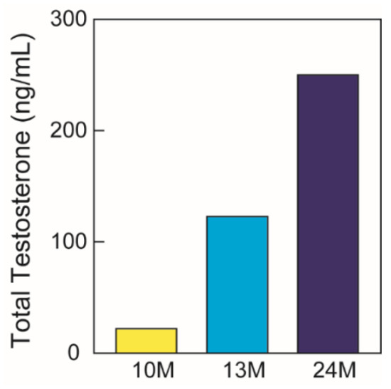
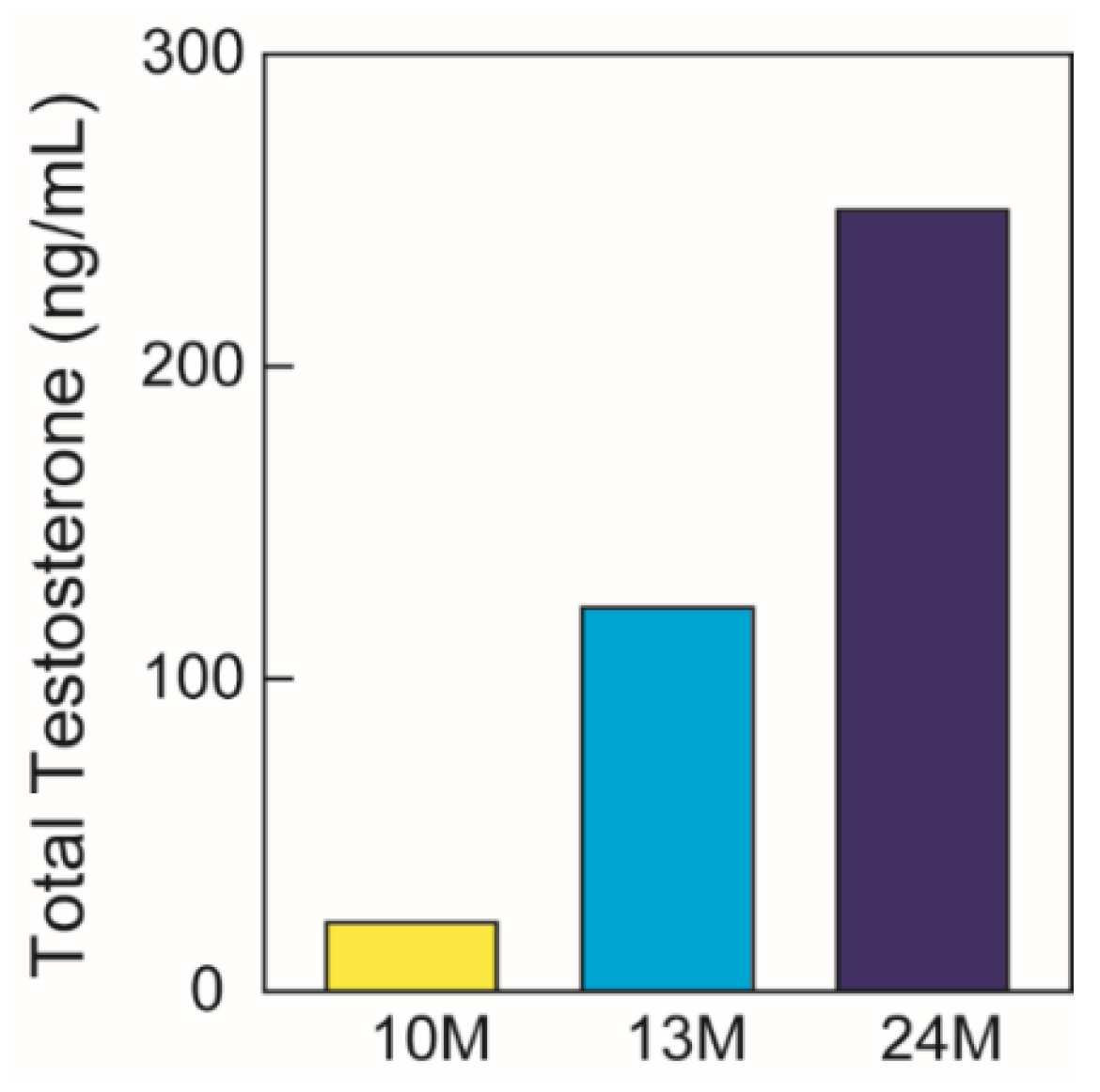
Testosterone levels are essential in defining the pubertal stages of boys. Liquid Chromatography–Mass Spectrometry (LC/MS) data showed that testosterone (T = 123 ng/dL) in PPP from a pubertal (13-year-old, 13M) male subject was markedly different from pre-pubertal (10M) (T = 22) and post-pubertal (24M) (T = 250) male subjects (Figure 1). In this pilot study, we used these PPP examples as representatives of different pubertal stages.
Figure 1.
Total testosterone levels in PPP from pre-pubertal (10-year-old male, 10M), pubertal (13M), and post-pubertal (24M) subjects.
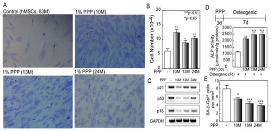
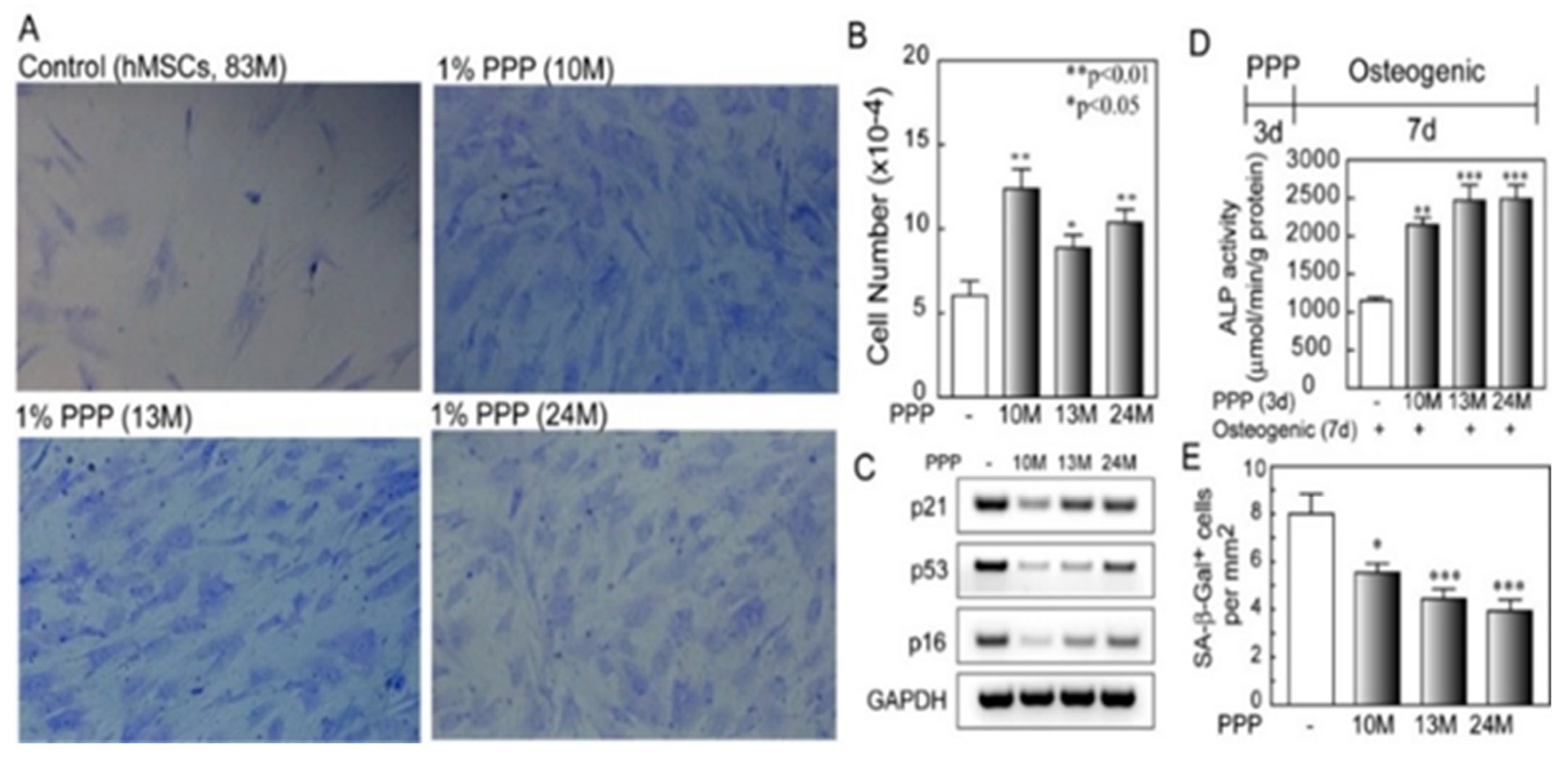
Because of the reported age-related decline in the potential of hMSCs to differentiate into osteoblasts [9,10], we tested whether PPP from different pubertal stage subjects had in vitro regenerative effects on hMSCs from elders. The effects of 1% PPP on cell proliferation, markers of cellular aging, and osteoblast differentiation were quantified (Figure 2). Three days after treatment, the 1% PPP from all males aged 10, 13, and 24 years significantly stimulated the proliferation of hMSCs obtained from an 83-year-old male (83M) (Figure 2A,B). One day of treatment showed striking declines in expression of p21, p53, and p16 in hMSCs from a 58-year-old male (58M); PPP obtained from a 10-year-old male subject (10M) was the most effective at inhibition of cell cycle inhibitory genes in hMSCs (58M) (Figure 2C). There was significant stimulation of osteoblast differentiation of MSCs from an 83-year-old male at day 7 in osteogenic medium following 3 days’ pre-treatment with PPP from younger subjects (Figure 2D). Another marker of cellular aging, senescence-associated β-galactosidase positive cells (SA-β-gal+) was significantly reduced by PPP in hMSCs from a 58-year-old male (Figure 2E). These findings indicate rejuvenating and pro-osteoblastogenic activities in pediatric PPP.
Figure 2.
Effects of PPP on hMSCs from old subjects. (A) Photomicrographs of toluidine blue-O-stained hMSCs (83-year-old male, 83M) in the Control, plus 1% PPP from 10, 13, or 24-y-o males (10M, 13M or 24M) at 3 days (Magnification 180×). (B) After 3 days, PPP stimulated proliferation of hMSCs obtained from 83M (** p = 0.0016, PPP from 10M, n = 6, vs. control, n = 9; * p = 0.031, PPP from 13M, n = 6, vs. controls; ** p = 0.0031, PPP from 24M, n = 6, vs. controls). (C) RT-PCR showed that 24 h treatments with 1% PPP downregulated p21, p53, and p16 in hMSCs (58M). (D) Effects of PPP on osteoblast differentiation in hMSCs (83M). After confluence, hMSCs (83M) were pre-treated for 3 days with 1% PPP in growth medium (α-MEM, 10% FBS) in a 24-well plate and changed to osteogenic medium (α-MEM, 1% FBS, 10 nM dexamethasone, 5 mM β-glycerophosphate, and 50 μg/mL ascorbate-2-phosphate) for 7 d. Pre-treatments with PPP significantly increased osteoblast differentiation in hMSCs (83M) as shown by Alkaline Phosphatase (ALP) enzyme activity (** p < 0.01, PPP of 10M vs. controls; *** p < 0.001, PPP of 13M vs. controls; *** p < 0.001, PPP of 24M vs. controls; n = 4, ANOVA). (E) Effects of PPP on SA-β-Gal+ cells in hMSCs from an older male (58M). After 7 days, PPP reduced SA-β-Gal+ cells. (* p < 0.05, PPP of 10M, n = 18, vs. controls, n = 16; *** p < 0.001, PPP of 13M n = 18, vs. controls; *** p < 0.001, PPP of 24M, n = 18, vs. controls; ANOVA).
3.2. Mass Spectrometry-Based (MS)-Proteomic Analyses of PPP
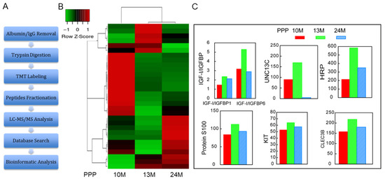
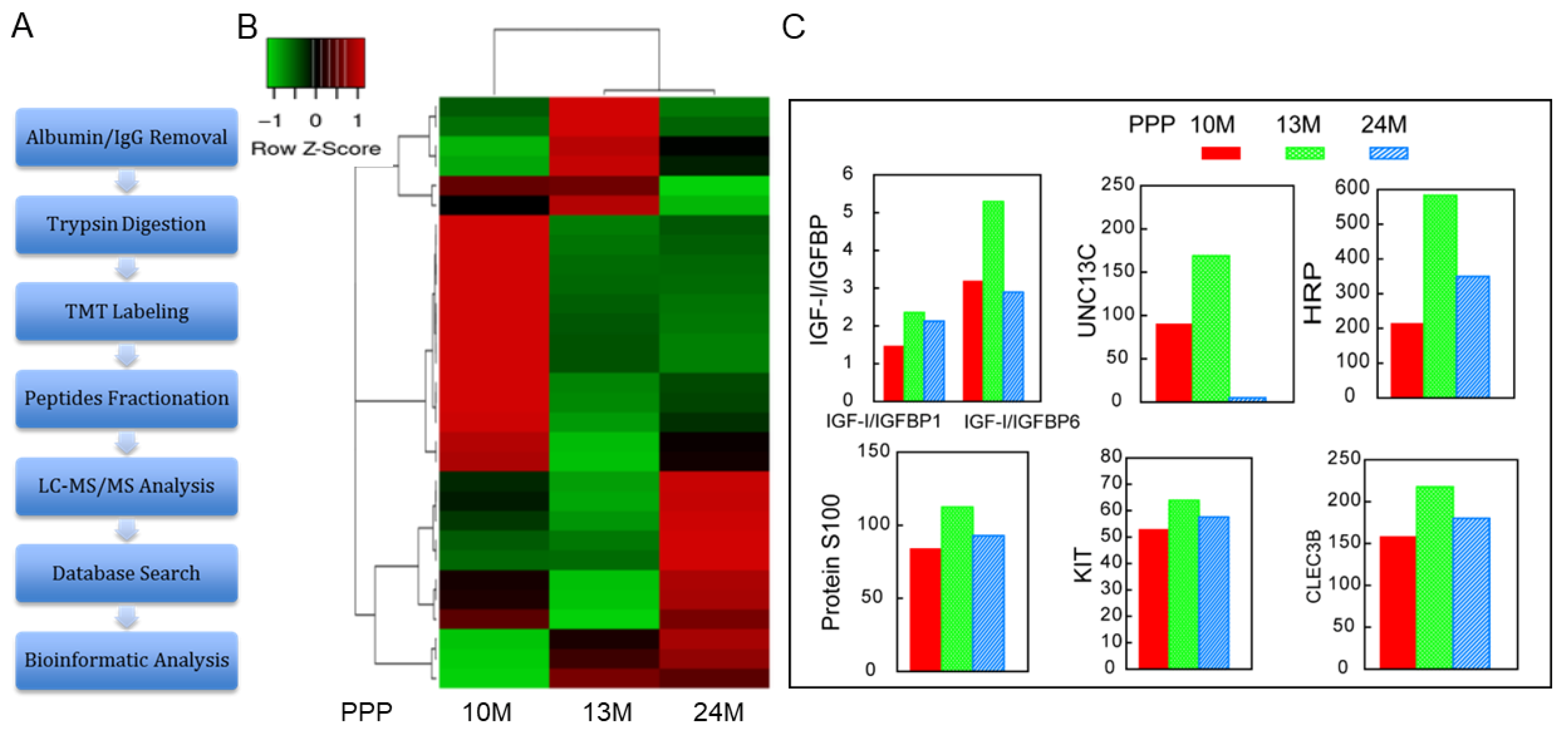
Biologic network analysis was accomplished with MS-based proteomics for specimens of PPP from males aged 10, 13, and 24 years (Figure 3). It is validating that IGF-I, a protein known as abundant in PRP, is present in these PPP samples (Table 1). Of 287 detectable serum proteins, 27 (9%) were higher and 112 (39%) lower in the 13M than in the 10M, with no difference in 148 proteins (52%). There were 30 proteins either two-fold > or < in 13M than their levels in 10M or 24M (Figure 3B, hierarchical cluster analysis of the 30 DEPs with a two-fold-change cutoff). Some proteins of potential importance in the skeletal growth spurt were found to be elevated in the pubertal specimen (13M) and lower in the samples from the pre-pubertal (10M) and post-pubertal (24M) subjects, such as IGF-I/IGFBP, UNC13C, HRP, Protein S100, Kit, and CLEC3B/tetranectin (Figure 3C). The fold-change in CLEC3B in 13M vs. 10M or 24M was 1.4- and 1.2-folds, respectively. The UNC13C level in the PPP of 13M was ≥2-fold compared with its levels in 10M or 24M (1.9- and 35.5-folds, respectively (Figure 3C). More details of examples of proteins distinctly different in PPP obtained from a pubertal male subject (13M) are shown in Table 1.
Figure 3.
MS-Proteomic analysis of PPP obtained from pre-pubertal (10-year-old male, 10M), pubertal (13M), and post-pubertal (24M) subjects. (A) Workflow for MS-Proteomics. (B) Hierarchical cluster analysis of MS data (30 DEPs were shown with a 2-fold-difference cutoff). (C) MS-proteomic data showed some proteins that were relatively higher in PPP from a 13-year-old male subject (13M).
Table 1.
Examples of protein levels in PPP obtained from prepubertal (10M), pubertal (13M), and post-pubertal (24M) male subjects.
3.3. Quantitative Differences in Gene Expression in hMSCs Obtained from Pre-Pubertal, Pubertal, and Post-Pubertal Subjects
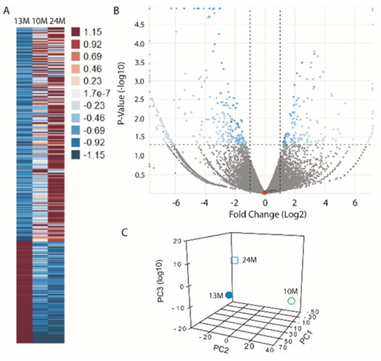
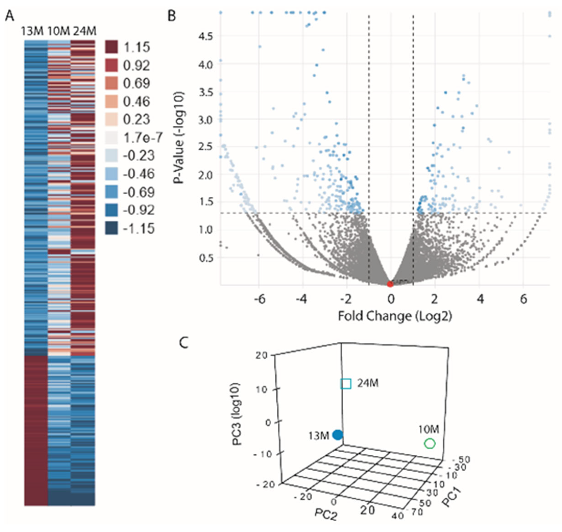
Next-generation RNA sequencing (RNA-Seq) analysis with Low Input Eukaryotic Smart-seq2 protocols was used to compare genes expressed in hMSCs from 10-, 13-, and 24-year-old males, shown respectively to represent pre-pubertal, pubertal, and post-pubertal status (Figure 4). There were 126 genes expressed predominantly in hMSCs obtained from a pubertal subject (13M), compared with pre-pubertal (10M) or post-pubertal subject (24M) (Table 2). The Heatmap and Volcano plot show the differential expression data in pubertal MSCs (Figure 4A,B, 13M vs. 10M and 24M). The expression count was analyzed by STAR and differential expression by DESeq with RNA-seq pipeline (Basepair Tech, Inc., New York, NY, USA). Many genes were not recognized as osteotropic, but the data indicate interesting bone-related hits for the pubertal specimen: Endothelin 1, Natriuretic Peptide B, SPARC/osteonectin, TGFα, TGFβI, FGF2, Col IV, ICAM1, Fibronectin, etc. (Table 3) and genes at low expression in the pubertal specimen, e.g., CCL8, MMP13, WSIP1, etc. (Table 4).
Figure 4.
RNA-Seq analysis of hMSCs obtained from pre-pubertal (10-year-old male, 10M), pubertal (13M), and post-pubertal (24M) subjects. (A) Heatmap shows the differential expression data of the 389 genes with ≥2-fold changes (up: crimson, 126 and down: blue, 263) in 13M vs. 10M and 24M. (B) Volcano plot of differential expression of 13M vs. 10M and 24M (housekeeping gene GAPDH is shown as a red dot; blue dots show significant differences). Expression count was analyzed by STAR and differential expression by DESeq with RNA-seq pipeline (Basepair, Inc., New York, NY, USA). (C) Clustering (principal coordinate analysis with DESeq2) showed the differences among these hMSCs.
Table 2.
Number of differentially expressed genes in hMSCs obtained from a pubertal male subject (13M) vs. pre-pubertal (10M) and/or post-pubertal (24M) subjects.
Table 3.
Examples of genes expressed at greater levels in hMSCs obtained from a pubertal male subject (13M) TPM: Transcripts Per Million.
Table 4.
Examples of genes expressed at lower levels in hMSCs obtained from a pubertal male subject (13M) TPM: Transcripts Per Million.
4. Discussion
Osteoporosis is a major cause of morbidity and mortality through its association with age-related fractures. Evidence shows that failure to gain sufficient bone during skeletal growth and maturation phases predisposes to the development of senile osteoporosis [2,3,4]. There are critical gaps in our knowledge about the mechanisms mediating the pubertal skeletal growth spurt and their relationships with detrimental skeletal changes that occur with aging. Our underlying hypothesis is that diminution or disappearance of Juvenile Protective Factors after a given developmental stage could contribute to the onset of age-related declines in a variety of physiological functions, including maintenance of bone mass. We further test the hypothesis with studies that suggest that precious, surgically discarded clinical specimens may be useful for proteomic and gene expression analysis. There are literature reports comparing osteoblast rejuvenation with plasma from young adults, cord blood, amnionic stem cells, and young animals. One study showed that exposure to a youthful circulation through heterochronic parabiosis did not reverse the diminished osteochondrogenic activity of aged skeletal stem cells or improve bone mass or skeletal healing parameters in aged mice [15]. Other studies using heterochronic parabiosis models showed that circulating juvenile factors did rejuvenate aged systems [16,17], including bone and mesenchymal stem cells [18,19].
This report appears to be the first to test the feasibility of examining plasma and MSCs from children for novel pubertal JPFs as an approach to discover new therapeutics for age-related bone loss. Our data substantiate the use of available PPP to test the hypothesis that puberty entails the production of circulating factors that stimulate skeletal growth. First, pediatric PPP rejuvenated age-related MSC functions in vitro. Second, proteomic analyses of PPP identified IGF-I, an osteotropic protein known to be abundant in PRP; other known osteotropic factors were detected. Third, proteomic and gene expression analyses identified factors not previously known to support bone growth.
Data from proteomic analysis of pediatric samples showed expected and unexpected proteins. Growth hormone (GH) is considered the primary regulator of pubertal growth by increasing serum insulin-like growth factor-I (IGF-I) [5]; the serum IGF-I/IGFBP ratio was shown to be the best marker for increased bone formation during puberty [8]. Our proteomic study determined that IGF-I/IGFBP1 and IGF-I/IGFBP6, which have potential importance in the skeletal growth spurt, were elevated in the pubertal specimen and lower in the samples from the pre-pubertal and post-pubertal subjects. These findings show expected results from studies with PRP. Our proteomic study revealed previously unexpected plasma proteins associated with puberty, such as CLEC3B/tetranectin and UNC-13 homolog C (UNC13C a.k.a MUNC13-3). CLEC3B/tetranectin was elevated in the pubertal stage; this is consistent with a report that stated that CLEC3B/tetranectin was associated with bone mineralization in mouse experiments [20]. Furthermore, a transitory increase in plasma CLEC3B/tetranectin was observed in early puberty, reaching its climax about the age of 11 to 12 in girls and 14 to 15 in boys [21]. Other animal studies showed that CLEC3B/tetranectin has a role in the positive regulation at the early stage of the fracture–healing process, which was reflected in the delayed fracture healing in CLEC3B-deficient mice [22]. Another animal study suggested that tetranectin is a candidate biomarker reflecting bone metabolic turnover [23]. Protein UNC-13 homolog C (UNC13C, a.k.a MUNC13-3) plays a role in the rapid communication between neurons [24,25]. Munc13-3 is almost exclusively expressed in the cerebellum and regulates cerebellar synaptic transmission and motor learning in mice [26]. Our data showed that the UNC13C/MUNC13-3 level is highest in PPP from the pubertal stage. The roles of UNC13C/MUNC13-3 in bone growth during puberty and adult bone homeostasis are unknown and need further study. These are examples of opportunities guided by proteomic studies.
Data from RNA-Seq analysis showed expected and unexpected gene expression. Mesenchymal stem cells (MSCs) undergo self-renewal and differentiation, contributing to speedy skeletal growth in early life; inducible premature MSC senescence during early puberty in mice leads to impaired osteoblastogenesis as well as bone loss in later adult life [4]. We used RNA-Seq analysis to compare genes expressed in hMSCs shown to represent pre-pubertal, pubertal, and post-pubertal status. These pilot RNA-Seq data suggest interesting bone-related hits, e.g., genes elevated in the pubertal subject: Endothelin 1, Natriuretic Peptide B, SPARC/osteonectin, TGFα, TGFβI, FGF2, Col IV, etc. (Table 3) and genes reduced in the pubertal subject, e.g., CCL8, MMP13, and WSIP1. 4). Endothelin-1 (EDN1) has a vital role in bone remodeling [27]. Endothelin-1 induces osteogenesis of bone marrow-derived mesenchymal stem cells [28,29]. Our pilot RNA-Seq data showed that EDN1/Endothelin-1 and its type B receptor, EDNRB, were expressed predominantly in hMSCs obtained from a pubertal specimen (Table 3), implying that EDN1/EDNRB may have a role in the pubertal growth spurt, but need further study. SPARC/osteonectin is one of the most abundant non-collagenous proteins expressed in mineralized tissues and plays a critical role in regulating bone remodeling and maintaining bone mass and quality [30]. Further studies are needed to determine whether SPARC/osteonectin is just a bone-forming biomarker or plays a role in the pubertal growth spurt.
Platelet-poor plasma has a low number of platelets, which are the source of numerous growth factors, but it was found suitable for proteomic analysis. PRP concentrate has been used for a number of applications, but there is an inconsistency in the clinical outcomes. Clinical benefits of PRP are attributed to regenerative effects of growth factors released from platelets, the most abundant being PDGF-AB, TGF-β1, and IGF-I [31]. There is, nevertheless, a striking lack of reproducibility in studies correlating growth factor content or bioactivity with platelet count, age, or sex of the donor [31,32,33,34,35]. There is a growing literature on the potential of banking umbilical cord blood and its derived MSCs for clinical uses [36]. While the efficacy of adult blood plasma is attributed to its reduction in Reactive Oxygen Species (ROS) [37,38,39], it has been shown that umbilical cord blood plasma’s effects go beyond ROS reduction to promote cellular repair and rejuvenation pathways [40]. Investigation of the detailed mechanisms of beneficial actions of pubertal PRP is likewise warranted.
There are limitations to this research. Additional markers of osteoblast differentiation are frequently used for in vitro experiments, e.g., quantitative mineralization assays and osteogenic marker expression. As a pilot study to test the feasibility of using available surgically discarded tissues (PPP and marrow-derived MSCs) for research on factors that mediate the pubertal growth spurt, it was not possible to assess the variability of proteomic and gene expression results in multiple samples from each peri-pubertal period. When these approaches become less expensive, larger R&D studies will become more feasible. This study design requires great cooperation between researchers and reconstructive surgeons willing to provide them with surgical waste materials. More information about the novel proteins and genes identified herein and their potential as preventive/therapeutic agents for skeletal aging and osteoporotic fracture prevention would be available from specific functional analyses in other model systems and from clustering or pathway enrichment analyses to support biological interpretation. We used specimens from only male subjects because of sex differences in timing and magnitude of the skeletal growth spurt at puberty [2]. Future comparison of tissues from males and females will be necessary.
5. Conclusions
Platelet-poor plasma from children had rejuvenating effects on hMSCs from elders, including an increase in osteoblast differentiation potential. This suggests that there may be circulating factors that contribute to the pubertal skeletal growth spurt. Some candidate proteins, both known and unknown to have osteotropic actions, were determined to be increased in pubertal plasma. These findings support the hypothesis that this research approach can identify novel factors that mediate the pubertal growth spurt. Initial RNA-Seq studies with RNA from prepubertal, pubertal, and post-pubertal MSCs revealed potentially significant bone-related hits. Although additional biological and reproducibility studies are needed to test the hypothesis that trophic factors or genes elevated in the blood or hMSCs, respectively, from subjects during the pubertal growth spurt are pro-osteogenic JPFs and have the potential to prevent or reverse age-associated declines in stem cell function, our exploratory research suggests that analysis of plasma and RNA from MSC in the peripubertal periods may represent a new approach to identify potential rejuvenating factors that could develop as therapeutic agents for age-related skeletal disorders.
Author Contributions
Conceptualization, J.G., S.Z. and B.L.P.; methodology, J.G. and S.Z.; validation, J.G. and S.Z.; formal analysis, S.Z.; resources, J.G., S.Z. and B.L.P.; data curation, S.Z.; writing—original draft preparation, J.G. and S.Z.; writing—review and editing, J.G. and S.Z.; visualization, S.Z.; supervision, J.G.; project administration, J.G. and B.L.P.; funding acquisition, J.G., S.Z. and B.L.P. All authors have read and agreed to the published version of the manuscript.
Funding
Departmental funds at Boston Children’s Hospital and Brigham and Women’s Hospital were used for this research.
Institutional Review Board Statement
The study was conducted according to the guidelines of the Declaration of Helsinki and approved by the Institutional Review Boards of Boston Children’s Hospital (protocol P00022614, 15 April 2016) and Brigham and Women’s Hospital (P99001235, 14 August 1998).
Data Availability Statement
All data supporting the findings of this study are available from the corresponding author upon reasonable request.
Conflicts of Interest
The authors declare no conflicts of interest.
References
- Zhu, J.; Chan, Y.M. Adult Consequences of Self-Limited Delayed Puberty. Pediatrics 2017, 139, e20163177. [Google Scholar] [CrossRef] [Scilit]
- Riggs, B.L.; Khosla, S.; Melton, L.J., 3rd. The assembly of the adult skeleton during growth and maturation: Implications for senile osteoporosis. J. Clin. Investig. 1999, 104, 671–672. [Google Scholar] [CrossRef] [Scilit]
- Elhakeem, A.; Frysz, M.; Tilling, K.; Tobias, J.H.; Lawlor, D.A. Association Between Age at Puberty and Bone Accrual from 10 to 25 Years of Age. JAMA Netw. Open 2019, 2, e198918. [Google Scholar] [CrossRef] [Scilit] [PubMed] [PubMed Central]
- Li, C.; Chai, Y.; Wang, L.; Gao, B.; Chen, H.; Gao, P.; Zhou, F.Q.; Luo, X.; Crane, J.L.; Yu, B.; et al. Programmed cell senescence in skeleton during late puberty. Nat. Commun. 2017, 8, 1312. [Google Scholar] [CrossRef] [Scilit] [PubMed]
- Farr, J.N.; Khosla, S. Skeletal changes through the lifespan—From growth to senescence. Nat. Rev. Endocrinol. 2015, 11, 513–521. [Google Scholar] [CrossRef] [Scilit] [PubMed] [PubMed Central]
- Juul, A.; Bang, P.; Hertel, N.T.; Main, K.; Dalgaard, P.; Jørgensen, K.; Müller, J.; Hall, K.; Skakkebaek, N.E. Serum insulin-like growth factor-I in 1030 healthy children, adolescents, and adults: Relation to age, sex, stage of puberty, testicular size, and body mass index. J. Clin. Endocrinol. Metab. 1994, 78, 744–752. [Google Scholar] [PubMed]
- Cole, T.J.; Ahmed, M.L.; Preece, M.A.; Hindmarsh, P.; Dunger, D.B. The relationship between Insulin-like Growth Factor 1, sex steroids and timing of the pubertal growth spurt. Clin. Endocrinol. 2015, 82, 862–869. [Google Scholar] [CrossRef] [Scilit] [PubMed Central]
- Kanbur, N.O.; Derman, O.; Kinik, E. The relationships between pubertal development, IGF-1 axis, and bone formation in healthy adolescents. J. Bone Miner. Metab. 2005, 23, 76–83. [Google Scholar] [CrossRef] [Scilit] [PubMed]
- D’Ippolito, G.; Schiller, P.C.; Ricordi, C.; Roos, B.A.; Howard, G.A. Age-related osteogenic potential of mesenchymal stromal stem cells from human vertebral bone marrow. J. Bone Miner. Res. 1999, 14, 1115–1122. [Google Scholar] [CrossRef] [Scilit] [PubMed]
- Zhou, S.; Greenberger, J.S.; Epperly, M.W.; Goff, J.P.; Adler, C.; Leboff, M.S.; Glowacki, J. Age-related intrinsic changes in human bone-marrow-derived mesenchymal stem cells and their differentiation to osteoblasts. Aging Cell 2008, 7, 335–343. [Google Scholar] [CrossRef] [Scilit] [PubMed] [PubMed Central]
- Ruggiero, B.; Padwa, B.L.; Christoph, K.M.; Zhou, S.; Glowacki, J. Vitamin D metabolism and regulation in pediatric MSCs. J. Steroid Biochem. Mol. Biol. 2016, 164, 287–291. [Google Scholar] [CrossRef] [Scilit] [PubMed]
- Li, J.; Padwa, B.L.; Zhou, S.; Mullokandova, J.; LeBoff, M.S.; Glowacki, J. Synergistic effect of 1α,25-dihydroxyvitamin D3 and 17β-estradiol on osteoblast differentiation of pediatric MSCs. J. Steroid Biochem. Mol. Biol. 2018, 177, 103–108. [Google Scholar] [CrossRef] [Scilit] [PubMed]
- Picelli, S.; Faridani, O.R.; Björklund, A.K.; Winberg, G.; Sagasser, S.; Sandberg, R. Full-length RNA-seq from single cells using Smart-seq2. Nat. Protoc. 2014, 9, 171–181. [Google Scholar] [CrossRef] [Scilit]
- Picelli, S.; Björklund, Å.K.; Faridani, O.R.; Sagasser, S.; Winberg, G.; Sandberg, R. Smart-seq2 for sensitive full-length transcriptome profiling in single cells. Nat. Methods 2013, 10, 1096–1098. [Google Scholar] [CrossRef] [Scilit]
- Ambrosi, T.H.; Marecic, O.; McArdle, A.; Sinha, R.; Gulati, G.S.; Tong, X.; Wang, Y.; Steininger, H.M.; Hoover, M.Y.; Koepke, L.S.; et al. Aged skeletal stem cells generate an inflammatory degenerative niche. Nature 2021, 597, 256–262. [Google Scholar] [CrossRef] [Scilit] [PubMed] [PubMed Central]
- Chu, X.; Subramani, K.; Thomas, B.; Terry, A.V., Jr.; Fulzele, S.; Raju, R.P. Juvenile Plasma Factors Improve Organ Function and Survival following Injury by Promoting Antioxidant Response. Aging Dis. 2022, 13, 568–582. [Google Scholar] [CrossRef] [Scilit] [PubMed] [PubMed Central]
- Conboy, I.M.; Conboy, M.J.; Wagers, A.J.; Girma, E.R.; Weissman, I.L.; Rando, T.A. Rejuvenation of aged progenitor cells by exposure to a young systemic environment. Nature 2005, 433, 760–764. [Google Scholar] [CrossRef] [Scilit] [PubMed]
- Baht, G.S.; Silkstone, D.; Vi, L.; Nadesan, P.; Amani, Y.; Whetstone, H.; Wei, Q.; Alman, B.A. Exposure to a youthful circulation rejuvenates bone repair through modulation of β-catenin. Nat. Commun. 2015, 6, 7131, Erratum in Nat Commun. 2015, 6, 7761. https://doi.org/10.1038/ncomms8761. [Google Scholar] [CrossRef] [Scilit] [PubMed] [PubMed Central]
- Pálovics, R.; Keller, A.; Schaum, N.; Tan, W.; Fehlmann, T.; Borja, M.; Kern, F.; Bonanno, L.; Calcuttawala, K.; Webber, J.; et al. Molecular hallmarks of heterochronic parabiosis at single-cell resolution. Nature 2022, 603, 309–314. [Google Scholar] [CrossRef] [Scilit] [PubMed] [PubMed Central]
- Wewer, U.M.; Ibaraki, K.; Schjørring, P.; Durkin, M.E.; Young, M.F.; Albrechtsen, R. A potential role for tetranectin in mineralization during osteogenesis. J. Cell Biol. 1994, 127, 1767–1775. [Google Scholar] [CrossRef] [Scilit] [PubMed] [PubMed Central]
- Jensen, B.A.; McNair, P.; Hyldstrup, L.; Clemmensen, I. Plasma tetranectin in healthy male and female individuals, measured by enzyme-linked immunosorbent assay. J. Lab. Clin. Med. 1987, 110, 612–617. [Google Scholar] [PubMed]
- Iba, K.; Abe, Y.; Chikenji, T.; Kanaya, K.; Chiba, H.; Sasaki, K.; Dohke, T.; Wada, T.; Yamashita, T. Delayed fracture healing in tetranectin-deficient mice. J. Bone Miner. Metab. 2013, 31, 399–408. [Google Scholar] [CrossRef] [Scilit] [PubMed]
- Sasaki, K.; Ozasa, Y.; Iba, K.; Wada, T.; Imai, S.; Matsumoto, K.; Sohma, H.; Aoshima, M.; Yamashita, T.; Kokai, Y. Significant increase of plasma tetranectin in ovx mice as defined by proteomics analysis. J. Orthop. Sci. 2014, 19, 809–819. [Google Scholar] [CrossRef] [Scilit] [PubMed]
- Vogl, C.; Cooper, B.H.; Neef, J.; Wojcik, S.M.; Reim, K.; Reisinger, E.; Brose, N.; Rhee, J.S.; Moser, T.; Wichmann, C. Unconventional molecular regulation of synaptic vesicle replenishment in cochlear inner hair cells. J. Cell Sci. 2015, 128, 638–644. [Google Scholar] [CrossRef] [Scilit] [PubMed]
- Padmanarayana, M.; Liu, H.; Michelassi, F.; Li, L.; Betensky, D.; Dominguez, M.J.; Sutton, R.B.; Hu, Z.; Dittman, J.S. A unique C2 domain at the C terminus of Munc13 promotes synaptic vesicle priming. Proc. Natl. Acad. Sci. USA 2021, 118, e2016276118. [Google Scholar] [CrossRef] [Scilit] [PubMed] [PubMed Central]
- Augustin, I.; Korte, S.; Rickmann, M.; Kretzschmar, H.A.; Südhof, T.C.; Herms, J.W.; Brose, N. The cerebellum-specific Munc13 isoform Munc13-3 regulates cerebellar synaptic transmission and motor learning in mice. J. Neurosci. 2001, 21, 10–17. [Google Scholar] [CrossRef] [Scilit] [PubMed] [PubMed Central]
- Kristianto, J.; Johnson, M.G.; Afzal, R.; Blank, R.D. Endothelin Signaling in Bone. Endocrinol. Metab. Clin. N. Am. 2017, 46, 51–62. [Google Scholar] [CrossRef] [Scilit] [PubMed Central]
- Hu, L.W.; Wang, X.; Jiang, X.Q.; Xu, L.Q.; Pan, H.Y. In vivo and in vitro study of osteogenic potency of endothelin-1 on bone marrow-derived mesenchymal stem cells. Exp. Cell Res. 2017, 357, 25–32. [Google Scholar] [CrossRef] [Scilit] [PubMed]
- Lee, M.S.; Wang, J.; Yuan, H.; Jiao, H.; Tsai, T.L.; Squire, M.W.; Li, W.J. Endothelin-1 differentially directs lineage specification of adipose- and bone marrow-derived mesenchymal stem cells. FASEB J. 2019, 33, 996–1007. [Google Scholar] [CrossRef] [Scilit] [PubMed Central]
- Rosset, E.M.; Bradshaw, A.D. SPARC/osteonectin in mineralized tissue. Matrix Biol. 2016, 52–54, 78–87. [Google Scholar] [CrossRef] [Scilit] [PubMed] [PubMed Central]
- Weibrich, G.; Kleis, W.K.; Hafner, G.; Hitzler, W.E. Growth factor levels in platelet-rich plasma and correlations with donor age, sex, and platelet count. J. Craniomaxillofac. Surg. 2002, 30, 97–102. [Google Scholar] [CrossRef] [Scilit] [PubMed]
- Evanson, J.R.; Guyton, M.K.; Oliver, D.L.; Hire, J.M.; Topolski, R.L.; Zumbrun, S.D.; McPherson, J.C.; Bojescul, J.A. Gender and age differences in growth factor concentrations from platelet-rich plasma in adults. Mil. Med. 2014, 179, 799–805. [Google Scholar] [CrossRef] [Scilit] [PubMed]
- Xiong, G.; Lingampalli, N.; Koltsov, J.C.B.; Leung, L.L.; Bhutani, N.; Robinson, W.H.; Chu, C.R. Men and Women Differ in the Biochemical Composition of Platelet-Rich Plasma. Am. J. Sports Med. 2018, 46, 409–419. [Google Scholar] [CrossRef] [Scilit] [PubMed] [PubMed Central]
- Guo, E.; Sun, L.; Chen, W.; Liu, C.; Chen, K.; Jiang, X.; Qin, X.; Su, J.; Yang, F.; Tian, H. Young human PRP promotes the rejuvenation of aged bone marrow mesenchymal stem cells and the therapeutic effect on ischemic heart disease. Eur. J. Pharmacol. 2023, 950, 175775. [Google Scholar] [CrossRef] [Scilit] [PubMed]
- Tian, J.; Li, X.J.; Ma, Y.; Mai, Z.; Yang, Y.; Luo, M.; Xu, W.; Chen, K.; Chen, X.; Tang, J.; et al. Correlation of bioactive components of platelet rich plasma derived from human female adult peripheral blood and umbilical cord blood with age. Sci. Rep. 2023, 13, 18428. [Google Scholar] [CrossRef] [Scilit] [PubMed] [PubMed Central]
- Ding, D.-C.; Chang, Y.-H.; Shyu, W.-C.; Lin, S.-Z. Human Umbilical Cord Mesenchymal Stem Cells: A New Era for Stem Cell Therapy. Cell Transplant. 2015, 24, 339–347. [Google Scholar] [CrossRef] [Scilit]
- Lu, T.; Finkel, T. Free radicals and senescence. Exp. Cell Res. 2008, 314, 1918–1922. [Google Scholar] [CrossRef] [Scilit] [PubMed] [PubMed Central]
- Almeida, M.; O’Brien, C.A. Basic biology of skeletal aging: Role of stress response pathways. J. Gerontol. Ser. A Biol. Sci. Med. Sci. 2013, 68, 1197–1208. [Google Scholar] [CrossRef] [Scilit] [PubMed] [PubMed Central]
- Li, Y.; Cheng, B.; Tian, J. Platelet-rich plasma may accelerate diabetic wound healing by modulating epithelial/endothelial-mesenchymal transition through inhibiting reactive oxygen species-mediated oxidative stress. Front. Bioeng. Biotechnol. 2025, 13, 1623780. [Google Scholar] [CrossRef] [Scilit] [PubMed] [PubMed Central]
- Jeong, M.; Lee, H.; Ko, T.H.; Choi, S.J.; Oh, W.; Kim, S. Umbilical Cord Blood Plasma Enhances Cellular Repair and Senescence Suppression in Human Dermal Fibroblasts Under Oxidative Stress. Rejuvenation Res. 2025, 28, 195–204. [Google Scholar] [CrossRef] [Scilit] [PubMed]

Disclaimer/Publisher’s Note: The statements, opinions and data contained in all publications are solely those of the individual author(s) and contributor(s) and not of MDPI and/or the editor(s). MDPI and/or the editor(s) disclaim responsibility for any injury to people or property resulting from any ideas, methods, instructions or products referred to in the content. |
© 2026 by the authors. Licensee MDPI, Basel, Switzerland. This article is an open access article distributed under the terms and conditions of the Creative Commons Attribution (CC BY) license.