


Abstract
Adult-onset cases of idiopathic hypogonadotropic hypogonadism (IHH) are characterized by partial or normal puberty development until adolescence and by the impairment of the hypothalamic–pituitary–gonadal (HPG) axis in adulthood. WDR11 and DCC genes are known to be involved in axonal development, particularly of hypothalamic GnRH neurons, and ciliogenesis. We report a female case of adult-onset hypogonadism and cerebellar ataxia, in which we identified two gene mutations. A panel of 48 genes was set up to search for variants in the causative genes of CHH. The variants found were analyzed following the American College of Medical Genetics and Genomics (ACMG) criteria to define their pathogenicity. We identified a missense heterozygous variant in the WDR11 gene NM_018117.12:c.2306T>G (p.Met769Arg) and a mutation in a second gene DCC resulting in amino acid substitutions NM_005215.4:c.3533C>T (p.Ser1178Phe). These variants were classified as being of uncertain clinical significance. We assume that there is a link between the variants found and the impairment of the gonadotrophic and neurological phenotype of the patient. Therefore, we propose the genetic test to identify the best therapeutic approach to identify infertility in female patients with IHH; we believe it is necessary to test WDR11 and DCC genes in larger populations with the same condition to introduce it in future protocols of assessment.
1. Introduction
Congenital hypogonadotropic hypogonadism (CHH) is a rare disorder with a genetic background characterized by the production, secretion or action deficit of gonadotropin-releasing hormone (GnRH), a key neuropeptide that regulates the reproductive competence of the hypothalamic–pituitary–gonadal axis. When it is associated with an impaired sense of smell, CHH is named Kallmann syndrome (KS) [1].
Over sixty genes have been implicated in the pathogenesis of the disease in approximately 50% of cases, as follows: KAL1 (ANOS1) with X-linked transmission; FGFR1 (encoding fibroblast growth factor receptor 1), FGF8, CHD7, HS6ST1 (encoding heparan-sulphate 6-O-sulphotransferase 1), SOX10, SEMA3A (encoding semaphorin-3A), IL17RD (encoding interleukin-17 receptor D) with autosomal dominant inheritance; and PROKR2 and/or PROK2, FEZF139 with autosomal recessive inheritance. GNRHR (encoding gonadotropin-releasing hormone receptor), GNRH1 (encoding gonadotropin-releasing hormone 1), KISS1R, KISS1, TACR3 and TAC3 were genes involved in CHH with a normal sense of smell (nCHH), while FGFR1 and PROKR2 can be implicated in both KS and nCHH patients [2]. The WD Repeat Domain 11 (WDR11) is one of the most recent genes identified in subjects with IHH and KS. It encodes for a protein with repeated WD domains, each one interacting with protein-binding partners that participate in a wide variety of cellular processes including the development of the central nervous system, particularly of hypothalamic GnRH neurons, and ciliogenesis in mice.
Kim et al., in 2010, focused their attention on a specific cell protein that interacts with WDR11, EMX1, a domain transcription factor involved in the development of olfactory neurons. Fifteen WDR11 mutations have been reported in CHH patients; they are all missense heterozygous, whereby some have a mutation in a second-known gene, and one is associated with clinical reversibility [3,4,5].
Deleted in colorectal cancer (DCC) is a gene known to be involved in axonal development in the brain [3] and encoding proteins containing Fibronectin type-III (FN3) domains, which are considered biologically relevant in GnRH neuron development. Defects in this gene have been identified in congenital mirror movement [4] and agenesis of the corpus callosum [5] and CHH [6]. Hearing loss, congenital mirror movement and other malformations are often CHH-associated phenotypes [7].
Clinically, CHH is characterized by absent or incomplete puberty and infertility. In the neonatal age, in the male, it can reveal itself with cryptorchidism and micropenis. In adults, males present absent or minimal virilization, poor libido and erectile dysfunction; females have primary amenorrhea. In addition, other developmental anomalies may be associated with so-called “non-reproductive” phenotypes, such as labiopalatoschisis, dental and renal agenesis, bimanual synkinesis, sensorineural deafness and skeletal abnormalities [1,8].
Adult-onset cases of idiopathic hypogonadotropic hypogonadism (IHH) with partial or normal puberty development until adolescence have been reported. Generally, these cases suggest a milder impairment of the HPG axis and a better fertility with gonadotrophin treatment.
In CHH females, the ovulation induction therapy must be personalized to increase fertility and pregnancy rates, but current evidence regarding the fertility treatment management is limited [8].
We describe a case of secondary amenorrhea and ataxia occurring late in the adulthood of a woman carrying compound heterozygosity of WDR11 and DCC missense mutations.
2. Case History
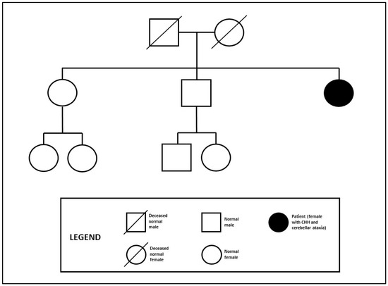
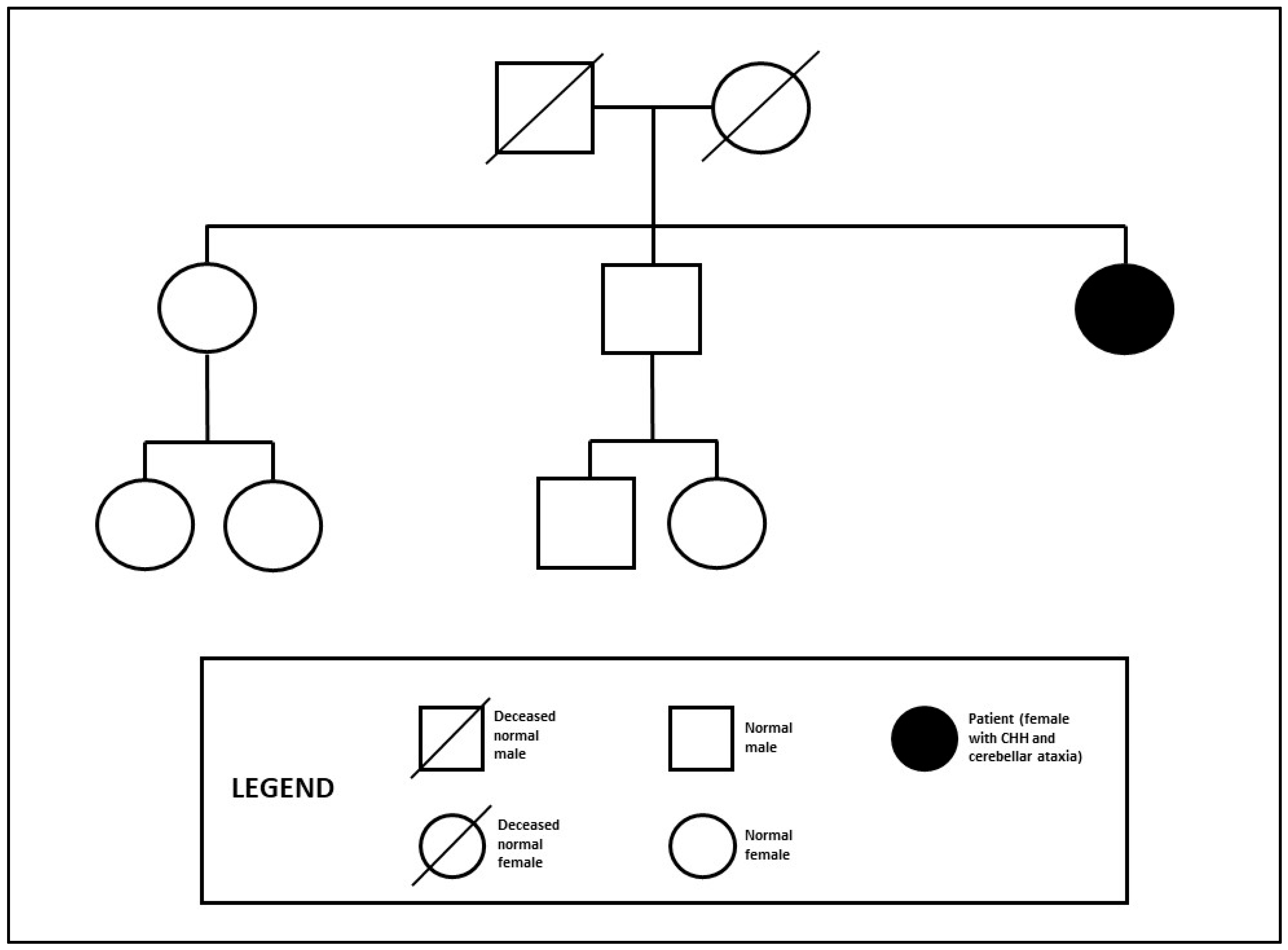
The patient, a 40-year-old female, was sent to our attention for consultation by neurologists because she was suffering from ataxia of recent onset and apparently secondary amenorrhea, which began immediately after menarche at 13 years old. She reported spontaneous pubertal development with no anosmia and acquired causes have been excluded. No family history of CHH has been reported; the patient has a sister and a brother who both had offspring [Figure 1].
Figure 1.
Genealogical tree of the patient.
From the age of 20, she started estrogen replacement therapy and at the age of 32 years, she underwent a cycle of ovulation induction therapy using human menopausal gonadotropin (HMG) and recombinant luteinizing hormone (LH) to promote fertility, but developed a severe ovarian hyperstimulation syndrome.
On physical examination, the patient showed bilateral breast enlargement and normal pubic hair distribution (Tanner stage B5 P5). She had a height of 167 cm and a weight of 67 kg with body mass index of 24 kg/m2.
We performed a complete hormonal evaluation; the main results are illustrated in Table 1. Hormone measurements were conducted using chemiluminescence assays on the Advia Centaur XP ® platform (Siemens Healthcare Diagnostics Inc., Tarrytown, NY, USA).
Table 1.
Hormone profiles of the patient (reference ranges in follicular phase of the menstrual cycle are reported). LH: luteinizing hormone; FSH: follicle-stimulating hormone; AMH: anti-mullerian hormone; TSH: thyroid-stimulating hormone; fT3: free triiodothyronine; fT4: free thyroxine; ACTH: adrenocorticotropic hormone.
The GnRH stimulation test (intravenous administration of 100 mcg of GnRH followed by blood samples at 15, 30, 60, 90 and 120 min) revealed a good LH and FSH response (respectively, 18 mIU/mL and 10 mIU/mL peaks).
On pelvic ultrasound, both ovaries appeared 5 mL in volume and the uterus showed a normal size and body/cervical ratio.
The brain Magnetic Resonance Imaging (MRI) showed wide-spread white matter changes with T2-weighted hyperintensities affecting deep and periventricular white matter and cerebellar atrophy; the pituitary gland appeared normal and free of focal alterations.
Chromosome karyotype analysis was normal.
Subject to informed consent, a peripheral venous blood sample (in EDTA) was performed. DNA was then extracted from lymphocytes (extraction KIT from Nuclear Laser Medicine S.r.l.) and it was quantified using Qubit. A panel of 48 genes was set up to search for variants in the causative genes of CHH (GNRHR, GNRH1, KISS1R, KISS1, TAC3, TACR3, FSHB, LHB, KAL1, FGFR1, FGF8, PROK2, PROKR2, CHD7, SEMA3A, HS6ST1, SOX10, SEMA7A, SEMA3E, IL17RD, KLBSPRY4, DUSP6, ESX1, FGF17, POLR3A, POLR3B, PNPLA6, PLXNA1, FLRT3, WDR11, NELF, NTN1, DCC, FEZF1, NROB1, NR5A1, HESX1, LHX4, PROP1, STUB1, LEP, LEPR, PCSK1, IGSF10, FTO, EAP1, OTUD4 and RNF216), on the ION S5 (Thermofisher) platform. Sanger sequencing was used as the gold standard to confirm nucleotide changes identified using next-generation sequencing (NGS). The variants found were analyzed following the ACMG criteria to define their pathogenicity.
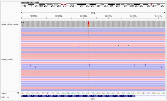
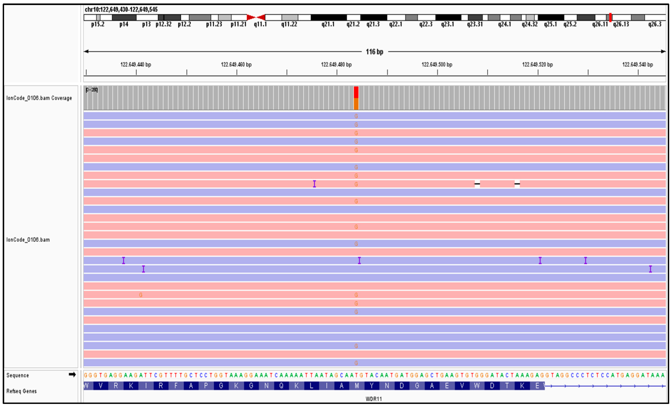
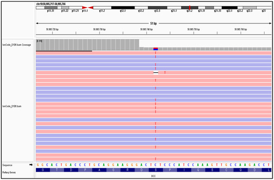
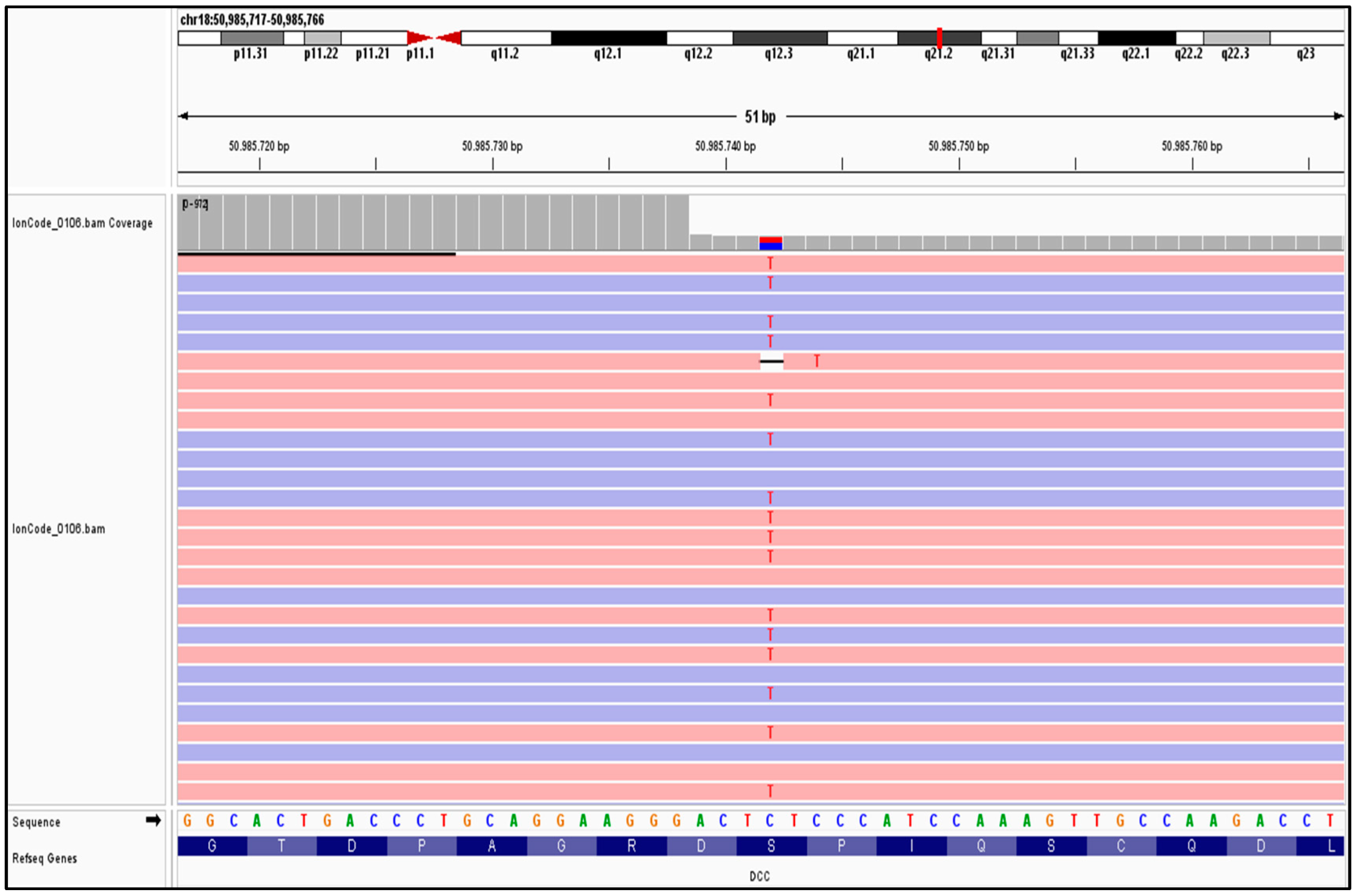
We identified a missense heterozygous variant in the WDR11 gene NM_018117.12:c.2306T>G (p.Met769Arg) [Figure 2]. A mutation in a second gene DCC was found, resulting in amino acid substitutions NM_005215.4:c.3533C>T (p.Ser1178Phe) [Figure 3]. These mutations were classified as variants of uncertain significance (VUSs), following ACMG criteria.
Figure 2.
Snapshot of Binary Alignment Map (BAM) file for c.2306T>G(p.Met769Arg) heterozygous variant in the WDR11 gene.
Figure 3.
Snapshot of BAM file for c.3533C>T (p.Ser1178Phe) heterozygous variant in the DCC gene.
We have therefore placed a diagnosis of congenital hypogonadotropic hypogonadism, supported by the presence of the genetic variants found. At present, we have continued with the hormone replacement therapy, proposing to make an attempt of therapeutic withdrawal in order to evaluate a potential reversibility, as reported in the literature in similar cases [9]. This attempt was unsuccessful.
3. Discussion
We report a case of hypogonadism associated with spontaneous puberty and late onset ataxia probably due to a missense heterozygous variant in the WDR11 gene NM_018117.12:c.2306T>G (p.Met769Arg) and a mutation in a second gene, DCC, resulting in amino acid substitutions NM_005215.4:c.3533C>T (p.Ser1178Phe). Kim et al., in 2010, focused their attention on a specific cell protein that interacts with WDR11, EMX1, a domain transcription factor involved in the development of olfactory neurons. Fifteen WDR11 mutations have been reported in CHH patients; they are all missense heterozygous, some with a mutation in a second known gene and only one is associated with clinical reversibility [10,11,12]. Heterozygous DCC mutations have been identified in KS and CHH probands [6].
In the present case, we highlight the relevance of a WDR11 gene mutation for its implication in the GnRH neuron migration and in the normal ciliogenesis, thus accounting for the failure in fertility restoration, leading to gonadotropin stimulation. In fact, it has been reported that WDR11 knockout mice show profound infertility with significantly fewer germ cells present in the gonads; WDR11-deficient ovaries are smaller than wild type and present with disproportionally higher numbers of oogonia or primordial follicles and a reduced number of mature follicles [11,13].
Generally, adult-onset hypogonadism cases are characterized by normal gonad development until early adulthood with the complete activation of the HPG axis at puberty, suggesting a milder GnRH deficiency. As in this case, this initial gonadic activation allows pubertal development and the definition of secondary sexual characteristics.
CHH is rare in females; estrogen–progestin hormone replacement therapy is necessary for the maintenance of bone health and female appearance, to improve sexual life and to promote a general sense of well-being. Because reversibility occurs in both male and female CHH cases (10–15%) [9], we have decided to implement periodic treatment withdrawal with close monitoring and follow-up. To date, the evidence for the restoration of the reproductive potential is limited. The therapeutic approach to stimulate fertility is not standardized but is believed to be personalized in terms of gonadotrophin type and dosage, especially in these patients. In our case, the patient performed only one cycle of ovarian stimulation, with HMG and LH, before coming to our attention with unsuccessful results due to a hyperstimulation syndrome.
Furthermore, our patient developed neurological symptoms of progressive severity in adulthood, as a possible expression of a neurodegenerative disorder of unknown origin. Considering that both WDR11 and DCC are involved in different brain development processes, we might hypothesize that there may be a link between the variants found and the neurological phenotype of the patient. Nowadays, POLR3A and POLR3B, OTUD4, STUB1, PNPLA6 and RNF216 are all genes associated with CHH syndromes and cerebellar ataxia with clinical manifestations that may vary and lead to an infant or adult onset [14,15,16,17,18,19]. The neurological management of these forms of ataxia is clearly not focused on correcting the causes, but on the symptoms, through neuromotor rehabilitation and speech therapy. Studies on large populations could also clarify if WDR11 and DCC can be causative of this clinical association.
4. Conclusions
We believe that the present case may be useful in broadening the spectrum of knowledge about CHH. We propose to include WDR11 and DCC genes among those associated with CHH and ataxia and to expand the genetic spectrum of patients affected with further studies on larger populations.
The management and therapeutic approach to female infertility of patients with IHH should be modified.
Author Contributions
Conceptualization, P.C.; methodology, A.A.; formal analysis, E.C., S.B. and R.I.; investigation, P.C., V.R. and R.I.; resources, P.C., R.I. and E.C.; data curation, A.A.; writing original draft preparation, P.C.; writing review and editing, P.C., G.S., V.R. and A.A.; visualization, P.C. and G.S.; supervision, A.A.; project administration, A.A. All authors have read and agreed to the published version of the manuscript.
Funding
This research received no external funding.
Institutional Review Board Statement
Ethical review and approval were waived for this study, dealing with a case report that was conducted according to Clinical Practice guidelines.
Informed Consent Statement
Written informed consent was obtained from the patient to publish this paper.
Data Availability Statement
Data supporting the reported results are available from the corresponding author upon reasonable request.
Conflicts of Interest
The authors declare no conflicts of interest.
References
- Pitteloud, N.; Durrani, S.; Raivio, T.; Sykiotis, G.P. Complex genetics of congenital hypogonadotropic hypogonadism. Front. Horm. Res. 2010, 39, 142–153. [Google Scholar] [CrossRef] [Scilit] [PubMed]
- Dodè, C.; Hardelin, J.P. Kallmann Syndrome. Eur. J. Hum. Genet. 2009, 17, 139–146. [Google Scholar] [CrossRef] [Scilit] [PubMed]
- Serafini, T.; Colamarino, S.A.; Leonardo, E.D.; Wng, H.; Beddington, R.; Skarnes, W.C.; Tessier-Lavigne, M. Netrin-1 is required for commissural axon guidance in the developing vertebrate nervous system. Cell 1996, 87, 1001–1014. [Google Scholar] [CrossRef] [Scilit] [PubMed]
- Srour, M.; Riviere, J.B.; Pham, J.M.; Dube, M.P.; Girard, S.; Morin, S.; Dion, P.A.; Asselin, G.; Rochefort, D.; Hince, P.; et al. Mutations in DCC gene cause congenital mirror movements. Science 2010, 328, 592. [Google Scholar] [CrossRef] [Scilit] [PubMed]
- Marsh, A.P.; Heron, D.; Edwards, T.J.; Quartier, A.; Galea, C.; Nava, C.; Rastetter, A.; Moutard, M.L.; Anderson, V.; Bitoun, P.; et al. Mutations in DCC cause isolated agenesis of the corpus callosus with incomplete penetrance. Nat. Genet. 2017, 49, 511–514. [Google Scholar] [CrossRef] [Scilit] [PubMed]
- Bouilly, J.; Messina, A.; Papadakis, G.; Cassatella, D.; Xu, C.; Acierno, J.S.; Tata, B.; Sykiotis, G.; Santini, S.; Sidis, Y.; et al. DCC/NTN1 complex mutations in patients with congenital hypogonadotropic hypogonadism impair GnRH neuron development. Hum. Mol. Genet. 2018, 27, 359–372. [Google Scholar] [CrossRef] [Scilit] [PubMed]
- Stamou, M.I.; Georgopoulos, N.A. Kallmann syndrome: Phenotype and genotype of hypogonadotropic hypogonadism. Metabolism 2017, 86, 124–134. [Google Scholar] [CrossRef] [Scilit] [PubMed]
- Hafiza, N.; Mariam, L.; Cheryl, F. Management of congenital hypogonadotropic hypogonadism in females. Hum. Fertil. 2021, 26, 622–631. [Google Scholar]
- Dwyer, A.A.; Raivio, T.; Pitteloud, N. MANAGEMENT OF ENDOCRINE DISEASE: Reversible hypogonadotropic hypogonadism. Eur. J. Endocrinol. 2016, 174, R267–R274. [Google Scholar] [CrossRef] [Scilit] [PubMed]
- Kim, H.G.; Ahn, J.-W.; Kurth, I.; Ullmann, R.; Kim, H.-T.; Kulharya, A.; Ha, K.-S.; Itokawa, Y.; Meliciani, I.; Wenzel, W.; et al. WDR11, a WD protein that interacts with trascription factor EMX1, is mutated in idiopathic hypogonadotropic hypogonadism and Kallmann Syndrome. Am. J. Hum. Genet. 2010, 87, 465–479. [Google Scholar] [CrossRef] [Scilit] [PubMed]
- Kim, Y.; Osborn, D.P.; Lee, J.; Araki, M.; Araki, K.; Mohun, T.; Känsäkoski, J.; Brandstack, N.; Miralles, F.; Kim, C.; et al. WDR11-mediated Hedgehog signalling defects underlie a new ciliopathy related to Kallmann syndrome. EMBO Rep. 2018, 19, 269–289. [Google Scholar] [CrossRef] [Scilit] [PubMed]
- Yamada, R.; Yamakita, N.; Yasuda, K.; Imai, A. Adult-onset reversible idiopathic hypogonadotropic hypogonadism in male adult carryng a WDR11 missense mutation. BMJ Case Rep. 2022, 15, e250444. [Google Scholar] [CrossRef] [Scilit] [PubMed]
- Lee, J.; Kim, Y.; Ataliotis, P.; Kim, H.G.; Kim, D.W.; Bennett, D.C.; Brown, N.A.; Layman, L.C.; Kim, S.H. Coordination of canonical and noncanonical Hedgehog signalling pathways mediated by WDR11 during primordial germ cell development. Sci. Rep. 2023, 13, 12309. [Google Scholar] [CrossRef] [Scilit] [PubMed]
- Bernard, G.; Chouery, E.; Putorti, M.L.; Tétreault, M.; Takanohashi, A.; Carosso, G.; Clément, I.; Boespflug-Tanguy, O.; Rodriguez, D.; Delague, V.; et al. Mutations of POLR3A encoding a catalytic subunit of RNA polymerase POL III cause a recessive hypomyelinating leukodystrophy. Am. J. Hum. Genet. 2011, 89, 415–423. [Google Scholar] [CrossRef] [Scilit] [PubMed]
- Tétreault, M.; Choquet, K.; Orcesi, S.; Tonduti, D.; Balottin, U.; Teichmann, M.; Fribourg, S.; Schiffmann, R.; Brais, B.; Vanderver, A.; et al. Recessive Mutations in POLR3B, Encoding the Second Largest Subunit of Pol III, Cause a Rare Hypomyelinating Leukodystrophy. Am. J. Hum. Genet. 2011, 89, 652–655. [Google Scholar] [CrossRef] [Scilit] [PubMed]
- Matthis, S.; Christoph, K.; Tobias, B.H.; Ludger, S. Ataxia meets chorioretinal dystrophy and hypogonadism: Boucher-Neuhäuser syndrome due to PNPLA6 mutations. J. Neurol. Neurosurg. Psychiatry A 2014, 86, 580–581. [Google Scholar] [CrossRef] [Scilit]
- Seminara, S.B.; Acierno, J.S., Jr.; Abdulwahid, N.A.; Crowley, W.F., Jr.; Margolin, D.H. Hypogonadotropic hypogonadism and cerebellar ataxia: Detailed phenotypic characterization of a large, extended kindred. J. Clin. Endocrinol. Metab. 2002, 87, 1607–1612. [Google Scholar] [CrossRef] [PubMed]
- Margolin, D.H.; Kousi, M.; Chan, Y.M.; Lim, E.T.; Schmahmann, J.D.; Hadjivassiliou, M.; Hall, J.E.; Adam, I.; Dwyer, A.; Plummer, L.; et al. Ataxia, dementia, and hypogonadotropism caused by disordered ubiquitination. N. Engl. J. Med. 2013, 368, 1992–2003. [Google Scholar] [CrossRef] [Scilit] [PubMed]
- De Roux, N.; Carel, J.C.; Léger, J. Congenital Hypogonadotropic Hypogonadism: A Trait Shared by Several Complex Neurodevelopmental Disorders. Endocr. Dev. 2016, 29, 72–86. [Google Scholar] [PubMed]

Disclaimer/Publisher’s Note: The statements, opinions and data contained in all publications are solely those of the individual author(s) and contributor(s) and not of MDPI and/or the editor(s). MDPI and/or the editor(s) disclaim responsibility for any injury to people or property resulting from any ideas, methods, instructions or products referred to in the content. |
© 2024 by the authors. Licensee MDPI, Basel, Switzerland. This article is an open access article distributed under the terms and conditions of the Creative Commons Attribution (CC BY) license (https://creativecommons.org/licenses/by/4.0/).