


Abstract
Lung transplantation remains a key treatment option for patients with pulmonary arterial hypertension (PAH) who continue to deteriorate despite targeted medical therapy. However, lung transplant remains underutilized in PAH, and these patients have among the highest waitlist mortality of any transplant indication. This narrative review, based on a PubMed search and author selection of the literature, synthesizes evidence on indications for lung transplant referral in PAH, risk stratification scores that provide insight into disease status, and the evolving medical therapies for PAH, including sotatercept, and their potential impact on referral and transplant practice. We then review preoperative and perioperative strategies for PAH patients, including hemodynamic support, primary graft dysfunction, extracorporeal membrane oxygenation, and the choice between bilateral lung and heart–lung transplantation, along with post-transplant outcomes. We also review geographic and institutional differences in organ availability and outcomes that may affect patients with PAH. Our findings suggest a need for earlier referral, research on how newer medications will affect referral and outcomes, and standardization of surgical and systemic practices to achieve good outcomes in this unique population.
1. Introduction
Pulmonary arterial hypertension (PAH) is a progressive pulmonary vascular disease defined hemodynamically as precapillary pulmonary hypertension (PH) with a mean pulmonary artery pressure (mPAP) > 20 mmHg, pulmonary artery wedge pressure (PAWP) ≤ 15 mmHg, and pulmonary vascular resistance (PVR) > 2 Wood units (WU), in the appropriate clinical context and in the absence of other causes of precapillary PH [1]. If not appropriately treated, it can lead to right heart failure and death [1]. Despite the advent of targeted therapies [2,3,4,5,6,7], a subset of patients continue to deteriorate, with worsening functional status, hemodynamic compromise, and progression to chronic right heart failure. Lung transplantation remains a key treatment option for these patients with progressive disease [1].
Lung transplantation for PAH presents distinct challenges, including high waitlist mortality, late referral, and perioperative right ventricular failure. In earlier US data, patients with idiopathic PAH carried a risk of waitlist mortality comparable to or higher than that of other indications [8]. Nevertheless, many patients who warrant lung transplantation for PAH are never referred or are referred too late to reach the waitlist [9,10]. These gaps have persisted despite advances in medical therapy for PAH and efforts to standardize listing criteria and organ allocation. There is also a knowledge gap. PH specialists have researched medical therapies and risk stratification extensively, while the transplant community has provided insights into perioperative and surgical strategies, post-transplant complications, and outcomes; however, these two bodies of work are often considered separately.
This review integrates evidence from the PAH perspective, particularly in the era of therapies that target pulmonary vascular remodeling beyond vasodilation, with evidence from the transplant perspective, where this patient population has distinct perioperative needs.
2. Methodology
This is a narrative review. A formal systematic review protocol was not used. We searched PubMed/MEDLINE from 1950 to 30 August 2026. Search terms included pulmonary arterial hypertension, lung transplantation, heart–lung transplantation, waitlist mortality, primary graft dysfunction, extracorporeal membrane oxygenation, and sotatercept. Additional targeted searches were performed for subtopics, including risk stratification, pericardial effusion, methamphetamine-associated PAH, and center volume. We included randomized controlled trials, prospective and retrospective cohort studies, registry analyses, meta-analyses, and guidelines or consensus documents published in English that addressed adult patients with PAH undergoing or being considered for lung or heart–lung transplantation or the medical therapies relevant to them. Where PAH-specific data were limited, we included studies from other populations (e.g., general lung transplant recipients, pulmonary embolism, heart failure), which are identified as such in the text. Additional sources included registry reports, conference abstracts, and reference lists of key articles. Smaller single-center studies were included when they provided insight into local and regional practices and outcomes. As this is a narrative review, study selection reflects the authors’ judgment of quality and relevance, and relevant studies may have been omitted.
3. Epidemiology and Rationale for Lung Transplantation in PAH
According to the 2024 Organ Procurement and Transplantation Network/Scientific Registry of Transplant Recipients (OPTN/SRTR) Annual Data Report, 152 of 3375 adult lung transplants (4.5%) performed in the United States in 2024 were for pulmonary vascular disease (diagnosis group B, a category broader than PAH), compared with 149 (5.5%) in 2019 [11]. The advent of PAH-targeted therapies, including prostacyclins, endothelin receptor antagonists, soluble guanylate cyclase stimulators, and phosphodiesterase-5 inhibitors, has improved functional capacity and delayed clinical worsening, with a survival benefit shown for epoprostenol [2,3,4,5,6,7], and may have postponed the need for transplantation in some patients. However, the long-term impact of these therapies on transplant utilization remains uncertain. They act primarily through vasodilation [12], although prostacyclin analogues also have antiproliferative effects [1]. Many patients do not reach a low-risk profile on combination therapy, and mortality remains high in intermediate- and high-risk patients [1]. Lung transplantation therefore remains an important option for patients with advanced PAH despite therapeutic advances over the past three decades. In 2024, the U.S. Food and Drug Administration (FDA) approved sotatercept, an activin signaling inhibitor that targets the proliferative vascular remodeling underlying PAH [12]. It may further change transplant epidemiology; Section 4.3 discusses this in more detail.
Registry data from the United States further describe transplant activity in this population. A United Network for Organ Sharing (UNOS) database study spanning June 2004 through September 2022 identified 914 first-time adult recipients with a primary diagnosis of PAH who underwent bilateral lung or heart–lung transplantation, averaging approximately 50 per year [13]; the cohort excluded congenital heart disease, connective tissue disease, retransplants and recipients on mechanical circulatory support. In the 2024 OPTN/SRTR report, 96 candidates with pulmonary vascular disease (10.1% of the waiting list) were waiting at the end of 2024, compared with 159 (11.2%) at the end of 2019 [11]. Because OPTN/SRTR groups PAH with other pulmonary vascular diseases, these figures likely overestimate PAH-specific volume [11]. Figure 1 provides an overview of the transplant pathway for patients with PAH, from diagnosis and medical therapy through referral, waitlisting, transplantation, and post-transplant outcomes.
Figure 1.
The transplant pathway in pulmonary arterial hypertension (PAH), from diagnosis and medical therapy through transplant discussion, referral for evaluation, listing, transplantation, and post-transplant outcomes. Treatment escalation at follow-up follows the 2022 ESC/ERS guidelines and the 2024 World Symposium algorithm, with sotatercept at high risk supported by the ZENITH trial; referral and listing criteria follow the 2022 ESC/ERS guidelines and the 2025 Delphi consensus. Initial dual oral therapy applies to idiopathic, heritable, or drug-associated PAH without cardiopulmonary comorbidities. Bilateral lung transplantation is the usual procedure; heart–lung transplantation is reserved for additional non-correctable cardiac disease. CPB, cardiopulmonary bypass; ECMO, extracorporeal membrane oxygenation; ESC/ERS, European Society of Cardiology/European Respiratory Society; PCH, pulmonary capillary hemangiomatosis; PVOD, pulmonary veno-occlusive disease; REVEAL, Registry to Evaluate Early and Long-term PAH Disease Management; RV, right ventricular; VA, veno-arterial; WHO, World Health Organization.
4. Candidate Evaluation and Pre-Transplant Management
4.1. Referral to Lung Transplantation and Waitlist Mortality
Relative to other major diagnoses, patients with idiopathic PAH (IPAH) listed for lung transplantation in the United States between 2002 and 2008 had a lower likelihood of transplantation and equal or higher waitlist mortality [8]. After implementation of the Lung Allocation Score (LAS), waitlist mortality decreased for every diagnosis except IPAH, for which the 12-month risk of death was 20% (14% before the LAS; p = 0.193) [8]. An analysis of REVEAL registry data found that the LAS underestimated mortality in PAH, particularly with mean right atrial pressure ≥ 14 mmHg or 6-min walk distance ≤ 300 m [14], whereas a UNOS analysis of 1430 candidates with IPAH found lower waitlist mortality and a higher incidence of transplantation after the LAS was introduced [15]. The LAS was revised in 2015 to raise the score for PAH; afterward, patients with PAH still had the highest risk of waitlist death (similar to interstitial lung disease) and the lowest transplant rate (similar to chronic obstructive pulmonary disease), so inequity persisted despite improvement [16].
In a study from the Toronto Lung Transplant and Pulmonary Hypertension Program published in 2012, 11% of patients with PAH referred for lung transplantation (a cohort that included a small number with chronic thromboembolic pulmonary hypertension) died before assessment [10]. Waitlist mortality also varied by subtype. Among listed patients, those with connective tissue disease (CTD)-associated PAH had higher waitlist mortality than those with other PAH subgroups (34% vs. 11%, p = 0.006) [10]. In IPAH, waitlist mortality fell from 25% (5 of 20 listed patients) in 1997–2004 to 0% (0 of 23) in 2005–2010, a period in which more patients were admitted to hospital to be bridged to transplant and extracorporeal life support (ECLS), mainly a pumpless pulmonary artery-to-left atrium oxygenator, was introduced [10]. These numbers are small, and the study was retrospective and from a single center.
More recently, an analysis of the US-based Pulmonary Hypertension Association Registry data published in 2026 showed that out of 1671 patients with Group 1 PAH, only 12% were referred for transplantation, although candidacy and patient preference were not captured [9]. Of those patients referred, 30% underwent lung transplantation and 21% died without transplantation [9]. Despite more than a decade since the Toronto publication showing high mortality rates and delayed referrals for patients with PAH, referral rates remain low and referrals appear to occur late [9].
Patients with PAH face challenges while awaiting transplantation, including inequitable access and higher perioperative risk compared with other diagnoses [9]. Early referral for transplant is essential to allow for thorough evaluation and ensure adequate education and patient/caregiver understanding of what transplant entails [9,10]. Patients with PAH can decompensate quickly, and early referral and assessment allow for more rapid evaluation and listing in the event of clinical deterioration [10]. Furthermore, there remain inequities in lung allocation for PAH, even with the implementation of the prior LAS and now the Composite Allocation Score (CAS) [8,9,16]. Since the CAS replaced the LAS in March 2023, waitlist mortality has fallen and transplant rates have risen among adult candidates overall [17], but PAH-specific CAS-era outcomes are not yet well described, and a national monitoring report cited by Kolaitis et al. found that PAH was the only diagnosis with worse removal rates for death or illness [9]. Early referral is therefore critical for timely listing and to maximize the likelihood of successful transplantation [9]. Importantly, patients should be referred, even if there may be perceived contraindications by the referring provider [9]. Many potential barriers are modifiable risk factors and can be further evaluated and optimized by an experienced multidisciplinary transplant team [9].
4.2. WHO Functional Class and Risk Stratification
All PAH patients should receive routine risk stratification at baseline and during follow-up [1,18]. Several risk assessment calculators have been externally validated [19], including Registry to Evaluate Early and Long-term PAH Disease Management (REVEAL) 2.0 [20], European Society of Cardiology/European Respiratory Society (ESC/ERS) [1], Comparative Prospective Registry of Newly Initiated Therapies for Pulmonary Hypertension (COMPERA) 2.0 [21], and the French noninvasive method [19,22]. Direct comparisons are limited. In one analysis within the REVEAL registry, REVEAL 2.0 discriminated better than the original COMPERA and French methods, although this was in its derivation cohort [20]. These tools all serve to identify patients who are at high risk of 1-year mortality, but clinical gestalt alone often disagrees with them: in a chart review of patients in World Health Organization (WHO) functional class II, concordance was 43–54%, with risk both underestimated and overestimated [23]. At diagnosis, patients who are low or intermediate risk are recommended dual oral combination therapy with a phosphodiesterase type 5 (PDE5) inhibitor and endothelin receptor antagonist (ERA) in idiopathic, heritable or drug-associated PAH without cardiopulmonary comorbidities [1,12,18]. Risk stratification is then repeated at follow-up (within 3–4 months of starting therapy [18]; 3–6 months in [12]) to determine if escalation of therapies to include a prostacyclin and/or activin signaling inhibitor is indicated. Patients who are high risk at time of diagnosis may require early initiation of parenteral prostacyclin in addition to a PDE5 inhibitor and an ERA, with close follow-up [12,18]. Observational data in idiopathic, heritable and anorexigen-associated PAH associate initial triple therapy including parenteral prostacyclin with higher survival in high-risk patients [24], but this recommendation rests on expert consensus and registry data rather than randomized trials [12].
WHO functional class is a core variable in the validated risk tools and an independent predictor of prognosis [19,22,25]. Improvement in functional class is associated with better survival [22,25], although the assessment is subjective [19,25]. The 2022 ESC/ERS guidelines recommend referral for lung transplant evaluation in patients at intermediate-high or high risk (or with a REVEAL score > 7) despite oral combination therapy, and listing for patients at high risk (or REVEAL score ≥ 10) despite optimized therapy that usually includes parenteral prostacyclin [1]. At the 2024 World Symposium on Pulmonary Hypertension, the treatment algorithm recommended considering referral for lung transplant evaluation in select high-risk patients at diagnosis and in intermediate-high or high-risk patients at first or subsequent follow-up [18]. Furthermore, the recently published Pulmonary Vascular Research Institute Delphi panel on lung transplantation in PAH also recommended using risk stratification to determine timing of lung transplant referral, using thresholds of intermediate-high risk by the ESC/ERS 4-stratum model or a REVEAL 2.0 score of 8 or higher [26]. The panel additionally agreed that patients who remain WHO functional class III or IV despite being on appropriate therapy should be referred. Separate criteria for waitlist listing were also proposed [26].
Despite these recommendations, patients are still being referred late. In the Pulmonary Hypertension Association Registry analysis described above, only 21% of patients with baseline WHO functional class IV and 18% of those with intermediate-high or high ESC/ERS risk were referred for transplant [9]. Additionally, only approximately 37.4% of patients whose risk never fell below intermediate-high despite parenteral prostacyclin were referred [9]. The overall data point to a need for heightened vigilance in transplant referral across PAH, including in subtypes with lower referral rates, such as drug- and toxin-associated PAH [9].
4.3. PAH-Targeted Therapies
Primary pulmonary hypertension was first described in the mid-20th century [27], with the first PAH-targeted therapy approved in 1995 in the form of intravenous (IV) epoprostenol. The landmark 12-week open-label randomized trial of 81 patients showed an improvement in exercise capacity through median six-minute walk distance (+31 m vs. −29 m, p < 0.002), quality of life (p < 0.01), and hemodynamics (mean pulmonary artery pressure: −8% vs. +3%, p < 0.002; pulmonary vascular resistance: −21% vs. +9%, p < 0.001), as well as a survival benefit (0 of 41 vs. 8 of 40 deaths; 0% vs. 20%, p = 0.003) with IV epoprostenol [2]. Following the introduction of IV epoprostenol, treatment decisions for PAH were fairly straightforward initially; the only options available were IV epoprostenol and/or transplantation. Even with the benefits of IV epoprostenol, mortality remained exceedingly high for PAH, and further research led to the development of more targeted PAH therapies. Since the 1990s, there have now been 11 FDA-approved PAH-targeted therapies, with the vast majority being pulmonary vasodilators that act on the nitric oxide pathway (PDE5 inhibitors, soluble guanylate cyclase stimulator), endothelin pathway (endothelin receptor antagonists), or prostacyclin pathway (synthetic prostacyclin, prostacyclin [IP] receptor agonists), and the most recent, sotatercept, acting on the activin signaling pathway [28,29].
In 2024, sotatercept was approved, representing the first activin signaling inhibitor, with antiproliferative rather than primarily vasodilatory effects [12]. It acts by inhibiting activin signaling to reduce pro-proliferative signaling and restore balance between the anti- and pro-proliferative signaling pathways of the pulmonary vasculature, which are disrupted in PAH [12]. In rodent models, sotatercept analogs improved hemodynamics and arteriolar remodeling [30] and, unlike sildenafil, reversed proinflammatory and proliferative gene expression [31].
In the STELLAR trial, sotatercept significantly improved the primary endpoint of six-minute walk distance by 40.8 m (p < 0.001) compared with placebo and markedly reduced the secondary endpoint of death or nonfatal clinical worsening (hazard ratio [HR] 0.16), with event curves separating by about week 10 [32]. The subsequent ZENITH trial showed that sotatercept produced a marked reduction in the composite end point of death, lung transplantation, or hospitalization for worsening PAH (HR 0.24) among high-risk patients already on maximum tolerated background therapy. This reduction was driven mainly by fewer hospitalizations for worsening PAH (8 vs. 43 patients). Lung transplantation was a component of the composite end point and was not analyzed on its own, but occurred in 1 patient in the sotatercept group and 6 in the placebo group [33]. These findings were extended to patients with intermediate- and high-risk PAH diagnosed within the previous year in the HYPERION trial, in which sotatercept significantly reduced clinical worsening compared with placebo (HR 0.24), driven mainly by less deterioration in exercise testing; deaths were similar between arms, and the trial was stopped early. There were no lung transplantation events in either treatment arm [34]. The benefits of sotatercept are therefore seen in both newly diagnosed and established PAH [32,33,34]. Definitive evidence of a sustained reduction in transplant utilization is still lacking, and the impact of sotatercept on transplant referral patterns and waitlist characteristics at a population level remains unknown.
Sotatercept has produced substantial hemodynamic improvement in some patients. In a small single-center series, selexipag was withdrawn without clinical deterioration in 7 of 7 patients, and parenteral treprostinil could be withdrawn in about a third of patients (10 of 27), although weaning failed in 4 of 14 attempts [35]. Sotatercept does have a unique side effect profile, including erythrocytosis, thrombocytopenia, and bleeding, especially epistaxis, which can be severe in select cases [32,33,34], and it has been associated with new or worsening pericardial effusions (Section 4.5). Therefore, although it is a generally well tolerated medication, not all patients may qualify for sotatercept due to this side effect profile.
These advancements have been transformative for the lives of patients but now make PAH treatment and transplantation decisions more nuanced. As discussed in Section 4.2, most PAH patients should be on at least double oral combination therapy, with those patients at high risk or not achieving an adequate low-risk profile at follow-up requiring additional therapy. In patients who require initiation of IV prostacyclin therapy, this should typically coincide with a discussion on potential lung transplantation in the future. There are some unique circumstances in which IV prostacyclin therapy may be contraindicated, such as patients with active drug use or poor social support. Patients with congenital heart disease-associated PAH with known right-to-left shunting may also be poor candidates for IV therapies, due to the theoretical risk of paradoxical emboli with indwelling central venous catheters [1].
Historically, in the absence of contraindications, patients listed for lung transplantation were generally expected to be taking three drug vasodilator therapies, including parenteral prostacyclin. The 2022 ESC/ERS guidelines recommend listing for high-risk patients despite optimized therapy that usually includes parenteral prostacyclin, and predate sotatercept [1]. In the current era, the 2024 World Symposium describes maximal medical therapy as four-drug therapy including sotatercept [18], but timing of referral, evaluation and listing depends on many factors, including local criteria and waitlist factors [18]. Neither document makes four-drug therapy a prerequisite for listing, sotatercept is not suitable for every candidate, and referral should not be delayed to complete a specific drug sequence [26]. Close follow-up after initiation or adjustment of PAH-targeted therapies is crucial to allow for repeat risk stratification and timely referral to lung transplantation if adequate risk reduction is not achieved. Importantly, for surgical planning, epoprostenol has anti-platelet effects [1] and sotatercept can cause thrombocytopenia [32], so these patients may have an increased bleeding risk during transplantation. Figure 2 summarizes these therapies by their mechanistic pathway and approximate era of approval.
Figure 2.
FDA-approved PAH-targeted therapies (11 drugs, 15 formulations) grouped by mechanistic pathway; positions indicate the approximate year of first approval, and brand names appear beneath each drug. cGMP, cyclic guanosine monophosphate; ERA, endothelin receptor antagonist; FDA, U.S. Food and Drug Administration; inh, inhaled; IP, prostacyclin receptor; IV, intravenous; NO, nitric oxide; PAH, pulmonary arterial hypertension; PDE5, phosphodiesterase type 5; PGI2, prostacyclin; sGC, soluble guanylate cyclase; SQ, subcutaneous.
4.4. Hemodynamic Support
Patients with advanced PAH are vulnerable to systemic hypotension, most commonly due to limited left ventricular preload in the setting of progressive right heart failure with right ventricular (RV) remodeling and reduced cardiac output. Low stroke volume and cardiac output from progressive RV dysfunction can contribute to underfilling of the left ventricle, while RV remodeling consisting of hypertrophy and dilation can lead to mechanical compression of the left ventricle resulting in impaired left ventricular structure and function [36]. In select cases of PAH, there may even be effects on coronary perfusion from compression of the left main coronary artery by a severely dilated pulmonary artery [26], which can further compromise myocardial perfusion and worsen cardiac dysfunction. As RV function deteriorates, patients often develop tachycardia as a compensatory mechanism to maintain cardiac output in the setting of reduced stroke volume. Therefore, although worsening volume overload and hypotension occur in advanced PAH, often the early signal of impending decompensation in PAH is tachycardia.
Shock index (heart rate divided by systolic blood pressure) can identify early shock before profound hypotension develops, but it has been studied mainly in acute pulmonary embolism [37,38] and has not been validated in PAH, so any role in PAH risk stratification is hypothesis-generating and cannot be recommended without supporting data.
In the setting of advanced or decompensated PAH, patients may need inotropic and/or vasopressor support. Dobutamine and milrinone are the most frequently used inotropes, and norepinephrine or vasopressin may be added for persistent hypotension [1]. Which agent is the inotrope or vasopressor of choice remains unclear, and dopamine has also been used [39,40]. Dopamine’s use is limited by tachycardia and arrhythmias, and norepinephrine is generally considered the preferred vasopressor, based on narrative review evidence [39]. The choice of agent should be individualized.
4.5. Signs of Advanced Disease: Ascites, Effusions, and Hemoptysis
Pericardial effusion is a sign of severe PAH and is included in the REVEAL 2.0 risk score, and multiple studies have shown an association between the presence of pericardial effusion in PAH and higher mortality [1,20,41]. Pericardial effusions develop in advanced PAH in the setting of RV failure with elevated right atrial pressure, although the pathogenesis is incompletely understood [41]. These pericardial effusions, therefore, reflect longstanding chronic RV failure and can serve as an indicator of PAH severity. PAH patients with moderate-severe pericardial effusion had a median survival of 11.3 months compared to 76.5 months for those without any pericardial effusion [42].
Management of larger effusions is controversial because of limited data. Drainage has been associated with high mortality in small series [41,43], so conservative management is preferred where possible. In most cases, there is gradual accumulation of pericardial fluid and overall low risk of tamponade due to the hypertrophied RV, protecting patients from the classic right atrial and right ventricular diastolic collapse. Cardiac tamponade, which is an indication for drainage, can develop in rare instances and may be characterized by isolated left sided chamber compression, rather than right sided collapse; pulsus paradoxus is usually absent as well [41]. When tamponade does occur, drainage has been performed successfully in selected patients with close hemodynamic monitoring during and after the procedure; rapid drainage has been proposed to cause abrupt enlargement of the right ventricle, shifting the interventricular septum leftward and compressing the left ventricle (LV), leading to hemodynamic collapse [41].
Notably, there have been emerging data suggesting that sotatercept may be associated with moderate to large pericardial effusions. In a multicenter case series of 391 patients, 20 (5.1%) developed new or worsening effusions, most often in connective tissue disease-associated PAH [44]. Unlike effusions of advanced right heart failure, these occurred despite improved hemodynamics and right heart function, arguing against worsening right heart failure as the main mechanism [44]. The underlying mechanism is unknown, with blockade of BMP9 and BMP10 signaling proposed, and causality cannot be established from retrospective data [44].
Similarly, the development of congestive hepatopathy and ascites typically reflects advanced right sided heart failure in longstanding severe PAH [36]. Liver stiffness assessed by elastography correlated with variables indicating right sided heart failure and was associated with severity of heart failure and also predicted worse outcomes in patients with heart failure [45]. In a small study of 37 patients with PAH, bilirubin > 1.2 mg/dL was associated with increased mortality [46].
Pleural effusions are less common in PAH compared to left-sided heart failure but can still develop in select cases [47,48]. In a single-center cohort of 191 patients, pleural effusion was present at baseline in 7.3%, and a new effusion developed in 27.5% of those followed; PAH-related new effusions were mostly bilateral (73.7%) and trace or small (94.7%), and a new effusion was associated with worse survival (adjusted HR 5.13) [48].
The development of pericardial effusion, pleural effusion, or ascites in PAH patients despite adequate PAH-targeted therapies signals worsening right heart failure and is associated with poor outcomes [41,48]. More than a trivial pericardial effusion and signs of hepatic dysfunction due to PAH are recognized criteria for referral for lung transplant evaluation [1,26].
Lastly, hemoptysis can occur in severe PAH, usually from bronchial collateral formation driven by pulmonary vascular and arteriolar remodeling and progressive obliteration of the distal vasculature. Bronchial artery embolization can serve as an effective temporizing measure, but re-bleeding is common [49], and in a small series hemoptysis in non-congenital-heart-disease PAH carried a mortality of 31% at 28 days and 54% at 1 year [49]. Patients who develop hemoptysis despite adequate PAH therapies should therefore be referred for lung transplantation [1,26].
4.6. Monitoring for Illicit Drug Use
Methamphetamine-associated PAH (meth-PAH) accounts for a notable proportion of PAH at expert US centers (22% of one registry cohort), is concentrated in the western United States [50], and is a growing subset [51]. Compared with idiopathic PAH, these patients present with more advanced heart failure symptoms, higher right atrial pressure, and lower stroke volume index [52]; a single-center abstract also reported less favorable hemodynamics than other Group 1 PAH [53]. They are less likely to be started on triple or parenteral therapy, although the reasons were not captured [50], and they have had worse event-free survival [52] and more emergency visits and hospitalizations than idiopathic PAH [50]. In a single-center prospective cohort treated with a protocolized subcutaneous treprostinil strategy, however, transplant-free survival was similar to idiopathic PAH (84.4% vs. 72.5%, over unequal follow-up), and continued methamphetamine use did not adversely affect progression or mortality; parenteral therapy was started in only 45% of meth-PAH patients, and these findings are hypothesis-generating and may not generalize to all centers [51]. Ongoing illicit drug use, which often is what precludes IV prostacyclin therapy, is also a contraindication to transplantation. Patients with a prior history of drug use, including those with meth-PAH, should be referred early so the transplant center can determine candidacy; candidacy typically requires at least six months of sobriety [26]. They should undergo routine monitoring for relapse with drug screens, and given how common methamphetamine use has become, drug screens should also be performed on any PAH patient who develops worsening RV failure symptoms.
5. Perioperative and Surgical Considerations
5.1. Primary Graft Dysfunction
Primary graft dysfunction (PGD) is a form of acute lung injury occurring within the first 72 h after lung transplantation, characterized by hypoxemia and diffuse alveolar infiltrates [54]. The 2016 International Society for Heart and Lung Transplantation (ISHLT) consensus grades PGD from 0 to 3 at T0 (within 6 h of final reperfusion), T24, T48 and T72 h, using chest radiograph edema and the PaO2/FiO2 ratio (grade 3: below 200; grade 2: 200–300). It counts extracorporeal lung support with bilateral edema as grade 3 and considers its use for non-hypoxic indications without edema ungradable [54]. PGD is the major cause of early complications and death after lung transplantation [55]; in a prospective multicenter cohort, grade 3 PGD increased 90-day mortality (relative risk 4.8) [56]. Patients with PAH undergoing lung transplantation are at an increased risk of developing PGD, with pulmonary artery pressure associated with PGD risk in a dose-dependent fashion (odds ratio [OR] 1.3 per 10 mmHg) [56]. In a multicenter cohort of 826 patients with pulmonary hypertension at listing (PAH or pulmonary hypertension from parenchymal lung disease), severe PGD was associated with higher listing mean pulmonary artery pressure and right atrial pressure [57]. According to a meta-analysis, 27% of lung transplant recipients develop grade 2 PGD, while 18% develop grade 3 PGD [58]. However, in lung transplant recipients with PAH, historically reported PGD rates run much higher. In a Belgian series conducted over 24 years, PGD grades 2–3 occurred significantly more frequently after bilateral lung transplant compared to heart–lung transplant for precapillary PH. Graft survival also improved over time, with reported 5-year graft survival of 42% for years 1991–1995 versus 87% for years 2010–2014 [59]. A more recent, very small Portuguese series (8 patients, 5 with pulmonary veno-occlusive disease, all supported with extracorporeal membrane oxygenation (ECMO)) still reported grade 3 PGD in 75% of patients, showing that outcomes may vary heavily between centers [60]. PAH remains one of the strongest recipient risk factors for PGD, with reported odds ratios of 2.2 to 3.7 across various studies (2.19 for pulmonary hypertension [58], 3.5 for PAH [56], 3.73 for primary pulmonary hypertension [61]).
Whether PGD rates have declined is unclear. Contemporary multicenter and national cohorts report grade 3 PGD in roughly 17% to 24% of recipients: 16.8% in the Lung Transplant Outcomes Group [56], about 21% in the UNOS registry from 2015 to 2019 [62], 23.8% in the donor management goal registry from 2015 to 2023 [63], and 24% in a 2026 cohort [64]. A single-center series of 2000 patients found grade 3 PGD essentially unchanged across eras (28.7% in 2001–2011 vs. 26.7% in 2012–2023) [65]. These cohorts differ in population and definition, so they cannot be read as a formal trend, and the 2016 ISHLT criteria detect more PGD than the 2005 criteria (37% vs. 26% in the same patients) [66].
The evidence on intraoperative support is clearer for cardiopulmonary bypass (CPB) than for ECMO. CPB has been the traditionally used modality. It requires full-dose systemic anticoagulation and provides cardiopulmonary support by diverting blood through an extracorporeal circuit. The blood-artificial surface contact associated with CPB is thought to activate inflammation, which eventually contributes to lung injury [67], although in a small single-center study, cytokine increases after reperfusion were similar with ECMO and CPB and greater than off-pump [68]. CPB is consistently associated with more PGD (OR 3.4 [56]; 2.38 [58]; 2.29 [61]). Central veno-arterial (VA)-ECMO has several advantages. In contrast to CPB, VA-ECMO uses a closed circuit, avoiding exposure to an external reservoir. Additionally, VA-ECMO can simply be continued in place in case post-transplant cardiopulmonary support is required without needing additional cannulation [69]. VA-ECMO also allows for controlled perfusion of the newly implanted pulmonary vasculature, and control over flow prevents exposure to abrupt increases in flow to the left ventricle [69]. In a Vienna series of 582 bilateral transplants, intraoperative ECMO was associated with better 1-, 3- and 5-year survival than no ECMO, persisting after propensity matching [70]. In a prospective single-center study of 159 patients on routine central VA-ECMO, grade 3 PGD at 72 h was 1.3% and 90-day mortality was 3.1% [71]. The American Association for Thoracic Surgery (AATS) 2022 consensus gives a Class I recommendation that prophylactic postoperative ECMO maintenance may be indicated in pulmonary hypertension patients to reduce PGD, and a Class I recommendation that patients with RV dysfunction should not be weaned off ECMO at the end of the procedure [72]. The Delphi panel did not reach consensus on routine postoperative ECMO and favored guiding it by hemodynamic response and transesophageal echocardiography [26].
ECMO versus off-pump comparisons are confounded by indication. In a multicenter registry of bilateral lung transplants at 8 high-volume centers, grade 3 PGD occurred in 42.7% of CPB-supported patients compared with 28.9% for ECMO and 12.1% off-pump (adjusted odds ratio 1.89 favoring ECMO over CPB), but severe pulmonary hypertension (mean pulmonary artery pressure ≥ 40 mmHg) was present in 26.9%, 20.2% and 4.5% of those groups, respectively [73]. These reports should be interpreted with caution because sicker recipients are often preferentially selected for ECMO support in the first place. A 2026 meta-analysis of six retrospective studies (1008 patients) comparing ECMO with off-pump transplantation found that ECMO use was associated with high-grade PGD at day 3 (OR 2.18) and poorer early survival (HR 1.555), which the authors examined in planned versus unplanned ECMO subgroups to address allocation bias [74]. This is different from post-transplant ECMO instituted as rescue for already established grade 3 PGD, a scenario that carries markedly worse survival than grade 3 PGD managed without ECMO (5-year survival 43% with VA-ECMO, 65% with veno-venous-ECMO (VV-ECMO) and 71% without ECMO) [75].
Evidence specific to PAH comes only from small single-center series. In Vienna, 41 patients with idiopathic PAH transplanted on prolonged postoperative VA-ECMO had 5 years survival of 87.4%, versus 77.4% in 31 patients (p = 0.189) [76]. A 20-patient Japanese cohort with prolonged central VA-ECMO and delayed chest closure reported no hemodynamic collapse and 100% survival at 90 days and 1 year, although 9 patients (45%) required hemothorax evacuation [77]. The AATS consensus attributes the PGD risk in PAH partly to left heart diastolic dysfunction and an underfilled left ventricle, which makes the reperfused lungs sensitive to any rise in left atrial pressure during the first 72 h, the physiologic basis for prophylactic ECMO [72]. Grading is also a problem. Because the ISHLT definition treats ECMO for non-hypoxic indications without edema as ungradable, the very low PGD rates in series using prolonged ECMO may partly reflect grading conventions; for example, 3.1% of Vienna patients were ungradable at 24 h [71]. A 2026 analysis also questions the validity of the current definition when ECMO is used [64].
Overall, the advantage of ECMO over CPB is consistent across multicenter and single-center data. There is little evidence for a genuine decline in PAH-specific PGD incidence and claims of PAH-specific improvement rest on small single-center series in which patient selection and grading conventions may contribute.
5.2. ECMO Bridge to Lung Transplant
VV-ECMO bridge-to-transplant is most commonly used for isolated respiratory failure, but PAH patients requiring bridging typically present with combined cardiac and respiratory failure secondary to progressive RV dysfunction. VA-ECMO is the most widely used form of mechanical circulatory support for RV failure [1], and the AATS consensus considers it ideally suited to hemodynamic deterioration with RV failure from raised pulmonary vascular resistance and a valuable bridge in selected patients with end-stage pulmonary hypertension [72]. In an early series of 25 bridged patients (mostly with interstitial lung disease, and mostly on VV-ECMO), RV systolic dysfunction on pre-ECMO echocardiography was a significant predictor of failure to reach transplant (OR 2.67, 95% CI 1.01–6.99) [78].
Configuration selection is not fixed at initiation; ECMO support can be adjusted based on how the patient responds, and VV-ECMO can be upgraded to VA-ECMO if needed. In a Korean cohort of 67 transplant candidates with RV dysfunction (not limited to PAH), VV-ECMO was used first in 79% and was maintained until transplant in 52%. Configuration was changed to VA-ECMO or an oxygenator-right ventricular assist device (RVAD) because of worsening RV dysfunction in 34%, and a further 21% started on VA-ECMO or an oxygenator-RVAD. There were no significant survival differences among the three strategies [79].
The most relevant outcomes data derive from a Hannover cohort of 123 patients with severe pulmonary hypertension listed for transplant, of whom 28 (23%) required ECMO bridging. The median duration of ECMO bridge to lung transplant was 16 days. Bridged and non-bridged patients had comparable rates of severe PGD (30% vs. 20%) and comparable graft survival at 1 year (82% vs. 88%) and 5 years (54% vs. 59%). However, ECMO bridge to lung transplant was associated with longer post-transplant intensive care unit (ICU) stays and higher rates of re-thoracotomy and vascular complications. These complications may reflect that bridged patients are sicker, and bridging potentially exposes patients to additional risk [80].
Another important factor that affects outcomes is the ability to remain functional and participate in physical rehabilitation while on ECMO support. In a cohort of 30 patients (80% on VV-ECMO, not PAH-specific) managed with early mobilization, 87% were successfully bridged to transplant, with 3-year post-transplant survival of 80% [81]. The AATS 2022 consensus gives a Class I recommendation that ambulation and rehabilitation be aggressively pursued during ECMO bridging [72]. The Delphi panel strongly agreed that ECMO can bridge refractory right heart failure and favored peripheral VA-ECMO among cannulation strategies [26]. Substantial variability in cannulation strategy for PH-driven right heart failure remains, and a Mayo Clinic pilot trial evaluating trans-septal ECMO cannulation in this population is currently recruiting (NCT06770023).
5.3. Bilateral Lung Transplant Versus Heart–Lung Transplantation
The surgical choice between bilateral lung transplantation versus heart–lung transplantation for PAH patients has remained a topic of debate for many years. This is largely because of PAH-associated RV dysfunction and the difficulty in determining whether it is reversible once afterload is corrected or reflects permanent damage warranting cardiac transplant. Current practice has strongly shifted in favor of bilateral lung transplantation. One Belgian series reported a drop in its heart–lung transplant utilization from 91.7% in 1991–1995 to 21.4% in 2010–2014 [59]. This shift is also reflected in the outcomes. In a UNOS registry analysis of 914 first-time recipients with a primary diagnosis of PAH transplanted between 2004 and 2022, 85% underwent bilateral lung transplantation and 15% underwent heart–lung transplantation, with no significant difference in 1- or 5-year survival after adjustment [13]. Heart–lung recipients in this cohort had lower cardiac index and higher pulmonary capillary wedge pressure and were more often bridged with ECMO, suggesting greater instability [13]. This supports the concept that RV dysfunction in PAH can recover after bilateral lung transplantation in appropriately selected patients, although these observational data are subject to selection bias [13].
Another SRTR registry analysis of 928 patients with idiopathic PAH (667 bilateral lung, 261 heart–lung) transplanted between 1987 and 2012 found similar adjusted overall survival with the two procedures; however, in a subset of patients requiring ICU admission prior to transplant, bilateral lung transplant was associated with worse survival (HR 1.83, 95% CI 1.02–3.28) [82]. This may reflect that patient acuity provides important clues about which patients will benefit from heart–lung transplantation compared to bilateral lung transplantation. Another important factor to consider is the era evaluated by this study, which may have affected practices impacting the outcomes; heart–lung recipients were more often transplanted in earlier eras, and the authors note that findings from this long retrospective period may not apply to current practice [82].
6. Outcome and Access
6.1. Survival Trends over Time
With the significant leaps made in surgical technique, preoperative and perioperative support, and allocation policy, it is important to examine how lung transplant outcomes have been affected. In the United States, 6-month mortality after adult lung transplantation fell from 8.6% for transplants in 2014 to 6.8% for transplants in 2023, and 1-year mortality has decreased in recent years, to 9.4% for 2023 transplants [11]. International registry data through 2008 similarly showed survival improving by era, driven largely by better 1-year survival [83]. Unfortunately, this trend is not uniform across all diagnoses. Idiopathic PAH carries a disproportionately high early mortality burden compared to other indications for transplantation. In a UNOS-based study of 693 adults with idiopathic PAH who underwent transplantation between 2005 and 2021, 90-day mortality was 10.2%, markedly higher than that of cystic fibrosis (3.65%), idiopathic pulmonary fibrosis (5.67%), and chronic obstructive pulmonary disease (COPD) (4.39%) [84]. Independent predictors of 90-day mortality included need for extracorporeal circulatory support and/or mechanical ventilation at the time of transplant (OR 3), higher pulmonary artery diastolic pressure (OR 1.3 per 10 mmHg), lower percent-predicted forced expiratory volume in 1 s (FEV1), elevated bilirubin (>2 mg/dL; OR 3), and prolonged allograft ischemic time (>6 h; OR 1.7) [84]. Several of these predictors reflect the severity of pulmonary vascular disease and right ventricular dysfunction, whereas ischemic time is potentially modifiable; a more individualized approach to this population is therefore warranted [84]. Long-term outcomes are better than early mortality alone suggests. Patients with PAH have the lowest survival at 1 year of any major indication, but their survival improves relative to other diseases over time. By 5 years it is similar to that of patients with COPD or interstitial lung disease, and by 10 years only patients with cystic fibrosis have better survival [85]. The 2022 ESC/ERS guidelines likewise describe good long-term outcomes for patients with PAH who survive the early post-transplant period, citing a conditional median survival of 10.0 years among recipients with idiopathic PAH who were alive at 1 year [1]. Single-center series report 5-year survival of 52% to 75% and 10-year survival of 45% to 66% [86], consistent with the Toronto cohort (61% at 5 years and 54% at 10 years), although 10-year survival there was lower in idiopathic PAH than in other PAH subgroups (42% vs. 70%) [10]. In experienced centers, 1-year survival after transplantation for pulmonary hypertension now exceeds 90% [87]. Interpretations of these data differ: one review describes less favorable long-term outcomes in PAH, yet its own registry data show that PAH survival crosses over that of other indications after the first years [85].
6.2. Geographic and Institutional Variations
Access to donor organs varies substantially by geography. A recent study of 3917 adult lung transplant candidates across 61 US centers found that donor availability varied significantly by region, with candidates listed at West Coast centers having approximately 30% lower effective donor availability than those at Midwest centers [88]. This may especially affect patients with PAH, who already face delayed referrals and higher waitlist mortality.
Data from the general lung transplant population suggest that systems and institutional practices, and surgeon expertise and experience, also tend to matter. A large UNOS-based study involving 10,007 patients at 71 centers showed that 1-year survival improved with increasing center volume up to a threshold of 33 cases annually, with no further gain above it; low-volume centers varied widely, and many performed well [89]. Another large study looking at bilateral lung transplant recipients with elevated pulmonary vascular resistance (≥6 Wood units, with or without parenchymal lung disease, so not limited to PAH), a physiologic marker closely tied to PAH severity, found that patients transplanted at lower-volume centers had a higher 1-year mortality than those transplanted at high-volume centers (HR 1.39) [90]. At the individual surgeon level, a national analysis looked at how surgeon practice patterns affect outcomes. Surgeons who performed only lung transplants had modestly but significantly lower 10-year mortality than surgeons who also performed heart transplants (HR 1.04), and surgeons performing more than 60 lung transplants had 7% lower hazard of death within 5 years than those performing 2 to 20 (adjusted HR 0.93) [91].
Although these data come largely from the general lung transplant population and are observational, they suggest that where and by whom a PAH patient is transplanted may matter, even after the decision to list has been made.
7. Conclusions and Future Directions
Patients with PAH have a narrow window to go through the evaluation process for lung transplantation. Unfortunately, a large number of patients who warrant an evaluation are not referred or are referred too late for lung transplant consideration. Earlier referral and consistent use of risk-stratification scores can allow for timely evaluation and successful listing. While on the waitlist, patients with PAH have among the highest mortality risk of any transplant indication. Because of this, even closer vigilance is warranted to allow these patients the maximum opportunity to succeed. Right ventricular dysfunction is not uniformly defined and can recover once pulmonary vascular afterload is relieved, so procedure selection should be individualized.
Future research is warranted to assess the real-world impact of newer therapeutic agents on the need for lung transplantation, referral practices, and waitlist outcomes, as well as long-term outcomes for patients bridged to transplant with ECMO. There is also a need for further refinement of the referral criteria and allocation scores to account for high waitlist mortality and geographic disparities, and to allow for a more just distribution of resources.
Author Contributions
Conceptualization, Z.D., J.Y., E.G. and K.A.; Methodology, Z.D. and J.Y.; Writing—Original Draft Preparation, Z.D. and J.Y.; Writing—Review & Editing, Z.D., J.Y., E.G. and K.A.; Supervision, E.G. and K.A. All authors have read and agreed to the published version of the manuscript.
Funding
This research received no external funding.
Institutional Review Board Statement
Not applicable.
Informed Consent Statement
Not applicable.
Data Availability Statement
No new data were created or analyzed in this study. Data sharing is not applicable to this article.
Conflicts of Interest
The authors declare no conflict of interest.
References
- Humbert, M.; Kovacs, G.; Hoeper, M.M.; Badagliacca, R.; Berger, R.M.F.; Brida, M.; Carlsen, J.; Coats, A.J.S.; Escribano-Subias, P.; Ferrari, P.; et al. 2022 ESC/ERS Guidelines for the diagnosis and treatment of pulmonary hypertension. Eur. Respir. J. 2023, 61, 2200879. [Google Scholar] [CrossRef] [Scilit] [PubMed]
- Barst, R.J.; Rubin, L.J.; Long, W.A.; McGoon, M.D.; Rich, S.; Badesch, D.B.; Groves, B.M.; Tapson, V.F.; Bourge, R.C.; Brundage, B.H.; et al. A Comparison of Continuous Intravenous Epoprostenol (Prostacyclin) with Conventional Therapy for Primary Pulmonary Hypertension. N. Engl. J. Med. 1996, 334, 296–301. [Google Scholar] [CrossRef] [Scilit] [PubMed]
- Pulido, T.; Adzerikho, I.; Channick, R.N.; Delcroix, M.; Galiè, N.; Ghofrani, H.-A.; Jansa, P.; Jing, Z.-C.; Le Brun, F.-O.; Mehta, S.; et al. Macitentan and Morbidity and Mortality in Pulmonary Arterial Hypertension. N. Engl. J. Med. 2013, 369, 809–818. [Google Scholar] [CrossRef] [Scilit]
- Sitbon, O.; Channick, R.; Chin, K.M.; Frey, A.; Gaine, S.; Galiè, N.; Ghofrani, H.-A.; Hoeper, M.M.; Lang, I.M.; Preiss, R.; et al. Selexipag for the Treatment of Pulmonary Arterial Hypertension. N. Engl. J. Med. 2015, 373, 2522–2533. [Google Scholar] [CrossRef] [Scilit]
- Galiè, N.; Barberà, J.A.; Frost, A.E.; Ghofrani, H.-A.; Hoeper, M.M.; McLaughlin, V.V.; Peacock, A.J.; Simonneau, G.; Vachiery, J.-L.; Grünig, E.; et al. Initial Use of Ambrisentan plus Tadalafil in Pulmonary Arterial Hypertension. N. Engl. J. Med. 2015, 373, 834–844. [Google Scholar] [CrossRef] [Scilit]
- Galiè, N.; Ghofrani, H.A.; Torbicki, A.; Barst, R.J.; Rubin, L.J.; Badesch, D.; Fleming, T.; Parpia, T.; Burgess, G.; Branzi, A.; et al. Sildenafil Citrate Therapy for Pulmonary Arterial Hypertension. N. Engl. J. Med. 2005, 353, 2148–2157, Erratum in N. Engl. J. Med. 2006, 354, 2400–2401. https://doi.org/10.1056/NEJMoa050010. [Google Scholar] [CrossRef] [Scilit]
- Ghofrani, H.-A.; Galiè, N.; Grimminger, F.; Grünig, E.; Humbert, M.; Jing, Z.-C.; Keogh, A.M.; Langleben, D.; Kilama, M.O.; Fritsch, A.; et al. Riociguat for the Treatment of Pulmonary Arterial Hypertension. N. Engl. J. Med. 2013, 369, 330–340. [Google Scholar] [CrossRef] [Scilit]
- Chen, H.; Shiboski, S.C.; Golden, J.A.; Gould, M.K.; Hays, S.R.; Hoopes, C.W.; De Marco, T. Impact of the Lung Allocation Score on Lung Transplantation for Pulmonary Arterial Hypertension. Am. J. Respir. Crit. Care Med. 2009, 180, 468–474. [Google Scholar] [CrossRef] [Scilit]
- Kolaitis, N.A.; Singer, J.P.; Bartolome, S.; Chakinala, M.M.; Grinnan, D.; Hemnes, A.R.; Heresi, G.A.; Leary, P.J.; Shlobin, O.A.; Ventetuolo, C.E.; et al. The landscape of referrals for lung transplantation in pulmonary arterial hypertension: A report from the Pulmonary Hypertension Association Registry. J. Heart Lung Transplant. 2026, 45, 125–136, Erratum in J. Heart Lung Transplant. 2026, 45, 1018. https://doi.org/10.1016/j.healun.2025.06.019. [Google Scholar] [CrossRef] [Scilit]
- De Perrot, M.; Granton, J.T.; McRae, K.; Pierre, A.F.; Singer, L.G.; Waddell, T.K.; Keshavjee, S. Outcome of patients with pulmonary arterial hypertension referred for lung transplantation: A 14-year single-center experience. J. Thorac. Cardiovasc. Surg. 2012, 143, 910–918. [Google Scholar] [CrossRef] [Scilit]
- Valapour, M.; Masotti, M.; Schladt, D.P.; Smith, J.M.; Swanner, K.; Poff, K.; Mupfudze, T.G.; Weiss, S.; Handarova, D.; Hart, A.; et al. OPTN/SRTR 2024 Annual Data Report: Lung. Am. J. Transplant. 2026, 26, S474–S550. [Google Scholar] [CrossRef] [Scilit]
- Sahay, S.; Chakinala, M.M.; Kim, N.H.; Preston, I.R.; Thenappan, T.; Mclaughlin, V.V. Contemporary Treatment of Pulmonary Arterial Hypertension: A U.S. Perspective. Am. J. Respir. Crit. Care Med. 2024, 210, 581–592, Erratum in Am. J. Respir. Crit. Care Med. 2024, 210, 1496. https://doi.org/10.1164/rccm.202405-0914SO. [Google Scholar] [CrossRef] [Scilit]
- Zarrabian, B.; Pennington, K.M.; Cajigas, H.R.; DuBrock, H.M.; Kennedy, C.; Frantz, R.; Kushwaha, S.; Villavicencio, M.A.; Saddoughi, S.A. Comparative outcomes of bilateral lung and heart-lung transplantation in primary pulmonary arterial hypertension: A UNOS database study. J. Heart Lung Transplant. 2026, 45, 634–644. [Google Scholar] [CrossRef] [Scilit]
- Benza, R.L.; Miller, D.P.; Frost, A.; Barst, R.J.; Krichman, A.M.; McGoon, M.D. Analysis of the Lung Allocation Score Estimation of Risk of Death in Patients With Pulmonary Arterial Hypertension Using Data From the REVEAL Registry. Transplantation 2010, 90, 298–305. [Google Scholar] [CrossRef] [Scilit]
- Schaffer, J.M.; Singh, S.K.; Joyce, D.L.; Reitz, B.A.; Robbins, R.C.; Zamanian, R.T.; Mallidi, H.R. Transplantation for Idiopathic Pulmonary Arterial Hypertension: Improvement in the Lung Allocation Score Era. Circulation 2013, 127, 2503–2513. [Google Scholar] [CrossRef] [Scilit]
- Kolaitis, N.A.; Chen, H.; Calabrese, D.R.; Kumar, K.; Obata, J.; Bach, C.; Golden, J.A.; Simon, M.A.; Kukreja, J.; Hays, S.R.; et al. The Lung Allocation Score Remains Inequitable for Patients with Pulmonary Arterial Hypertension, Even after the 2015 Revision. Am. J. Respir. Crit. Care Med. 2023, 207, 300–311. [Google Scholar] [CrossRef] [Scilit]
- Cho, P.D.; White, J.P.; Kim, S.T.; Zappacosta, H.; McKay, S.; Kim, H.-J.; Abramov, A.; Daniel, M.; Biniwale, R.; Sayah, D.; et al. Early outcomes of lung transplantation under the composite allocation score system. J. Thorac. Cardiovasc. Surg. 2025, 170, S0022522325003034. [Google Scholar] [CrossRef] [Scilit]
- Chin, K.M.; Gaine, S.P.; Gerges, C.; Jing, Z.-C.; Mathai, S.C.; Tamura, Y.; McLaughlin, V.V.; Sitbon, O. Treatment algorithm for pulmonary arterial hypertension. Eur. Respir. J. 2024, 64, 2401325. [Google Scholar] [CrossRef] [Scilit]
- Dardi, F.; Boucly, A.; Benza, R.; Frantz, R.; Mercurio, V.; Olschewski, H.; Rådegran, G.; Rubin, L.J.; Hoeper, M.M. Risk stratification and treatment goals in pulmonary arterial hypertension. Eur. Respir. J. 2024, 64, 2401323. [Google Scholar] [CrossRef] [Scilit]
- Benza, R.L.; Gomberg-Maitland, M.; Elliott, C.G.; Farber, H.W.; Foreman, A.J.; Frost, A.E.; McGoon, M.D.; Pasta, D.J.; Selej, M.; Burger, C.D.; et al. Predicting Survival in Patients With Pulmonary Arterial Hypertension. Chest 2019, 156, 323–337. [Google Scholar] [CrossRef] [Scilit]
- Hoeper, M.M.; Pausch, C.; Olsson, K.M.; Huscher, D.; Pittrow, D.; Grünig, E.; Staehler, G.; Vizza, C.D.; Gall, H.; Distler, O.; et al. COMPERA 2.0: A refined four-stratum risk assessment model for pulmonary arterial hypertension. Eur. Respir. J. 2022, 60, 2102311. [Google Scholar] [CrossRef] [Scilit]
- Hoeper, M.M.; Pausch, C.; Olsson, K.M.; Huscher, D.; Pittrow, D.; Grünig, E.; Staehler, G.; Vizza, C.D.; Gall, H.; Distler, O.; et al. Prognostic value of improvement endpoints in pulmonary arterial hypertension trials: A COMPERA analysis. J. Heart Lung Transplant. 2022, 41, 971–981. [Google Scholar] [CrossRef] [Scilit]
- Sahay, S.; Tonelli, A.R.; Selej, M.; Watson, Z.; Benza, R.L. Risk assessment in patients with functional class II pulmonary arterial hypertension: Comparison of physician gestalt with ESC/ERS and the REVEAL 2.0 risk score. PLoS ONE 2020, 15, e0241504. [Google Scholar] [CrossRef] [Scilit]
- Boucly, A.; Savale, L.; Jaïs, X.; Bauer, F.; Bergot, E.; Bertoletti, L.; Beurnier, A.; Bourdin, A.; Bouvaist, H.; Bulifon, S.; et al. Association between Initial Treatment Strategy and Long-Term Survival in Pulmonary Arterial Hypertension. Am. J. Respir. Crit. Care Med. 2021, 204, 842–854. [Google Scholar] [CrossRef] [Scilit]
- Barst, R.J.; Chung, L.; Zamanian, R.T.; Turner, M.; McGoon, M.D. Functional Class Improvement and 3-Year Survival Outcomes in Patients With Pulmonary Arterial Hypertension in the REVEAL Registry. Chest 2013, 144, 160–168. [Google Scholar] [CrossRef] [Scilit]
- Kolaitis, N.A.; Barnes, H.; Levine, D.J.; Castillo, H.; Arcasoy, S.M.; Bacchetta, M.; Benvenuto, L.; Berman-Rosenzweig, E.; Cevasco, M.; Demarest, C.T.; et al. Approach to Lung Transplantation in Pulmonary Arterial Hypertension: A Delphi Consensus on Behalf of the Transplant Task Force of the Pulmonary Vascular Research Institute. Pulm. Circ. 2025, 15, e70088. [Google Scholar] [CrossRef] [Scilit]
- Dresdale, D.T.; Schultz, M.; Michtom, R.J. Primary pulmonary hypertension. Am. J. Med. 1951, 11, 686–705. [Google Scholar] [CrossRef] [Scilit]
- Taichman, D.B.; Leopold, J.A.; Elliott, G. Continued Progress in Therapy for Pulmonary Arterial Hypertension. N. Engl. J. Med. 2023, 388, 1524–1526. [Google Scholar] [CrossRef] [Scilit]
- Ghofrani, H.-A.; Gomberg-Maitland, M.; Zhao, L.; Grimminger, F. Mechanisms and treatment of pulmonary arterial hypertension. Nat. Rev. Cardiol. 2025, 22, 105–120. [Google Scholar] [CrossRef] [Scilit]
- Yung, L.-M.; Yang, P.; Joshi, S.; Augur, Z.M.; Kim, S.S.J.; Bocobo, G.A.; Dinter, T.; Troncone, L.; Chen, P.-S.; McNeil, M.E.; et al. ACTRIIA-Fc rebalances activin/GDF versus BMP signaling in pulmonary hypertension. Sci. Transl. Med. 2020, 12, eaaz5660. [Google Scholar] [CrossRef] [Scilit]
- Joshi, S.R.; Liu, J.; Bloom, T.; Karaca Atabay, E.; Kuo, T.-H.; Lee, M.; Belcheva, E.; Spaits, M.; Grenha, R.; Maguire, M.C.; et al. Sotatercept analog suppresses inflammation to reverse experimental pulmonary arterial hypertension. Sci. Rep. 2022, 12, 7803. [Google Scholar] [CrossRef] [Scilit]
- Hoeper, M.M.; Badesch, D.B.; Ghofrani, H.A.; Gibbs, J.S.R.; Gomberg-Maitland, M.; McLaughlin, V.V.; Preston, I.R.; Souza, R.; Waxman, A.B.; Grünig, E.; et al. Phase 3 Trial of Sotatercept for Treatment of Pulmonary Arterial Hypertension. N. Engl. J. Med. 2023, 388, 1478–1490. [Google Scholar] [CrossRef] [Scilit] [PubMed]
- Humbert, M.; McLaughlin, V.V.; Badesch, D.B.; Ghofrani, H.A.; Gibbs, J.S.R.; Gomberg-Maitland, M.; Preston, I.R.; Souza, R.; Waxman, A.B.; Moles, V.M.; et al. Sotatercept in Patients with Pulmonary Arterial Hypertension at High Risk for Death. N. Engl. J. Med. 2025, 392, 1987–2000. [Google Scholar] [CrossRef] [Scilit]
- McLaughlin, V.V.; Hoeper, M.M.; Badesch, D.B.; Ghofrani, H.A.; Gibbs, J.S.R.; Gomberg-Maitland, M.; Preston, I.R.; Souza, R.; Waxman, A.B.; Kopeć, G.; et al. Sotatercept for Pulmonary Arterial Hypertension within the First Year after Diagnosis. N. Engl. J. Med. 2025, 393, 1599–1611. [Google Scholar] [CrossRef] [Scilit]
- Olsson, K.M.; Fuge, J.; Park, D.-H.; Kamp, J.C.; Hoeper, M.M. Withdrawal of prostacyclin pathway therapies after initiation of sotatercept treatment in patients with pulmonary arterial hypertension. Eur. Respir. J. 2025, 65, 2500064. [Google Scholar] [CrossRef] [Scilit]
- Rosenkranz, S.; Howard, L.S.; Gomberg-Maitland, M.; Hoeper, M.M. Systemic Consequences of Pulmonary Hypertension and Right-Sided Heart Failure. Circulation 2020, 141, 678–693. [Google Scholar] [CrossRef] [Scilit] [PubMed]
- Keller, K.; Coldewey, M.; Geyer, M.; Beule, J.; Balzer, J.O.; Dippold, W. Shock index for outcome and risk stratification in acute pulmonary embolism✩. Artery Res. 2016, 15, 30. [Google Scholar] [CrossRef] [Scilit]
- Elmoniem, A.E.; Shaban, L.; Elkholy, M.; Kotb, K.; Abo Saif, S. Shock index as a prognostic value in risk stratification of patients with acute pulmonary embolism. Eur. Respir. J. 2013, 42, P2443. [Google Scholar] [CrossRef] [Scilit]
- Coz Yataco, A.; Aguinaga Meza, M.; Buch, K.P.; Disselkamp, M.A. Hospital and intensive care unit management of decompensated pulmonary hypertension and right ventricular failure. Heart Fail. Rev. 2016, 21, 323–346. [Google Scholar] [CrossRef] [Scilit]
- Vonk-Noordegraaf, A.; Haddad, F.; Chin, K.M.; Forfia, P.R.; Kawut, S.M.; Lumens, J.; Naeije, R.; Newman, J.; Oudiz, R.J.; Provencher, S.; et al. Right Heart Adaptation to Pulmonary Arterial Hypertension. J. Am. Coll. Cardiol. 2013, 62, D22–D33. [Google Scholar] [CrossRef] [Scilit]
- Sahay, S.; Tonelli, A.R. Pericardial Effusion in Pulmonary Arterial Hypertension. Pulm. Circ. 2013, 3, 467–477. [Google Scholar] [CrossRef] [Scilit]
- Fenstad, E.R.; Le, R.J.; Sinak, L.J.; Maradit-Kremers, H.; Ammash, N.M.; Ayalew, A.M.; Villarraga, H.R.; Oh, J.K.; Frantz, R.P.; McCully, R.B.; et al. Pericardial Effusions in Pulmonary Arterial Hypertension. Chest 2013, 144, 1530–1538. [Google Scholar] [CrossRef] [Scilit]
- Hemnes, A.R.; Gaine, S.P.; Wiener, C.M. Poor Outcomes Associated with Drainage of Pericardial Effusions in Patients with Pulmonary Arterial Hypertension. South Med. J. 2008, 101, 490–494. [Google Scholar] [CrossRef] [Scilit]
- Sahay, S.; Villasmil Hernandez, N.; Al Aaraj, Y.; Argula, R.G.; Bernardo, R.J.; Chavarria, M.; Ibecheozor, C.; Coons, J.C.; Domsic, R.T.; Fortin, T.; et al. Pericardial effusions and sotatercept therapy in pulmonary arterial hypertension: A multicentre, real-world experience. Eur. Respir. J. 2025, 66, 2501040. [Google Scholar] [CrossRef] [Scilit]
- Taniguchi, T.; Ohtani, T.; Kioka, H.; Tsukamoto, Y.; Onishi, T.; Nakamoto, K.; Katsimichas, T.; Sengoku, K.; Chimura, M.; Hashimoto, H.; et al. Liver Stiffness Reflecting Right-Sided Filling Pressure Can Predict Adverse Outcomes in Patients With Heart Failure. JACC Cardiovasc. Imaging 2019, 12, 955–964. [Google Scholar] [CrossRef] [Scilit]
- Takeda, Y.; Takeda, Y.; Tomimoto, S.; Tani, T.; Narita, H.; Kimura, G. Bilirubin as a prognostic marker in patients with pulmonary arterial hypertension. BMC Pulm. Med. 2010, 10, 22. [Google Scholar] [CrossRef] [Scilit]
- Luo, Y.; Robbins, I.M.; Karatas, M.; Brixey, A.G.; Rice, T.W.; Light, R.W. Frequency of Pleural Effusions in Patients With Pulmonary Arterial Hypertension Associated With Connective Tissue Diseases. Chest 2011, 140, 42–47. [Google Scholar] [CrossRef] [Scilit]
- Chandel, A.; Verster, A.; Rahim, H.; Khangoora, V.; Nathan, S.D.; Ahmad, K.; Aryal, S.; Bagnola, A.; Singhal, A.; Brown, A.W.; et al. Incidence and prognostic significance of pleural effusions in pulmonary arterial hypertension. Pulm. Circ. 2021, 11, 20458940211012366. [Google Scholar] [CrossRef] [Scilit]
- Cantu, J.; Wang, D.; Safdar, Z. Clinical implications of haemoptysis in patients with pulmonary arterial hypertension: Clinical implications of haemoptysis in patients with pulmonary arterial hypertension. Int. J. Clin. Pract. 2012, 66, 5–12. [Google Scholar] [CrossRef] [Scilit]
- Kolaitis, N.A.; Zamanian, R.T.; De Jesus Perez, V.A.; Badesch, D.B.; Benza, R.L.; Burger, C.D.; Chakinala, M.M.; Elwing, J.M.; Feldman, J.; Lammi, M.R.; et al. Clinical Differences and Outcomes between Methamphetamine-associated and Idiopathic Pulmonary Arterial Hypertension in the Pulmonary Hypertension Association Registry. Ann. Am. Thorac. Soc. 2021, 18, 613–622, Erratum in Ann. Am. Thorac. Soc. 2022, 19, 1243–1244. https://doi.org/10.1513/AnnalsATS.202007-774OC. [Google Scholar] [CrossRef] [Scilit]
- Uhland, C.; Gibbs, A.; Insel, M.; Kubba, S.; Rischard, F.P. Treatment Response and Survival in Methamphetamine-Associated Pulmonary Arterial Hypertension. J. Am. Heart Assoc. 2025, 14, e042673. [Google Scholar] [CrossRef] [Scilit]
- Zamanian, R.T.; Hedlin, H.; Greuenwald, P.; Wilson, D.M.; Segal, J.I.; Jorden, M.; Kudelko, K.; Liu, J.; Hsi, A.; Rupp, A.; et al. Features and Outcomes of Methamphetamine-associated Pulmonary Arterial Hypertension. Am. J. Respir. Crit. Care Med. 2018, 197, 788–800. [Google Scholar] [CrossRef] [Scilit]
- Vasti, E.; Papolos, A.; Kolaitis, N.A.; Mayfield, J.; Tison, G.H.; De Marco, T. Patients with Methamphetamine-Associated Pulmonary Arterial Hypertension Have Less Favorable Hemodynamics Than Other Patients with Group 1 PAH. J. Heart Lung Transplant. 2020, 39, S18–S19. [Google Scholar] [CrossRef] [Scilit]
- Snell, G.I.; Yusen, R.D.; Weill, D.; Strueber, M.; Garrity, E.; Reed, A.; Pelaez, A.; Whelan, T.P.; Perch, M.; Bag, R.; et al. Report of the ISHLT Working Group on Primary Lung Graft Dysfunction, part I: Definition and grading—A 2016 Consensus Group statement of the International Society for Heart and Lung Transplantation. J. Heart Lung Transplant. 2017, 36, 1097–1103. [Google Scholar] [CrossRef] [Scilit] [PubMed]
- Christie, J.D.; Van Raemdonck, D.; Fisher, A.J. Lung Transplantation. N. Engl. J. Med. 2024, 391, 1822–1836. [Google Scholar] [CrossRef] [Scilit]
- Diamond, J.M.; Lee, J.C.; Kawut, S.M.; Shah, R.J.; Localio, A.R.; Bellamy, S.L.; Lederer, D.J.; Cantu, E.; Kohl, B.A.; Lama, V.N.; et al. Clinical Risk Factors for Primary Graft Dysfunction after Lung Transplantation. Am. J. Respir. Crit. Care Med. 2013, 187, 527–534. [Google Scholar] [CrossRef] [Scilit]
- Porteous, M.K.; Lee, J.C.; Lederer, D.J.; Palmer, S.M.; Cantu, E.; Shah, R.J.; Bellamy, S.L.; Lama, V.N.; Bhorade, S.M.; Crespo, M.M.; et al. Clinical Risk Factors and Prognostic Model for Primary Graft Dysfunction after Lung Transplantation in Patients with Pulmonary Hypertension. Ann. Am. Thorac. Soc. 2017, 14, 1514–1522. [Google Scholar] [CrossRef] [Scilit]
- Pan, J.; Zhang, J.; Lu, X. Incidence of primary graft dysfunction and its associated risk factors after lung transplantation: A systematic review and meta-analysis. Surg. Today 2025, 55, 1783–1802. [Google Scholar] [CrossRef] [Scilit]
- Brouckaert, J.; Verleden, S.E.; Verbelen, T.; Coosemans, W.; Decaluwé, H.; De Leyn, P.; Depypere, L.; Nafteux, P.; Van Veer, H.; Meyns, B.; et al. Double-lung versus heart-lung transplantation for precapillary pulmonary arterial hypertension: A 24-year single-center retrospective study. Transpl. Int. 2019, 32, 717–729. [Google Scholar] [CrossRef] [Scilit]
- Matias, M.V.; Cruz, Z.; Figueiredo, C.; Moita, C.; Roxo, M.; Reis, J.E.; Costa, A.R.; Silva, J.S.; Barbosa, J.M.; Calvinho, P.; et al. Lung Transplantation in Pulmonary Arterial Hypertension: The Portuguese Experience. Transplant. Proc. 2024, 56, 1115–1120. [Google Scholar] [CrossRef] [Scilit]
- Liu, Y.; Liu, Y.; Su, L.; Jiang, S. Recipient-Related Clinical Risk Factors for Primary Graft Dysfunction after Lung Transplantation: A Systematic Review and Meta-Analysis. PLoS ONE 2014, 9, e92773. [Google Scholar] [CrossRef] [Scilit] [PubMed]
- Jawitz, O.K.; Raman, V.; Bryner, B.S.; Klapper, J.; Hartwig, M.G. Center volume and primary graft dysfunction in patients undergoing lung transplantation in the United States—A cohort study. Transpl. Int. 2021, 34, 194–203. [Google Scholar] [CrossRef] [Scilit]
- Alderete, I.S.; Medina, C.K.; Pontula, A.; Halpern, S.E.; Soto, A.L.; Patel, K.J.; Klapper, J.A.; Hartwig, M.G. Donor and recipient factors associated with primary graft dysfunction following lung transplantation: A donor management goal registry analysis. J. Thorac. Cardiovasc. Surg. 2025, 170, 405–412. [Google Scholar] [CrossRef] [Scilit]
- Anderson, M.R.; Gallop, R.; Localio, A.R.; Oyster, M.; Kalman, L.; Griffiths, S.; Hartwig, M.; Snyder, L.D.; Shashaty, M.; Ahya, V.; et al. Re-evaluating the role of oxygen delivery and radiographs in the definition of primary graft dysfunction after lung transplantation: A Lung Transplant Outcomes Group cohort study. J. Heart Lung Transplant. 2026, S1053249826020693. [Google Scholar] [CrossRef] [Scilit]
- Liu, C.R.; Terada, Y.; Takahashi, T.; Shepherd, H.M.; Tague, L.; Bery, A.I.; Witt, C.A.; Guillamet, R.V.; Byers, D.E.; Halverson, L.; et al. A single-center experience of 2000 adult lung transplants. J. Thorac. Cardiovasc. Surg. 2026, 172, S002252232600958X. [Google Scholar] [CrossRef] [Scilit]
- Daoud, D.; Chacon Alberty, L.; Wei, Q.; Hochman Mendez, C.; Virk, M.H.M.; Mase, J.; Jindra, P.; Cusick, M.; Choi, H.; Debolske, N.; et al. Incidence of primary graft dysfunction is higher according to the new ISHLT 2016 guidelines and correlates with clinical and molecular risk factors. J. Thorac. Dis. 2021, 13, 3426–3442. [Google Scholar] [CrossRef] [Scilit]
- Laskey, D.; Housman, B.; Dawodu, G.; Scheinin, S. Intraoperative Extracorporeal Support during Lung Transplantation: Not Just for the High-Risk Patient. J. Clin. Med. 2023, 13, 192. [Google Scholar] [CrossRef] [Scilit]
- Chacon-Alberty, L.; Ye, S.; Elsenousi, A.; Hills, E.; King, M.; D’Silva, E.; Leon, A.P.; Salan-Gomez, M.; Li, M.; Hochman-Mendez, C.; et al. Effect of intraoperative support mode on circulating inflammatory biomarkers after lung transplantation surgery. Artif. Organs 2023, 47, 749–760. [Google Scholar] [CrossRef] [Scilit]
- Dell’Amore, A.; Campisi, A.; Congiu, S.; Mazzarra, S.; Pastore, S.; Dolci, G.; Baiocchi, M.; Frascaroli, G. Extracorporeal life support during and after bilateral sequential lung transplantation in patients with pulmonary artery hypertension. Artif. Organs 2020, 44, 628–637. [Google Scholar] [CrossRef] [Scilit]
- Hoetzenecker, K.; Schwarz, S.; Muckenhuber, M.; Benazzo, A.; Frommlet, F.; Schweiger, T.; Bata, O.; Jaksch, P.; Ahmadi, N.; Muraközy, G.; et al. Intraoperative extracorporeal membrane oxygenation and the possibility of postoperative prolongation improve survival in bilateral lung transplantation. J. Thorac. Cardiovasc. Surg. 2018, 155, 2193–2206.e3. [Google Scholar] [CrossRef] [Scilit]
- Hoetzenecker, K.; Benazzo, A.; Stork, T.; Sinn, K.; Schwarz, S.; Schweiger, T.; Klepetko, W.; Kifjak, D.; Baron, D.M.; Hager, H.; et al. Bilateral lung transplantation on intraoperative extracorporeal membrane oxygenator: An observational study. J. Thorac. Cardiovasc. Surg. 2020, 160, 320–327.e1. [Google Scholar] [CrossRef] [Scilit]
- Hartwig, M.; Van Berkel, V.; Bharat, A.; Cypel, M.; Date, H.; Erasmus, M.; Hoetzenecker, K.; Klepetko, W.; Kon, Z.; Kukreja, J.; et al. The American Association for Thoracic Surgery (AATS) 2022 Expert Consensus Document: The use of mechanical circulatory support in lung transplantation. J. Thorac. Cardiovasc. Surg. 2023, 165, 301–326. [Google Scholar] [CrossRef] [Scilit]
- Loor, G.; Huddleston, S.; Hartwig, M.; Bottiger, B.; Daoud, D.; Wei, Q.; Zhang, Q.; Ius, F.; Warnecke, G.; Villavicencio, M.A.; et al. Effect of mode of intraoperative support on primary graft dysfunction after lung transplant. J. Thorac. Cardiovasc. Surg. 2022, 164, 1351–1361.e4. [Google Scholar] [CrossRef] [Scilit]
- Passos, F.S.; Carvalho e Silva, M.C.; Rodrigues, F.T.D.O.; Oliveira, R.E.N.D.N.; Pessoa, B.M.; Treml, R.E.; Heim, C.; Siemeni, T.; Kirov, H.; Doenst, T.; et al. Extracorporeal Membrane Oxygenation Versus Off-pump Technique in Lung Transplantation: A Meta-analysis With Reconstructed Time-to-event Data. Transplantation 2026, 110, e472–e480. [Google Scholar] [CrossRef] [Scilit]
- Takahashi, T.; Terada, Y.; Pasque, M.K.; Nava, R.G.; Kozower, B.D.; Meyers, B.F.; Patterson, G.A.; Kreisel, D.; Puri, V.; Hachem, R.R. Outcomes of Extracorporeal Membrane Oxygenation for Primary Graft Dysfunction After Lung Transplantation. Ann. Thorac. Surg. 2023, 115, 1273–1280. [Google Scholar] [CrossRef] [Scilit]
- Moser, B.; Jaksch, P.; Taghavi, S.; Muraközy, G.; Lang, G.; Hager, H.; Krenn, C.; Roth, G.; Faybik, P.; Bacher, A.; et al. Lung transplantation for idiopathic pulmonary arterial hypertension on intraoperative and postoperatively prolonged extracorporeal membrane oxygenation provides optimally controlled reperfusion and excellent outcome. Eur. J. Cardiothorac. Surg. 2018, 53, 178–185. [Google Scholar] [CrossRef] [Scilit]
- Kawashima, M.; Ijiri, N.; Konoeda, C.; Toyokawa, G.; Nakao, K.; Cong, Y.; Hino, H.; Kurosawa, H.; Kashiwa, K.; Ushio, M.; et al. Extended Use of Central Veno-Arterial Extracorporeal Membrane Oxygenation in Lung Transplantation for Patients with Pulmonary Arterial Hypertension. Eur. J. Cardiothorac. Surg. 2025, 67, ezaf256. [Google Scholar] [CrossRef] [Scilit]
- Banga, A.; Batchelor, E.; Mohanka, M.; Bollineni, S.; Kaza, V.; Mullins, J.; Tran, M.; Bajona, P.; Peltz, M.; Wait, M.; et al. Predictors of outcome among patients on extracorporeal membrane oxygenation as a bridge to lung transplantation. Clin. Transplant. 2017, 31, e12990. [Google Scholar] [CrossRef] [Scilit]
- Lee, S.Y.; Ahn, J.H.; Kim, H.C.; Shim, T.S.; Kang, P.-J.; Lee, G.D.; Choi, S.H.; Jung, S.-H.; Park, S.-I.; Hong, S.-B. Outcomes of Lung Transplantation in Patients With Right Ventricular Dysfunction: A Single-Center Retrospective Analysis Comparing ECMO Configurations in a Bridge-to-Transplant Setting. Transpl. Int. 2024, 37, 12657. [Google Scholar] [CrossRef] [Scilit]
- Kruszona, S.; Aburahma, K.; Wand, P.; De Manna, N.D.; Avsar, M.; Bobylev, D.; Müller, C.; Carlens, J.; Weymann, A.; Schwerk, N.; et al. Extracorporeal membrane oxygenation as a bridge to transplant in severe pulmonary hypertension. Eur. J. Cardiothorac. Surg. 2024, 66, ezae420. [Google Scholar] [CrossRef] [Scilit]
- Hakim, A.H.; Ahmad, U.; McCurry, K.R.; Johnston, D.R.; Pettersson, G.B.; Budev, M.; Murthy, S.; Blackstone, E.H.; Tong, M.Z. Contemporary Outcomes of Extracorporeal Membrane Oxygenation Used as Bridge to Lung Transplantation. Ann. Thorac. Surg. 2018, 106, 192–198. [Google Scholar] [CrossRef] [Scilit]
- Hill, C.; Maxwell, B.; Boulate, D.; Haddad, F.; Ha, R.; Afshar, K.; Weill, D.; Dhillon, G.S. Heart–lung vs. double-lung transplantation for idiopathic pulmonary arterial hypertension. Clin. Transplant. 2015, 29, 1067–1075. [Google Scholar] [CrossRef] [Scilit]
- Christie, J.D.; Edwards, L.B.; Kucheryavaya, A.Y.; Aurora, P.; Dobbels, F.; Kirk, R.; Rahmel, A.O.; Stehlik, J.; Hertz, M.I. The Registry of the International Society for Heart and Lung Transplantation: Twenty-seventh official adult lung and heart-lung transplant report—2010. J. Heart Lung Transplant. 2010, 29, 1104–1118. [Google Scholar] [CrossRef] [Scilit]
- Girgis, R.E.; Manandhar-Shrestha, N.K.; Krishnan, S.; Murphy, E.T.; Loyaga-Rendon, R. Predictors of early mortality after lung transplantation for idiopathic pulmonary arterial hypertension. Pulm. Circ. 2024, 14, e12371. [Google Scholar] [CrossRef] [Scilit]
- Kolaitis, N.A. Lung Transplantation for Pulmonary Arterial Hypertension. Chest 2023, 164, 992–1006. [Google Scholar] [CrossRef] [Scilit]
- Galiè, N.; Corris, P.A.; Frost, A.; Girgis, R.E.; Granton, J.; Jing, Z.C.; Klepetko, W.; McGoon, M.D.; McLaughlin, V.V.; Preston, I.R.; et al. Updated Treatment Algorithm of Pulmonary Arterial Hypertension. J. Am. Coll. Cardiol. 2013, 62, D60–D72. [Google Scholar] [CrossRef] [Scilit]
- Hoeper, M.M.; Benza, R.L.; Corris, P.; De Perrot, M.; Fadel, E.; Keogh, A.M.; Kühn, C.; Savale, L.; Klepetko, W. Intensive care, right ventricular support and lung transplantation in patients with pulmonary hypertension. Eur. Respir. J. 2019, 53, 1801906. [Google Scholar] [CrossRef] [Scilit]
- Valapour, M.; Gunsalus, P.R.; Rose, J.; Lehr, C.J.; Baker, S.L.; Dalton, J.E. Geography as a Determinant of Donor Access for Lung Transplantation in the United States. Chest 2026, S0012369226006525. [Google Scholar] [CrossRef] [Scilit]
- Yang, Z.; Subramanian, M.P.; Yan, Y.; Meyers, B.F.; Kozower, B.D.; Patterson, G.A.; Nava, R.G.; Hachem, R.R.; Witt, C.A.; Pasque, M.K.; et al. The Impact of Center Volume on Outcomes in Lung Transplantation. Ann. Thorac. Surg. 2022, 113, 911–917. [Google Scholar] [CrossRef] [Scilit]
- Maligireddy, A.; Jabri, A.; Rojulpote, C.; Alhuneafat, L.; Aronow, H.; Haft, J.; Villablanca, P.; Awdish, R.; Kelly, B.; Franco-Palacios, D.; et al. Lung Transplantation With Elevated Pulmonary Vascular Resistance: Insights From the United Network for Organ Sharing Database. Ann. Thorac. Surg. Short Rep. 2025, 3, 784–790. [Google Scholar] [CrossRef] [Scilit]
- Ruck, J.M.; Feng, S.N.; Bowring, M.G.; Zhou, A.L.; Ha, J.S.; Polanco, A.; Merlo, C.A.; Bush, E.L. Lung transplant outcomes by surgeon practice and volume. JHLT Open 2025, 8, 100237. [Google Scholar] [CrossRef] [Scilit]
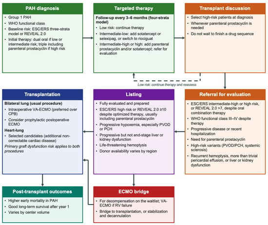
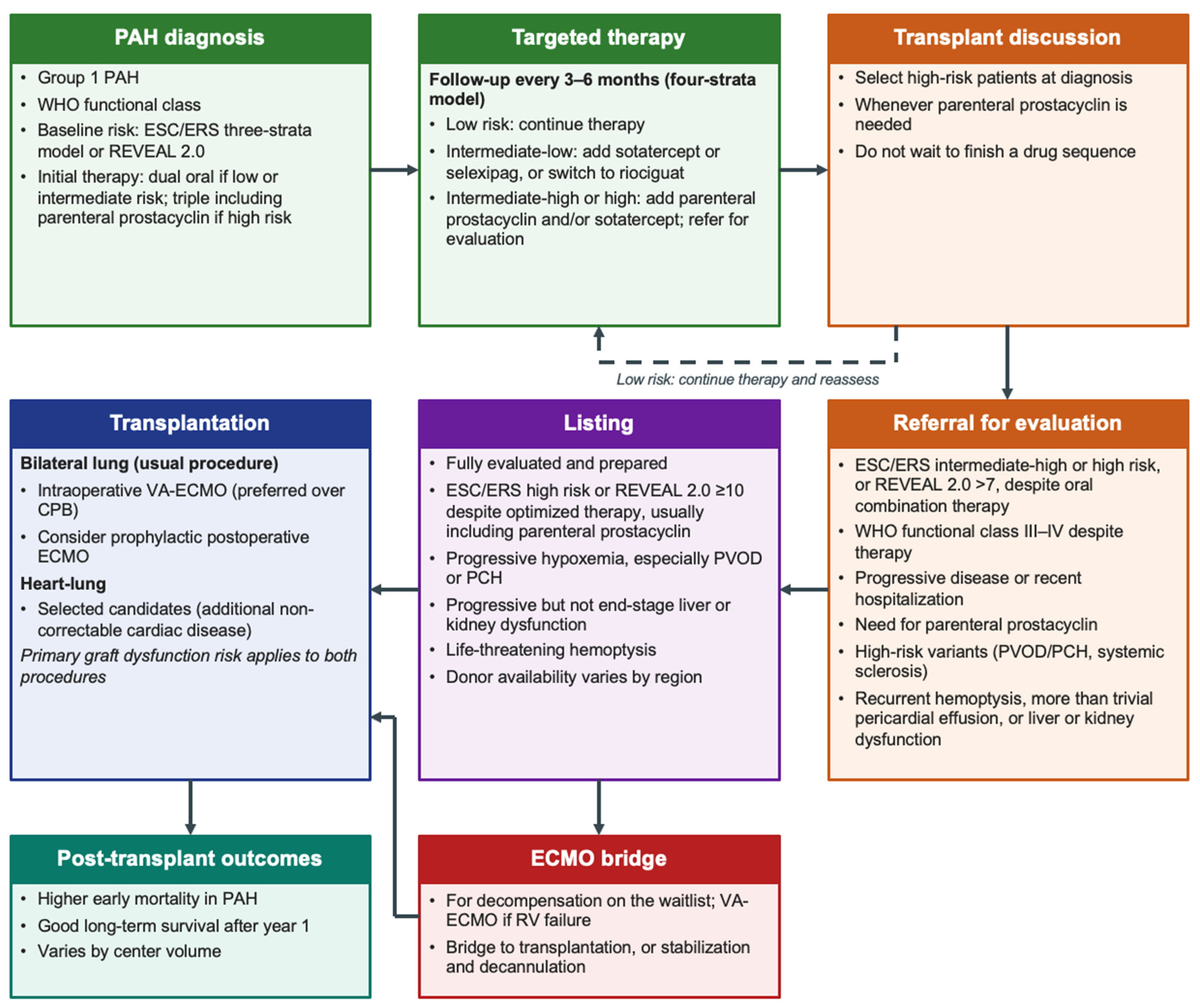
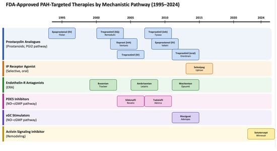
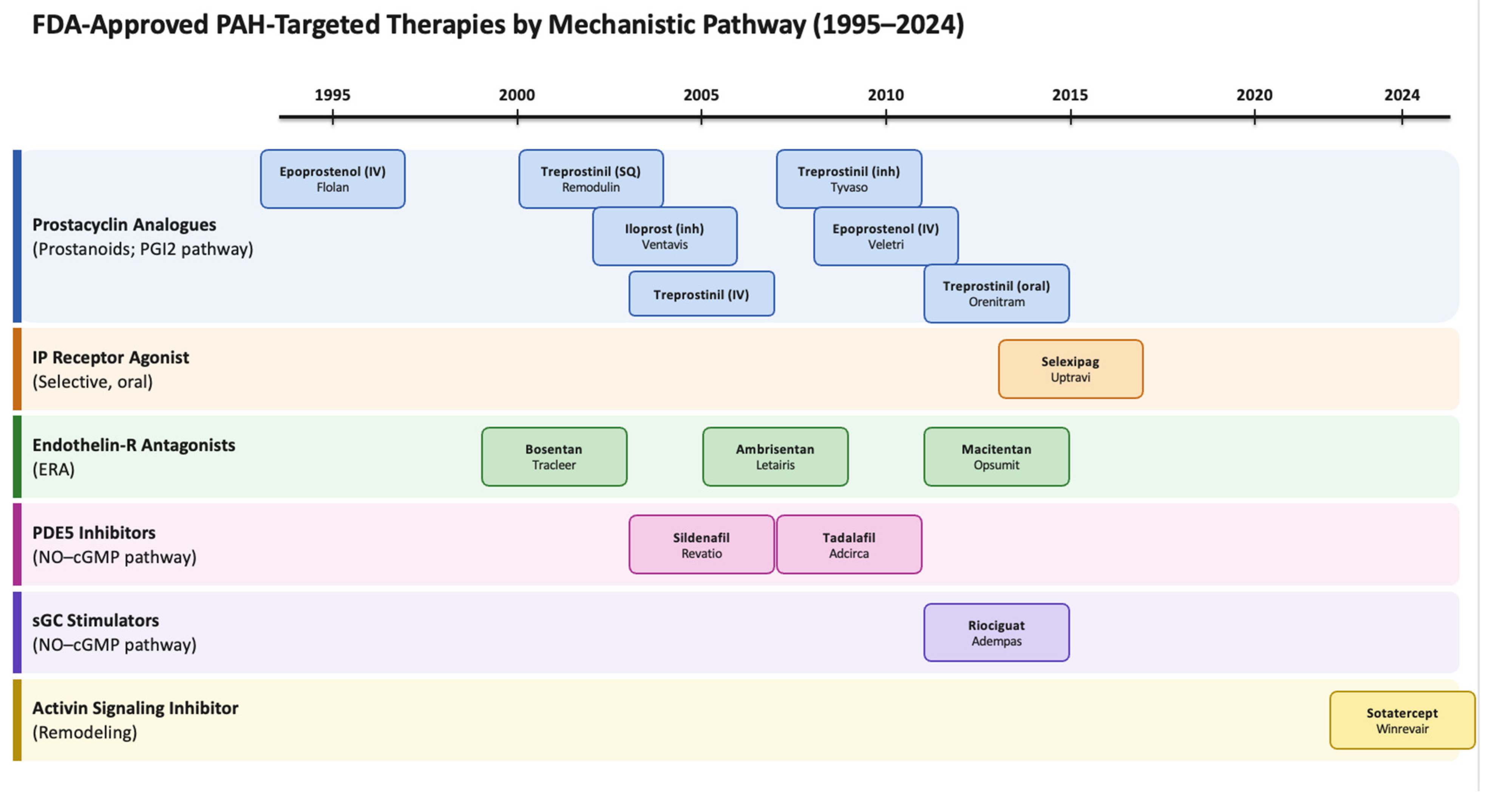
Disclaimer/Publisher’s Note: The statements, opinions and data contained in all publications are solely those of the individual author(s) and contributor(s) and not of MDPI and/or the editor(s). MDPI and/or the editor(s) disclaim responsibility for any injury to people or property resulting from any ideas, methods, instructions or products referred to in the content. |
© 2026 by the authors. Licensee MDPI, Basel, Switzerland. This article is an open access article distributed under the terms and conditions of the Creative Commons Attribution (CC BY) license.