


Abstract
Nocardia is an opportunistic pathogen associated with life-threatening infections, particularly in solid organ transplant recipients. In this population, combination therapy with two or more antibiotics and prolonged treatment duration are generally recommended to minimize the risk of relapse, although tolerability issues may adversely affect clinical outcomes. Herein, we present two cases of Nocardia infection in heart transplant recipients treated with short-term treatment (<180 days), including tedizolid. This treatment was effective in treating pulmonary nocardiosis in a heart transplant recipient, while remodulation of the immunosuppressive regimen to restore immune function with a longer course of antimicrobial therapy (7 months) contributed to successful treatment in the second case. These two cases are anecdotal, and there are currently only a few case series reported in the literature to support recommending a short course of treatment for Nocardia infection in SOT recipients. Nevertheless, the occurrence of adverse effects or tolerability issues during prolonged treatment may make a shorter treatment approach an interesting option to explore.
1. Introduction
Nocardia is an opportunistic pathogen responsible for potentially life-threatening infections, particularly in immunocompromised individuals. Recipients of solid organ or hematopoietic stem cell transplantation are at increased risk, with prevalence higher in heart and lung recipients (1–3.5%) compared with liver and kidney (<1%) [1].
Predisposing factors include a high blood level of calcineurin inhibitors in the preceding month, corticosteroid dose and use of tacrolimus, and recent CMV infection or other opportunistic infections occurring within six months before the diagnosis of nocardiosis [2,3,4].
The complexity of Nocardia infection treatment in SOT patients is related to multiple factors, mainly the length of antibiotic therapy, the emergence of drug resistance, and drug–drug interactions with concomitant immunosuppressive regimens and other co-medications [5].
Combination therapy is recommended, and the length of treatment is prolonged to minimize risk of relapse up to 6 to 12 months after antimicrobial therapy [6]. Shorter treatment durations have been proposed for non-immunosuppressed patients with a single pulmonary lesion not caused by N. farcinica [7]. Shorter treatment has also been sporadically reported for patients with solid organ transplant who received 3–4 weeks of i.v. bactericidal antibiotics (mainly a beta-lactam in combination with amikacin or ciprofloxacin), followed by a relatively short (2–3 month) oral course [8] or, in select cases, with a short course of antimicrobial treatment [2].
Here, we report two cases of heart transplant recipients with nocardial infection successfully treated with a tedizolid-based regimen: one received a short-course therapy, while the other was successfully treated following a recurrence.
2. Case Reports
2.1. Case Report 1
A 57-year-old man who had undergone orthotopic heart transplantation three months earlier for ischemic cardiomyopathy and post-myocardial infarction heart failure, and who was receiving tacrolimus, mycophenolate mofetil, and prednisone (20 mg/day), was admitted in October 2021 with CMV enterocolitis due to viral reactivation, which was successfully treated.
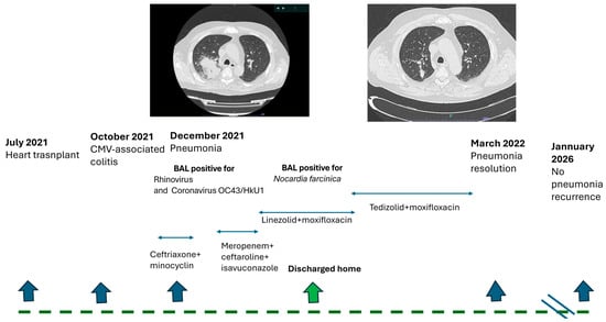
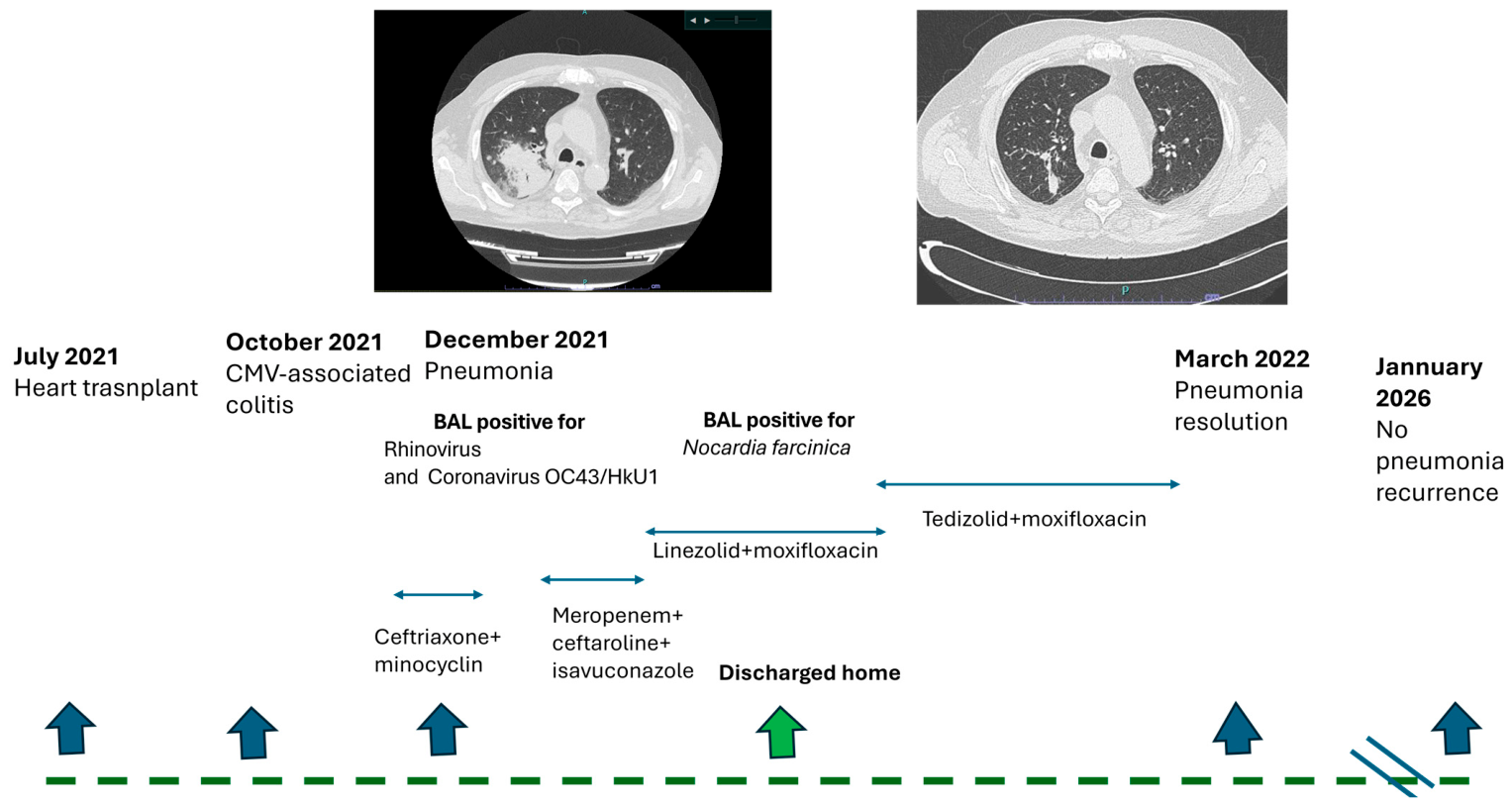
In December 2021, a right perihilar consolidation was detected on chest X-ray, associated with cough and dyspnea, anemia, neutrophilic leukocytosis, and increased C-reactive protein on blood tests. A bronchoalveolar lavage was then performed and turned positive for Rhinovirus and Coronavirus OC43/HkU1. We initiated empiric antibiotic therapy including ceftriaxone and minocycline, though persisting fever and dyspnea required oxygen therapy. Blood and urine cultures were consistently negative, while a lung CT scan documented large and irregular parenchymal opacity with air bronchograms and ground-glass opacities in the right lung; further small opacities were in both right and left lobes (Figure 1). Although a broad-spectrum antibiotic combination therapy with ceftaroline and meropenem was started in association with isavuconazole, he continued to be feverish with worsening respiratory distress and increasing inflammatory markers. On day 8 from incubation, Nocardia farcinica was isolated from BAL culture (Table 1). The antimicrobial susceptibility test results showed third-generation cephalosporin and carbapenem resistance, while trimethoprim/sulfamethoxazole, linezolid, and fluoroquinolone maintained their activity. Definitive antimicrobial treatment with 600 mg of linezolid twice daily and 400 mg of moxifloxacin once daily was started, resulting in rapid clinical improvement and allowing discharge after ten days of therapy. Considering the expected prolonged treatment duration, linezolid was subsequently switched to 200 mg of tedizolid once daily during outpatient follow-up after three weeks of treatment, owing to its more favorable safety and tolerability profile.
Figure 1.
Clinical course of patient 1.
Table 1.
Antimicrobial Susceptibility Testing Results.
The patient reported subjective well-being and good tolerance to the ongoing therapy. Clinical and radiological follow-up was performed two months later, in March 2022, with chest CT demonstrating near-complete resolution of the lesions. Antimicrobial therapy was subsequently discontinued after a total treatment duration of three months. Follow-up chest radiography and laboratory tests performed in September 2022 were unremarkable, and the patient remained clinically stable without evidence of recurrence through January 2026.
2.2. Case Report 2
A 59-year-old male patient underwent emergency orthotopic heart transplantation in January 2023 because of refractory cardiogenic shock. The post-transplant course was complicated by multiple episodes of CMV reactivation treated with ganciclovir. The last episode of CMV reactivation occurred in June 2023, with a blood CMV DNA level of 828,000 copies/mL.
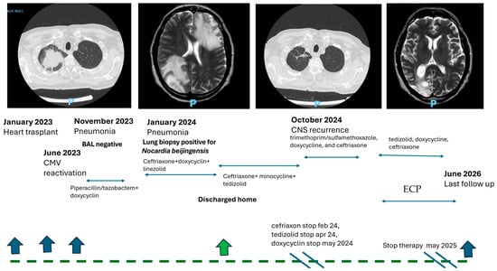
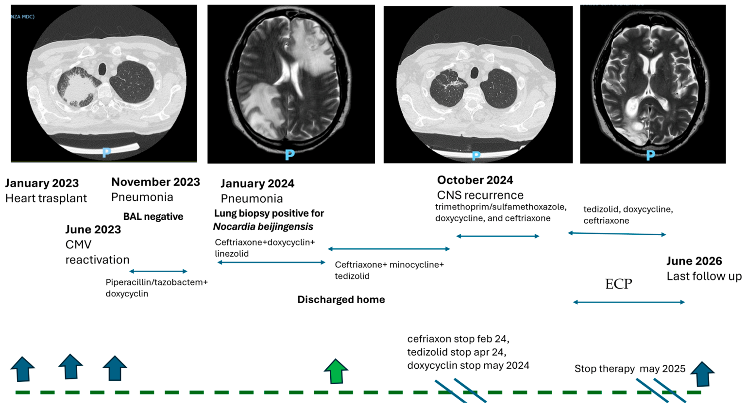
In November 2023, the patient was hospitalized for right apical pneumonia without microbiological isolation and was treated with piperacillin/tazobactam and doxycycline. Follow-up chest CT showed a large right upper lobe apical consolidation (56 × 38 mm) with spiculated margins, heterogeneous contrast enhancement, and multiple surrounding centrilobular micronodules with tree-in-bud appearance extending to the right lower lobe and left lung. Nocardia beijingensis, resistant to amoxicillin clavulanate, fluoroquinolones, and carbapenems (Table 1), was isolated from the microbiological culture of a CT-guided lung biopsy performed in January 2024. Brain MRI, performed to assess possible central nervous system involvement, revealed a left inferior fronto-subcortical lesion suggestive of cerebral nocardiosis. The patient was discharged on ceftriaxone, doxycycline, and tedizolid.
Ceftriaxone was discontinued on 26 February, tedizolid on 16 April, and doxycycline on 15 May 2024 (total duration of antimicrobial therapy, 4 months). Follow-up imaging in March 2024 and June 2024 showed radiological improvement on chest CT and reduction in the cerebral lesion to a residual 4 mm fronto-opercular cortical–subcortical lesion.
In October 2024, the patient was readmitted with probable recurrence of cerebral nocardiosis. In fact, three days before admission, the patient developed a headache and fever responsive to paracetamol. On the day prior to admission, an episode of confusion was reported by his wife, prompting presentation to the Emergency Department.
Contrast-enhanced brain CT revealed multiple cortico-subcortical expansive lesions involving the left frontal and right parieto-occipital regions, the largest located in the right occipital area and measuring approximately 31 × 25 mm. These lesions demonstrated central hypodensity with peripheral ring enhancement, highly suggestive of abscess formation. Marked surrounding vasogenic edema extended into the splenium and anterior portion of the corpus callosum, resulting in a mild mass effect on adjacent structures, compression of the lateral ventricles, effacement of the involved cortical sulci, and an approximately 2 mm leftward midline shift.
Given the previous isolation of Nocardia beijingensis from lung biopsy, antimicrobial therapy with trimethoprim/sulfamethoxazole, doxycycline, and ceftriaxone was initiated.
An extensive microbiological and virological workup was performed and yielded negative results, except for CMV replication (around 5000 to 10,000 copies/mL). A brain MRI confirmed multiple clustered cortico-subcortical expansive lesions in the left frontal and right parieto-occipital regions, considered highly suggestive of nocardial abscesses based on the patient’s history.
Neurosurgical consultation was requested to assess the feasibility of stereotactic brain biopsy; however, no indication for neurosurgical intervention was identified except for diagnostic biopsy purposes. It was agreed to reassess the patient with contrast-enhanced brain MRI after two weeks of antibiotic therapy. A lumbar puncture was performed, and microbiological examination of cerebrospinal fluid was negative. The antimicrobial regimen was subsequently modified by discontinuing trimethoprim/sulfamethoxazole and introducing tedizolid. As part of the diagnostic workup, high-resolution chest CT demonstrated a marked improvement in pulmonary parenchymal findings. Follow-up brain MRI performed two weeks apart showed a reduction in the size of the previously identified cortico-subcortical lesions in the left frontal and right parieto-occipital regions.
Progressive improvement in visual function and gait was observed, with no residual neurological deficits. Continuation of tedizolid therapy in combination with doxycycline and ceftriaxone was agreed upon; throughout the follow-up period, the complete blood count remained within normal ranges. Concomitantly, off-line ECP was initiated, with a gradual reduction in the daily dose of immunosuppressive drugs. Since June 2026, the patient has undergone a total of 19 treatment cycles (one procedure every 2 weeks for 6 months and then one procedure every month for 12 months).
A follow-up brain MRI performed in March 2025 demonstrated reduced contrast enhancement in the previously identified right parieto-occipital lesions, with substantial stability of the associated cortico-subcortical lesions. Reduced enhancement was also observed in the left frontal lesion. In May 2025, antimicrobial treatment was discontinued after a total treatment duration of 7 months, and follow-up MRI showed no evidence of new lesions. The patient remained clinically stable and asymptomatic, with no neurological symptoms at a follow-up evaluation in June 2026; he continues plasmapheresis on a monthly basis (Figure 2).
Figure 2.
Clinical course of patient 2.
Antimicrobial susceptibility testing was performed using the Sensititre RAPMYCOI panel (Thermo Scientific, Waltham, MA, USA) by the broth microdilution method. Results were interpreted according to the Clinical and Laboratory Standards Institute (CLSI) 2018 criteria.
3. Discussion
Nocardia infection can be challenging to diagnose, and clinicians should be aware of the microbiological issues associated with Nocardia growth in culture, as these organisms can be easily missed in routine microbiology workflows because of their slow growth rates, even on enriched media [9].
In the present cases, Nocardia farcinica was isolated after eight days of incubation and Nocardia beijingensis also grew in culture after 8 days of incubation. Culture methods remain the gold standard for Nocardia spp. isolation [9], as molecular methods do not currently replace culture-based methods, which remain the gold standard for the diagnosis of Nocardia infections [5].
Broth microdilution is the recommended method for susceptibility testing according to the Clinical and Laboratory Standards Institute, although clinical data supporting Nocardia-specific breakpoints remain limited [5]. Nocardia exhibits variable antimicrobial susceptibility patterns in vitro; therefore, when selecting a treatment, it is important to consider the site and severity of the infection, as well as the potential for drug interactions and/or adverse effects [6,10].
Due to the lack of evidence from randomized clinical trials, the choice of therapy requires careful consideration of both treatment tolerability and duration. Consequently, management strategies for nocardial infections should be tailored to the individual patient [2].
Trimethoprim-sulfamethoxazole has been the treatment of choice for nocardiosis for the past 50 years. However, despite its preserved antibacterial activity, its adverse effects, including agranulocytosis, aplastic anemia, neutropenia, and thrombocytopenia, may be particularly problematic in solid organ transplant recipients receiving immunosuppressive therapy [10]. Similarly, linezolid, one of the most active agents against Nocardia, is also associated with hematological toxicity, particularly thrombocytopenia and pancytopenia, especially during prolonged treatment courses [11]. Tedizolid is a second-generation oxazolidinone with proven efficacy, oral bioavailability, a good pharmacokinetic profile, and a lower burden of adverse effects (i.e., reduced myelosuppression) [12]; however, data on the clinical use of tedizolid are scarce [13].
In vitro data demonstrated that tedizolid is two-to-three-fold more effective than linezolid for most Nocardia species and non-inferior to linezolid in treating N. nova complex and N. brasiliensis [14].
Cases reporting the switch from linezolid- to tedizolid-based therapy in immunosuppressed and myelosuppressed patients with poor tolerance to linezolid, treated for a long period of time of up to 200 days, have shown good therapeutic results [13,15].
Indeed, tedizolid has a lower potential for myelosuppression and peripheral neuropathy than linezolid during prolonged treatment courses, likely due to its reduced mitochondrial toxicity [16]. Although lower CNS penetration of tedizolid compared with linezolid has been reported [17], available data on tedizolid pharmacokinetics—particularly CNS penetration—remain limited and heterogeneous and should be considered in the decision algorithm.
Tedizolid’s cerebrospinal fluid (CSF) penetration ratios range from approximately 2% in healthy rats to over 50% in some clinical estimates, with discrepant findings attributed to variable protein binding, active efflux transporters, and methodological differences across studies [18,19,20]. Clinicians should weigh the limited and heterogeneous CSF penetration data when considering tedizolid for CNS infections, and further studies with direct CSF measurement are needed before firm recommendations can be made. SOT patients are routinely exposed to medications with potential myelosuppressive effects, including mycophenolate, (val)ganciclovir, and trimethoprim-sulfamethoxazole. Consequently, the prolonged use of linezolid in this setting may be associated with an increased risk of bone marrow toxicity. Tedizolid was therefore selected as an alternative, given its more favorable hematological safety profile and lower potential for myelotoxicity compared with linezolid. Neither of the two patients described in this report experienced adverse events related to tedizolid treatment.
Current guidelines regarding Nocardia infection in the transplanted population suggest the use of trimethoprim/sulfamethoxazole in various combinations, mainly with carbapenems and/or aminoglycosides or linezolid, considering a length of treatment of 6–12 months, according to the clinical picture [2]. Shorter treatment durations have also been reported in a few SOT patients who received 3–4 weeks of intravenous bactericidal antibiotics (β-lactams and aminoglycosides) [8], as well as in those without systemic involvement [2].
In the first patient described in this report, who presented with a solitary pulmonary lesion, a 3-month course of antimicrobial therapy was sufficient to successfully treat nocardiosis despite infection with Nocardia farcinica, which typically has an unfavorable susceptibility profile when compared to other species (i.e., N. nova and N. abscessus) [21], and despite the patient being immunosuppressed.
In the second case presented here, recurrence of CNS infection occurred after a 4-month course of combination therapy in the setting of severe immunosuppression, as evidenced by concurrent CMV reactivation. A subsequent 7-month combination regimen including tedizolid, minocycline, and ceftriaxone, along with a reduction in immunosuppressive burden, was associated with a successful outcome. Nocardia infection was associated with other opportunistic infections in 50% of cases in SOT, such as CMV, aspergillosis, and non-tubercular mycobacteriosis [4]. The diagnosis of a concurrent fungal infection during the 6 months preceding the diagnosis of Nocardia infection [2], as well as CMV infection [3], was associated with an increased risk of nocardiosis. In addition, elevated blood levels of calcineurin inhibitors during the preceding month, higher corticosteroid doses, and tacrolimus use have been independently associated with the development of nocardiosis [2].
The clinical course of the second patient was particularly complex, making it difficult to establish a definitive cause for the relapse. The immunosuppressive regimen, which was associated with recurrent CMV reactivation, may have contributed to disease recurrence. The immunomodulatory effects of apheresis, together with prolonged antibiotic therapy and a tapering immunosuppressive regimen, may have contributed to the favorable clinical outcome. ECP has been shown to reduce mortality and to allow tapering of immunosuppressive therapy in most cases [22,23,24]. Moreover, it has shown promising results in heart transplant recipients at high risk of infection by preventing graft rejection while minimizing the risk of infection, thereby enabling a reduction in immunosuppressive therapy [25]. The treatment consists of mononuclear cell collection using a cell separator device, cell irradiation by UV-A after 8-methxypsoralen addition, followed by reinfusion to the patient. The treatment protocol was based on the one followed at our center for heart and lung rejection. Since June 2025, the patient has undergone a total of 19 treatment cycles (one procedure every 2 weeks for 6 months and then one procedure every month for 12 months). The procedures have been well tolerated with no additional infectious risk.
4. Conclusions
In conclusion, short-term treatment (<180 days) including tedizolid was effective in treating pulmonary nocardiosis in a heart transplant recipient, while remodulation of the immunosuppressive regimen to restore immune function with a longer course of antimicrobial therapy (7 months) contributed to successful treatment in the second case.
These two cases are anecdotal, and there are currently only a few case series reported in the literature to support recommending a short course of treatment for Nocardia infection in SOT recipients. Nevertheless, the occurrence of adverse effects or tolerability issues during prolonged treatment may make a shorter treatment approach an interesting option to explore.
Author Contributions
Conceptualization, E.S.; methodology, P.G.; software, C.D.F.; validation, C.D.F.; investigation, B.C. and C.B.; formal analysis, P.C.; writing—original draft, C.B. and P.G.; writing—review and editing, E.S. and R.B.; visualization, P.C.; project administration, B.C.; data curation, C.D.F.; funding acquisition, not applicable; supervision, R.B. All authors have read and agreed to the published version of the manuscript.
Funding
This research received no external funding.
Institutional Review Board Statement
Ethical approval was not necessary for this single-patient case report in accordance with local policy.
Informed Consent Statement
Informed consent was obtained from the patient for participation in this study.
Data Availability Statement
Data are available upon request.
Conflicts of Interest
The authors declare no conflicts of interest.
Abbreviations
The following abbreviations are used in this manuscript:
| CMV | Cytomegalovirus |
| SOT | Solid Organ Transplant recipient |
| BAL | Bronchoalveolar lavage |
| MRI | Magnetic Resonance Imaging |
| ECP | Extracorporeal Photopheresis |
References
- Pieruzzi, M.; Matucci, T.; Raccagni, A.R.; Antinori, S.; Riccardi, N.; Nozza, S.; Giacomelli, A. How we treat Nocardia infections in transplant recipients and people with HIV. Clin. Microbiol. Infect. 2026, 32, 760–766. [Google Scholar] [CrossRef] [Scilit] [PubMed]
- Lebeaux, D.; Freund, R.; van Delden, C.; Guillot, H.; Marbus, S.D.; Matignon, M.; Van Wijngaerden, E.; Douvry, B.; De Greef, J.; Vuotto, F.; et al. Outcome and Treatment of Nocardiosis After Solid Organ Transplantation: New Insights from a European Study. Clin. Infect. Dis. 2017, 64, 1396–1405, Erratum in Clin. Infect. Dis. 2017, 65, 1431. https://doi.org/10.1093/cid/cix124. [Google Scholar] [CrossRef] [Scilit] [PubMed]
- Peleg, A.Y.; Husain, S.; Qureshi, Z.A.; Silveira, F.P.; Sarumi, M.; Shutt, K.A.; Kwak, E.J.; Paterson, D.L. Risk factors, clinical characteristics, and outcome of Nocardia infection in organ transplant recipients: A matched case-control study. Clin. Infect. Dis. 2007, 44, 1307–1314. [Google Scholar] [CrossRef] [Scilit] [PubMed]
- Bini Viotti, J.; Simkins, J.; Reynolds, J.M.; Ciancio, G.; Guerra, G.; Abbo, L.; Anjan, S. Nocardiosis in Solid Organ Transplant Recipients: 10-Year Single Center Experience and Review of Literature. Microorganisms 2024, 12, 1156. [Google Scholar] [CrossRef] [Scilit] [PubMed]
- Yetmar, Z.A.; Marty, P.K.; Clement, J.; Miranda, C.; Wengenack, N.L.; Beam, E. State-of-the-Art Review: Modern Approach to Nocardiosis-Diagnosis, Management, and Uncertainties. Clin. Infect. Dis. 2025, 80, e53–e64. [Google Scholar] [CrossRef] [Scilit] [PubMed]
- Coussement, J.; Lebeaux, D.; Rouzaud, C.; Lortholary, O. Nocardia infections in solid organ and hematopoietic stem cell transplant recipients. Curr. Opin. Infect. Dis. 2017, 30, 545–551. [Google Scholar] [CrossRef] [Scilit] [PubMed]
- Attias, N.H.; Schlaeffer-Yosef, T.; Zahavi, I.; Hasson, N.; Ari, Y.B.; Darawsha, B.; Levitan, I.; Goldberg, E.; Landes, M.; Litchevsky, V.; et al. Shorter vs. standard-duration antibiotic therapy for nocardiosis: A multi-center retrospective cohort study. Infection 2025, 53, 1115–1127. [Google Scholar] [CrossRef] [Scilit] [PubMed]
- Tripodi, M.F.; Durante-Mangoni, E.; Fortunato, R.; Cuccurullo, S.; Mikami, Y.; Farina, C.; Utili, R. In vitro activity of multiple antibiotic combinations against Nocardia: Relationship with a short-term treatment strategy in heart transplant recipients with pulmonary nocardiosis. Transpl. Infect. Dis. 2011, 13, 335–343. [Google Scholar] [CrossRef] [Scilit] [PubMed]
- McTaggart, L.R.; Chen, Y.; Poopalarajah, R.; Kus, J.V. Incubation time and culture media impact success of identification of Nocardia spp. by MALDI-ToF mass spectrometry. Diagn. Microbiol. Infect. Dis. 2018, 92, 270–274. [Google Scholar] [CrossRef] [Scilit] [PubMed]
- Margalit, I.; Lebeaux, D.; Tishler, O.; Goldberg, E.; Bishara, J.; Yahav, D.; Coussement, J. How do I manage nocardiosis? Clin. Microbiol. Infect. 2021, 27, 550–558. [Google Scholar] [CrossRef] [Scilit] [PubMed]
- Shen, T.; Wu, L.; Geng, L.; Wei, Z.; Zheng, S. Successful treatment of pulmonary Nocardia farcinica infection with linezolid: Case report and literature review. Braz. J. Infect. Dis. 2011, 15, 486–489. [Google Scholar] [CrossRef] [Scilit] [PubMed]
- Kisgen, J.J.; Mansour, H.; Unger, N.R.; Childs, L.M. Tedizolid: A new oxazolidinone antimicrobial. Am. J. Health Syst. Pharm. 2014, 71, 621–633. [Google Scholar] [CrossRef] [Scilit] [PubMed]
- Chomei, Y.; Nishimura, S.; Iwata, K. Long-term use of tedizolid for pulmonary nocardiosis. J. Infect. Chemother. 2022, 28, 1172–1176. [Google Scholar] [CrossRef] [Scilit] [PubMed]
- Brown-Elliott, B.A.; Wallace, R.J., Jr. In Vitro Susceptibility Testing of Tedizolid against Isolates of Nocardia. Antimicrob. Agents Chemother. 2017, 61, e01537-17. [Google Scholar] [CrossRef] [Scilit] [PubMed]
- Matin, A.; Sharma, S.; Mathur, P.; Apewokin, S.K. Myelosuppression-sparing treatment of central nervous system nocardiosis in a multiple myeloma patient utilizing a tedizolid-based regimen: A case report. Int. J. Antimicrob. Agents 2017, 49, 488–492. [Google Scholar] [CrossRef] [Scilit] [PubMed]
- Flanagan, S.; McKee, E.E.; Das, D.; Tulkens, P.M.; Hosako, H.; Fiedler-Kelly, J.; Passarell, J.; Radovsky, A.; Prokocimer, P. Nonclinical and pharmacokinetic assessments to evaluate the potential of tedizolid and linezolid to affect mitochondrial function. Antimicrob. Agents Chemother. 2015, 59, 178–185. [Google Scholar] [CrossRef] [Scilit] [PubMed]
- Olivier, V.; Margaux, B.; David, L.; Anne Christine, J.; Heidi, W.; Christophe, A.; Laure, A.; Alexia, C. Optimizing Tedizolid Dosing in Cerebral Nocardiosis: Clinical Impact of Direct Unbound Concentration Measurement and Population PK Modelling in Two Cases. JAC Antimicrob. Resist. 2026, 8, dlag004. [Google Scholar] [CrossRef] [Scilit] [PubMed]
- Lahouati, M.; Oudart, M.; Alzieu, P.; Chapouly, C.; Petitcollin, A.; Xuereb, F. Penetration of linezolid and tedizolid in cerebrospinal fluid of mouse and impact of blood–brain barrier disruption. Clin. Transl. Sci. 2025, 18, e70100. [Google Scholar] [CrossRef] [Scilit] [PubMed]
- Brousse, X.; Lê, M.P.; Lahouati, M.; Petitgas, P. Insufficient cerebrospinal fluid penetration of tedizolid: A case report. JAC Antimicrob. Resist. 2025, 7, dlaf100. [Google Scholar] [CrossRef] [Scilit] [PubMed]
- Gu, L.; Ma, M.; Zhang, Y.; Zhang, L.; Zhang, S.; Huang, M.; Zhang, M.; Xin, Y.; Zheng, G.; Chen, S. Comparative pharmacokinetics of tedizolid in rat plasma and cerebrospinal fluid. Regul. Toxicol. Pharmacol. 2019, 107, 104420. [Google Scholar] [CrossRef] [Scilit] [PubMed]
- Schlaberg, R.; Fisher, M.A.; Hanson, K.E. Susceptibility profiles of Nocardia isolates based on current taxonomy. Antimicrob. Agents Chemother. 2014, 58, 795–800. [Google Scholar] [CrossRef] [Scilit] [PubMed]
- Barr, M.L.; Meiser, B.M.; Eisen, H.J.; Roberts, R.F.; Livi, U.; Dall’Amico, R.; Dorent, R.; Rogers, J.G.; Radovancević, B.; Taylor, D.O.; et al. Photopheresis for the prevention of rejection in cardiac transplantation. Photopheresis Transplantation Study Group. N. Engl. J. Med. 1998, 339, 1744–1751. [Google Scholar] [CrossRef] [Scilit] [PubMed]
- Barten, M.J.; Sax, B.; Schopka, S.; Amarelli, C.; Epailly, E.; Natali, B.; Teszák, T.; Gökler, J.; Borchert, K.; Theil, J.; et al. European multicenter study on the real-world use and clinical impact of extracorporeal photopheresis after heart transplantation. J. Heart Lung Transplant. 2023, 42, 1131–1139. [Google Scholar] [CrossRef] [Scilit] [PubMed]
- Teszak, T.; Assabiny, A.; Kiraly, A.; Tarjanyi, Z.; Parazs, N.; Szakal-Toth, Z.; Hartyanszky, I.; Szabolcs, Z.; Racz, K.; Reti, M.; et al. Extracorporeal photopheresis in the treatment of cardiac allograft rejection: A single-centre experience. Transpl. Immunol. 2023, 79, 101853. [Google Scholar] [CrossRef] [Scilit] [PubMed]
- Gökler, J.; Aliabadi-Zuckermann, A.; Zuckermann, A.; Osorio, E.; Knobler, R.; Moayedifar, R.; Angleitner, P.; Leitner, G.; Laufer, G.; Worel, N. Extracorporeal Photopheresis with Low-Dose Immunosuppression in High-Risk Heart Transplant Patients-A Pilot Study. Transpl. Int. 2022, 35, 10320. [Google Scholar] [CrossRef] [Scilit] [PubMed]

Disclaimer/Publisher’s Note: The statements, opinions and data contained in all publications are solely those of the individual author(s) and contributor(s) and not of MDPI and/or the editor(s). MDPI and/or the editor(s) disclaim responsibility for any injury to people or property resulting from any ideas, methods, instructions or products referred to in the content. |
© 2026 by the authors. Licensee MDPI, Basel, Switzerland. This article is an open access article distributed under the terms and conditions of the Creative Commons Attribution (CC BY) license.