


Abstract
Background: Male infertility affects millions of couples, accounting for 50 percent of cases. Despite such a major contribution of the male factor, it is not properly evaluated and is often overlooked in infertility assessments. Semen analysis, which is routinely performed to assess infertility status, is unable to assess the defects at the molecular level which are important to assess the fertilizing capacity of the sperm. This study aims to determine the utility of sperm function tests as biomarkers for male infertility in addition to standard semen analysis. Methods: Thirty-five men (aged 25–45 years) were recruited and divided into two groups: those with at least one altered semen parameter (infertile group) and those with normal semen parameters but unable to conceive after more than one year of unprotected intercourse (unexplained male infertility group). The DNA Fragmentation Index (DFI), Nuclear Chromatin Decondensation Test (NCDT) and Hypoosmotic Swelling Test (HOS) were used in diagnosing sperm dysfunction in both groups. The Mann–Whitney U testand Spearman’s rank correlation were used for analyzing the parameters of the groups. A p value < 0.05 was considered statistically significant. Results: While motility and vitality were nearly identical in both groups, the infertile group showed more morphological abnormalities. The DFI was higher in the unexplained male infertility group (UMI) (82%) than in the infertile group (36%). Poor decondensation capacity was present in 27% of the unexplained male infertility group and 60% of the infertile group. Both groups’ hypoosmotic swelling values fell within the usual range. Spearman correlation analysis revealed that the NCDT showed significant positive correlations with sperm vitality (r = 0.36; p = 0.02) and morphology (r = 0.53; p = 0.001). The DFI demonstrated significant negative correlations with vitality (r = −0.45; p = 0.006) and motility (r = −0.39; p = 0.01). HOS was significantly positively correlated with motility (r = 0.56; p = 0.0004) and vitality (r = 0.57; p = 0.0003). Additionally, the NCDT and DFI showed a significant inverse correlation (r = −0.33; p = 0.04). Conclusions: These findings highlight the potential of sperm function tests as valuable diagnostic tools alongside conventional semen analysis for a more comprehensive assessment of male fertility.
1. Introduction
Infertility is a disease of the reproductive system due to failure to conceive even after being sexually active for one year or more of regular intercourse without any contraceptive use [1]. Male infertility poses a significant challenge globally, affecting millions of couples. It is said that 15% of couples worldwide experience infertility. Males are found to be solely responsible for 20–30% of infertility cases and contribute to 50% of cases overall [2]. Despite its prevalence, the male factor remains understudied and is often overlooked in infertility evaluations. Sperm has continuously been the target of microbial attack and oxidative damage, leading to both structural and functional deficit and hence male infertility [3,4,5,6]. Testicular spermatogenesis, along with the development and function of other accessory sex glands such as the prostate and seminal vesicles, is essential for normal seminal plasma components, normal sperm parameters and normal sperm function [7,8,9,10]. Semen analysis has always been considered as a pillar for assessing male infertility and is performed worldwide according to WHO guidelines [11,12]. Semen analysis involves the assessment of both qualitative (e.g., semen viscosity and sperm morphology) and quantitative (e.g., semen volume and sperm count) parameters of semen [13]. While conventional semen analysis remains a cornerstone, its reliance on descriptive parameters overlooks crucial aspects of sperm function essential for fertilization. Besides this, its standards and reference ranges were determined by examining only a fertile population, and its assessment involves large inter-laboratory and intra-individual variability [14,15,16]. A major limitation of conventional semen analysis which cannot be overlooked is that it can only assess extracellular defects such as motility, morphology, and viscosity but does not involve the assessment of intracellular components such as DNA and its packaging, centrioles, RNA, or antioxidant capacity [17,18,19]. This limitation emphasizes the need for better diagnostic tools that will take into account the spermatozoa’s functional capacity. Male infertility causes can be broadly divided into two major domains: (1) Male-factor infertility, where the male partner has abnormal semen parameters according to the reference limits set by the WHO. This domain can be further divided into two categories: the first is explained male infertility, where the cause for abnormal semen parameters is known; explained male infertility can be further subdivided into three main subcategories, anatomical, hormonal, and genetics and environmental. The second category under male infertility causes is idiopathic male infertility, where the cause for abnormal semen parameters is not known (infertile group) [20].
Unexplained male infertility, which includes males from couples who have all the normal semen parameters but still are not able to conceive and the cause for this is not known, and both partners show normal parameters in their fertility evaluation (unexplained male infertility group). The reason for this could be that these males possess defects in intracellular sperm components which are not affecting the semen parameters, thus resulting in a normal semen analysis [20]. Intracellular sperm components like DNA fragmentation, improper packaging, inability to handle oxidative stress and centriole dysfunction could be the mechanisms of unexplained male infertility [21,22]. There have been continuous efforts in understanding pathophysiology and searching for the biomarkers depicting fertility status in female reproductive disorders such as polycystic ovary syndrome, premature ovarian failure, and preeclampsia [23,24,25,26,27,28]. The focus has been on molecular cytogenetics, immunohistochemistry and cell morphology-based techniques in ovulatory or non-ovulatory disorders in women [29,30,31]. These techniques usually differentiate the effects of cell damage mediators in exposed and non-exposed cells and therefore the use of such techniques has been given importance for identifying potential biomarkers [32,33,34]. Similarly, in infertile males, the search for potential diagnostic biomarkers affecting sperm function has been emphasized [35,36,37]. Therefore, the current diagnostic landscape demands the development of tests that encompass both the intracellular and extracellular components of sperm during fertility evaluation, especially for cases with unexplained male infertility. Sperm function tests assess the reproductive potential of sperm in vitro, estimate fertilization and pregnancy rates, and diagnose sperm-specific dysfunction. A single test is not a good predictor of fertility outcome; it is always the combination of assessments encompassing all functional parameters of sperm which gives us an accurate evaluation of fertility potential [38]. Whether sperm function tests are to be incorporated along with routine semen analysis while performing the fertility assessment needs to be confirmed further. In this study, the following three sperm morphology-based tests were evaluated in both infertile and unexplained male infertility populations: the Nuclear Chromatin Decondensation Test (NCDT), the Hypoosmotic Swelling Test (HOST) and the DNA Fragmentation Index (DFI). These are advanced sperm function tests that provide information beyond routine semen parameters necessary for evaluating the intracellular component of sperm. The NCDT evaluates the degree of sperm chromatin compaction and its ability to properly decondense after fertilization, a critical step for male pronucleus formation and normal embryo development, with normal samples possessing more than 70% NCDT-positive cells [38,39]. The HOST assesses the structural and functional integrity of the sperm plasma membrane, with a cut-off of more than 50% HOS-reacted sperm considered normal, which is essential for sperm survival, motility, and interaction within the female reproductive tract [38]. The DFI determines the extent of DNA damage in sperm, reflecting the genetic quality required for successful fertilization, implantation, and healthy pregnancy outcomes [40,41]. According to the previous literature, a DFI of more than 20% was shown to affect fertility and pregnancy outcomes, so based on this, a value of 20% was considered as a cut-off for this study [42,43]. Collectively, these tests offer a comprehensive evaluation of sperm functional competence by examining chromatin integrity, membrane viability, and DNA stability, thereby improving the assessment of male reproductive potential.
2. Material and Methods
2.1. Study Design
In total, 35 men aged 25–45 years were recruited from the Andrology Lab (Department of Reproductive Biology, AIIMS, New Delhi) and divided into two groups based on their semen parameters—those with any one altered semen parameter (infertile group, n = 24) and those with normal semen parameters but unable to conceive even after more than one year of unprotected intercourse (unexplained male infertility group, n = 11). For the unexplained male infertility group, female-factor infertility was excluded; all female partners underwent thorough gynecological evaluation, and only couples with normal female fertility parameters were included. Exclusion criteria for both groups included any known cause of genital infection, endocrinological disorders, genital tract obstruction, varicocele, anti-sperm antibodies, chromosomal aneuploidy, smoking, and alcohol intake. A formal power calculation was not conducted for this investigation, as it was designed as a pilot study to evaluate the feasibility of sperm functional assessments and to estimate effect sizes for future larger trials. The sample size of n = 35 was selected based on the pilot nature of the study and the objective of recruiting a strictly defined, homogeneous population of infertile males to minimize confounding variables. The protocol of the study was approved by the Institute Human Ethics Committee (IEC-406/06.05.2022), and written consent was obtained from all cases and controls. Routine semen analysis was performed according to WHO guidelines (2021), along with sperm function tests (DFI, NCDT, and HOS). A minimum sperm count of 39 million/ejaculate, sperm concentration of 16 million/mL, total motility of 42%, viability of 54%, and normal morphology of 4% were considered within the normal range. In addition to conventional semen parameters, the Teratozoospermia Index (TZI) was calculated as an indicator of the average number of morphological defects present per abnormal spermatozoon. TZI was determined according to WHO 2021 guidelines using the following formula:
TZI (Teratozoospermia Index) = (Total number of head defects + midpiece defects + tail defects + excess residual
cytoplasm defects)/(Total number of abnormal spermatozoa)
cytoplasm defects)/(Total number of abnormal spermatozoa)
A TZI value approaching 1 indicates that most abnormal spermatozoa possess a single defect, whereas higher values reflect the presence of multiple morphological abnormalities within individual spermatozoa.
2.2. DNA Fragmentation Index (DFI)
The DFI was performed using the Halosperm G2 kit (HT-HSG2R; Halotech DNA SL, Madrid, Spain) according to the manufacturer’s instructions. Agarose contained in an Eppendorf tube was melted at 98 °C in a water bath or thermoshaker. Semen sample was diluted with PBS up to 20 million sperm per ml. Immediately after, 50 µL of the semen sample was transferred to the Eppendorf tube containing melted agarose and mixed gently; formation of bubbles was prevented. Then, 10 µL of the cell suspension was placed onto the center of a sample well and covered with a coverslip. The slide was placed on a cold surface and placed in the fridge at 2–8 °C for 7 min to solidify the agarose. The slide was taken out of the fridge, and the coverslip was removed by sliding it off gently. Solution 1 (denaturation solution) was added onto the sample well and incubated for 7 min. The solution was then removed by tilting. Solution 2 (lysis solution) was applied and incubated for 20 min. Positive and negative controls used for DFI analysis are provided in Supplementary Figure S1. The slides were washed for 5 min with distilled water and then dehydrated with 70% and 100% ethanol for 2 min each. The drained slides were allowed to air-dry. The slides were then stained with solution 3 (SSA) and solution 4 (SSB) respectively and incubated for 7 min each. Excess stain was removed, and the slides were visualized under the microscope.
Sperm DNA fragmentation was calculated by the formula: SDF% = (Fragmented + degraded) × 100/Total number of counted cells. A minimum number of 200 sperms were counted.
2.3. Nuclear Chromatin Decondensation Test (NCDT)
The NCDT was performed using the HI-Tech NCDT kit (HI-Tech Solutions, New Delhi, India) according to the manufacturer’s instructions. In total, 500 µL of wash buffer was added to 100 µL of liquified semen sample, mixed well, and centrifuged for 5 min at 1500 g. The supernatant was discarded and 200 µL of NCD solution was added to the pellet, mixed well, and incubated at 37 °C for 30 min. Then, 50 µL of stop solution was added, and the mixture was incubated again at 37 °C for 5 min. A drop of mixture solution was transferred onto the glass slide and examined under the microscope. A minimum number of 200 condensed and decondensed heads were counted.
2.4. Hypoosmotic Swelling Test (HOS)
In total, 1.0 mL of hypoosmotic (HOS) solution containing 1.5 mM sodium citrate and 1.5 mM fructose (SRL Pvt. Ltd., Mumbai, India) was incubated for 10 min at 37 °C. The tube containing the HOS solution was gently mixed with 0.1 mL of completely mixed, liquid semen. The mixture of semen and HOS solution was incubated at 37 °C for at least 30 min. The contents of the tube were gently mixed after being incubated at 37 °C. A drop was then put onto a microscopic slide, covered with a coverslip, and examined under a microscope to check for spermatozoa with curled tails. A minimum number of 200 sperms were counted.
2.5. Statistical Analysis
The data was analyzed using Spearman’s rank correlation, and the Mann–Whitney U test was used to compare the semen parameters between cases and controls using GraphPad Prism8 version 8.0.1 (GraphPad Software, Inc., San Diego, CA, USA). A p value < 0.05 was considered statistically significant.
3. Results
3.1. Comparison of Conventional Semen Parameters
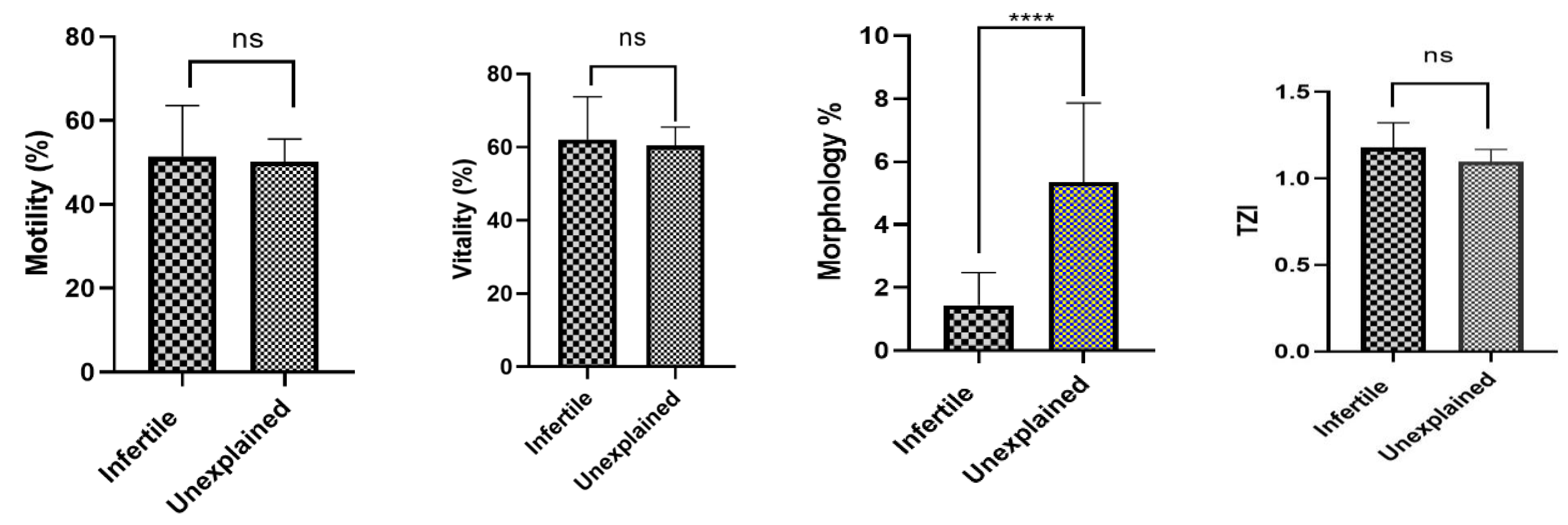
Detailed analysis of semen parameters was done (Table 1 and Table 2) followed by a comparative analysis of semen parameters between the unexplained and infertile groups performed using the Mann–Whitney U test. While motility and vitality showed no statistically significant differences between the two cohorts (p = 0.40; p = 0.29), a highly significant difference was observed in sperm morphology (p < 0.0001). This indicates that despite similar functional profiles in motility and vitality, the structural integrity of the sperm differed markedly between the groups. However, the Teratozoospermia Index did not differ significantly between the two cohorts (p = 0.13),suggesting that although overall morphology differed, the distribution of specific morphological defects per spermatozoon remained comparable between groups (Figure 1).
Table 1.
Semen parameters along with sperm function tests of the recruited infertile patients.
Table 2.
Semen parameters along with sperm function tests of the recruited unexplained male infertility patients.
Figure 1.
Comparison of conventional semen parameters between infertile and UMI cohorts. Asterisks denote statistical significance calculated via the Mann–Whitney U test (p < 0.0001), and ns denotes a non-significant difference between the two cohorts.
3.2. Comparison of Sperm Function Tests Between Groups
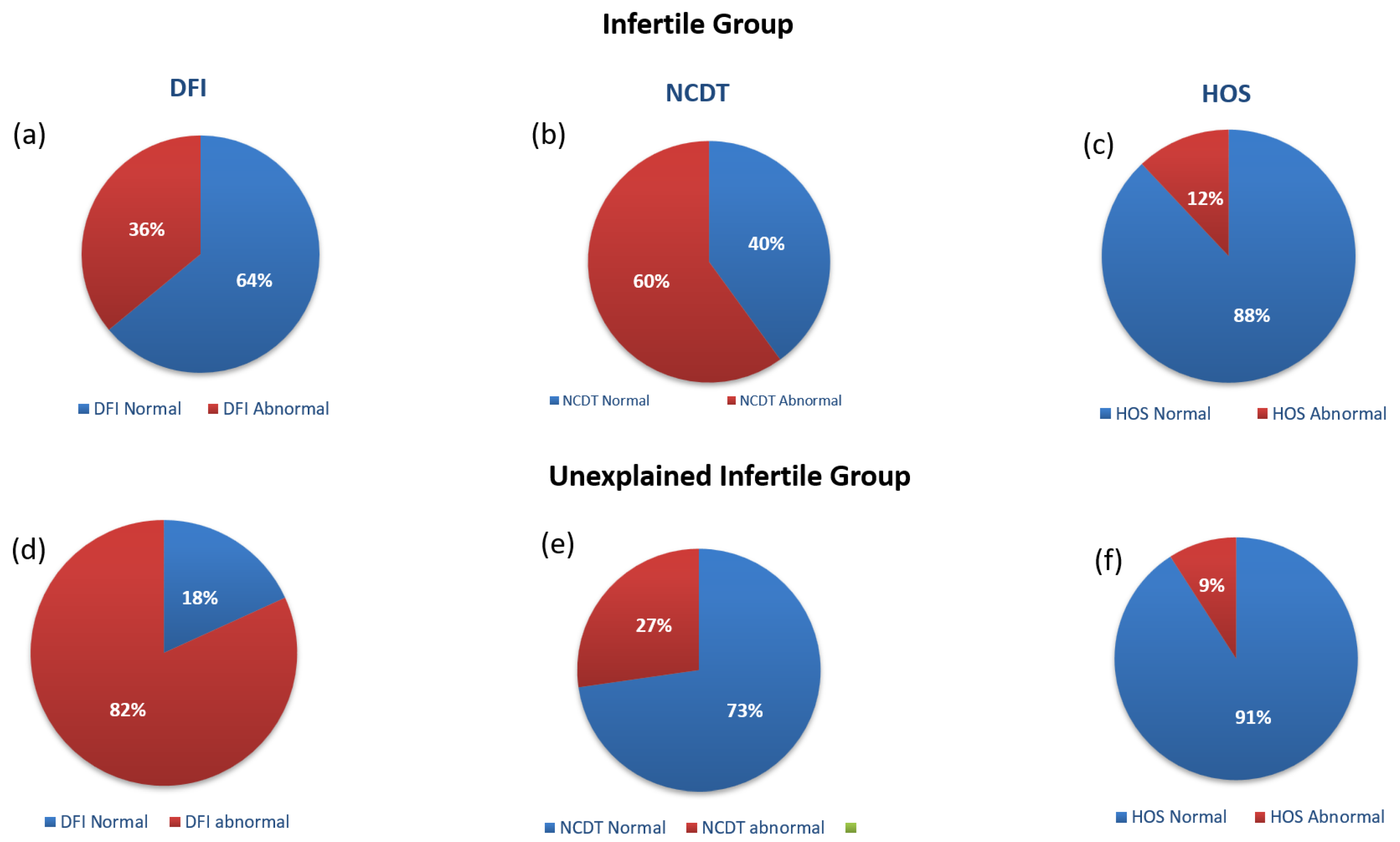
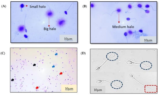
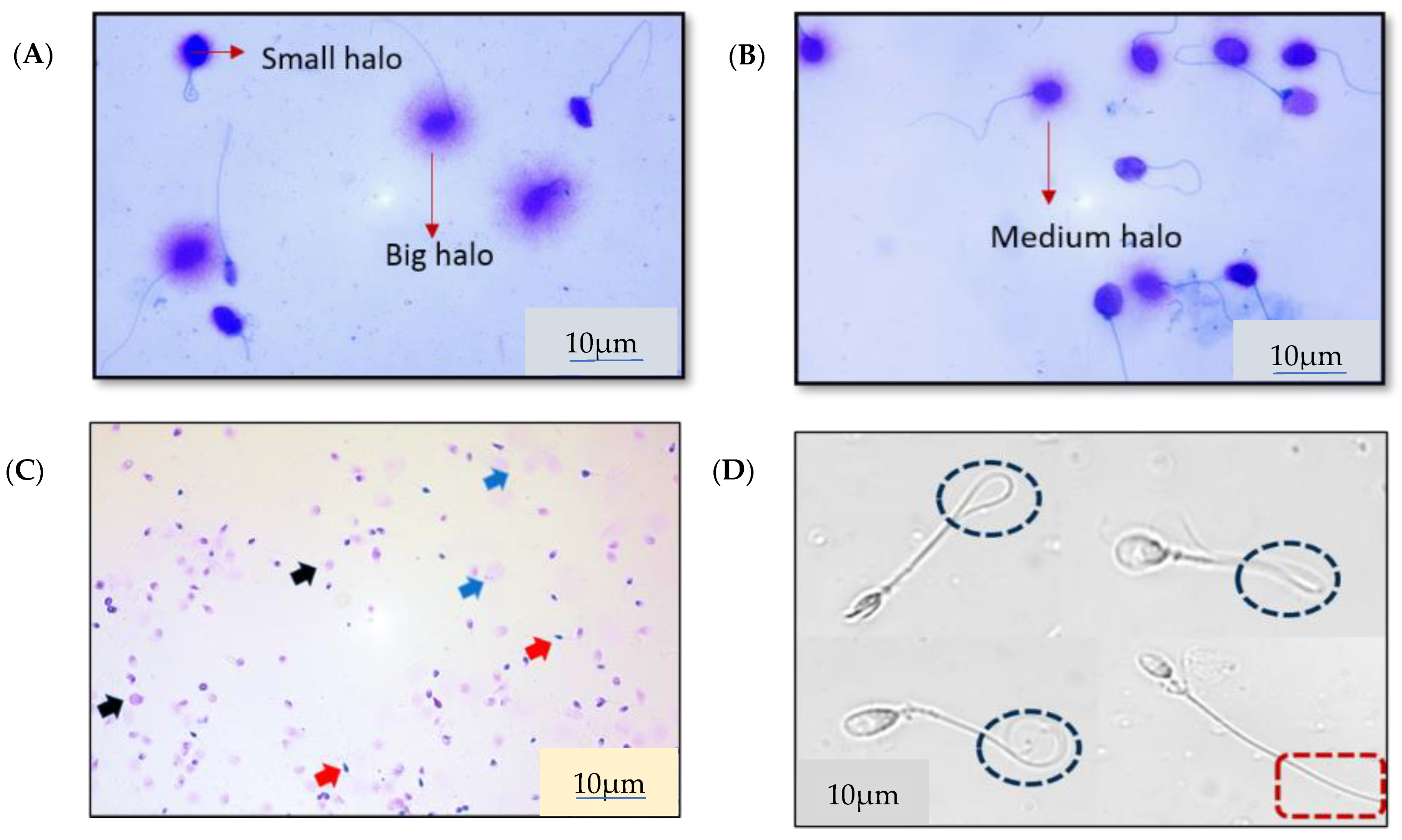
The three sperm function tests revealed distinct physiological profiles for each group. The unexplained male infertility (UMI) group exhibited a substantially higher prevalence of abnormal DNA fragmentation (82%) compared to the infertile group (36%). Regarding chromatin integrity, poor decondensation capacity was more prevalent in the infertile group (60%) than in the UMI group (27%). The results for the HOS test were largely within the normal range for both cohorts, with only 12% of the infertile group and 9% of the UMI group failing to meet the osmotic stress threshold (Figure 2a–f). Representative microscopic images of DFI, NCDT, and HOS assays demonstrating the characteristic staining and structural patterns observed during sperm functional assessment are presented in Figure 3A–D.
Figure 2.
Comparison of different sperm function tests between the infertile and unexplained male infertility group. (a,d) compare the DFI between the infertile and unexplained male infertility groups: 36% has an abnormal DFI in the infertile group as compared to 82% in the unexplained male infertility group; 64% has a normal DFI in the infertile group as compared to only 18% in the unexplained male infertility group [cut-off value, 20%]. (b,e) compare the NCD values between the two groups: 60% has abnormal decondensation capacity in the infertile group as compared to 27% in the unexplained male infertility group; 40% has normal decondensation capacity in the infertile group as compared to 73% in the unexplained male infertility group [cut-off value, 70%]. (c,f) compare the HOS values between the two groups: 88% has normal HOS values in the infertile group as compared to 91% in the unexplained male infertility group; 12% has abnormal HOS values in the infertile group as compared to 9% in the unexplained male infertility group [cut-off value, 50%].
Figure 3.
Representative images of the sperm functional assays. (A,B) depict the DNA Fragmentation Index, where large and medium halos indicate non-fragmented DNA, while small halos signify fragmented DNA. (C) illustrates the Nuclear Chromatin Decondensation Test, with arrows identifying varying degrees of decondensation: blue for high, black for moderate, and red for non-decondensed sperm. (D) represents the Hypoosmotic Swelling Test, where coiled tails indicate intact, functional membranes capable of withstanding osmotic stress, whereas straight tails represent non-functional membranes.
3.3. Inter-Correlation of Sperm Function Tests
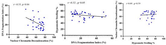
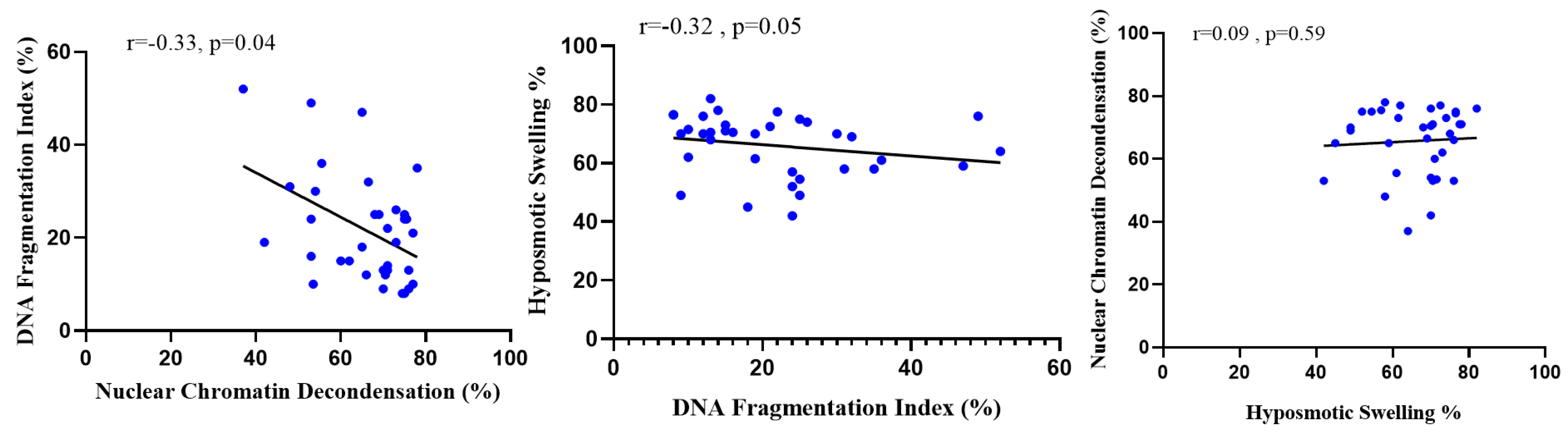
Spearman correlation analysis was used to evaluate the relationships between the three functional assays. Analysis of the relationships between the functional assays showed that the NCDT and DFI exhibited a significant negative correlation (r = −0.33; p = 0.04), suggesting that increased DNA damage is linked to impaired chromatin decondensation. HOS exhibited a non-significant negative correlation with the DFI (r =−0.32; p = 0.05) and a very weak positive correlation with the NCDT (r = 0.09; p = 0.59). (Figure 4).
Figure 4.
Correlation analysis of sperm function tests, highlighting the significant negative correlation between DNA fragmentation and chromatin decondensation (r = −0.33; p = 0.04).
3.4. Correlation of Sperm Function Tests with Semen Parameters
Each functional test was correlated with semen parameters to identify potential biomarkers of sperm quality.
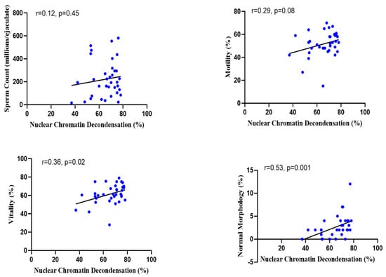
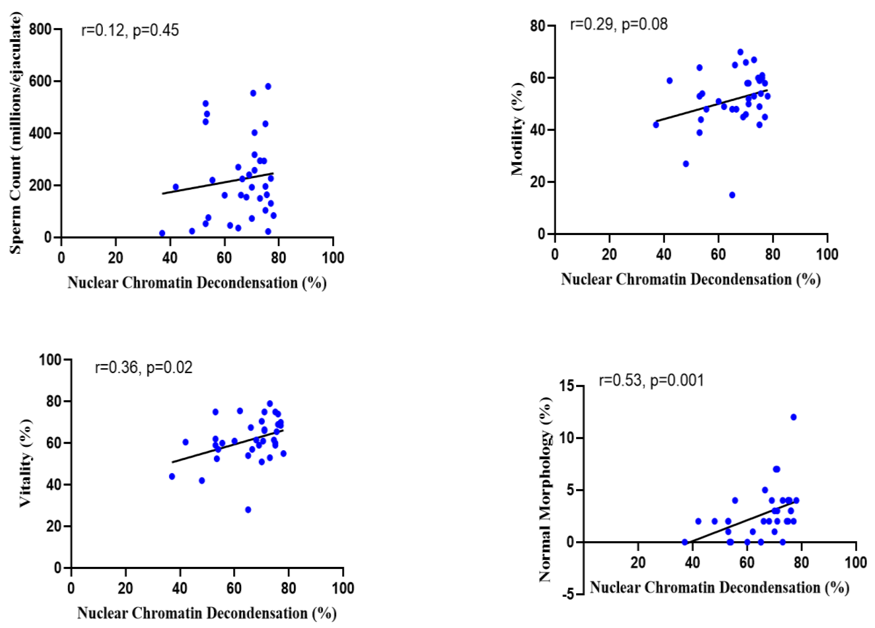
- NCDT: Showed a significant positive correlation with vitality (r = 0.36; p = 0.02) and morphology (r = 0.53; p = 0.001). Correlations with sperm count (r = 0.12; p = 0.45) and motility (r = 0.29; p = 0.08) were positive but not statistically significant (Figure 5).
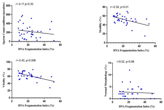
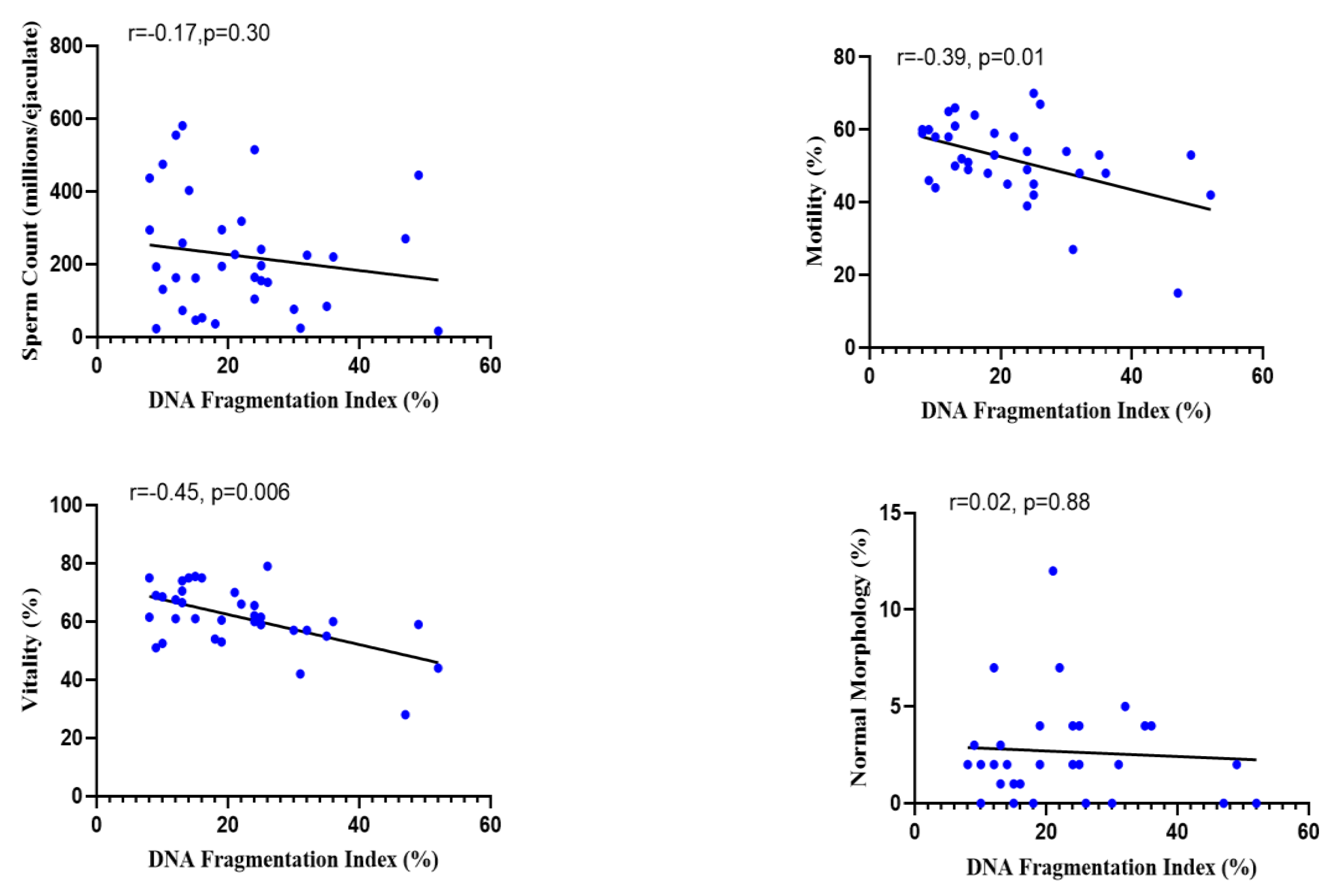
- DFI: Demonstrated a significant inverse relationship with vitality (r = −0.45; p = 0.006) and motility (r = −0.39; p = 0.01). No significant correlations were found with sperm count or morphology (r = −0.17; p = 0.30; r = 0.02; p = 0.88) (Figure 6).
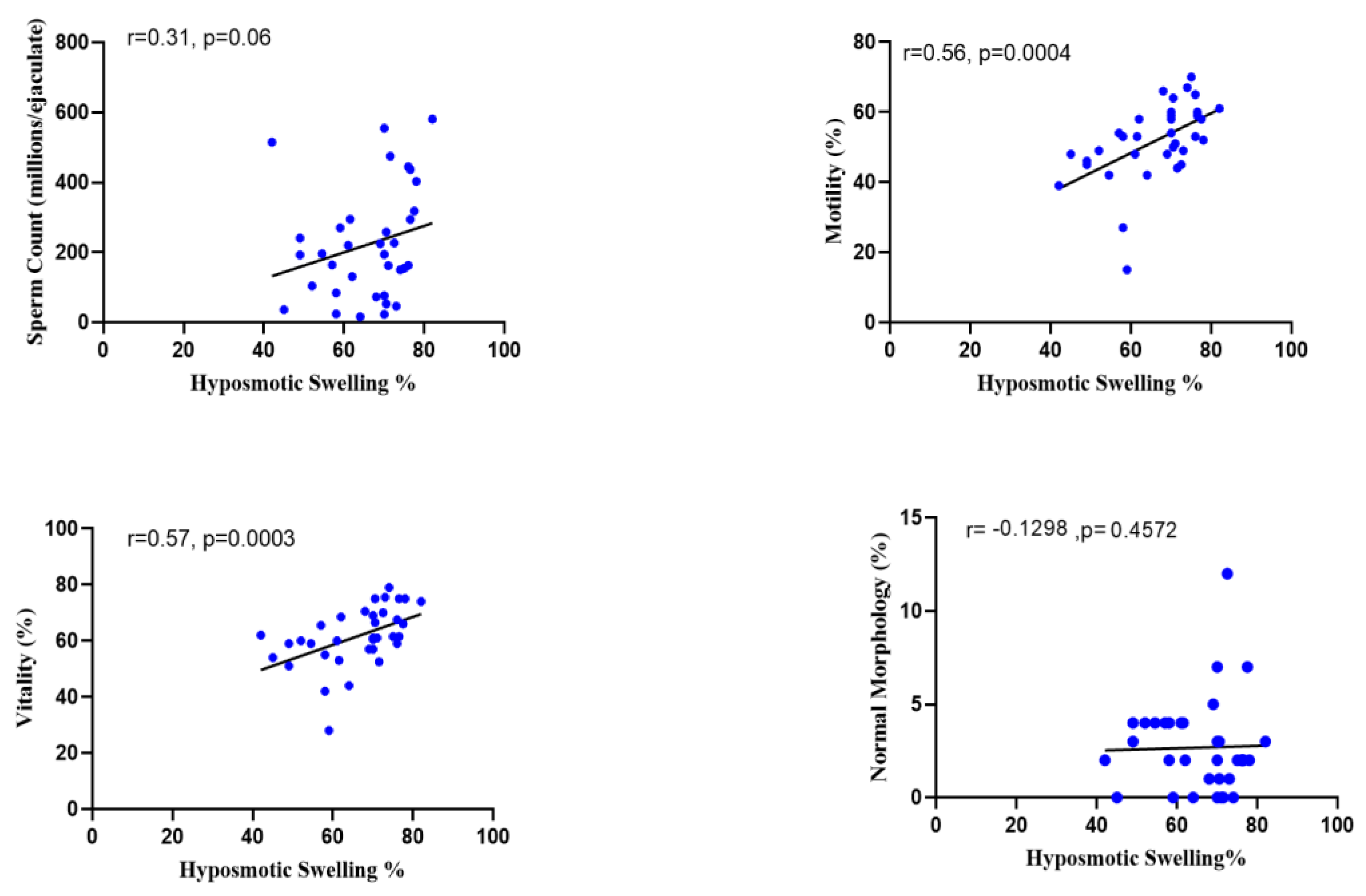
- HOS: Positively and significantly correlated with motility (r = 0.56; p = 0.0004) and vitality (r = 0.57; p = 0.0003). It showed no significant correlation with morphology or sperm count (r = 0.12; p = 0.45; r = 0.31; p = 0.06) (Figure 7).
Figure 5.
Correlation analysis of NCDT with semen parameters, showing significant relationships with vitality and morphology.
Figure 6.
Correlation analysis of DFI with semen parameters, indicating significant inverse relationships with motility and vitality.
Figure 7.
Correlation analysis of HOS with semen parameters, indicating significant relationships with motility and vitality.
4. Discussion
Male fertility displays a complex interplay of genetic, environmental, and physiological factors. Understanding limitations pertaining to diagnostics and comprehending sperm function are critical in improving infertility management. Even after advancements, there are gaps which cannot be ignored for understanding the complex molecular interactions underlying fertilization. Advance usage of predictive and sensitive diagnostic tests needs to be carried out, taking into consideration the ultimate goal of detecting molecular defects in sperm function and tailoring therapies accordingly.
Our findings indicate that the infertile group has a higher incidence of morphological defects, averaging 98.54%, as compared to 94.64% in the unexplained male infertility group. Our findings are consistent with the previous literature that highlights the important role of sperm morphology in male infertility assessment and diagnosis [44,45,46]. Furthermore, the higher occurrence of these defects in the infertile group aligns with data in previous studies that show that morphological abnormalities, particularly sperm head defects, can impair fertilization capacity, emphasizing the continued relevance of this parameter in clinical evaluation [47]. Our results reinforce the idea that even subtle differences in morphology can have major implications on reproductive success. Although conventional sperm morphology differed significantly between the two cohorts, the Teratozoospermia Index (TZI) remained comparable, indicating that the average number of defects per abnormal spermatozoon was similar between infertile and unexplained male infertility individuals. The TZI reflects the structural complexity of sperm abnormalities rather than the proportion of abnormal sperm itself. Therefore, the absence of a significant difference suggests that the severity of multiple morphological deformities within individual spermatozoa may not independently distinguish unexplained male infertility from classical male-factor infertility. Previous studies have similarly reported that although abnormal sperm morphology is associated with impaired fertility potential, indices assessing morphological defect burden such as the TZI or the sperm deformity index demonstrate limited discriminatory capacity when evaluated independently of functional sperm parameters and DNA integrity assessments [48,49,50]. These findings further support the possibility that intracellular sperm dysfunction, particularly DNA fragmentation and chromatin instability, may contribute more substantially to unexplained infertility than morphology severity alone. In spite of the differences observed in morphology between the two groups, both groups showed very similar sperm motility and vitality. This finding highlights the commonly faced challenge in male infertility diagnosis wherein sperm motility and vitality, which are undeniably crucial for fertilizing capacity [51,52], have similar values between groups, indicating that these conventional parameters alone may not fully explain the observed infertility. Our results reinforce the need to incorporate advanced sperm function tests to identify the underlying causes of infertility, especially when conventional parameters appear to be in the normal range [53]. Sperm chromatin decondensation is vital for the formation of male pronuclei—a critical stage in fertilization. It involves both the loss of zinc from sperm and disulphide reduction by glutathione reductase present in oocytes [54,55]. In vitro decondensed sperms are characterized by swollen heads, and this is achieved by SDS and EDTA [56]. A notable percentage in both cohorts exhibited limited ability for decondensation, indicating improper packaging of chromatin. In total, 27% of the unexplained male infertility group and 60% of the infertile group exhibited low decondensation capacity. Our results align with previous studies which link inadequate decondensation capacity with male infertility [57].
Inadequate decondensation has been associated with poor embryonic prognosis in assisted reproductive technology [58]. Research also indicates that proper sperm chromatin condensation is decisive for the fertilization ability of sperm [59] and improper decondensation disrupts the embryo development process [60]. Therefore, our results, wherein 60% of the infertile group exhibit inadequate decondensation capacity, align with these studies, indicating a link between poor decondensation capacity and fertility issues. While 60% of the infertile group in our study exhibited inadequate decondensation capacity, the remaining 40% where this factor was not contributing suggest that other mechanisms are at play, highlighting the multifactorial nature of male infertility. In these cases, high sperm DNA fragmentation may be the primary driver, as proper chromatin packaging does not always guarantee DNA integrity. This fragmentation is frequently driven by excessive reactive oxygen species production and subsequent oxidative stress, alongside chromosomal abnormalities, gene mutations, or epigenetic alterations [61,62]. Our results also indicate that some proportion of the UMI group (27%) exhibits inadequate decondensation capacity, but it becomes more pronounced and a significant contributing factor in infertile individuals. These findings highlight the need to use the NCDT as a diagnostic marker which can help in identifying specific nuclear defects contributing to infertility [53]. Indeed, 82% of our UMI group exhibited increased DFI compared to only 36% of the infertile cohort, providing a potential explanation for their inability to conceive despite having normal conventional parameters. Beyond oxidative stress, this DNA damage may also originate from apoptosis during spermatogenesis, defective chromatin remodeling during spermiogenesis, or post-testicular damage occurring during epididymal transit [63]. The results of the HOS test in both groups were within the anticipated range, indicating that the functional integrity of the plasma membrane was maintained in our study participants. Both HOS and eosin–nigrosine staining are used to assess sperm viability, but they work on different principles. Eosin–nigrosine tells us about the structural integrity of the membrane and follows the dye exclusion principle where nonviable spermatozoa exhibit deficient cell membrane structures, permitting eosin to enter the cells and stain them, whereas viable spermatozoa resist the dyes and remain unstained [64]. In contrast, the HOS test tells us about the functional integrity of the membrane. When sperm are placed in a hypotonic medium, those with functional and intact membrane will swell and exhibit a characteristic tail coiling to achieve osmotic equilibrium. Because the HOS test does not use any dye, it has an added advantage over the eosin–nigrosine test in terms of the selection of viable immotile sperm for ART techniques like ICSI [64].
A positive correlation was found between HOS and vitality (r = 0.57), which is consistent with previous results and indicates that the sperm population assessed in our study possesses membranes that are both structurally and functionally intact [65]. An intact plasma membrane is important for maintaining both its structural integrity and its ability to withstand osmotic changes. Therefore, the positive correlation we found between these two parameters reinforces the idea that both can be used as indicators of sperm health. As expected, both the NCDT and HOS showed positive correlations with all semen parameters, as both tests evaluate sperm functional integrity and better functional integrity generally correlates to better overall semen parameters; they also evaluate the decondensation capacity of sperm, an important step for fertilization [66]. Sperm with proper chromatin packaging exhibits overall good morphology, motility and vitality [67]. The HOS test assesses the functional integrity of the sperm plasma membrane. Our data is supported by a previous study which states that membrane integrity is linked to overall higher semen quality [68].
Our results indicate a negative correlation between the DFI and vitality (r = −0.45). Our findings are supported by the previous literature which found an inverse relationship between sperm DNA fragmentation and vitality [69,70]. High levels of sperm DNA fragmentation have also been correlated with reduced sperm viability, impaired fertilization, poor embryo development, recurrent pregnancy loss, and lower success rates in assisted reproductive technologies [71]. The findings of our study and previous studies support the notion that compromised DNA integrity often leads to decreased sperm vitality, thus impacting overall fertilization potential.
The DFI showed negative correlations with all semen parameters (motility, count, morphology and vitality), as expected and as supported by previous studies [69,70]. Increased sperm DNA fragmentation compromises the sperm function and thus leads to reduced semen parameters. A recent study shows that the DFI is considered an important tool for the diagnosis of male infertility, providing insights beyond routine semen analysis [72]. Increased DFI levels have also been identified as one of the factors for unexplained recurrent spontaneous abortion and are also negatively correlated with sperm viability, as seen in our study [73]. While increased DFI is known to affect the semen parameters, its direct association with IVF success rates is still an active area of research [74]. TUNEL, the Sperm Chromatin Dispersion Test, the Sperm Chromatin Structure Assay and the comet assay are the various DFI assays available, each with its own advantages and disadvantages [75]. DFI tests have high sensitivity (79%) and specificity (86%) for the detection of infertility, and they also have higher accuracy than conventional methods for sperm evaluation [76]. The novelty of this research lies in the integrated application of functional testing to a strictly defined cohort of unexplained male infertility. While these individuals meet standard WHO 2021 criteria, our comparative analysis identifies underlying functional defects—specifically in DNA integrity and chromatin decondensation—that are frequently missed by conventional analysis. Furthermore, we demonstrate that specialized diagnostic insights can be achieved using resource-efficient assays. By utilizing accessible methodologies rather than complex molecular equipment, this study provides a practical diagnostic framework for standard laboratories to improve their evaluation of male-factor infertility beyond basic semen analysis.
Study Limitations: This study has two primary limitations. A key limitation of this study is the absence of a proven fertile control group, which prevents the establishment of a population-specific ‘normal’ baseline for sperm functional parameters. While we utilized established thresholds based on the literature—such as DFI > 20% and HOS > 50%—these benchmarks may not fully account for the physiological variations unique to our demographic. Consequently, our findings are best generalized as a method for identifying functional defects within infertile populations rather than defining absolute diagnostic cut-offs for the general public. For instance, the significant gap in DFI between the unexplained group (82%) and the known infertile group (36%) clearly identifies DNA damage as a major factor in unexplained cases. However, the predictive power of these values for achieving natural conception still needs to be validated against a fertile benchmark. Future large-scale studies including fertile controls will be essential to transition these functional tests into a reliable, population-wide diagnostic tool. Another limitation of this study is the relatively small sample size (n = 35), as it was designed as an exploratory pilot investigation. However, a posthoc assessment of our statistical power confirms the robustness of the primary findings. With n = 35 and p = 0.05, the study was sufficiently powered (80%) to detect moderate-to-strong correlations (r ≥ 0.45) [77]. This is reflected in our significant findings, such as the strong correlations observed between HOS and motility (r = 0.56) and vitality (r = 0.57), and between the NCDT and morphology (r = 0.53), as well as the significant inverse relationship between the DFI and vitality (r = −0.45). While this study may have been underpowered to detect weaker associations, such as that between the DFI and sperm count (r = −0.17), the current results provide the essential foundational data and effect size estimates required to power future large-scale clinical validation studies [77].
5. Conclusions
In conclusion, these results suggest that sperm function tests provide a more com-prehensive assessment of male fertility than standard semen analysis alone. While our findings indicate their potential diagnostic value, further large-scale studies are required before these tests can be recommended for routine clinical use.
Supplementary Materials
The following supporting information can be downloaded at: https://www.mdpi.com/article/10.3390/reprodmed7030028/s1, Figure S1: Positive and negative controls used for DFI analysis demonstrating halo formation patterns following denaturation and lysis treatment conditions.
Author Contributions
Conceptualization, M.S.; methodology, A.T., T.G. and A.G.; formal analysis, A.T., M.S. and N.D.; data curation, A.T. and N.D.; investigation, A.T., B.S., T.G. and A.G.; Writing, A.T. and T.G.; final manuscript review, M.S.; supervision, M.S. and A.H. Infrastructure. A.H.; Patient selection, J.B.; All authors have read and agreed to the published version of the manuscript.
Funding
This research was funded by the Indian Council of Medical Research (ICMR), Grant No. I-1452.
Institutional Review Board Statement
The study was conducted in accordance with the Declaration of Helsinki and approved by the Institute Human Ethics Committee of the All India Institute of Medical Sciences (AIIMS), New Delhi (IEC-406/06.05.2022).
Informed Consent Statement
Written informed consent was obtained from all participants involved in the study.
Data Availability Statement
The data presented in this study are available on reasonable request from the corresponding author. The data are not publicly available due to ethical and privacy restrictions.
Acknowledgments
The authors would like to sincerely thank Pradeep, Arokiadas and other members of Andrology team for their support in semen analysis and patient recruitment. We also extend our special thanks to Kajal Sihag for her assistance with microscopy work.
Conflicts of Interest
The authors declare no conflicts of interest.
Abbreviations
The following abbreviations are used in this manuscript:
| ART | Assisted Reproductive Technology |
| DFI | DNA Fragmentation Index |
| HOS | Hypoosmotic Swelling |
| IEC | Institutional Ethics Committee |
| NCDT | Nuclear Chromatin Decondensation Test |
| WHO | World Health Organization |
References
- Björndahl, L.; Brown, J.K. The sixth edition of the WHO Laboratory Manual for the Examination and Processing of Human Semen: Ensuring quality and standardization in basic examination of human ejaculates. Fertil. Steril. 2022, 117, 246–251. [Google Scholar] [CrossRef] [Scilit] [PubMed]
- Agarwal, A.; Mulgund, A.; Hamada, A.; Chyatte, M.R. A unique view on male infertility around the globe. Reprod. Biol. Endocrinol. 2015, 13, 1–9. [Google Scholar] [CrossRef] [Scilit]
- Ankur Sharma, M.; Halder, A.; Dada, R.; Mahey, R.; Kumar, N. Effect of Oxidative Stress on Sperm TERRA Expression and Chromatin Function in Infertile Males: A Preliminary Finding. J. Sci. Res. Rep. 2024, 30, 947–959. [Google Scholar] [CrossRef] [Scilit]
- John, A.; Sharma, M.; Dada, R.; Mahey, R.; Halder, A.; Pandey, D. Effect of Oxidative Stress on Sperm Shelterin TRF1 Expression and Chromatin Function in Infertile Males: Preliminary Findings. Glob. J. Reprod. Med. 2024, 10, 555800. [Google Scholar] [CrossRef]
- Dipankar, S.P.; Kumar, T.; Begum, H.A.; Itagi Naik, B.N.; Kumar, Y.; Sharma, M.; Asim Sarfaraz, A.; Kumari, A. Semen Quality in Males Suffering from COVID-19: A Pilot Study. Cureus 2022, 14, e31776. [Google Scholar] [CrossRef] [Scilit] [PubMed]
- Asuri, K.; Mohammad, A.; Prajapati, O.M.; Sagar, R.; Kumar, A.; Sharma, M.; Chaturvedi, P.K.; Gupta, S.V.; Rai, S.K.; Misra, M.C.; et al. A prospective randomized comparison of sexual function and semen analysis following laproscopic totally extraperitoneal inguinal hernia repair. Surg. Endosc. 2021, 35, 2936–2941. [Google Scholar] [CrossRef] [Scilit] [PubMed]
- Sharma, M.; Gupta, S.; Dhole, B.; Kumar, A. The Prostate gland. In Basics of Human Andrology, 1st ed.; Kumar, A., Sharma, M., Eds.; Springer Nature: Singapore, 2017; pp. 17–35. [Google Scholar]
- Gupta, S.; Sharma, M.; Kumar, A. Seminal Vesicles. In Basics of Human Andrology, 1st ed.; Kumar, A., Sharma, M., Eds.; Springer Nature: Singapore, 2017; pp. 37–45. [Google Scholar]
- Sharma, M.; Kumar, A. Neurovascular supply and lymphatic drainage of male reproductive organs. In Basics of Human Andrology, 1st ed.; Kumar, A., Sharma, M., Eds.; Springer Nature: Singapore, 2017; pp. 55–65. [Google Scholar]
- Sharma, M.; Kumar, A. The Sperm. In Basics of Human Andrology, 1st ed.; Kumar, A., Sharma, M., Eds.; Springer Nature: Singapore, 2017; pp. 171–203. [Google Scholar]
- Lipshultz, L.I.; Howards, S.S.; Niederberger, C.S. (Eds.) Infertility in the Male; Cambridge University Press: Cambridge, UK, 2009. [Google Scholar]
- Barratt, C.L. Semen analysis is the cornerstone of investigation for male infertility. Practitioner 2007, 251, 8–17. [Google Scholar] [PubMed]
- Goyal, R.; Kotru, M.; Gogia, A.; Sharma, S. Qualitative defects with normal sperm counts in a patient attending infertility clinic. Indian. J. Pathol. Microbiol. 2018, 61, 233–235. [Google Scholar] [CrossRef] [Scilit] [PubMed]
- Barbăroșie, C.; Agarwal, A.; Henkel, R. Diagnostic value of advanced semen analysis in evaluation of male infertility. Andrologia 2021, 53, e13625. [Google Scholar] [PubMed]
- Barroso, G.; Mercan, R.; Ozgur, K.; Morshedi, M.; Kolm, P.; Coetzee, K.; Kruger, T.; Oehninger, S. Intra-and inter-laboratory variability in the assessment of sperm morphology by strict criteria: Impact of semen preparation, staining techniques and manual versus computerized analysis. Human. Reprod. 1999, 14, 2036–2040. [Google Scholar] [CrossRef] [Scilit]
- Moazzam, A.; Choudhary, M.N.; Muhammad, I.; Sarwat, J.; Ijaz, A. From basic to contemporary semen analysis: Limitations and variability. J. Anim. Plant Sci. 2015, 25, 328–336. [Google Scholar]
- Brazil, C. Practical semen analysis: From A to Z. Asian J. Androl. 2010, 12, 14. [Google Scholar] [CrossRef] [Scilit] [PubMed]
- Guzick, D.S.; Overstreet, J.W.; Factor-Litvak, P.; Brazil, C.K.; Nakajima, S.T.; Coutifaris, C.; Carson, S.A.; Cisneros, P.; Steinkampf, M.P.; Hill, J.A.; et al. Sperm morphology, motility, and concentration in fertile and infertile men. N. Engl. J. Med. 2001, 345, 1388–1393. [Google Scholar] [CrossRef] [Scilit] [PubMed]
- Smith, K.D.; Rodriguez-Rigau, L.J.; Steinberger, E. Relation between indices of semen analysis and pregnancy rate in infertile couples. Fertil. Steril. 1977, 28, 1314–1319. [Google Scholar] [CrossRef] [Scilit] [PubMed]
- Pandruvada, S.; Royfman, R.; Shah, T.A.; Sindhwani, P.; Dupree, J.M.; Schon, S.; Avidor-Reiss, T. Lack of trusted diagnostic tools for undetermined male infertility. J. Assist. Reprod. Genet. 2021, 38, 265–276. [Google Scholar] [CrossRef] [Scilit] [PubMed]
- Henkel, R.; Panner Selvam, M.K.; Ambar, R.F. Etiologies of sperm DNA damage and its impact on male infertility. Andrologia 2021, 53, e13706. [Google Scholar] [CrossRef] [Scilit] [PubMed]
- Avidor-Reiss, T.; Carr, A.; Fishman, E.L. The sperm centrioles. Mol. Cell. Endocrinol. 2020, 518, 110987. [Google Scholar] [CrossRef] [Scilit] [PubMed]
- Tholiya, S.; Kumari, A.; Mahey, R.; Kalaivani, M.; Sati, H.C.; Sharma, M.; Jayraj, A.K.; Malhotra, N. Evaluation of predictors of response to ovulation induction using letrozole in women with polycystic ovary syndrome: A prospective cohort study. J. Hum. Reprod. Sci. 2024, 17, 240–245. [Google Scholar] [CrossRef] [Scilit] [PubMed]
- Sihag, K.; Sharma, M.; Halder, A.; Sharma, J.B.; Ara, S. Mitochondria-Endoplasmic Reticulum Cross Talk in Premature Ovarian Insufficiency. J. Reprod. Med. Gynecol. Obstet. 2024, 9, 165. [Google Scholar] [CrossRef] [Scilit]
- Halder, A.; Kumar, H.; Sharma, M.; Jain, M.; Kaur, A.K.; Pandey, S. Serum anti-Müllerian hormone: A potential biomarker for polycystic ovary syndrome. Indian. J. Med. Res. 2023, 158, 1–10. [Google Scholar] [CrossRef] [Scilit]
- Kumar, H.; Halder, A.; Sharma, M.; Jain, M.; Kalsi, A.K. Dihydrotestosterone- A Potential Biomarker of Hyperandrogenaemia in Polycystic Ovary Syndrome: A Case-control Study from North India. J. Clin. Diagn. Res. 2022, 16, QC09–QC14. [Google Scholar] [CrossRef] [Scilit]
- Sharma, M.; Sihag, K.; Halder, A.; Sharma, J.B. Premature Ovarian Insufficiency in Adolescents. J. Obstet. Gynecol. 2023, 13. [Google Scholar]
- Rishi, I.; Halder, A.; Sharma, J.; Jain, M.; Sharma, M. Single Strand Conformation Polymorphism and Sequencing of HS6ST2 Gene in Patients of Idiopathic Premature Ovarian Failure. J. Clin. Diagn. Res. 2020, 14, GC01–GC08. [Google Scholar] [CrossRef] [Scilit]
- Sharma, M.; Kumar, R.; Bhatla, N.; Dhingra, R. A Comparative study of apoptosis in placentas of normal and preeclamptic Indian pregnant women by TUNEL assay and M30 immunostaining. J. Clin. Lab. Anal. 2012, 26, 459–466. [Google Scholar] [CrossRef] [Scilit] [PubMed]
- Sharma, M.; Kumar, R.; Dhingra, R. An immunohistochemical study of the syncytial knots in the preeclamptic placentas. Int. J. Pharm. Res. Biosci. 2012, 1, 228–239. [Google Scholar] [CrossRef] [Scilit]
- Sharma, M.; Kumar, R.; Dhingra, R. Apoptosis: Searching for the detection techniques. Int. J. Biol. Med. Res. 2012, 3, 2172–2175. [Google Scholar] [CrossRef] [Scilit]
- Alaee, S.; Khodabandeh, Z.; Dara, M.; Elham, H.; Sharma, M. Curcumin Mitigates Acrylamide-Induced Ovarian Antioxidant Disruption and Apoptosis in Female Balb/c Mice: A Comprehensive Study on Gene and Protein Expressions. Food Sci. Nutr. 2024, 12, 4160–4172. [Google Scholar] [CrossRef] [Scilit] [PubMed]
- Rana, R.; Halder, A.; Gupta, S.; Sharma, M. An invitro analysis of L-Carnitine mediated rescue of TNF-α induced apoptosis in mice oocytes. Int. J. Clin. Biochem. Res. 2022, 9, 218–223. [Google Scholar] [CrossRef] [Scilit]
- Rana, R.; Sharma, M. A Timeline Morphological Study of TNF-α Induced Changes in Mice Oocytes. J. Infertil. Reprod. Biol. 2018, 6, 16–18. [Google Scholar] [CrossRef] [Scilit]
- Sihag, K.; Halder, A.; Sharma, M. Metabolic markers in male infertility: A pilot study. J. Men’s Health 2024, 20, 138–145. [Google Scholar] [CrossRef] [Scilit]
- Sharma, Y.; Sharma, M.; Halder, A.; Mahey, R.; Kumar, N.; Dipankar, S. Temporal analysis of cryopreservation effects on human sperm phospholipase C zeta expression profile: Laboratory preliminary findings. J. Lab. Physicians 2024, 16, 328–337. [Google Scholar] [CrossRef] [Scilit]
- Joginder Halder, A.; Sharma, J.B.; Mahey, R.; Gupta, S.; Sharma, M. A preliminary study of human sperm citrate synthase expression in patients with failed ICSI cycles. J. Res. Biol. 2020, 10, 001–011. [Google Scholar]
- Talwar, P.; Hayatnagarkar, S. Sperm function test. J. Hum. Reprod. Sci. 2015, 8, 61–69. [Google Scholar] [CrossRef] [Scilit] [PubMed]
- Gopalkrishnan, K.; Hinduja, I.N.; Anand Kumar, T.C. In vitro decondensation of nuclear chromatin of human spermatozoa: Assessing fertilizing potential. Arch. Androl. 1991, 27, 43–50. [Google Scholar] [CrossRef] [Scilit] [PubMed]
- Osman, A.; Alsomait, H.; Seshadri, S.; El-Toukhy, T.; Khalaf, Y. The effect of sperm DNA fragmentation on live birth rate after IVF or ICSI: A systematic review and meta-analysis. Reprod. Biomed. Online 2015, 30, 120–127. [Google Scholar] [CrossRef] [Scilit] [PubMed]
- Fernández, J.L.; Muriel, L.; Rivero, M.T.; Goyanes, V.; Vazquez, R.; Alvarez, J.G. The sperm chromatin dispersion test: A simple method for the determination of sperm DNA fragmentation. J. Androl. 2003, 24, 59–66. [Google Scholar] [CrossRef] [Scilit] [PubMed]
- Oleszczuk, K.; Augustinsson, L.; Bayat, N.; Giwercman, A.; Bungum, M. Prevalence of high DNA fragmentation index in male partners of unexplained infertile couples. Andrology 2013, 1, 357–360. [Google Scholar] [PubMed]
- Malm, G.; Rylander, L.; Giwercman, A.; Haugen, T.B. Association between semen parameters and chance of fatherhood-a long-term follow-up study. Andrology 2019, 7, 76–81. [Google Scholar] [PubMed]
- Moretti, E.; Signorini, C.; Noto, D.; Corsaro, R.; Collodel, G. The relevance of sperm morphology in male infertility. Front. Reprod. Health 2022, 4, 945351. [Google Scholar] [CrossRef] [Scilit] [PubMed]
- Oehninger, S.; Kruger, T.F. Sperm morphology and its disorders in the context of infertility. FS Rev. 2021, 2, 75–92. [Google Scholar] [CrossRef] [Scilit]
- Fu, L.; Zhou, F.; Chen, G.; Yuan, R.; Li, W.; Qiu, S.; Tang, L.; Liu, W.; Gu, Y.; Lu, W. Morphological Parameters of 29994 sperm in a fertile male population—Based on Papanicolaou staining and SSA-II Plus. Front. Endocrinol. 2025, 16, 1546290. [Google Scholar] [CrossRef] [Scilit]
- Deventhiran, R.A.; Ramanathan, K.U.; Nandakumar, N.A. Prevalance of male infertility in India: Studies on the effects of gonadotropin releasing hormones. Asian J. Pharm. Clin. Res. 2017, 10, 208–210. [Google Scholar] [CrossRef] [Scilit]
- Kruger, T.F.; Acosta, A.A.; Simmons, K.F.; Swanson, R.J.; Matta, J.F.; Oehninger, S. Predictive value of abnormal sperm morphology in in vitro fertilization. Fertil. Steril. 1988, 49, 112–117. [Google Scholar] [CrossRef] [Scilit] [PubMed]
- Menkveld, R.; Wong, W.Y.; Lombard, C.J.; Wetzels, A.M.; Thomas, C.M.; Merkus, H.M.; Steegers-Theunissen, R.P. Semen parameters, including WHO and strict criteria morphology, in a fertile and subfertile population: An effort towards standardization of in-vivo thresholds. Hum. Reprod. 2001, 16, 1165–1171. [Google Scholar] [CrossRef] [Scilit] [PubMed]
- Aziz, N.; Buchan, I.; Taylor, C.; Kingsland, C.R.; Lewis-Jones, I. The sperm deformity index: A reliable predictor of the outcome of oocyte fertilization in vitro. Fertil. Steril. 1996, 66, 1000–1008. [Google Scholar] [CrossRef] [Scilit] [PubMed]
- Ameen, E.M.; Sabir, F.N.; Mohammed, S.I. Relationships between semen quality and fertility in a population of infertile men in Erbil city. Cell. Mol. Biol. 2022, 68, 63–68. [Google Scholar] [CrossRef] [Scilit] [PubMed]
- Homonnai, Z.T.; Paz, G.; Weiss, J.N.; David, M.P. Quality of semen obtained from 627 fertile men. Int. J. Androl. 1980, 3, 217–228. [Google Scholar] [CrossRef] [Scilit] [PubMed]
- Sanyal, D.; Arya, D.; Nishi, K.; Balasinor, N.; Singh, D. Clinical utility of sperm function tests in predicting male fertility: A systematic review. Reprod. Sci. 2024, 31, 863–882. [Google Scholar] [PubMed]
- Kvist, U. Importance of spermatozoal zinc as temporary inhibitor of sperm nuclear chromatin decondensation ability in man. Acta Physiol. Scand. 1980, 109, 79–84. [Google Scholar] [CrossRef] [Scilit] [PubMed]
- Huret, J.L. Variability of the chromatin decondensation ability test on human sperm. Arch. Androl. 1983, 11, 1–7. [Google Scholar] [CrossRef] [Scilit] [PubMed]
- Calvin, H.I.; Bedford, J.M. Stimulation of actinomycin-D-binding to eutherian sperm chromatin by reduction of disulphide bonds. J. Reprod. Fertil. 1974, 36, 225–229. [Google Scholar] [CrossRef] [Scilit] [PubMed]
- Menezo, Y.; Clement, P.; Amar, E. Evaluation of sperm DNA structure, fragmentation and decondensation: An essential tool in the assessment of male infertility. Transl. Androl. Urol. 2017, 6, S553. [Google Scholar] [CrossRef] [Scilit] [PubMed]
- Alfred, Y.; Simon, A.; Bruno, A.; Patrice, D.; Boula, S.S.; Jean-Paul, D.; Gangbo, F.; Anatole, L. Assessment of Spermatic Chromatin Decondensation by the Toluidine Blue Assay in Infertile Patients in Cotonou. Adv. Reprod. Sci. 2022, 10, 37–48. [Google Scholar] [CrossRef]
- Llavanera, M.; Ribas-Maynou, J.; Delgado-Bermúdez, A.; Recuero, S.; Muiño, R.; Hidalgo, C.O.; Tamargo, C.; Bonet, S.; Mateo-Otero, Y.; Yeste, M. Sperm chromatin condensation as an in vivo fertility biomarker in bulls: A flow cytometry approach. J. Anim. Sci. Biotechnol. 2021, 12, 115. [Google Scholar] [CrossRef] [Scilit] [PubMed]
- Jumeau, F.; Rives, N.; Lechevallier, P.; Boniou, C.; Letailleur, M.; Réal-Lhommet, A.; Feraille, A. Sperm chromatin condensation defect accelerates the kinetics of early embryonic development but does not modify ICSI outcome. Int. J. Mol. Sci. 2022, 24, 393. [Google Scholar] [CrossRef] [Scilit] [PubMed]
- Marinaro, J.A.; Schlegel, P.N. Sperm DNA damage and its relevance in fertility treatment: A review of recent literature and current practice guidelines. Int. J. Mol. Sci. 2023, 24, 1446. [Google Scholar] [CrossRef] [Scilit] [PubMed]
- Rashki Ghaleno, L.; Alizadeh, A.; Drevet, J.R.; Shahverdi, A.; Valojerdi, M.R. Oxidation of sperm DNA and male infertility. Antioxidants 2021, 10, 97. [Google Scholar] [CrossRef] [Scilit] [PubMed]
- Andreu-Noguera, J.; López-Botella, A.; Sáez-Espinosa, P.; Gómez-Torres, M.J. Epigenetics role in spermatozoa function: Implications in health and evolution—An overview. Life 2023, 13, 364. [Google Scholar] [CrossRef] [Scilit] [PubMed]
- Katkov, I. (Ed.) Current Frontiers in Cryobiology; InTech: Singapore, 2012. [Google Scholar] [CrossRef] [Scilit] [PubMed]
- Ramu, S.; Jeyendran, R.S. The hypo-osmotic swelling test for evaluation of sperm membrane integrity. In Spermatogenesis: Methods and Protocols; Humana Press: Totowa, NJ, USA, 2012; pp. 21–25. [Google Scholar]
- Vladimirov, I.K. (Ed.) IVF Technologies and Infertility—Current Practices and New Perspectives; IntechOpen: London, UK, 2023. [Google Scholar] [CrossRef] [Scilit]
- Hekmatdoost, A.; Lakpour, N.; Sadeghi, M.R. Sperm chromatin integrity: Etiologies and mechanisms of abnormality, assays, clinical importance, preventing and repairing damage. Avicenna J. Med. Biotechnol. 2009, 1, 147. [Google Scholar] [PubMed]
- Rajashri, M.; Reddy, K.R.; Kumari, G.A.; Kumari, N.N.; Kesharwani, S. Correlation between Hypo-osmotic swelling test (HOST) and other seminal characteristics of Deccani ram semen. J. Exp. Biol. Agric. Sci. 2017, 5, 195–200. [Google Scholar] [CrossRef] [Scilit]
- Du, C.; Tuo, Y. Correlation of DNA fragments with routine semen parameters and lifestyle and their impact on assisted reproductive outcomes. Rev. Int. Androl. 2023, 21, 100337. [Google Scholar] [CrossRef] [Scilit] [PubMed]
- Samplaski, M.K.; Dimitromanolakis, A.; Lo, K.C.; Grober, E.D.; Mullen, B.; Garbens, A.; Jarvi, K.A. The relationship between sperm viability and DNA fragmentation rates. Reprod. Biol. Endocrinol. 2015, 13, 42. [Google Scholar] [CrossRef] [Scilit] [PubMed]
- Liang, X. Detection and Clinical Application of Sperm DNA Fragmentation. In Andrology Insights-Understanding Male Reproductive Health and Diseases: Understanding Male Reproductive Health and Diseases; IntechOpen: London, UK, 2025; p. 71. [Google Scholar]
- Pozzi, E.; Fallara, G.; Belladelli, F.; Corsini, C.; Raffo, M.; Candela, L.; Schifano, N.; d’Arma, A.; Capogrosso, P.; Boeri, L.; et al. Clinical parameters associated with altered sperm DNA fragmentation index among primary infertile men: Findings from a real-life cross-sectional study. Andrology 2023, 11, 1694–1701. [Google Scholar] [CrossRef] [Scilit] [PubMed]
- Wijaya, R.A.; Kusumaatmaja, A.; Rizal, D.M. Predicting the value of sperm analysis using an electronic nose. Indones. J. Electr. Eng. Comput. Sci. 2022, 28, 174–182. [Google Scholar] [CrossRef] [Scilit]
- Lin, M.; Zhang, Y.; Wang, H.; Wang, Y.; Wang, Y.; Feng, N.; He, Q. Multivariate analyses on male factors and construction of a nomogram for predicting low in vitro fertilization rate. Heliyon 2024, 10, e29271. [Google Scholar] [CrossRef] [Scilit] [PubMed]
- Hallak, J. Utility of sperm DNA fragmentation testing in different clinical scenarios of male reproductive abnormalities and its influence in natural and assisted reproduction. Transl. Androl. Urol. 2017, 6, S509. [Google Scholar] [CrossRef] [Scilit] [PubMed]
- Tanga, B.M.; Qamar, A.Y.; Raza, S.; Bang, S.; Fang, X.; Yoon, K.; Cho, J. Semen evaluation: Methodological advancements in sperm quality-specific fertility assessment—A review. Anim. Biosci. 2021, 34, 1253. [Google Scholar] [CrossRef] [Scilit] [PubMed]
- Kunselman, A.R. A brief overview of pilot studies and their sample size justification. Fertil. Steril. 2024, 121, 899–901. [Google Scholar] [CrossRef] [Scilit] [PubMed]

Disclaimer/Publisher’s Note: The statements, opinions and data contained in all publications are solely those of the individual author(s) and contributor(s) and not of MDPI and/or the editor(s). MDPI and/or the editor(s) disclaim responsibility for any injury to people or property resulting from any ideas, methods, instructions or products referred to in the content. |
© 2026 by the authors. Licensee MDPI, Basel, Switzerland. This article is an open access article distributed under the terms and conditions of the Creative Commons Attribution (CC BY) license.