


Abstract
Background/Objectives: Myocardial bridging is a common anatomic variant in which an epicardial coronary artery takes an intramyocardial course. A true intracameral, or intracavitary, coronary course is rare. On coronary CT angiography (CTA), multiplanar and curved planar reformations answer most of these questions, but some cases stay equivocal because of partial-volume effects, cardiac motion, and loss of depth cues. A vessel next to a thin or fat-infiltrated wall can also mimic an intracameral course. Cinematic rendering (CR) is a photorealistic three-dimensional post-processing method that uses Monte Carlo path tracing with global illumination to reproduce natural depth and soft-tissue shading. Methods: In this retrospective, single-center, four-patient case series, we describe patients in whom CR was used alongside standard two-dimensional and reformatted CTA after the planar reconstructions had been read as equivocal. The cases were purposively selected as illustrative examples rather than consecutively enrolled, and the report is intended to be hypothesis-generating. Results: In two patients with left anterior descending (LAD) myocardial bridging, CR showed the tunneled segment with depth and was useful for discussion with referring clinicians and for teaching. In two patients whose coronary segment looked intracameral on planar images, CR showed intact overlying myocardium, which favored an intramyocardial course over chamber entry. Conclusions: In these selected patients, CR did not add anatomic information that was unavailable on expert reformations. It appeared to improve perceived depth and reader confidence in these equivocal cases and was useful for communication and teaching; no quantitative image analysis or formal reader study was performed. Because this is a small, retrospective case series without a comparison group, these observations are illustrative and hypothesis-generating and cannot be extrapolated to coronary CTA in general; prospective comparative studies are needed to determine whether CR adds diagnostic value over standard reconstructions.
1. Introduction
In myocardial bridging, a normally epicardial coronary artery runs through the myocardium, most often in the mid-left anterior descending artery (LAD) [1]. Reported prevalence depends heavily on detection method, ranging from roughly 15 to 85% at autopsy to about 5 to 40% on CTA [2]; in a CTA cohort of 1000 patients without obstructive coronary artery disease, 22% had a bridged course with a benign long-term prognosis, so most bridging is incidental [3]. Deep (>2 mm) or long (>25 mm) tunneled segments can nonetheless cause ischemia through systolic compression and delayed diastolic relaxation [4], so depth, length, and location matter mainly in symptomatic or anatomically severe cases.
Standard two-dimensional (2D) CTA, read together with multiplanar reformation (MPR) and curved planar reformation (CPR), confirms most myocardial bridges. In a smaller number of cases the diagnosis stays equivocal, because partial-volume averaging, cardiac motion, and the loss of depth information on planar images can suggest bridging that is not there or hide a true intramyocardial segment [5]. A less common but related problem occurs when a coronary vessel runs near a thin-walled structure such as the right ventricular (RV) free wall. Here, fat infiltration, motion, or poor tissue contrast can make the vessel look intracameral even though it stays within the myocardium [6,7].
This matters because a true intracameral (intracavitary) coronary course is rare: CTA series report an overall prevalence of about 0.36 to 1.3%, most often in the LAD and RCA [8], with an intra-atrial right coronary artery the commonest variant at about 0.15 to 0.29% [9,10] and a true intra-ventricular course rarer still, described mainly in isolated reports and surgical series. These variants are usually benign, but they are worth recognizing before radiofrequency ablation, right-heart catheterization, device implantation, or valve surgery, since an intracavitary vessel can be injured during these procedures [8,11]. Because a true intracameral course is so uncommon, a segment that looks intracameral on 2D CTA is usually an artifact. In practice, the reader is trying to rule out a rare but serious mimic, not to diagnose a common finding.
Cinematic rendering (CR) is a photorealistic three-dimensional (3D) post-processing method. It uses Monte Carlo path tracing with global illumination and material-specific optical properties to reproduce natural shadowing and soft-tissue boundaries [12,13]. Conventional volume rendering (VR) uses simpler local shading, while CR restores depth cues that can help the reader judge overlying tissue planes [14]. In this series we show how CR was used together with standard reformatted CTA in four patients whose planar reconstructions were equivocal for myocardial bridging or for a suspected intracameral segment. We do not claim that CR is needed for routine bridge assessment or that it adds anatomic information beyond expert reformations. Our aim is to show its illustrative and communicative role in selected cases and to point out where formal comparison is still needed.
Three-dimensional visualization of cardiac CT has progressed from GPU-accelerated volume rendering to photorealistic cinematic rendering [15,16], and prior CR work in cardiovascular imaging has largely addressed congenital heart disease, postoperative vascular anatomy, complex aortic disease, and cardiac devices, where the technique conveys spatial relationships more naturally than conventional volume rendering [17,18,19,20,21]. The specific problem addressed here has not, to our knowledge, been examined: coronary CTA studies that remain equivocal after expert multiplanar and curved planar reformation, in which the unresolved question is whether a vessel lies within myocardium or within a cardiac chamber. That is the gap this series addresses, and the question we ask is deliberately narrow—whether the depth cues that CR restores help a reader adjudicate the vessel–myocardium–chamber relationship in such cases. This extends the existing cinematic rendering literature from anatomy that is already conspicuous to anatomy that is genuinely ambiguous, while framing the answer as illustrative rather than diagnostic.
2. Materials and Methods
2.1. Patients, Ethics, and Case Selection
This retrospective series comprises four patients who underwent clinically indicated ECG-gated coronary CTA as part of routine care; no additional imaging or experimental procedures were performed for this report. Under the Columbia University Human Research Protection Office (HRPO) Case Report Policy and the federal definition of human-subjects research at 45 CFR 46.102(L), a report of this nature does not constitute human-subjects research and did not require Institutional Review Board approval or informed consent; all images were de-identified prior to analysis. The corresponding statements appear in the back matter.
Case selection. The four cases were identified retrospectively from clinically indicated coronary CTA examinations interpreted at our institution as part of routine clinical care. They were not a consecutive cohort; they were purposively chosen as instructive examples in which the coronary course remained equivocal after standard interpretation, and this convenience sampling is a source of selection bias (see Section Limitations). Because the cases arose from routine clinical practice rather than from a predefined consecutive series, we are not able to report a screening interval or a denominator of coronary CTA studies reviewed, and no formal screening log was maintained; this limits the transparency of case ascertainment and is acknowledged as such in the Limitations. A study was considered equivocal when the interpreting cardiothoracic radiologist, after reviewing the axial source images together with multiplanar and curved planar reformations and thin-slab maximum-intensity projections, could not confidently distinguish an intramyocardial (bridged) course from an epicardial course or, in Cases 3 and 4, from an intracameral-appearing course, owing to partial-volume averaging, cardiac motion, or indistinct tissue planes.
Image interpretation. For each case the standard two-dimensional and reformatted images were reviewed first and classified as equivocal by the interpreting cardiothoracic radiologist during clinical reporting; cinematic rendering was then reviewed by the study authors with knowledge of the standard reconstructions. Interpretation was therefore sequential and unblinded rather than independent or randomized in presentation order, and no separate blinded or formally adjudicated consensus read was performed. Because the CR images were assessed alongside, rather than independently of, the standard reconstructions, and because reader confidence was recorded qualitatively rather than with a formal rating instrument, the confidence impressions reported here are descriptive; the study design needed to quantify them is outlined in the Discussion.
2.2. CT Acquisition
All four patients had clinically indicated ECG-gated coronary CTA on our standard cardiac protocol. Scans were acquired at a tube voltage of 120 kVp with automated tube-current modulation. Intravenous iodinated contrast (Omnipaque 350, iohexol, sourced from GE HealthCare, Chicago, IL, USA) was administered as a 60 mL bolus followed by a saline chase, with acquisition triggered by region-of-interest (ROI) bolus tracking set to a threshold of 140 HU in the ascending aorta. Thin-section images were reconstructed at 0.6 mm slice thickness for isotropic three-dimensional post-processing and reviewed in mid-diastole.
2.3. Cinematic Rendering Post-Processing
CR was applied to the same thin-section source data used for the standard 2D and conventional 3D reconstructions, so no change to the scan was needed. Rendering was done on Siemens syngo.via (version VB80) with the Cinematic VRT (cVRT) engine (Siemens Healthineers, Forchheim, Germany). For each case we adjusted opacity, windowing, and clip-plane (cut-plane) settings interactively to remove overlying tissue and to show how the vessel related to the myocardium and to the chamber. In Case 2 we used a sequential anterior-to-posterior “front cut plane” to show the tunneled segment at increasing depth. Post-processing took roughly 10 to 30 min per case, including loading, any segmentation, and creating the static and dynamic images. This added time is why CR is best kept for specific questions rather than run on every case. Interactive cinematic rendering also depends on GPU-capable hardware and a workstation licensed for the cVRT engine, neither of which is universal in general cardiac CT practice, and the quality of a rendering depends on operator familiarity with preset selection, opacity, and clip-plane placement rather than on the acquisition itself. In our experience a period of hands-on practice was needed before renderings were reliably informative. These hardware, licensing, and training requirements, together with the added post-processing time, are the main practical constraints on transferring this workflow to other centers and platforms, and are considered further in the Limitations.
2.4. Technical Background
CR handles light and tissue differently from conventional VR. VR uses uniform shading based on local surface gradients and gives few realistic depth cues. Maximum-intensity projection (MIP) makes vessels more conspicuous but flattens 3D information into one plane, so it is hard to judge how deep a vessel sits relative to nearby structures [14]. CR instead gives each voxel optical properties (absorption, scattering, subsurface reflectance) based on its material type and traces many simulated photons through the volume, which produces soft shadows, ambient occlusion, and continuous surfaces [12,13]. For the questions here, this shadowing helps show the boundary between overlying myocardium and a tunneled segment, and it helps separate myocardium from nearby epicardial fat and from the blood pool in the chamber. Monte Carlo path tracing is computationally heavy, so interactive CR is practical only on GPU-accelerated workstations. Lower-end or CPU-only systems render too slowly for interactive cut-plane, windowing, and viewpoint changes [15].
3. Case Series
3.1. Case 1: LAD Myocardial Bridging, Confirmation of a Suspected Tunnel
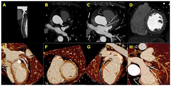
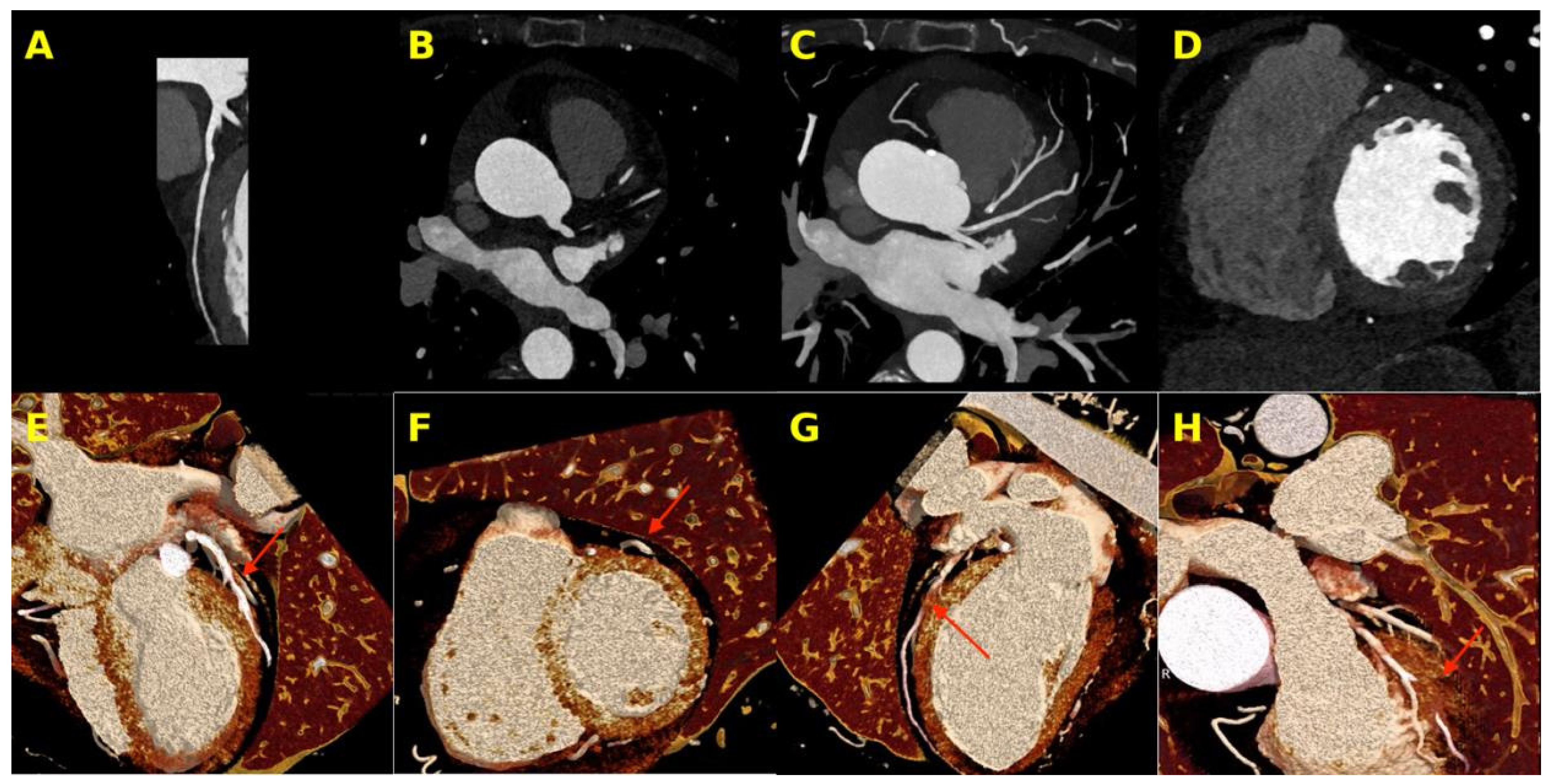
This coronary CTA was done for atypical chest pain. On grayscale 2D images the mid-LAD seemed to dip beneath the left ventricular myocardium, which suggested bridging, but the 3D relationship was hard to pin down on curved planar, axial, and MIP reconstructions. CR restored depth and surface shading and showed the LAD running through an intramyocardial tunnel, which supported bridging rather than a surface impression (Figure 1).
Figure 1.
Grayscale 2D CTA (A–D) and cinematic rendering (E–H) in suspected LAD myocardial bridging. (A–D) Curved planar, axial, axial MIP, and cardiac short-axis images show an apparent dip of the mid-LAD into adjacent myocardium, with limited depth cues. (E–H) CR depicts the LAD within an intramyocardial tunnel (red arrows), best appreciated in panel (H), where the tunnel is seen from the surface, restoring volumetric context lost on planar imaging. Key: LAD, left anterior descending artery; MIP, maximum-intensity projection; red arrows, tunneled segment. What was equivocal: on the planar images (A–D) the apparent dip of the mid-LAD could not be separated with confidence from an epicardial vessel lying against myocardium, because planar reformations carry no depth information. What CR adds: in (E–H) myocardium is resolved above the vessel, establishing a true intramyocardial tunnel rather than a surface groove.
3.2. Case 2: Mid-LAD Intramyocardial Tunnel with Depth-Resolved 3D Depiction
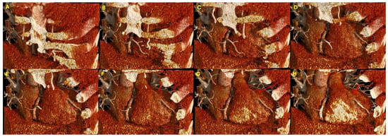
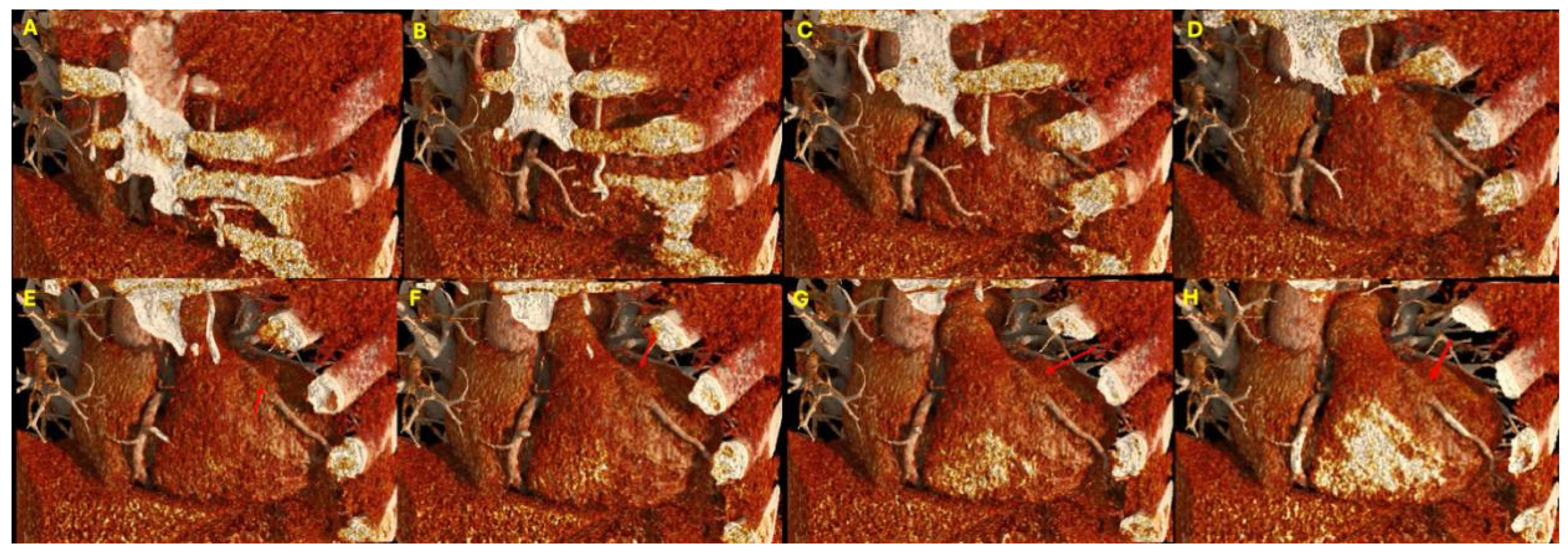
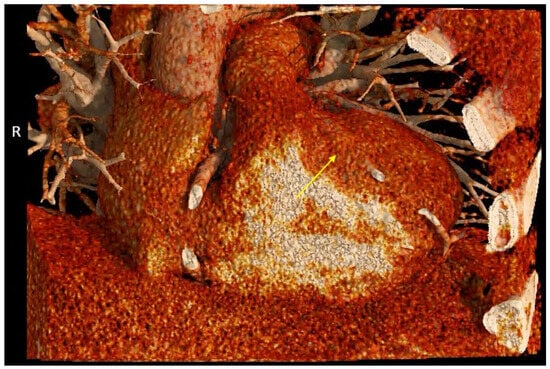
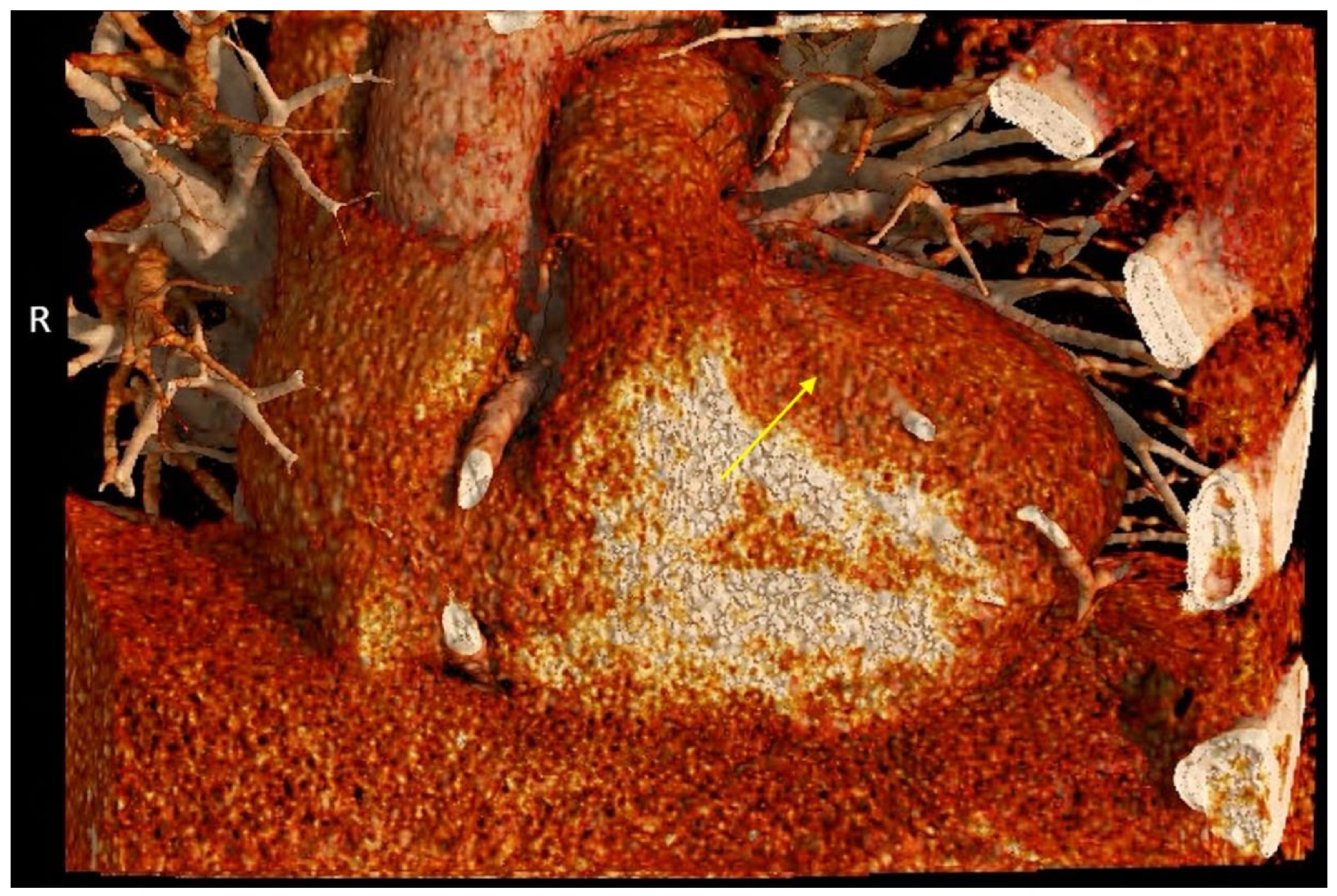
2D CTA showed mid-LAD bridging, but the 3D shape of the tunneled segment was hard to assess. We sectioned the volume from anterior to posterior with a sequential “front cut plane,” which showed the LAD beneath the myocardial surface and then re-emerging onto the epicardium distally. These views were helpful when discussing the case with the ordering clinician and when teaching trainees (Figure 2 and Figure 3).
Figure 2.
Sequential cinematic-rendered views of a mid-LAD intramyocardial tunnel. (A–C) A coronally oriented 3D volume is shown with a “front cut plane” progressively removing tissue from anterior to posterior. The mid-LAD emerges from beneath the bridge in panels (D–H) (tunneled segment, red arrows in (E–H), with overlying myocardium appreciable above the vessel). Anterior chest-wall structures, including ribs, are retained for anatomic orientation. Key: LAD, left anterior descending artery; red arrows (E–H), tunneled segment. What was equivocal: conventional 3D reconstructions display the vessel surface without conveying how much tissue lies above it, so the depth of the bridge cannot be judged. What CR adds: the progressive cut-plane sequence removes myocardium layer by layer, so the thickness of tissue overlying the vessel—and therefore the depth of the bridge—is appreciated directly.
Figure 3.
Close-up cinematic rendering of the mid-LAD myocardial bridge. The tunneled segment is highlighted (yellow arrow), with the distal LAD emerging from the tunnel onto the epicardial surface. Key: LAD, left anterior descending artery; yellow arrow, tunneled segment. The “R” in the figure is a standard orientation marker indicating the right side of the body. What was equivocal: on planar reformations the tunneled and epicardial portions of the vessel appear continuous and equivalent, so the point of emergence is not identifiable. What CR adds: the transition at which the vessel leaves myocardium and reaches the epicardial surface is shown directly, defining the distal extent of the bridge.
3.3. Case 3: RCA Appearing Intracameral on 2D but Bridging Within a Fat-Infiltrated RV Wall
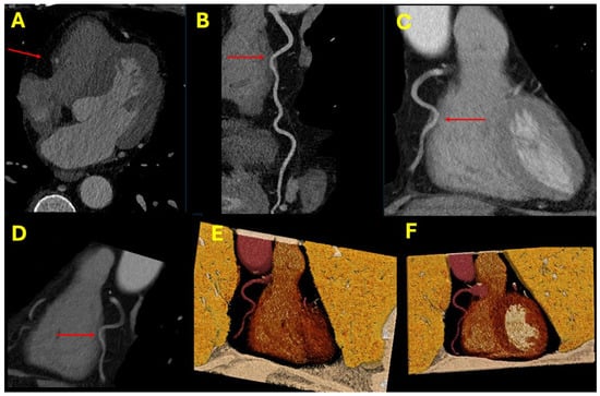
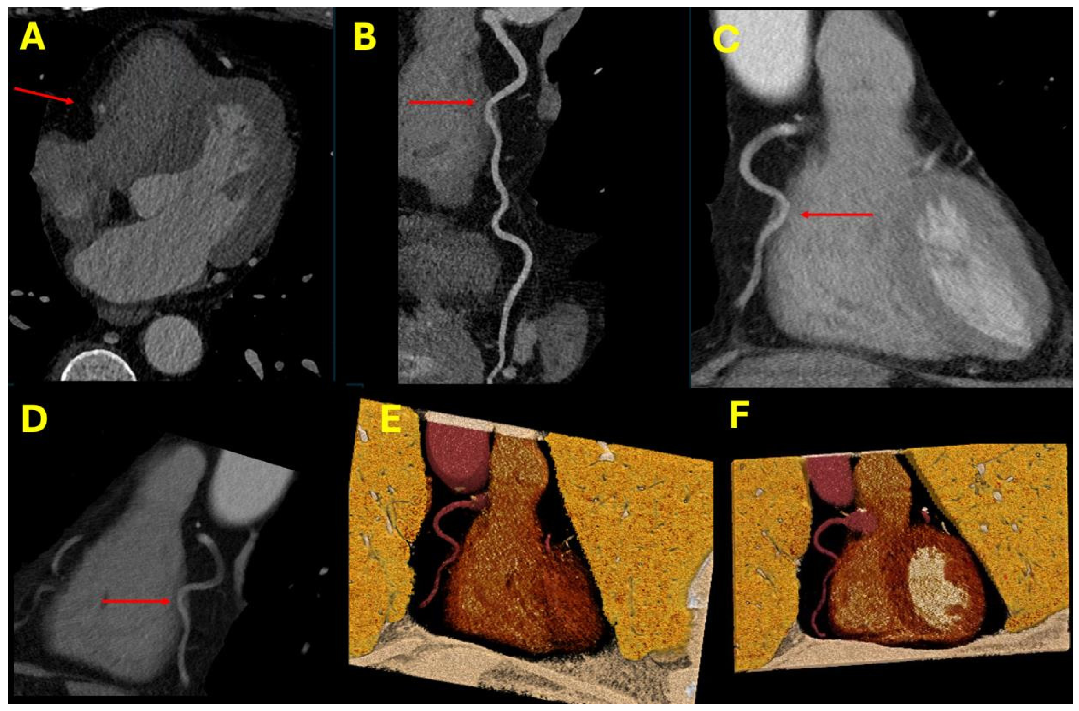
On 2D CTA the mid-RCA seemed to enter the right ventricular cavity, which raised concern for an intracameral course. The appearance came from fat infiltration of the RV free wall together with gating artifact, which blurred the boundary between the RV wall and the chamber. When we adjusted windowing and opacity to remove the RV free wall, CR showed the RCA running inside the fat-rich free wall without entering the chamber, which supported an extra-cavitary course rather than a true intracameral one (Figure 4). A true intra-ventricular course would be very rare [8], and the CR appearance fit the more common, artifactual explanation.
Figure 4.
2D CTA (A–D) and CR (E,F) in a suspected intracameral RCA. (A–D) Axial, curved planar, and thin-slab MIP images suggest that the mid-RCA might enter the right ventricular cavity (red arrows). (E,F) CR displays the RCA coursing within a fat-rich portion of the RV free wall without entering the chamber or blood pool. Key: RCA, right coronary artery; RV, right ventricle; MIP, maximum-intensity projection. Relative to the planar images (A–D), on which the vessel–chamber boundary is indistinct, CR (E,F) resolves the intact fat-rich RV free wall interposed between the RCA (arrows) and the ventricular blood pool, so that the vessel is seen to remain outside the chamber.
3.4. Case 4: LAD with a Thin Myocardial Bridge Versus a Suspected Intracameral Segment
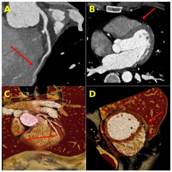
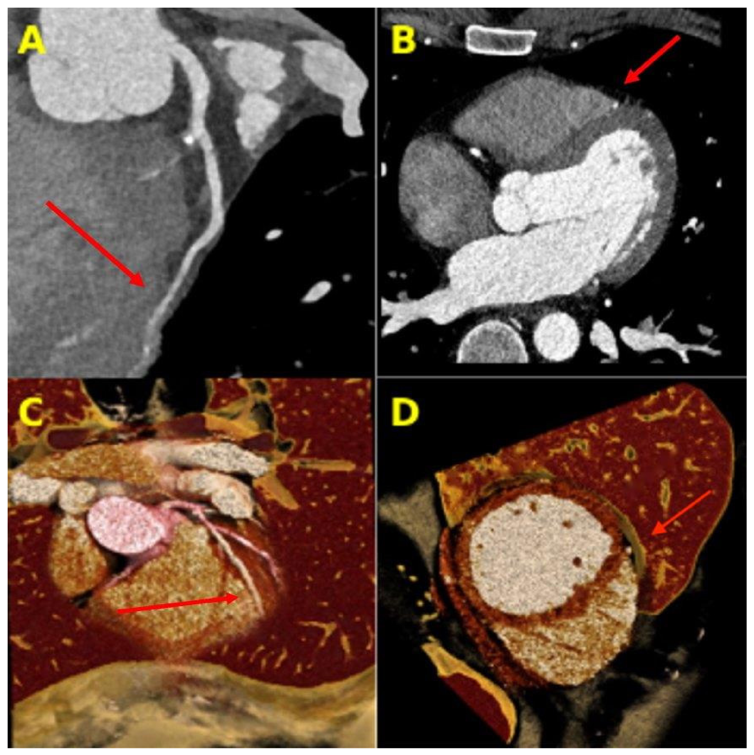
2D CTA raised concern for a small intracameral LAD segment next to the RV, and the boundary between the vessel and the myocardium was unclear on curved planar and axial images. CR showed a thin but definite layer of myocardium wrapping the LAD all the way around, which supported a thin intramyocardial course rather than chamber entry (Figure 5).
Figure 5.
Evaluation of a suspected intracameral LAD segment. (A,B) Curved planar and axial images show the LAD approaching the right ventricular cavity (red arrows). (C,D) CR demonstrates thin overlying and underlying myocardial tissue (red arrows), supporting an intramyocardial rather than intracameral course. Key: LAD, left anterior descending artery; RV, right ventricle. On the planar images (A,B) the vessel appears to abut the right ventricular cavity, whereas CR (C,D) demonstrates a continuous thin rim of myocardium encircling the vessel (arrows), indicating an intramyocardial rather than intracameral course.
4. Discussion
This series shows CR used for two related questions in coronary CTA: confirming and showing myocardial bridging, and assessing a segment that looks intracameral on planar images. In the first two cases, CR gave depth-resolved views of a tunneled LAD. The sequential “front cut plane” was useful for conveying bridge depth and shape, which matter in the minority of hemodynamically important bridges [4]. In the last two cases, CR showed intact myocardium around vessels that looked like they entered a chamber on 2D images, which supported an intramyocardial course.
We want to be accurate about what this adds. Good MPR and CPR already confirm most myocardial bridges, and in these cases CR did not reveal anatomy that was unavailable on expert reformations. CR improved depth perception, raised the reader’s confidence in equivocal cases, and produced images that referring clinicians and trainees could read easily. The point is that CR presented existing information more clearly rather than adding new anatomy. That difference should guide when CR is used and how any validation study is designed. We therefore frame CR here as a communicative and educational aid rather than a determinant of diagnostic accuracy; whether it changes diagnostic performance is a separate question that this series cannot answer and that would require formal reader studies. Consistent with this, CR did not alter the final diagnostic report in any of the four patients, and no management decision was taken on the basis of the rendered images; in each case the interpretation reached on the standard reconstructions stood unchanged. The contribution of CR was confined to communication and teaching—conveying the tunneled segment, or the intact myocardium overlying a vessel that had appeared intracameral, more readily to referring clinicians and trainees than the planar images allowed. We state this explicitly because the absence of any diagnostic or management change is itself an honest result of this series and bounds the claims that can be drawn from it.
The intracameral question calls for caution. A true intracavitary coronary course is rare, on the order of 0.36 to 1.3% overall on CTA. The intra-atrial RCA is its most common form, at about 0.15 to 0.29%, and a true intra-ventricular course is rarer still [8,9,10]. So when a segment looks intracameral on 2D images, as in Cases 3 and 4, the odds strongly favor an artifact from a thin or fat-infiltrated wall, from motion, or from partial-volume effects. Here CR helps by making the far more likely intramyocardial course easy to see, which helps rule out a rare but serious mimic that matters before ablation, device implantation, and valve surgery [8,11]. We are careful not to overstate how often this question comes up.
These findings agree with earlier work on the value of CR in cardiovascular imaging, including congenital heart disease, postoperative vascular anatomy, and complex aortic disease [16,17,18,19,20]. This series adds myocardial bridging and the separation of bridging from intracameral-appearing segments to that literature, while being clear about what a small, uncontrolled series can and cannot show.
Limitations
This is a small, retrospective, single-center series with no comparison group. We reviewed the CR images next to the standard reconstructions rather than on their own, so we cannot say how often CR changes interpretation or confidence compared with MPR and CPR alone. We also cannot estimate how often CR would change management in routine practice, though it is probably a small fraction. We chose cases for their instructive appearance, which introduces selection bias. Because they were not drawn from a consecutive series, we also cannot report the number of coronary CTA studies screened or the interval over which they were collected, so the frequency with which such equivocal cases arise cannot be estimated from this report. Access is a further constraint: CR requires GPU-capable hardware and a licensed rendering workstation, and it requires operator training that is not yet part of routine cardiac CT practice. CR is still operator-dependent, has no standardized protocol across vendors [12,13], and adds post-processing time, so it fits selected cases rather than routine use. Because the appearance of a cinematic-rendered image depends on operator-chosen presets, opacity, windowing, and clip-plane placement, the same dataset can yield different renderings in different hands, which limits reproducibility across readers and centers; standardized, vendor-neutral CR protocols and presets would reduce this variability and make the technique more transferable. Our workflow was implemented on a single vendor platform (Siemens syngo.via cVRT), and equivalent presets are not yet standardized across CR-capable systems. Multicenter, multi-vendor studies using agreed CR parameters will be needed before the technique can be compared fairly across platforms or recommended for general use. The 10 to 30 min of additional post-processing per case is also non-trivial in a high-volume practice, where applying CR to every study is impractical; in our view CR is best reserved for the small number of cases that remain equivocal after expert reformation, or for teaching and communication, rather than added to routine reporting. Establishing added value would take a prospective, adequately powered reader study that compares accuracy and confidence with and without CR, ideally with an independent reference standard and a report of how much CR adds over expert reformations [21].
Future directions: A definitive assessment of CR would require a prospective, adequately powered study rather than a descriptive series. An ideal design would consecutively enroll patients undergoing coronary CTA in whom standard interpretation is equivocal for myocardial bridging or an intracameral course, using pre-specified criteria for equivocality. Multiple readers of differing experience would interpret each study in two sessions separated by a washout interval and presented in counterbalanced order: standard reconstructions alone versus standard reconstructions supplemented by CR. Pre-specified endpoints would include diagnostic accuracy against an independent reference standard, such as invasive coronary angiography with intravascular ultrasound or surgical correlation where available, together with reader confidence on a defined Likert scale, inter-reader agreement, and reading time. The sample size would be powered for the primary accuracy or agreement endpoint, and a standardized CR protocol (fixed preset, opacity, and windowing with documented, case-specific clip-plane adjustments) would be used to support reproducibility. Such a design would separate any genuine gain in diagnostic performance from the communicative and educational benefits described here.
5. Conclusions
In these four patients, cinematic rendering was a useful addition to standard reformatted coronary CTA, both for showing myocardial bridging and for assessing segments that looked intracameral on planar images. It improved depth perception and reader confidence in selected equivocal cases and worked well for communication and teaching. It did not add anatomy beyond what expert MPR and CPR already showed. Because true intracameral courses are rare and this series had no comparison group, prospective comparative studies are needed to show whether CR adds diagnostic value over conventional reconstructions. This is an exploratory, small-sample case series: its findings illustrate a potential role for CR in selected equivocal cases but cannot be extrapolated to all coronary CTA examinations and require confirmation in prospective, controlled studies.
Author Contributions
Conceptualization, Y.S., A.A. and M.U.; methodology, Y.S. and A.A.; image post-processing and figure preparation, Y.S. and A.A.; writing, original draft preparation, Y.S. and A.A.; writing, review and editing, Y.S., A.A. and M.U.; supervision, M.U. All authors have read and agreed to the published version of the manuscript.
Funding
This research received no external funding.
Institutional Review Board Statement
This report was determined not to constitute human-subjects research under the Columbia University Human Research Protection Office (HRPO) Case Report Policy and the federal regulatory definition of human-subjects research at 45 CFR 46.102(L). Accordingly, Institutional Review Board approval was not required.
Informed Consent Statement
Patient consent for publication was waived. Under the Columbia University Human Research Protection Office (HRPO) Case Report Policy and the federal definition of human-subjects research (45 CFR 46.102(L)), this report was determined not to constitute human-subjects research, so neither Institutional Review Board approval nor informed consent was required. All images and clinical details were fully de-identified before analysis, and no information that could identify individual patients is included in this report.
Data Availability Statement
The imaging data supporting this case series are not publicly available to protect patient privacy; de-identified data may be available from the corresponding author on reasonable request, subject to institutional approval.
Conflicts of Interest
The authors declare no conflicts of interest.
References
- Tarantini, G.; Migliore, F.; Cademartiri, F.; Fraccaro, C.; Iliceto, S. Left Anterior Descending Artery Myocardial Bridging: A Clinical Approach. J. Am. Coll. Cardiol. 2016, 68, 2887–2899. [Google Scholar] [CrossRef] [Scilit] [PubMed]
- Evbayekha, E.O.; Nwogwugwu, E.; Olawoye, A.; Bolaji, K.; Adeosun, A.A.; Ajibowo, A.O.; Nsofor, G.C.; Chukwuma, V.N.; Shittu, H.O.; Onuegbu, C.A.; et al. A Comprehensive Review of Myocardial Bridging: Exploring Diagnostic and Treatment Modalities. Cureus 2023, 15, e43132. [Google Scholar] [CrossRef] [Scilit] [PubMed]
- Dimitriu-Leen, A.C.; van Rosendael, A.R.; Smit, J.M.; van Elst, T.; van Geloven, N.; Maaniitty, T.; Jukema, J.W.; Delgado, V.; Scholte, A.J.H.A.; Saraste, A.; et al. Long-Term Prognosis of Patients with Intramural Course of Coronary Arteries Assessed with CT Angiography. JACC Cardiovasc. Imaging 2017, 10, 1451–1458. [Google Scholar] [CrossRef] [Scilit] [PubMed]
- Bourassa, M.G.; Butnaru, A.; Lespérance, J.; Tardif, J.C. Symptomatic Myocardial Bridges: Overview of Ischemic Mechanisms and Current Diagnostic and Treatment Strategies. J. Am. Coll. Cardiol. 2003, 41, 351–359. [Google Scholar] [CrossRef] [Scilit] [PubMed]
- Kim, S.Y.; Seo, J.B.; Do, K.H.; Heo, J.N.; Lee, J.S.; Song, J.W.; Choe, Y.H.; Kim, T.H.; Yong, H.S.; Choi, S.I.; et al. Coronary Artery Anomalies: Classification and ECG-Gated Multi-Detector Row CT Findings with Angiographic Correlation. Radiographics 2006, 26, 317–333. [Google Scholar] [CrossRef] [Scilit] [PubMed]
- Schmitt, R.; Froehner, S.; Brunn, J.; Wagner, M.; Brunner, H.; Cherevatyy, O.; Gietzen, F.; Christopoulos, G.; Kerber, S.; Fellner, F. Congenital Anomalies of the Coronary Arteries: Imaging with Contrast-Enhanced Multidetector Computed Tomography. Eur. Radiol. 2005, 15, 1110–1121. [Google Scholar] [CrossRef] [Scilit] [PubMed]
- Angelini, P. Coronary Artery Anomalies: Current Clinical Issues: Definitions, Classification, Incidence, Clinical Relevance, and Treatment Guidelines. Tex. Heart Inst. J. 2002, 29, 271–278. [Google Scholar] [CrossRef] [Scilit] [PubMed]
- Gać, P.; Siudek, B.; Głuszczyk, A.; Plizga, J.; Grajnert, F.; Poręba, R. Computed Tomography Angiography as a Method for Diagnosing Intracavitary Coronary Arteries. Diagnostics 2024, 14, 1798. [Google Scholar] [CrossRef] [Scilit] [PubMed]
- Ganga, K.P.; Ojha, V.; Goyal, A.; Deepti, S.; Kumar, S. Intra-Atrial Right Coronary Artery on Dual-Source CT: Prevalence and Characteristics. Diagn. Interv. Radiol. 2021, 27, 595–598. [Google Scholar] [CrossRef] [Scilit] [PubMed]
- Opolski, M.P.; Pręgowski, J.; Kruk, M.; Staruch, A.D.; Witkowski, A.; Demkow, M.; Hryniewiecki, T.; Michałek, P.; Rużyłło, W.; Kępka, C. The Prevalence and Characteristics of Intra-Atrial Right Coronary Artery Anomaly in 9,284 Patients Referred for Coronary Computed Tomography Angiography. Eur. J. Radiol. 2014, 83, 1129–1133. [Google Scholar] [CrossRef] [Scilit] [PubMed]
- Rosamond, T.L.; Wetzel, L.H.; Lakkireddy, D.; Ferrell, R.; Tadros, P. Intracameral Right Coronary Artery: Detection by 64-Slice Coronary Computed Tomographic Angiography and Implications for Radiofrequency Ablation of Atrial Dysrhythmias. Pacing Clin. Electrophysiol. 2007, 30, 1571–1574. [Google Scholar] [CrossRef] [Scilit] [PubMed]
- Eid, M.; De Cecco, C.N.; Nance, J.W., Jr.; Caruso, D.; Albrecht, M.H.; Spandorfer, A.J.; De Santis, D.; Varga-Szemes, A.; Schoepf, U.J. Cinematic Rendering in CT: A Novel, Lifelike 3D Visualization Technique. AJR Am. J. Roentgenol. 2017, 209, 370–379. [Google Scholar] [CrossRef] [Scilit] [PubMed]
- Dappa, E.; Higashigaito, K.; Fornaro, J.; Leschka, S.; Wildermuth, S.; Alkadhi, H. Cinematic Rendering: An Alternative to Volume Rendering for 3D Computed Tomography Imaging. Insights Imaging 2016, 7, 849–856. [Google Scholar] [CrossRef] [Scilit] [PubMed]
- Calhoun, P.S.; Kuszyk, B.S.; Heath, D.G.; Carley, J.C.; Fishman, E.K. Three-Dimensional Volume Rendering of Spiral CT Data: Theory and Method. Radiographics 1999, 19, 745–764. [Google Scholar] [CrossRef] [Scilit] [PubMed]
- Zhang, Q.; Eagleson, R.; Peters, T.M. Dynamic Real-Time 4D Cardiac MDCT Image Display Using GPU-Accelerated Volume Rendering. Comput. Med. Imaging Graph. 2009, 33, 461–476. [Google Scholar] [CrossRef] [Scilit] [PubMed]
- Fellner, F.A. Introducing Cinematic Rendering: A Novel Technique for Post-Processing Medical Imaging Data. J. Biomed. Sci. Eng. 2016, 9, 170–175. [Google Scholar] [CrossRef]
- Rowe, S.P.; Johnson, P.T.; Fishman, E.K. Initial Experience with Cinematic Rendering for Chest Cardiovascular Imaging. Br. J. Radiol. 2018, 91, 20170558. [Google Scholar] [CrossRef] [Scilit] [PubMed]
- Rowe, S.P.; Johnson, P.T.; Fishman, E.K. Cinematic Rendering of Cardiac CT Volumetric Data: Principles and Initial Observations. J. Cardiovasc. Comput. Tomogr. 2018, 12, 56–59. [Google Scholar] [CrossRef] [Scilit] [PubMed]
- Röschl, F.; Purbojo, A.; Rüffer, A.; Cesnjevar, R.; Dittrich, S.; Glöckler, M. Initial Experience with Cinematic Rendering for the Visualization of Extracardiac Anatomy in Complex Congenital Heart Defects. Interact. Cardiovasc. Thorac. Surg. 2019, 28, 916–921. [Google Scholar] [CrossRef] [Scilit] [PubMed]
- Yasrab, M.; Rizk, R.C.; Chu, L.C.; Fishman, E.K. Cinematic Rendering of Non-Traumatic Thoracic Aorta Emergencies: A New Look at an Old Problem. Emerg. Radiol. 2024, 31, 269–276. [Google Scholar] [CrossRef] [Scilit] [PubMed]
- Layden, N.; Brassil, C.; Jha, N.; Saundankar, J.; Yim, D.; Andrews, D.; Patukale, A.; Srigandan, S.; Murray, C.P. Cinematic versus Volume Rendered Imaging for the Depiction of Complex Congenital Heart Disease. J. Med. Imaging Radiat. Oncol. 2023, 67, 487–491. [Google Scholar] [CrossRef] [Scilit] [PubMed]
Disclaimer/Publisher’s Note: The statements, opinions and data contained in all publications are solely those of the individual author(s) and contributor(s) and not of MDPI and/or the editor(s). MDPI and/or the editor(s) disclaim responsibility for any injury to people or property resulting from any ideas, methods, instructions or products referred to in the content. |
© 2026 by the authors. Licensee MDPI, Basel, Switzerland. This article is an open access article distributed under the terms and conditions of the Creative Commons Attribution (CC BY) license.