


Abstract
Background: Accurate acetabular component positioning is critical to the success of total hip arthroplasty (THA). Mixed reality (MR)-based simulation offers a novel approach to surgical training by providing real-time feedback within immersive learning environments. This study evaluated the impact of an MR-based simulation on technical performance, as well as its usability and user experience. Methods: A prospective pre–post pilot educational study was conducted in a controlled simulation laboratory using a physical anatomical model. Forty-six final-year medical students with no prior experience in MR technology participated. Participants performed acetabular component positioning tasks before and after MR-assisted training. Technical accuracy was assessed by measuring deviations in inclination and anteversion. Usability, workload, presence, and cybersickness were evaluated using validated questionnaires. Results: MR-assisted training significantly improved technical accuracy. Median absolute deviation in inclination decreased from 1.0° (interquartile range [IQR], 1.0–3.5) to 0.5° (IQR, 0.0–2.0) (p = 0.0008, r = 0.56), while anteversion deviation decreased from 4.5° (IQR, 2.0–7.0) to 2.0° (IQR, 1.0–4.0) (p < 0.0001, r = 0.66). User experience scores ranked the application among top-performing systems in benchmark comparisons. Participants reported high levels of presence and usability, low workload (all NASA-TLX domains <36/100), and minimal cybersickness (mean Simulator Sickness Questionnaire score = 0.26). Conclusions: MR-assisted simulation significantly improved short-term technical performance in novice trainees and was associated with high user acceptance and minimal adverse effects. These findings support the use of MR as a promising adjunct in early surgical training, enabling integration of cognitive and psychomotor skills through real-time feedback. Further studies are warranted to evaluate skill retention and transfer to clinical practice.
1. Introduction
Total hip arthroplasty (THA) is one of the most successful and commonly performed procedures in orthopaedic surgery, with an increasing demand driven by the growing prevalence of osteoarthritis [1]. Complications such as instability and dislocation remain clinically relevant and are closely associated with acetabular component positioning [2,3].
Accurate orientation of the acetabular component is a critical step in THA, directly influencing implant stability and long-term outcomes. However, achieving target inclination and anteversion remains challenging, as conventional non-navigated techniques rely heavily on visual estimation. Although target orientations of approximately 45° inclination and 20° anteversion are commonly used in simulation and educational settings, the concept of a universal “safe zone” remains debated. Contemporary evidence suggests that optimal component orientation should be individualized according to patient anatomy, spinopelvic mobility, surgical approach, and implant design. In the present study, these target angles were selected as standardized educational reference values to facilitate reproducible comparison across participants rather than to represent universally optimal clinical targets. Variability in component positioning has been reported even among experienced surgeons, highlighting the influence of surgical expertise and the limitations of current techniques [4].
Surgical training in orthopaedics is heterogeneous, with variability in operative exposure and access to simulation tools across training programs globally. As a result, early skill acquisition may be inconsistent, emphasizing the need for standardized and reproducible training methods that allow novice surgeons to develop technical competence in a controlled environment.
Simulation-based training may be a promising strategy to address these challenges. Virtual reality (VR) platforms have demonstrated effectiveness in improving surgical performance and visuospatial skills across multiple domains [5,6,7]. In the context of THA, VR-based simulation has been associated with improved accuracy in acetabular component positioning 6. However, these systems lack tactile feedback, which is an essential component of surgical skill acquisition. Unlike virtual reality, which completely replaces the physical environment, augmented reality overlays digital information onto the real world, whereas mixed reality additionally allows spatial interaction between virtual objects and the physical environment.
Mixed reality (MR) integrates virtual elements within a real physical environment, enabling simultaneous interaction with digital guidance and real surgical instruments. This hybrid approach may enhance spatial understanding and support the acquisition of motor skills in simulated surgical tasks. MR has shown promising applications in orthopaedic surgery, including anatomical visualization and surgical planning [8,9]. Nevertheless, its application as a simulation-based guidance system in THA remains limited [10,11].
An MR application designed to provide real-time holographic guidance for acetabular component positioning was used in this study. Although primarily intended for intraoperative assistance, its potential use as a simulation-based guidance system in a simulated environment has not been explored [12].
The aim of this study was to evaluate the effect of MR guidance on acetabular component positioning accuracy in a simulated THA setting and to assess secondary usability outcomes. We hypothesized that MR guidance would reduce deviation from target inclination and anteversion angles and support the acquisition of spatial orientation skills relevant to THA.
2. Materials and Methods
2.1. Study Design and Setting
This was a prospective pre–post pilot simulation study designed to evaluate the effect of MR guidance on acetabular component positioning accuracy and secondary usability outcomes. The study was conducted in a controlled simulation laboratory environment using a physical anatomical model.
2.2. Participants
A total of 46 novice trainees participated in the study. The mean age was 25.26 years (SD = 1.78), including 26 women and 20 men.
Participation was voluntary, and no prior experience with mixed reality technology was required. Participants were final-year medical students with no previous experience in total hip arthroplasty, arthroplasty simulation, or immersive simulation technologies. All participants underwent the same standardized training protocol. The study was approved by the Ethics Committee of Hospital Universitari i Politècnic La Fe, Valencia, Spain (approval code GAN-1; approved on 12 April 2023).
2.3. Intervention
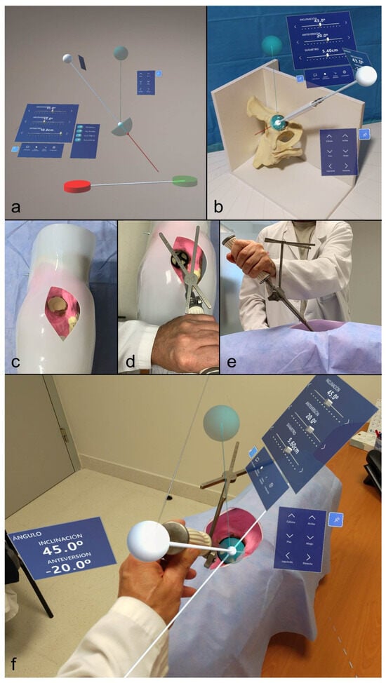
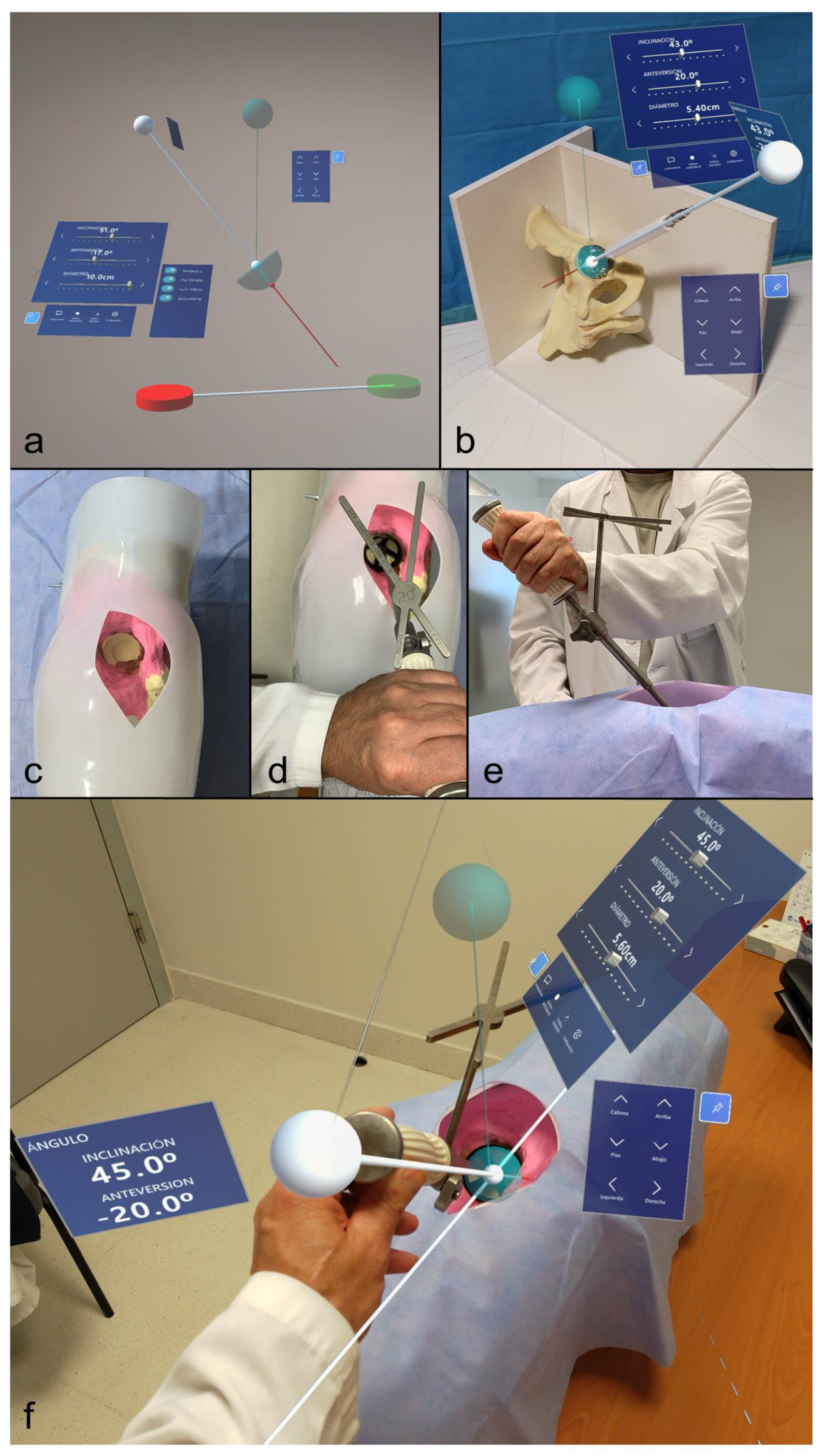
Participants underwent a structured MR-based training session using the MR application on the HoloLens 2 headset (Microsoft, Redmond, WA, USA). The application provides a spatially registered holographic guide to assist in acetabular component positioning, displaying real-time inclination and anteversion angles aligned with the physical model (Figure 1a,b).
Figure 1.
(a) Mixed reality interface displayed through the headset, showing the calibration guides (red and green disc-shaped guides placed on the surgical table to define the reference plane for angle measurements), the holographic acetabular cup (to be positioned within the physical acetabulum), the holographic alignment guide (the white sphere is interactive and allows modification of cup angulation), the main control panel displaying cup diameter and inclination and anteversion angles (which can be adjusted by the user), and a secondary panel enabling fine adjustments of the cup’s position. (b) Interface overlaid onto a pelvic model, with the calibration guides already positioned and hidden, and the holographic acetabular cup placed within the physical acetabulum. Inclination and anteversion angles measurements are displayed. (c) Custom anatomical model simulating a posterolateral approach. (d) Mechanical guide positioned to achieve the desired 45° and 20°. (e) Alternate perspective of the mechanical guide. (f) MR interface overlaid onto the custom anatomical model, with the guide set to 45° inclination and 20° anteversion. This was the setup for the training procedure.
The training intervention consisted of a guided hands-on simulation in which participants interacted with holographic elements using hand gestures, allowing real-time adjustment of cup orientation while maintaining tactile feedback through standard surgical instrumentation.
2.4. Simulation Model and Equipment
Training was performed using a custom-built anatomical hip model simulating a posterolateral approach. The model consisted of a thermoplastic external shell representing soft tissue, with an internal composite pelvic bone model and surrounding simulated musculature. The pelvis was fixed in a lateral decubitus position on a wooden frame, aligned parallel to the horizontal plane to replicate standard surgical positioning (Figure 1c).
A Stryker Trident® Tritanium® acetabular system and corresponding mechanical alignment guide were used to reproduce realistic surgical conditions (Figure 1d,e).
2.5. Study Procedure
All participants received a standardized 10 min theoretical introduction covering the surgical approach and the principles of acetabular component positioning, including target angles of 45° inclination and 20° anteversion.
The experimental protocol consisted of three sequential phases:
- Baseline Assessment:
Participants positioned the acetabular component using a conventional mechanical alignment guide without MR assistance. The achieved inclination and anteversion angles were recorded.
- 2.
- Mixed Reality-Assisted Training:
Participants completed a single standardized 15 min MR-assisted training session. During this period, they were allowed to freely manipulate and repeatedly adjust the acetabular component using the holographic guidance in order to familiarize themselves with the system. No formal repeated test attempts were recorded during the training session. Following completion of this single training session, participants performed one post-training assessment without MR assistance [12]. Before each training session, the MR system was calibrated according to the protocol previously described by Juan et al., ensuring alignment between the holographic guidance system and the physical pelvic model. All participants underwent the same standardized training protocol under the supervision of the same instructor. The instructor provided guidance during the training session but did not intervene while participants performed the positioning task, ensuring independent task execution. Only one MR-assisted practice session was permitted. During training, the holographic alignment guide displayed the current inclination and anteversion angles in real time as participants manipulated the acetabular component, allowing continuous visual feedback to facilitate adjustment toward the target orientation (Figure 1f). Participants were not informed of their baseline performance before the post-training assessment; performance feedback was provided only after completion of the entire study protocol.
- 3.
- Post-Training Assessment:
Participants performed a final attempt using the mechanical guide alone, without MR assistance. Final orientation angles were recorded.
After the procedure, participants completed a set of standardized questionnaires.
2.6. Outcome Measures
2.6.1. Primary Outcome
The primary outcome was accuracy of acetabular component positioning, defined as the absolute deviation (in degrees) from the target angles of 45° inclination and 20° anteversion. Measurements were obtained before and after the MR intervention. The measurement accuracy of the mixed reality application used in this study has been previously validated in an experimental setting by comparison with independent ground-truth measurements. In that validation study, acetabular component orientation angles provided by the application were compared against manually obtained values using calibrated physical instruments, including a goniometer and angle protractor. The reported mean absolute error was 0.375° (SD 0.483) for anteversion and 0.1° (SD 0.308) for inclination, with maximum discrepancies not exceeding 1°. These results indicate a high level of accuracy within clinically acceptable thresholds, supporting the validity of the application for angular measurement in simulation-based training contexts [12].
2.6.2. Secondary Outcomes
Secondary outcomes included subjective measures of user experience and application usability:
- Cybersickness, assessed using selected items from the Simulator Sickness Questionnaire (SSQ) [13].
- Presence, measured with an adapted augmented reality presence questionnaire [14].
- Workload, assessed using the NASA Task Load Index (NASA-TLX) [15].
- User experience, evaluated using the User Experience Questionnaire (UEQ) [16,17].
- Additional custom questions assessing perceived usability, efficiency, and satisfaction.
2.6.3. Data Collection
All angular measurements were obtained using the MR application and recorded by a single trained researcher. Although this approach may introduce measurement bias, the application has previously been validated against calibrated physical instruments for angle assessment [12]. Questionnaire responses were collected immediately after completion of the simulation session. The evaluator was not blinded to the study phase, as measurements were recorded during the simulation session.
2.6.4. Statistical Analysis
Data distribution was assessed using the Shapiro–Wilk test. As post-intervention inclination data were not normally distributed, non-parametric tests were used for paired comparisons.
Pre-intervention and post-intervention differences in absolute deviation for inclination and anteversion were analyzed using the Wilcoxon signed-rank test.
Effect sizes (r) were calculated for non-parametric comparisons to estimate the magnitude of observed differences. Statistical significance was set at p < 0.05.
UEQ responses were analyzed using the standardized UEQ Data Analysis Tool and compared against the established benchmark dataset.
3. Results
3.1. Primary Outcome: Accuracy of Acetabular Component Positioning
A total of 46 participants completed the study.
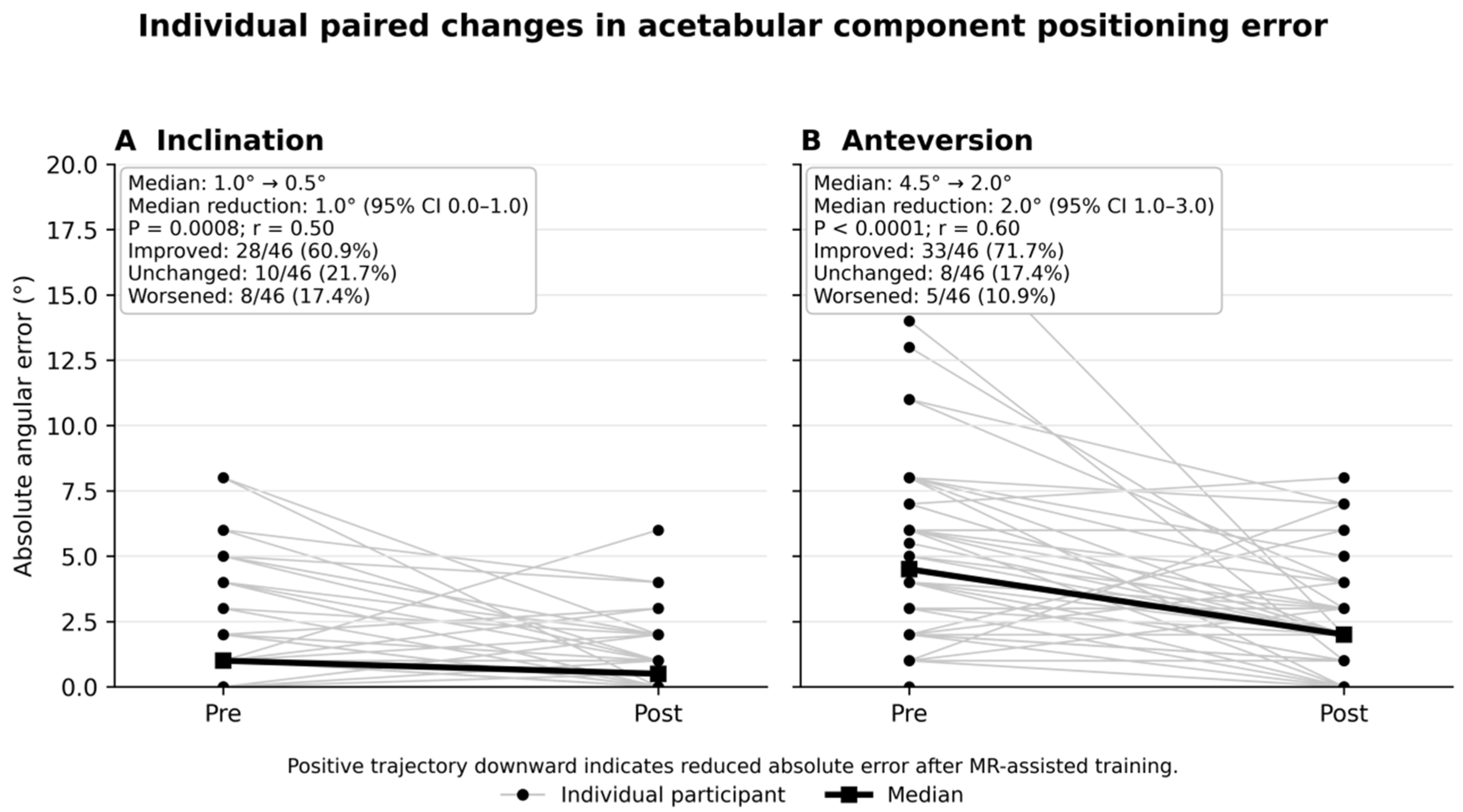
Median absolute deviation from the target inclination (45°) decreased from 1.0° (IQR 1.0–3.5) pre-intervention to 0.5° (IQR 0.0–2.0) post-intervention. This reduction was statistically significant (Wilcoxon V = 121.0, p = 0.00075, Z = 3.37, r = 0.497).
Similarly, median absolute deviation from the target anteversion (20°) decreased from 4.5° (IQR 2.0–7.0) pre-intervention to 2.0° (IQR 1.0–4.0) post-intervention. This improvement was also statistically significant (Wilcoxon V = 90.5, p < 0.0001, Z = 4.06, r = 0.599) (Table 1).
Table 1.
Primary outcome: Acetabular component positioning accuracy before and after MR-assisted training.
Both effect sizes indicate a substantial intervention effect.
Values are presented as median (interquartile range). Absolute error was calculated as the absolute deviation from the target orientation (45° inclination and 20° anteversion). Differences between pre- and post-training measurements were analyzed using the Wilcoxon signed-rank test. Effect size (r) was calculated as Z/√N. Median paired reduction represents the median of the individual paired pre–post differences rather than the arithmetic difference between the reported medians.
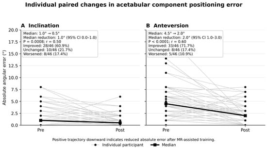
Individual participant trajectories are shown in Figure 2, illustrating that improvement was observed in most participants, particularly for anteversion.
Figure 2.
Individual paired changes in acetabular component positioning error before and after MR-assisted training. Absolute errors relative to the target orientation (45° inclination and 20° anteversion) are shown for (A) inclination and (B) anteversion. Gray lines connect paired measurements from each participant, black circles indicate individual observations, and the solid black line represents the group median. Lower values indicate greater positioning accuracy.
3.2. Secondary Outcomes
- User Experience
User experience ratings were high, with mean scores approaching 6 out of 7 across all items. UEQ analysis placed the application within the top 10% of benchmarked products in five of six domains [13,14].
- 2.
- Presence
Mean presence scores ranged from 5.27 to 6.07 (7-point scale), indicating high perceived immersion [15].
- 3.
- Workload
NASA-TLX scores were low across all domains (all means <36/100), with particularly low frustration levels [16].
- 4.
- Cybersickness
Participants reported minimal symptoms of cybersickness. Mean SSQ score was 0.26 (SD = 0.59) [17].
3.3. Summary of Findings
Mixed reality-assisted training significantly improved acetabular component positioning accuracy, with large effect sizes. Secondary usability outcomes indicated high user satisfaction, low workload, and minimal adverse effects. A summary of the secondary usability outcomes, including user experience, presence, workload, and cybersickness, is provided in Supplementary Figure S1.
4. Discussion
In this prospective pilot study, MR guidance significantly improved acetabular component positioning accuracy in a simulated THA environment. Participants demonstrated reduced deviation from target inclination and anteversion following a single MR-guided session, with large effect sizes observed for both parameters. Our findings suggest that MR-assisted training may improve immediate technical performance during simulated acetabular component positioning. Whether these improvements translate into enhanced implant positioning, implant longevity, or clinical outcomes remains to be established.
4.1. Primary Outcome: Accuracy of Acetabular Component Positioning
The primary finding of this study is that a single MR-guided session resulted in a statistically significant improvement in acetabular component positioning accuracy. This is particularly relevant given that accurate acetabular orientation is a critical determinant of total hip arthroplasty outcomes, influencing instability and revision risk, and is known to vary with surgical experience [3,4]. It should also be recognized that optimal acetabular component positioning extends beyond achieving target inclination and anteversion angles. Conventional freehand cup implantation has been shown to exhibit approach-specific orientation biases, and accurate reconstruction of the hip center of rotation (COR) represents another important determinant of joint biomechanics, implant stability, and restoration of hip function. These considerations become particularly relevant in patients with complex anatomy, dysplasia, or altered spinopelvic mechanics, where individualized component positioning may be required rather than adherence to fixed angular targets. Although the present simulation focused on standardized angular orientation as an educational endpoint, future MR-based guidance systems could integrate additional anatomical parameters to support more comprehensive implant positioning [18]. The magnitude of improvement observed suggests that MR guidance could produce a substantial effect in early-stage trainees, who typically rely on visual estimation during initial training. Although both inclination and anteversion improved significantly following the intervention, the absolute reduction in inclination error was modest and should be interpreted cautiously. By contrast, the larger improvement observed for anteversion may represent the more educationally meaningful finding of this pilot study. As this was a simulation-based study, the clinical relevance of these angular improvements remains to be established. These findings are consistent with previous literature demonstrating the effectiveness of immersive technologies in surgical training. Virtual reality–based training has been shown to improve surgical performance and visuospatial skills across multiple domains [5,6,7]. In the context of total hip arthroplasty, VR simulation has been associated with improved acetabular component positioning accuracy among residents [6]. Unlike VR, which lacks haptic feedback, MR enables real-time visual guidance while preserving interaction with physical instruments and anatomical models, allowing simultaneous integration of visuospatial information and tactile interaction, potentially enhancing the acquisition of spatial orientation skills. This aligns with previous studies highlighting the potential of MR for anatomical visualization and intraoperative guidance in orthopaedics [8,9]. However, the educational benefit observed should be attributed to the complete MR-assisted training intervention rather than to mixed reality technology in isolation. The intervention incorporated several educational elements, including holographic visualization, continuous numerical feedback of inclination and anteversion, gesture-based interaction, and tactile feedback through conventional surgical instruments. Because these components were delivered simultaneously, their individual contributions cannot be distinguished. Future studies should compare MR guidance with alternative feedback modalities, such as conventional navigation or digital inclinometer-based systems, to identify the specific mechanisms responsible for the observed learning effect. Furthermore, systematic reviews on extended reality technologies in orthopaedics have emphasized their growing role in surgical training while also noting the limited availability of MR-specific applications in THA [10,11]. Our study contributes to addressing this gap by providing empirical evidence supporting the feasibility and effectiveness of MR-based simulation in this context. Given the variability in early surgical exposure, MR-based platforms may represent a promising educational adjunct for practicing component positioning skills in a standardized simulated environment. By enabling repeated practice in a controlled environment with immediate feedback, MR may facilitate more efficient acquisition of orientation skills relevant to THA component placement. Accordingly, this study should be interpreted as demonstrating improvement after exposure to an MR-assisted training package rather than proving superiority of MR over conventional training. The present findings only demonstrate immediate post-training improvement. Whether these gains persist over time or transfer to more realistic surgical environments remains unknown.
4.2. Secondary Outcomes: User Experience, Presence, Workload, and Cybersickness
In addition to improvements in technical performance, participants reported high levels of usability, presence, and overall user experience, along with low perceived workload. User experience scores ranked the application among top-performing systems in benchmark comparisons, supporting the usability and acceptability of the MR platform. High scores for presence, usability, and satisfaction indicate that participants perceived the simulation as immersive, which is an important factor in effective simulation-based learning. The low NASA-TLX scores across all domains suggest that the MR system did not impose excessive cognitive or physical demands. Participants reported minimal symptoms of cybersickness, with very low Simulator Sickness Questionnaire scores. This finding is important, as discomfort and visual fatigue are potential barriers to the adoption of immersive technologies. The negligible levels of cybersickness observed in this study suggest that the MR application is well tolerated. These findings support feasibility and user acceptability of MR implementation.
4.3. Limitations
This study has several limitations. Firstly, the sample size was relatively small and included only novice trainees, which may limit generalizability to residents or more experienced surgeons. Furthermore, the pre–post design without a control group limits causal inference and does not allow direct comparison with conventional or alternative training modalities. In addition, the intervention evaluated in this study consisted of an integrated MR-assisted training package that combined holographic visualization, real-time numerical feedback of inclination and anteversion, gesture-based interaction, and tactile manipulation of real surgical instruments. Consequently, the present study cannot determine which individual component was primarily responsible for the observed improvement. In particular, the contribution of real-time numerical feedback cannot be separated from the immersive MR environment itself. Although the magnitude of improvement and large effect sizes suggest a meaningful intervention effect, a learning effect from repeated task performance cannot be excluded. Outcomes were assessed immediately after the intervention and therefore do not provide information on long-term skill retention. Because all participants completed the tasks in the same fixed sequence (baseline assessment, MR-assisted training, and post-training assessment), part of the observed improvement may reflect practice effects, increasing familiarity with the simulation model, recall of the target angles, or greater confidence using the mechanical alignment guide rather than a specific effect of mixed reality itself. Therefore, causal attribution to the MR intervention should be interpreted with caution. In addition, all angular measurements were obtained using the same MR application employed during the intervention and were recorded by a single, non-blinded evaluator. Although the application has previously been validated against independent physical measurements, the use of the same system for both training and outcome assessment may have introduced measurement bias. Future studies should incorporate an independent reference standard, such as optical tracking, CT-based measurements, a validated navigation system, or calibrated digital instruments, together with blinded outcome assessment whenever feasible. Finally, although the simulation model approximates surgical conditions, it cannot fully replicate the complexity of real operative environments, and transferability of skills to clinical practice remains to be established.
Furthermore, because patient-specific spinopelvic parameters were not incorporated into the simulation model, impingement-free range of motion could not be evaluated. Consequently, the study was limited to assessment of component orientation accuracy and did not investigate the functional consequences of implant positioning with respect to hip biomechanics or impingement risk.
Despite its potential, mixed reality also presents several practical limitations. Similar to augmented reality systems, accurate hologram registration depends on robust calibration and tracking, and misalignment between virtual overlays and the physical environment may reduce guidance accuracy. In addition, current head-mounted displays have restricted fields of view, may increase cognitive demand during prolonged use, and remain dependent on hardware availability and cost. These technical limitations should be considered when interpreting the potential role of MR as an adjunct to surgical training and future intraoperative applications [19].
4.4. Future Directions
Future research should include randomized controlled trials comparing MR-guidance with conventional and alternative simulation modalities to better establish comparative educational performance for improving component positioning accuracy. Longitudinal studies are needed to evaluate skill retention and the transfer of acquired skills to cadaveric and clinical settings.
Further development of MR applications incorporating complete procedural workflows, along with validation in more realistic training environments, may help define the role of MR within comprehensive surgical training programs.
5. Conclusions
Mixed reality-assisted training significantly improved short-term acetabular component positioning accuracy in a simulated total hip arthroplasty environment. Participants demonstrated reduced deviations from the target inclination and anteversion angles immediately after a single training session, indicating improved technical performance in this simulated task.
These findings support the feasibility and educational potential of an integrated MR-assisted training approach for early surgical simulation. However, the present study evaluated a specific MR-assisted training package, and the individual contribution of its different components cannot be determined. Furthermore, the observed improvements reflect immediate post-training performance and should not be interpreted as evidence of improved operative performance or clinical outcomes.
Further randomized controlled studies incorporating independent outcome measurements, delayed skill retention assessments, and transfer to cadaveric or clinical settings are required to establish the educational value and clinical relevance of this approach.
Supplementary Materials
The following supporting information can be downloaded at: https://www.mdpi.com/article/10.3390/prosthesis8070078/s1, Supplementary Figure S1. Secondary usability outcomes following MR-assisted training. Summary of user experience (UEQ), presence questionnaire, NASA Task Load Index (NASA-TLX), and Simulator Sickness Questionnaire (SSQ) results. UEQ scores are presented according to the published benchmark categories, while presence, workload, and cybersickness results are displayed using descriptive statistics. Higher UEQ and presence scores indicate a more favorable user experience, whereas lower NASA-TLX and SSQ scores indicate lower workload and minimal simulator-related discomfort.
Author Contributions
Conceptualization: J.H.-B., D.M. and M.-C.J.; methodology: D.M., A.A.V.-N. and M.-C.J.; data curation: D.M. and J.H.-B.; formal analysis: J.H.-B., D.M. and M.-C.J.; writing—original draft preparation: J.H.-B.; writing—review and editing: J.H.-B., D.M., A.A.V.-N. and M.-C.J. Project administration: D.M. and M.-C.J.; Funding acquisition: D.M. and M.-C.J. All authors have read and agreed to the published version of the manuscript.
Funding
This research was funded by the FISABIO (Fundació per al Foment de la Investigació Sanitària i Biomèdica de la Comunitat Valenciana) POLISABIO 2023—PI2 RMArthro.
Institutional Review Board Statement
The study was conducted in accordance with the Declaration of Helsinki and approved by the Ethics Committee of Hospital U. y P. LA FE de Valencia (Spain) (protocol code GAN-1 and date of approval 12 April 2023).
Informed Consent Statement
Informed consent was obtained from all subjects involved in the study prior to participation. Written informed consent has been obtained from the patients to publish this paper.
Data Availability Statement
The datasets generated and analyzed during the current study are available from the corresponding author upon reasonable request.
Acknowledgments
During the preparation of this manuscript/study, the authors used Google NotebookLM (free version) for the purposes of creating the Graphical Abstract. The authors have reviewed and edited the output and take full responsibility for the content of this publication.
Conflicts of Interest
The authors declare no conflicts of interest.
References
- Cross, M.; Smith, E.; Hoy, D.; Nolte, S.; Ackerman, I.; Fransen, M.; Bridgett, L.; Williams, S.; Guillemin, F.; Hill, C.L.; et al. The global burden of hip and knee osteoarthritis: Estimates from the Global Burden of Disease 2010 study. Ann. Rheum. Dis. 2014, 73, 1323–1330. [Google Scholar] [CrossRef] [Scilit] [PubMed]
- Lewinnek, G.E.; Lewis, J.L.; Tarr, R.; Compere, C.L.; Zimmerman, J.R. Dislocations after total hip-replacement arthroplasties. J. Bone Jt. Surg. Am. 1978, 60, 217–220. [Google Scholar] [CrossRef] [Scilit]
- Rowan, F.E.; Benjamin, B.; Pietrak, J.R.; Haddad, F.S. Prevention of dislocation after total hip arthroplasty. J. Arthroplast. 2018, 33, 1316–1324. [Google Scholar] [CrossRef] [Scilit] [PubMed]
- Grammatopoulos, G.; Alvand, A.; Monk, A.P.; Mellon, S.; Pandit, H.; Rees, J.; Rees, J.; Gill, H.S.; Murray, D.W. Surgeons’ accuracy in achieving their desired acetabular component orientation. J. Bone Jt. Surg. Am. 2016, 98, e72. [Google Scholar] [CrossRef] [Scilit] [PubMed]
- Mao, R.Q.; Lan, L.; Kay, J.; Lohre, R.; Ayeni, O.R.; Goel, D.P.; de Sa, D. Immersive virtual reality for surgical training: A systematic review. J. Surg. Res. 2021, 268, 40–58. [Google Scholar] [CrossRef] [Scilit] [PubMed]
- Hooper, J.; Tsiridis, E.; Feng, J.E.; Schwarzkopf, R.; Waren, D.; Long, W.J.; Poultsides, L.; Macaulay, W.; Papagiannakis, G.; Kenanidis, E.; et al. Virtual reality simulation facilitates resident training in total hip arthroplasty: A randomized controlled trial. J. Arthroplast. 2019, 34, 2278–2283. [Google Scholar] [CrossRef] [Scilit]
- Logishetty, K.; Gofton, W.T.; Rudran, B.; Beaulé, P.E.; Cobb, J.P. Fully immersive virtual reality for total hip arthroplasty. J. Bone Jt. Surg. Am. 2020, 102, e27. [Google Scholar] [CrossRef] [Scilit] [PubMed]
- Kolac, U.C.; Paksoy, A.; Akgün, D. Three-dimensional planning, navigation, patient-specific instrumentation and mixed reality in shoulder arthroplasty: A digital orthopedic renaissance. EFORT Open Rev. 2024, 9, 517–527. [Google Scholar] [CrossRef] [Scilit] [PubMed]
- Chytas, D.; Nikolaou, V.S. Mixed reality for visualization of orthopedic surgical anatomy. World J. Orthop. 2021, 12, 727–731. [Google Scholar] [CrossRef] [Scilit] [PubMed]
- Luo, F.; Hu, Y.; Liu, S.; Yang, R. XR (extended reality: Virtual reality, augmented reality, mixed reality) technology applications in orthopedic field. Medicine 2025, 104, e42956. [Google Scholar] [CrossRef] [Scilit] [PubMed]
- Su, S.; Wang, R.; Zhou, R.; Chen, Z.; Zhou, F. The effectiveness of virtual reality, augmented reality, and mixed reality training in total hip arthroplasty: A systematic review and meta-analysis. J. Orthop. Surg. Res. 2023, 18, 121. [Google Scholar] [CrossRef] [Scilit] [PubMed]
- Juan, M.; Hidaldo, C.; Mifsut, D. A mixed reality application for total hip arthroplasty. Virtual Real. 2024, 28, 39. [Google Scholar] [CrossRef] [Scilit]
- Laugwitz, B.; Held, T.; Schrepp, M. Construction evaluation of a user experience questionnaire. In HCI and Usability for Education and Work (USAB 2008); Lecture Notes in Computer Science; Holzinger, A., Ed.; Springer: Berlin/Heidelberg, Germany, 2008; Volume 5298, pp. 63–76. [Google Scholar] [CrossRef] [Scilit]
- Schrepp, M.; Hinderks, A.; Thomaschewski, J. Construction of a Benchmark for the User Experience Questionnaire (UEQ). Int. J. Interact. Multimed. Artif. Intell. 2017, 4, 40–44. [Google Scholar] [CrossRef] [Scilit]
- Regenbrecht, H.; Schubert, T. Measuring presence in augmented reality environments: Design and a first test of a questionnaire. In Proceedings of the Fifth Annual International Workshop Presence 2002, Porto, Portugal, 9–11 October 2002; pp. 138–144. Available online: http://matthewlombard.com/ISPR/Proceedings/2002/Regenbrecht%20and%20Schubert.pdf (accessed on 8 January 2026).
- Hart, S.G.; Staveland, L.E. Development of NASA-TLX (Task Load Index): Results of Empirical and Theoretical Research. In Human Mental Workload, ser. Advances in Psychology; Hancock, P.A., Meshkati, N., Eds.; Elsevier: Amsterdam, The Netherlands, 1988; Volume 52, pp. 139–183. [Google Scholar] [CrossRef] [Scilit]
- Kennedy, R.S.; Lane, N.E.; Berbaum, K.S.; Lilienthal, M.G. Simulator Sickness Questionnaire: An Enhanced Method for Quantifying Simulator Sickness. Int. J. Aviat. Psychol. 1993, 3, 203–220. [Google Scholar] [CrossRef] [Scilit] [PubMed]
- Avram, G.M.; Prill, R.; Gurau, C.D.; Georgeanu, V.; Deleanu, B.; Russu, O.; Becker, R.; Predescu, V. Acetabular cup placement and offset control in robotic total hip arthroplasty performed through the modified anterolateral approach. Int. Orthop. 2023, 47, 2265–2273. [Google Scholar] [CrossRef] [Scilit] [PubMed]
- Ishida, T.; Tateiwa, T.; Takahashi, Y.; Seki, T.; Masaoka, T.; Shishido, T.; Yamamoto, K. Error and risk factors in augmented reality-guided cup placement during total hip arthroplasty. J. Orthop. Sci. 2026, in press. [Google Scholar] [CrossRef] [Scilit] [PubMed]

Disclaimer/Publisher’s Note: The statements, opinions and data contained in all publications are solely those of the individual author(s) and contributor(s) and not of MDPI and/or the editor(s). MDPI and/or the editor(s) disclaim responsibility for any injury to people or property resulting from any ideas, methods, instructions or products referred to in the content. |
© 2026 by the authors. Licensee MDPI, Basel, Switzerland. This article is an open access article distributed under the terms and conditions of the Creative Commons Attribution (CC BY) license.