


Abstract
Background/Objectives: The stability of complete dentures is strongly influenced by the biomechanical properties of the oral mucosa, whose heterogeneity results in non-uniform load distribution, while its clinical evaluation remains predominantly qualitative. This article proposes a theoretical differential hypothesis aimed at providing a conceptual mathematical framework for interpreting the relationship between mucosal resilience and load distribution in complete dentures. Methods: The denture-mucosa system was represented along a one-dimensional coordinate, defining resilience R(x) and pressure P(x) as continuous functions related by a first-order differential equation, interpreted through elementary principles of differential calculus. Results: A theoretical simulation based on physiological parameters (F = 50 N, Young’s modulus 19.75 MPa, R = 2 mm) highlights that areas of thinner mucosa tend to behave as stress concentration points, while spatial variability of resilience generates deformation gradients potentially associated with prosthetic instability. Conclusions: The model, although simplified and non-predictive, provides a coherent interpretative framework and can support the integration of biomechanical parameters into clinical reasoning and prosthetic planning. No clinical recommendations should be derived from this model until experimental validation has been performed.
1. Introduction
Complete dentures remain a fundamental rehabilitative solution for edentulous patients, but they continue to present significant challenges in terms of stability, comfort, and functional adaptation. These issues arise from the interaction between a rigid structure, the prosthetic base, and a deformable and non-uniform biological substrate represented by the oral mucosa [1]. In recent years, research has placed increasing attention on the biomechanical role of the supporting soft tissues, highlighting how their properties—particularly thickness, resilience, and viscoelastic behavior—directly influence the distribution of pressure and stress induced by the prosthesis [2]. Therefore, it is not simply a passive support, but a dynamic system whose response varies locally and affects the entire prosthetic balance. A clinically crucial aspect is the marked heterogeneity of the oral mucosa. The attached (keratinized) gingiva, generally located at the level of the edentulous ridge, shows a greater capacity to withstand compressive loads, while the non-keratinized mucosal areas are more sensitive and less suitable to sustain prolonged pressure [3]. This structural discontinuity inevitably leads to a non-uniform distribution of loads when the prosthesis is placed in function. Recent literature has also highlighted a significant relationship between the distribution of prosthetic pressures and the onset of pain, instability and mucosal lesions, underlining how localized load peaks represent a determining factor in the clinical failure of complete dentures [4]. In this context, the ability to predict and control such stress concentrations becomes a priority objective. Despite this evidence, in daily clinical practice the evaluation of mucosal resilience remains predominantly qualitative and based on the operator’s experience. This approach limits the possibility of standardizing the diagnostic process and translating biological information into reproducible design choices. In parallel, advanced numerical models, including those based on biomechanical simulations, have demonstrated how the variation of soft tissue properties significantly influences the behavior of the total prosthesis [2,5]. However, these tools often remain confined to academic contexts and are rarely directly applied in clinical practice. Although the current model focuses on the mucosal supporting surface rather than the implant-bone interface, the principle of spatially variable load distribution applies to both contexts. Some authors [6] have demonstrated, through a systematic review, that mandibular flexion during function generates non-uniform stress at the implant-prosthesis interface, with implications for structural fatigue and long-term stability [6]. Other authors have further confirmed, in a five-year retrospective cohort study on tilted distal implants, that load asymmetry influences prosthetic outcomes even under controlled clinical conditions. Although both studies deal with implant-supported rehabilitations, their findings reinforce the biomechanical relevance of spatially heterogeneous load distribution as a common challenge in full-arch prosthetic planning [7]. In light of these considerations, the need for an intermediate approach emerges, capable of linking the mathematical description of the prosthesis-mucosa system with simple and applicable clinical indications. From this perspective, the introduction of a differential model of mucosal resilience allows for a continuous description of the variation in tissue properties along the support surface, moving beyond a purely static and geometric view.
Clinically, this implies moving from empirical assessment toward systematic data collection—for example, through quantitative evaluation of mucosal deformability in the crestal areas prior to prosthetic design. This change allows prosthetic design to be guided not only by the morphology of the alveolar ridge but also by the mechanical behavior of the supporting soft tissues. The aim of this article is to propose a hypothesis for a differential model of mucosal resilience, formulated in mathematical terms, aimed at continuously describing the biomechanical behavior of supporting tissues and its potential integration into the clinical planning of complete dentures.
2. Mathematical Model
For readers without formal mathematical training, the model can be intuitively understood as follows: when the thickness of the oral mucosa varies along the arch, the prosthesis fails to distribute the load uniformly. Thinner, less deformable areas tend to concentrate greater pressure, while thicker areas absorb stress more effectively. The following equations provide a structured way to express and quantify this clinical observation. This study proposes a differential mathematical model to describe the load distribution in complete dentures in relation to the spatial variability of mucosal resilience. The objective is not to predict overload areas directly, but to provide a conceptual mathematical representation of how spatial variations in mucosal resiliencemay influence load redistribution across the denture-bearing surface. From a methodological perspective, the prosthesis-mucosa system was modeled along a one-dimensional coordinate x, which represents the path from the posterior to the anterior region of the arch, in order to continuously analyze the biomechanical variation of the support surface. The main variable of the model is mucosal resilience R(x), defined as the vertical deformation of the tissue under controlled load. The pressure exerted by the prosthesis is indicated as P(x) and represents the local response of the system to functional stress. Under physiological conditions, the average total masticatory load in complete denture wearers was considered to be approximately 50 N, a value consistent with the clinical literature for edentulous subjects during a standardized masticatory function [8]. This load was used as input for model verification, in order to simulate a realistic functional condition. To illustrate the implications of the proposed framework, representative anatomical values were considered in three different reference areas of the support surface: anterior crestal region, posterior crestal region and palatal region. The mucosal thickness values hypothesized and used for the model were respectively equal to 1.3 mm, 1.2 mm and 1.0 mm, in agreement with clinically observable ranges in the oral mucosa of the edentulous patient, the Young’s modulus of 19.75 MPa (average range condition) for the attached gingiva [9]. These three thickness values were selected to represent a clinically plausible gradient across the anterior crestal area, reflecting the progressive reduction in mucosal thickness commonly observed from posterior to anterior regions in edentulous ridges [3]. They are not intended to represent any specific patient, but rather to illustrate realistic spatial variability within the range documented in the available literature. Young’s modulus of 19.75 MPa was adopted as a representative mid-range value from the dataset reported by Goktas et al. [9], which documents values ranging from approximately 5 to 70 MPa depending on tissue site and measurement conditions. It is assumed that the mucosa exhibits non-homogeneous mechanical behavior along the arch and that these variations in thickness directly influence the local deformation capacity of the tissue, resulting in a non-uniform distribution of resilience R(x). The prosthesis is considered a rigid structure with respect to soft tissues, so the distribution of pressures depends mainly on the local biomechanical characteristics of the mucosa. The relationship between pressure and resilience has therefore been described as:
assuming a local inverse relationship between pressure and resilience, i.e., as resilience increases a reduction in local pressure is observed. It is important to clarify, in simple terms, that the proposed differential formulation is not meant to define a new direct mathematical law linking pressure and mucosal resilience in an algebraic way. Instead, it should be understood as a way of describing how small local changes in tissue properties are associated with changes in how pressure is redistributed along the denture-bearing surface. In this sense, the model focuses on spatial gradients rather than on a direct functional relationship between variables. The integrated linear form can be seen as a simplified limiting case of this behavior and does not capture the full meaning of the model, which lies in the spatial variation of the system rather than in a final closed-form equation. It is explicitly recognized that integrating the differential equation dP/dx = −k dR/dx yields the algebraic relation P = −kR + C, which, considered in isolation, does not introduce any new predictive content. The theoretical contribution of the model lies not in this integrated form, but rather in the explicit formalization of the spatial coupling between resilience gradients and pressure redistribution. The differential formulation emphasizes that local variations in tissue properties determine local variations in load distribution—a relationship that, although linear in its simplest form, is intended to be extended to heterogeneous and nonlinear tissue behavior in future experimental implementations. The current formulation should therefore be considered a first-order approximation within a hypothesis-generating framework, not as a complete biomechanical model. The spatial variation of the system has therefore been formalised using the differential equation:
where k represents a positive biomechanical coefficient describing the degree of adaptation of the prosthesis–mucosa system. At present, k should be interpreted as an empirical biomechanical coupling coefficient describing the relationship between spatial variations in mucosal resilience and the corresponding pressure redistribution along the prosthesis support area. Since this study is intended as a theoretical and hypothesis-generating framework, no numerical value is assigned to k. Its determination and experimental calibration remain the subject of future investigations. From a dimensional analysis perspective, since P(x) has units of pressure [Pa] and R(x) has units of displacement [mm], k has units of [Pa/mm] or equivalently [MPa/m]. A possible experimental approach for calibrating k could involve controlled indentation measurements on oral mucosal samples, correlating the pressure measured at a known contact area with the corresponding tissue deformation, in order to derive a patient-specific coupling value. Until such calibration is performed, k should be treated as an unknown scaling parameter and the model remains a qualitative analytical tool rather than a quantitative predictive tool. This limitation is explicitly acknowledged.
P(x) = f(R(x))
dP(x)/dx = −k · dR(x)/dx,
Model Assumptions
The proposed formulation is based on a series of simplifying assumptions designed to facilitate the conceptual description of the interaction between the prosthesis and the mucosa. First, the prosthesis support area is represented as a one-dimensional spatial domain along the arch. Second, loading conditions are assumed to be static, neglecting the time-dependent effects associated with chewing. Third, a local linear inverse relationship between pressure and mucosal resilience is assumed. Fourth, the prosthesis base is assumed to be rigid relative to the supporting soft tissues. Finally, the viscoelastic behavior of tissues and patient-specific calibration parameters are not included in this formulation.
The rationale behind the selected numerical values is as follows. The masticatory force of 50 N reflects published bite force measurements in edentulous subjects during standardized masticatory function [8]. The Young’s modulus of 19.75 MPa represents an intermediate estimate from the dataset of Goktas et al. [9], chosen to model an average mechanical scenario rather than an extreme case. The mucosal thickness values of 1.0, 1.2, and 1.3 mm reflect the commonly documented decreasing gradient from posterior to anterior crestal regions in edentulous ridges [3] and are intended to illustrate clinically plausible spatial variability, not to represent any individual patient. These assumptions intentionally prioritize conceptual clarity and biomechanical interpretability over predictive accuracy and must therefore be considered when interpreting the model results. From an analytical perspective, the spatial behavior of the pressure function P(x) is interpreted using elementary concepts of differential calculus. In particular, under the assumption of continuity and differentiability, the condition dP/dx = 0 identifies potential stationary points that may correspond to local extremes of the pressure distribution. This observation, consistent with the standard formulation of Fermat’s theorem for functions differentiable on open intervals, provides an interpretative—not predictive—criterion for identifying regions of potential biomechanical relevance. Similarly, the mean value of the strain gradient over a given spatial interval is evaluated using the classical mean-value theorem (Lagrange), which quantifies the average rate of change of strain between two anatomical regions. It is explicitly stated that these applications are elementary and that the resulting calculations—identification of the point of minimum thickness and calculation of a difference quotient—do not constitute independent mathematical analysis. They are used solely as a structured interpretative vocabulary to describe phenomena already observable from the input data.
3. Theoretical Application of the Model
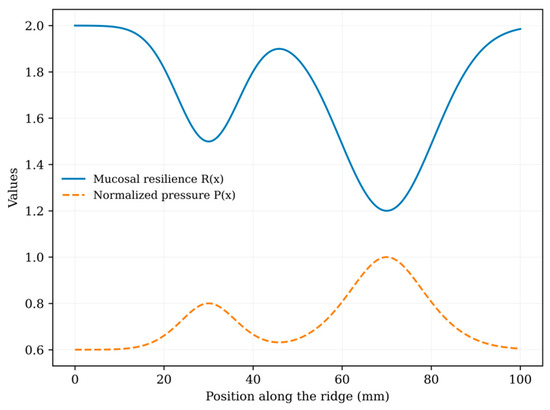
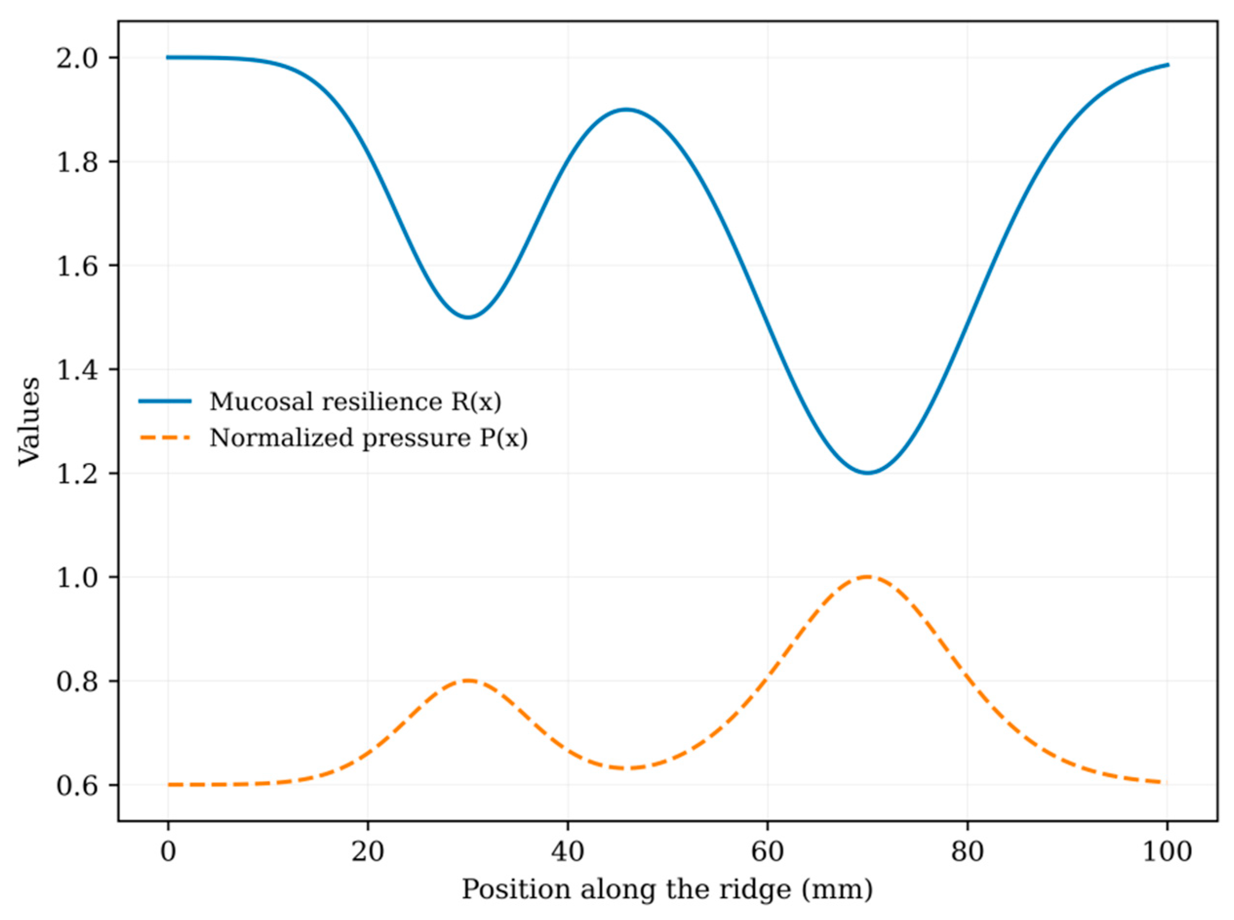
To illustrate the implications of the proposed framework, a simplified numerical example based on representative clinical parameters was considered: the application of a total masticatory force equal to approximately 50 N on a lower complete denture. This force determines a pressure distribution on the support surface, which is a function of the contact area and the local biomechanical properties of the mucosa. The spatial adaptation of the resilience R(x) and the consequent distribution of the normalized pressure P(x) along the arch are illustrated in Figure 1, where the inverse proportionality relationship between tissue deformability and local load can be noted. The force is distributed along the edentulous crest, where the mucosa presents variable thicknesses in three zones (1.3 mm, 1.2 mm, 1 mm), while in the rest of the arch the thickness remains at a standard value of 2 mm. It is acknowledged that this numerical example is based on a single representative set of input parameters and does not constitute a validated simulation. No sensitivity analysis was performed to assess how variations in input values (e.g., applied force, contact area, mucosal thickness, or Young’s modulus) influence the model outputs. As a qualitative indication, it is noted that the calculated stress σ = F/A scales linearly with F and inversely with A; consequently, a 20% change in contact area (from 27 mm2 to approximately 22 or 32 mm2) would produce a proportional change in σ of approximately ±20%. Similarly, the strain ε = σ/E is directly sensitive to the assumed Young’s modulus: the value of 19.75 MPa used here represents an average estimate from a limited data set, and values reported in the literature range from approximately 5 to 70 MPa depending on the tissue site and measurement technique [9]. These ranges indicate that the model results are sensitive to input assumptions and should be interpreted with caution. A formal sensitivity analysis and comparison with finite element results in the literature are considered necessary steps for any future experimental validation of this model. For the calculations, we used the average Young’s modulus of 19.75 MPa, assumed as a representative value of the attached gingiva based on data reported in the literature.
Figure 1.
Graphical representation of the differential model. The blue curve (Mucosal resilience) shows the variation in mucosal thickness/deformability along the ridge (x); the dashed orange curve (normalized pressure) indicates the load distribution. Note how the local pressure maxima coincide with the mucosal resilience minima, highlighting the areas at greatest risk of mechanical overload.
3.1. Local Pressure Concentration Analysis
The pressure function P(x) can be examined to identify stationary points corresponding to local extrema of pressure distribution. Calculation of the stress (σ) on the contact surface of the analyzed points (A = 27 mm2):
σ = F/A = 50 N/27 mm2
Converting the area into SI units (27 mm2 = 27 × 10−6 m2)
σ = 1.85 MPa
From this calculation, we obtain the strain (ε), or the percentage of relative deformation of the tissue with respect to its original thickness:
ε = σ/E = 1.85/19.75 ≈ 0.094 (approximately 9.4%)
The strain value represents a relative deformation of 9.4%, consistent with the elastic behavior of the mucosa under controlled physiological conditions. The interpretation of the local extremes is consistent with the classical conditions described by Fermat’s theorem. Within the proposed framework, the regions corresponding to mucosal thickness minima may coincide with areas of local pressure concentration and can therefore be considered clinically relevant zones for biomechanical assessment. In the model considered, the point with minimum thickness (1 mm) represents the region in which the stress tends to concentrate, representing a clinically critical zone. Under these conditions, the mucosa exhibits a theoretically reduced capacity to dissipate elastic stresses. Within the proposed model, this region could represent a biomechanically relevant area that deserves greater clinical attention. However, it should be noted that this model does not measure or predict clinical phenomena such as pain, ischemia, or prosthesis loosening. These remain theoretical inferences that require independent experimental and clinical verification before any such interpretation can be considered evidence-based.
3.2. Deformation Gradient Analysis
The mean variation in deformation along the arch can be evaluated by comparing the deformation observed in different anatomical regions. Local sagging is defined as:
where the deformation is:
ΔL = L0 · ε
ε = σ/E = 1.85/19.75 ≈ 0.094
Substituting the values in the different zones:
Standard mucosa (2 mm) ΔL = 2 · 0.094 = 0.188
Posterior zone (1.3 mm) ΔL = 1.3 · 0.094 = 0.122
Intermediate zone (1.2 mm) ΔL = 1.2 · 0.094 = 0.113
Anterior zone (1 mm) ΔL = 1 · 0.094 = 0.094
Considering the interval between the standard mucosa and the thinnest zone (d = 20 mm), the average variation in sagging is:
ΔLstd − ΔLmin/d = 0.188 − 0.094/20 mm = 0.0047 mm−1
This value represents the average deformation gradient along the arch. The presence of a deformation gradient indicates that the mucosa does not deform uniformly under load. Since the denture base can be considered rigid compared to the soft tissues, this difference in behavior can translate into a slight inclination of the denture during function. This condition could theoretically cause micro-discontinuities in the contact between the prosthesis and the mucosa, potentially affecting the adhesion mechanisms related to the salivary film. This conclusion is derived exclusively from gradient analysis and does not constitute a direct measurement of suction loss or clinical instability. In particular, the thicker areas tend to compress more, while the thinner areas offer greater relative resistance, generating a condition of mechanical imbalance in the load distribution. From a clinical point of view, this phenomenon can contribute to the reduction of prosthetic stability and the appearance of areas of localized overload (denture lesions), especially in areas with thinner mucosa. In the presence of a rigid prosthetic structure, even modest gradients can translate into functional discontinuities in the contact, amplifying the biomechanical effects at the clinical level. The main biomechanical parameters derived from the application of the model and their correlations with prosthetic stability and tissue health are summarised in Table 1.
Table 1.
Biomechanical parameters derived from the theoretical application of the differential model, based on a single representative numerical example. All values are illustrative and should not be interpreted as clinically predictive.
4. Discussion
4.1. Biomechanical Implications
The application of elementary concepts of differential calculus, in particular the conditions associated with Fermat’s theorem for stationary points and the mean value theorem (Lagrange), provides an interpretive framework that shows how the stability of a complete denture is not defined exclusively by the morphology of the ridge, but also by the management of gingival pressure gradients. In daily clinical practice, prosthetic stability depends on the patient’s anatomical conditions, perfect occlusal adaptation, correction of the prosthetic edges and the prosthetic seal; small, uncontrolled variations often lead to damage of the gingival tissue [10,11]. The results of our model show that a non-uniform distribution of the load generates local differences in deformation. In areas with a thinner mucosa, the increase in stress translates into a more critical deformation gradient compared to the tissue’s response capacity [12,13]. This condition may favor the onset of mucosal lesions, suggesting that the excess load associated with deformation represents a possible biomechanical mechanism underlying denture lesions. The values obtained allow us to quantify and understand that, given a similar relative deformation (ε ≈ 0.094), the absolute sagging varies as a function of the mucosal thickness, ranging from approximately 0.188 mm in the thickest areas to approximately 0.094 in the thinnest areas. The calculated mean gradient (0.0047 mm−1) does not represent a direct clinical value, but rather how the sagging changes between adjacent areas. In the presence of a rigid prosthetic base, even variations of this magnitude can translate into functional micro-misalignments during loading, contributing to the loss of prosthesis-mucosa contact. In this context, the model allows us to introduce a quantitative interpretation of clinically observable but rarely expressed phenomena, transforming apparently modest differences into interpretable biomechanical variables. The references to Fermat’s and Lagrange’s theorems are intended only to provide an intuitive mathematical interpretation of the pressure and strain distributions [14,15]. It should be noted that neither theorem establishes a causal relationship between the identified mathematical conditions and a specific clinical outcome. Their application in this context is purely descriptive and interpretive. This integration of mathematical reasoning and clinical interpretation provides a simplified framework for discussing the biomechanical phenomena associated with complete prostheses. In this context, the two theorems can be considered complementary interpretive tools for describing both the localization and variation of stresses within the proposed framework. Furthermore, the model may offer a theoretical interpretive perspective on certain clinically observed phenomena, such as localized tissue remodeling and the need for relining. The relationship between the pressure gradients described by this model and long-term bone resorption, however, remains speculative and cannot be established from a purely analytical framework. Any association between the strain gradients described here and clinically observed bone remodeling should be considered a hypothesis to be tested, not a conclusion of this study.
4.2. Clinical Implications
A further clinically relevant element concerns occlusal stability; an unbalanced distribution of dental contacts can amplify the pressure gradients already present, accentuating the differences in deformation between areas of different mucosal thickness [16]. A non-homogeneous distribution of pressure over time can in fact determine conditions of chronic overload, favouring processes of remodelling of the supporting tissue [17,18]. In this sense, relining can be interpreted as a clinical adaptation to the biomechanical variations that progressively develop over time [19].
4.3. Comparison with Finite Element Analysis
The proposed conceptual framework is not intended to replace finite element analysis. Rather, it represents a simplified analytical approach that can facilitate biomechanical reasoning and support the interpretation of load redistribution mechanisms. FEA allows a detailed and three-dimensional simulation of the stress distribution, taking into account complex geometries and heterogeneous material properties [20]. However, these approaches require high technical skills and computational tools that are not always available in daily clinical practice. The presented differential model, despite its simplification, allows obtaining useful information on the load distribution starting from clinically accessible parameters, configuring itself as a possible intermediate tool between empirical evaluation and advanced numerical simulation. A direct quantitative comparison between the outputs of the present model and published finite element analyses of complete prosthetic systems [5,13] was not performed, since the simplifying assumptions of the present framework (one-dimensional domain, static loading, linear material behavior) are not directly comparable with the three-dimensional, viscoelastic, patient-specific conditions modeled in the FEA studies. Such a comparison would require a systematic reformulation of the model under corresponding boundary conditions, which is beyond the scope of this conceptual article and is identified as a priority for future work. Overall, the proposed model is configured as a useful theoretical tool for connecting the mathematical description to clinical practice, offering a more integrated reading of the behavior of the prosthesis-mucosa system. The integration of the proposed differential model within modern digital systems, implementation in the CAD/CAM flow, would allow overcoming the limit of manual empirical retouching. Such integration remains a speculative long-term prospect that would require prior experimental validation, calibration of the model parameters based on patient needs, and clinical studies evaluating its impact on prosthetic outcomes. No clinical benefit can be attributed to this model until these steps are completed.
4.4. Limitations
This study presents several limitations that must be clearly acknowledged. First, the model is one-dimensional and therefore unable to capture the three-dimensional complexity of the real denture-mucosa interface. Second, all loading conditions are assumed to be static, while masticatory forces are inherently dynamic and cyclic. Third, the mucosal tissue is modeled as a linear elastic material, neglecting its well-documented viscoelastic and anisotropic behavior. Fourth, the biomechanical coefficient k is currently undefined and cannot be determined from the data presented in this study. Fifth, the numerical example is based on a single set of representative parameters, and no formal sensitivity analysis was conducted. Sixth, the model has not been validated against experimental measurements or finite element analysis results. These limitations collectively restrict the model to the role of a conceptual, hypothesis-generating framework and preclude any direct clinical application at this stage. This work should not be interpreted as a validated predictive model, but rather as a hypothesis-generating analytical framework that aims to stimulate future experimental and computational investigations.
4.5. Future Perspectives
Future validation of this framework should proceed along four main directions: (1) controlled indentation measurements on oral mucosal samples to calibrate the biomechanical coefficient k; (2) systematic comparison of model results with published finite element data under equivalent boundary conditions; (3) prospective clinical studies correlating quantitative measurements of mucosal resilience with prosthetic outcomes in complete denture wearers; (4) extension of the one-dimensional formulation to a three-dimensional spatial domain, capable of capturing the full geometric and mechanical complexity of the denture bearing surface.
5. Conclusions
This work proposes a differential theoretical framework to describe the relationship between mucosal resilience and pressure distribution in complete dentures. Rather than constituting a predictive biomechanical model, the proposed formulation should be considered a hypothesis-generating approach, aimed at translating a clinically recognized phenomenon into a continuous mathematical representation. The study’s main contribution lies in conceptualizing mucosal resilience as a spatially variable that can influence load redistribution along the prosthetic support area. This perspective suggests that prosthetic evaluation should not be based solely on geometric or anatomical considerations but should also take into account the mechanical variability of supporting tissues, even within a simplified analytical framework. From a clinical perspective, the proposed approach can support biomechanical reasoning during prosthetic design. In digital workflows, mucosal resilience information could be integrated into CAD/CAM procedures to guide local modifications of the prosthetic base. This possibility, although conceptually well-founded, remains entirely prospective and depends on the experimental validation and clinical calibration of the model parameters described in this study.
Similarly, in conventional techniques, selective compensatory strategies could be considered in areas characterized by reduced mucosal thickness. Although the model is based on simplifying assumptions and has not yet been calibrated or experimentally validated, it may provide a useful theoretical basis for future research integrating clinical measurements, finite element analyses, and patient-specific biomechanical data. It must be explicitly acknowledged that the current model has not been validated experimentally, in vitro, or clinically. The theoretical predictions presented here should therefore be interpreted as conceptual hypotheses rather than clinically applicable results. Comparison with finite element analysis results and correlation with clinical observations are considered necessary steps before any practical application of this model can be considered. Further studies will be needed to define the biomechanical coefficient k, evaluate the sensitivity of the model and verify the correspondence between the proposed theoretical framework and experimental observations.
Author Contributions
Conceptualization and methodology, S.C. and F.C.; writing—original draft preparation and writing—review and editing, A.B. and D.L.; supervision, S.C. and D.L.; validation, G.C. and F.C. G.C. and F.C. contributed equally to this work. All authors have read and agreed to the published version of the manuscript.
Funding
This research received no external funding.
Institutional Review Board Statement
Not applicable.
Informed Consent Statement
Not applicable.
Data Availability Statement
Data from this study are available upon reasonable request by writing to the corresponding author.
Conflicts of Interest
The authors declare no conflicts of interest.
Abbreviations
| Symbols | |
| X | spatial coordinate along the arch (mm) |
| P(x) | local pressure exerted by the prosthesis (Pa or MPa) |
| R(x) | mucosal resilience (vertical deformation under load) (mm) |
| k | biomechanical coupling coefficient [Pa/mm or MPa/m]; describes the proportionality between the spatial gradients of mucosal resilience and pressure redistribution; currently not numerically defined and subject to future experimental calibration |
| F | masticatory force |
| E | Young’s modulus |
| A | contact area |
| ε | strain (relative deformation) |
| σ | stress (tension) |
| L0 | initial thickness of the mucosa |
| ΔL | absolute deformation |
| d | distance between two points of the arch |
| Legends to formulas | |
| P(x) | f(R(x)); pressure-resilience relationship |
| dP(x)/dx | −k · dR (x)/dx; differential equation of the modulus |
| ΔL | L0 · ε; Absolute strain |
| ΔL1 − ΔL2/d | mean strain rate |
| σ | F/A; stress |
| ε | σ/E; Hooke’s law |
References
- Paras, A.; Ma, S.; Waddell, J.N.; Choi, J.J.E. Denture–Mucosa Pressure Distribution and Pressure–Pain Threshold in In Vivo, In Vitro and In Silico Studies: A Literature Review. Oral 2022, 2, 112–125. [Google Scholar] [CrossRef] [Scilit]
- Paras, A.; Ma, S.; Waddell, J.N.; Choi, J.J.E. Real-time in vitro measurement of denture-mucosa pressure distribution in a typical edentulous patient with and without implants: Development of a methodology. J. Mech. Behav. BioMed Mater. 2021, 119, 104531. [Google Scholar] [CrossRef] [Scilit] [PubMed]
- Haruta, A.; Matsushita, Y.; Tsukiyama, Y.; Sawae, Y.; Sakai, N.; Koyano, K. Effects of mucosal thickness on the stress distribution and denture stability of mandibular implant-supported overdentures with unsplinted attachments in vitro. J. Dent. Biomech. 2011, 2011, 894395. [Google Scholar] [CrossRef] [Scilit] [PubMed]
- Wang, C.X.; Rong, Q.G.; Zhu, N.; Ma, T.; Zhang, Y.; Lin, Y. Finite element analysis of stress in oral mucosa and titanium mesh interface. BMC Oral Health 2023, 23, 25. [Google Scholar] [CrossRef] [Scilit] [PubMed]
- Lee, J.-H.; Seo, J.-H.; Park, S.-W.; Kim, W.-G.; Jung, T.-G.; Lee, S.-J. A Finite Element Analysis Study of Edentulous Model with Complete Denture to Simulate Masticatory Movement. Bioengineering 2024, 11, 336. [Google Scholar] [CrossRef] [Scilit] [PubMed]
- Caggiano, M.; D’Ambrosio, F.; Acerra, A.; Giudice, D.; Giordano, F. Biomechanical Implications of Mandibular Flexion on Implant-Supported Full-Arch Rehabilitations: A Systematic Literature Review. J. Clin. Med. 2023, 12, 5302. [Google Scholar] [CrossRef] [Scilit] [PubMed] [PubMed Central]
- Giordano, F.; Di Spirito, F.; Acerra, A.; Rupe, A.; Cirigliano, G.; Caggiano, M. The outcome of tilted distal implants immediately loaded under screw-retained cross-arch prostheses. A 5-year retrospective cohort study. J. Osseointegration 2024, 16, 31–38. [Google Scholar] [CrossRef]
- Shala, K.; Tmava-Dragusha, A.; Dula, L.; Pustina-Krasniqi, T.; Bicaj, T.; Ahmedi, E.; Lila, Z. Evaluation of Maximum Bite Force in Patients with Complete Dentures. Open Access Maced. J. Med. Sci. 2018, 6, 559–563. [Google Scholar] [CrossRef] [Scilit] [PubMed] [PubMed Central]
- Goktas, S.; Dmytryk, J.J.; McFetridge, P.S. Biomechanical behavior of oral soft tissues. J. Periodontol. 2011, 82, 1178–1186. [Google Scholar] [CrossRef] [Scilit] [PubMed]
- Alzoubi, N.S.; Kuraitby, S.; Almustafa, A. Investigation of traumatic ulcer locations and healing progression during denture adjustment periods in Syrian complete denture patients. Sci. Rep. 2025, 15, 23890. [Google Scholar] [CrossRef] [Scilit] [PubMed] [PubMed Central]
- Findler, M.; Perzon, O.; Almoznino, G.; Zini, A.; Sharav, Y.; Czerninski, R.; Aframian, D.J.; Haviv, Y. Unveiling denture-induced oral lesions: A comprehensive study on classification and pain assessment. J. Oral Rehabil. 2024, 51, 931–937. [Google Scholar] [CrossRef] [Scilit] [PubMed]
- Chen, J.; Ahmad, R.; Li, W.; Swain, M.; Li, Q. Biomechanics of oral mucosa. J. R. Soc. Interface 2015, 12, 20150325. [Google Scholar] [CrossRef] [Scilit] [PubMed] [PubMed Central]
- Fatola, D.; Nasution, I.D.; Sabri, M.; Chairunnisa, R. Pain-related analysis on a resorbed ridge with various denture occlusal schemes using finite element method. Braz. Dent. J. 2024, 35, 5798. [Google Scholar] [CrossRef] [Scilit] [PubMed] [PubMed Central]
- Hunter, P.J.; Nielsen, P.M.; Smaill, B.H.; LeGrice, I.J.; Hunter, I.W. An anatomical heart model with applications to myocardial activation and ventricular mechanics. Crit. Rev. Biomed. Eng. 1992, 20, 403–426. [Google Scholar] [PubMed]
- Zhao, H.; Kamensky, D.; Hwang, J.T.; Chen, J.S. Automated shape and thickness optimization for non-matching isogeometric shells using free-form deformation. Eng. Comput. 2024, 40, 3495–3518. [Google Scholar] [CrossRef] [Scilit] [PubMed] [PubMed Central]
- Wang, Z.; Su, Y.; Wang, J.; Liu, Y.; Xing, W. Occlusal parameters and wear of artificial teeth in complete dentures with lingualized versus bilateral balanced occlusion: A randomized clinical trial. BMC Oral Health 2024, 24, 1405. [Google Scholar] [CrossRef] [Scilit] [PubMed] [PubMed Central]
- Aslan, E.; Dundar, N.; Mutlu, O. Quantitative Assessment of Long-Term Changes in Peri-Implant Bone Structure in Patients Using Fixed and Removable Prostheses. Int. J. Oral Maxillofac. Implant. 2025, 1–24. [Google Scholar] [CrossRef] [Scilit] [PubMed]
- Pham, N.Q.; Gonda, T.; Maeda, Y.; Ikebe, K. Average rate of ridge resorption in denture treatment: A Systematic Review. J. Prosthodont. Res. 2021, 65, 429–437. [Google Scholar] [CrossRef] [Scilit] [PubMed]
- Kuralt, M.; Selmani Bukleta, M.; Kuhar, M.; Fidler, A. Bone and soft tissue changes associated with a removable partial denture. A novel method with a fusion of CBCT and optical 3D images. Comput. Biol. Med. 2019, 108, 78–84. [Google Scholar] [CrossRef] [Scilit] [PubMed]
- Suenaga, H.; Chen, J.; Yamaguchi, K.; Li, W.; Sasaki, K.; Swain, M.; Li, Q. Mechanobiological bone reaction quantified by positron emission tomography. J. Dent. Res. 2015, 94, 738–744. [Google Scholar] [CrossRef] [Scilit] [PubMed]

Disclaimer/Publisher’s Note: The statements, opinions and data contained in all publications are solely those of the individual author(s) and contributor(s) and not of MDPI and/or the editor(s). MDPI and/or the editor(s) disclaim responsibility for any injury to people or property resulting from any ideas, methods, instructions or products referred to in the content. |
© 2026 by the authors. Licensee MDPI, Basel, Switzerland. This article is an open access article distributed under the terms and conditions of the Creative Commons Attribution (CC BY) license.