


Abstract
Background: Elderly patients often experience difficulty adapting to occlusal reconstruction. Therefore, accurate bite registration is mandatory to ensure precise transfer of the established maxillomandibular relationship to the definitive prosthesis. However, conventional bite registration methods may compromise reproducibility because of deformation of registration materials and instability of mucosa-supported record bases. Methods: A 65-year-old woman with the primary complaints of unstable occlusion and difficulty in mastication underwent occlusal reconstruction. After occlusal stabilization using provisional crowns, bridges, and removable partial dentures, definitive impressions were made with and without the provisional restorations. The casts were scanned, and the digital datasets were superimposed to reproduce the established occlusal morphology of the provisional restorations. This occlusal morphology was used to design a tooth-supported computer-aided design–computer-aided manufacturing (CAD–CAM)-fabricated milled provisional restoration. Following intraoral verification of occlusal stability, the milled provisional restoration served as a mounting guide for the working casts on an articulator. Definitive crowns, bridges, and removable partial dentures were then fabricated. Results: Following comprehensive prosthodontic rehabilitation with definitive prostheses, occlusal stability and masticatory function improved, and the patient was satisfied with the functional outcomes of treatment. Conclusions: A tooth-supported CAD–CAM-fabricated milled provisional restoration used as a bite registration device enables potentially more consistent transfer of the maxillomandibular relationship while avoiding mucosal displacement and material deformation. This technique, which integrates digital and conventional workflows, may provide a new option for addressing adaptation challenges in occlusal reconstruction.
1. Introduction
Occlusal reconstruction plays a critical role in restoring oral function and quality of life in elderly patients with extensive tooth loss [1]. However, successful outcomes depend not only on the fabrication of definitive prostheses but also on the patient’s ability to adapt to the reconstructed occlusion. Age-related changes in neuromuscular coordination and sensory feedback may compromise adaptation to new occlusal schemes, making elderly patients particularly susceptible to occlusal instability and functional discomfort following occlusal reconstruction [2,3].
Accurate bite registration is therefore essential in occlusal reconstruction to ensure precise transfer of the established maxillomandibular relationship to the definitive prostheses [4]. In clinical practice, provisional restorations are often used to stabilize occlusion over an extended period, allowing neuromuscular adaptation and functional harmonization [5]. The occlusal morphology achieved during this provisional phase represents a clinically validated reference that should ideally be reproduced in the definitive prosthetic reconstruction [6].
Conventional bite registration techniques, however, present inherent limitations in this context. Registration materials are susceptible to deformation, and mucosa-supported record bases may lack stability, particularly in patients with extensive edentulism [7,8]. These factors can reduce the reproducibility of the established occlusal relationship and introduce discrepancies during articulator mounting, potentially undermining the functional success of the definitive prostheses.
Recent advances in digital dentistry have enabled the integration of computer-aided design–computer-aided manufacturing (CAD–CAM) technology into prosthodontic workflows, offering improved precision and reproducibility [9,10,11]. In particular, digitally designed and milled provisional restorations provide a tooth-supported and dimensionally stable alternative that may overcome the limitations of conventional bite registration methods [12]. By capturing and transferring the occlusal morphology established during the provisional phase, such restorations may serve not only as interim prostheses but also as reliable bite registration devices.
The purpose of this case report is to describe a digital–conventional workflow in which a tooth-supported CAD–CAM-fabricated milled provisional restoration was used as a bite registration device in occlusal reconstruction. This approach facilitates more consistent transfer of the established maxillomandibular relationship to the definitive prostheses while potentially reducing sources of error associated with mucosal displacement and material deformation.
2. Case Report
2.1. Patient Information and Initial Examination
A 65-year-old woman visited our hospital with primary complaints of unstable occlusion and mastication difficulty.
At the initial examination, the occlusal condition was systematically assessed in terms of occlusal contacts, occlusal plane, and occlusal relationship in maximum intercuspation. Bilaterally uneven occlusal contacts were observed, and disruption of the occlusal plane due to extrusion of the mandibular left second premolar and the maxillary right second molar was identified. In the anterior region, disharmony of the incisal edge position and tooth arrangement resulted in inadequate anterior guidance, accompanied by esthetic discrepancies in the anterior restorations.
The existing removable partial dentures showed insufficient support and stability due to the absence of a rest on the mandibular right first premolar, as well as poor adaptation of the intaglio surface. As a result, denture displacement occurred during function, causing pain in the underlying mucosa and contributing to masticatory instability. Muscles or temporomandibular joint symptoms were not observed.
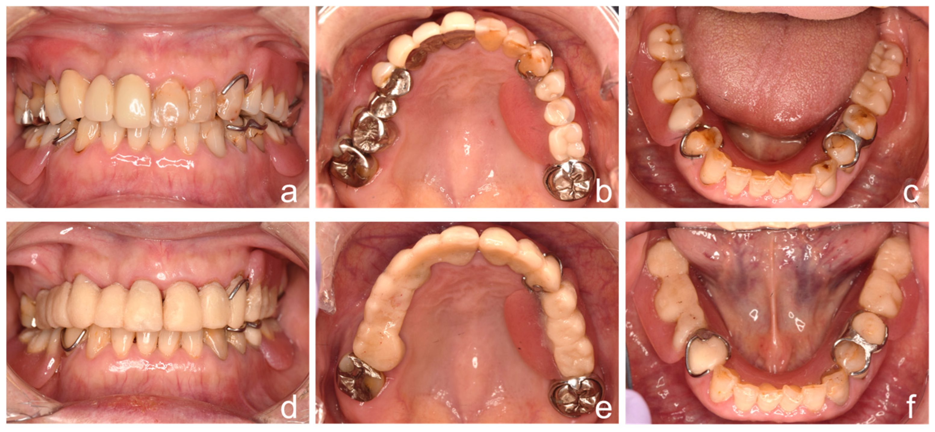
The vertical dimension of occlusion was evaluated using the Willis method and assessment of the interocclusal rest space, which was measured at approximately 3 mm and considered within the normal range. Therefore, alteration of the vertical dimension was not required (Figure 1a–c).
Figure 1.
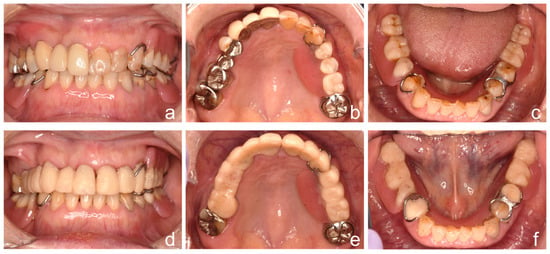
Fabrication of provisional prostheses and modification of existing dentures. (a) Intraoral frontal, (b) maxillary occlusal, and (c) mandibular occlusal views with existing dentures at the initial examination. (d) Intraoral frontal view after occlusal stabilization using provisional restorations and modified existing dentures. (e) Maxillary occlusal view after occlusal stabilization. (f) Mandibular occlusal view after occlusal stabilization.
2.2. Fabrication of Provisional Prostheses and Modification of Existing Dentures
The right maxillary first premolar was extracted due to a root fracture. Centric relation was recorded using a bimanual manipulation technique, with care taken to maintain a consistent natural head position because head position may influence occlusal contact during bite registration procedures [13]. The resulting maxillomandibular relationship was then used for articulator mounting, followed by diagnostic waxing. Based on the diagnostic waxing, provisional crowns and bridges were fabricated and placed intraorally. The existing dentures were modified using acrylic resin (UNIFAST II; GC Corp., Tokyo, Japan) at chairside (Figure 1d–f), including occlusal surface adjustment, relining of the intaglio surface, and addition of a rest.
Following these modifications, mucosal pain was reduced, and an improvement in patient comfort was observed. Consequently, the patient demonstrated improved masticatory performance, particularly when chewing harder and fibrous foods. The patient also reported improved esthetic satisfaction and no phonetic impairment.
Masticatory function was objectively evaluated using a glucose-containing gummy (Glucorum; GC Corp., Tokyo, Japan) and a glucose analyzer (Glucosensor GS-IIN; GC Corp., Tokyo, Japan) [14], demonstrating an increase from 77 to 183 mg/dL. The measured glucose concentration reflects the amount of glucose released from the test material into a buffer solution and serves as an index of masticatory function [15].
2.3. Milled Provisional Restorations for Bite Registration
The transferred maxillomandibular relationship was defined as the functionally adapted occlusal position established during the provisional phase after clinical stabilization and was no longer coincident with the initial centric relation record.
After occlusal stabilization, two definitive impressions of the maxilla were made using a silicone elastomeric impression material (Examix Fine; GC Corp., Tokyo, Japan). First, an impression was taken with the provisional crowns and bridges in place together with the existing removable partial denture. This impression was used to fabricate a stone cast representing the intraoral condition with the provisional prostheses in situ. Second, an impression of the prepared abutment teeth was taken to fabricate the working cast.
Both impressions were made using the same silicone elastomeric material and standardized procedures to minimize variability related to soft tissue displacement, thereby ensuring consistency between the datasets used for subsequent digital superimposition.
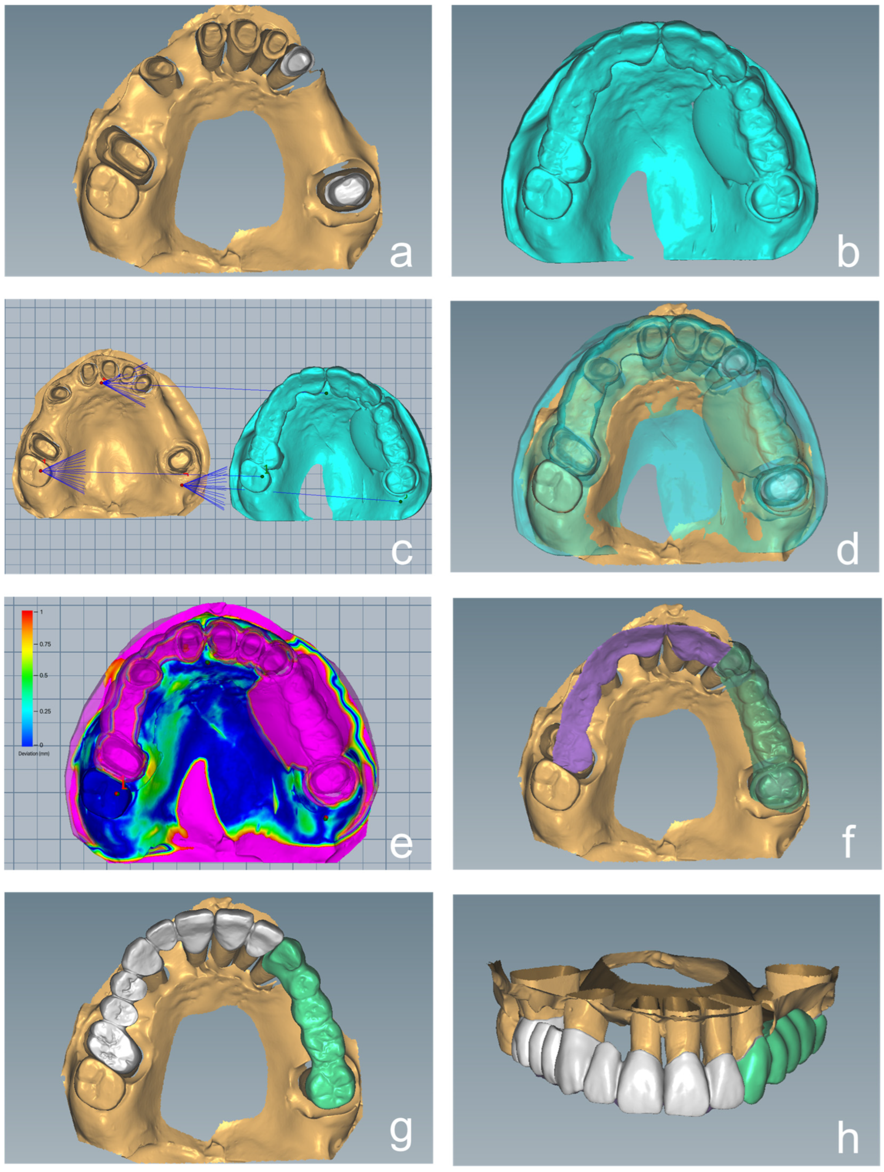
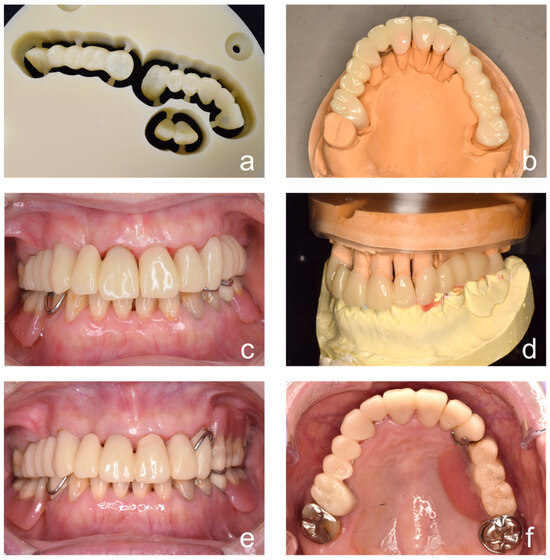
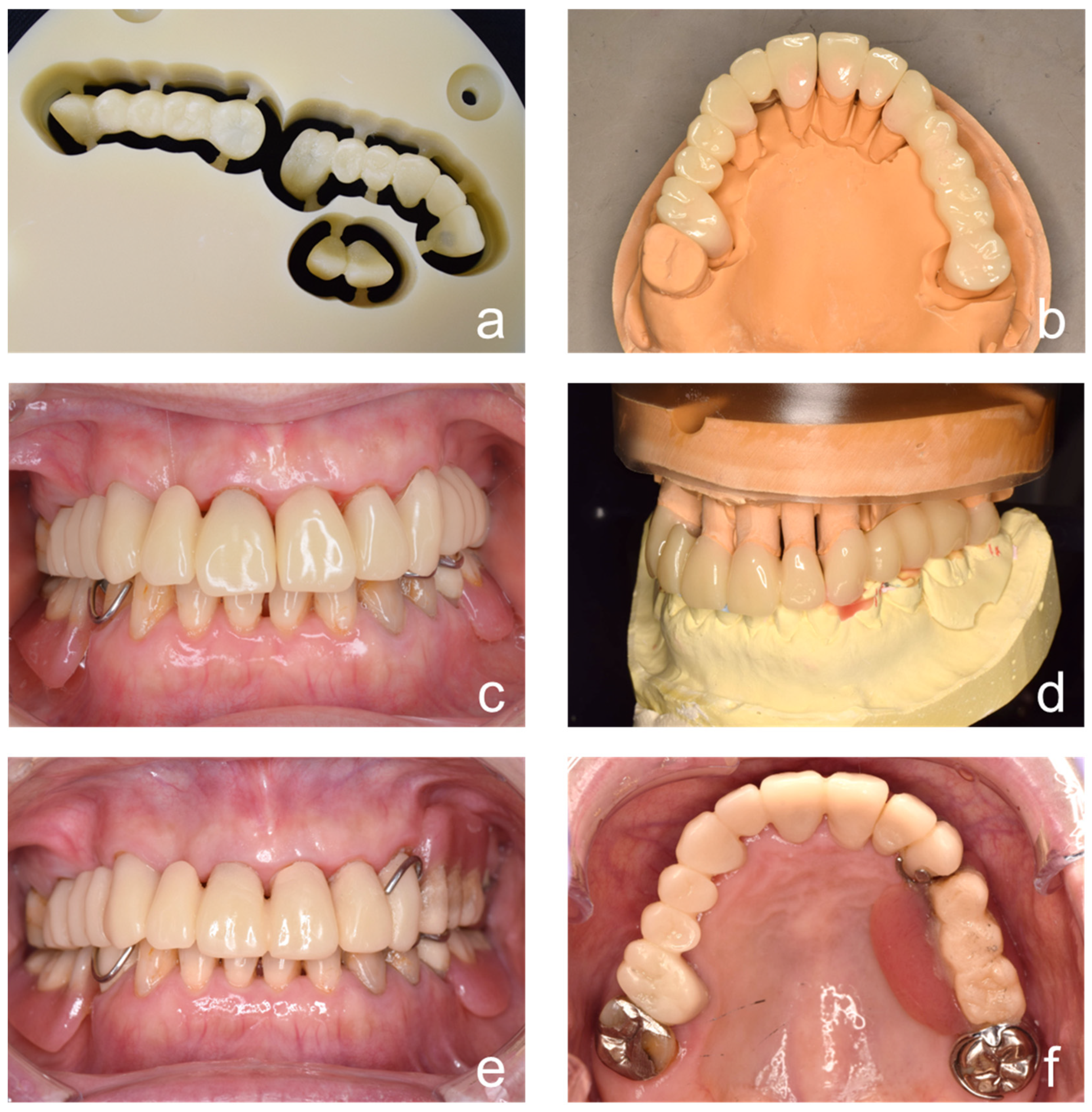
The working cast and cast of the provisional prostheses were scanned using a desktop laboratory scanner (T-Series; Medit, Seoul, Republic of Korea), and the resulting datasets were imported into CAD software (Ceramill Mind, version 1.0; Amann Girrbach, Maeder, Austria) (Figure 2a,b). Initial alignment was performed using three reference points with minimal displacement between the datasets: the mesiolingual cusp tip of the maxillary right second molar, the center of the incisive papilla, and the left maxillary tuberosity (Figure 2c). Although extrusion of the maxillary right second molar was observed at the initial examination, occlusal adjustment had already been completed before both scan acquisitions. This was followed by global best-fit matching using an iterative closest point algorithm based on stable anatomical structures shared between the datasets, including the palatal vault, incisive papilla, maxillary tuberosity, and residual non-restored tooth surfaces (Figure 2d). The agreement between the datasets was assessed using a color-coded surface deviation map (Figure 2e), and the analysis demonstrated close correspondence between the aligned datasets, with a root mean square error (RMS) of 0.160 mm, a mean deviation of 0.159 mm, and a 95th percentile deviation of 0.439 mm. The occlusal surfaces of the provisional crowns, as well as those of the fixed and removable partial dentures, were digitally extracted (Figure 2f). Provisional restorations incorporating the digitally extracted occlusal surfaces were designed (Figure 2g,h) and then milled from a polymethyl methacrylate disk (Asahi Roentgen) and a milling machine (Ceramill Motion 2; Amann Girrbach, Maeder, Austria; Figure 3a,b).
Figure 2.
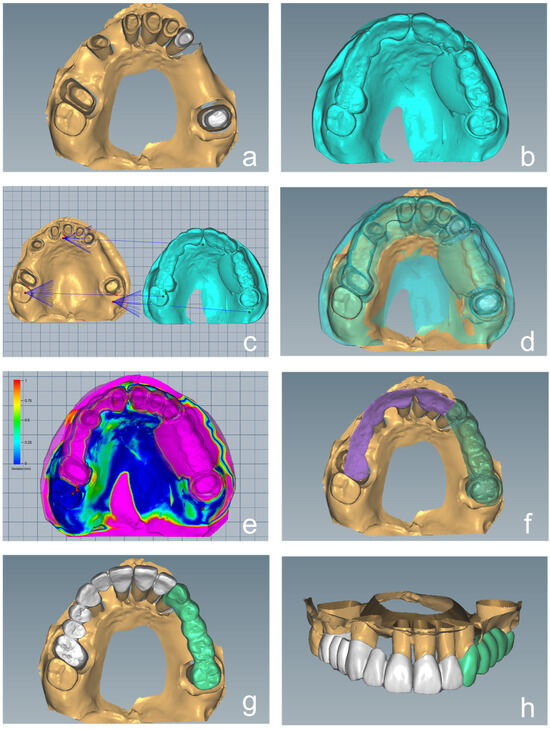
Design of the milled provisional restorations. (a) Digitally scanned maxillary working cast. (b) Digitally scanned cast of the maxillary provisional prostheses. (c) Initial alignment using three anatomical reference points (mesiolingual cusp tip of the maxillary right second molar, center of the incisive papilla, and left maxillary tuberosity). (d) Digital superimposition of the maxillary working cast and the cast of the maxillary provisional prostheses. (e) Color-coded surface deviation map demonstrating the agreement between the superimposed datasets. The color scale indicates deviation magnitude in millimeters (mm), with blue representing minimal deviation and red representing larger deviation values. Pink regions indicate areas outside the comparison range or non-overlapping regions. (f) Digital extraction of the occlusal surface of the maxillary provisional prostheses. (g) Occlusal view of the digitally designed provisional restorations incorporating the extracted occlusal surfaces. (h) Frontal view of the digitally designed provisional restorations incorporating the extracted occlusal surfaces.
Figure 3.
Milled provisional restorations for bite registration. (a) Milling of the digitally designed provisional restorations. (b) Occlusal view of the milled provisional restorations. (c) Intraoral confirmation of the occlusal stability of the milled provisional restorations. (d) Mounted working casts using the milled provisional restorations. (e) Intraoral frontal and (f) intraoral occlusal views with the milled provisional restorations and modified existing denture.
The milled provisional restorations were placed intraorally onto the abutment teeth, and occlusal stability was confirmed (Figure 3c). The maxillary cast was mounted on a semi-adjustable articulator (Proarch III EG; Shofu Inc., Kyoto, Japan) using a face-bow transfer. The mandibular cast was then mounted using the occlusal surfaces of the milled provisional restorations, which determined the maxillomandibular relationship (Figure 3d, Supplementary Figure S1). The removable partial denture component of the milled provisional restoration was removed, and the existing denture was reinserted and used together with the remaining fixed restorations as provisional prostheses (Figure 3e,f).
2.4. Fabrication of the Definitive Prostheses
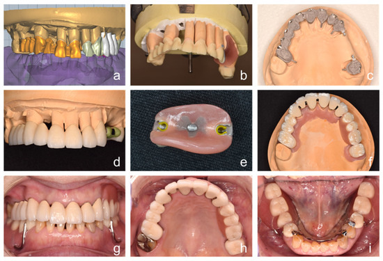
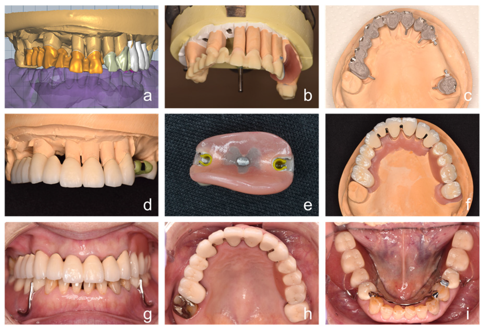
To fabricate the metal substructures of the definitive crowns and fixed partial dentures, digital cutback was performed on the provisional restorations (Figure 4a). Wax patterns with the cutback design were milled from wax disks (Japan Dental Supply, Tokyo, Japan) (Figure 4b) and converted into gold-silver palladium alloy-based substructures (Figure 4c). After extracoronal attachments (Mini-SG attachment; Cendres + Métaux, Biel, Switzerland) had been soldered to the metal substructures, resin veneering was performed using a hybrid composite resin (GRADIA; GC Corp., Tokyo, Japan) (Figure 4d). A full-contour provisional restoration fabricated by CAD–CAM was used to create a silicone index, which served as a guide for controlling veneering thickness and final morphology.
Figure 4.
Fabrication of the definitive prostheses. (a) Digital cutback performed on the provisional restorations. (b) Milled wax patterns with the cutback design. (c) Metal substructures incorporating extracoronal attachments. (d) Resin-veneered crowns. (e) Removable maxillary partial denture with extracoronal attachments. (f) Definitive fixed and removable maxillary prostheses. (g) Intraoral frontal, (h) maxillary occlusal, and (i) mandibular occlusal views with definitive prostheses.
Using a refractory cast, a removable partial denture incorporating the female components of the extracoronal attachments was fabricated (Figure 4e,f). After delivery of the definitive maxillary prostheses, a mandibular removable partial denture was fabricated using the altered cast technique.
2.5. Clinical Outcome
The patient was satisfied with the functional outcomes of the treatment and reported no complications (Figure 4g–i). The patient did not experience difficulty in adapting to the definitive prostheses. The patient’s score on the Japanese version of the Oral Health Impact Profile (OHIP-J54) [16] improved from 105 before treatment to 17 after treatment. Additionally, the patient’s results on the masticatory function test, performed using a glucose-containing gummy and glucose analyzer as described in Section 2.2, increased from 77 mg/dL before treatment to 183 mg/dL after occlusal stabilization with provisional restorations and modified dentures, and further improved to 251 mg/dL after delivery of the definitive prostheses. The measured glucose concentration reflects the amount of glucose released from the test material into a buffer solution and serves as an index of masticatory function rather than a systemic blood glucose value.
3. Discussion
The present technique focuses on the transfer of the established maxillomandibular relationship to the definitive prosthesis to support neuromuscular coordination in elderly patients undergoing occlusal reconstruction. In this case, occlusal stability was first achieved using provisional restorations, allowing the patient to adapt functionally to a harmonized mandibular position. The established maxillomandibular relationship was subsequently transferred to the definitive prostheses using a tooth-supported CAD–CAM-milled provisional restoration as a bite registration device.
A key advantage of this approach is the use of a rigid, tooth-supported structure for bite registration. In contrast to conventional methods that rely on mucosa-supported record bases, which are susceptible to displacement and deformation, the present technique provides a stable reference based on abutment teeth. This structural stability potentially reduces sources of error associated with soft tissue resilience and material distortion [17,18], thereby supporting more consistent transfer of the maxillomandibular relationship during articulator mounting. As a result, the neuromuscularly adapted mandibular position established during the provisional phase may contribute to more consistent transfer to the definitive prosthesis.
Clinically, this technique may be particularly beneficial in elderly patients, in whom adaptation to new occlusal conditions can be compromised [19]. Transferring the functionally established maxillomandibular relationship may facilitate smoother adaptation and reduce post-treatment discomfort, consistent with the improved functional outcomes observed in this case. Hence, this technique, which integrates digital and conventional workflows, may provide a new option for addressing the adaptation challenges experienced by older adults during occlusal reconstruction.
CAD–CAM-milled provisional restoration offers practical advantages in terms of duplication and remanufacturing [20]. This reproducibility is supported by the favorable marginal and internal fit reported for CAD–CAM-fabricated provisional restorations compared with conventionally fabricated restorations [21]. In addition, because the design is stored as digital data, the restoration can be readily reproduced in the event of fracture or loss, without interrupting the treatment sequence. Furthermore, the same dataset can be used to fabricate metal substructures through a digital cutback workflow [22], enhancing consistency between provisional and definitive prostheses.
Although a similar concept has been previously reported [12], in which occlusal information from provisional restorations is transferred within a fully digital workflow, the present technique differs in its clinical implementation. Digital workflows offer clinical advantages but may have limitations in capturing certain anatomical details, particularly subgingival finish lines and edentulous ridge morphology. The present technique addresses these limitations by integrating digital and conventional procedures. The use of conventional impression techniques enables reliable acquisition of clinically relevant anatomical information, while model and articulator-based workflows ensure compatibility with established laboratory procedures, including the lost-wax method required for cases involving extracoronal attachments. As a result, the applicability of occlusal transfer workflows may be extended to a wider range of cases, including those involving both fixed and removable prosthodontic components. In addition, future integration with artificial intelligence-based approaches for occlusal prediction and mandibular position analysis may further improve the efficiency and predictability of digital prosthodontic workflows [23].
A limitation of this study is the lack of quantitative evaluation of the fit of the milled provisional restorations and the accuracy of occlusal transfer. Detailed measurement of marginal and internal fit and quantitative evaluation of occlusal transfer were not performed in this report. Further studies incorporating quantitative analyses are warranted to validate the accuracy of this approach.
Another limitation of the present study is the potential inaccuracy associated with the ICP-based superimposition process. Although standardized impression procedures were used, residual differences in mucosal compression at the palatal vault between the two impressions may have influenced the ICP-based superimposition process and represent a potential source of alignment error. Therefore, further development of clinical procedures that minimize impression-related soft tissue deformation and improve the reliability of digital superimposition is warranted.
In the present study, the agreement between the datasets was assessed using a color-coded surface deviation map, which demonstrated an RMS of 0.160 mm, a mean deviation of 0.159 mm, and a 95th percentile deviation of 0.439 mm. The RMS and mean deviation values obtained in the present study were generally consistent with previously reported alignment accuracy ranges [24]. A previous study evaluating superimposition accuracy between CBCT datasets and digital dental casts has reported 95th percentile deviation values ranging from 0.30 to 1.90 mm in complex digital workflows [25]. In addition, another study comparing digital registration methods and CBCT-based evaluation for implant position assessment has suggested that a 95% Hausdorff Distance below 0.53 mm may represent a clinically acceptable threshold [26]. Although the 95% Hausdorff Distance and the 95th percentile deviation used in the present study are not identical metrics, both reflect localized large-deviation characteristics within aligned datasets.
On the other hand, a previous investigation evaluating long-span intraoral scanning and digital alignment has demonstrated that stitching-related and spatial alignment errors may accumulate during full-arch or large-span workflows, potentially increasing localized deviation values, including the 95th percentile deviation [27]. Therefore, superimposition accuracy should be interpreted comprehensively using multiple quantitative parameters rather than a single metric alone. Taken together, the quantitative superimposition results in the present study suggested reasonable overall agreement between the aligned datasets.
This report is based on a single case; therefore, statistical analysis was not performed and the findings cannot be generalized. The pre- and post-treatment OHIP-J scores and masticatory function test results are presented descriptively to illustrate the clinical changes observed in this case. The observed improvements should be interpreted in the context of comprehensive prosthodontic rehabilitation rather than being solely attributed to the specific technique. While these findings suggest favorable clinical outcomes, further studies with larger sample sizes are required to confirm its clinical applicability.
Supplementary Materials
The following supporting information can be downloaded at: https://www.mdpi.com/article/10.3390/prosthesis8060053/s1, Figure S1: Procedure for bite registration and articulator mounting.
Author Contributions
Conceptualization, clinical treatment, writing—original draft preparation, T.O.; conceptualization, writing—original draft preparation, T.K.; technical fabrication of the prosthesis, T.H.; writing—review and editing, supervision, H.E. All authors have read and agreed to the published version of the manuscript.
Funding
This research received no external funding.
Institutional Review Board Statement
According to the Japanese Ethical Guidelines for Medical and Biological Research Involving Human Subjects, Chapter 1, Section 2–8 (Definitions), which are followed by Tohoku University, individual case reports conducted for educational or academic purposes are not considered “research” under these Guidelines and are therefore exempt from institutional review board approval.
Informed Consent Statement
Informed consent was obtained from all subjects involved in the study. Written informed consent has been obtained from the patient to publish this paper.
Data Availability Statement
The raw data supporting the conclusions of this article will be made available by the authors on request.
Acknowledgments
The authors thank the team at the Dental Laboratory of Tohoku University Hospital for providing professional technical services.
Conflicts of Interest
The authors declare no conflicts of interest.
References
- Ohtake, T.; Kondo, T.; Bhattarai, S.; Harata, T.; Egusa, H. A technique for denture fabrication using 3D-printed duplicated removable partial dentures. Int. J. Prosthodont. 2025. Epub ahead of printing. [Google Scholar] [CrossRef] [Scilit]
- Sessle, B.J. The implications of changing demographics and brain plasticity to prosthodontics. J. Prosthodont. Res. 2024, 68, vi. [Google Scholar] [CrossRef] [Scilit]
- Müller, F. A Lifelong “dental identity”: New opportunities in the era of digital dentistry. Int. J. Prosthodont. 2025, 38, 7–8. [Google Scholar] [CrossRef]
- Ghazal, M.; Albashaireh, Z.; Kern, M. The ability of different materials to reproduce accurate records of interocclusal relationships in the vertical dimension. J. Oral Rehabil. 2008, 35, 816–820. [Google Scholar] [CrossRef] [Scilit]
- Heckmann, S.M.; Heussinger, S.; Linke, J.J.; Graef, F.; Pröschel, P. Improvement and long-term stability of neuromuscular adaptation in implant-supported overdentures. Clin. Oral Implant. Res. 2009, 20, 1200–1205. [Google Scholar] [CrossRef] [Scilit]
- Burns, D.R.; Beck, D.A.; Nelson, S.K. A review of selected dental literature on contemporary provisional fixed prosthodontic treatment: Report of the Committee on Research in Fixed Prosthodontics of the Academy of Fixed Prosthodontics. J. Prosthet. Dent. 2003, 90, 474–497. [Google Scholar] [CrossRef] [Scilit]
- Vergos, V.K.; Tripodakis, A.P. Evaluation of vertical accuracy of interocclusal records. Int. J. Prosthodont. 2003, 16, 365–368. [Google Scholar]
- Wieckiewicz, M.; Grychowska, N.; Zietek, M.; Wieckiewicz, W. Evaluation of the elastic properties of thirteen silicone interocclusal recording materials. BioMed Res. Int. 2016, 2016, 7456046. [Google Scholar] [CrossRef] [Scilit]
- Kondo, T.; Bhattarai, S.; Hatakeyama, M.; Egusa, H. CAD/CAM-fabricated zirconia removable dental prosthesis with telescopic crowns: A case report. Int. J. Prosthodont. 2026, 39, 89–93. [Google Scholar] [CrossRef] [Scilit] [PubMed]
- Auduc, C.; Douillard, T.; Nicolas, E.; El Osta, N. Fully digital workflow in full-arch implant rehabilitation: A descriptive methodological review. Prosthesis 2025, 7, 85. [Google Scholar] [CrossRef] [Scilit]
- Ahmed, M.K.; Rauf, A.M. Fit accuracy and shear peel bond strength of CAD/CAM-fabricated versus conventional stainless steel space maintainers: In vitro comparative study. Prosthesis 2025, 7, 159. [Google Scholar] [CrossRef] [Scilit]
- Kurian, N.; Varghese, K.G.; Wadhwa, S.; Daniel, A.Y.; Thomas, P.M. Digital superimposition of scans for recording and transferring vertical relation from provisional restorations when rehabilitating worn-out dentition: A dental technique. J. Prosthodont. 2025, 34, 216–220. [Google Scholar] [CrossRef] [Scilit]
- Kahya Karaca, S.; Hekimoglu, C.; Akca, K. Digital occlusal analysis of bite registration with anteroposterior inclined head positions. J. Prosthodont. Res. 2025, 69, 562–568. [Google Scholar] [CrossRef] [Scilit]
- Akema, S.; Mameno, T.; Nakagawa, T.; Inagaki, H.; Hatta, K.; Murotani, Y.; Tsujioka, Y.; Hagino, H.; Higashi, K.; Takahashi, T.; et al. Masticatory performance indicates the development of psychological frailty: A 6-year prospective cohort study by the SONIC study group. J. Prosthodont. Res. 2025, 69, 294–302. [Google Scholar] [CrossRef] [Scilit]
- Uesugi, H.; Shiga, H. Relationship between masticatory performance using a gummy jelly and masticatory movement. J. Prosthodont. Res. 2017, 61, 419–425. [Google Scholar] [CrossRef] [Scilit] [PubMed]
- Yamazaki, M.; Inukai, M.; Baba, K.; John, M.T. Japanese version of the Oral Health Impact Profile (OHIP-J). J. Oral Rehabil. 2007, 34, 159–168. [Google Scholar] [CrossRef] [Scilit] [PubMed]
- Wu, J.; Xie, H.; Sadr, A.; Chung, K.H. Evaluation of internal fit and marginal adaptation of provisional crowns fabricated with three different techniques. Sensors 2021, 21, 740. [Google Scholar] [CrossRef] [Scilit]
- Kang, S.Y.; Park, J.H.; Kim, J.H.; Kim, W.C. Accuracy of provisional crowns made using stereolithography apparatus and subtractive technique. J. Adv. Prosthodont. 2018, 10, 354–360. [Google Scholar] [CrossRef] [Scilit]
- Müller, F. From prosthodontics to gerodontics—Challenges in a super-aging society. J. Prosthodont. Res. 2025, 69, vi–vii. [Google Scholar] [CrossRef] [Scilit]
- Sidhom, M.; Zaghloul, H.; Mosleh, I.E.; Eldwakhly, E. Effect of different CAD/CAM milling and 3D printing digital fabrication techniques on the accuracy of PMMA working models and vertical marginal fit of PMMA provisional dental prosthesis: An in vitro study. Polymers 2022, 14, 1285. [Google Scholar] [CrossRef] [Scilit] [PubMed]
- Yılmaz, B.K.; Kızılırmak, K.T.; Ocak, M.; Tamam, E. Micro-CT evaluation of marginal and internal fit of provisional fixed dental prostheses produced with additive and subtractive manufacturing. BMC Oral Health 2025, 25, 797. [Google Scholar] [CrossRef] [Scilit]
- Ntovas, P.; Ladia, O.; Barmak, B.; Fehmer, V.; Sailer, I. Impact of material and silicone index support using additively manufactured trays on the reproducibility of diagnostic trial restorations (mock-up): A comparative in-vitro study. J. Dent. 2025, 162, 106013. [Google Scholar] [CrossRef] [Scilit]
- Wu, J.; Yuen, K.H.; Lee, Y.H.; Liu, Y.; Tsoi, J.K.H.; Lam, W.Y.H. The use of artificial intelligence in predicting maximal intercuspal position: A feasibility study. J. Prosthodont. Res. 2025, 69, 389–393. [Google Scholar] [CrossRef] [Scilit]
- Han, X.; Wei, D.; Jiang, X.; Di, P.; Yi, C.; Lin, Y. Digital registration versus cone-beam computed tomography for evaluating implant position: A prospective cohort study. BMC Oral Health 2024, 24, 304. [Google Scholar] [CrossRef] [Scilit] [PubMed]
- Rangel, F.A.; Maal, T.J.J.; de Koning, M.J.J.; Bronkhorst, E.M.; Bergé, S.J.; Kuijpers-Jagtman, A.M. Integration of digital dental casts in cone beam computed tomography scans-a clinical validation study. Clin. Oral Investig. 2018, 22, 1215–1222. [Google Scholar] [CrossRef] [Scilit] [PubMed]
- Fagundes, F.B.; Papasratorn, D.; Elgarba, B.M.; Fontenele, R.C.; Neves, F.S.; Jacobs, R. AI-driven gingival segmentation on CBCT: Validation using delineation by intraoral scanning and CBCT-based cotton roll separation. J. Dent. 2026, 166, 106331. [Google Scholar] [CrossRef] [Scilit]
- Kernen, F.; Schlager, S.; Seidel Alvarez, V.; Mehrhof, J.; Vach, K.; Kohal, R.; Nelson, K.; Flügge, T. Accuracy of intraoral scans: An in vivo study of different scanning devices. J. Prosthet. Dent. 2022, 128, 1303–1309. [Google Scholar] [CrossRef] [Scilit] [PubMed]
Disclaimer/Publisher’s Note: The statements, opinions and data contained in all publications are solely those of the individual author(s) and contributor(s) and not of MDPI and/or the editor(s). MDPI and/or the editor(s) disclaim responsibility for any injury to people or property resulting from any ideas, methods, instructions or products referred to in the content. |
© 2026 by the authors. Licensee MDPI, Basel, Switzerland. This article is an open access article distributed under the terms and conditions of the Creative Commons Attribution (CC BY) license.