


Abstract
In this study, a fully digital workflow enabling the alignment of pre- and postoperative mandibular intraoral scans in complete-arch implant rehabilitation using intraosseous fiducial reference markers is presented. A prosthetically driven digital workflow was implemented for mandibular complete-arch rehabilitation in which two cylindrical intraosseous pins were placed in a median or paramedian mandibular region and used as fiducial reference markers to align pre- and postoperative intraoral scans. Pin osteotomies were prepared using a calibrated drilling protocol and preserved throughout surgery to allow for the exact repositioning of the pins. Implant positions were recorded using photogrammetry, while postoperative intraoral scans were acquired after suturing with the pins reinserted. Scan alignment was performed using a best-fit algorithm based exclusively on the fiducial pin geometry. Standardized convergent scanbodies without undercuts were used for intraoral scanning. The proposed workflow allowed for the alignment of pre- and postoperative mandibular scans without additional radiographic acquisitions and was compatible with both guided and free-hand implant placement, regardless of the degree of mandibular atrophy. Within the limitations of a proof-of-concept report, the use of intraosseous fiducial reference markers represents a minimally invasive and clinically applicable solution to a critical limitation of fully digital mandibular rehabilitation workflows, without the quantitative validation of accuracy, trueness, or reproducibility.
1. Introduction
Digital workflows have progressively transformed complete-arch implant rehabilitation, allowing for prosthetically driven planning, integration of facial and intraoral data, and computer-aided design (CAD) and manufacturing (CAM) of provisional and definitive restorations [1,2]. Compared with conventional impression techniques, digital approaches may reduce clinical time, improve patient comfort, and enhance the standardization of prosthetic workflows [3,4,5].
However, the accuracy of digital impressions for complete-arch implant-supported prostheses remains a critical concern. Multiple systematic reviews and comparative studies have consistently shown that intraoral scanners (IOSs) exhibit decreasing trueness and precision as the scanning span increases, with cumulative stitching errors and the lack of rigid reference structures negatively affecting scan reliability [3,4,6,7,8,9,10]. These limitations are amplified in complete-arch scenarios compared with partially dentate situations, in which residual teeth provide stable landmarks for scan registration [6,7,8].
To overcome these limitations, several strategies have been proposed to improve the accuracy of complete-arch digital impressions. For example, using auxiliary geometric devices and artificial landmarks has been shown to increase scan trueness by introducing additional reference geometry during acquisition [11,12,13,14,15].
Photogrammetry has been introduced as an alternative or complementary technique for recording implant positions in complete-arch rehabilitations. Both in vitro and clinical studies have reported superior accuracy and repeatability of photogrammetric systems compared with IOSs, particularly for defining inter-implant relationships [16,17,18]. Despite these advantages, photogrammetry primarily provides implant-level positional data and does not inherently solve the problem of aligning preoperative prosthetic planning datasets with postoperative intraoral scans within a comprehensive virtual patient.
Preliminary attempts to register patient-relevant reference structures in edentulous mandibles using intraoral scanners have been described in conference settings; however, these approaches have not resulted in standardized or clinically transferable workflows [19].
The integration of multiple digital datasets—including facial and intraoral scans and radiographic images—requires accurate data registration and alignment. Reviews on digital data registration in implant dentistry emphasize that reliable alignment depends on the availability of stable, non-deformable reference structures and on the registration strategy adopted [20,21]. In edentulous arches, the absence of rigid anatomical landmarks significantly complicates this process, particularly when the morphology of the soft tissues is altered by surgical procedures.
Although several techniques have been proposed to facilitate alignment in the maxilla—where palatal structures may serve as relatively stable reference areas—the mandible represents a substantially more challenging scenario. Mandibular deformation during mouth opening, functional movements, and surgical manipulation, combined with the lack of a broad, rigid reference surface, has been identified as a major source of inaccuracy in complete-arch digital workflows [19,22]. Consequently, reliable pre- and postoperative scan alignment is particularly difficult to achieve in mandibular rehabilitations.
More invasive approaches have been described to address this problem, including workflows based on additional postoperative cone-beam computed tomography (CBCT) acquisitions to register pre- and postoperative datasets using osseous landmarks [20,22,23]. While such methods may improve alignment accuracy, they increase radiation exposure and procedural complexity, limiting their routine clinical applicability.
Therefore, despite extensive research on IOS accuracy, auxiliary geometry, photogrammetry, and digital data registration, a clinically applicable and minimally invasive method for reliably aligning pre- and postoperative mandibular scans in complete-arch implant rehabilitation remains lacking [20,21,22,23,24]. The aim of this proof-of-concept study is to present a fully digital workflow incorporating intraosseous fiducial reference markers to enable accurate mandibular scan alignment without additional radiographic acquisitions.
2. Technique
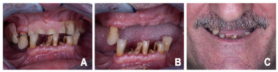
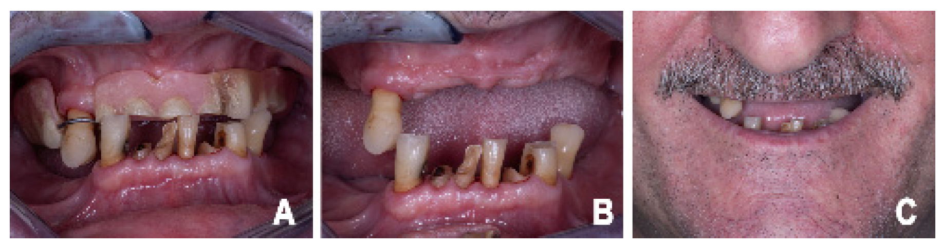
A clinical case involving a 56-year-old male patient with good systemic health was examined to exemplify the described protocol (Figure 1). Under controlled periodontal conditions, the patient underwent bimaxillary complete-arch implant rehabilitation. During the surgery, performed under intravenous sedation, the compromised residual teeth were extracted. Four cylindrical 3.75 mm titanium implants with internal conical connections (Youse CC, Sweden & Martina, Due Carrare, Italy) were placed both in the maxilla and the mandible and immediately loaded following multi-unit abutment (MUA) connection.
Figure 1.
Initial clinical situation. (A) Intraoral frontal view showing compromised dentition and altered occlusal relationships. (B) Intraoral occlusal view highlighting residual teeth scheduled for extraction. (C) Extraoral smile view documenting the initial compromised esthetic condition.
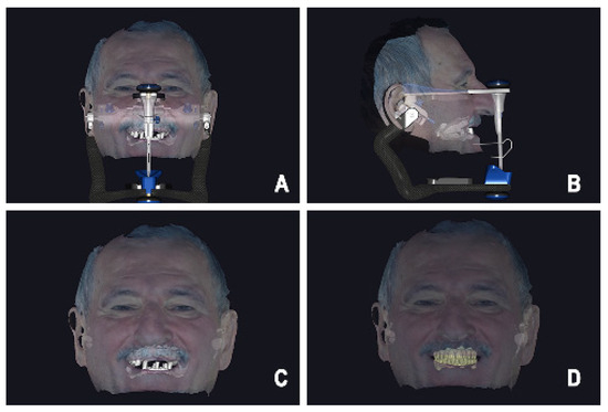
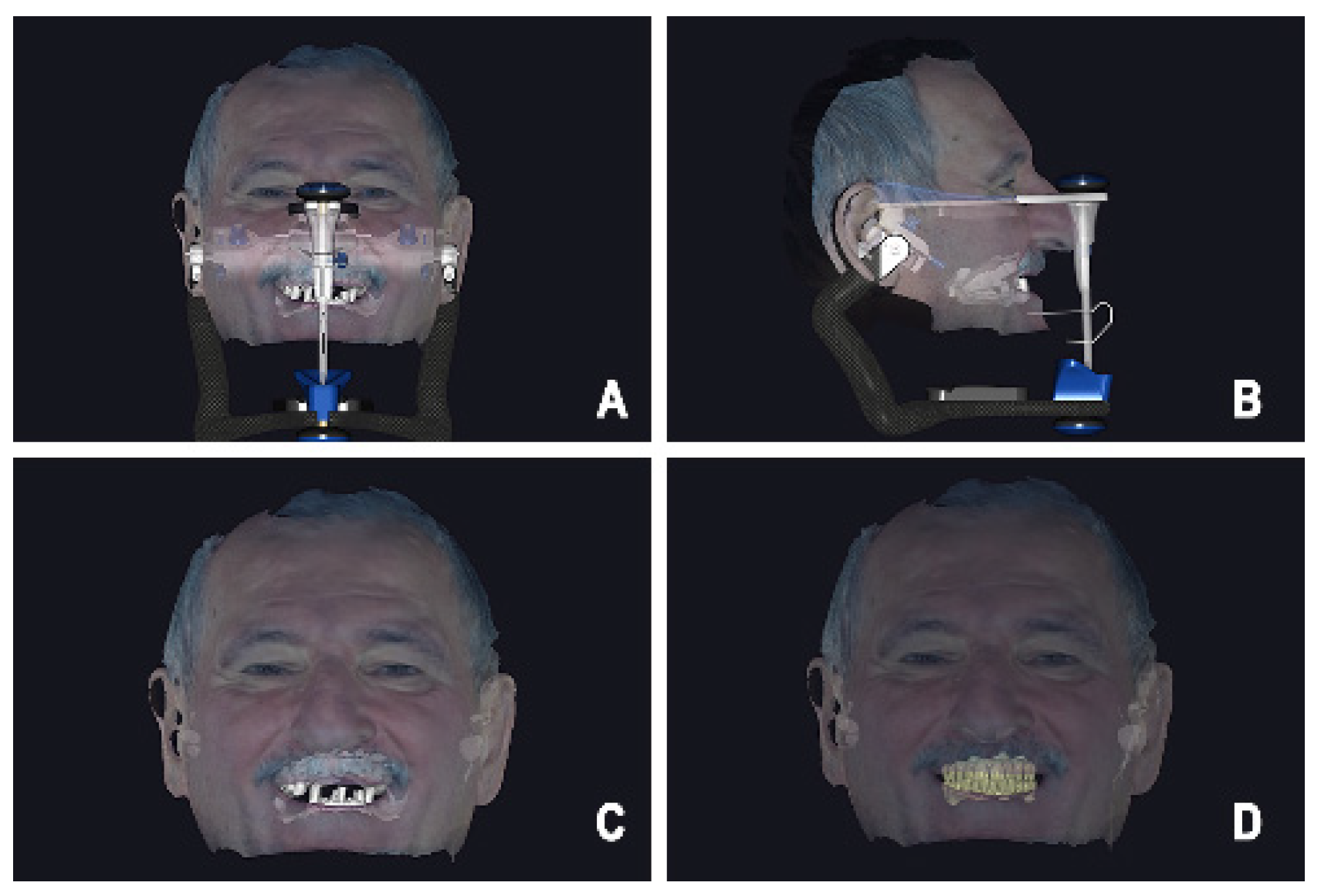
The prosthetic workflow started with a prosthetically driven diagnostic phase. Facial scanning, intraoral scanning, and digital centric relation registration were performed to generate a virtual patient (Figure 2). When required, the vertical dimension of occlusion was digitally increased using a virtual articulator, as previously described [24]. Based on these data, a computer-aided design (CAD) of the provisional complete-arch prosthesis was developed, focusing on esthetic, functional, and phonetic parameters. At this stage, the prosthetic design was finalized independently of the definitive implant positions (Figure 2D).
Figure 2.
Virtual patient. (A) Frontal view in the virtual articulator showing alignment according to the bipupillary plane. (B) Lateral view in the virtual articulator showing alignment according to the Camper plane. (C) Virtual patient showing the initial clinical condition. (D) Virtual patient with the digital prosthetic design of the provisional restorations.
To enable accurate alignment between pre- and postoperative mandibular intraoral scans, two cylindrical intraosseous pins used as fiducial reference markers were employed. The use of two reference elements is mandatory to allow full spatial registration of the datasets, as a single axisymmetric reference would constrain alignment only along its longitudinal axis.
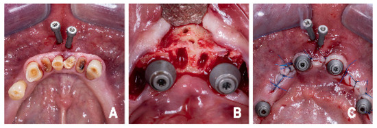
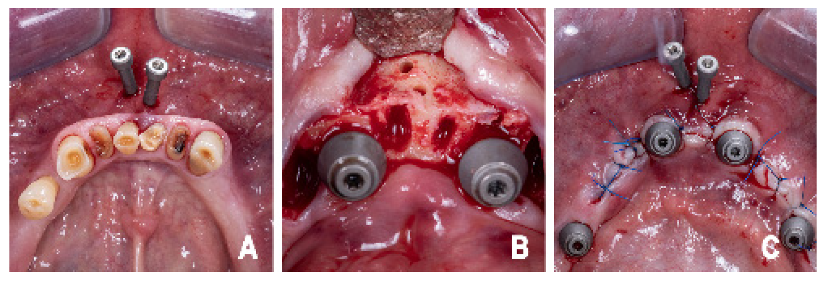
The pins were positioned in a paramedian region of the mandible in areas not intended for implant placement (Figure 3A). When teeth scheduled for extraction were present in the paramedian region and osteotomy was required, the pins were positioned apical to the root apices. This ensured that the osteotomy geometry would not be altered during extraction or implant site preparation and could therefore be reused as a stable reference throughout the procedure (Figure 3B).
Figure 3.
Intraosseous fiducial pin positioning and surgical phase. (A) Occlusal view showing the placement of two cylindrical intraosseous fiducial pins in the paramedian mandibular region. (B) Surgical view after flap elevation showing preserved pin osteotomies and implant site preparation. (C) Sutured surgical site with implants and fiducial pins repositioned in the original osteotomies.
The pins were cylindrical and axisymmetric; therefore, rotational orientation did not influence the alignment process. The accuracy of repositioning depended exclusively on the preservation of the osteotomy geometry, allowing for the reinsertion of the pins in the identical spatial position relative to the mandibular bone. The pins could be positioned either in a vertically stacked or horizontally adjacent configuration, depending on anatomical and operative considerations.
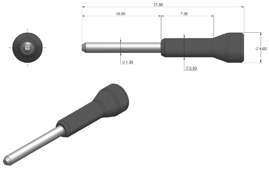
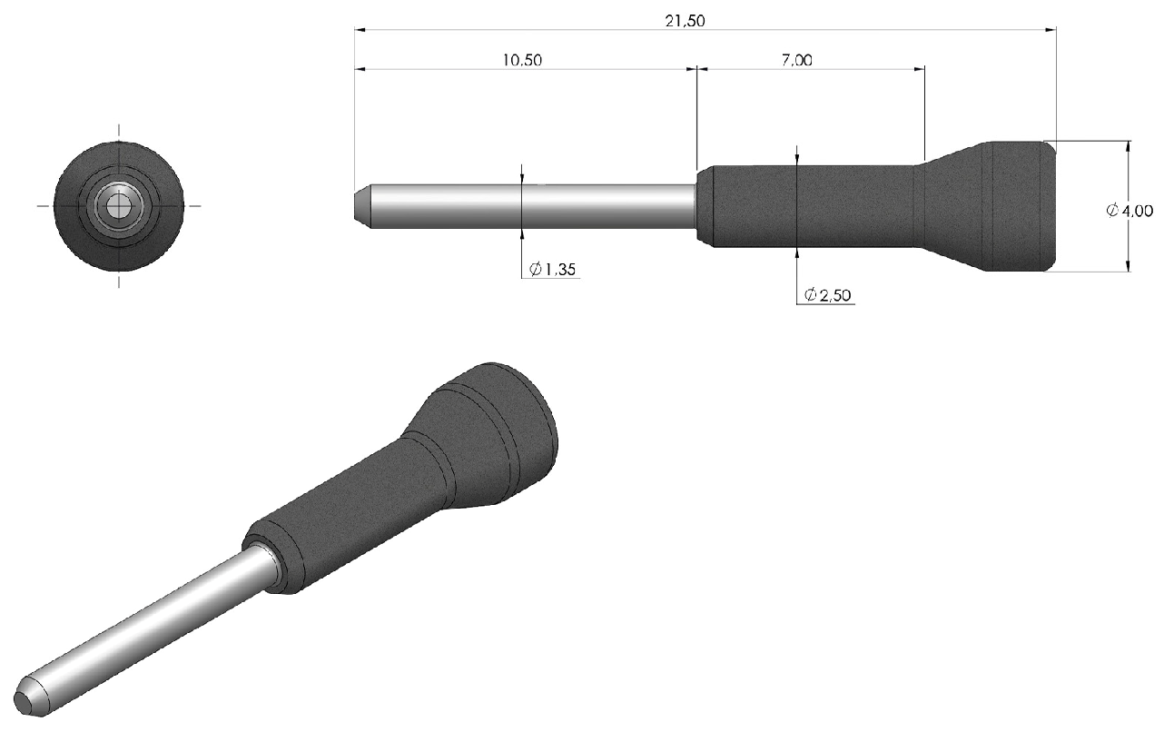
Each pin was manufactured from titanium grade 4 (ASTM F67) and consisted of a cylindrical intraosseous shaft (diameter 1.35 mm; length 10.5 mm) connected to a coronal reference head with an enlarged diameter (maximum diameter 4.0 mm). The total length of the pin was 21.5 mm. The dimensional tolerances were compliant with ISO standards for precision mechanical components (Figure 4).
Figure 4.
Technical drawing of the cylindrical intraosseous fiducial pin used for mandibular scan alignment. Dimensional specifications of the pin, including the intraosseous cylindrical shaft (diameter 1.35 mm; length 10.5 mm), the coronal reference head with enlarged diameter (maximum diameter 4.0 mm), and the total length (21.5 mm). The pin is manufactured in titanium grade 4 (ASTM F67) with dimensional tolerances compliant with ISO standards for precision mechanical components.
Pin osteotomies were prepared using a drill calibrated to the diameter of the intraosseous shaft (diameter 1.4 mm), allowing for passive insertion without translational play between the pin and the osteotomy walls. No reproducibility of osteotomy position, angulation, or depth was required between patients; the only critical requirement was that the osteotomy geometry remained unchanged during surgery.
A preoperative mandibular intraoral scan was acquired with the pins in position. During surgery, the pins were temporarily removed to avoid interference with flap elevation and implant placement.
Following implant insertion, an implant-level photogrammetric impression was acquired to define the inter-implant relationships with high precision. Photogrammetric acquisition was performed before reinserting the pins and wound closure.
After photogrammetric acquisition, the pins were reinserted into the original osteotomies by passing them through the pre-existing perforations in the soft tissues. The surgical site was then sutured, and a postoperative intraoral scan was acquired with the pins in position.
The protocol was independent of the implant placement technique and could be applied to both guided and free-hand surgery. The workflow was also scanner-agnostic and could be implemented using any intraoral scanning system.
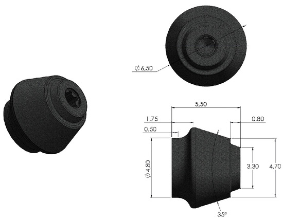
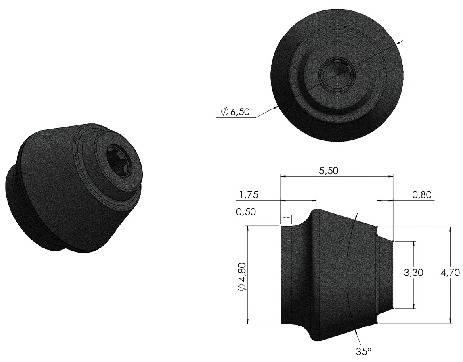
The implants were intraorally scanned using newly designed standard scanbodies suitable for all patients. The scanbodies were characterized by a fully convergent geometry (35°) without undercuts, allowing for complete optical acquisition from a single occlusal scanning path (Figure 5). This design enabled the scanbodies to be captured in one continuous pass, minimizing interruptions in scanner tracking and reducing distortions related to soft tissue displacement or postoperative bleeding at the crestal level.
Figure 5.
Technical drawing of the newly designed standard scanbodies characterized by a fully convergent geometry without undercuts, allowing complete optical acquisition from a single occlusal scanning path.
Two versions of the scanbodies were available, differing in height, and were selected according to the residual thickness of the peri-implant soft tissues following surgery.
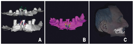
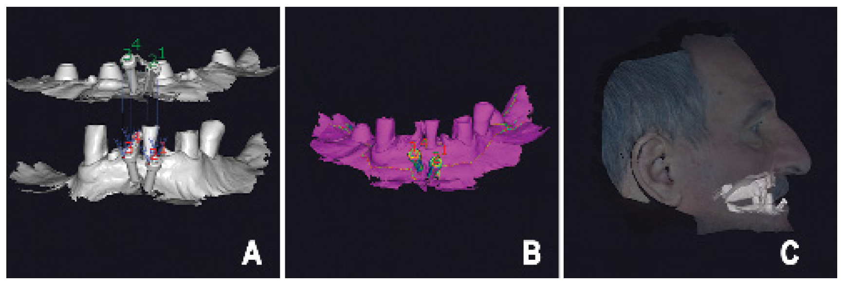
Pre- and postoperative mandibular scans were imported into a dental CAD environment (exocad GmbH, Darmstadt, Germany). Scans were aligned using a best-fit registration algorithm based exclusively on the geometric surfaces of the intraosseous fiducial reference pins. Soft tissues and other deformable structures were intentionally excluded from the alignment process (Figure 6).
Figure 6.
Scan alignment workflow based on fiducial reference pins. (A) Superimposition of pre- and postoperative mandibular scans using best-fit alignment based exclusively on the fiducial pin geometry. (B) Highlighted alignment of the fiducial pins confirming stable registration between datasets. (C) Reconstruction of the postoperative virtual patient after scan alignment.
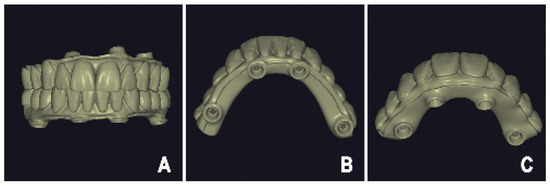
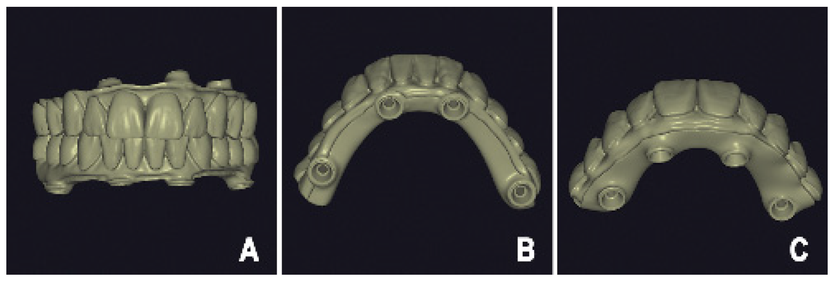
The aligned datasets were integrated with the photogrammetric impression to reconstruct the postoperative virtual patient. The predesigned digital provisional prosthesis was then inserted into the aligned postoperative dataset without modifying its original geometry (Figure 7).
Figure 7.
Basal view of the prosthesis–implant interface geometry. (A) Prosthetic design showing the occlusal relationship. (B) Basal view of the mandibular prosthesis with highlighted prosthesis–implant interface geometry. (C) Basal view of the maxillary prosthesis with highlighted prosthesis–implant interface geometry.
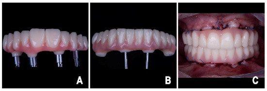
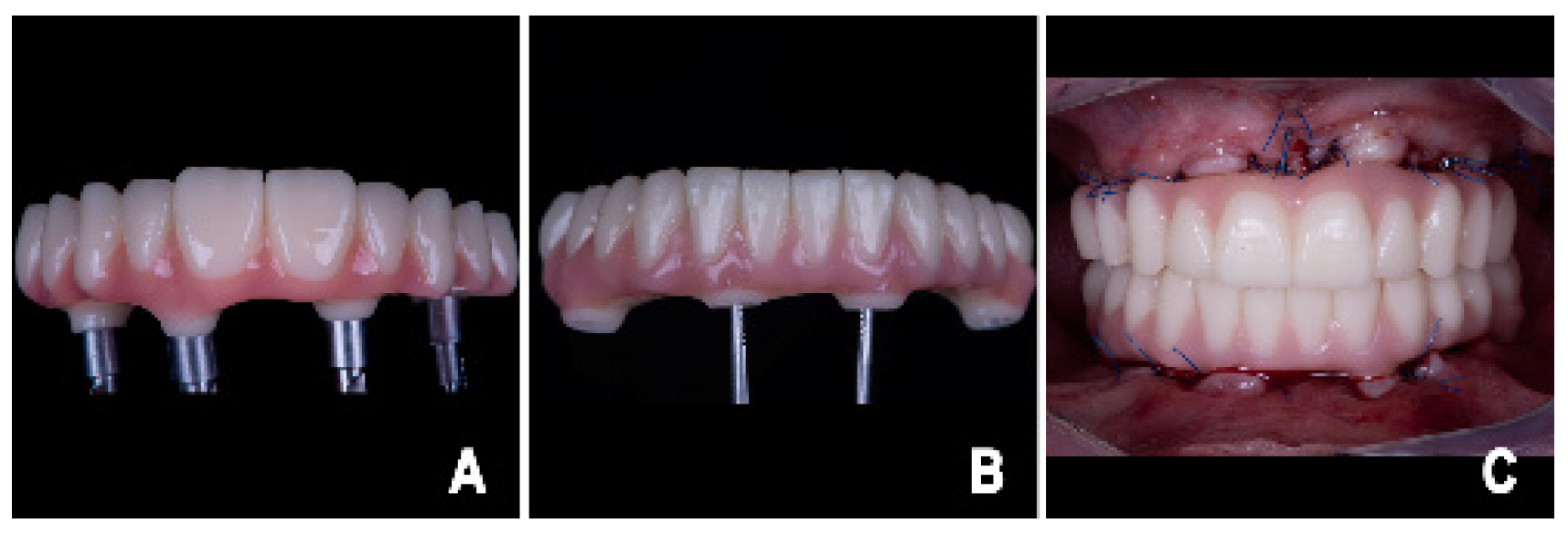
After refining the prosthesis–soft tissue interface and verifying implant-related geometries, the provisional prosthesis was manufactured using additive manufacturing. The restoration was printed using a dual-build platform approach with a desktop 3D printer (SprintRay Inc., Los Angeles, CA, USA) and a high-strength resin material (Onyx 2, SprintRay Inc., Los Angeles, CA, USA). Tooth- and gingival-colored components were printed simultaneously, subsequently bonded, and delivered within a few hours after surgery (Figure 8 and Figure 9).
Figure 8.
Printed provisional complete-arch prostheses and clinical delivery. (A) Frontal view of the upper provisional prosthesis. (B) Frontal view of the lower provisional prosthesis. (C) Intraoral frontal view after delivery of the provisional prostheses before occlusal adjustments, showing the accurate interarch adaptation achieved through the dual-pin alignment technique.
Figure 9.
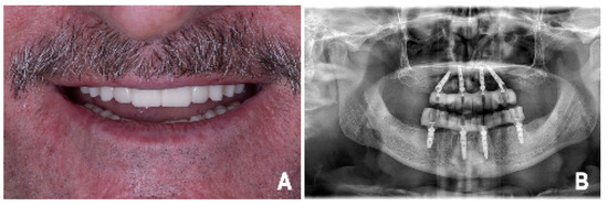
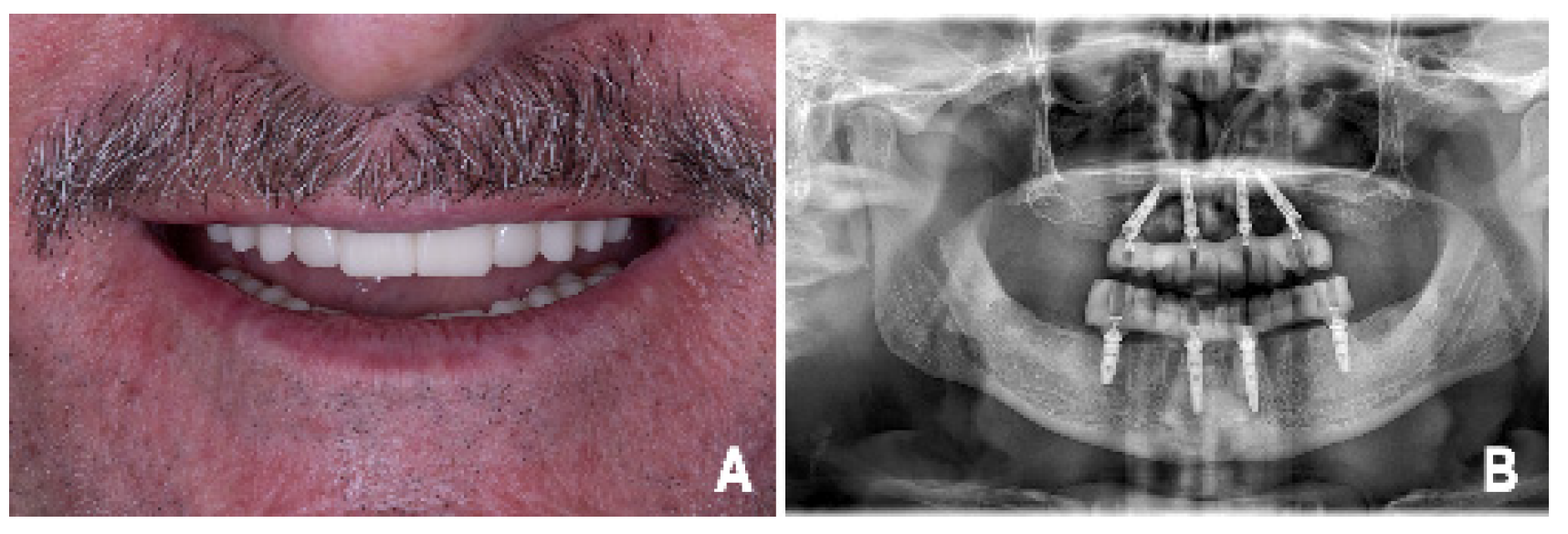
Final clinical and radiographic outcome. (A) Extraoral frontal view showing the patient’s smile after delivery of the complete-arch provisional prostheses, highlighting esthetic integration and lip support. (B) Postoperative panoramic radiograph confirming implant positioning and the correct fit of the provisional complete-arch prostheses on the implants.
Following application of the proposed fully digital workflow, the pre- and postoperative mandibular intraoral scans were aligned using the calibrated dual-pin reference system. Preserving the pin osteotomy geometry throughout the surgical procedure allowed for accurate and reproducible repositioning of the fiducial references, enabling stable scan alignment independent of soft tissue morphology changes induced by surgery.
The protocol proved to be simple and standardized, requiring no additional radiographic acquisitions and no complex operator-dependent steps. Scan alignment was performed using only the geometric surfaces of the fiducial pins, resulting in a reproducible procedure with a reduced risk of operator-related variability.
The workflow was applicable across different clinical scenarios, including varying degrees of mandibular atrophy and both dentate and edentulous conditions. Implants could be positioned using either guided or free-hand techniques without impeding the effectiveness of the alignment protocol. Furthermore, the method was compatible with different intraoral scanning systems and seamlessly integrated with photogrammetric acquisition for implant position recording.
Overall, the proposed approach resulted in a time-efficient and clinically transferable protocol, enabling the reconstruction of a coherent postoperative virtual patient and insertion of the predesigned digital provisional prosthesis without modifying its original geometry.
3. Discussion
The alignment of preoperative prosthetic planning data with postoperative intraoral scans represents a critical step in fully digital workflows for complete-arch implant rehabilitation, particularly in the mandible. The absence of rigid anatomical landmarks, combined with mandibular deformation during functional movements and surgical manipulation, makes reliable registration of digital datasets inherently challenging [3,4,7,8,9,23].
Several strategies have been proposed to mitigate these limitations. Auxiliary geometric devices and artificial landmarks have been shown to improve the trueness of complete-arch intraoral scans (IOSs) by introducing additional reference geometry during acquisition [11,12,13]. However, these solutions primarily improve the scanning accuracy and do not fully address the problem of aligning pre- and postoperative datasets once soft tissue morphology has been altered by surgery.
Photogrammetry has been widely reported to provide superior accuracy in defining inter-implant relationships compared with IOSs in complete-arch scenarios [16,17,18,19].
Nevertheless, photogrammetric acquisition focuses on implant-level positional information and does not inherently enable the alignment of postoperative implant data with preoperative prosthetic designs within a comprehensive virtual patient.
More invasive workflows based on additional postoperative cone-beam computed tomography (CBCT) acquisitions have been described to facilitate dataset registration using osseous landmarks [21,22]. Although these approaches may improve alignment accuracy, they increase radiation exposure and procedural complexity, limiting their routine applicability in fully digital, chairside-oriented workflows. A comparative overview of the main alignment strategies currently described in the literature is provided in Table 1.
Table 1.
Comparison of different alignment strategies in complete-arch digital workflows.
Recently proposed workflows based on intraosseous pins or reference markers have been described primarily in the context of maxillary rehabilitations, where intraoral scanning is subject to reduced deformation and stable anatomical areas are available for pin placement without interference from the surgical procedure. These conditions are not directly transferable to the mandible, particularly in cases of advanced mandibular atrophy requiring flap elevation, bone reduction, or extensive osteotomy. In the mandible, suitable regions for pin placement that remain unaffected throughout surgery are extremely limited. Although the retromolar trigones may represent a theoretical option, their distance from the implant-bearing area may introduce distortion and compromise the accuracy of pre- and postoperative dataset alignment. Consequently, workflows developed for the maxilla cannot be directly extrapolated to the atrophic mandible without addressing these specific anatomical challenges [14,15]. This distinction highlights that previously described pin-based alignment strategies, primarily developed for maxillary applications, cannot be directly translated to the atrophic mandible without modifications to ensure stable and reproducible intraosseous reference positioning.
Previously, protocols have been described that use surgical anchor pins as fiducial markers to facilitate the alignment of complete-arch datasets. For example, Papaspyridakos et al. [5] reported a workflow utilizing anchor pins from a stackable surgical guide to register intraoral scans. However, this approach presents specific limitations that the present protocol aims to overcome.
Surgical anchor pins are designed for fixation rather than optical acquisition. Their reflective metallic surface often requires intraoral scanning powder [4], and their geometry—frequently characterized by threads or undercuts—can complicate mesh acquisition, particularly in anatomically challenging areas such as the vestibule. Moreover, this approach is inherently limited to guided surgery workflows. In contrast, the fiducial pins proposed in the present study are specifically engineered for digital workflows, featuring a matte surface and a simplified convex geometry optimized for optical acquisition. This design enables scanning even in complex soft tissue conditions and allows the protocol to be applied in both guided and free-hand implant placement procedures.
The key concept of the proposed method is preservation of the pin osteotomy geometry throughout surgery, allowing for the fiducial references to be reinserted in an identical spatial position relative to the mandibular bone. The simple cylindrical geometry of the pins is well suited to best-fit alignment strategies, reducing ambiguity during surface-based registration and improving the robustness of scan superimposi-tion. However, this approach assumes that the osteotomy geometry remains un-changed throughout the surgical procedure. Inadvertent enlargement or modification of the osteotomy may compromise the reproducible positioning of the pin, potentially affecting alignment accuracy.
An additional advantage of the proposed workflow is its compatibility with different digital acquisition systems, as alignment is based exclusively on the fiducial pin geometry and is largely independent of the intraoral scanner used. Integration with photogrammetric acquisition allows for accurate inter-implant relationships to be combined with global scan alignment.
Recent evidence has shown that operator experience can significantly influence the quality and efficiency of digital impressions [25]. In this context, a simple, standardized, and non–skill-sensitive protocol such as the one proposed may represent a relevant advantage in clinical practice.
Despite its advantages, the present study has limitations. Specifically, as a proof-of-concept investigation, the workflow was not validated through large-scale quantitative accuracy measurements. No quantitative assessment of trueness, precision, or reproducibility was performed in the present study. In addition, the proposed workflow involves the placement of intraosseous fiducial pins, which introduces an additional surgical step and may be associated with increased procedural invasiveness. Although the pins are positioned in areas not intended for implant placement, potential risks related to their insertion, such as local bone trauma or interference with surgical access, should be considered. Furthermore, the use of only two fiducial markers may theoretically allow minimal rotational discrepancies during alignment, particularly in the absence of additional geometric constraints. These aspects should be taken into account when interpreting the results and when applying the protocol in different clinical scenarios.
Future studies should focus on quantitatively assessing the alignment accuracy and reproducibility across different clinical scenarios. Nevertheless, the proposed approach offers a solution to a recognized limitation of fully digital mandibular rehabilitation workflows.
4. Summary
This proof-of-concept study presents a fully digital workflow for mandibular complete-arch implant rehabilitation based on the use of intraosseous fiducial reference markers to enable pre- and postoperative intraoral scan alignment. By preserving the geometry of the pin osteotomies throughout the surgical procedure, the proposed protocol provides reference structures that are designed to minimize the influence of soft tissue morphology, implant positioning, and surgical approach. The method integrates seamlessly with existing digital tools, including intraoral scanning and photogrammetry, without requiring additional radiographic acquisitions. Within its limitations, this approach represents a clinically applicable and minimally invasive solution to a critical challenge in full-digital mandibular rehabilitation workflows, although no quantitative assessment of accuracy, trueness, or reproducibility was performed and the assumption of preserved osteotomy geometry may represent a potential limitation.
Author Contributions
Conceptualization, F.T.; methodology, F.T. and E.R.; validation, F.T., S.S. and E.R.; formal analysis, F.T.; investigation, F.T.; resources, F.T. and S.S.; data curation, F.T. and E.R.; writing—original draft preparation, F.T.; writing—review and editing, F.T.; visualization, F.T. and E.R.; supervision, F.T., S.S. and E.R.; project administration, F.T. and S.S. All authors have read and agreed to the published version of the manuscript.
Funding
This research received no external funding.
Institutional Review Board Statement
Ethical review and approval were waived for this study because it describes a single-case clinical report performed as part of routine clinical care, without experimental intervention or prospective data collection, in accordance with applicable national regulations.
Informed Consent Statement
Written informed consent was obtained from the patient for treatment and for the anonymous use of clinical data and images for scientific publication.
Data Availability Statement
The original contributions presented in this study are included in the article. Further inquiries can be directed to the corresponding authors.
Conflicts of Interest
The authors declare no conflicts of interest.
References
- Wulfman, C.; Naveau, A.; Rignon-Bret, C. Digital scanning for complete-arch implant-supported restorations: A systematic review. J. Prosthet. Dent. 2020, 124, 161–167. [Google Scholar] [CrossRef] [Scilit]
- Pozzi, A.; Carosi, P.; Gallucci, G.O.; Nagy, K.; Nardi, A.; Arcuri, L. Accuracy of complete-arch digital implant impression with intraoral optical scanning and stereophotogrammetry: An in vivo prospective comparative study. Clin. Oral Implant. Res. 2023, 34, 1106–1117. [Google Scholar] [CrossRef] [Scilit]
- Flügge, T.; van der Meer, W.J.; Gonzalez, B.G.; Vach, K.; Wismeijer, D.; Wang, P. The accuracy of different dental impression techniques for implant-supported dental prostheses: A systematic review and meta-analysis. Clin. Oral Implant. Res. 2018, 29, 374–392. [Google Scholar] [CrossRef] [Scilit]
- Çakmak, G.; Yilmaz, H.; Santos, A.T.; Kökat, A.M.; Yilmaz, B. Effect of Scanner Type and Scan Body Location on the Accuracy of Mandibular Complete-Arch Digital Implant Scans: An In Vitro Study. J. Prosthodont. 2022, 31, 419–426. [Google Scholar] [CrossRef] [Scilit] [PubMed]
- Papaspyridakos, P.; De Souza, A.; Bathija, A.; Kang, K.; Chochlidakis, K. Complete Digital Workflow for Mandibular Complete-arch Implant Rehabilitation in 3 Appointments. J. Prosthodont. 2021, 30, 548–552. [Google Scholar] [CrossRef] [Scilit] [PubMed]
- Andriessen, F.S.; Rijkens, D.R.; van der Meer, W.J.; Wismeijer, D.W. Applicability and accuracy of an intraoral scanner for scanning multiple implants in edentulous mandibles: A pilot study. J. Prosthet. Dent. 2014, 111, 186–194. [Google Scholar] [CrossRef] [Scilit] [PubMed]
- Di Fiore, A.; Meneghello, R.; Graiff, L.; Savio, G.; Vigolo, P.; Monaco, C.; Stellini, E. Full arch digital scanning systems performances for implant-supported fixed dental prostheses: A comparative study of 8 intraoral scanners. J. Prosthodont. Res. 2019, 63, 396–403. [Google Scholar] [CrossRef] [Scilit]
- Müller, P.; Ender, A.; Joda, T.; Katsoulis, J. Impact of digital intraoral scan strategies on the impression accuracy using the TRIOS Pod scanner. Quintessence Int. 2016, 47, 343–349. [Google Scholar] [CrossRef] [Scilit]
- Denneulin, T.; Rignon-Bret, C.; Ravalec, G.; Tapie, L.; Bouter, D.; Wulfman, C. Accuracy of Complete-Arch Implant Digital Scans: Effect of Scanning Protocol, Number of Implants, and Scan Body Splinting. Int. J. Prosthodont. 2023, 36, 219–227. [Google Scholar] [CrossRef] [Scilit]
- Vag, J.; Stevens, C.D.; Badahman, M.H.; Ludlow, M.; Sharp, M.; Brenes, C.; Mennito, A.; Renne, W. Trueness and precision of complete arch dentate digital models produced by intraoral and desktop scanners: An ex-vivo study. J. Dent. 2023, 139, 104764. [Google Scholar] [CrossRef] [Scilit]
- Iturrate, M.; Eguiraun, H.; Etxaniz, O.; Solaberrieta, E. Accuracy analysis of complete-arch digital scans in edentulous arches when using an auxiliary geometric device. J. Prosthet. Dent. 2019, 121, 447–454. [Google Scholar] [CrossRef] [Scilit]
- Kanjanasavitree, P.; Thammajaruk, P.; Guazzato, M. Comparison of different artificial landmarks and scanning patterns on the complete-arch implant intraoral digital scans. J. Dent. 2022, 125, 104266. [Google Scholar] [CrossRef] [Scilit]
- Huang, R.; Liu, Y.; Huang, B.; Zhang, C.; Chen, Z.; Li, Z. Improved scanning accuracy with newly designed scan bodies: An in vitro study comparing digital versus conventional impression techniques for complete-arch implant rehabilitation. Clin. Oral Implant. Res. 2020, 31, 625–633. [Google Scholar] [CrossRef] [Scilit]
- Serri, M.; Ruggiero, G.; Serri, M.; Mastrosimone, A.; Zarone, F.; Sorrentino, R. A fully-digital rehabilitation for immediately loaded fixed in-terim complete-arch prosthesis: A case report. J. Osseointegr. 2024, 16, 8–14. [Google Scholar] [CrossRef]
- Espona, J.; Roig, E.; Ali, A.; Roig, M. Immediately loaded interim complete-arch implant-supported fixed dental prostheses fabricated with a completely digital workflow: A clinical technique. J. Prosthet. Dent. 2020, 124, 423–427. [Google Scholar] [CrossRef] [Scilit] [PubMed]
- Ma, B.; Yue, X.; Sun, Y.; Peng, L.; Geng, W. Accuracy of photogrammetry; intraoral scanning, and conventional impression techniques for complete-arch implant rehabilitation: An in vitro comparative study. BMC Oral Health 2021, 21, 636. [Google Scholar] [CrossRef] [Scilit]
- Zhang, Y.J.; Qian, S.J.; Lai, H.C.; Shi, J.Y. Accuracy of photogrammetric imaging versus conventional impressions for complete arch implant-supported fixed dental prostheses: A comparative clinical study. J. Prosthet. Dent. 2023, 130, 212–218. [Google Scholar] [CrossRef] [Scilit] [PubMed]
- Hussein, M.O. Photogrammetry technology in implant dentistry: A systematic review. J. Prosthet. Dent. 2023, 130, 318–326. [Google Scholar] [CrossRef] [Scilit]
- Chen, Y.; Gusmao, I.G.; Papaspyridakos, P.; Att, W. Registration of patient-relevant-references in edentulous jaws using intraoral scanners: Introduction of two novel techniques. Int. J. Esthet. Dent. 2019, 14, 394. [Google Scholar]
- Pedrinaci, I.; Gallucci, G.O.; Lanis, A.; Friedland, B.; Pala, K.; Hamilton, A. Computer-Assisted Implant Planning: A Review of Data Registration Techniques. Int. J. Periodontics Restor. Dent. 2025, 45, 652–665. [Google Scholar] [CrossRef] [Scilit]
- Joda, T.; Gallucci, G.O. The virtual patient in dental medicine. Clin. Oral Implant. Res. 2015, 26, 725–726. [Google Scholar] [CrossRef] [Scilit]
- Hamilton, A.; Akhondi, S.; Pala, K.; Luz, D.; Gallucci, G.O. Using postoperative CBCT to enhance preoperative planning integration in complete arch implant rehabilitation. J. Prosthet. Dent. 2025, 134, 580–585. [Google Scholar] [CrossRef] [Scilit]
- Cuellar, L.R.; Valencia-Niño, C.H.; López-Vaca, O.R.; Martínez-Vega, R.A. Workflow for guided implant placement based on anatomic landmarks and the CAD-CAM fabrication of a prosthetic framework for an implant-supported fixed dental prosthesis. J. Prosthet. Dent. 2025, 135, 694–699. [Google Scholar] [CrossRef] [Scilit]
- Tirone, F.; Salzano, S.; Pirja, D.A. Optimizing passive fit and esthetic results of screw-retained zirconia implant-supported fixed complete dentures designed and produced digitally. Digit. Dent. J. 2025, 2, 100023. [Google Scholar] [CrossRef] [Scilit]
- Limones, A.; Morton, D.; Sallorenzo, A.; Lin, W.S.; Sadid-Zadeh, R.; Phasuk, K.; Revilla-León, M.; Gómez-Polo, M. Impact of operator experience on intraoral digital scanning: A systematic review, meta-analysis, and meta-regression. Report of the Committee on Research in Fixed Prosthodontics of the American Academy of Fixed Prosthodontics. J. Prosthet. Dent. 2025, 135, 297.e1–297.e13. [Google Scholar] [CrossRef] [Scilit]

Disclaimer/Publisher’s Note: The statements, opinions and data contained in all publications are solely those of the individual author(s) and contributor(s) and not of MDPI and/or the editor(s). MDPI and/or the editor(s) disclaim responsibility for any injury to people or property resulting from any ideas, methods, instructions or products referred to in the content. |
© 2026 by the authors. Licensee MDPI, Basel, Switzerland. This article is an open access article distributed under the terms and conditions of the Creative Commons Attribution (CC BY) license.