


Abstract
Fermentation-derived fruit-peel formulations have potential as alternative denture cleansers, but their development requires evidence that they can provide antifungal activity without compromising denture-base materials during repeated use. Objectives: To evaluate the antifungal activity of six-month-fermented mixed pineapple–orange and papaya-based effective eco-produce formulations (M-EEP and P-EEP, respectively) against Candida albicans and determine their time-dependent effects on heat-polymerised polymethyl methacrylate (PMMA) surface roughness and topography. Methods: Antifungal activity against Candida albicans was assessed using broth microdilution to determine the minimum inhibitory concentration and minimum fungicidal concentration. A peroxide-based denture cleanser (PDC) served as the commercial reference. PMMA specimens were allocated to PDC, M-EEP, P-EEP or distilled water (n = 7). Arithmetic mean roughness (Ra) and root mean square roughness (Rq) were measured at baseline, 7, 14, 28 and 90 days using atomic force microscopy. Surface topography was evaluated qualitatively, and longitudinal data were analysed using generalised estimating equations. Results: Both EEP formulations inhibited visible fungal growth when tested as prepared and prevented colony recovery after subculture, demonstrating fungicidal activity. Significant time and medium x time effects were observed for Ra and Rq (p < 0.05). M-EEP showed no significant within-group roughness change, whereas P-EEP and PDC showed early reductions that were not sustained by day 90. No medium produced progressive roughening, and all Ra values remained below the commonly cited 0.2 µm plaque-retentive threshold. Conclusions: Six-month-fermented EEP formulations demonstrated antifungal activity against planktonic Candida albicans ATCC 10231 without progressive increases in mean PMMA surface roughness, thereby addressing both biological efficacy and material compatibility within the same experimental framework.
1. Introduction
Effective denture hygiene is essential for maintaining oral health, comfort and continued prosthesis use among removable denture wearers. Denture surfaces may retain food debris, salivary deposits, and microorganisms, and inadequate cleansing is associated with denture stomatitis and other inflammatory oral conditions [1]. Polymethyl methacrylate (PMMA), which remains widely used for denture-base fabrication, provides a non-shedding surface on which microorganisms, including Candida albicans, may persist [1,2]. Mechanical cleaning is therefore commonly supplemented with chemical denture cleansers, particularly among older adults and individuals with reduced manual dexterity or difficulty accessing retentive denture surfaces, for whom brushing alone may be inadequate [3].
Conventional chemical denture cleansers include peroxide-based, hypochlorite-based, acidic, and enzymatic formulations, which act through different mechanisms to facilitate deposit removal and microbial control. Peroxide-based products are among the most widely used commercial options because their effervescent, oxidative and surfactant actions support routine denture cleansing. However, repeated exposure to chemical cleansers may alter PMMA properties, including surface roughness, colour stability, hardness, and mechanical behaviour [4,5,6]. These changes are clinically relevant because surface roughness and other surface characteristics can influence microbial adhesion and biofilm retention on denture-base materials, although roughness alone may not fully determine Candida albicans adhesion [7,8,9]. A candidate cleanser should therefore demonstrate relevant antimicrobial activity without progressively compromising the denture-base material.
Interest in plant-derived oral care agents has encouraged the investigation of fruit-processing residues as sources of biologically active constituents. Fruit peels are commonly discarded despite containing organic acids, phenolic compounds, flavonoids, enzymes and other constituents with antimicrobial potential. Their utilisation may convert abundant organic residues into value-added raw materials for oral care development. Fermentation provides one approach for transforming these residues into chemically complex liquid formulations through the release, modification and generation of substrate-derived metabolites [10,11,12,13,14]. Such preparations have been described as eco-enzyme, garbage enzyme, or effective eco-produce (EEP). In the present study, EEP refers to filtered formulations produced through prolonged fermentation of fruit peels, molasses and water.
Previous dental studies have reported antibacterial activity of fruit-waste fermentation-derived preparations against Enterococcus faecalis, Streptococcus mutans, and Lactobacillus acidophilus, together with preliminary cytotoxicity findings [12,13,14]. Antifungal activity against Candida albicans has also been reported for other fruit-waste fermentation-derived formulations. Collectively, these findings provide a growing biological basis for investigating such preparations in dentistry. Nevertheless, the available evidence remains centred largely on biological screening, with limited assessment of compatibility with prosthodontic materials. However, previous studies of fermentation-derived fruit-waste preparations have focused predominantly on biological activity, with limited evaluation of their compatibility with prosthodontic materials. Conversely, studies that have assessed antimicrobial performance together with denture-base material properties have largely involved conventional or other non-fermentation-derived cleansing agents [15,16]. Thus, researchers have largely investigated the biological efficacy and material compatibility of fermentation-derived fruit-waste formulations separately. This distinction is important because organic acids and other fermentation-derived metabolites associated with antifungal activity may also interact with PMMA during repeated exposure. Furthermore, most material studies have compared surface roughness only before and after a predetermined exposure period. Although such endpoint assessments identify the final net effect, they may overlook transient, progressive, or non-linear changes occurring during repeated exposure. A longitudinal assessment at multiple time points therefore provides additional information on PMMA’s temporal response and allows biological activity and material compatibility to be considered within the same experimental framework.
Against this background, it remains unclear whether the specific six-month-fermented papaya-based and mixed pineapple–orange EEP formulations can demonstrate antifungal activity while maintaining PMMA surface characteristics during repeated exposure. Because formulation composition varies with fruit substrate and fermentation conditions, findings from one preparation cannot be assumed to apply directly to another. The present study addresses this gap by combining antifungal assessment with repeated longitudinal evaluation of PMMA surface roughness and topography over 90 days. Unlike previous work centred primarily on biological screening or endpoint material assessment, this longitudinal approach links biological performance with the time-dependent response of a clinically relevant denture-base material, enabling evaluation of transient, progressive, or non-linear surface changes. This combined biological–material assessment represents an important translational step in evaluating fermentation-derived fruit-waste formulations as potential denture cleansers.
Accordingly, this study aimed to evaluate the antifungal performance of six-month-fermented, as-prepared papaya-based and mixed pineapple–orange EEP formulations against Candida albicans and to determine their time-dependent effects on the surface roughness and topography of heat-polymerised PMMA over 90 days. A peroxide-based denture cleanser and distilled water were included as commercial and aqueous reference media, respectively. The null hypotheses were that: (1) the tested EEP formulations would not differ in antifungal performance against Candida albicans and (2) immersion medium, time, and their interaction would have no significant effect on PMMA surface roughness.
2. Materials and Methods
2.1. Study Design, Setting and Ethical Considerations
This in vitro experimental study evaluated the antifungal activity of six-month-fermented mixed pineapple–orange effective eco-produce (M-EEP) and papaya-based effective eco-produce (P-EEP) against Candida albicans and their time-dependent effects on heat-polymerised PMMA. Laboratory procedures were conducted in the Microbiology and Dental Materials Laboratories, Faculty of Dentistry, The National University of Malaysia, Kuala Lumpur, Malaysia, and the Biomaterials Research Laboratory, University of Malaya, Kuala Lumpur, Malaysia.
Ethical approval was obtained from the Research Ethics Committee of The National University of Malaysia (UKM PPI/111/8/JEP-2023-754). No human participants or animals were involved; therefore, informed consent and clinical trial registration were not applicable. Microbial culture, handling, and disposal procedures complied with institutional biosafety and research-governance requirements.
2.2. Preparation of EEP Formulations and Reference Media
The EEP formulations were prepared using a protocol adapted from Arun and Sivashanmugam [10]. M-EEP contained 45 g of pineapple peel and 30 g of orange peel in a 6:4 ratio by weight, whereas P-EEP contained 75 g of unripe papaya peel. Each formulation was prepared at a 3:1:10 ratio of fruit-peel waste, molasses, and tap water (75 g: 25 g: 250 mL) in a disinfected 600 mL airtight plastic container. The mixtures were stirred, sealed and stored in the dark at room temperature for six months. During early fermentation, the containers were opened periodically to release accumulated gas, then resealed.
After six months, the mixtures were filtered to remove coarse residues and centrifuged at 10,000 rpm for 6 min using an Eppendorf centrifuge (Eppendorf SE, Hamburg, Germany). The supernatants were sequentially filtered through 0.45 µm and 0.22 µm polyethersulfone membrane filters (Bioflow Lifescience Sdn Bhd, Ampang, Selangor, Malaysia). Three independent batches of each formulation were prepared under identical conditions.
A peroxide-based denture cleanser (PDC), Polident® (PTI Royston LLC, Royston, GA, USA), served as the commercial reference. It was prepared according to the manufacturer’s instructions by dissolving one tablet in 200 mL of sterile distilled water at room temperature. A fresh solution was prepared before each exposure cycle. Distilled water served as the aqueous reference medium.
2.3. Characterisation of Immersion Media
The initial pH values of all immersion media, including PDC, M-EEP, P-EEP, and DW, were measured at room temperature using a calibrated bench-top pH meter (BP3001, Trans Instruments, Singapore). Measurements were performed in triplicate and recorded as mean ± standard deviation.
Qualitative metabolite profiling was performed for the EEP formulations to provide compositional context. M-EEP and P-EEP samples were analysed using an ultra-performance liquid chromatography coupled with high-resolution mass spectrometry (UPLC-HRMS) system (Dionex Ultimate 3000 RS UPLC coupled to a Q Exactive Orbitrap hybrid tandem mass spectrometer, Thermo Fisher Scientific, Waltham, MA, USA). Before analysis, 1 mL of each filtered sample was passed through a 0.45 µm polyethersulfone (PES) membrane filter.
Putative compound annotation was performed using Compound Discoverer software version 3.3 (Thermo Fisher Scientific, Waltham, MA, USA), with database matching against mzCloud, ChemSpider, and Metabolika. The UPLC-HRMS data were used only for qualitative formulation context; metabolite abundance was not quantified or correlated with PMMA surface roughness outcomes.
2.4. Candida albicans Strain and Inoculum Preparation
Candida albicans ATCC 10231 (American Type Culture Collection, Manassas, VA, USA) was used. The organism was cultured on Sabouraud dextrose agar and incubated aerobically at 37 °C for 24–48 h. Culture purity was confirmed by colony morphology and microscopic examination.
The inoculum was prepared using a method adapted from the Clinical and Laboratory Standards Institute M27 procedure [17]. Approximately five colonies at least 1 mm in diameter were suspended in 5 mL of sterile distilled water and adjusted spectrophotometrically to the equivalent of a 0.5 McFarland standard at 530 nm. The suspension was diluted 1:50 and then 1:20 in Sabouraud dextrose broth to produce a final inoculum of approximately 0.5 × 10³ to 2.5 × 10³ cells/mL.
2.5. Antifungal Susceptibility Testing
The antifungal activity of the six-month-fermented M-EEP and P-EEP formulations was evaluated against planktonic Candida albicans ATCC 10231 using a broth microdilution procedure adapted from the Clinical and Laboratory Standards Institute M27 protocol [17]. Three independently prepared batches of each formulation were evaluated, with each batch tested in three technical replicate wells.
Two-fold serial dilutions of each formulation, ranging from 100% to 0.39%, were prepared in Sabouraud dextrose broth. Each test preparation was inoculated with the fungal suspension and incubated aerobically at 37 °C for 24 h. Formulation-specific uninoculated blanks were included to correct for the intrinsic colour and turbidity of the EEP preparations. Inoculated broth without a test formulation served as the growth control, while uninoculated broth served as the sterility control.
Fungal growth was assessed visually and by optical-density measurement at 530 nm. Blank-corrected OD530 was calculated by subtracting the corresponding uninoculated formulation-blank reading from each inoculated well reading. The minimum inhibitory concentration (MIC) was defined as the lowest tested concentration showing no visible fungal growth.
To determine the minimum fungicidal concentration (MFC), aliquots from wells without visible growth were subcultured onto Sabouraud dextrose agar and incubated at 37 °C for 24–48 h. The MFC was defined as the lowest tested concentration from which no fungal colonies were recovered. The 100% (undiluted) formulations were selected for the PMMA immersion experiment because this was the common MFC for both formulations.
2.6. PMMA Specimen Fabrication and Sample Size
The sample size was calculated using Power and Sample Size software version 3.0 (Vanderbilt University, Nashville, TN, USA). Based on a previous related study [18], 28 specimens were required, with seven specimens per group, to provide 80% power at α = 0.05 for detecting a 0.05 µm difference in arithmetic mean roughness (Ra).
PMMA specimens measuring 10 × 10 × 2 mm were fabricated from heat-polymerised acrylic resin (Metrocryl, Metrodent Ltd., Huddersfield, England, UK) using conventional compression moulding. Wax patterns were flasked and dewaxed, and the PMMA powder and monomer were mixed according to the manufacturer’s instructions. The material was packed into the mould, trial closures were performed, and a final hydraulic pressure of approximately 9.8 MPa was applied. Polymerisation was performed at 70 °C for 6 h, followed by boiling for 2 h. The flasks were cooled to room temperature overnight before deflasking.
One surface of each specimen was finished with fine-grit abrasive paper and polished with pumice slurry and SHERA polishing cream (SHERA Werkstoff-Technologie GmbH & Co. KG, Lemförde, Germany) using a wet rag wheel, following established PMMA finishing and polishing procedures [19]. Specimens with visible porosity, cracks, distortion or dimensional defects were excluded. Before baseline measurement, all specimens were stored in distilled water at room temperature for 24 h. Ra was the primary surface-roughness outcome used for sample-size determination, whereas Rq was included as a complementary secondary roughness measure.
2.7. Repeated-Exposure Protocol
The 28 specimens were randomly allocated to four groups (n = 7): M-EEP, P-EEP, PDC, and distilled water. Specimens were coded before measurement. Each specimen underwent three consecutive 8-h exposure cycles per 24-h period for 90 days. The assigned medium was renewed after every 8-h cycle to maintain consistent exposure. Between cycles, specimens were removed, rinsed under running tap water, and transferred to freshly prepared medium of the same type. Exposure was conducted at approximately 25 °C. Surface assessments were performed at baseline and after 7, 14, 28, and 90 days.
2.8. Atomic Force Microscopy and Surface Roughness
Surface roughness and topography were assessed using a Dimension Icon VT-1000 atomic force microscope (Bruker Corporation, Billerica, MA, USA). A silicon probe was operated in tapping mode with a spring constant of approximately 10 N/m and a resonance frequency of 250 kHz. Each scan covered a 10 × 10 µm2 area.
Each specimen was divided into three equal vertical sections using reference markings, and one scan was acquired from the centre of each section at every time point. The same designated regions were used throughout the study to support longitudinal comparison. The three readings were averaged to obtain specimen-level Ra and Rq. Ra represented the mean absolute deviation of surface heights from the mean plane, whereas Rq represented the root mean square of height deviations and was more sensitive to isolated peaks and valleys. Two-dimensional and three-dimensional AFM images were also examined qualitatively to describe changes in surface topography.
2.9. Field-Emission Scanning Electron Microscopy
After the 90-day exposure period, one representative specimen from each group was examined qualitatively using a Clara field-emission scanning electron microscope (FESEM) (Tescan Orsay Holding, a.s., Brno, Czech Republic). Specimens were sectioned with a precision cutting machine, mounted on aluminium stubs, and sputter-coated with platinum. Cross-sectional images were acquired at an accelerating voltage of 2 kV and a magnification of ×24,000 using an Everhart-Thornley detector (field of view, 10.97 µm). FESEM observations were exploratory and were not included in quantitative statistical analysis.
2.10. Statistical Analysis
Statistical analyses were performed using IBM SPSS Statistics version 29 for antifungal data and version 21 for PMMA roughness data (IBM Corp., Armonk, NY, USA). Statistical significance was set at α = 0.05.
For each EEP formulation, three independently prepared batches were tested, with each batch assessed in three technical replicate wells. The OD530 reading of the corresponding uninoculated formulation blank was subtracted from each inoculated well reading. The three blank-corrected technical replicate values were then averaged to obtain one OD530 value for each independent batch. Batch-level OD530 values at the nominal 100% condition were summarised as the mean ± standard deviation.
An exact two-sided Mann–Whitney U test was used to compare batch-level OD530 values between M-EEP and P-EEP at the nominal 100% condition. MIC, MFC, visible-growth findings, and colony recovery after subculture were determined according to predefined microbiological criteria and reported descriptively.
For longitudinal PMMA data, separate generalised estimating equation models were constructed for Ra and Rq to account for repeated measurements within specimens. Immersion medium, time, and the medium x time interaction were entered as fixed effects, and an autoregressive first-order working correlation structure was used. Where significant main or interaction effects were detected, estimated marginal means were compared with Bonferroni adjustment. Effect estimates were reported with 95% confidence intervals.
3. Results
3.1. Antifungal Activity of the Six-Month-Fermented EEP Formulations
The six-month-fermented EEP formulations demonstrated different inhibitory profiles against Candida albicans (Table 1). M-EEP prevented visible fungal growth at 100%, but growth was observed at 50%; therefore, its MIC was 100%. P-EEP prevented visible growth at both 50% and 100%, giving an MIC of 50%.
Table 1.
Antifungal activity of six-month-fermented EEP formulations against Candida albicans ATCC 10231.
Following subculture, no fungal colonies were recovered from M-EEP or P-EEP at 100%. Colony recovery occurred following exposure to P-EEP at 50%, indicating that the inhibitory effect at this concentration was not fungicidal. The MFC was therefore 100% for both formulations. Accordingly, the 100% (undiluted) formulations were selected for the subsequent PMMA immersion experiment as the common fungicidal condition.
At the selected 100% condition, the mean blank-corrected OD530 values were −0.0077 ± 0.0101 for M-EEP and 0.0002 ± 0.0042 for P-EEP. No statistically significant difference in OD530 was detected between the formulations at this concentration (exact two-sided Mann–Whitney U = 2.0, p = 0.400). The small negative corrected values reflected minor measurement variation around zero after subtracting the corresponding formulation blank and were not interpreted as indicating negative fungal growth.
The peroxide-based denture cleanser prevented visible fungal growth, whereas the inoculated broth control showed growth. The sterility control showed no growth.
MIC and MFC were determined from the complete two-fold serial dilution series. Blank-corrected OD530 values are presented only for the 100% condition subsequently used in the PMMA immersion experiment. Values represent the mean ± standard deviation of three independently prepared formulation batches; each batch value was obtained by averaging three technical replicate wells.
3.2. Immersion-Media Characterisation
Table 2 summarises the initial pH values of the immersion media. PDC had a near-neutral pH, while DW served as the aqueous reference medium. M-EEP and P-EEP were acidic, with initial pH values of 3.14 ± 0.11 and 3.03 ± 0.03, respectively.
Table 2.
Initial pH values of the denture-cleansing and reference media.
Qualitative UPLC-HRMS profiling detected 60 putatively annotated metabolites in each EEP formulation. The detected compounds were classified as organic acids, amino acids and peptide derivatives, phenolic compounds, sugar-related metabolites, fatty-acid derivatives, nitrogen-containing metabolites, and other detected compounds, as summarised in Table 3. These findings provided compositional context and did not represent quantitative comparisons of metabolite abundance.
Table 3.
Metabolite-class distribution of six-month-fermented M-EEP and P-EEP based on putative UPLC-HRMS annotations.
3.3. Surface-Roughness Evaluation
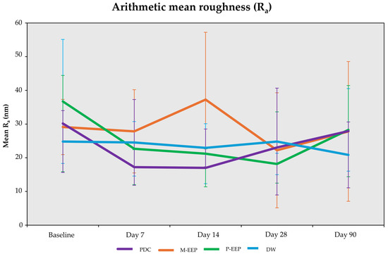
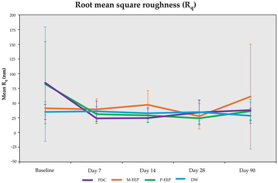
Surface roughness was assessed using Ra and Rq over 90 days in PMMA specimens immersed in PDC, M-EEP, P-EEP, and DW. Figure 1 and Figure 2 illustrate temporal trends. The GEE model findings are summarised in Table 4, while change-from-baseline values are presented in Table 5.
Figure 1.
Changes in arithmetic mean roughness (Ra) of heat-polymerised PMMA during 90-day immersion in denture-cleansing media. Data are presented as mean ± SD (n = 7 per group).
Figure 2.
Changes in root mean square roughness (Rq) of heat-polymerised PMMA during 90-day immersion in denture-cleansing media. Data are presented as mean ± SD (n = 7 per group).
Table 4.
Generalised estimating equation analysis of the effects of immersion medium, time, and medium x time interaction on PMMA surface roughness.
Table 5.
Changes from baseline in PMMA surface roughness during 90-day immersion in denture-cleansing media.
At baseline, Rq values were higher and more variable in the P-EEP and PDC groups than in the M-EEP and DW groups. Because of this baseline variation, subsequent reporting considered both absolute roughness values and within-specimen changes from baseline. GEE analysis showed significant time effects for both Ra and Rq. Significant medium × time interactions were also detected for both parameters, indicating that roughness changes differed across immersion media over time.
DW showed minimal fluctuation in Ra and Rq throughout the 90-day period, with no significant difference from baseline at any follow-up time point. M-EEP also showed no significant change from baseline for either parameter, although Rq values were more dispersed at Day 90. P-EEP showed significant reductions in Ra at Day 14 and Day 28 compared with baseline, but no significant difference was detected at Day 90. Rq values showed numerical reductions at follow-up time points, but these were not statistically significant after adjustment.
PDC showed significant reductions in Ra at Day 7 and Day 14 compared with baseline. Rq showed a similar early reduction at Day 7 and Day 14. For both parameters, Day 90 values were not significantly different from baseline. Change-from-baseline analysis showed that mean Ra values at Day 90 were not higher than baseline in any group. For Rq, M-EEP showed a numerical increase at Day 90, whereas P-EEP, PDC, and DW showed lower mean values than baseline; however, none of these Day-90 changes was statistically significant.
Between-medium comparisons at individual time points showed limited differences among groups. After Bonferroni adjustment, the only significant between-medium difference was observed for Ra at Day 14, where M-EEP showed higher roughness than PDC (mean difference = 20.277 nm, 95% CI: 6.051 to 34.502; p = 0.031). No significant between-medium differences were detected for Rq.
3.4. Surface Topography Evaluation
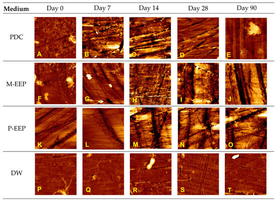
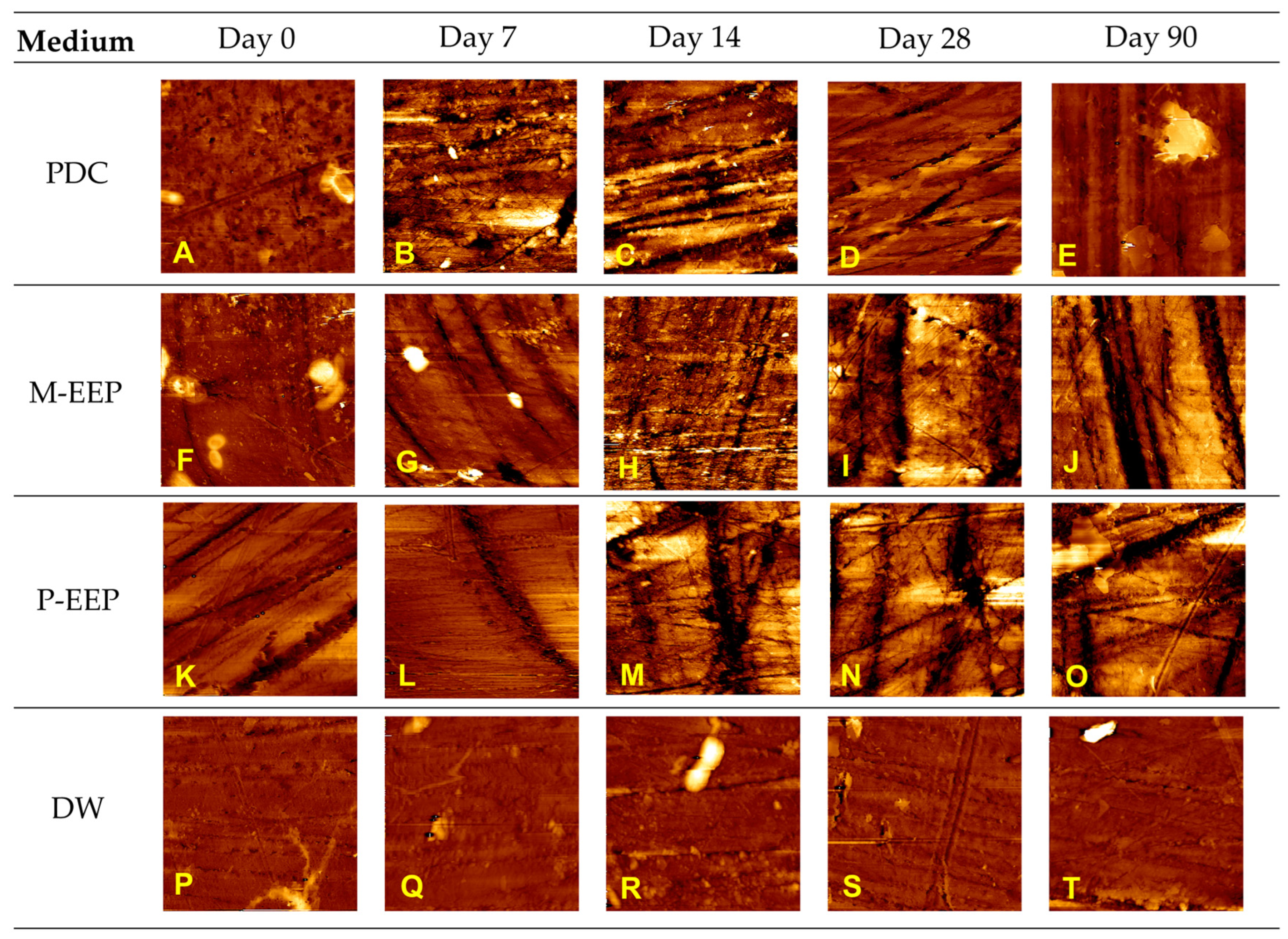
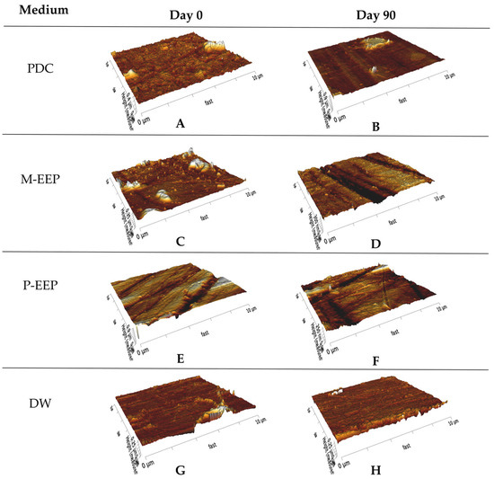
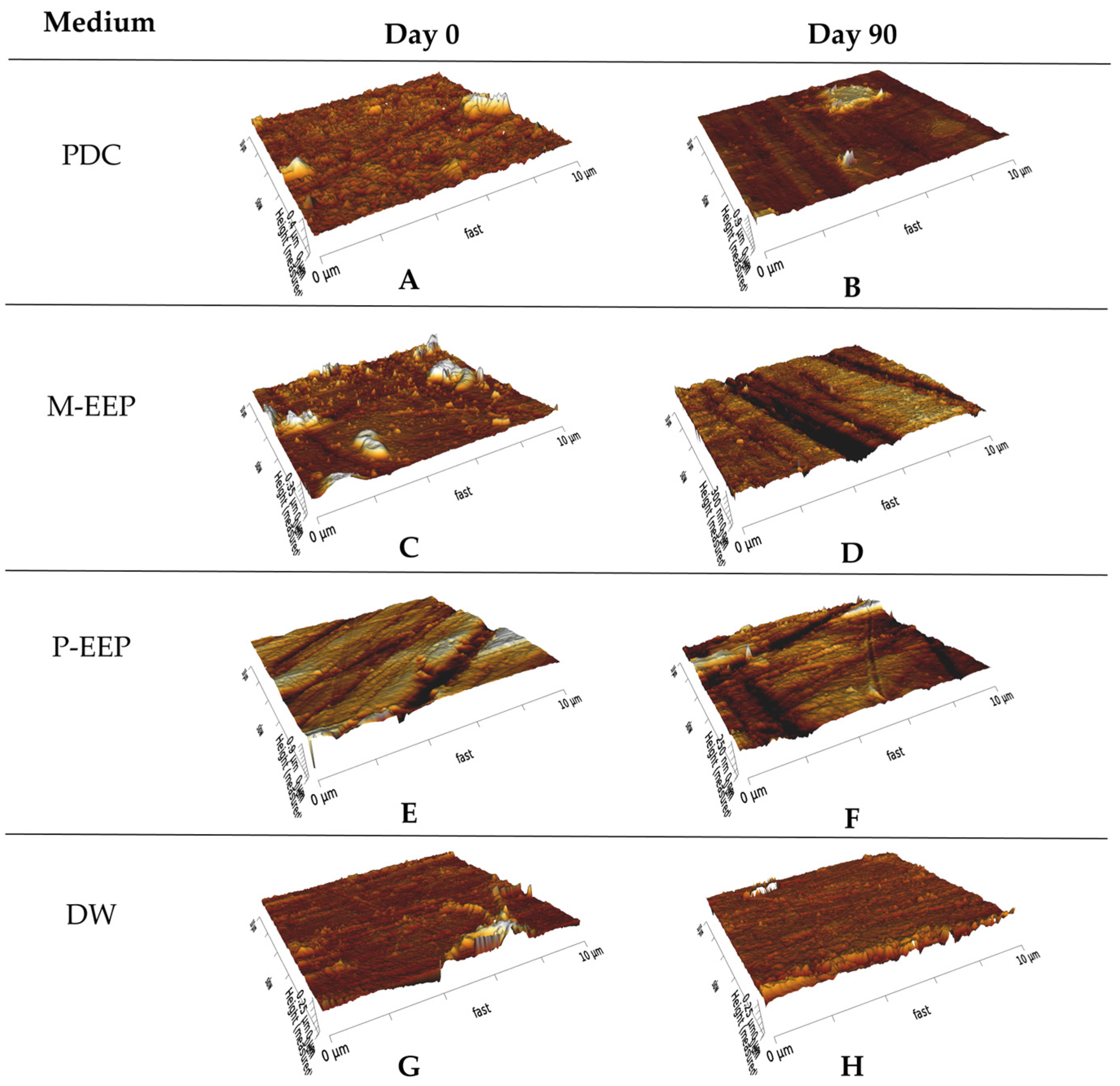
Two-dimensional AFM height maps were used to qualitatively assess PMMA surface topography across the immersion period (Figure 3). Three-dimensional AFM reconstructions provided supplementary visual support for surface contours and height variation (Figure 4).
Figure 3.
Representative two-dimensional atomic force microscopy height maps of heat-polymerised PMMA during 90-day immersion in denture-cleansing media: (A) PDC, Day 0; (B) PDC, Day 7; (C) PDC, Day 14; (D) PDC, Day 28; (E) PDC, Day 90; (F) M-EEP, Day 0; (G) M-EEP, Day 7; (H) M-EEP, Day 14; (I) M-EEP, Day 28; (J) M-EEP, Day 90; (K) P-EEP, Day 0; (L) P-EEP, Day 7; (M) P-EEP, Day 14; (N) P-EEP, Day 28; (O) P-EEP, Day 90; (P) DW, Day 0; (Q) DW, Day 7; (R) DW, Day 14; (S) DW, Day 28; (T) DW, Day 90. Each image represents a 10 × 10 µm2 scan area. Images are shown for qualitative assessment of surface topography.
Figure 4.
Representative three-dimensional atomic force microscopy reconstructions of heat-polymerised PMMA at Day 0 and Day 90 following immersion in denture-cleansing media: (A) PDC, Day 0; (B) PDC, Day 90; (C) M-EEP, Day 0; (D) M-EEP, Day 90; (E) P-EEP, Day 0; (F) P-EEP, Day 90; (G) DW, Day 0; (H) DW, Day 90. Each reconstruction represents a 10 × 10 µm2 scan area and is shown for qualitative assessment of surface topography.
At baseline, specimens from all groups showed relatively uniform surfaces with fine polishing features and minimal height contrast (Figure 3A,F,K,P). Across follow-up time points, surface appearance differed among immersion media. In the PDC group, surface irregularities became more apparent at intermediate time points, followed by a Day 90 appearance closer to baseline (Figure 3A–E).
M-EEP and P-EEP specimens showed more heterogeneous topography over time, including localised linear markings and alternating raised and depressed areas (Figure 3F–O). These features were more evident at later time points, although the AFM images did not show a clear qualitative distinction between the two EEP groups at Day 90.
Specimens immersed in DW maintained a comparatively uniform appearance throughout the 90-day period, with polishing features remaining visible and limited changes in height contrast (Figure 3P–T). Overall, AFM observations demonstrated localised and time-dependent changes in surface topography that were not fully reflected by the mean Ra and Rq values.
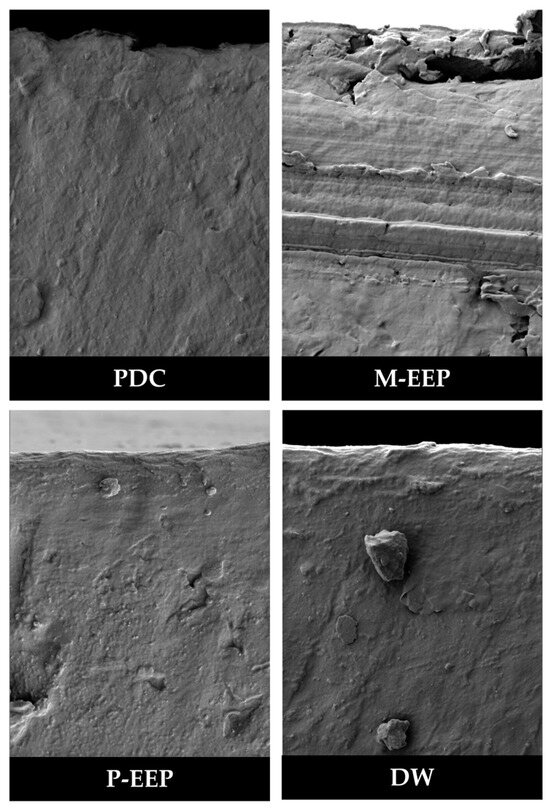
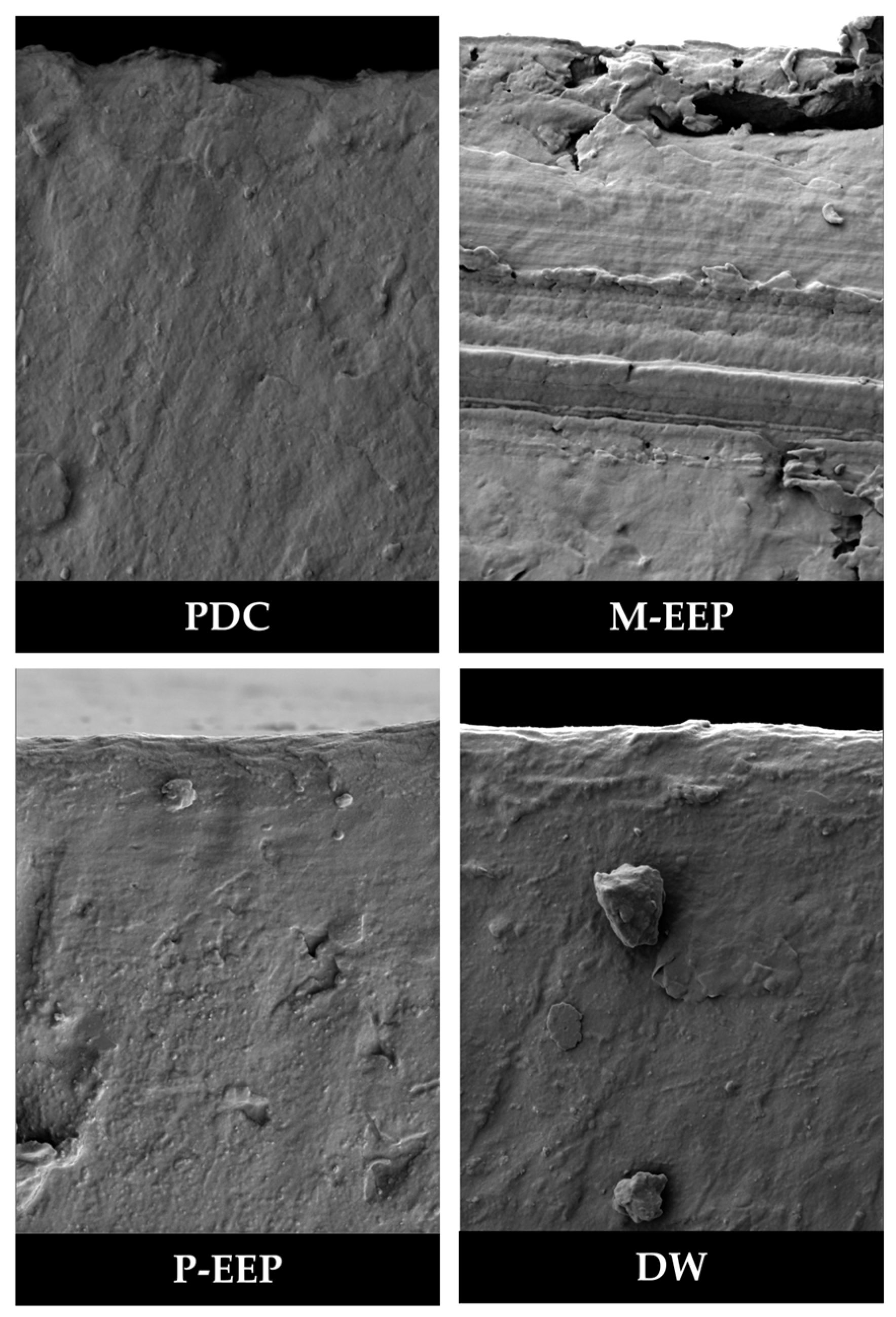
Exploratory FESEM cross-sectional images at Day 90 are provided in Figure 5. A visible discontinuity was observed in the representative M-EEP specimen, whereas the representative P-EEP, PDC, and DW specimens showed more continuous cross-sectional structures. As FESEM was performed on one representative specimen per group, these findings were considered exploratory and were not included in the quantitative surface-roughness analysis.
Figure 5.
Representative field-emission scanning electron microscopy images of sectioned heat-polymerised PMMA specimens after 90-day immersion in denture-cleansing media. One representative specimen was examined per group; the images are qualitative and exploratory and were not included in the quantitative surface-roughness analysis.
4. Discussion
The combined biological and material findings indicate that the six-month-fermented EEP formulations achieved antifungal activity without producing progressive roughening of heat-polymerised PMMA during prolonged exposure. P-EEP showed a lower MIC than M-EEP, indicating greater inhibitory potency across the tested concentrations, while both formulations were fungicidal at full strength. This common fungicidal condition was therefore selected for the PMMA experiment, allowing material compatibility to be evaluated at a concentration with confirmed biological activity.
At its manufacturer-recommended working concentration, PDC also prevented visible fungal growth. Because PDC was not subjected to the serial dilution procedure used for the EEP formulations, its relative antifungal potency could not be compared directly with their MIC values. Nevertheless, full-strength M-EEP, full-strength P-EEP, and prepared PDC all achieved the categorical outcome of no visible growth and no colony recovery under their respective test conditions. PDC consequently provided a clinically established reference against which the biological performance of the EEP formulations could be interpreted.
The antifungal pattern differed from that previously reported against Enterococcus faecalis. Earlier work showed that pineapple–orange and papaya EEP inhibited Enterococcus faecalis at 50% and were bactericidal at full strength [12]. In the present study, P-EEP inhibited Candida albicans at 50%, whereas M-EEP required full strength. This difference illustrates that the antimicrobial activity of a fermentation-derived preparation may depend on the target organism. Fungal cells differ from bacteria in their cell-wall organisation, sterol-containing membranes, metabolic regulation, and adaptive stress responses; antibacterial findings should therefore not be assumed to predict antifungal potency.
The distinct inhibitory profiles of M-EEP and P-EEP were unlikely to be attributable to acidity alone. Both formulations had similarly acidic pH values, yet P-EEP retained inhibitory activity at a greater dilution. Organic acids may contribute to fungal inhibition when their undissociated forms cross the cell membrane and subsequently dissociate within the relatively neutral cytoplasm, disrupting intracellular pH regulation, solute transport and metabolic enzyme activity [10,11,14]. However, the effectiveness of an acidic formulation also depends on the type and concentration of organic acids, buffering capacity, and the presence of other biologically active constituents.
Qualitative UPLC-HRMS profiling provided further context for the observed difference. P-EEP showed greater representation of phenolic compounds and amino acid and peptide derivatives, whereas M-EEP contained more organic acids and sugar-related metabolites. Among the putatively identified constituents, hesperetin and sorbic acid provide plausible literature-supported links to antifungal activity. Hesperetin, detected in P-EEP, has been reported to inhibit Candida growth and biofilm formation, including Candida albicans, with proposed mechanisms involving oxidative stress, membrane disruption and apoptosis-like cell death [20]. Sorbic acid, detected in M-EEP, is a recognised inhibitor of yeasts, including Candida albicans, with activity influenced by its dissociated and undissociated forms [21]. Other phenolic constituents identified in the formulations may also contribute to antifungal activity; for example, propyl gallate has been reported to enhance azole activity against Candida albicans, although its independent contribution within the present formulation cannot be established [22]. The antifungal effect observed in the present study is therefore more plausibly attributed to the combined activity of multiple metabolites, together with the acidic formulation environment, than to a single dominant compound. However, because metabolite abundance was not quantified and individual compounds were not tested independently, the relative contribution of each constituent cannot be determined. These proposed mechanisms should therefore be regarded as literature-supported interpretations of whole-formulation activity rather than experimentally confirmed mechanisms in the present study.
The PMMA findings showed that surface response depended on both the immersion medium and exposure time. Significant medium x time interactions for Ra and Rq indicated dynamic, formulation-specific patterns rather than a simple ranking from least to most damaging. PDC and P-EEP showed early reductions in roughness that were no longer significant at Day 90, whereas M-EEP and DW showed no significant within-group changes. The principal quantitative material finding was the absence of cumulative increases in Ra and Rq throughout the observation period, despite transient changes at earlier time points.
The prolonged immersion schedule was deliberately designed as an accelerated material-ageing model rather than as a simulation of the manufacturer-recommended three-minute daily use of PDC. Continuous, repeated exposure increased the opportunity for water sorption, leaching, and cleanser–polymer interactions to become detectable within a practical experimental period. Applying the same intensified chemical challenge to PDC, M-EEP, P-EEP, and DW also enabled controlled comparison of their time-dependent material effects. PDC therefore served as a material-response benchmark under the standardised accelerated exposure conditions used in this study, rather than as a simulation of its prescribed clinical use. Accordingly, differences observed between PDC and the EEP formulations under this intensified protocol should not be interpreted as evidence of comparative material performance during routine clinical cleansing.
This model provides a conservative assessment of whether small initial surface variations develop into cumulative changes during extended chemical contact. Under this intensified exposure, none of the cleansing media produced progressive PMMA roughening. The EEP findings are therefore notable because no progressive increase in mean Ra or Rq was observed despite a substantially more demanding exposure condition than would ordinarily be encountered during routine cleansing. The protocol should not, however, be converted into an exact number of clinical years because chemical and water-mediated changes may not accumulate linearly.
Several near-surface processes may account for the temporary reductions observed with PDC and P-EEP. Water uptake can plasticise the superficial polymer region and alter the local distribution of peaks and valleys. Surfactant, effervescent, or oxidative components in PDC may remove loosely retained polishing residues, while soluble constituents in the EEP formulations may modify the hydrated surface environment. Polymer relaxation and variation in the AFM scanning region may also influence measured topography [23,24,25,26]. These processes could produce small fluctuations in Ra and Rq without representing either genuine surface improvement or progressive disruption of the PMMA matrix. Rq is particularly responsive to isolated peaks and depressions, which may explain its greater variability in some groups.
The different media pH values did not correspond to a predictable PMMA response. M-EEP and P-EEP were similarly acidic but produced different temporal patterns, while the near-neutral PDC also showed early changes. Surface behaviour was therefore more likely governed by interactions among overall formulation chemistry, polymer characteristics, and exposure duration than by acidity alone. This finding also cautions against assuming that either a natural origin or an established commercial composition guarantees complete material inertness.
Between-medium comparisons did not reveal a consistent hierarchy. The only significant adjusted difference occurred for Ra at Day 14, when M-EEP showed a higher value than PDC. This difference was not sustained at subsequent assessments and was not accompanied by significant differences in Rq. It is therefore more consistent with a time-specific surface response than with persistent damage caused by M-EEP or superior material protection by PDC.
The present pattern accords with the more favourable section of the natural-denture-cleanser literature. Grapefruit seed extract, for example, reduced Candida albicans biofilm without significantly altering PMMA roughness, hardness, or flexural strength during repeated exposure [27]. Geraniol- and thymol-based formulations similarly reduced multispecies biofilm under a prolonged-use simulation without clinically concerning changes in roughness or colour [16]. These studies, together with the present results, suggest that selected naturally derived formulations can achieve biological activity while maintaining relevant PMMA properties.
Such compatibility is not universal. Other natural preparations have increased surface roughness, reduced hardness, or altered colour when tested at higher concentrations or after prolonged exposure [28,29,30]. The contrast indicates that natural derivatives cannot be treated as a uniform category. Their material effects depend on formulation composition, concentration, exposure protocol, and the acrylic resin evaluated. The value of the present EEP formulations therefore lies not merely in their natural origin or antifungal activity, but in achieving that activity without progressive roughness changes under an accelerated exposure model.
AFM imaging demonstrated medium- and time-related variations in surface topography, including greater heterogeneity in the EEP groups at selected intervals. These qualitative changes were not always reflected by corresponding changes in mean Ra and Rq, indicating that average roughness parameters may not capture localised morphological features. Although three predefined regions were examined for each specimen at every time point, each AFM scan represented a limited surface area. Therefore, the absence of progressive increases in mean Ra and Rq should not be interpreted as evidence that surface morphology remained unchanged. Rather, the findings indicate localised and time-dependent surface responses without progressive mean roughening over the 90-day exposure period.
FESEM examination revealed a discontinuity in the representative M-EEP specimen at Day 90. Because only one specimen per group was examined by FESEM, this observation is exploratory and cannot determine whether the feature was associated with EEP exposure or specimen-specific variation. Confirmation would require assessment of multiple independent specimens and multiple regions.
The principal material–biological finding is that antifungal activity against the tested planktonic strain was achieved without progressive increases in mean PMMA roughness. This balance matters because a cleanser that reduces fungal viability but gradually damages the denture base could make the prosthesis harder to maintain and may facilitate subsequent microbial retention. Under the conditions tested, both EEP formulations avoided this undesirable biological–material trade-off, and their surface responses remained within the range observed for PDC under the same accelerated challenge.
P-EEP warrants particular attention for further formulation development because its inhibitory activity extended to a lower concentration than that of M-EEP. Full strength was nevertheless required for fungicidal activity in both formulations. Optimisation should therefore determine whether fungicidal performance can be retained by adjusting concentration, contact time, or formulation delivery while improving practicality, stability, and acceptability for repeated denture use.
All measured roughness values remained below the commonly cited 0.2 µm threshold associated with increased plaque retention [31]. This supports the surface compatibility of the formulations within the present model. However, microbial adhesion is also influenced by surface chemistry, surface free energy, hydrophobicity, salivary pellicle formation, and microbial phenotype [32,33,34,35]. Maintaining roughness below this threshold therefore supports cleanability but does not, by itself, confirm resistance to recolonisation or established denture-biofilm removal.
The findings support progression to clinically representative evaluation rather than immediate replacement of PDC. Their practical value will depend on whether the antifungal–material balance is retained against mature denture biofilms and under conditions incorporating saliva, mechanical cleaning, thermal variation, and repeated microbial challenge. More broadly, the study demonstrates the value of selecting candidate cleansers through concurrent biological and material assessment: antimicrobial potency is insufficient if the active regimen compromises the denture base, while material compatibility has limited value in the absence of meaningful fungal control.
The findings are limited to planktonic Candida albicans ATCC 10231, one heat-polymerised PMMA resin, and an immersion-based ageing model. Baseline roughness values, particularly Rq, showed greater magnitude and variability in the P-EEP and PDC groups. Although within-specimen changes from baseline were considered, this variability remains a limitation when interpreting between-group differences. The modest number of specimens per group may also limit the precision of the GEE estimates, particularly for the medium x time interaction. Because GEE inference relies on large-sample approximations across independent clusters, these interaction findings should be interpreted cautiously and considered alongside the estimated changes, confidence intervals and observed longitudinal patterns, rather than statistical significance alone. PDC was evaluated only at its manufacturer-recommended working concentration and was not subjected to the serial-dilution protocol used for the EEP formulations; consequently, direct MIC-based comparison of antifungal potency between PDC and the EEP formulations was not possible. The separate contributions of acidity and individual metabolites could not be established, and the pH of the immersion media was measured only at initial preparation rather than throughout the repeated 8-h exposure cycles. Material outcomes were confined to surface roughness and qualitative morphology. Future studies should therefore include mature Candida biofilms formed directly on PMMA, clinical isolates, pH-controlled formulations, cytotoxicity assessment, combined brushing–immersion protocols, and broader material properties such as colour stability, hardness, and strength.
Overall, the six-month-fermented EEP formulations demonstrated antifungal activity without progressive increases in mean PMMA surface roughness during prolonged exposure. P-EEP showed greater inhibitory potency, while both formulations exhibited time-dependent surface changes on qualitative imaging. These findings support further investigation of EEP as a denture-cleansing candidate, with additional studies required to characterise its surface effects more comprehensively and under clinically relevant conditions.
5. Conclusions
Within the limitations of this in vitro accelerated-exposure model, six-month-fermented M-EEP and P-EEP demonstrated antifungal activity against the tested planktonic Candida albicans ATCC 10231 without progressive increases in mean PMMA surface roughness over 90 days. The present study advances earlier biological screening of fermentation-derived fruit-waste formulations by incorporating longitudinal assessment of a clinically relevant denture-base material. Qualitative imaging nevertheless demonstrated localised surface changes, indicating that the absence of progressive increases in mean roughness should not be interpreted as evidence that surface morphology remained unchanged. These findings support further formulation optimisation, broader surface-characterisation studies, safety assessment, and evaluation under clinically relevant cleansing conditions.
Author Contributions
Conceptualization, H.A.K.M.; methodology, D.S., H.A.K.M., H.Z.Y., Z.S. and A.M.; software, D.S.; validation, D.S., H.A.K.M., H.Z.Y., Z.S. and A.M.; formal analysis, D.S. and H.Z.Y.; investigation, D.S.; resources, H.A.K.M.; data curation, D.S. and H.Z.Y.; writing—original draft preparation, D.S.; writing—review and editing, D.S. and H.Z.Y.; visualization, D.S., H.A.K.M., H.Z.Y., Z.S. and A.M.; supervision, H.Z.Y.; project administration, D.S., H.A.K.M., and H.Z.Y.; funding acquisition, H.A.K.M. All authors have read and agreed to the published version of the manuscript.
Funding
This study was supported by The National University of Malaysia under the Geran Galakan Penyelidik Muda (GGPM), grant no. GGPM-2023-004. The funding body had no role in the study design, data collection, data analysis or interpretation, manuscript preparation, or the decision to submit the article for publication.
Institutional Review Board Statement
The study was approved by the Research Ethics Committee of The National University of Malaysia (UKM PPI/111/8/JEP-2023-754; approval date: 26 October 2023). No human participants or animals were involved in this study.
Informed Consent Statement
Not applicable. This study did not involve human participants.
Data Availability Statement
The data presented in this study are available from the corresponding author upon reasonable request. The data are not publicly available because related analyses are being prepared for publication.
Acknowledgments
The authors acknowledge The National University of Malaysia for its support and the technical assistance provided throughout this study.
Conflicts of Interest
The authors declare no conflicts of interest.
Abbreviations
The following abbreviations are used in this manuscript:
| AFM | Atomic force microscopy |
| ATCC | American Type Culture Collection |
| CI | Confidence interval |
| DW | Distilled water |
| EEP | Effective eco-produce |
| FESEM | Field-emission scanning electron microscopy |
| GEE | Generalised estimating equation |
| M-EEP | Mixed pineapple–orange effective eco-produce |
| MFC | Minimum fungicidal concentration |
| MIC | Minimum inhibitory concentration |
| OD | Optical density |
| OD530 | Optical density measured at 530 nm |
| PDC | Peroxide-based denture cleanser |
| PEG-180 | Polyethylene glycol 180 |
| PES | Polyethersulfone |
| PMMA | Polymethyl methacrylate |
| P-EEP | Papaya-based effective eco-produce |
| Ra | Arithmetic mean roughness |
| Rq | Root mean square roughness |
| SD | Standard deviation |
| SPSS | Statistical Package for the Social Sciences |
| TAED | Tetraacetylethylenediamine |
| UPLC-HRMS | Ultra-performance liquid chromatography–high-resolution mass spectrometry |
| VP/VA | Vinylpyrrolidone/vinyl acetate |
References
- Gendreau, L.; Loewy, Z.G. Epidemiology and etiology of denture stomatitis. J. Prosthodont. 2011, 20, 251–260. [Google Scholar] [CrossRef] [Scilit] [PubMed]
- Kuhar, M.; Funduk, N. Effects of polishing techniques on the surface roughness of acrylic denture base resins. J. Prosthet. Dent. 2005, 93, 76–85. [Google Scholar] [CrossRef] [Scilit] [PubMed]
- Glass, R.T.; Conrad, R.S.; Bullard, J.W.; Goodson, L.B.; Mehta, N.; Lech, S.J.; Loewy, Z.G. Evaluation of microbial flora found in previously worn prostheses from the Northeast and Southwest regions of the United States. J. Prosthet. Dent. 2010, 103, 384–389. [Google Scholar] [CrossRef] [Scilit] [PubMed]
- Alqanas, S.S.; Alfuhaid, R.A.; Alghamdi, S.F.; al-Qarni, F.D.; Gad, M.M. Effect of denture cleansers on the surface properties and color stability of 3D printed denture base materials. J. Dent. 2022, 120, 104089. [Google Scholar] [CrossRef] [Scilit] [PubMed]
- Çakan, U.; Kara, O.; Kara, H. Effects of various denture cleansers on surface roughness of hard permanent reline resins. Dent. Mater. J. 2015, 34, 246–251. [Google Scholar] [CrossRef] [Scilit] [PubMed][Green Version]
- Costa, R.T.F.; Pellizzer, E.P.; Vasconcelos, B.C.d.E.; Gomes, J.M.L.; Lemos, C.A.A.; de Moraes, S.L.D. Surface roughness of acrylic resins used for denture base after chemical disinfection: A systematic review and meta-analysis. Gerodontology 2021, 38, 242–251. [Google Scholar] [CrossRef] [Scilit] [PubMed]
- Arslan, E.; Akay, C.; Erdönmez, D.; Avukat, E.N. Evaluation of the effect of new generation denture base materials aged in artificial saliva at different pH levels on surface roughness and Candida albicans adhesion. BMC Oral Health 2025, 25, 356. [Google Scholar] [CrossRef] [Scilit] [PubMed]
- Fouda, S.M.; Gad, M.M.; Abualsaud, R.; Ellakany, P.; AlRumaih, H.S.; Farooqi, F.A.; Matin, A.; Al-Eraky, D.M.; Al-Qarni, F.D.; Al-Harbi, F.A. In Vitro Evaluation of Candida albicans Adhesion and Related Surface Properties of CAD/CAM Denture Base Resins. Eur. J. Dent. 2024, 18, 579–586. [Google Scholar] [CrossRef] [Scilit] [PubMed]
- Sultana, N.; Ahmed, S.; Nandini, V.V.; Lathief, J.; Boruah, S. An In Vitro Comparison of Microbial Adhesion on Three Different Denture Base Materials and Its Relation to Surface Roughness. Cureus 2023, 15, e37085. [Google Scholar] [CrossRef] [Scilit] [PubMed]
- Arun, C.; Sivashanmugam, P. Study on optimization of process parameters for enhancing the multi-hydrolytic enzyme activity in garbage enzyme produced from preconsumer organic waste. Bioresour. Technol. 2017, 226, 200–210. [Google Scholar] [CrossRef] [Scilit] [PubMed]
- Indrani, B.; Shrijona, H.; Jyotchna, G.; Nayan, T. A Systematic Review on Enzyme Extraction from Organic Wastes and its Application. J. Biochem. Technol. 2022, 13, 32–37. [Google Scholar] [CrossRef] [Scilit]
- Mavani, H.A.K.; Tew, I.M.; Wong, L.; Yew, H.Z.; Mahyuddin, A.; Ahmad Ghazali, R.; Pow, E.H.N. Antimicrobial Efficacy of Fruit Peels Eco-Enzyme against Enterococcus faecalis: An In Vitro Study. Int. J. Environ. Res. Public Health 2020, 17, 5107. [Google Scholar] [CrossRef] [Scilit] [PubMed]
- Ng, M.Y.; Shafiei, Z.; Rahman, A.; Sockalingam, S.N.M.; Zakaria, A.S.I.; Mahyuddin, A. Antibacterial effects of effective ecoproduce on Enterococcus faecalis: An in vitro Study. J. Int. Dent. Med. Res. 2020, 13, 861–867. [Google Scholar] [CrossRef] [Scilit] [PubMed]
- Tallei, T.E.; Fatimawali; Niode, N.J.; Alsaihati, W.M.; Salaki, C.L.; Alissa, M.; Kamagi, M.; Rabaan, A.A. Antibacterial and antioxidant activity of ecoenzyme solution prepared from papaya, pineapple, and kasturi orange fruits: Experimental and molecular docking studies. J. Food Process. Preserv. 2023, 2023, 5826420. [Google Scholar] [CrossRef] [Scilit]
- Ferro, A.C.; Spavieri, J.H.P.; Ribas, B.R.; Scabelo, L.; Jorge, J.H. Do denture cleansers influence the surface roughness and adhesion and biofilm formation of Candida albicans on acrylic resin? Systematic review and meta-analysis. J. Prosthodont. Res. 2022, 67, 164–172. [Google Scholar] [CrossRef] [Scilit] [PubMed]
- Kaypetch, R.; Rudrakanjana, P.; Tua-Ngam, P.; Tosrisawatkasem, O.; Thairat, S.; Tonput, P.; Tantivitayakul, P. Effects of two novel denture cleansers on multispecies microbial biofilms, stain removal and the denture surface: An in vitro study. BMC Oral Health 2023, 23, 852. [Google Scholar] [CrossRef] [Scilit] [PubMed]
- Rex, J.H.; Clinical and Laboratory Standards Institute. Reference Method for Broth Dilution Antifungal Susceptibility Testing of Yeasts: Approved Standard, 3rd ed.; Clinical and Laboratory Standards Institute: Wayne, PA, USA, 2008. [Google Scholar]
- Köroğlu, A.; Sahin, O.; Dede, D.; Deniz, S.; Karacan Sever, N.; Özkan, S. Efficacy of denture cleaners on the surface roughness and Candida albicans adherence of sealant agent coupled denture base materials. Dent. Mater. J. 2016, 35, 810–816. [Google Scholar] [CrossRef] [Scilit] [PubMed]
- Berger, J.C.; Driscoll, C.F.; Romberg, E.; Luo, Q.; Thompson, G. Surface roughness of denture base acrylic resins after processing and after polishing. J. Prosthodont. 2006, 15, 180–186. [Google Scholar] [CrossRef] [Scilit] [PubMed]
- Carević, T.; Kostić, M.; Nikolić, B.; Stojković, D.; Soković, M.; Ivanov, M. Hesperetin-Between the Ability to Diminish Mono- and Polymicrobial Biofilms and Toxicity. Molecules 2022, 27, 6806. [Google Scholar] [CrossRef] [Scilit] [PubMed]
- Eklund, T. The antimicrobial effect of dissociated and undissociated sorbic acid at different pH levels. J. Appl. Bacteriol. 1983, 54, 383–389. [Google Scholar] [CrossRef] [Scilit] [PubMed]
- D’Auria, F.D.; Tecca, M.; Strippoli, R.; Simonetti, N. In vitro activity of propyl gallate-azole drug combination against fluconazole- and itraconazole-resistant Candida albicans strains. Lett. Appl. Microbiol. 2001, 32, 220–223. [Google Scholar] [CrossRef] [Scilit] [PubMed]
- Degirmenci, K.; Hayati Atala, M.; Sabak, C. Effect of different denture base cleansers on surface roughness of heat polymerised acrylic materials with different curing process. Odovtos Int. J. Dent. Sci. 2020, 22, 145–153. [Google Scholar] [CrossRef] [Scilit]
- Felton, D.; Cooper, L.; Duqum, I.; Minsley, G.; Guckes, A.; Haug, S.; Meredith, P.; Solie, C.; Avery, D.; Deal Chandler, N. Evidence-based guidelines for the care and maintenance of complete dentures: A publication of the American College of Prosthodontists. J. Prosthodont. 2011, 20, S1–S12. [Google Scholar] [CrossRef] [Scilit] [PubMed]
- Kiesow, A.; Sarembe, S.; Pizzey, R.L.; Axe, A.S.; Bradshaw, D.J. Material compatibility and antimicrobial activity of consumer products commonly used to clean dentures. J. Prosthet. Dent. 2016, 115, 189–198.e188. [Google Scholar] [CrossRef] [Scilit] [PubMed]
- Ozyilmaz, O.Y.; Akin, C. Effect of cleansers on denture base resins’ structural properties. J. Appl. Biomater. Funct. Mater. 2019, 17, 2280800019827797. [Google Scholar] [CrossRef] [Scilit] [PubMed]
- Tsutsumi-Arai, C.; Takakusaki, K.; Arai, Y.; Terada-Ito, C.; Takebe, Y.; Imamura, T.; Ide, S.; Tatehara, S.; Tokuyama-Toda, R.; Wakabayashi, N.; et al. Grapefruit seed extract effectively inhibits the Candida albicans biofilms development on polymethyl methacrylate denture-base resin. PLoS ONE 2019, 14, e0217496. [Google Scholar] [CrossRef] [Scilit] [PubMed]
- Heidrich, D.; Fortes, C.B.B.; Mallmann, A.T.; Vargas, C.M.; Arndt, P.B.; Scroferneker, M.L. Rosemary, castor oils, and propolis extract: Activity against Candida albicans and alterations on properties of dental acrylic resins. J. Prosthodont. 2019, 28, e863–e868. [Google Scholar] [CrossRef] [Scilit] [PubMed]
- Rachmadi, P.; Firdaus, A.K.; Wayan, I.; Sukmana, B.I.; Aspriyanto, D.; Puspitasari, D.; Darmawan, H.; Singgih, M.F.; Ramadhany, S.; Sinarruli, N.B. The effect of immersion of 12.5% basil leaves and 25% mauli banana stem mixture extracts on surface hardness, surface roughness and discoloration of acrylic resin. Syst. Rev. Pharm. 2020, 11, 281–287. [Google Scholar] [CrossRef] [Scilit]
- Sugiaman, V.K.; Saputri, R.I.; Naliani, S.; Amalia, J. Lemongrass (Cymbopogon citratus) oil nanoparticle synthesis, characteristic, and evaluation of antibacterial and antifungal effects and the influence on hardness of acrylic resin. J. Pharm. Pharmacogn. Res. 2024, 12, 1156–1169. [Google Scholar] [CrossRef] [Scilit]
- Bollen, C.M.; Lambrechts, P.; Quirynen, M. Comparison of surface roughness of oral hard materials to the threshold surface roughness for bacterial plaque retention: A review of the literature. Dent. Mater. 1997, 13, 258–269. [Google Scholar] [CrossRef] [Scilit] [PubMed]
- Nevzatoğlu, E.U.; Ozcan, M.; Kulak-Ozkan, Y.; Kadir, T. Adherence of Candida albicans to denture base acrylics and silicone-based resilient liner materials with different surface finishes. Clin. Oral Investig. 2007, 11, 231–236. [Google Scholar] [CrossRef] [Scilit] [PubMed]
- Radford, D.R.; Challacombe, S.J.; Walter, J.D. Denture plaque and adherence of Candida albicans to denture-base materials in vivo and in vitro. Crit. Rev. Oral Biol. Med. 1999, 10, 99–116. [Google Scholar] [CrossRef] [Scilit] [PubMed]
- Radford, D.R.; Sweet, S.; Challacombe, S.; Walter, J. Adherence of Candida albicans to denture-base materials with different surface finishes. J. Dent. 1998, 26, 577–583. [Google Scholar] [CrossRef] [Scilit] [PubMed]
- Yuzugullu, B.; Acar, O.; Cetinsahin, C.; Celik, C. Effect of different denture cleansers on surface roughness and microhardness of artificial denture teeth. J. Adv. Prosthodont. 2016, 8, 333–338. [Google Scholar] [CrossRef] [Scilit] [PubMed]

Disclaimer/Publisher’s Note: The statements, opinions and data contained in all publications are solely those of the individual author(s) and contributor(s) and not of MDPI and/or the editor(s). MDPI and/or the editor(s) disclaim responsibility for any injury to people or property resulting from any ideas, methods, instructions or products referred to in the content. |
© 2026 by the authors. Licensee MDPI, Basel, Switzerland. This article is an open access article distributed under the terms and conditions of the Creative Commons Attribution (CC BY) license.