


Abstract
Human milk is the gold standard for infant nutrition; however, infant formulas (IFs) remain the main alternative when breastfeeding is not possible. Cow and goat milk-based IFs are well established, whereas sheep milk IFs have attracted increasing interest. IFs are among the most studied food products; nevertheless, little is known about the evolution of small molecules during digestion. This study monitored the metabolomic changes in sheep and goat IFs during simulated digestion using the dynamic in vitro gastrointestinal digestion system (DIDGI®). Using an untargeted GC–MS approach, a total of 51 metabolites were identified in samples collected throughout the digestion process. The results of multivariate statistics indicated that the gastric and the intestinal digestion stages, rather than milk origin, were the primary sources of metabolic variation. However, different sheep and goat IF metabolite fingerprints were detected and persisted throughout digestion. Temporal profile analysis showed limited amino acid release during gastric digestion followed by a rapid increase during intestinal transition, reflecting extensive proteolysis. Similarly, massive lipolysis led to the accumulation in the intestine of free fatty acids, including medium-chain fatty acids derived from milk fat. These findings provide the first untargeted GC–MS characterization of the metabolomic evolution of sheep and goat milk IFs during dynamic in vitro gastrointestinal digestion.
Keywords:
infant formulas; dynamic digestion; GC-MS; metabolomics; free amino acids; sheep milk; goat milk 1. Introduction
Human milk is widely recognized as the gold standard for infant nutrition, providing the optimal balance of nutrients and bioactive compounds required for neonatal growth, immune maturation, and cognitive development during early life [1]. However, when breastfeeding is not possible or insufficient, infant formulas (IFs) represent the primary nutritional alternative and are manufactured to reproduce, as closely as possible, the composition and functional properties of human milk [2]. Recent metabolomics studies have demonstrated that different infant feeding regimens can significantly influence the metabolic profile of neonates. Breastfed infants exhibit a more gluconeogenic metabolic profile, characterized by higher concentrations of Krebs cycle intermediates (e.g., fumaric acid and succinic acid) [3]. In contrast, IF consumption has been associated with the excretion of metabolites derived from specific formulation ingredients, such as D-glucitol and 4-deoxytetronic acid [3]. Most commercial IFs are traditionally based on bovine milk ingredients supplemented with a blend of vegetable oils and micronutrients to better mimic the fatty acid profile of human milk. Considerable compositional variability has also been reported among commercial IFs, largely influenced by the presence of prebiotic ingredients such as galacto-oligosaccharides (GOSs) and fructo-oligosaccharides (FOSs). In general, IFs tend to contain higher levels of simple sugars, including glucose, fructose, galactose, and mannitol, whereas human milk is characterized by a higher abundance of free amino acids (e.g., threonine, valine, alanine, and serine) and inositols. Exclusive metabolites such as orotic acid and isomaltulose have been reported in infant formulas but are typically absent in human milk, reflecting differences in ingredient composition and industrial processing [4]. Importantly, the manufacturing processes applied to IFs, particularly thermal treatments, can modify the structural organization and digestive behavior of milk proteins. Whey proteins, either alone or in association with caseins, may form heat-induced aggregates, thereby affecting protein solubility and susceptibility to gastrointestinal proteolysis [5]. Moreover, the supramolecular organization of IF emulsions differs from that of human milk and may influence both lipid and protein digestion. In particular, the adsorption of caseins and whey proteins at the emulsion interface can modulate their accessibility to gastric proteases such as pepsin [6,7].
In recent years, increasing attention has been directed toward small-ruminant milk-based IFs, particularly those derived from goat and sheep milk, due to their distinctive nutritional and functional characteristics [8]. Goat milk-based IFs are already widely commercialized and well established in infant nutrition, whereas sheep milk-based IFs are currently authorized only in selected countries, including New Zealand and China, and remain prohibited in the European Union [9]. Nevertheless, sheep milk has attracted growing scientific interest because of its higher fat and protein content, as well as its unique lipid and casein composition, compared with bovine and goat milk [10]. Previous studies have demonstrated the feasibility of producing nutritionally balanced IFs using sheep milk-derived ingredients [11,12,13]. Moreover, sheep milk-based infant formula contains higher levels of medium-chain fatty acids than goat milk IF, whereas proteins undergo more extensive gastrointestinal digestion, resulting in a higher number of released peptides and greater protein sequence coverage [11,12]. Protein composition is another important determinant of IF digestion. Ruminant milk is generally characterized by a casein-to-whey protein ratio of approximately 80:20. This ratio is commonly adjusted through the addition of whey proteins to approach the approximately 40:60 casein-to-whey protein ratio of human milk [13]. Whey protein ingredients used for this purpose are standardized dairy fractions obtained through whey processing and are widely used in IF manufacture [13,14]. Compared with bovine milk, goat milk is generally characterized by a lower proportion of αs1-casein and larger casein micelles, whereas sheep milk contains higher levels of total solids, proteins, and minerals [10,15,16].
Goat milk is generally considered more easily digestible than sheep milk, as the higher protein and mineral content of sheep milk may promote the formation of firmer protein gels in the gastric environment, potentially affecting gastric protein digestion [16,17]. Previous characterization of the same goat and sheep milk-based IFs investigated in the present study identified 37 bioactive peptides derived from casein hydrolysis during gastrointestinal digestion [12]. The lipid fraction of IFs consists predominantly of triacylglycerols containing short-, medium-, and long-chain fatty acids. In ruminant milk, short-chain fatty acids (SCFAs) are preferentially esterified at the sn-1 and sn-3 positions of the glycerol backbone [18]. This positional distribution makes these fatty acids more susceptible to enzymatic hydrolysis, thereby facilitating their release during gastrointestinal digestion. This feature may be particularly relevant in infant nutrition, as short- and medium-chain fatty acids are more readily absorbed than long-chain fatty acids and can therefore represent a readily available source of energy for infants [19]. Previous characterization of the IFs investigated in the present study showed that the sheep milk-based IF contained higher levels of short-chain fatty acids (SCFAs) than the goat milk-based IF [11].
Beyond lipid composition and protein digestibility, the metabolite profile arising during gastrointestinal digestion also represents an important aspect of the nutritional quality of IFs. During gastric digestion, casein micelles undergo destabilization due to acidification and pepsin-mediated proteolysis, leading to the formation of protein coagulates that may also entrap fat globules [20]. The structural properties of these coagulates can strongly influence gastric digestion kinetics, gastric emptying, and the subsequent intestinal release and absorption of amino acids [21]. Previous investigations based on in vitro digestion models demonstrated that casein coagulation behavior is affected by multiple factors, including processing-induced protein modifications, food matrix composition, and species-dependent differences in milk proteins [20,22,23,24,25,26]. During gastrointestinal digestion, digestive enzymes such as pepsin and trypsin hydrolyze milk proteins, generating peptides and low-molecular-weight metabolites, including free amino acids [4]. These metabolites may originate from the native milk metabolome [27,28], ingredients intentionally added during manufacturing, by-products of ingredient production (e.g., fructo-oligosaccharides) [29], or may be newly generated during digestion. Some of these metabolites may be of nutritional relevance for infants. However, it remains unclear whether differences in milk origin influence the generation of small molecules during digestion and, consequently, their potential bioaccessibility. Dynamic in vitro digestion systems, such as DIDGI®, provide a physiologically relevant platform for investigating infant gastrointestinal digestion because they reproduce dynamic changes in pH, enzyme secretion, gastric emptying, and intestinal transit under controlled conditions [30]. Despite the growing interest in small-ruminant milk-based IFs, the evolution of small molecules during dynamic in vitro gastrointestinal digestion remains poorly characterized. To the best of our knowledge, this is the first study to investigate the fate of sheep and goat milk-based IFs during gastrointestinal digestion using an untargeted metabolomics approach, providing novel insights into their metabolic transformation and potential bioaccessibility. We hypothesized that the different protein and lipid composition of sheep and goat milk-based IFs would result in distinct metabolomic profiles during dynamic in vitro gastrointestinal digestion. Therefore, the aim of the present study was to (i) investigate and compare the metabolomic evolution of sheep and goat milk-based IFs during dynamic in vitro infant digestion using an untargeted GC–MS approach, and (ii) characterize the temporal evolution of metabolites and amino acid release throughout gastrointestinal digestion.
2. Materials and Methods
2.1. Chemicals
LC-grade solvents, including methanol, chloroform, and n-hexane (GC/MS grade, >99%), were obtained from Sigma-Aldrich (Milan, Italy). Potassium chloride (analytical grade) was purchased from Carlo Erba (Val de Reuil Cedex, France). Ultrapure water was produced using a Milli-Q purification system (Millipore, Milan, Italy). Succinic acid-2,2,3,3-d4, anhydrous pyridine (99.8%), amino acid standard mixture (2.5 mM each), saturated alkane calibration mixture (C7–C40), O-methylhydroxylamine hydrochloride, N-methyl-N-(trimethylsilyl) trifluoroacetamide, and 2-dodecanone were supplied by Sigma-Aldrich (Milan, Italy). Porcine pepsin (P6887), porcine pancreatin (P7545), and bovine bile extract (B3883) were acquired from Sigma Aldrich (St. Quentin Fallavier, France), together with the enzyme inhibitors pepstatin A (P5318), pefabloc (76307) and 4-bromophenyl boronic acid (B75956). Rabbit gastric extract (RGE), 74 U/mg of lipase and 754 U/mg of pepsin were supplied by Lipolytech (Marseille, France).
2.2. Infant Formulas
Powdered infant formulas (IFs) intended for infants aged 0–6 months were kindly supplied by Blue River Dairy LP (Invercargill, New Zealand). Sheep and goat milk IFs were formulated using whole milk (10–20%), whey ingredients (55–65%), and a vegetable oil blend (approximately 20%) composed of canola, sunflower, and coconut oils. Both formulations had a casein-to-whey protein ratio of 40:60. The formulations also contained galacto-oligosaccharides (GOSs), fructo-oligosaccharides (FOSs), vitamins, and nucleotides, as declared on the product labels. According to the manufacturer’s specifications, both IFs contained 10–11% total protein, with whey proteins accounting for 6.7% of dry matter. The sheep milk IF contained 25.2% fat and 55.6% carbohydrates and provided 2082 kJ/100 g, whereas the goat milk IF contained 26.0% fat and 54.4% carbohydrates and provided 2096 kJ/100 g. Prior to the in vitro digestion experiments, the powdered IFs were reconstituted with warm boiled drinking water according to the preparation instructions provided on the product labels.
2.3. In Vitro Dynamic Digestion
Dynamic gastrointestinal digestion of sheep and goat milk IFs was carried out using the DIDGI® dynamic digestion system (INRAE, Paris, France), following the methodology previously described and validated by [30]. Digestive parameters for both gastric and intestinal phases were selected to reproduce, as closely as possible, the gastrointestinal conditions of full-term four-week-old infants fed human milk, according to the protocol adapted from [31]. To better simulate the gastric emptying kinetics associated with IF consumption, the gastric emptying half-time was adjusted to 78 min, as previously proposed by [32]. The DIDGI® system was operated using the StoRM® software (INRA, Grignon, France), which enables the continuous monitoring and regulation of key digestive parameters, including gastric acidification kinetics, gastric and intestinal emptying rates, and enzyme secretion flow. Detailed gastrointestinal digestion parameters are summarized in [11].
Digestion experiments were conducted over a total period of 3 h using three independent digestion replicates for each IF formulation. Samples were collected from both gastric (G) and intestinal (I) compartments at 40, 80, 120, and 180 min of digestion. Overall, 27 samples were obtained for each experimental group (sheep and goat IFs), and samples were labeled from G0 to I180 according to digestion compartment and collection time. To prevent further enzymatic hydrolysis after sampling, protease inhibitors were immediately added prior to storage. Specifically, gastric samples received 10 μL mL−1 of 0.72 mM Pepstatin A, whereas intestinal digesta were supplemented with 50 μL mL−1 of 0.1 M Pefabloc (4-(2-aminoethyl)-benzolsulfonyl fluoride hydrochloride) and 50 μL mL−1 of 0.1 M 4-bromophenyl boronic acid. Samples were subsequently stored at −20 °C until analysis.
2.4. Untargeted Metabolomic Analysis by GC–MS
Metabolite extraction was performed according to a modified Folch method [33]. Briefly, 200 µL of digesta sample was transferred into Eppendorf tubes and mixed with 600 µL of a chloroform/methanol solution (2:1, v/v). The mixture was vortexed and subjected to sonication for 30 min in an ice bath to enhance metabolite extraction. Samples were then stored at 4 °C for 1 h, with agitation every 15 min. Subsequently, 100 µL of an aqueous 0.88 M KCl solution and 50 µL of internal standard solution (succinic acid-2,2,3,3-d4 at 250 mg/L) were added. The isotopically labeled succinic acid was used as the quantitative internal standard to correct for extraction and instrumental variability. After centrifugation at 4 °C for 20 min at 24,104 rcf, the aqueous phase was carefully collected (300 µL) and transferred into glass vials. The aqueous fractions were then dried using a Concentrator Plus (Eppendorf, Hamburg, Germany).
The dried aqueous phase was derivatized following a two-step procedure involving methoximation and silylation (as reported in [34]). After derivatization, 160 µL of n-hexane containing a secondary internal standard (2-dodecanone) was added prior to analysis. The latter compound was used as a secondary instrumental standard to monitor injection stability and chromatographic performance throughout the analytical sequence.
GC–MS analyses were carried out under the same chromatographic and mass spectrometric conditions reported in [34]. Briefly, 1 μL of each derivatized sample was introduced into an Agilent 6850 gas chromatograph coupled to a 5973-mass selective detector (Agilent Technologies Inc., Santa Clara, CA, USA) using splitless injection. The injector temperature was maintained at 200 °C. Separation was achieved on an HP-5MS Ultra Inert capillary column (30 m × 0.25 mm i.d., 0.25 μm film thickness; J&W Scientific, CA, USA), with helium as the carrier gas at a constant flow rate of 0.8 mL/min. The oven temperature program started at 70 °C with a 1 min hold, increased to 260 °C at 10 °C/min and was held for 2 min, then to 280 °C at 30 °C/min with a further 2 min hold, and finally to 330 °C at 50 °C/min, where it was maintained for 5 min. A solvent delay of 5.20 min was applied. The transfer line and ion source temperatures were set to 280 °C and 180 °C, respectively. Mass spectrometric detection was performed under electron ionization (70 eV), acquiring spectra over an m/z range of 50–550 at a scan rate of 1.6 scans/s.
The analytical sequence was organized as follows: (i) five consecutive procedural blank injections were initially performed to stabilize the system and evaluate background noise; (ii) at least five consecutive pooled QC injections were used to condition the chromatographic system and ensure stable retention times, peak shapes, and signal reproducibility [35]. Quality control (QC) samples were prepared by pooling equal aliquots of all digestion samples prior to extraction. The pooled QC sample was subjected to the same extraction, drying, derivatization, and GC–MS analytical procedures as the study samples.
Metabolite identification was performed using Agilent MassHunter Unknowns Analysis (version 12.1, Agilent Technologies, CA, USA). Data were processed by chromatographic deconvolution (normal sensitivity), comparison of mass spectra and retention indices with those reported in the NIST20 Mass Spectral Library (minimum match factor = 70), and blank subtraction using a 5× FWHM retention time window. When available, compound annotations were confirmed through the injection of authentic standards. Metabolite identification confidence was assigned according to the Metabolomics Standards Initiative (MSI) criteria [36]. Compounds confirmed by comparison with authentic standards analyzed under the same experimental conditions were classified as Level 1 (identified compounds), whereas compounds annotated based on retention information and mass spectral matching against the NIST 2020 library, without confirmation by authentic standards, were classified as Level 2 (putatively annotated compounds). Unidentified spectral features were classified as Level 4 (unknown compounds).
Peak integration was conducted using Agilent MassHunter Quantitative Analysis (version 12.1). Data normalization was achieved by dividing each metabolite peak area by the internal standard (succinic acid-2,2,3,3-d4). Linear retention indices (LRIs) were calculated using a homologous series of n-alkanes (Table S1), according to standard procedures [34].
2.5. Statistical Analysis
GC-MS metabolomic data were normalized using the internal standard method to correct instrumental response and subsequently were further subjected to total-sum normalization. This approach was adopted to minimize the influence of differences in overall signal intensity arising from the dynamic nature of the digestion model, including progressive gastric emptying, transfer of digesta between compartments, and dilution caused by the addition of digestive fluids. Normalized data were processed using SIMCA-P+ (version 14.1, Sartorius Stedim Data Analytics AB, Umeå, Sweden) for multivariate statistical analysis. In this work, different tools were used: (i) the unsupervised Principal Component Analysis (PCA) for data set overview; (ii) Partial Least Squares (PLS) analysis to investigate the relationship between the metabolomic profiles and the response variables; and (iii) Orthogonal Partial Least Squares (OPLS), an extension of PLS that separates predictive variation from orthogonal (non-predictive) variation, thereby improving model interpretation and facilitating the identification of metabolites associated with the response variables. Variables were mean-centered, and unit variance was scaled column-wise. The quality of the models and the optimum number of principal components were evaluated based on the cumulative parameters R2X and R2Y (goodness of fit) and their analogs in cross-validation Q2X and Q2Y (goodness of prediction) and tested for overfitting using a Y-table permutation test (n = 600). Model accuracy was also evaluated through the relatively low Root Mean Square Error (RMSEE) and the Root Mean Square Error of Cross-Validation (RMSECV). Model validity and predictive performance were assessed using cross-validation analysis of variance (CV-ANOVA). The Variable Importance in Projection (VIP) scores describe the metabolite influence on sample classification, and VIPcvSE refers to the Jack-knife standard error of the VIP computed from all rounds of cross-validation; only variables having a VIP score > 1.0 were taken into consideration and annotated. Statistical differences between sheep and goat milk-based IFs were assessed using a mixed-effects model fitted by restricted maximum likelihood (REML). Milk type (sheep and goat milk-based IF), digestion time, and their interaction (milk type × time) were included as fixed effects, while digestion replicate was included as a random effect to account for repeated measurements over time. For each metabolite, the overall effects of milk type, digestion time, and their interaction were evaluated, and the corresponding global p-values were reported. Statistical analyses were performed using GraphPad Prism (version 10.1, Boston, MA, USA).
Temporal profile of amino acids was visualized using interpolated curves generated in MATLAB R2025b (MathWorks, Natick, MA, USA) using a piecewise cubic Hermite interpolation (PCHIP) algorithm.
3. Results and Discussion
3.1. Untargeted GC-MS Analysis
Untargeted GC–MS metabolomic analysis allowed for the detection of a broad range of low-molecular-weight compounds associated with the simulated gastrointestinal digestion of sheep and goat milk IFs. Following methoximation and trimethylsilylation, metabolites belonging to different chemical classes, including amino acids, organic acids, sugars, sugar alcohols, fatty acids, and phosphorylated intermediates, were successfully separated and annotated based on retention indices and comparison of mass spectra with spectral databases and authentic standards (Table S2).
The fate of fatty acids during gastrointestinal digestion has already been comprehensively described in our previous study [11]. Accordingly, the present work focuses on the untargeted metabolomic characterization of IF during gastrointestinal digestion, while the fatty acid data are provided only for completeness.
Amino acids represented one of the predominant metabolite classes detected throughout digestion. Several amino acids were detected as multiple derivatized forms, reflecting the presence of different reactive functional groups and derivatization products (e.g., valine). The GC–MS analysis also revealed several lipid-derived metabolites associated with the hydrolysis of complex lipids, including medium- and long-chain fatty acids. Glycerol and glycerol phosphate were additionally identified, reflecting progressive lipolysis and phospholipid hydrolysis during digestion. Carbohydrate-related compounds represented another important metabolite group.
3.2. Multivariate Statistical Analysis
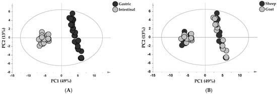
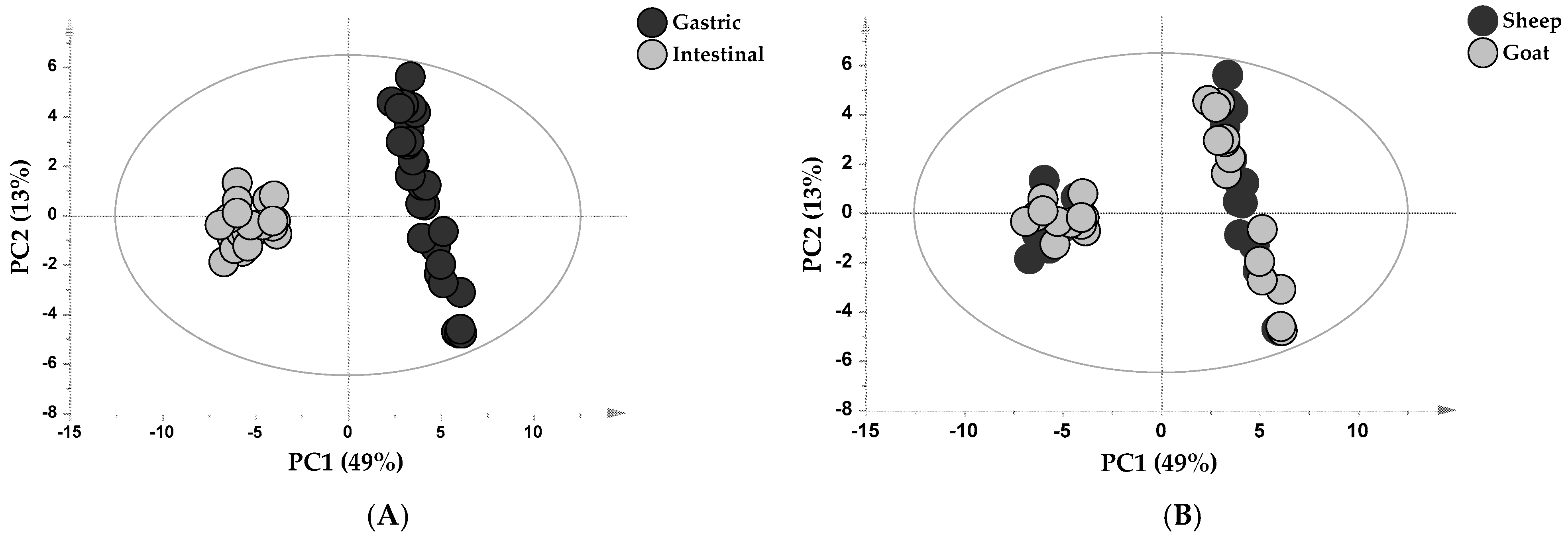
Unsupervised PCA was initially performed to investigate the global structure of the metabolomic dataset and to evaluate the impact of digestion on the overall metabolic profiles. As shown in the score plot of Figure 1A, a clear separation between gastric and intestinal samples was observed along the first principal component (PC1), demonstrating that digestion stage represented the major source of variability within the dataset. Based on the milk origin of IF, no discernible sample grouping was observed (Figure 1B).
Figure 1.
Principal Component Analysis (PCA) score plots of sheep and goat IF digested samples during in vitro dynamic digestion (n = 54; two groups, nine time points, three sample replicates). Samples were differently colored depending on digestion tract (A) and milk origin of IF (sheep and goat) (B). R2X = 0.61 and Q2 = 0.54.
Gastric samples exhibited a broader dispersion along the second principal component (PC2) compared with intestinal samples. This wider distribution likely reflects the progressive release of intermediate digestion products resulting from extensive enzymatic hydrolysis. In contrast, the tighter clustering of intestinal samples indicates more similar metabolomic profiles following digestion.
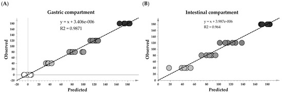
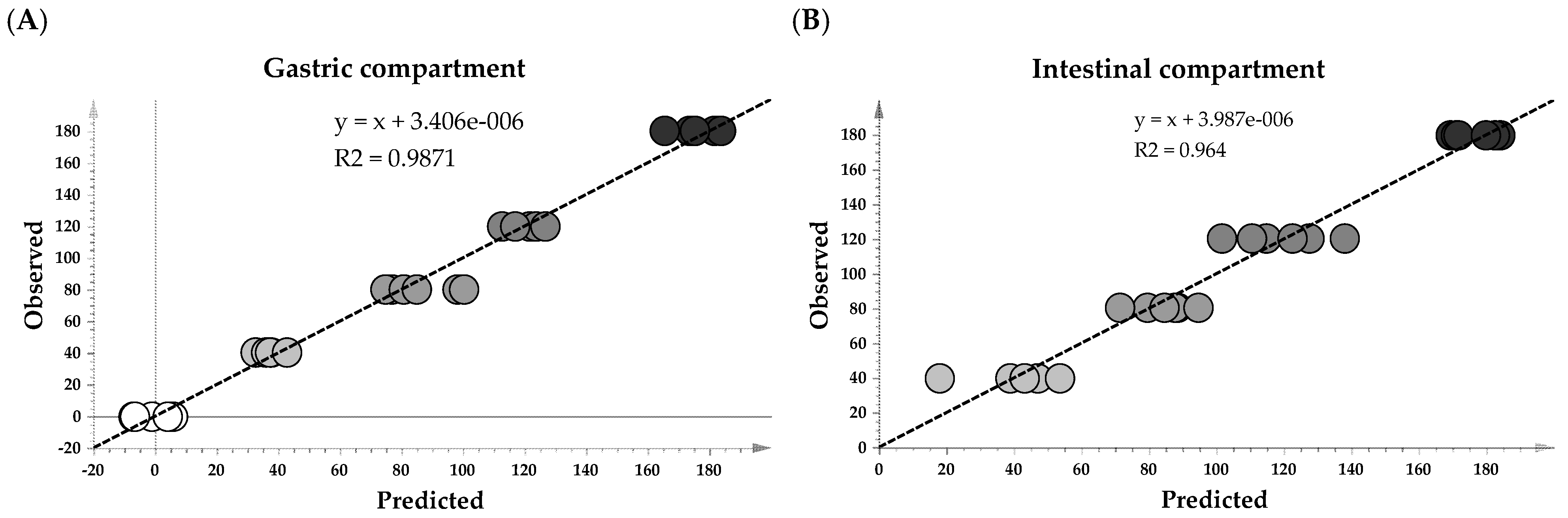
To further evaluate the evolution of the metabolomic profile during digestion, separate PLS models were generated for sheep and goat milk IF samples in the gastric and intestinal compartments, using sampling time as the response variable (Y). Both models showed excellent performance, as demonstrated by the strong linear relationship between observed and predicted values (Figure 2). The coefficients of determination (R2 = 0.984 and 0.982 for the gastric and intestinal compartments, respectively) indicated that approximately 98% of the variability in the response variable was explained by the models. The relatively low RMSEE values (7.62 and 10.75, respectively) further supported model accuracy, whereas the higher RMSECV values (12.95 and 23.05, respectively) reflected model performance during cross-validation. The models also exhibited high explanatory power, with R2X and R2Y values of 0.55 and 0.98 for the gastric compartment and 0.49 and 0.96 for the intestinal compartment, respectively. Cross-validation yielded Q2 values of 0.96 for the gastric model and 0.86 for the intestinal model, indicating excellent predictive ability.
Figure 2.
Observed vs. predicted plots from the PLS model developed for the gastric (A) and intestinal (B) compartment. The dashed line represents the linear regression between observed and predicted values. R2Y = 0.99 and 0.96; RMSEE = 7.62 and 10.75; RMSECV = 12.95 and 23.05; R2X = 0.55 and 0.49; Q2 = 0.96 and 0.82; permutation test n = 600; R2 = 0.51 and 0.54; Q2 = −0.40 and −0.44 for the gastric and intestinal models, respectively. The gastric and intestinal models were built using 30 and 24 metabolomic observations, respectively, obtained from six independent digestion runs (three sheep and three goat milk infant formula digestions).
To identify the metabolites contributing most strongly to the observed metabolic changes during digestion, Variable Importance in Projection scores (VIP > 1) were extracted from the predictive component of the OPLS models. The analysis highlighted distinct metabolic trajectories during the gastric and intestinal phases of digestion. In the gastric compartment, the progressive decrease in glyceric acid, citric acid, ribonic acid, glycerol phosphate, galactose, xylitol, lactic acid, and trihydroxybutyric acid reflects progressive changes in carbohydrate-related and intermediary metabolites during gastric digestion (Table 1). The reduction in citric and lactic acids may partly reflect gastric emptying in the dynamic digestion model. Because metabolomic data were normalized to 100, these variations should be interpreted as relative changes in metabolite abundance, reflecting both the progressive transfer of gastric contents and chemical transformations occurring during digestion.
Table 1.
Metabolites mostly correlated with time identified by OPLS analysis during the progression of in vitro dynamic digestion in the gastric and in the intestinal compartments.
To exclude potential confounding effects associated with total-sum normalization, the temporal evolution of citric and lactic acids throughout digestion was also evaluated using GC–MS peak areas normalized to the internal standard (Figure S1). This complementary approach confirmed the same temporal trend, with both metabolites showing a progressive decrease throughout digestion.
Conversely, the increase in sucrose, glucopyranose, glycerol, and several amino acids, including leucine, isoleucine, tyrosine, tryptophan, and valine, is consistent with the progressive release of soluble nutrients from the food matrix. The accumulation of amino acids is consistent with the proteolytic activity of pepsin, which initiates protein hydrolysis and generates low-molecular-weight peptides and free amino acids. Because pepsin acts primarily as an endopeptidase, gastric proteolysis predominantly generates peptides rather than free amino acids. Therefore, the relatively low levels of free amino acids detected during the gastric phase are consistent with the known proteolytic activity of pepsin. Similarly, the increase in glycerol and fatty acids (e.g., stearic and decanoic acids) is consistent with the onset of lipid hydrolysis and the release of lipid-derived compounds from vegetable oil and from the small portion of milk fat used as ingredients.
The intestinal compartment was characterized by more pronounced hydrolytic processes. Several carbohydrates and carbohydrate-derived metabolites, including glucopyranose, talopyranose, and xylitol, showed a progressive decline, likely reflecting their further transformation during digestion. In contrast, glycerol emerged as the most influential metabolite (VIP value = 2.34), accompanied by significant increases in essential amino acids such as threonine and methionine, as well as medium- and long-chain fatty acids (i.e., decanoic, dodecanoic, and linoleic acids). These changes are consistent with pancreatin-mediated hydrolysis of proteins and lipids, facilitated by bile salts [11]. Pancreatin contains a complex mixture of digestive enzymes (e.g., trypsin, amylase and lipase, ribonuclease, and protease) responsible for the extensive hydrolysis observed during the intestinal phase. The release of essential amino acids confirms the efficient proteolytic activity of pancreatic enzymes, whereas the accumulation of free fatty acids reflects the action of pancreatic lipase on milk triglycerides. Medium-chain fatty acids also increased during intestinal digestion. These fatty acids are naturally abundant in small-ruminant milk and are recognized for their rapid intestinal absorption and efficient utilization as energy substrates.
A limitation of the present study is that the in vitro gastrointestinal digestion model does not reproduce intestinal absorption. Consequently, the metabolites detected after digestion represent the luminal metabolite profile generated during the digestive process, but do not necessarily reflect the metabolites that would be present in the intestinal lumen or systemic circulation in vivo after absorption. This limitation is particularly relevant for free amino acids and fatty acids, which would normally undergo intestinal absorption in vivo, thereby reducing their luminal concentrations during gastrointestinal transit. Consequently, their accumulation in the intestinal compartment of the present model should primarily be interpreted as reflecting their progressive release during proteolysis and lipolysis in the absence of intestinal uptake and may therefore overestimate their luminal persistence compared with in vivo conditions. Therefore, the metabolomic profile obtained should be interpreted as a representation of the metabolic transformations occurring during digestion rather than the actual metabolite profile that would be observed in vivo. Future studies combining in vitro digestion models with intestinal absorption systems, such as Caco-2 cell models or advanced gut-on-a-chip platforms, could provide a more comprehensive understanding of the gastrointestinal fate, intestinal absorption, and bioavailability of IF-derived metabolites. In addition, the model does not account for microbial metabolism in the distal intestine, which may further modify the metabolite composition before excretion or absorption.
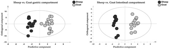
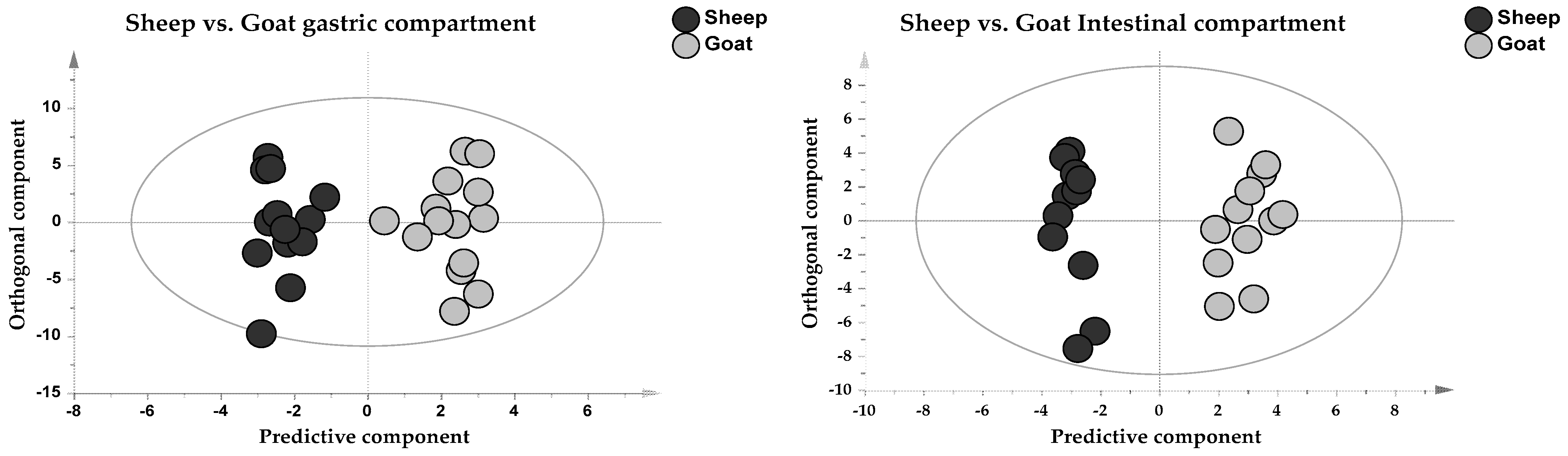
Although digestion stage represented the dominant source of variation, species-specific differences remained evident. To highlight these differences, two pairwise OPLS-DA models were constructed, comparing sheep and goat milk IFs separately for the gastric and intestinal compartments. The score plots revealed a clear separation between sheep and goat milk IF samples along the predictive component in both compartments (Figure 3). Although sheep and goats share broadly similar digestive physiology as ruminants, species-specific differences in the composition of sheep and goat milk-based infant formulas have been previously reported, particularly with respect to lipid and protein composition [10,11,12]. These compositional differences persisted throughout gastrointestinal digestion, resulting in distinct metabolomic signatures, as demonstrated by the OPLS-DA models.
Figure 3.
Pairwise OPLS-DA score plots of IF samples during in vitro dynamic digestion. Gastric compartment: n = 30, component 1 + 1, R2Y = 0.93, Q2 = 0.88, CV-ANOVA, p < 0.0001, permutation test (n = 600), R2 = 0.35, Q2 = −0.56. Intestinal compartment: n = 24, component 1 + 1, R2Y = 0.96, Q2 = 0.87, CV-ANOVA, p < 0.0001, permutation test (n = 600), R2 = 0.42, Q2 = −0.60.
VIP scores extracted from the predictive component of the OPLS-DA models highlighted discriminant metabolites between sheep- and goat-based IFs during gastric and intestinal digestion, as reported in Table 2, while in Table S3 the mean ± standard deviation (SD) and statistical parameters have been reported. In the gastric compartment, goat IF samples were characterized by higher levels of urea and ornithine than sheep IFs. Urea is the final product of the urea cycle, the major pathway responsible for the detoxification of excess ammonia generated during amino acid catabolism [37,38]. In ruminants, milk urea nitrogen (MUN) is widely recognized as an indicator of blood urea nitrogen (BUN) and has been used as a biomarker of protein metabolism and nitrogen utilization efficiency [39,40,41]. In the present study, urea was already more abundant in goat IF than in sheep IF at G0, before the addition of digestive enzymes, indicating that differences in urea abundance between the two formulas were present before enzymatic digestion (Figure S2). However, since the metabolomic compositions of the original IF powders were not determined, the origin of urea detected throughout digestion cannot be unequivocally attributed to the original milk or formula composition. Similarly, the higher ornithine abundance is consistent with species-dependent differences in nitrogen-related metabolites, although the biological origin of these differences cannot be established from the present data. Ornithine is a key intermediate of the urea cycle and plays a central role in nitrogen metabolism [42]. Uridine was identified as a characteristic metabolite of sheep IFs. This finding agrees with previous reports showing that uridine and uridine monophosphate are among the predominant nucleotides in ruminant milk, where they contribute to nucleotide metabolism and may support neonatal intestinal development and cellular growth [43]. Notably, several discriminant metabolites identified between sheep- and goat-based IFs were detected in both the gastric and intestinal compartments, including urea, citric acid, phosphate, and uridine. This observation indicates that species-specific metabolomic signatures persisted throughout gastrointestinal digestion despite exposure to the same digestive conditions. Because previous studies reported broadly similar protein digestibility [4] and lipid hydrolysis [11] for sheep- and goat-based IFs, the persistence of these discriminant metabolites is more likely attributable to intrinsic compositional differences between the two formulas than to differences in digestive susceptibility. These findings demonstrate that milk origin leaves a persistent metabolomic fingerprint that remains detectable throughout dynamic in vitro digestion.
Table 2.
Discriminant metabolites between sheep and goat IF samples in the gastric and intestinal compartment identified by OPLS-DA.
3.3. Temporal Evolution of Free Amino Acids During In Vitro Digestion
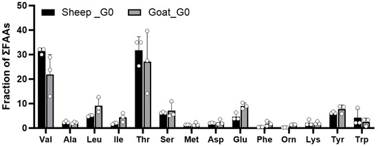
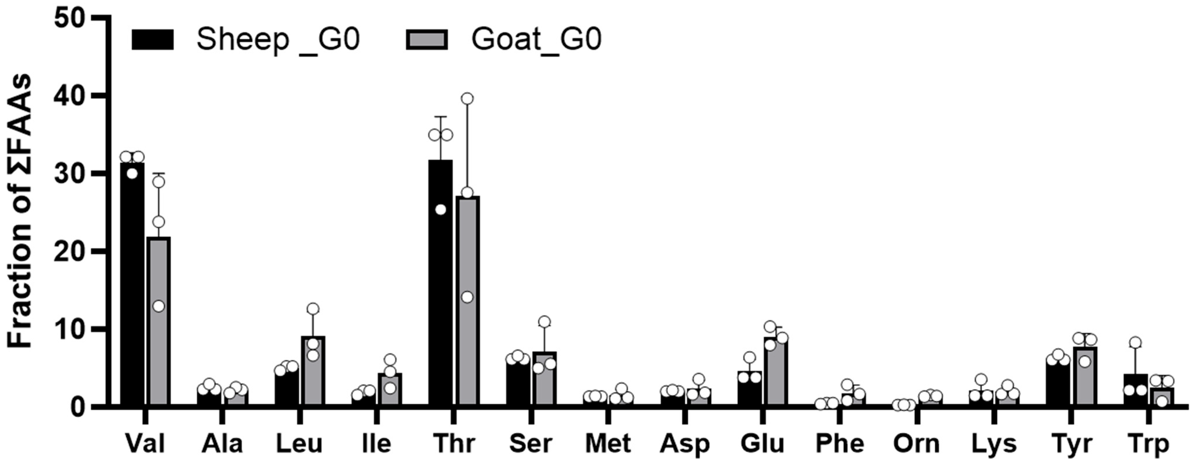
Dietary proteins cannot be absorbed in their intact form by enterocytes and therefore require enzymatic hydrolysis to generate free amino acids (FAAs) and peptides, which are subsequently absorbed in the small intestine [44]. Because milk naturally contains free amino acids and peptides [27], the initial FAA composition (G0) was used as a baseline to evaluate the release of individual amino acids during the gastrointestinal digestion of sheep and goat milk-based IFs. The relative abundance of each FAA was expressed as a fraction of total FAAs (ΣFAAs), and the results are shown in Figure 4. Both infant formulas exhibited a free amino acid profile dominated by threonine and valine, which together accounted for more than 30% of the total FAA fraction. The high proportion of threonine likely reflects both the intrinsic amino acid composition of milk proteins and proteolytic events occurring during the manufacture of dairy ingredients. Whey powder and whey protein concentrates derived from sweet (rennet) whey retain glycomacropeptide (GMP), a bioactive C-terminal fragment released from κ-casein during cheese manufacture. GMP is a highly hydrophilic peptide consisting of 64 amino acid residues and containing up to four O-linked carbohydrate chains composed of sialic acid, galactose, and N-acetylgalactosamine attached to threonine residues [45]. Consequently, GMP is exceptionally rich in threonine, which accounts for approximately 19% of its amino acid composition [46]. Previous peptidomic analyses of digested IFs revealed extensive hydrolysis of glycomacropeptide-derived peptides, particularly in sheep milk-based IFs, where numerous GMP-derived peptides were detected from the early gastric phase [12]. Accordingly, the high abundance of threonine observed at G0 in the present study is consistent with the composition of dairy ingredients [47].
Figure 4.
Free amino acid profile of sheep and goat milk-based infant formulas at the beginning of gastric digestion (G0), determined by GC–MS and expressed as the fraction of total free amino acids (ΣFAAs; mean ± SD). Dots represent individual replicates. Mean differences between formulas were assessed using pairwise multiple t-tests, with p-values adjusted for multiple comparisons using the two-stage step-up false discovery rate (FDR) procedure of Benjamini, Krieger, and Yekutieli.
Likewise, the high proportion of valine is consistent with the abundance of branched-chain amino acids in ruminant milk proteins. No significant differences were detected between sheep and goat milk-based formulas in the relative distribution of free amino acids at G0 (p > 0.05). Overall, the observed profile represents the initial pool of free amino acids available prior to digestion and provides a baseline for assessing amino acid release during subsequent gastrointestinal digestion.
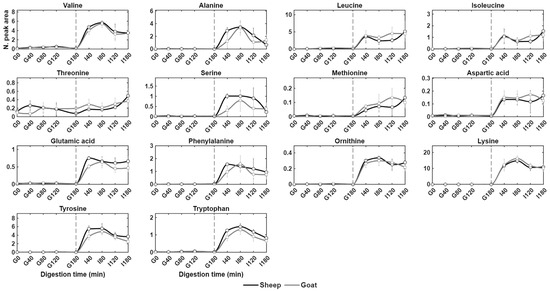
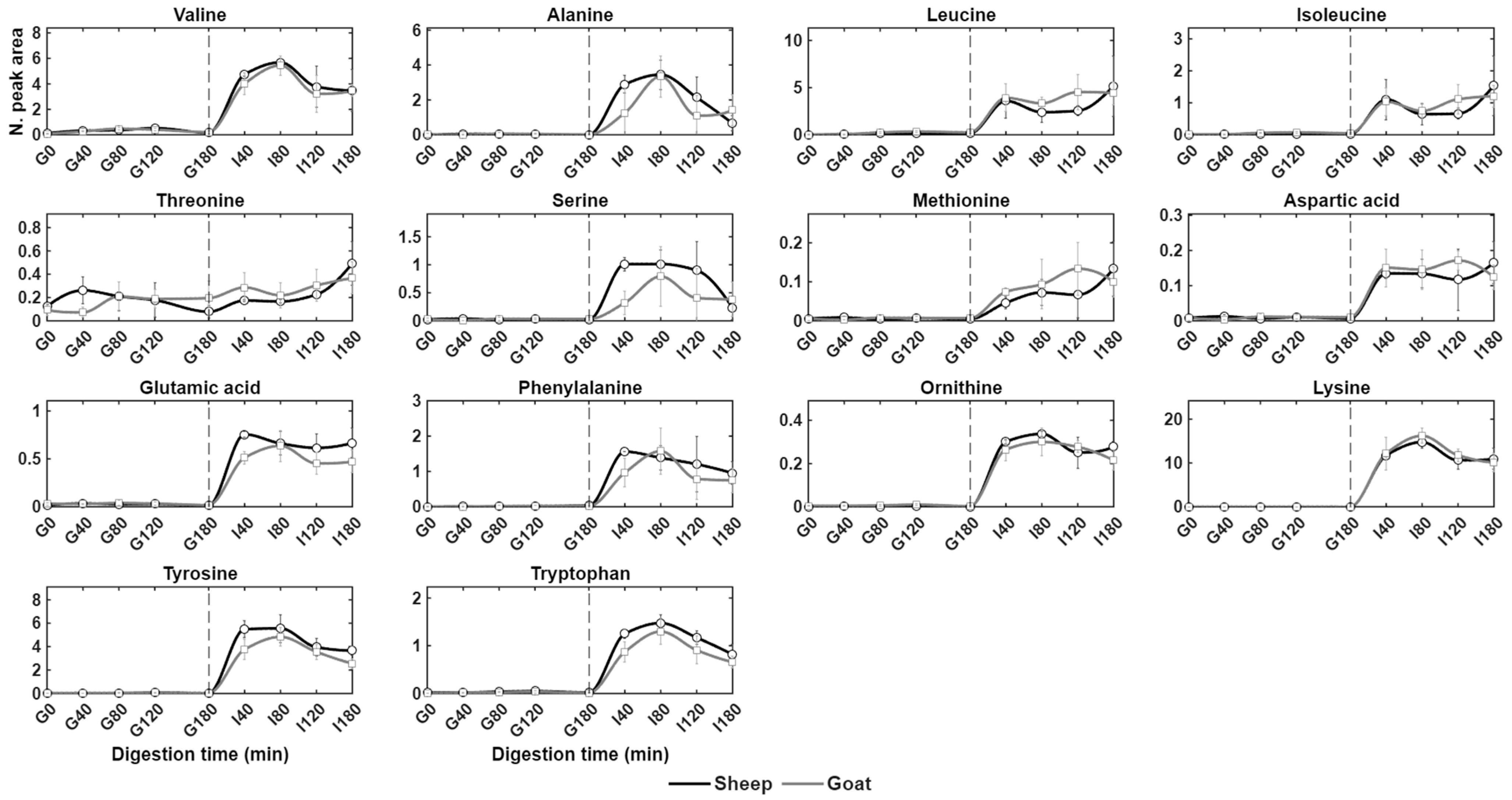
During in vitro gastrointestinal digestion, protein hydrolysis was initiated by pepsin in the gastric compartment and further intensified by pancreatin during the intestinal phase. Accordingly, mean normalized peak areas obtained at each digestion time point were used to construct continuous kinetic curves for both sheep and goat milk-based infant formulas using the PCHIP algorithm. As shown in Figure 5, the gastric phase was characterized by low and relatively stable FAA levels in both sheep and goat milk IFs (except for threonine), whereas a marked increase was observed immediately after the transition to the intestinal phase (I40). This trend indicates that, during the intestinal phase of digestion, amino acids mainly derive from the continuous hydrolysis of peptides generated during the gastric phase. These observations are consistent with our previous proteomic characterization of digested sheep and goat milk-based IFs performed by UHPLC-Orbitrap-HRMS, which demonstrated extensive peptide hydrolysis during intestinal digestion [12].
Figure 5.
Temporal evolution of normalized free amino acid peak areas (N. peak area) during dynamic in vitro gastrointestinal digestion of sheep milk-based (black line) and goat milk-based (gray line) infant formulas (n = 54). Continuous kinetic profiles were generated using PCHIP based on normalized GC–MS peak areas. The dashed vertical line indicates the transition from the gastric phase (G0–G180) to the intestinal phase (I40–I180).
Relatively few studies have directly compared protein digestion between human milk and Ifs, e.g., [47,48]. Different digestive behavior has been reported for human milk (HM) and conventional bovine IF. During the gastric phase, HM formed larger aggregates and exhibited a different protein distribution, with a higher proportion of proteins remaining in the serum phase compared with bovine IF. During the intestinal phase, contrasting results regarding protein hydrolysis kinetics have been reported. Abrahamse et al. [47] observed slower protein hydrolysis for HM, whereas Maathuis et al. [49] reported different kinetics of nitrogen bioaccessibility among HM, goat milk-based IF (GIF), and conventional cow milk-based IF (CIF). In particular, nitrogen bioaccessibility from GIF showed kinetics similar to those observed for HM, whereas CIF exhibited a delayed pattern.
Branched-chain amino acids (BCAAs, i.e., valine, leucine, and isoleucine) displayed distinct release patterns. Valine exhibited a rapid increase at the beginning of the intestinal phase followed by a progressive decrease toward I180 (I180/I40: sheep = 0.73; goat = 0.86), whereas isoleucine and leucine increased more gradually throughout the intestinal phase following an initial rise at I40. Previous in vitro, animal [50], and human studies have consistently shown that rapid protein hydrolysis initially generates medium-sized peptides, which are subsequently degraded into smaller peptides and free amino acids during intestinal digestion [51]. However, several peptides, particularly proline-rich sequences, may exhibit partial resistance to gastrointestinal enzymes [52]. Consistent with this observation, no release of free proline was detected throughout the digestion process in the present study.
Charton et al. [53] reported that both apparent and true digestibility of total nitrogen were lower for HM than for IF in the mid-jejunum, ileum, and caecum, whereas no significant differences were observed in the proximal jejunum and colon. Differences were observed at the level of individual amino acids. Apparent ileal digestibility did not differ between HM and IF for histidine, lysine, arginine, glutamic acid, and glycine, whereas it was lower for HM for the other amino acids investigated. When true ileal digestibility was considered, lysine showed slightly higher digestibility in HM, whereas phenylalanine, threonine, valine, alanine, proline, and serine showed higher digestibility in IF. These findings highlight that differences in protein digestion between HM and IF are not uniform across the amino acid pool but are amino acid-specific, with individual amino acids exhibiting distinct digestibility patterns. Similarly, in the present study, individual amino acids showed distinct release kinetics during the intestinal digestion of sheep and goat milk-based IFs. Although Maathuis et al. [49] suggest that protein quality did not differ among GIF, CIF, and HM, the kinetics of protein digestion of GIF were more comparable to those of HM than to those of CIF. Moreover, since HM was not included as a direct experimental comparator in the present study, our results do not allow us to establish whether sheep or goat milk-based IF more closely resembles HM in terms of amino acid release or availability.
The marked increase in free amino acids during the intestinal phase is of nutritional relevance because the small intestine is the primary site of amino acid absorption and metabolism [54,55,56]. Once absorbed, these amino acids can support protein synthesis, energy production, and the biosynthesis of other biologically active compounds [57].
The release of threonine during the final phase of intestinal digestion can be explained by a combination of compositional, structural, and enzymatic factors that act synergistically to maximize amino acid bioaccessibility. First, milk proteins are intrinsically rich in threonine [58]. In particular, β-casein and κ-casein, together with whey proteins such as β-lactoglobulin, contain a relatively high proportion of threonine residues [59]. This compositional abundance provides a substantial reservoir from which free threonine can be released and contributes to the high biological value of milk proteins, which supply essential amino acids in proportions that closely match human nutritional requirements [58]. Second, the structural organization of caseins promotes enzymatic accessibility. During gastric digestion, caseins undergo extensive hydrolysis owing to their flexible, loosely organized conformation, which renders them highly susceptible to pepsin activity [60]. Unlike globular proteins, caseins are largely intrinsically disordered and lack rigid secondary and tertiary structures [61,62]. Consequently, threonine residues may be located within exposed and flexible regions of the polypeptide chain, where peptide bonds are readily accessible to pepsin in the stomach and subsequently to pancreatic proteases and brush-border peptidases in the small intestine [63]. Consistent with this mechanism, Figure 5 shows a marked increase in the abundance of individual free amino acids (FAAs) following the transition from the gastric to the intestinal phase. Similar increases in FAA concentrations during intestinal digestion have previously been reported in static in vitro digestion models of pasteurized milk [64] and pseudocereal protein matrices [65], indicating that extensive proteolysis primarily occurs during the intestinal stage. Likewise, a comparison between static in vitro digestion and in vivo piglet digestion of skim milk powder demonstrated that FAA concentrations measured in intestinal digesta generated in vitro closely resembled those observed in the in vivo intestinal compartment. In both systems, only limited amounts of FAAs and small peptides were detected during gastric digestion, supporting the physiological relevance of the in vitro digestion model [66]. Collectively, these findings reinforce that the intestinal phase is the principal site of free amino acid generation and that pancreatic proteases, together with brush-border peptidases, play a central role in maximizing amino acid bioaccessibility.
4. Conclusions
This study provides an untargeted GC–MS metabolomic characterization of sheep and goat milk-based infant formulas during dynamic in vitro infant gastrointestinal digestion. The metabolomic profiles were strongly influenced by the digestive stage, with a clear separation between gastric and intestinal digesta. Intestinal digestion was characterized by a marked accumulation of free amino acids, glycerol phosphate, and medium- and long-chain fatty acids, reflecting the extensive proteolytic and lipolytic activities promoted by pancreatic enzymes and bile salts. Likewise, the progressive release of medium-chain fatty acids, such as hexanoic, octanoic, and decanoic acids, highlighted the efficient digestion of milk lipids during intestinal phase. Temporal analysis revealed that free amino acid release was limited during gastric digestion but increased rapidly following the transition to the intestinal compartment, confirming that the intestinal phase is the principal site of amino acid generation. Although digestion stage represented the major source of metabolomic variation, species-dependent differences were also observed. Overall, these findings provide new insights into the digestive metabolomic trajectories of small-ruminant milk-based infant formulas under dynamic in vitro gastrointestinal digestion.
Supplementary Materials
The following supporting information can be downloaded at: https://www.mdpi.com/article/10.3390/dairy7050073/s1, Table S1: Retention times (RTs, in min) and theoretical Kovats retention indices (RIs) for the n-alkanes detected and used as reference standards in this study. Only those alkanes effectively detected under the analytical conditions are included in the table; Table S2: GC-MS characteristics of identified metabolites in sheep and goat digesta samples. Table S3: Mean ± standard deviation (SD) of metabolites identified by GC–MS in goat and sheep milk-based IFs. Statistical differences were assessed using a mixed-effects model fitted by restricted maximum likelihood (REML), with milk type, digestion time, and their interaction (milk type × time) as fixed effects and digestion replicate as a random effect. For each metabolite, the overall effects of milk type, digestion time, and their interaction were evaluated, and the corresponding global p-values are reported; Figure S1. Temporal evolution of lactic and citric acids during gastrointestinal digestion of sheep and goat milk-based infant formulas. GC–MS peak areas were normalized to the internal standard area without applying total-sum; Figure S2. Temporal evolution of urea during gastrointestinal digestion of sheep and goat milk-based infant formulas. GC–MS peak areas were normalized to the internal standard area.
Author Contributions
Conceptualization, M.C., P.S., D.D., O.M. and P.C.; methodology, M.C.;. software, M.C.; validation, M.C., P.S. and P.C.; formal analysis, M.C., A.D. and C.M.; investigation, M.C., P.S. and P.C.; resources, P.C.; data curation, M.C. and P.S.; writing—original draft preparation, M.C. and P.S.; writing—review and editing, M.C., P.S., D.D. and P.C.; visualization, M.C., P.S. and P.C.; supervision, P.S. and P.C. All authors have read and agreed to the published version of the manuscript.
Funding
This research received no external funding.
Data Availability Statement
The original contributions presented in this study are included in the article/Supplementary Materials. Further inquiries can be directed to the corresponding author.
Conflicts of Interest
The authors declare no conflicts of interest.
References
- Zou, X.; Ali, A.H.; Abed, S.M.; Guo, Z. Current knowledge of lipids in human milk and recent innovations in infant formulas. Curr. Opin. Food Sci. 2017, 16, 28–39. [Google Scholar] [CrossRef] [Scilit]
- Song, X.; Wang, X.; Yang, M.; Acevedo-Fani, A.; Singh, H.; Ye, A. Dynamic in vitro gastric digestion behaviour of commercial infant formulae made with cow, goat and sheep milk. Foods 2024, 13, 1286. [Google Scholar] [CrossRef] [Scilit] [PubMed]
- Barbosa, A.M.C.; Cortes, S.C.; Pulido, N.; Parra, M.Y.; Rodríguez-López, A.; Guevara-Morales, J.; Echeverri-Peña, O.Y. Metabolic impact of infant formulas in young infants: An outlook from the urine metabolome. Heliyon 2022, 8, e10432. [Google Scholar] [CrossRef] [Scilit] [PubMed]
- Scano, P.; Murgia, A.; Demuru, M.; Consonni, R.; Caboni, P. Metabolite Profiles of Formula Milk Compared to Breast Milk. Food Res. Int. 2016, 87, 76–82. [Google Scholar] [CrossRef] [Scilit] [PubMed]
- Dupont, D.; Mandalari, G.; Mollé, D.; Jardin, J.; Rolet-Répécaud, O.; Duboz, G.; Léonil, J.; Mills, C.E.N.; Mackie, A.R. Food processing increases casein resistance to simulated infant digestion. Mol. Nutr. Food Res. 2010, 54, 1677–1689. [Google Scholar] [CrossRef] [Scilit] [PubMed]
- Bourlieu, C.; Ménard, O.; Bouzerzour, K.; Mandalari, G.; Macierzanka, A.; Mackie, A.R.; Dupont, D. Specificity of infant digestive conditions: Some clues for developing relevant in vitro models. Crit. Rev. Food Sci. Nutr. 2014, 54, 1427–1457. [Google Scholar] [CrossRef] [Scilit] [PubMed]
- Macierzanka, A.; Sancho, A.I.; Mills, E.C.; Rigby, N.M.; Mackie, A.R. Emulsification alters simulated gastrointestinal proteolysis of β-casein and β-lactoglobulin. Soft Matter 2009, 5, 538–550. [Google Scholar] [CrossRef] [Scilit]
- Liu, W.; Li, J.; Ma, J.; Xun, Y.; Feng, X.; Ning, Y.; Deng, Z. Digestibility of Proteins and Lipids in Infant Formulas: A Comparison of Cow and Goat Milk-Based Products with Human Milk. Food Biosci. 2025, 67, 107363. [Google Scholar] [CrossRef] [Scilit]
- Maryniak, N.Z.; Sancho, A.I.; Hansen, E.B.; Bøgh, K.L. Alternatives to Cow’s Milk-Based Infant Formulas in the Prevention and Management of Cow’s Milk Allergy. Foods 2022, 11, 926. [Google Scholar] [CrossRef] [Scilit] [PubMed]
- Park, Y.W.; Juárez, M.; Ramos, M.; Haenlein, G.F.W. Physico-chemical characteristics of goat and sheep milk. Small Rumin. Res. 2007, 68, 88–113. [Google Scholar] [CrossRef] [Scilit]
- Casula, M.; Manis, C.; Menard, O.; Tolle, G.; Cochet, M.F.; Dupont, D.; Scano, P.; Garau, V.; Caboni, P. Lipidomics of Sheep and Goat Milk-Based Infant Formulae during In Vitro Dynamic Digestion. Food Chem. 2024, 461, 140850. [Google Scholar] [CrossRef] [Scilit] [PubMed]
- Scano, P.; Casula, M.; Ménard, O.; Dupont, D.; Manis, C.; Serrao, S.; Manconi, B.; Caboni, P. Simulated Gastrointestinal Protein Digestion of Sheep and Goat Milk Infant Formulae. Int. Dairy J. 2025, 162, 106162. [Google Scholar] [CrossRef] [Scilit]
- Lai, G.; Caboni, P.; Piras, C.; Pes, M.; Sitzia, M.; Addis, M.; Pirisi, A.; Scano, P. Development and Chemico-Physical Characterization of Ovine Milk-Based Ingredients for Infant Formulae. Appl. Sci. 2023, 13, 653. [Google Scholar] [CrossRef] [Scilit]
- Lucena, M.E.; Alvarez, S.; Menéndez, C.; Riera, F.A.; Alvarez, R. Beta-lactoglobulin removal from whey protein concentrates: Production of milk derivatives as a base for infant formulas. Sep. Purif. Technol. 2006, 52, 310–316. [Google Scholar] [CrossRef] [Scilit]
- Raynal-Ljutovac, K.; Lagriffoul, G.; Paccard, P.; Guillet, I.; Chilliard, Y. Composition of goat and sheep milk products: An update. Small Rumin. Res. 2008, 79, 57–72. [Google Scholar] [CrossRef] [Scilit]
- Storry, J.E.; Grandison, A.S.; Millard, D.; Owen, A.J.; Ford, G.D. Chemical composition and coagulating properties of renneted milks from different breeds and species of ruminant. J. Dairy Res. 1983, 50, 215–229. [Google Scholar] [CrossRef] [Scilit]
- Domagała, J. Instrumental texture, syneresis and microstructure of yoghurts prepared from goat, cow and sheep milk. Int. J. Food Prop. 2009, 12, 605–615. [Google Scholar] [CrossRef] [Scilit]
- Teng, F.; Reis, M.G.; Yang, L.; Ma, Y.; Day, L. Structural characteristics of triacylglycerols contribute to the distinct in vitro gastric digestibility of sheep and cow milk fat prior to and after homogenisation. Food Res. Int. 2020, 130, 108911. [Google Scholar] [CrossRef] [Scilit] [PubMed]
- Lindquist, S.; Hernell, O. Lipid digestion and absorption in early life: An update. Curr. Opin. Clin. Nutr. Metab. Care 2010, 13, 314–320. [Google Scholar] [CrossRef] [Scilit] [PubMed]
- Van Eijnatten, E.J.; Camps, G.; Rombouts, W.; Pellis, L.; Smeets, P.A. Gastric Digestion and Changes in Serum Amino Acid Concentrations after Consumption of Casein from Cow and Goat Milk: A Randomized Crossover Trial in Healthy Men. J. Nutr. 2025, 155, 3374–3383. [Google Scholar] [CrossRef] [Scilit] [PubMed]
- Huppertz, T.; Chia, L.W. Milk Protein Coagulation under Gastric Conditions: A Review. Int. Dairy J. 2021, 113, 104882. [Google Scholar] [CrossRef] [Scilit]
- Ceballos, L.S.; Morales, E.R.; de la Torre Adarve, G.; Castro, J.D.; Martínez, L.P.; Sampelayo, M.R.S. Composition of Goat and Cow Milk Produced under Similar Conditions and Analyzed by Identical Methodology. J. Food Compos. Anal. 2009, 22, 322–329. [Google Scholar] [CrossRef] [Scilit]
- Mulet-Cabero, A.I.; Mackie, A.R.; Wilde, P.J.; Fenelon, M.A.; Brodkorb, A. Structural Mechanism and Kinetics of In Vitro Gastric Digestion Are Affected by Process-Induced Changes in Bovine Milk. Food Hydrocoll. 2019, 86, 172–183. [Google Scholar] [CrossRef] [Scilit]
- Wang, X.; Ye, A.; Lin, Q.; Han, J.; Singh, H. Gastric Digestion of Milk Protein Ingredients: Study Using an In Vitro Dynamic Model. J. Dairy Sci. 2018, 101, 6842–6852. [Google Scholar] [CrossRef] [Scilit] [PubMed]
- Almaas, H.; Cases, A.L.; Devold, T.G.; Holm, H.; Langsrud, T.; Aabakken, L.; Aadnøy, T.; Vegarud, G.E. In Vitro Digestion of Bovine and Caprine Milk by Human Gastric and Duodenal Enzymes. Int. Dairy J. 2006, 16, 961–968. [Google Scholar] [CrossRef] [Scilit]
- Hettinga, K.; Pellis, L.; Rombouts, W.; Du, X.; Grigorean, G.; Lönnerdal, B. Effect of pH and Protein Composition on Proteolysis of Goat Milk Proteins by Pepsin and Pancreatin. Food Res. Int. 2023, 173, 113294. [Google Scholar] [CrossRef] [Scilit] [PubMed]
- Caboni, P.; Murgia, A.; Porcu, A.; Manis, C.; Ibba, I.; Contu, M.; Scano, P. A Metabolomics Comparison between Sheep’s and Goat’s Milk. Food Res. Int. 2019, 119, 869–875. [Google Scholar] [CrossRef] [Scilit] [PubMed]
- Scano, P.; Carta, P.; Ibba, I.; Manis, C.; Caboni, P. An Untargeted Metabolomic Comparison of Milk Composition from Sheep Kept under Different Grazing Systems. Dairy 2020, 1, 30–41. [Google Scholar] [CrossRef] [Scilit]
- Bode, L.; Kuhn, L.; Kim, H.Y.; Hsiao, L.; Nissan, C.; Sinkala, M.; Chipepo, K.; Mwiya, M.; Thea, D.M.; Aldrovandi, G.M. Human Milk Oligosaccharide Concentration and Risk of Postnatal Transmission of HIV through Breastfeeding. Am. J. Clin. Nutr. 2012, 96, 831–839. [Google Scholar] [CrossRef] [Scilit] [PubMed]
- Menard, O.; Cattenoz, T.; Guillemin, H.; Souchon, I.; Deglaire, A.; Dupont, D.; Picque, D. Validation of a New In Vitro Dynamic System to Simulate Infant Digestion. Food Chem. 2014, 145, 1039–1045. [Google Scholar] [CrossRef] [Scilit] [PubMed]
- de Oliveira, S.C.; Bellanger, A.; Menard, O.; Pladys, P.; Le Gouar, Y.; Dirson, E.; Kroell, F.; Dupont, D.; Deglaire, A.; Bourlieu, C. Impact of Human Milk Pasteurization on Gastric Digestion in Preterm Infants: A Randomized Controlled Trial. Am. J. Clin. Nutr. 2017, 105, 379–390. [Google Scholar] [CrossRef] [Scilit] [PubMed]
- Le Roux, L.; Ménard, O.; Chacon, R.; Dupont, D.; Jeantet, R.; Deglaire, A.; Nau, F. Are faba bean and pea proteins potential whey protein substitutes in infant formulas? An in vitro dynamic digestion approach. Foods 2020, 9, 362. [Google Scholar] [CrossRef] [Scilit] [PubMed]
- Folch, J.; Lees, M.; Sloane Stanley, G.H. A Simple Method for the Isolation and Purification of Total Lipides from Animal Tissues. J. Biol. Chem. 1957, 226, 497–509. [Google Scholar] [CrossRef] [Scilit]
- Casula, M.; Scano, P.; Manis, C.; Scano, E.A.; Caboni, P. Comprehensive Multi-Technique Analysis of Raw Cuttlefish Ink and Its Shelf-Stable Food Product Derivative. J. Food Compos. Anal. 2025, 148, 108181. [Google Scholar] [CrossRef] [Scilit]
- González-Domínguez, Á.; Estanyol-Torres, N.; Brunius, C.; Landberg, R.; González-Domínguez, R. QC Omics: Recommendations and Guidelines for Robust, Easily Implementable and Reportable Quality Control of Metabolomics Data. Anal. Chem. 2024, 96, 1064–1072. [Google Scholar] [CrossRef] [Scilit] [PubMed]
- Sumner, L.W.; Amberg, A.; Barrett, D.; Beale, M.H.; Beger, R.; Daykin, C.A.; Fan, T.W.-M.; Fiehn, O.; Goodacre, R.; Griffin, J.L.; et al. Proposed minimum reporting standards for chemical analysis: Chemical Analysis Working Group (CAWG) Metabolomics Standards Initiative (MSI). Metabolomics 2007, 3, 211–221. [Google Scholar] [CrossRef] [Scilit] [PubMed]
- Harmeyer, J.; Martens, H. Aspects of urea metabolism in ruminants with reference to the goat. J. Dairy Sci. 1980, 63, 1707–1728. [Google Scholar] [CrossRef] [Scilit] [PubMed]
- Patra, A.K.; Aschenbach, J.R. Ureases in the gastrointestinal tracts of ruminant and monogastric animals and their implication in urea-N/ammonia metabolism: A review. J. Adv. Res. 2018, 13, 39–50. [Google Scholar] [CrossRef] [Scilit] [PubMed]
- Broderick, G.A.; Clayton, M.K. A statistical evaluation of animal and nutritional factors influencing concentrations of milk urea nitrogen. J. Dairy Sci. 1997, 80, 2964–2971. [Google Scholar] [CrossRef] [Scilit] [PubMed]
- Schepers, A.J.; Meijer, R.G.M. Evaluation of the utilization of dietary nitrogen by dairy cows based on urea concentration in milk. J. Dairy Sci. 1998, 81, 579–584. [Google Scholar] [CrossRef] [Scilit] [PubMed]
- Zhao, X.; Zheng, N.; Zhang, Y.; Wang, J. The role of milk urea nitrogen in nutritional assessment and its relationship with phenotype of dairy cows: A review. Anim. Nutr. 2025, 20, 33–41. [Google Scholar] [CrossRef] [Scilit] [PubMed]
- Stewart, G.S.; Smith, C.P. Urea nitrogen salvage mechanisms and their relevance to ruminants, non-ruminants and man. Nutr. Res. Rev. 2005, 18, 49–62. [Google Scholar] [CrossRef] [Scilit] [PubMed]
- Plakantara, S.; Michaelidou, A.M.; Polychroniadou, A.; Menexes, G.; Alichanidis, E. Nucleotides and nucleosides in ovine and caprine milk during lactation. J. Dairy Sci. 2010, 93, 2330–2337. [Google Scholar] [CrossRef] [Scilit] [PubMed]
- Qiao, K.; Zhou, X.; Wu, H.; Zhang, L.; Liang, L.; Zhang, Y. Research on the release and absorption regularities of free amino acids and peptides in vitro digestion of yeast protein. Food Chem. 2025, 482, 144176. [Google Scholar] [CrossRef] [Scilit] [PubMed]
- Córdova-Dávalos, L.E.; Jiménez, M.; Salinas, E. Glycomacropeptide bioactivity and health: A review highlighting action mechanisms and signaling pathways. Nutrients 2019, 11, 598. [Google Scholar] [CrossRef] [Scilit] [PubMed]
- Nakano, T.; Betti, M. Isolation of κ-casein glycomacropeptide from bovine whey fraction using food grade anion exchange resin and chitin as an adsorbent. J. Dairy Res. 2020, 87, 127–133. [Google Scholar] [CrossRef] [Scilit] [PubMed]
- Rigo, J.; Boehm, G.; Georgi, G.; Jelinek, J.; Nyambugabo, K.; Sawatzki, G.; Studzinski, F. An infant formula free of glycomacropeptide prevents hyperthreoninemia in formula-fed preterm infants. J. Pediatr. Gastroenterol. Nutr. 2001, 32, 127–130. [Google Scholar] [CrossRef] [Scilit] [PubMed]
- Abrahamse, E.; Huybers, S.; Alles, M.S.; Renes, I.B.; Knol, J.; Bouritius, H.; Ludwig, T. Fermented infant formula increases ileal protein digestibility and reduces ileal proteolytic activity compared with standard and hydrolyzed infant formulas in piglets. J. Nutr. 2015, 145, 1423–1428. [Google Scholar] [CrossRef] [Scilit] [PubMed]
- Maathuis, A.; Havenaar, R.; He, T.; Bellmann, S. Protein digestion and quality of goat and cow milk infant formula and human milk under simulated infant conditions. J. Pediatr. Gastroenterol. Nutr. 2017, 65, 661–666. [Google Scholar] [CrossRef] [Scilit] [PubMed]
- Barbe, F.; Ménard, O.; Le Gouar, Y.; Buffière, C.; Famelart, M.-H.; Laroche, B.; Le Feunteun, S.; Dupont, D.; Rémond, D. The heat treatment and the gelation are strong determinants of the kinetics of milk proteins digestion and of the peripheral availability of amino acids. Food Chem. 2013, 136, 1203–1212. [Google Scholar] [CrossRef] [Scilit] [PubMed]
- Sanchón, J.; Fernández-Tomé, S.; Miralles, B.; Hernández-Ledesma, B.; Tomé, D.; Gaudichon, C.; Recio, I. Protein degradation and peptide release from milk proteins in human jejunum. Comparison with in vitro gastrointestinal simulation. Food Chem. 2018, 239, 486–494. [Google Scholar] [CrossRef] [Scilit] [PubMed]
- Vanhoof, G.; Goossens, F.; De Meester, I.; Hendriks, D.; Scharpé, S. Proline motifs in peptides and their biological processing. FASEB J. 1995, 9, 736–744. [Google Scholar] [CrossRef] [Scilit]
- Charton, E.; Henry, G.; Cahu, A.; Le Gouar, Y.; Dahirel, P.; Moughan, P.J.; Montoya, C.A.; Bellanger, A.; Dupont, D.; Le Huërou-Luron, I.; et al. Ileal digestibility of nitrogen and amino acids in human milk and an infant formula as determined in neonatal minipiglets. J. Nutr. 2023, 153, 1063–1074. [Google Scholar] [CrossRef] [Scilit] [PubMed]
- Wu, G. Intestinal mucosal amino acid catabolism. J. Nutr. 1998, 128, 1249–1252. [Google Scholar] [CrossRef] [Scilit] [PubMed]
- Rhoads, J.M.; Wu, G. Glutamine, arginine, and leucine signaling in the intestine. Amino Acids 2009, 37, 111–122. [Google Scholar] [CrossRef] [Scilit] [PubMed]
- Lallès, J.P.; Boudry, G.; Favier, C.; Le Floc’h, N.; Luron, I.; Montagne, L.; Oswald, I.P.; Pié, S.; Piel, C.; Sève, B. Gut function and dysfunction in young pigs: Physiology. Anim. Res. 2004, 53, 301–316. [Google Scholar] [CrossRef] [Scilit] [PubMed]
- Stoll, B. Intestinal uptake and metabolism of threonine: Nutritional impact. Adv. Pork Prod. 2006, 17, 257–263. [Google Scholar]
- Moughan, P.J. Milk Proteins: A Rich Source of Bioactives for Developing Functional Foods. In Milk Proteins; Boland, M., Singh, H., Eds.; Academic Press: Cambridge, MA, USA, 2020; pp. 633–649. [Google Scholar] [CrossRef] [Scilit]
- Lacroix, M.; Bos, C.; Léonil, J.; Airinei, G.; Luengo, C.; Daré, S.; Benamouzig, R.; Fouillet, H.; Fauquant, J.; Tomé, D.; et al. Compared with Casein or Total Milk Protein, Digestion of Milk Soluble Proteins Is Too Rapid to Sustain the Anabolic Postprandial Amino Acid Requirement. Am. J. Clin. Nutr. 2006, 84, 1070–1079. [Google Scholar] [CrossRef] [Scilit] [PubMed]
- Salelles, L.; Floury, J.; Le Feunteun, S. Pepsin activity as a function of pH and digestion time on caseins and egg white proteins under static in vitro conditions. Food Funct. 2021, 12, 12468–12478. [Google Scholar] [CrossRef] [Scilit] [PubMed]
- Dunker, A.K.; Brown, C.J.; Lawson, J.D.; Iakoucheva, L.M.; Obradović, Z. Intrinsic Disorder and Protein Function. Biochemistry 2002, 41, 6573–6582. [Google Scholar] [CrossRef] [Scilit] [PubMed]
- Uversky, V.N. What Does It Mean to Be Natively Unfolded? Eur. J. Biochem. 2002, 269, 2–12. [Google Scholar] [CrossRef] [Scilit] [PubMed]
- Holt, C.; Carver, J.A.; Ecroyd, H.; Thorn, D.C. Invited Review: Caseins and the Casein Micelle: Their Biological Functions, Structures, and Behavior in Foods. J. Dairy Sci. 2013, 96, 6127–6146. [Google Scholar] [CrossRef] [Scilit] [PubMed]
- Kopf-Bolanz, K.A.; Schwander, F.; Gijs, M.; Vergères, G.; Portmann, R.; Egger, L. Validation of an In Vitro Digestive System for Studying Macronutrient Decomposition in Humans. J. Nutr. 2012, 142, 245–250. [Google Scholar] [CrossRef] [Scilit] [PubMed]
- Serena-Romero, G.; Ignot-Gutiérrez, A.; Conde-Rivas, O.; Lima-Silva, M.Y.; Martínez, A.J.; Guajardo-Flores, D.; Cruz-Huerta, E. Impact of In Vitro Digestion on the Digestibility, Amino Acid Release, and Antioxidant Activity of Amaranth (Amaranthus cruentus L.) and Cañihua (Chenopodium pallidicaule Aellen) Proteins in Caco-2 and HepG2 Cells. Antioxidants 2023, 12, 2075. [Google Scholar] [CrossRef] [Scilit] [PubMed]
- Egger, L.; Schlegel, P.; Baumann, C.; Stoffers, H.; Guggisberg, D.; Brügger, C.; Dürr, D.; Stoll, P.; Vergères, G.; Portmann, R. Physiological Comparability of the Harmonized INFOGEST In Vitro Digestion Method to In Vivo Pig Digestion. Food Res. Int. 2017, 102, 567–574. [Google Scholar] [CrossRef] [Scilit] [PubMed]

Disclaimer/Publisher’s Note: The statements, opinions and data contained in all publications are solely those of the individual author(s) and contributor(s) and not of MDPI and/or the editor(s). MDPI and/or the editor(s) disclaim responsibility for any injury to people or property resulting from any ideas, methods, instructions or products referred to in the content. |
© 2026 by the authors. Licensee MDPI, Basel, Switzerland. This article is an open access article distributed under the terms and conditions of the Creative Commons Attribution (CC BY) license.