


Abstract
The use of different marine resources, including various colouring agents, is rooted in human history. They have been used as nutrients and medicaments and in luxury products. Readily available marine biological pigments (MBPs) are considered inexpensive materials, since they can be used as colourants. MBPs form part of the ‘’blue technology’’ approach for industrial colouring applications, including staining, textile dyeing, and uses to give fashionable colours to luxury products. They serve as nutrient additives, cosmetic ingredients, and as parts of beauty products. Today, they still are broadly used in the context of diverse new applications. MBPs can be concentrated differently on the bodies of marine creatures, providing each pigment its unique colour and properties. MBPs like carotenoids can complement important cell activities, like photosynthesis. They can function independently or support other micro- and macromolecules. MBPs are essential for their host’s survival. Outside their primary hosts (in the context of uses on and in consumers’ corpora), they either stay unmodified or can be changed by association with specific micro- and macromolecules. A few types of MBPs have been extracted, purified, identified, and formulated as drugs or pure chemicals. MBPs share common properties, such as functioning as antioxidants, can protect against sunlight and UV waves, and can improve vision. They take part in health protection and disease treatment, including anticancer, anti-inflammatory, anti-neurodegeneration, anti-ageing, and anti-wrinkle functions, and can function as an antimicrobial. With many undiscovered properties, MBPs represent a source for de novo applications with innumerable chances that might offer mastery of the fields of colour-based applications. This review summaries the importance of MBPs and addresses important applicable properties that make them attractive for nutraceutical, medicinal, pharmaceutical, and cosmeceutical uses, in addition to various industrial applications, and discusses historical and contemporary facts that have attracted human attention, both in the past and today, along with setting out expectations for the future of MBPs.
1. Introduction
1.1. Pigments, Dyes, Colours, Colourants
The primary difference between pigments and dyes is their solubility. Pigments are solid powder particles, soluble in organic acids, fats, and oils, and in certain cases, they can dissolve slowly in water. Collectively, they dissolve in organic solvents (polar and non-polar). Dyes are water-soluble colourants that are chemically bound to materials. In the biological system, pigments are essential parts of their primary hosts. For example, they are part of their hosts’ essential biological survival activities, like in photosynthesis. They are essential for consumers as well (like in food, colouring agents, and drugs). Many pigments are found in edible foods, including marine products [1]. Pigments can be found in safe resources, like edible marine products [2]. Historically pigments have been used as medicine [3], cosmetics [2], and as food additives [4,5,6,7]. Natural pigments, at least those derived from edible resources, are safer than synthetic ones. As a parts of these natural pigments, MBPs are consumed through food, and they are also purified and used in pharmaceutical products that are applied to body parts (like skin contact/absorption) [8,9]. They are active molecules that can accomplish crucial functions or complement the activity of certain macromolecules. For that reason, they are essential for their primary hosts (producers) and can be essential (healthy) for their secondary hosts (consumers). MBPs are produced by biological structures at the beginning of food chains or by any of their members. If they are edible and safe, humans can consume any of them. For example, a human can consume primary hosts (like edible algae, such as Spirulina platensis) or a secondary host that comes at the end of the food chain, like fishes (salmon) and crabs. Meanwhile, the closest consumers to the MBPs primary hosts are more adapted to collect MBPs in their corpora (like in their cuticles and exoskeletons).
1.2. Natively Coloured, Acquired, Collected, and Changed MBPs, and Pigment-Less Phenomena
Materials (including MBPs) have different capacities to absorb light [10,11]. The wavelengths that are not adsorbed will give our eyes the objects’ colours. For that, the amount, type, and intensity of MBPs is important. The amount of the existing sunlight in a particular area influences the production of MBPs. For example, their amounts can be increased to increase photosynthesis ability or to protect against excess sunlight. MBPs are responsible not only for photosynthesis, colouring, and hiding and protection, but also for other essential activities. They can interact as sensors against any change in sunlight intensity, enabling better adaptation and survival. They are compounds that can go up/go down as a response to different changes in the surrounding environment, like salt, pH, oxygen, organic materials, and water depth. In macroalgae heterotrophic growth, they even become colourless. Colours, sometimes, do not exist as a built-in compound(s) in the exoskeletons of marine creatures. Transparent/semi-transparent exoskeletons can reflect the underlying body’s colour. Transparent/semi-transparent exoskeletons, in particular those of marine creatures, turn coloured after feeding on an MBPs producer(s). The consumed pigments will be collected in the corpora of the consumers as it is, or after being changed. Carotenoprotein pigments like α-Crustacyanin include a group of eight β-Crustacyanin protein dimers, which encompass sixteen non-covalently associated Astaxanthin molecules. The terminal location (in their new host corpus) can be responsible for this change, as with other MBP types, which could change the final colour. Not only the exoskeleton, but also the body itself can collect the pigments. So, as for the consumers that feed on MBP producers, their final colours can differ from one location to another based on the utilised foods that have MBPs. Cuticle (cuticula) is the superficial, noncellular layer secreted by the hypodermis covering certain organisms. As an exoskeleton, it can be coloured, transparent or semi-transparent [12,13,14]. This is the main difference between the outer covers of some shrimps and coloured crabs, which can be gene-dependent, feed-dependent, or both. One important criterion of MBPs is their ability to be collected in some bio-structures [15]. Feeding animals with food with MBPs has proved to improve the final products [16,17,18,19]. To that end, varying food resources and consuming appropriate colourant marine creatures are essential. MBPs’ ability to be collected in different creatures’ corpus units is not restricted to marine species but can also be seen in terrestrial creatures. For example, lesser flamingos (Phoeniconaias minor Geoffroy) in the Ngorongoro Crater, Tanzania, get their pink colour, which is common to wild flamingos, from the Astaxanthin carotenoid that they absorb from their diet of brine shrimp [20]. If fed a carotene-free diet, they would become white. Other species feed on Artemia like Flamingolepis liguloides and Anomotaenia tringae [21]. This observation encourages the use of MBPs from safe producers, or purified MBPs, as food additives where the desired result is not restricted to their health-based and nutritive properties but extends to their ability to introduce certain colours to the final products, like egg yolk [22,23].
1.3. Kindly Ensure Your Food Quality, Because You Will Become, Finally, My Food
Throughout history, folk beliefs came to understand that although one cannot consume certain toxic medicinal plants, one could access their useful active ingredients if they were consumed by more resilient creatures, such as a camel. Milk gotten from camels fed on desert plants is considered more effective [24,25]. Similarly, marine invertebrates and vertebrates can access resources that humans cannot access or can access only with difficulty. Marine shrimps are always expensive and difficult to obtain from their natural ecosystems. Mullus barbatus feeds on shrimps and gains special taste when cooked properly. M. surmuletus (striped, red mullet) shows a typical red, orange, and yellowish coloration with longitudinal stripes that can change through the contraction and expansion of chromatophores, specialised pigment cells. The fish’s vibrant vermilion colour intensifies when stressed or upon death. Common MBPs involved in M. surmuletus coloration include Melanin, carotenoids, and pteridines. One rare M. surmuletus strain showing abnormally pigmented specimens (e.g., bluish and metallic colouring) was reported in the Mediterranean (İzmir Bay) [26]. It might be that MBPs are dependent on exo-resources! In Alexandria (Egypt), the inhabitants believe that M. barbatus can be a suitable substitute for shrimp, when fished during shrimp reproduction season. The consumption of M. surmuletus at specific times of the year reflects a deep human understanding of how differences in colour, taste, and the food’s ultimate utility can vary with diet.
1.4. Even Isolated Lakes Become Coloured
Some marine ecosystems (like the Red Sea, the White Sea, and the Black Sea) have been given the name of a certain colour based on their general appearance. Many issues, including MBPs’ producers, their macro/microbiota, and the compounds excreted or discharged into marine water, if they have colour, will affect the final marine ecosystem colour. Water ecosystems can enable a unique auto-selection for specific algal species like S. platensis (Arthrospira fusiformis). In the Middle East, there is a common belief that ‘’from water, every living creature was created’’. For wherever water exists, living structures exist. Deep in the desert, near Shad lake, with its first microscopic identification, A. fusiformis was reintroduced to the globe by Dangeard (1940) from a sample collected by Mr. C’reach (a pharmacist) from a local market [27]. A. fusiformis can dominate alkaline lakes where alkalinity [27] and salinity [20] levels become high when achieving high function, by the mechanisms of natural selection. Again, such changes in pH (alkalinity) and salinity, as well as those in other factors, like the temperature, enable physiological changes, including changes in the amounts of MBPs. One could observe such variations even within one species, like in A. fusiformis produced in different environmental locations and conditions. If samples are collected from different markets worldwide, they will show colour variations before and after the extraction processes [28,29]. Observing migrant birds eat Arthrospira, humans understood that it was safe and edible. In Ethiopia, farmers and herders living in areas close to the soda lakes make their cattle drink Arthrospira water about once a month, believing that it has therapeutic effects and health rewards with respect to some shortages of dietary food [30]. Rich (1931) reported it as controlling and dominating phytoplankton in lakes in the Rift Valley of East Africa [20]. A. fusiformis gives Lake Shad its green colour. Shad lake’s colour, size, salinity, alkalinity, and biodiversity are changed based on the amount of rain/year. A. fusiformis dominates the lakes with dense growth, which encourages their collection. The wet biomass is filtered on the desert sand. Under the desert sun, it dries safely and quickly. After repeating this process, the algae biomass becomes a cake that is cut into small pieces (like biscuits). They may have been the first produced edible cake/biscuits on the earth. Nowadays A. fusiformis is a one-unit food/feed and pharmacological product with many useful, amazing products. It has been used to produce cyanocobalamin (B12) and antioxidant MBPs like β-carotene, tocopherols, and γ-linolenic acid [31]. Its deep blue colour, from phycocyanin and other extractable MBPs, comprising myxoxanthophyll and Zeaxanthin, has been widely used, these being safe colourants for food additive purposes [32].
1.5. Let US Bring Nature to Our Houses
Outside of their natural habitats, many producers of MBPs can be cultivated in photobioreactors if the correct axenic culture is used and allowed to grow as a single cell. In such cases, a pharmaceutical grade product can be gotten. For more details, refer to Amara and Steinbüchel (2013) [20]. Producing unique algal species like Arthrospira in highly pure form will enable their use in many nutraceutical and food/feed applications and will guarantee better drugs and purely extracted pharmaceutical compounds. Understanding the process of MBP production within the context of the ecosystem’s effect on certain MBP producers will enable the design of the correct environment for single cell production with specific properties. The starting point might be in the selection of medium components, which can be changed or used as-is or modified, to optimise the production process. Using a double concentration of the designed selective medium by mistake enabled the elimination of all other microalgae and bacterial strains, as reported by Amara and Steinbüchel, where a new medium was introduced for producing pharmaceutical grade Arthrospira [20]. Phycocyanin and Arthrospira’s exopolysaccharides have anticancer agents, antioxidants, antivirals [33,34], and anti-inflammatories, and can be a tonic agent for the immune system [32]. A. fusiformis has a potent antiviral activity against herpes simplex virus [33,34]. The proof of its effect on herpes simplex encourages its use as an antiviral drug with other viruses. The importance of A. fusiformis and other macroalgae has encouraged scientists and investors to build artificial habitats, labs, and photobioreactors to enlarge and control their production.
1.6. A Need for Regular Data Updates
One can observe the increase in the number of newly identified marine resources, including MBPs’ producers and their different chemical structures, by observing new names found in recently published encyclopaedias or major databases. The MBPs are the norm. New structures are regularly discovered [35]. MBPs even decorate the sea environment, and they are an essential part of several different biological pathways [36]. These MBPs have roles in their hosts’ survival mechanisms, photosynthesis, photoprotection, camouflage, and attraction of partners. The colour intensity is changed by changes in the surrounding environment. Macro/microbiota colours are usually blue, green, and red. Even in deep water, biota can produce their colours. After Türkiye’s earthquakes (2023), the Alexandria (Egypt) beach turned brownish because of the colour of floating brown algae. In a city like Alexandria, one can see assorted colours (all at once) in different aquatic areas. Green/brown algae are on the beach, and the sea (the Mediterranean) is white/blue. In its southeast, where Lake Mariout is nearby, its colour is faint green. Between the city of Alexandria and Lake Mariout, discrete saline spots exist with brackish water and brines and became red to deep red after the rainy season. Each gains most or some of its final colour from its existing MBPs producers. Worldwide, algae produce assorted colours, while macroalgae, which are rich in chlorophyll-a and chlorophyll-b, seem green. Greenish-brown algae are attributed to Fucoxanthin. The red colour of algae is caused by chlorophyll-a, -c, and -d, and phycobilins. On the producers’ level, specific MBPs give their hosts certain capacities, as they can absorb light in deep water efficiently. Conditions could change the water’s ability to pass light to marine creatures, like water depth, quality, pH, salinity, and floating contents. Such conditions interact with the marine biota that can produce MBPs and could influence any ecosystem’s general colour. In addition, they give their consumers many privileges, including colour change, protection, and many other factors, as highlighted throughout the text.
2. The Classification of MBPs Based on Their Producers
MBPs can be classified based on various criteria. One of them is categorisation based on their producers. Chlorophylls are green MBPs essential for the photosynthesis process and they exist in marine plants. Chlorophyll-a (blue-green) [37,38,39] exists in all phytoplankton, red seaweed, and green algae. Chlorophyll-b (green, yellow) is exclusive to green algae (Chlorophyta). Chlorophyll-c (yellow green) exists in brown algae (Phaeophyta) and diatoms. Phycobiliproteins are water-soluble MBPs found in cyanobacteria and red algae, comprising phycocyanin and phycoerythrin. Phycoerythrin (red) is dominated by red algae (Rhodophyta), allowing them to flourish at great depths. Phycocyanin (blue) exists in blue-green algae (Cyanophyta) as A. fusiformis. Carotenoids are variants with diverse colours: Fucoxanthin (brown/orange) exists in brown algae (like kelp and Sargassum), Astaxanthin (red-pink) is extracted from Haematococcus pluvialis (green microalgae) [40], and β-carotene (yellow-orange) is produced by Dunaliella salina [41]. Many of the MBPs from marine vertebrates and invertebrates are based on exo-source consumption, which is followed by accumulation and subjected to internal modification/adaptation. Some marine creatures have brilliant colours because of complex/specific metabolic pathways. They include Astaxanthin (pink, red) which is the elementary pigment in salmon, shrimp, lobster, and crayfish, and is stored as carotenoproteins. Crustacyanin is a pigment–protein complex that turns red Astaxanthin into blue in live lobster shells. Echinochrome [42] (deep brown/red) is from quinone MBPs in sea urchins [43]. Tunaxanthin [44] (bright yellow) exists in fish skins and fins (as in yellowtail). Tetrapyrrole (blue), like Phycoerythrobilin [45], is a pigment found in some nudibranchs (marine molluscs). MBPs from bacteria and microorganisms produce a broad range of colours with different antimicrobial properties. They include Prodigiosin (deep red), which is produced by Serratia and Vibrio bacteria [46,47,48,49,50,51]. Violacein (violet/purple) is produced by Chromobacterium violaceum [52]. Indigoidine (deep blue) [53] is produced by Phaeobacter [54] and Rheinheimera. Alteromonas produce deep brown/black Melanin [55]. Scytonemin (yellow green) is a sheathed pigment, dominant among cyanobacteria used to protect against UV lights. Glaukothalin (deep blue) is a specific pigment in marine bacteria in the Wadden Sea [56]. The above simple categorisation is useful in designating the right natural colouring producers, especially those that are safe and edible for nutrients or those that can be used as animal feed additives. Meanwhile, there is a large hindrance and overlapping based on the MBPs’ original backbone, namely, nature: how they are changed, or collected in certain parts of the cells/tissues/organs/whole organisms and their co-structure, like their associations with different proteins [57].
Based on pigment contents, algae can be categorised into three groups: Chlorophyceae (green algae), Phaeophyceae (brown algae), and Rhodophyceae (red algae). For more details, kindly refer to Table 1 and Figure 1.
Table 1.
Photosynthetic pigments in algae.
Figure 1.
The main MBPs involved in photosynthesis processes. There are three main groups: Chlorophylls, Carotenoids and Phycobilins. Each group is branched to give a certain MPB. Colour is designated as the major common colour, and in case of the presence of more than one common colour, the letters indicate the different colours.
3. Classification of MBPs Based on Their Chemical Structure
In this review, the classification of MBPs will be based on their chemical structures, which is driven by a specific function(s) indicating the first choice. MBPs with vital crucial biological activities can be classified into three basic classes.
3.1. The First Class: Chlorophylls
Chlorophylls are greenish lipid-soluble natural MBPs with a porphyrin ring, like chlorophyll-a, -b, -c, -d, and -e, plus other types [58]. Like in terrestrial plants, chlorophylls are the most important marine pigments and the main agents responsible for the photosynthesis process. Chlorophylls alone are sufficient to accomplish a correct process of photosynthesis. In addition, they can get support from other structures, particularly carotenoids. In marine environments, chlorophylls exist in phytoplankton, algae, and cyanobacteria. Marine chlorophylls differ from terrestrial ones in the former’s diversity, collaboration (with other structures), and adaptation to aquatic light and structural variants. Marine chlorophylls are fine-tuned to absorb blue-green light (447–520 nm), which penetrates deepest in the ocean (land plants use red and blue light). Marine chlorophylls make up roughly half of the global photosynthetic oxygen production. Their levels are highly variable based on nutrients and season, with high concentrations in cold, nutrient-rich polar waters (yellow) and low concentrations in nutrient-poor subtropical regions (blue). Marine cyanobacteria, such as Prochlorococcus, have unique chlorophyll variants (divinyl chlorophyll-a and -b) and some are even found to have chlorophyll-d or f for low light, far-red absorption. Like terrestrial chlorophyll, the marine variants have a chlorine ring structure with a magnesium atom (Mg2+) at their core, which is essential for capturing light energy. They are labile (unstable) and easily degraded into pheophytins by acidic conditions (loss of Mg2+) or by chlorophyllase enzyme action. They are commonly bound to special pigment–protein complexes to promote the efficiency of light-harvesting, specifically in deep/sunk sea environments where light is scarce. Chlorophylls that were extracted specifically from microalgae are recognised for their potent antioxidant, anti-inflammatory, and neuroprotective properties [59,60,61,62,63]. For more details, refer to Table 2 and Figure 2.
Table 2.
Some important MBPs, their colours, and producers.
Figure 2.
The chemical structures of Chlorophyll-a, -b, -c, and -d.
3.1.1. Chlorophyll-a
Chlorophyll-a is known as a universal photosynthetic pigment, present in marine oxygenic photosynthesis in all marine phytoplankton, algae, and cyanobacteria. It converts light energy through its biological system to chemical energy. Chlorophyll-a is the pigment which acts as the primary electron donor (in the electron transport chain). High concentrations can indicate healthy, productive ecosystems, but high levels of growth are a sign of eutrophication (oxygen depletion). Water, temperature, and nutrient levels, especially nitrogen, phosphorus, and salinity, significantly influence its distribution. Chlorophyll-a in the ocean is a key proxy for measuring phytoplankton biomass and estimating primary productivity. It includes a chlorine ring, featuring four nitrogen atoms surrounding a central magnesium ion, with a long hydrophobic hydrocarbon tail (phytol) that anchors it to thylakoid membranes. It is essential for absorbing sunlight in the blue (~430~430–450 nm) and red (~662~662–680 nm) spectra and driving photosynthesis. Satellite remote sensing measures surface chlorophyll-a concentrations to estimate the phytoplankton biomass and track ocean productivity and ecosystem health [59,60,61,62,63].
3.1.2. Chlorophyll-b
Chlorophyll-b is an accessory photosynthetic pigment primarily found in marine green algae (Chlorophyta), some marine cyanobacteria (prochlorophytes), and Prochlorococcus, broadening the light absorption range. Chlorophyll-b stabilises the major light-harvesting complex II (LHC II) in green algae. It absorbs light at different wavelengths better than chlorophyll-a, primarily in the blue range (about 455 nm) and the orange red (about 642 nm), transferring this energy to chlorophyll-a, enhancing photosynthetic efficiency in the ocean. In shaded or deep water, marine organisms increase their chlorophyll-b specifically to absorb the blue light that penetrates deeper. Bryopsis corticulans show that dissociating chlorophyll-b from the LHC acts as a photoprotective mechanism against high light irradiation [59,60,61,62,63].
3.1.3. Chlorophyll-c
Chlorophyll-c is a blue-green accessory photosynthetic pigment that lacks reduced ring D and the long hydrophobic terpenoid phytol chain (phytol alcohol) imprint of chlorophyll-a and -b, making it more polar and structurally unique. Unlike chlorophyll-a and -b, chlorophyll-c acts as Mg-porphyrins rather than Mg-chlorins, with an unsaturated porphyrin ring. In land plants, chlorophyll-b is common. Several marine phytoplanktons, specifically photosynthetic Chromista (such as diatoms and dinoflagellates) [114] and haptophytes (such as Emiliania huxleyi), have significant chlorophyll-c (types c1, c2 and c3). Chlorophyll-c acts as a light-harvesting accessory pigment, transferring its energy to chlorophyll-a in the LHC. Chlorophyll-c1 has absorption peaks at 444–447 nm, 577–579 nm, and 626–629 nm [114]. Chlorophyll-c2 has absorption peaks at 447–450 nm, 580–581 nm, and 627–629 nm [114]. Chlorophyll-c3 has absorption peaks at 452 nm, 585 nm, and 625–627 nm [115]. At least eight other subtypes exist [116]. In solution, it presents a blue-green colour. It features an acrylic acid side chain, which has led to some compounds in this group being named chlorophyllides. Its absorption properties, specifically combined with MBPs like Fucoxanthin, allow marine organisms to efficiently capture the blue-green light common in marine habitats. It is incorporated into the Fucoxanthin-Chlorophyll-a/-c protein (FCPs) complex in diatoms and brown algae, where it helps in light-harvesting and photoprotection.
3.1.4. Chlorophyll-d
Chlorophyll-d is an accessory photosynthetic pigment, characterised by Harold Strain and Winston Manning in 1943 [151]. It was uniquely known in Acaryochloris marina in the 1990s [117]. It exists in cyanobacteria, which use energy captured from sunlight for photosynthesis. Chlorophyllide-d is made from Chlorophyllide-a [118]. Chlorophyll-d absorbs far-red light at the 710 nm wavelength, just outside the visual range. An organism with chlorophyll-d is adapted to an environment such as moderately deep water, where it can use infrared light for photosynthesis.
3.2. The Biggest Class: The Carotenoids
Carotenoids are lipophilic linear polyenes, and can be divided into two groups: Group I, which includes Carotenes (α-, γ-, β-) and lycopenes (when the chain ends with a cyclic group, containing only carbon and hydrogen atoms); Group II, which includes Xanthophylls or oxycarotenoids (like Fucoxanthin, Violaxanthin, Antheraxanthin, Zeaxanthin, Lutein, and Neoxanthin), which have at least one oxygen atom as a hydroxyl group, an oxy-group, or a combination of both [152]. Based on their colour, α- and β-carotene, Lutein, and Zeaxanthin are present in red seaweed; β-carotene, Lutein, Violaxanthin, Neoxanthin, and Zeaxanthin are in green seaweed species; and β-carotene, Violaxanthin, Pheophytins, and Fucoxanthin are in brown algae [153]. For more details refer to Figure 3 and Table 2 and Table 3.
Figure 3.
The chemical structures of some carotenoids.
Carotenoids are used as dietary supplements, fortified foods, food dyes, animal feed additives, pharmaceuticals, and additives to cosmetic products. They have strong antioxidant properties with anticancer activity. Carotenoids have antioxidants, quench singlet oxygen, and undergo oxidation, isomerise and scavenge free radicals. They are used to treat ophthalmologic diseases [154]. The term carotene (also carotin, from the Latin carota, “carrot”) is used for many related unsaturated hydrocarbon substances. Carotenes are yellow, orange or red lipid-soluble, natural MBPs of C40 terpenes (having the formula C40Hx [155]). Carotenes are isomers of an unsaturated hydrocarbons that act as photosynthetic MBPs without oxygen atoms and exist at lower trophic levels [156,157]. Primary carotenoids are needed for photosynthesis. They include β-carotene, Violaxanthin, and Neoxanthin. Secondary carotenoids include α-carotene, Zeaxanthin, Antheraxanthin, and β-Cryptoxanthin [156,157]. Carotenes send the light energy they absorb into chlorophyll. One classification for carotenoids is based on the end groups: (1) apo/diapo carotenoids (in which one or two terminal fragments were removed); (2) homo carotenoids (with over forty atoms of carbon, often 50); and (3) norcarotenoids (C37, C38, and C39). They can occur in other organisms because of various kinds of uptake. Miller et al. (1996) [158] evaluated carotenoid antioxidants against radicals and established the following order of decreasing activity: Lycopene > β-Cryptoxanthin > Lutein = Zeaxanthin > α Carotene > Echineone > Canthaxanthin = Astaxanthin. Some carotenoids can be associated with proteins. When this connotation is in stoichiometric proportion, it is named a true carotenoprotein [159]. Carotenoids isolated from different algal species include Chlorococcum humicola [119], Dunaliella salina [160], Haematococcus pluvialis [161], Odontella aurita, Phaeodactylum tricornutum [162], A. fusiformis [163], Himanthalia elongata [164], and Chlorella vulgaris [165].
3.2.1. Astaxanthin
Astaxanthin is a keto carotenoid that fits in the group of terpenes. It is formed from five carbon precursors, isopentenyl diphosphate and dimethylallyl diphosphate. It acts as a potent antioxidant [166]. It is produced by some algae (mainly microalgae) [69], plants, bacteria, and fungi. Chlorella zofingiensis; (green macroalgae) can produce it through heterotrophic growth. Astaxanthin is found in marine products, such as fish (marine salmon) [167] and freshwater fish [168]. The microalgae most used for its production are H. pluvialis and Chlorella zofingiensis [64]. Its valuable aspects are in preventing and treating common systemic diseases and disorders [169]. It is available in various forms, including oils, tablets, capsules, syrups, soft gels, creams, biomass, and powder [66]. Astaxanthin enhances mitochondrial function by reducing mitochondrial reactive oxygen species (ROS) while improving ATP production [19]. Its conjugated double bond (CDB) in its polyene backbone explains its ability to quench singlet oxygen [170]. Unlike most antioxidants, which work on the inner (e.g., vitamin E and β-carotene) or the outer sides of membranes (e.g., vitamin C), Astaxanthin stretches through the bilayer membrane, conferring anti-ageing potential [171]. It is a dietary supplement for health applications [172], including encapsulating oxidative stress from physical exercise in athletes [173]. It is safe as a feed additive in aquaculture and in animal feed industries [174]. Astaxanthin is affected by pH, heat, and light. It blocks cytokine production and reduces inflammation [175], exhibits antimicrobial activity against Helicobacter pylori, and reduces gastric inflammation [176]. It protects skin against different illnesses, like rotenone-induced neurotoxicity in Parkinson’s disease [177], traumatic brain injury, hepatic encephalopathy, and neuroinflammation in the brain [178]. It improves blood status [179], can cross the blood–brain barrier, and reduces neuronal inflammation [180,181,182]. It is sixty-five times more potent than ascorbic acid; ten times more potent than β-carotene, Canthaxanthin, Lutein, and Zeaxanthin; and a hundred times more potent than α-tocopherol [102]. Astaxanthin acts against cardiovascular diseases [183] and exhibits anticancer [169], anti-obesity [169], anti-TB [169], antiviral [169], and nephroprotective effects [169], as well as fertility-enhancing properties [169], along with other applications. It has antidiabetic effects [169], and prevents diet-induced insulin resistance [174]. Astaxanthin has anti-ageing properties [184], protects cognitive function in ageing and neurodegeneration [185]. It delays the progression of metabolic cataracts [186] and contributes to cataract prevention [187]. Supplement consumption represents no toxicity risk. Animals and humans can consume it [66]. Astaxanthin can be converted to other carotenoid forms through isomerisation, aggregation, or esterification. It scavenges superoxide anion radicals (O2−) [188] and prevents lipid peroxidation [173]. Astaxanthin in an aqueous carotenoprotein form is able to bind to photooxidative stress-inducers that was isolated from eukaryotic macroalga [189]. It is neuroprotective [190], and reduces peroxidation and oxidative DNA damage [70]. Natural Astaxanthin is more active than synthetic Astaxanthin [190] and is classified as ‘’Generally Recognised as Safe’’ (GRAS) by the FDA. It is stable at temperatures of 70 °C to 90 °C, making it suitable for food and nutraceutical applications [191]. Interest in natural Astaxanthin is rising substantially. It is extracted as a lipophilic compound, and this can be performed with organic solvents and oils [66]. In 2019, the European Food Safety Authority (EFSA) set up an acceptable intake of 0.2 mg/day/kg body weight [192].
3.2.2. Crustacyanin (Carotenoprotein Pigment)
Crustacyanin is a unique water-soluble carotenoprotein pigment producing typical blue, purple, or green–brown hues in the exoskeletons of marine crustaceans, such as lobsters and blue crabs, accounting for their blue colouration [160]. It generates shell hues and designs in crustaceans, which are essential for survival, camouflage, mating choice, and communication. It aids in stabilising the otherwise unstable free Astaxanthin within the exoskeleton. α-Crustacyanin comprises eight β-Crustacyanin protein dimers. The native protein, α-Crustacyanin, is a substantial, 320 kDa (atomic mass) multi-subunit assembly made up of eight heterodimeric β-Crustacyanin subunits encompassing sixteen non-covalently associated Astaxanthin molecules [161]. β-Crustacyanin has two Astaxanthin carotenoids stacked, absorbing light at 580–590 nm [162]. The peak wavelength for the β-Crustacyanin dimer is at 580 nm, while α-Crustacyanin shows a bathochromic shift to 632 nm [161]. Its binding to protein shifts the absorption peak from 472 nm (red) to about 591 nm (β-Crustacyanin, blue purple) or 632 nm (α-Crustacyanin, blue). Crustacyanins are part of the lipocalin protein family. The functional dimeric units (β-Crustacyanin) are made up of two different protein subunits (usually A1/A3 or C1/C2 pairs) that bind with Astaxanthin molecules. The bonds that connect the protein and carotenoids can be easily broken by heat. When cooked, the Crustacyanin denatures, allowing the Astaxanthin to be released and transforming the shell from blue to vibrant red (the original colour of free Astaxanthin). Crustacyanins are water-soluble. Crustacyanin contributes to the antioxidant defence mechanisms of the crustaceans’ shells, protecting against lipid peroxidation. Crustacyanin gene expression increases in response to heavy metal or hypoxia stress as a defence mechanism. Crustacyanin is used technically as an Astaxanthin-binding protein, to solubilise and concentrate Astaxanthin molecules [94] (Table 2).
3.2.3. Lutein (Greenish Yellow)
Algae is a reservoir of Lutein. Esteban et al. (2009) [193] reported that red algae (Rhodophyta) show a common carotenoid pattern of β-carotene and one to three xanthophylls: Lutein, Zeaxanthin, or Anteraxanthin. Lutein is a polyisoprenoid with forty carbon atoms and cyclic structures at each end of its conjugated chain [194]. H. pluvialis, Scenedesmus spp. (Scenedesmus almeriensis), Chlorella spp., Rhodophyta spp., Spirulina spp. D. salina, and Galdieria sulphuraria have high Lutein content [86]. Lutein is a yellow carotenoid pigment reported to have beneficial effects in humans, especially as to the eye. Lutein promotes the regeneration of normal retinal blood vessels and prevents the effects of age-related macular degeneration (AMD) and cataracts [165]. Its production by microalgae has been optimised [195]. It was proposed that sunlight induces the conversion of Lutein to redder MBPs (like Astaxanthin). Lutein prevents Melanin deposits and whitens the skin [150]. Temperature harms Lutein content (higher degradation at 35 °C), and some have observed that forming 13-cis-Lutein is favoured. For healthy people, food is a proper source of Lutein [195]. Lutein is used in cosmetics, pharmaceuticals, and food, primarily because of its colour and bioactivities with anticancer properties. Oral Lutein supplementation reduces the influence of ultraviolet irradiation [196]. It can repair the skin barrier, capillaries and photo-ageing skin, reduces erythema and telangiectasia, and lightens skin wrinkles. Lutein has a big market share [197]. It has several protective effects, which include antioxidant, anticancer, anti-inflammatory, and cardioprotective activities [198].
3.2.4. Zeaxanthin (Xanthophyll) (Isomer of Lutein)
Zeaxanthin is a strong carotenoid (xanthophyll) extract from marine macroalgae, cyanobacteria, and microalgae [199]. Zeaxanthin is a structural isomer of Lutein. Zeaxanthin, along with its isomer meso-Zeaxanthin, has a key role in eye health, specifically as the dominant pigment in the central part of the human retina, the fovea. It is a lipophilic compound that exists primarily as (3R,3′R)-Zeaxanthin, with 11 CDB. It consists of a polyene chain with 11 CDB and ionone rings. Humans cannot synthesise Zeaxanthin but can consume it from exo-sources. Algae Zeaxanthin is found in free-form, while in plants it presents as mono and diesters [200]. Zeaxanthin decreases health problems associated with AMD and cataracts. It protects against blue light, acts as an anti-inflammatory, improves vision, quenches singlet oxygen and lipid peroxy radicals, protects the retina from oxidative stress, and works towards the prevention and treatment of eye diseases, such as the development of macular degeneration and cataracts [89]. Zeaxanthin is a natural sunblock, absorbing harmful high-energy, short-wavelength blue light (400–500 nm) before it damages the photoreceptor cells in the retina. It is produced technically from D. salina [88], A. fusiformis, Corallina officinalis, Cyanophora paradoxa, Glaucocystis nostochinearum [88] and Chlorella ellipsoidea. It has shown usefulness in some applications, like skin whitening agents [201], chemotherapy [202], nutraceutical implications and cancer prevention. The ionone rings have a hydroxyl group that can attach to the fatty acids during esterification [203]. This compound and some of its derivatives, like meso-Zeaxanthin, have a high antioxidant effect.
3.2.5. Fucoxanthin (Xanthophyll)
Fucoxanthin is present in the chloroplasts of eukaryotic algae and participates in photosynthesis [204]. This molecule makes up to 10% of total carotenoids [205]. Fucoxanthin has the formula C42H58O6 [206] and is more water-soluble than β-carotene. It is a secondary metabolite ranging from orange to brown, responsible for colouring algae from the Phaeophyceae family. It was first isolated from Fucus, Dictyota, and Laminaria by Willstätter and Page in 1914 [207]. It is a xanthophyll pigment present in golden-brown unicellular microalgae, such as Undaria pinnatifida (wakame), Laminaria japonica (kombu), Sargassum, Eisenia, Himathalia, Alaria, and Cystoseira [208]. It is found in diatoms and most other heterokonts, giving them a brown or olive-green colour. Fucoxanthin belongs to non-provitamin-A carotenoids. It occurs in different marine species such as Alaria crassifolia, Ascophyllum nodosum, Chaetoseros sp., Cladosiphon okamuranus, Cylindrotheca closterium, Cystoseira hakodatensis, Ecklonia stolonifera, Eisenia bicyclis, Fucus serratus, Hijikia fusiformis, Himanthalia elongata, Ishige okamurae and Fucus vesiculosus [209]. Diatoms have up to four times more Fucoxanthin than seaweed [210]. In diatoms such as Phaeodactylum tricornutum, Fucoxanthin is protein-bound along with chlorophyll to form a light-harvesting protein complex [211], and is responsible for up to 60% of energy transfer to chlorophyll-a [212]. This compound has functional groups, including hydroxyl, carboxyl, epoxy, and carbonyl moieties, and has an allenic bond [207]. It absorbs blue and green light at bandwidths 450–540 nm, imparting a brownish-olive colour to algae. Fucoxanthin absorbs light primarily in the blue-green to yellow–green part of the visible spectrum, peaking at 510–525 nm. When bound to protein, the absorption spectrum of Fucoxanthin expands from 450–540 nm to 390–580 nm, a range useful in aquatic environments [213]. Fucoxanthin acts like an antenna for light-harvesting and energy transfer in the photosystem LHC [214]. It transfers energy to the chlorophyll–protein complexes with high efficiency [215]. Fucoxanthin is chemically unstable against heat, light, and oxygen, which generates rapid degradation. When ingested, it is hydrolysed in the gastrointestinal tract to fucoxanthinol (the primary metabolite) and metabolised in the liver to Amarouciaxanthin-A. It has antidiabetic properties that can decrease the side effects of insulin resistance and reduce blood glucose levels. It exhibits anticancer, antitumor, anti-inflammatory, antimicrobial, antihypertensive, anti-obesity (well regarded as a “fat-burning” compound), anti-angiogenic, photoprotective, and neuroprotective effects [216]. Fucoxanthin protects against neuronal damage associated with Alzheimer’s disease, traumatic brain injury, cerebral ischaemia, and hepatic encephalopathy, improving neurological function and reducing oxidative stress and brain inflammation [182]. It scavenges ROS, specifically under low-oxygen (anoxic) conditions. It has low toxicity and is non-genotoxic, even at high doses. It is effective against Gram-positive bacteria like Staphylococcus aureus, Staphylococcus epidermidis, and Streptococcus agalactiae; and Gram-negative bacteria like Acinetobacter lwoffii, Escherichia coli, Klebsiella oxytoca, Klebsiella pneumoniae, Proteus mirabilis, Pseudomonas aeruginosa, and Serratia marcescens. The Fucoxanthin market was estimated to be US 120 million dollars in 2022 [217,218]. Fucoxanthin is used in food as an antidiabetic agent.
3.2.6. Scytonemin, a High-Energy, Radiation-Shielding Yellow–Brown Pigment
Scytonemin is a high-energy, radiation-shielding yellow–brown pigment produced by marine and terrestrial cyanobacteria, comprising Nostoc, Scytonema, Calothrix, Lyngbya, Rivularia, Chlorogloeopsis, and Hyella [96], first discovered in 1849 by Swiss botanist Carl Nägeli [219]. Its structure was solved in 1993 [97]. Scytonemin is a secondary metabolite and an extracellular matrix (sheath) pigment. The biosynthesis in Lyngbya aestuarii was discovered by Balskus, Case and Walsh. It is an aromatic indole alkaloid built from two identical condensation products of tryptophanyl- and tyrosyl-derived subunits linked through a carbon–carbon bond [97]. Based on the redox conditions, it can exist in two interconvertible forms: The more common oxidised yellow–brown form is insoluble in water and slightly soluble in organic solvents, such as pyridine. Its reduced form, with a bright red colour, is more soluble in organic solvents [220]. Three Scytonemin biosynthetic enzymes are needed (ScyA, ScyB, and ScyC) [221]. Scytonemin is a lipophilic dimeric indole alkaloid in the extracellular polysaccharide sheath (EPS) that acts as an efficient biomolecule against the harmful effects of sunlight. It can filter up to 90% of incident UV-A radiation. Its biosynthesis in cyanobacteria is triggered by exposure to UV-A, UV-B, and UV-C wavelengths [222] across the violet-blue spectral region, with an in vivo maximum absorption at 370 nm and an in vitro maximum absorption at 386 nm and 252 nm, and with smaller peaks at 212 nm, 278 nm, and 300 nm [96]. Cyanobacterial soil crusts warm the soil surface by as much as 10 °C through production and collection of Scytonemin [223]. This effect is caused by the dissipation of photons absorbed by the Scytonemin molecules into heat. It often persists in the sheaths of desiccated or dead cyanobacteria, making it a valuable biomarker for paleo-climatological studies. It allows the transmittance of wavelengths needed for photosynthesis [224], but it is upregulated in response to oxidative stress, elevated temperatures and salinity stress. Cyanobacteria that can produce Scytonemin inhabit highly exposed terrestrial, freshwater, and coastal environments. Such ecosystems include deserts, semideserts, rocks, cliffs, marine intertidal flats, and hot springs. The cyclisation of the resultant β-ketoacid yields a tricyclic ketone. Oxidation and dimerisation yield to the completed natural product. It acts as a potent antioxidant, scavenging radicals to decrease the formation of ROS and thymine dimers. It protects DNA from damage. It moves forward by the conversion of L-tryptophan to 3-indole pyruvic acid, followed by coupling to p-hydroxyphenylpyruvic acid. Scytonemin has some important natural derivatives, such as dimethoxyscytonemin, tetramethoxyscytonemin, and scytonin. It protects the cell passively, without requiring continuous metabolic investment, during stress conditions like desiccation. Scytonemin shows promising anti-inflammatory and antiproliferative properties.
3.2.7. Tunaxanthin (Carotene) Yellow Pigment
Tunaxanthin is a specific carotenoid pigment, carotene, present in the skin and fins of marine fishes, especially those of the Perciformes order (such as yellowtails). Marine fishes (Perciformes) transform food-derived Astaxanthine and Lutein into Tunaxanthine through intermediates such as Zeaxanthine and β-carotene triol. It often occurs in esterified forms in fish. Tunanxanthin is considered a specific carotenoid metabolic pathway marker in fish and is repeatedly analysed alongside Lutein and Astaxanthin. Tunaxanthin is widespread in yellow-pigmented marine fishes, including the yellowtail (Seriola quinqueradiata), the Red Sea bream and the black bass. Tunaxanthin is naturally produced in several stereoisomers, an unusual form of the fish’s skin. Tunaxanthin acts as an important antioxidant and helps to protect marine fishes from oxidative stress. It offers a greater variety of structures than terrestrial plant carotenoids. Tunaxanthin is part of carotenoids with acetyl or allenic bonds. Tunaxanthin is produced by the metabolic conversion of other carotenoids in the diet [44].
3.2.8. Echinochrome-A
Echinochrome-A is regarded as a safe substance and has been approved as a medication in Russia for many years. It is known as Spinochrome and is a marine phenol compound with potential pharmacological effects [106]. It has a deep red colour in its sodium salt form and serves as the active part in the Russian-approved drug Histochrome®, used for cardiac (cardiovascular problems) and eye-related treatments. It is used for reducing the size of necrotic areas in myocardial infarction. It safeguards the mitochondrial membrane potential and helps maintain ATP levels in heart cells during periods of stress. It reduces cardiomyocyte death by reducing the rapid increase in ROS and preventing mitochondrial dysfunction when blood flow is restored. It was originally isolated from the sea urchin Scaphechinus mirabilis [225]. It has counteracted ROS, including superoxide anion radicals, peroxyl radicals, and nitric oxide. Echinochrome-A helps against inflammatory bowel disease, and it is one of the strongest natural antioxidants [107]. It plays a role in protecting the retina from damage during proliferative and diabetic retinopathy. It can bind to metal ions; particularly iron (Fe2+), which stops the start of lipid peroxidation. Echinochrome-A helps reduce inflammatory signals like IL-1β and IL-6 while boosting the anti-inflammatory signal IL-10. It discourages the development of M1 macrophages, which cause inflammation, and encourages M2 macrophages. Because of its lipophilic ethyl side chain, Echinochrome-A can reduce swelling in the cornea and speed up the healing of the corneal surface after chemical burns. It can get into cell membranes and shield them from harm. Echinochrome-A is typically harvested from the shells, spines, and reproductive organs of sea urchins, such as the Scaphechinus mirabilis species. Echinochrome-A acts on traumatic hemophthalmia, hyphema (blood in the anterior chamber), and retinal haemorrhages. It strengthens the mitochondrial-DNA regulatory genes. It can be adapted to oral use through specialised formulations that increase water solubility. It increases mitochondrial content in cells. Echinochrome-A destroys the radicals and actively improves cell energy production. It was found that it targets different diseases by targeting specific molecular signals through biological functions. Echinochrome-A is a nutraceutical used to reduce glucose levels, cholesterol, and triglycerides. It has low toxicity and good bioavailability.
3.2.9. β-Carotene (Carotene)
β-carotene acts as a precursor to vitamin A (retinol), with a conversion ability important for vision, immune function, and skin health. β-carotene has two retinyl groups and is metabolised in the mucosa of the human small intestine by β-carotene 15,15′-monooxygenase into the retinal, a form of vitamin A for humans and some mammals. Cats can convert a little β-carotene to retinol, but this quantity is not enough for their daily requirements [226]. Marine-derived β-carotene is often favoured over synthetic alternatives because of its safety, environmental friendliness, and isomers that enhance biological activity. While synthetic β-carotene is exclusively in the all-transform, marine β-carotene holds a mix of all-trans-and 9-cis isomers. β-carotene can be stored in the liver and body fat and converted to retinal as needed. β-carotene serves as an effective scavenger of ROS and acts as a quencher for singlet oxygen, which aids in stabilising biological membranes and shielding them from lipid peroxidation. Marine resources, like microalgae such as D. salina, can have β-carotene to levels of 10–13% of their dry weight when faced with environmental stresses like high salinity, intense light, extreme temperatures, and nutrient scarcity. Marine β-carotene has shown anti-inflammatory properties and may lower the risk of cancers, including hepatocellular carcinoma.
3.2.10. β-Cryptoxanthin (Rare Xanthophyll Carotenoid)
β-Cryptoxanthin, a xanthophyll, is a carotenoid pigment that does not dissolve in water. Structurally, it is like β-carotene, differing by a hydroxyl group. Pure β-cryptoxanthin appears as a red crystalline solid with a metallic sheen. Within the human body, it is transformed into vitamin A (retinol) and is classified as provitamin A. As an antioxidant, β-cryptoxanthin offers protection against free radical damage to cells and DNA; it promotes the repair of DNA oxidative damage [227]. This substance is a food colouring agent (INS number 161c). Australia and New Zealand have approved it. The research in β-Cryptoxanthin shows a positive correlation between its intake and protective effects against some diseases. It has antitumor activity [217] and anti-inflammatory effects (stronger than other carotenoids). It was found in algae, primarily in red algae [228]. Its concentration depends on environmental factors such as season, processing techniques, and storage temperatures [217].
3.2.11. Siphonaxanthin (Rare Xanthophyll Carotenoid)
This compound is present in species of the Siphonales order, green algae that thrive in deep waters [163]. Chemically, it is a tetraterpene featuring a carotenoid β-cycle with a hydroxyl group at C3 on one end and simple hydration of the most distant double bond. Siphonaxanthin keto carotenoid (rare) [19-(trans-Δ 2-dodecenoate)], is a carotenoid present in digestible seaweed, Codium fragile (a staple diet in Japan), Caulerpa lentillifera, and Umbraulva japonica [229]. Aasen and Jensen first found and described it in Saprospira grandis [163]. It has anti-angiogenic activity. Siphonaxanthin suppresses the viability of human leukaemia HL-60 cells [229]. Siphonaxanthin may show a greater ability to inhibit growth in cancer cells [230]. This strong pro-apoptotic activity is linked to lower expression of Bcl-2, activation of caspase-3, and increased levels of death receptor 5 (DR5) expression [231]. Siphonaxanthin has applications in health care and the food industry [232]. Unlike Fucoxanthin, Siphonaxanthin lacks epoxide or an allenic bond in its chemical structure; however, it features an extra hydroxyl group on the 19th carbon, which may enhance its significant ability to induce apoptosis. It promotes effective energy transfer between carotenoids and chlorophylls [233]. It has an essential role in light-harvesting [231]. Siphonaxanthin changes the activities of lipid rafts by locating in the cell membrane [234]. Siphonaxanthin showed significant anti-angiogenic activity [235]. Siphonaxanthin suppresses the mRNA expression of fibroblast growth factor 2 (FGF-2), its receptor FGFR-1, and their trans-activation factor (EGR-1) [236]. Algae produce it as a defence against activated oxygen generated by light [237], making Siphonaxanthin a potent antioxidant. It has anti-angiogenic and anti-inflammatory properties [234]. Siphonaxanthin extracted from the green algae Codium fragile was found to inhibit cell proliferation in HepG2, AGS, and HCT-15 and HL-60 cells by inducing caspase-dependent apoptosis [109,238].
3.2.12. Saproxanthin (Rare Xanthophyll Carotenoid)
Saproxanthin (xanthophyll) is a rare carotenoid found in algae, bacteria, and archaea. This tetraterpene features a carotenoid cycle structure hydroxylated at the C3 position on one end group and with a simple hydration of the farthest double bond at the other termination of the compound [134]. Saproxanthin is classified as a xanthophyll. It was first found and described by Aasen and Jensen in Saprospira grandis [66]. It is known for its potent antioxidant properties. Algae produce Saproxanthin to shield themselves from ROS generated by light [237]. In vitro research has shown that pure Saproxanthin shows strong antioxidant activity against lipid peroxidation in rat brain homogenates and offers neuroprotection against L-glutamate toxicity [93].
3.2.13. Myxol (Rare Xanthophyll Carotenoid) Variant of γ-Carotene
Myxol principally exists in marine environments but has been discovered within freshwater algal species [163]. It is a variant of γ-carotene, either appearing in natural forms, in its free state or bound with fucosides or nitrogenous groups. This pigment is glycosylated at the 2′-OH position rather than the typical 1′-OH location in the molecule [92]. The primary organisms that produce this substance are cyanobacteria, which are collectively called myxophyceae [239]. Both Anabaena and Nostoc are responsible for synthesising Myxol [90]. In addition to free Myxol, freshwater algae such as Oscillatoria limosa incorporate several bound forms of Myxol, including derivatives such as pro-2′-O-methyl-methylpentoside and 4-keto-Myxol-2′-methylpentoside [91]. Variants of Myxol show antioxidant capabilities that surpass those of Zeaxanthin and β-carotene [240]. Myxol has the potential to enhance biological membranes, making them less permeable to oxygen [240]. The Synechococcus sp. strain PCC7002 synthesises a unique monocyclic myxoxanthophyll identified as Myxol-2 Fucoside [92]. The 3R,2 S form of Myxol was detected in the Flavobacteriaceae family and in the cyanobacterium Anabaena variabilis [237]. If the enzyme 2-hydroxylase acts on Saproxanthin, this carotenoid transforms into Myxol. The antioxidant properties of Myxol were shown through its ability to inhibit lipid peroxidation catalysed by free radicals in a rat brain homogenate and through its protective effect against L-glutamate toxicity in a neuronal hybridoma cell line. Supplementing with Myxol may contribute to the strengthening and stabilisation of biological membranes.
3.2.14. Diatoxanthin (Rare Xanthophyll Carotenoid) Analogue of Zeaxanthin
Diatoxanthin, an analogue of Zeaxanthin, is a type of xanthophyll found within diatoms and phytoplankton. Diatoms are frequently called golden-brown microalgae because of their pigment features, including diatoxanthin [241]. Diatoxanthin contributes to the algae’s protective mechanisms against the harmful effects caused by excessive light exposure. Algae can quickly adjust to fluctuations in light intensity while maintaining their essential biological processes without disruption [242]. Such traits can be harnessed to increase the yield of diatoxanthin. It enhances blue light exposure, but within limits, such as 300 µmol photons m−2·s−1 for Euglena gracilis [85].
3.2.15. Diadinoxanthin (Rare Xanthophyll Carotenoid)
Diadinoxanthin is like diatoxanthin and is found in some groups of algae. This MBP is a diatom-specific carotenoid [243]. Diadinoxanthin is the inactive form of diatoxanthin and it quickly changes into the active form when subjected to high levels of light stress. Diadinoxanthin comes from Neoxanthin. It is basically a simple isomerisation of one of the allenic double bonds of the Neoxanthin molecule [244]. The antioxidant effect comes from a deep oxidation process that changes Diadinoxanthin into diatoxanthin, which helps reduce harmful singlet oxygen inside the cell and prevents damage to the cell [245].
Carotenoids Analysis
Most carotenoids absorb light between 400 and 500 nm. Carotenoids are analysed using chromatographic methods, such as liquid chromatography. Gas chromatography is not appropriate, because of their heat lability [246,247]. PDA and mass spectrometry (MS) are the most popular detectors. They offer valuable mass and spectroscopic data [246,247,248,249]. Normal and reversed-phase columns are used for separating mixtures of carotenoids. The reversed-phase column is more common. Molecular weight and fragmentation patterns are two pieces of information from MS. Compounds that do not fit the expected mass are ruled out using molecular weight. More information is provided by fragmentation patterns to help to identify chemical bonds and functional groups. For MS analysis, a variety of ionisation sources is available. Electrospray ionisation (ESI) and atmospheric pressure chemical ionisation (APCI) are pertinent. Molecular or protonated molecular ions (positive ion mode) are produced when ESI is applied to carotenes and xanthophylls. The positive-mode molecular ions and protonated molecules are seen, while molecular ions and deprotonated molecules are in negative mode [157,247,249]. In APCI the solvent and mobile phases used affect the proportion of the fragments. Polar solvents, such as alcohols, raise the protonated carotenoids. Polar solvents ease protonated ions [250]. Other analytical parameters are equal between ESI and APCI. APCI yields higher linearity of detector response over carotenoid concentrations. It reaches three orders of magnitude, which is relevant when the measurement is intended [246]. Photodiode array detectors (PDA) are useful in the analysis, which is explained by CDB (it makes up the chromophore). The choice of columns is important. Some columns can separate isomers as contaminants. The C18 column allows the separation of several carotenoids without isomers [248]. The C30 column enables the separation of geometrical and structural isomers. Xanthophylls (Polar) elute first, and then the carotenoids (less polar) [246,251]. They absorb ultraviolet, violet, and blue light, and scatter orange or red light and yellow light (in low concentrations). The UV-Vis spectra require interpretation as to the shape and maxima of the spectrum. CDB affects the position of long-wave absorbance bands. A rise in this number raises the wavelength of maximal absorption. Most UV spectra display three distinct absorption peaks (maxima) in the visible region (400–500 nm). The relative proportion of the highest band (III) and the middle band (II) is used to identify carotenoids. Carbonyl function in the end group in conjugation with the polyene chain causes the loss of the fine structure and a single, sometimes symmetrical, peak is observed. A bathochromic shift can be found. This structure is responsible for generating the typical UV-Vis spectra with three absorption maxima.
3.3. Class III Phycobilins (Phycobiliprotein)
Class III: Phycobilins (Phycobiliproteins) [154] are water-soluble and natural fluorescent proteins divided into three types, namely, type I, phycocyanin (blue pigment); type II, phycoerythrin (red pigment); and type III, allophycocyanins (light blue pigment), with phycoerythrin being abundant in several red macroalgae species [252]. Bilin, Cyanophycin, and Phycocyanobilin structures are represented in Figure 4. Algae, like A. fusiformis [253], Botryococcus, Chlorella [254], Dunaliella [41], Haematococcus [40], and Nostoc [90], have been recognised as outstanding sources of phycobiliproteins. These MBPs have interesting medicinal applications, like antioxidant, anticancer, anti-inflammatory, anti-obesity, anti-angiogenic, and neuroprotective functions [152].
Figure 4.
The chemical structures of Cyanophycin and Bilin, (2R,3Z)-Phycocyanobilin, Phylloerythrin, Phycobilin and 21H-Biline.
All phycobiliproteins are water-soluble [255]. They cannot exist in the membrane like carotenoids. Phycobiliproteins aggregate to form clusters that adhere to the membrane and are called phycobilisomes. Phycobiliproteins have fluorescent properties used in immunoassay kits. Phycobiliproteins include a protein, covalently linked to chromophores, named phycobilins (i.e., Phycocyanin and Phycoerythrin) [256]. These water-soluble protein–chromophore complexes are good antioxidants and food colourants [257].
3.3.1. Phycocyanin
The name ‘Phycocyanin’ is from the Greek term “phyco”, meaning “algae” and the English word “cyan”, which conventionally means a shade of blue-green (close to “aqua”) and comes from the Greek “kyanos” which means a somewhat different colour: “deep blue”. Phycocyanin is a pigment–protein complex. It is an accessory pigment to chlorophyll. Phycocyanin is typically a light blue colour, absorbing orange and red light, particularly 620 nm, and it emits fluorescence. Phycocyanins are present in cyanobacteria (also called blue-green algae) [258]. The product of Aphanizomenon flosaquae and A. fusiformis, phycocyanin is a colouring agent known as “Lina Blue” or “EXBERRY Shade Blue’ and is used in sweets and ice cream. The phycobiliproteins are made of two subunits (alpha and beta) having a protein backbone to which 1–2 linear tetrapyrrole chromophores are covalently bound. C-phycocyanin is often found in cyanobacteria, which thrive around hot springs, as it can be stable up to 70 °C, with identical spectroscopic (light-absorbing) behaviours at 20 and 70 °C. Thermophiles contain slightly different amino acid sequences [259]. Phycocyanin is produced by photoautotrophic cyanobacteria [260]. A. platensis is a microalga that produces C-phycocyanin [261]. Phycocyanin is an oligomeric protein, forming equal numbers of α and β-subunits (with molecular weights of about 18 and 21 kDa, respectively) [217]. The αβ-pairs build the pigment as a trimer α3β3 or hexamer α6β6. Both α and β-subunits have bilin chromophores, which have linear tetrapyrrole rings attached to the cysteine amino acid of the apoprotein by thioether linkages [219]. Marine phycocyanin is a potent antioxidant that neutralises free radicals, decreases ROS, and boosts antioxidant enzymes such as SOD and catalase. It suppresses production of TNF-α, NO and other pro-inflammatory cytokines, making it effective in mitigating inflammation. It shows potential to inhibit tumour cell proliferation, induce apoptosis, and function as a photosensitiser in photodynamic therapy (PDT). It was shown to offer protection against neurodegenerative diseases by decreasing Alzheimer’s-related amyloid-beta production and offering liver protection against toxicity. Some marine strains, such as Synechococcus sp., produce specific types (like R-phycocyanin II) that can have slightly different molecular weights and spectral features compared to freshwater species. Phycocyanin is in high demand in the food and beverage industry for items like candies, ice cream, and dairy products. It is used in anti-ageing products and skin-brightening creams, offering soothing benefits. Because of its fluorescence, it finds applications with flow cytometry, fluorescence microscopy, and immunological assays. Phycocyanin plays a key role in the LHC of photosynthesis and is recognised for its potent antioxidant properties [262]. Medical applications of phycocyanin are of interest because of its anti-inflammatory, antiviral, anticancer, immunostimulatory, and antioxidant properties [87]. Phycocyanin revealed a significant inhibitory effect on the growth of cancer cells in a time- and dose-dependent manner. Multiple mechanisms were found, inducing apoptosis, cell cycle arrest, inhibiting DNA replication, and the generation of ROS [263]. While apoptosis was significantly increased in cancerous cells, phycocyanin had much lower toxicity in cells from healthy tissues, which makes it a proper candidate for chemotherapeutic applications [73].
3.3.2. Phycoerythrin
Rhodophyta (red algae) and cyanobacteria (blue-green algae) are known for their phycobilin MBPs. Phycoerythrin of cyanobacteria Lyngbya spp. showed anticancer properties against A549 lung cancer cells [93]. An important source is Porphyridium purpureum, which has a phycoerythrin content of 5–10% of dry weight. Other marine producers include red macroalgae (Gracilaria sp.) and red microalgae (as Porphyridium sp.). By using fresh medium replenishment to prevent metabolic inhibition by secreted exopolysaccharides, high-efficiency production is achieved, increasing yields by over 200%. There are two types of phycobilins: (α) phycocyanin, which is blue and gives the water its colour, and (β) phycoerythrin, which is red and is found in picocyanobacteria, which are tiny picoplanktonic algae that are common in oligotrophic blue ocean waters. Red seaweeds are the main source of R-Phycoerythrin, which has different absorption peaks (498, 540 and 565 nm). Bangiales (red microalgae) often have β-phycoerythrin, which typically has peaks at 495 and 545 nm. It functions as an accessory pigment in photosynthesis, allowing algae to thrive in deep water. Phycoerythrin has potent antioxidant potential, and is capable of scavenging free radicals, decreasing oxidative stress, and acting as a neuroprotective/hepatoprotective agent. Phycoerythrin has anticancer, anti-ageing, and anti-allergic properties, resists photooxidative damage, and its non-toxic pigment has been used in food (yogurt, milkshakes) and cosmetics. It has properties protective against ROS-related damage. Because of its high fluorescence, R-Phycoerythrin is a popular fluorescent probe in immunology, flow cytometry, and fluorescence microscopy.
3.3.3. Allophycocyanins
Allophycocyanin absorbs and emits at longer wavelengths than phycocyanin C or phycocyanin R. It is water soluble, functions by red light absorption, and is used in the photosynthesis of cyanobacteria and red algae. It is an accessory pigment to chlorophyll. Allophycocyanin is typically a trimeric protein with the structure (α/β)3, composed of three α and three β subunits. Each unit has a phcocyanobilin chromophore, a linear tetrapyrrole responsible for light absorbing and fluorescent properties. While both Allophycocyanin and phycocyanin are phycobiliproteins, Allophycocyanin absorbs at longer wavelengths (650–660 nm) appearing red or orange, while phycocyanin absorbs at shorter wavelengths (620–640 nm) appearing blue or green [125,126].
3.3.4. The Bilins of Phycobilins
Bilins are a group of open chain tetrapyrroles that play a crucial role in light-harvesting in cyanobacteria. They are covalently attached to phycobiliproteins. Cyanobacteria bilins are important for capturing light energy and help to form phycobilisomes, which are light-harvesting complexes of these organisms. The primary bilin names in cyanobacteria include the following: (1) Phycocyanobilin is a blue bilin associated with phycocyanin which absorbs orange and red light. In the mixotrophic culture, the sum of heterotrophic and autotrophic growth separately equals the mixotrophic growth. Phycocyanobilin is synthesised from haem and inserted into the C-phycocyanin apoprotein in three enzymatic steps. Cyclic haem is oxidised to linear biliverdin IXα by haem oxygenase and converted to 3Z-Phycocyanobilin, the dominant Phycocyanobilin isomer, by 3Z-Phycocyanobilin: ferredoxin oxidoreductase. (2) Phycoerythrobilin is a red bilin associated with phycoerythrin which absorbs blue and green light. (3) Phycobiliviolin a bilin that helps cyanobacteria adapt to specific light conditions, absorbing a mix of green and yellow light. (4) Phycourobilin acts as an accessory pigment absorbing highly penetrating blue and green light in deep water. Amara (2017) reported a proposed beneficial use of some bilins in correcting protein folding to decrease the side effects of mis-folded proteins [264]. They include red bilin; 2.2 1H-bilin-1(22H)-one, 21H-bilin-1(24H)-one; 1H-bilin-1-one; and 22H-Biline [264].
3.4. Other MBP Variants
3.4.1. Melanins
Melanin, a heterogeneous biopolymer that appears deep brown or black, derives its name from “melanos” an ancient Greek word for black [265]. Melanins are prone to structural degradation, namely, decarboxylation by acid treatment, oxidation of catechol moieties by oxygen and ring fission upon alkali treatment [266]. It is produced through oxidative polymerisation involving Phenolic or indolic molecules [267] (Figure 5). The monomers can be diverse, but indole quinone and dihydroxyindole are repeatedly recur. By changing the proportion and bonding pattern, many Melanins are generated [268]. Deep brown Melanins can be found in the ink/teguments of individual marine species, being responsible for the deep colours of Octopus sp., as well as sea cucumbers, sand dollars, and other species belonging to the phylum Echinodermata [78]. Melanins are black, brown, or yellowish pigments that biosynthetically result from oxidation of Phenolic metabolites, chiefly tyrosine. Melanin is a structurally complex and functionally diverse natural pigment with diverse applications [77]. Based on its chemical structure, Melanin is generally classified into Eumelanin, Pyomelanin, Pheomelanin, Neuromelanin, and Allomelanin [267]. Actinomycetes produce microbial Melanin. Rare marine actinomycetes of the genera Nocardiopsis, Micromonospora, Dietzia, and Salinispora are potential sources of secondary metabolites with varied functions and distinctive molecular structures [269]. Melanin (Black/Deep brown) from marine bacteria provides significant free radical scavenging. Melanin pigment isolated from aquatic fungi importantly decreases the viability of lung cancer cells. Melanin from Halomonas venusta and Streptomyces bellus is used as colour and UV protection in products such as lip balms. Melanin from aquatic bacteria, algae, fungi and invertebrates offers antioxidant, radioprotective, and immunomodulatory properties with applications in medical, food, and cosmetic products [270]. Different microorganisms produce diverse types of Melanin including Eumelanin, through a pathway similar to the mammalian Melanin pathway [122]. The marine sponge-associated actinomycete was isolated from the marine sponge Smenospongia sp., from Pramuka Island, Kepulauan Seribu, Indonesia [80]. Fungal Melanin has particle sizes in the nanogranule range, while bacterial Melanin has comparatively small dimensions. Eumelanins are black/deep brown nitrogen-bearing molecules, and pheomelanins display a yellowish/brown colour and contain nitrogen and at least one sulphur atom [80]. A third group, allomelanins, was proposed, comprising deep non-nitrogenous MBPs from fungal and bacterial sources [81].
Figure 5.
The chemical structures of Violacein, Prodigiosin, Scytonemin, Echinochrome-A, Melanin, and Azulene.
3.4.2. Indigoids “Antique Purple”
Indigoids are known as “Antique purple” or “Tyrian purple”. They are the oldest known marine-derived MBPs. Records show that they were used in pre-Roman times and were first used commercially in the 13th century BC [271]. 6,60-dibromoindigo is the most well-known indigoid, separated from Nucella lapillus by Edward Schunck [131]. This pigment from Murex brandaris L. was first described by Friedlander (1909) [132]. Marine invertebrates were reported to have it in an oxidised, coloured form, known as the keto form. Indigoids are insoluble in water and are repeatedly called vat dyes. The purple pigment found in shellfish is produced after the shellfish dies, because of a ruptured hypobranchial gland. This process produces a mixture of indoxyl sulphates and a desulphating enzyme, oxidising when exposed to air, and occasionally sunlight, to produce indigos. In addition to 6,6′-dibromoindigo, there is shellfish purple 6-bromoindigo. Murex trunculus has indirubin and monobromoindirubins 6,6′-dibromoindirubin [272,273].
3.4.3. Azulenes (Carotenoproteins) Deep Blue
Azulene is an aromatic organic compound and an isomer of naphthalene. Naphthalene is colourless, but Azulene is deep blue. The compound is named after its colour, as “azul” is Spanish for blue. There are two terpenoids, Vetivazulene (4,8-dimethyl-2-isopropylazulene) and Guaiazulene (1,4-dimethyl-7-isopropylazulene), which feature the Azulene skeleton and are found in some marine invertebrates. Azulenes produce the striking blue colours of several marine organisms (notably tropical species), but do not correspond to carotenoproteins. The first identified azulenes were Linderazulene and guaiazulene, isolated from the Gorgonians Paramuricea chamaeleon Koch [274] and Euplexaura erecta Kü [275], respectively. Several shades of blue and purple result from the presence of guaiazulene and related sesquiterpenoid derivatives. It has been shown that Azulene’s absorption spectrum features an uncommonly low-lying, first excited state S1 (a result of lower repulsive energy between the two electrons in the almost orthogonal HOMO and LUMO orbitals) [276,277], an uncommonly large S1–S2 energy gap [278], and a rather broad region of transparency between 380 and 480 nm. Other examples include echinofuran, from the gorgonian Echinogorgia praelonga (Ridley) [279], its related compound iso-echinofuran, and GUT from Echinogorgia complexa Nutting [280] Several dihydro derivatives of Linderazulene were described, namely 2,3-dihydrolinderazulene from the gorgonian Acalycigorgia sp. [281] and 8,9-dihydrolinderazulene from E. complexa [280]. Like carotenoids, Azulenes are highly sensitive to photooxidation, with some degradation products described [282].
3.4.4. Violacein Deep Violet (Dye for Textiles)
Some marine bacterial strains, such as Chromobacterium, Duganella, Pseudoalteromonas luteoviolacea, Pseudoalteromonas sp. (in deep-sea waters) [98], Janthinobacterium species [99], Iodobacter, Rugamonas [100], and Massilia [101], produce the deep violet pigment Violacein as a secondary metabolite. It is a dye for textiles (such as silk and wool) because of its vivid colour. Violacein production is regulated by quorum sensing using acyl-homoserine lactones (AHLs) [283]. The genes necessary for its production, vioABCDE, and the regulatory mechanisms used were investigated in a few Violacein-producing strains [284]. Violacein functions as an immunomodulator and shields cells from heavy metals. It is used in lotions and anti-ageing creams as an antimicrobial and antioxidant. Despite issues with solubility and low production yields, marine-derived Violacein is a promising natural compound for pharmaceutical applications. Violacein has potent antineoplastic properties and low toxicity to normal cells. Violacein has additional intriguing commercial applications, in textile manufacturing, medicines, and cosmetics. Five proteins are needed for the enzymatic condensing of two tryptophan molecules to produce Violacein. Genetic engineering and synthetic biology are being used to increase the fermentative yields of Violacein [284]. It has antibacterial activity against Staphylococcus aureus [285] and other Gram-positive pathogens [286]. It is effective against antibiotic-resistant Gram-positive MRSA. It has fungicidal properties and acts against pathogenic fungi like Batrachochytrium dendrobatidis. It has antiviral, antitumor [287], antiprotozoal, antiparasitic, antimalarial (Plasmodium falciparum), and antileishmanial (Leishmania amazonensis) properties. Violacein exhibits strong toxicity against a variety of human malignant tumour cell lines, including leukaemia, breast, colon, and melanoma. While normal lymphocytes are unaffected, it causes specific apoptosis in malignant tumour cells.
3.4.5. Prodigiosin (Tripyrrole Molecule) (Red Pigment)
Prodigiosin is a red pigment (Figure 5) with the chemical formula C20H25N3O. It comes from secondary metabolites of microorganisms, especially Serratia marcescens. It is a tripyrrole molecule made up of pyrrole (ring A), 3-methoxypyrrole (ring B), and 2-methyl-3-pentylpyrrole (ring C). It is produced by S. nematodiphila, S. plymuthica, S. rubidaea, Pseudoalteromonas rubra, Vibrio sp., Janthino bacterium, Pseudomonas putida, Streptomyces coelicolor, and Hahella chejuensis [123]. It is an alkaloid with significant pharmacological and industrial potential. It is a linear tripyrrole of three pyrrole rings (A, B, and C). Despite being light-sensitive, the pigment shows a clear red hue in acidic environments (maximum absorbance at 535 nm) and turns orange yellow in alkaline ones. Prodigiosin is insoluble in water but easily dissolved in organic solvents. It acts as a potent pro-apoptotic agent against some cancer cell lines (like leukaemia, gastric, and colon). It causes DNA cleavage, induces apoptosis, and shows broad-spectrum antibiotic action against Gram-positive bacteria (like Staphylococcus aureus) and some Gram-negative strains. It is an effective algicide against harmful algal blooms (HABs), functioning by inducing ROS. It can suppress T-cell proliferation. It is a natural dye and food colourant with anti-spoilage effects.
3.4.6. Glaukothalin (Blue, Marine Bacteria)
Marine bacteria belonging to the genus Rheinheimera strains, which are found in the German Wadden Sea and the Danish Øresund, produce the deep blue, non-polar pigment known as glaukothalin. The Greek terms glaukos (blue) and thalatta (sea) are the source of the name “glaukothalin”. In its pure state, it has a molecular weight of 584.85 g/mol and a vivid blue colour. The formula C34H56N4O4 was discovered through chemical analysis using NMR and MS. It is produced by Rheinheimera species and is often observed with marine organic particles and diatom aggregates. The structure, which has two conjugated heterocyclic halves joined by aliphatic side chains, is symmetrical. Glaukothalin production is significantly increased by adding the amino acid arginine in the growth medium with low salinity. Co-cultivation and interaction with other bacterial strains affect pigment synthesis. It shows an inhibitory effect against some marine bacterial strains belonging to the Bacillus/Clostridium and Cytophaga–Flavobacter–Bacteroides groups. It has strong activity against the crustacean Artemia salina (100% mortality at 0.1 mg/mL). Due to that, it is considered an efficient cytotoxic agent. It has an antagonistic effect against other surrounding bacterial strains in the marine environment [56].
3.4.7. Tetrapyrroles
Tetrapyrroles, known as the “MBPs of life”, are abundant in marine environments. They play essential roles in photosynthesis, respiration, and light sensing. Marine cyanobacteria have specialised mechanisms to regulate tetrapyrrole biosynthesis in response to fluctuating oxygen levels, often using specialised enzymes (e.g., HemN) that function under anaerobic conditions. Marine diatoms were found to produce tetrapyrrole ligands that complex copper (Cu), mitigating its toxicity in seawater. Marine organisms produce novel tetrapyrrolic compounds (e.g., in Polychaeta worms) that are distinct from those in terrestrial environments, often serving as MBPs for colour and protection. Marine tetrapyrroles display high levels of structural diversity, comprising cyclic types (chlorophyll and haem derivatives) and linear types (phycobilins). Several marine-derived tetrapyrroles are highly photoactive. They can absorb light energy and generate ROS in the presence of oxygen, making them effective photosensitisers. Unique structures, such as tolyporphins (bacteriochlorins), were identified in marine cyanobacteria. They often provide protection against UV-induced damage. Their ability to generate ROS upon light activation makes them excellent candidates for targeted tumour destruction. These molecules chelate metal ions; in their centre, there is magnesium in chlorophylls and iron in haem, and these are important for their function in electron transfer. They are used as natural dyes in food and cosmetic products. They have four pyrrolic rings and include macrocycles like chlorophyll and haem and linear types such as phycobilins, and are often characterised by strong absorption in the UV and visible light regions [288,289,290].
Table 3.
Different MPBs with their different applications.
4. Future Prospects and Suggestions Regarding MBPs
MBPs are diverse due to the great biodiversity of the marine ecosystem. Collectively, they share some properties, like their colour effects, chemical structures, sensitivity to light, sunlight and UV protection ability, antioxidant properties, and many other features. The MBPs, in addition to their colours, have other functions, like being part of different camouflage mechanisms, attracting partners and facilitating hunting. A single marine algal strain could have different pigments that respond in different ways to the light applied. They are produced in quantities based on their hosts’ genetic basis as well as different environmental changes and effects. Understanding the growth conditions for MBP producer strains could enable favouring producing one pigment type over other ones. Pure MBP production was achieved by using a pure axenic single-strain culture in controlled production conditions using closed photobioreactors. Pharmaceutical grade single cell MBP production is a crucial issue both for producing pharmaceutical grade single cell products and for the purity of the extracted MBPs. The FDA (Table 2) has approved several MBPs; the most famous one is the Spirulina which has already been used through the ages by humans. MBPs have diverse applications (Table 3). Extensive clinical trials have resulted in success in commercialising some pure marine pigments, like Astaxanthin, Phycocyanin, β-Carotene, Fucoxanthin, Lutein, Zeaxanthin, Canthaxanthin, and Mycosporine-like amino acids (commercialised in some cosmetic products). MBPs have unique, promising cosmetic and body-care properties that enable many interesting applications [2,9,14,79,104,145,150,201]. MBPs have demonstrated anti-ageing agent properties, like those of Fucoxanthin, Astaxanthin, and Mycosporine-like amino acids. Most MBPs are antioxidants which enable the formulation of UV-absorbing and skin care cosmetic products. Carotenoids can be used as moisturising agents. Some MBPs, such as Flavonoids, Tannins, Terpenoids, and Phenols, are interesting whitening agents. Some are of interest for ointment formulation because of their anti-inflammatory properties, such as those of Phenolic compounds (e.g., Phlorotannins) and Phycobiliproteins and their anti-cellulite agents, like those of Flavonoids, Quercetin and Phlorotannins. Other clinical trials are presently running, attempting to commercialise other MBPs. Each pigment can be a base for one unique product or for multiple products. MBPs should receive more attention, and more in vivo and in vitro studies should be applied to screen the active MBPs and their proposed applications. Types consumed within edible food should be safe and their bio-toxicity should be well-identified. There is a real limitation in studying the genes that are responsible for the production of MBPs. More research should be conducted to satisfy our demand for understanding the ways in which such diverse pigments, and so many, could exist in a microscopic organism and interact based on the surrounding environment based on light intensity and some other essential important variables, like water, salinity, and pH. Commercialising marine pigments will be faster in some sectors than others, where, for example, in the medicinal sector a new pigment needs more validation through long clinical trial processes, while in other sectors, like in cosmetics and food production, the process can go faster. Building knowledge and databases, and even mapping which species are already consumed traditionally by humans (including their content of MBPs), will direct us to the correct possible applications of these MBPs [292] (Figure 6). Drug discovery; synthetic biology; metabolic engineering; proteome, genome, and omics analysis; experimental design; classical, random and directed mutagenesis; strain improvement; media improvement; using photobioreactors; in vivo, in vitro and clinical studies; and many other tactics, all collectively can flourish the production and the commercialisation of a certain pigment. There is no known limit to the number of MBPs that can be found in marine resources, and because of that, one could not exclusively map all the possible future expected applications, but this treasure should be studied more deeply, and with great attention from various experts in different disciplines.
Figure 6.
MBPs start from the sunlight, pass to different hosts in marine ecosystems, transfer from one host to another one, and are processed in different applications like foods, pigments, and drug-based applications.
5. Conclusions
The MBPs vary, but can be classified as chlorophylls, carotenoids and phycobiliproteins. MBPs gave marine structures their colours, but their key role is in the process of photosynthesis. For their consumers they are a source of active gradients. They have been used with food and feeds. The number of discovered MBPs is norm, but relatively few have been identified. Some have been produced synthetically. MBPs are used as probes in diagnostic kits. MBPs are a worldwide treasure that can add economic values and support sectors like food, feed, drugs, medicines, pharmaceuticals, nutraceuticals, cosmeceuticals, and the like. Based on light intensity, marine algae have colour variations. Each MBP is a part of marine food chains and transferred after its production within them. MBPs enable fast tracking, isolation, purification, and analysis. MBPs give us incentives to think and to discover useful resources. MBPs are potent antioxidants; thus, they have been reported as anti-inflammatory, anticancer, anti-perforation, anti-ageing and anti-obesity. Humans can eat MBPs producers, such as a single-cell A. platensis, but also can consume other creatures that collect them in their corpora, like salmon, tuna fish, shrimps, crabs, etc. There are other issues about MBPs, which could not be covered in a single review, but this review was designed to give a range of facts where each fact could be something to consider.
Funding
This research received no external funding.
Data Availability Statement
All data are included in this review.
Acknowledgments
The author acknowledges the staff of Colgate-Palmolive, USA/Egypt, Alexandria-Egypt specially those who encouraged him to master much knowledge and many tactics for many products; cosmetics; quality control, and the like during his work for Colgate-Palmolive-USA/Egypt as a staff member in different positions in the quality lab and the production department from 1990–1998. The memory of that time encouraged him to write this review.
Conflicts of Interest
The author declares no conflicts of interest.
Abbreviations
| AMD | Age-related macular degeneration |
| CDB | Conjugated double bond |
| EFSA | European Food Safety Authority |
| EPS | Extracellular polysaccharide sheath |
| FCP | Fucoxanthin Chlorophyll-a/-c Protein |
| GRAS | Generally recognised as safe |
| HAB | Harmful algal blooms |
| LHC | Light-harvesting complex |
| MBP | Marine biological pigments |
| MS | Mass spectrometry |
| PDT | Photodynamic therapy |
| ROS | Reactive oxygen species |
References
- Ramos-Romero, S.; Torrella, J.R.; Pagès, T.; Viscor, G.; Torres, J.L. Edible Microalgae and Their Bioactive Compounds in the Prevention and Treatment of Metabolic Alterations. Nutrients 2021, 13, 563. [Google Scholar] [CrossRef] [Scilit]
- Kalasariya, H.S.; Yadav, V.K.; Yadav, K.K.; Tirth, V.; Algahtani, A.; Islam, S.; Gupta, N.; Jeon, B.H. Seaweed-Based Molecules and Their Potential Biological Activities: An Eco-Sustainable Cosmetics. Molecules 2021, 26, 5313. [Google Scholar] [CrossRef] [Scilit]
- Cadar, E.; Popescu, A.; Dragan, A.M.; Pesterau, A.M.; Pascale, C.; Anuta, V.; Prasacu, I.; Velescu, B.S.; Tomescu, C.L.; Bogdan-Andreescu, C.F.; et al. Bioactive Compounds of Marine Algae and Their Potential Health and Nutraceutical Applications: A Review. Mar. Drugs 2025, 23, 152. [Google Scholar] [CrossRef] [Scilit]
- Arena, R.; Renda, G.; Ottaviani Aalmo, G.; Debeaufort, F.; Messina, C.M.; Santulli, A. Valorization of the Invasive Blue Crabs (Callinectes sapidus) in the Mediterranean: Nutritional Value, Bioactive Compounds and Sustainable by-Products Utilization. Mar. Drugs 2024, 22, 430. [Google Scholar] [CrossRef] [Scilit]
- Peng, J.; Yuan, J.P.; Wang, J.H. Effect of Diets Supplemented with Different Sources of Astaxanthin on the Gonad of the Sea Urchin Anthocidaris crassispina. Nutrients 2012, 4, 922–934. [Google Scholar] [CrossRef] [Scilit]
- Podgórska-Kryszczuk, I. Spirulina-an Invaluable Source of Macro-and Micronutrients with Broad Biological Activity and Application Potential. Molecules 2024, 29, 5387. [Google Scholar] [CrossRef] [Scilit]
- Xu, J.; Liao, W.; Liu, Y.; Guo, Y.; Jiang, S.; Zhao, C. An Overview on the Nutritional and Bioactive Components of Green Seaweeds. Food Prod. Process. Nutr. 2023, 5, 18. [Google Scholar] [CrossRef] [Scilit]
- Thiyagarasaiyar, K.; Goh, B.H.; Jeon, Y.J.; Yow, Y.Y. Algae Metabolites in Cosmeceutical: An Overview of Current Applications and Challenges. Mar. Drugs 2020, 18, 323. [Google Scholar] [CrossRef] [Scilit]
- Zhao, W.; Yang, A.; Wang, J.; Huang, D.; Deng, Y.; Zhang, X.; Qu, Q.; Ma, W.; Xiong, R.; Zhu, M.; et al. Potential Application of Natural Bioactive Compounds as Skin-Whitening Agents: A Review. J. Cosmet. Dermatol. 2022, 21, 6669–6687. [Google Scholar] [CrossRef] [Scilit]
- Endo, H.; Moriyama, H.; Okumura, Y. Photoinhibition and Photoprotective Responses of a Brown Marine Macroalga Acclimated to Different Light and Nutrient Regimes. Antioxidants 2023, 12, 357. [Google Scholar] [CrossRef] [Scilit]
- Travesso, M.; Missionário, M.; Cruz, S.; Calado, R.; Madeira, D. Combined Effect of Marine Heatwaves and Light Intensity on the Cellular Stress Response and Photophysiology of the Leather Coral Sarcophyton cf. glaucum. Sci. Total Environ. 2023, 861, 160460. [Google Scholar] [CrossRef] [Scilit]
- Chiou, T.H.; Place, A.R.; Caldwell, R.L.; Marshall, N.J.; Cronin, T.W. A Novel Function for a Carotenoid: Astaxanthin Used as a Polarizer for Visual Signalling in a Mantis Shrimp. J. Exp. Biol. 2012, 215, 584–589. [Google Scholar] [CrossRef] [Scilit]
- Heingård, M.; Sjövall, P.; Schultz, B.P.; Sylvestersen, R.L.; Lindgren, J. Preservation and Taphonomy of Fossil Insects from the Earliest Eocene of Denmark. Biology 2022, 11, 395. [Google Scholar] [CrossRef] [Scilit]
- Manivasagan, P.; Venkatesan, J.; Sivakumar, K.; Kim, S.K. Actinobacterial Melanins: Current Status and Perspective for the Future. World J. Microbiol. Biotechnol. 2013, 29, 1737–1750. [Google Scholar] [CrossRef] [Scilit]
- Corato, A.; Le, T.T.; Baurain, D.; Jacques, P.; Remacle, C.; Franck, F. A Fast-Growing Oleaginous Strain of Coelastrella Capable of Astaxanthin and Canthaxanthin Accumulation in Phototrophy and Heterotrophy. Life 2022, 12, 334. [Google Scholar] [CrossRef] [Scilit]
- Feijão, E.; Cruz de Carvalho, R.; Duarte, I.A.; Matos, A.R.; Cabrita, M.T.; Novais, S.C.; Lemos, M.F.L.; Caçador, I.; Marques, J.C.; Reis-Santos, P.; et al. Fluoxetine Arrests Growth of the Model Diatom Phaeodactylum tricornutum by Increasing Oxidative Stress and Altering Energetic and Lipid Metabolism. Front. Microbiol. 2020, 11, 1803. [Google Scholar] [CrossRef] [Scilit]
- Khan, F. Astaxanthin in Aquaculture: Enhancing Abalone Health through Oxidative Stress Management. Fish. Shellfish Immunol. 2025, 165, 110502. [Google Scholar] [CrossRef] [Scilit]
- Lin, S.; Chen, M.; Chen, X.; Li, Y.; Liu, Y.; Zhang, P.; Hou, X.; Tan, B.; Niu, J. Supplemental Effects of Haematococcus pluvialis in a Low-Fish Meal Diet for Litopenaeus vannamei at Varying Temperatures: Growth Performance, Innate Immunity and Gut Bacterial Community. Front. Immunol. 2024, 15, 1501753. [Google Scholar] [CrossRef] [Scilit]
- Liu, P.; Huang, C.; Shen, Q.; Luo, Q.; Yang, R.; Chen, H.; Wu, W.; Chen, J. Dietary Supplementation with Algae Powders and Carotenoids Enhances Growth Performance and Tissue-Specific Carotenoid Accumulation in Penaeus Vannamei. Animals 2025, 15, 1550. [Google Scholar] [CrossRef] [Scilit]
- Amara, A.A.; Steinbüchel, A. New Medium for Pharmaceutical Grade Arthrospira. Int. J. Bacteriol. 2013, 2013, 203432. [Google Scholar] [CrossRef] [Scilit]
- Sánchez, M.I.; Paredes, I.; Lebouvier, M.; Green, A.J. Functional Role of Native and Invasive Filter-Feeders, and the Effect of Parasites: Learning from Hypersaline Ecosystems. PLoS ONE 2016, 11, e0161478. [Google Scholar] [CrossRef] [Scilit]
- Toyes-Vargas, E.; Ortega-Pérez, R.; Espinoza-Villavicencio, J.L.; Arellano-Pérez, M.; Civera, R.; Palacios, E. Effect of Marine by-Product Meals on Hen Egg Production Parameters, Yolk Lipid Composition and Sensory Quality. J. Anim. Physiol. Anim. Nutr. 2018, 102, 462–473. [Google Scholar] [CrossRef] [Scilit]
- Zhao, Y.C.; Li, X.Y.; Wang, C.C.; Yang, J.Y.; Xue, C.H.; Zhang, T.T.; Wang, Y.M. Free Astaxanthin-Rich Diets Enhanced Astaxanthin Accumulation in Egg Yolks Compared to Esterified Astaxanthin-Rich Diets. Food Chem. 2023, 405, 134872. [Google Scholar] [CrossRef] [Scilit]
- El-Baky, N.A.; Amara, A.A. Natural Antimicrobial Therapeutic Peptides: Milk Lactoferricin and Spirulina Platensis Peptides. In Antimicrobials in Pharmaceutical and Medicinal Research; CRC Press: Boca Raton, FL, USA, 2023; pp. 19–47. [Google Scholar]
- Amara, A.A. The Folk Memory and Believe: One Candle Eradicate the Small Box; What Else? Int. J. Vaccine Res. 2017, 2, 14. [Google Scholar] [CrossRef] [Scilit]
- Kucuksezgin, F.; Kontas, A.; Altay, O.; Uluturhan, E.; Darilmaz, E. Assessment of Marine Pollution in Izmir Bay: Nutrient, Heavy Metal and Total Hydrocarbon Concentrations. Environ. Int. 2006, 32, 41–51. [Google Scholar] [CrossRef] [Scilit]
- Ciferri, O. Spirulina, the Edible Microorganism. Microbiol. Rev. 1983, 47, 551–578. [Google Scholar] [CrossRef] [Scilit]
- Hamad, G.M.; Abd El-Baky, N.; Sharaf, M.M.; Amara, A.A. Volatile Compounds, Fatty Acids Constituents, and Antimicrobial Activity of Cultured Spirulina (Arthrospira fusiformis) Isolated from Lake Mariout in Egypt. Sci. World J. 2023, 2023, 9919814. [Google Scholar] [CrossRef] [Scilit]
- El-Baky, A.; Rezk, N.M.F.; Amara, A.A. Arthrospira Platensis Variants: A Comparative Study Based on C-Phycocyanin Gene and Protein, Habitat, and Growth Conditions. J. Mar. Sci. Eng. 2023, 11, 663. [Google Scholar] [CrossRef] [Scilit]
- Kebede, E. Response of Spirulina Platensis (=Arthrospira fusiformis) from Lake Chitu, Ethiopia, to Salinity Stress from Sodium Salts. J. Appl. Phycol. 1997, 9, 551–558. [Google Scholar] [CrossRef] [Scilit]
- Ortega-Calvo, J.J.; Mazuelos, C.; Hermosin, B.; Saiz-Jimenez, C. Chemical Composition Ofspirulina and Eukaryotic Algae Food Products Marketed in Spain. J. Appl. Phycol. 1993, 5, 425–435. [Google Scholar] [CrossRef] [Scilit]
- Kato, T. Blue Pigment from Spirulina. New Food Ind. 1994, 29, 17–21. [Google Scholar]
- Sharaf, M.; Amara, A.; Aboul-Enein, A.; Helmi, S.; Ballot, A.; Astani, A.; Schnitzler, P. Molecular Authentication and Characterization of the Antiherpetic Activity of the Cyanobacterium Arthrospira fusiformis. Pharmazie 2010, 65, 132–136. [Google Scholar] [CrossRef] [Scilit]
- Sharaf, M.; Amara, A.; Aboul-Enein, A.; Helmi, S.; Ballot, A.; Schnitzler, P. Antiherpetic Efficacy of Aqueous Extracts of the Cyanobacterium Arthrospira fusiformis from Chad. Pharmazie 2013, 68, 376–380. [Google Scholar] [CrossRef] [Scilit]
- Lai, J.X.; Yu, Z.M.; Song, X.X.; Han, X.T.; Cao, X.H.; Yuan, Y.Q. Phytoplankton Pigment Patterns and Community Structure in the Yangtze Estuary and Its Adjacent Areas. Huan Jing Ke Xue 2013, 34, 3405–3415. [Google Scholar]
- Yokono, M.; Tomo, T.; Nagao, R.; Ito, H.; Tanaka, A.; Akimoto, S. Alterations in Photosynthetic Pigments and Amino Acid Composition of D1 Protein Change Energy Distribution in Photosystem II. Biochim Biophys. Acta 2012, 1817, 754–759. [Google Scholar] [CrossRef] [Scilit]
- Kolluru, S.; Tiwari, S.P. Modeling Ocean Surface Chlorophyll-a Concentration from Ocean Color Remote Sensing Reflectance in Global Waters Using Machine Learning. Sci. Total Environ. 2022, 844, 157191. [Google Scholar] [CrossRef] [Scilit]
- Levi, E.E.; Jeppesen, E.; Nejstgaard, J.C.; Davidson, T.A. Chlorophyll-a Determinations in Mesocosms under Varying Nutrient and Temperature Treatments: In-Situ Fluorescence Sensors Versus in-Vitro Measurements. Open Res. Eur. 2024, 4, 69. [Google Scholar] [CrossRef] [Scilit]
- Zeng, C.; Xu, H.; Fischer, A.M. Chlorophyll-a Estimation around the Antarctica Peninsula Using Satellite Algorithms: Hints from Field Water Leaving Reflectance. Sensors 2016, 16, 2075. [Google Scholar] [CrossRef] [Scilit]
- Satoh, A.; Tsuji, S.; Okada, Y.; Murakami, N.; Urami, M.; Nakagawa, K.; Ishikura, M.; Katagiri, M.; Koga, Y.; Shirasawa, T. Preliminary Clinical Evaluation of Toxicity and Efficacy of a New Astaxanthin-Rich Haematococcus pluvialis Extract. J. Clin. Biochem. Nutr. 2009, 44, 280–284. [Google Scholar] [CrossRef] [Scilit]
- Jin, E.; Feth, B.; Melis, A. A Mutant of the Green Alga Dunaliella salina Constitutively Accumulates Zeaxanthin under All Growth Conditions. Biotechnol. Bioeng. 2003, 81, 115–124. [Google Scholar] [CrossRef] [Scilit]
- Artyukov, A.A.; Zelepuga, E.A.; Bogdanovich, L.N.; Lupach, N.M.; Novikov, V.L.; Rutckova, T.A.; Kozlovskaya, E.P. Marine Polyhydroxynaphthoquinone, Echinochrome A: Prevention of Atherosclerotic Inflammation and Probable Molecular Targets. J. Clin. Med. 2020, 9, 1494. [Google Scholar] [CrossRef] [Scilit]
- Brasseur, L.; Hennebert, E.; Fievez, L.; Caulier, G.; Bureau, F.; Tafforeau, L.; Flammang, P.; Gerbaux, P.; Eeckhaut, I. The Roles of Spinochromes in Four Shallow Water Tropical Sea Urchins and Their Potential as Bioactive Pharmacological Agents. Mar. Drugs 2017, 15, 179. [Google Scholar] [CrossRef] [Scilit]
- Miki, W.; Yamaguchi, K.; Konosu, S. Comparison of Carotenoids in the Ovaries of Marine Fish and Shellfish. Comp. Biochem Physiol. B 1982, 71, 7–11. [Google Scholar] [CrossRef] [Scilit]
- Dammeyer, T.; Frankenberg-Dinkel, N. Insights into Phycoerythrobilin Biosynthesis Point toward Metabolic Channeling. J. Biol. Chem. 2006, 281, 27081–27089. [Google Scholar] [CrossRef] [Scilit]
- D’Aoust, J.Y.; Gerber, N.N. Isolation and Purification of Prodigiosin from Vibrio psychroerythrus. J. Bacteriol. 1974, 118, 756–757. [Google Scholar] [CrossRef] [Scilit]
- Gerber, N.N. Prodigiosin-Like Pigments. CRC Crit. Rev. Microbiol. 1975, 3, 469–485. [Google Scholar] [CrossRef] [Scilit]
- Kanagasabhapathy, M.; Yamazaki, G.; Ishida, A.; Sasaki, H.; Nagata, S. Presence of Quorum-Sensing Inhibitor-Like Compounds from Bacteria Isolated from the Brown Alga Colpomenia sinuosa. Lett. Appl. Microbiol. 2009, 49, 573–579. [Google Scholar] [CrossRef] [Scilit]
- Li, P.; He, S.; Zhang, X.; Gao, Q.; Liu, Y.; Liu, L. Structures, Biosynthesis, and Bioactivities of Prodiginine Natural Products. Appl. Microbiol. Biotechnol. 2022, 106, 7721–7735. [Google Scholar] [CrossRef] [Scilit]
- Padmavathi, A.R.; Abinaya, B.; Pandian, S.K. Phenol, 2,4-Bis(1,1-Dimethylethyl) of Marine Bacterial Origin Inhibits Quorum Sensing Mediated Biofilm Formation in the Uropathogen Serratia marcescens. Biofouling 2014, 30, 1111–1122. [Google Scholar] [CrossRef] [Scilit]
- Yamazaki, G.; Nishimura, S.; Ishida, A.; Kanagasabhapathy, M.; Zhou, X.; Nagata, S.; Morohoshi, T.; Ikeda, T. Effect of Salt Stress on Pigment Production of Serratia rubidaea N-1: A Potential Indicator Strain for Screening Quorum Sensing Inhibitors from Marine Microbes. J. Gen. Appl. Microbiol. 2006, 52, 113–117. [Google Scholar] [CrossRef] [Scilit]
- Durán, N.; Justo, G.Z.; Durán, M.; Brocchi, M.; Cordi, L.; Tasic, L.; Castro, G.R.; Nakazato, G. Advances in Chromobacterium violaceum and Properties of Violacein-Its Main Secondary Metabolite: A Review. Biotechnol. Adv. 2016, 34, 1030–1045. [Google Scholar] [CrossRef] [Scilit]
- Breider, S.; Sehar, S.; Berger, M.; Thomas, T.; Brinkhoff, T.; Egan, S. Genome Sequence of Epibacterium ulvae Strain Dsm 24752(T), an Indigoidine-Producing, Macroalga-Associated Member of the Marine Roseobacter Group. Environ. Microbiome 2019, 14, 4. [Google Scholar] [CrossRef] [Scilit]
- Beyersmann, P.G.; Chertkov, O.; Petersen, J.; Fiebig, A.; Chen, A.; Pati, A.; Ivanova, N.; Lapidus, A.; Goodwin, L.A.; Chain, P.; et al. Genome Sequence of Phaeobacter caeruleus Type Strain (Dsm 24564(T)), a Surface-Associated Member of the Marine Roseobacter clade. Stand. Genom. Sci. 2013, 8, 403–419. [Google Scholar] [CrossRef] [Scilit]
- Ghattavi, S.; Homaei, A.; Kamrani, E. Innovative Cuo-Melanin Hybrid Nanoparticles and Polytetrafluoroethylene for Enhanced Antifouling Coatings. Colloids Surf. B Biointerfaces 2025, 246, 114387. [Google Scholar] [CrossRef] [Scilit]
- Grossart, H.P.; Thorwest, M.; Plitzko, I.; Brinkhoff, T.; Simon, M.; Zeeck, A. Production of a Blue Pigment (Glaukothalin) by Marine Rheinheimera spp. Int. J. Microbiol. 2009, 2009, 701735. [Google Scholar] [CrossRef] [Scilit]
- Schwarz, J.N. Dynamic Partitioning of Tropical Indian Ocean Surface Waters Using Ocean Colour Data—Management and Modelling Applications. J. Environ. Manag. 2020, 276, 111308. [Google Scholar] [CrossRef] [Scilit]
- Scheer, H. An Overview of Chlorophylls and Bacteriochlorophylls: Biochemistry, Biophysics, Functions and Applications. In Advances in Photosynthesis and Respiration; Springer: Dordrecht, The Netherlands, 2006; pp. 1–26. [Google Scholar]
- Lewin, R.A. Prochloron—A Status Report. Phycologia 1984, 23, 203–208. [Google Scholar]
- Hedley, J.D.; Mumby, P.J. Biological and Remote Sensing Perspectives of Pigmentation in Coral Reef Organisms. Adv. Mar. Biol. 2002, 43, 277–317. [Google Scholar] [CrossRef] [Scilit]
- Gould, S.B.; Waller, R.F.; McFadden, G.I. Plastid Evolution. Annu. Rev. Plant Biol. 2008, 59, 491–517. [Google Scholar] [CrossRef] [Scilit]
- Saide, A.; Lauritano, C.; Ianora, A. Pheophorbide A: State of the Art. Mar. Drugs 2020, 18, 257. [Google Scholar] [CrossRef] [Scilit]
- Bhagooli, R.; Mattan-Moorgawa, S.; Kaullysing, D.; Louis, Y.D.; Gopeechund, A.; Ramah, S.; Soondur, M.; Pilly, S.S.; Beesoo, R.; Wijayanti, D.P.; et al. Chlorophyll Fluorescence—A Tool to Assess Photosynthetic Performance and Stress Photophysiology in Symbiotic Marine Invertebrates and Seaplants. Mar. Pollut. Bull. 2021, 165, 112059. [Google Scholar] [CrossRef] [Scilit]
- Han, D.; Li, Y.; Hu, Q. Astaxanthin in Microalgae: Pathways, Functions and Biotechnological Implications. Algae 2013, 28, 131–147. [Google Scholar] [CrossRef] [Scilit]
- Suhnel, S.; Lagreze, F.; Ferreira, J.F.; Campestrini, L.H.; Maraschin, M. Carotenoid Extraction from the Gonad of the Scallop Nodipecten Nodosus (Linnaeus, 1758) (Bivalvia: Pectinidae). Braz. J. Biol. 2009, 69, 209–215. [Google Scholar] [CrossRef] [Scilit]
- Ambati, R.R.; Phang, S.M.; Ravi, S.; Aswathanarayana, R.G. Astaxanthin: Sources, Extraction, Stability, Biological Activities and Its Commercial Applications—A Review. Mar. Drugs 2014, 12, 128–152. [Google Scholar] [CrossRef] [Scilit]
- Liu, Z.; Liu, Q.; Zhang, D.; Wei, S.; Sun, Q.; Xia, Q.; Shi, W.; Ji, H.; Liu, S. Comparison of the Proximate Composition and Nutritional Profile of Byproducts and Edible Parts of Five Species of Shrimp. Foods 2021, 10, 2603. [Google Scholar] [CrossRef] [Scilit]
- Abdelazim, K.; Ghit, A.; Assal, D.; Dorra, N.; Noby, N.; Khattab, S.N.; El Feky, S.E.; Hussein, A. Production and Therapeutic Use of Astaxanthin in the Nanotechnology Era. Pharmacol. Rep. 2023, 75, 771–790. [Google Scholar] [CrossRef] [Scilit]
- Davinelli, S.; Nielsen, M.E.; Scapagnini, G. Astaxanthin in Skin Health, Repair, and Disease: A Comprehensive Review. Nutrients 2018, 10, 522. [Google Scholar] [CrossRef] [Scilit]
- Si, P.; Zhu, C. Biological and Neurological Activities of Astaxanthin (Review). Mol. Med. Rep. 2022, 26, 300. [Google Scholar] [CrossRef] [Scilit]
- Nisticò, D.M.; Piro, A.; Oliva, D.; Osso, V.; Mazzuca, S.; Fagà, F.A.; Morelli, R.; Conidi, C.; Figoli, A.; Cassano, A. A Combination of Aqueous Extraction and Ultrafiltration for the Purification of Phycocyanin from Arthrospira maxima. Microorganisms 2022, 10, 308. [Google Scholar] [CrossRef] [Scilit]
- Chen, Z.; Wu, W.; Wen, Y.; Zhang, L.; Wu, Y.; Farid, M.S.; El Seedi, H.R.; Capanoglu, E.; Zhao, C. Recent Advances of Natural Pigments from Algae. Food Prod. Process. Nutr. 2023, 5, 39. [Google Scholar] [CrossRef] [Scilit]
- Fernández-Rojas, B.; Hernández-Juárez, J.; Pedraza-Chaverri, J. Nutraceutical Properties of Phycocyanin. J. Funct. Foods 2014, 11, 375–392. [Google Scholar] [CrossRef] [Scilit]
- Yin, S.; Niu, L.; Shibata, M.; Liu, Y.; Hagiwara, T. Optimization of Fucoxanthin Extraction Obtained from Natural by-Products from Undaria pinnatifida Stem Using Supercritical CO2 Extraction Method. Front. Nutr. 2022, 9, 981176. [Google Scholar] [CrossRef] [Scilit]
- Muñoz-Miranda, L.A.; Iñiguez-Moreno, M. An Extensive Review of Marine Pigments: Sources, Biotechnological Applications, and Sustainability. Aquat. Sci. 2023, 85, 68. [Google Scholar] [CrossRef] [Scilit]
- Barbosa, M.; Valentão, P.; Andrade, P.B. Astaxanthin and Fucoxanthin: Promising Marine Xanthophylls with Therapeutic Potential. Mar. Biotechnol. 2020, 3, 1391–1426. [Google Scholar]
- Gosset, G. Biotechnological Production of Melanin with Microorganisms. In Bio-Pigmentation and Biotechnological Implementations; Ov, S., Ed.; John Wiley & Sons, Inc.: New York, NY, USA, 2017. [Google Scholar]
- Bandaranayake, W.M. The Nature and Role of Pigments of Marine Invertebrates. Nat. Product. Rep. 2006, 23, 223–255. [Google Scholar] [CrossRef] [Scilit]
- Michalak, I.; Dmytryk, A.; Chojnacka, K. Algae Cosmetics. In Encyclopedia of Marine Biotechnology; Kim, S.-K., Ed.; John Wiley & Sons: New York, NY, USA, 2020. [Google Scholar]
- Pisano, M.A.; Sommer, M.J.; Lopez, M.M. Application of Pretreatments for the Isolation of Bioactive Actinomycetes from Marine Sediments. Appl. Microbiol. Biotechnol. 1986, 25, 285–288. [Google Scholar] [CrossRef] [Scilit]
- D’Ischia, M.; Wakamatsu, K.; Napolitano, A.; Briganti, S.; Garcia-Borron, J.; Kovacs, D.; Meredith, P.; Pezzella, A.; Picardo, M.; Sarna, T.; et al. Melanins and Melanogenesis: Methods, Standards, Protocols. Pigment Cell Melanoma Res. 2013, 26, 616–633. [Google Scholar] [CrossRef] [Scilit]
- Ito, S.; Wakamatsu, K.; D’ischia, M.; Napolitano, A.; Pezzella, A. Structure of Melanins. In Melanins and Melanosomes; Wiley: New York, NY, USA, 2011; pp. 167–185. [Google Scholar]
- Muthezhilan, R.; Ragul, R.; Pushpam, A.C.; Narayanan, R.L.; Hussain, A.J. Isolation, Optimization and Extraction of Microbial Pigments from Marine Yeast Rhodotorula sp. (Amby109) as Food Colourants. Biosci. Biotechnol. Res. Asia 2014, 11, 271–278. [Google Scholar] [CrossRef] [Scilit]
- Takaichi, S. Distribution, Biosynthesis, and Function of Carotenoids in Oxygenic Phototrophic Algae. Mar. Drugs 2025, 23, 62. [Google Scholar] [CrossRef] [Scilit]
- Tanno, Y.; Kato, S.; Takahashi, S.; Tamaki, S.; Takaichi, S.; Kodama, Y.; Sonoike, K.; Shinomura, T. Light Dependent Accumulation of Β-Carotene Enhances Photo-Acclimation of Euglena gracilis. J. Photochem Photobiol. B 2020, 209, 111950. [Google Scholar] [CrossRef] [Scilit]
- Sun, Z.; Li, T.; Zhou, Z.G.; Jiang, Y. Microalgae as a Source of Lutein: Chemistry, Biosynthesis, and Carotenogenesis. Adv. Biochem. Eng. Biotechnol. 2016, 153, 37–58. [Google Scholar] [CrossRef] [Scilit]
- Liu, Q.; Huang, Y.; Zhang, R.; Cai, T.; Cai, Y. Medical Application of Spirulina Platensis Derived C-Phycocyanin. Evid. Based Complement. Altern. Med. 2016, 2016, 7803846. [Google Scholar] [CrossRef] [Scilit]
- Sajilata, M.G.; Singhal, R.S.; Kamat, M.Y. The Carotenoid Pigment Zeaxanthin—A Review. Compr. Rev. Food Sci. Food Saf. 2008, 7, 29–49. [Google Scholar] [CrossRef] [Scilit]
- Ma, L.; Lin, X.M. Effects of Lutein and Zeaxanthin on Aspects of Eye Health. J. Sci. Food Agric. 2010, 90, 2–12. [Google Scholar] [CrossRef] [Scilit]
- Marasco, E.K.; Vay, K.; Schmidt-Dannert, C. Identification of Carotenoid Cleavage Dioxygenases from Nostoc sp. Pcc 7120 with Different Cleavage Activities. J. Biol. Chem. 2006, 281, 31583–31593. [Google Scholar] [CrossRef] [Scilit]
- Francis, G.W.; Hertzberg, S.; Andersen, K.; Liaaen-Jensen, S. New Carotenoid Glycosides from Oscillatoria limosa. Phytochemistry 1970, 9, 629–635. [Google Scholar] [CrossRef] [Scilit]
- Graham, J.E.; Bryant, D.A. The Biosynthetic Pathway for Myxol-2′ Fucoside (Myxoxanthophyll) in the Cyanobacterium Synechococcus sp. Strain Pcc 7002. J. Bacteriol. 2009, 191, 3292–3300. [Google Scholar] [CrossRef] [Scilit]
- Shindo, K.; Kikuta, K.; Suzuki, A.; Katsuta, A.; Kasai, H.; Yasumoto-Hirose, M.; Matsuo, Y.; Misawa, N.; Takaichi, S. Rare Carotenoids, (3r)-Saproxanthin and (3r,2′s)-Myxol, Isolated from Novel Marine Bacteria (Flavobacteriaceae) and Their Antioxidative Activities. Appl. Microbiol. Biotechnol. 2007, 74, 1350–1357. [Google Scholar] [CrossRef] [Scilit]
- Hara, K.Y.; Yagi, S.; Hirono-Hara, Y.; Kikukawa, H. A Method of Solubilizing and Concentrating Astaxanthin and Other Carotenoids. Mar. Drugs 2021, 19, 462. [Google Scholar] [CrossRef] [Scilit]
- Rastogi, R.P.; Sinha, R.P.; Moh, S.H.; Lee, T.K.; Kottuparambil, S.; Kim, Y.J.; Rhee, J.S.; Choi, E.M.; Brown, M.T.; Häder, D.P.; et al. Ultraviolet Radiation and Cyanobacteria. J. Photochem Photobiol. B 2014, 141, 154–169. [Google Scholar] [CrossRef] [Scilit]
- Sinha, R.P.; Häder, D.-P. Uv-Protectants in Cyanobacteria. Plant Sci. 2008, 174, 278–289. [Google Scholar] [CrossRef] [Scilit]
- Proteau, P.J.; Gerwick, W.H.; Garcia-Pichel, F.; Castenholz, R. The Structure of Scytonemin, an Ultraviolet Sunscreen Pigment from the Sheaths of Cyanobacteria. Experientia 1993, 49, 825–829. [Google Scholar] [CrossRef] [Scilit]
- Yada, S.; Wang, Y.; Zou, Y.; Nagasaki, K.; Hosokawa, K.; Osaka, I.; Arakawa, R.; Enomoto, K. Isolation and Characterization of Two Groups of Novel Marine Bacteria Producing Violacein. Mar. Biotechnol. 2007, 10, 128–132. [Google Scholar] [CrossRef] [Scilit]
- Ambrožič Avguštin, J.; Bertok, D.Ž.; Kostanjšek, R.; Avguštin, G. Isolation and Characterization of a Novel Violacein-Like Pigment Producing Psychrotrophic Bacterial Species Janthinobacterium svalbardensis sp. Nov. Antonie Van. Leeuwenhoek 2013, 103, 763–769. [Google Scholar] [CrossRef] [Scilit]
- Sedláček, I.; Holochová, P.; Sobotka, R.; Busse, H.J.; Švec, P.; Králová, S.; Šedo, O.; Pilný, J.; Staňková, E.; Koublová, V.; et al. Classification of a Violacein-Producing Psychrophilic Group of Isolates Associated with Freshwater in Antarctica and Description of Rugamonas violacea sp. Nov. Microbiol. Spectr. 2021, 9, e0045221. [Google Scholar] [CrossRef] [Scilit]
- Sedláček, I.; Holochová, P.; Busse, H.J.; Koublová, V.; Králová, S.; Švec, P.; Sobotka, R.; Staňková, E.; Pilný, J.; Šedo, O.; et al. Characterisation of Waterborne Psychrophilic Massilia Isolates with Violacein Production and Description of Massilia antarctica sp. Nov. Microorganisms 2022, 10, 704. [Google Scholar] [CrossRef] [Scilit]
- Alem, D.; Marizcurrena, J.J.; Saravia, V.; Davyt, D.; Martinez-Lopez, W.; Castro-Sowinski, S. Production and Antiproliferative Effect of Violacein, a Purple Pigment Produced by an Antarctic Bacterial Isolate. World J. Microbiol. Biotechnol. 2020, 36, 120. [Google Scholar] [CrossRef] [Scilit]
- Williams, S.T. Molluscan Shell Colour. Biol. Rev. Camb. Philos. Soc. 2017, 92, 1039–1058. [Google Scholar] [CrossRef] [Scilit]
- Gastineau, R.; Turcotte, F.; Pouvreau, J.B.; Morançais, M.; Fleurence, J.; Windarto, E.; Prasetiya, F.S.; Arsad, S.; Jaouen, P.; Babin, M.; et al. Marennine, Promising Blue Pigments from a Widespread Haslea Diatom Species Complex. Mar. Drugs 2014, 12, 3161–3189. [Google Scholar] [CrossRef] [Scilit]
- Shikov, A.N.; Flisyuk, E.V.; Obluchinskaya, E.D.; Pozharitskaya, O.N. Pharmacokinetics of Marine-Derived Drugs. Mar. Drugs 2020, 18, 557. [Google Scholar] [CrossRef] [Scilit] [PubMed]
- Jeong, S.H.; Kim, H.K.; Song, I.S.; Lee, S.J.; Ko, K.S.; Rhee, B.D.; Kim, N.; Mishchenko, N.P.; Fedoryev, S.A.; Stonik, V.A.; et al. Echinochrome a Protects Mitochondrial Function in Cardiomyocytes against Cardiotoxic Drugs. Mar. Drugs 2014, 12, 2922–2936. [Google Scholar] [CrossRef] [Scilit]
- Khalaf, M.L.; Soliman, A.M.; Fahmy, S.R.; Mohamed, A.S. Anti-Thrombotic Mechanisms of Echinochrome a on Arterial Thrombosis in Rats: In-Silico, in-Vitro and in-Vivo Studies. Cardiovasc. Hematol. Agents Med. Chem. 2025, 23, 143–160. [Google Scholar] [CrossRef] [Scilit]
- Uju, N.P.S.U.K.D.; Santoso, J.; Setyaningsih, I.; Hardingtyas, S.D.; Yopi. Extraction of Phycoerythrin from Kappaphycus alvarezii Seaweed Using Ultrasonication. IOP Conf. Ser. Earth Environ. Sci. 2020, 414, 012028. [Google Scholar] [CrossRef] [Scilit]
- Jain, A.; Sirisha, V.L. Algal Carotenoids: Understanding Their Structure, Distribution and Potential Applications in Human Health. In Encyclopedia of Marine Biotechnology; Wiley: New York, NY, USA, 2020; Volume 1, pp. 33–64. [Google Scholar]
- Wu, Y.C.; Wang, X.B.; Li, H.J. Marine Natural Pigments: Bioactivities, Production and Application. Encycl. Mar. Biotechnol. 2020, 3, 1493–1520. [Google Scholar]
- Peng, Y.; Hu, J.; Yang, B.; Lin, X.P.; Zhou, X.F.; Yang, X.W.; Liu, Y. Chemical Composition of Seaweeds. In Seaweed Sustainability; Academic Press: Cambridge, MA, USA, 2015; pp. 79–124. [Google Scholar]
- Fernandes, A.S.; Nogara, G.P.; Menezes, C.R.; Cichoski, A.J.; Mercadante, A.Z.; Jacob-Lopes, E.; Zepka, L.Q. Identification of Chlorophyll Molecules with Peroxyl Radical Scavenger Capacity in Microalgae Phormidium autumnale Using Ultrasound-Assisted Extraction. Food Res. Int. 2017, 99, 1036–1041. [Google Scholar] [CrossRef] [Scilit]
- Cho, M.; Lee, H.S.; Kang, I.J.; Won, M.H.; You, S. Antioxidant Properties of Extract and Fractions from Enteromorpha prolifera, a Type of Green Seaweed. Food Chem. 2011, 127, 999–1006. [Google Scholar] [CrossRef] [Scilit]
- Dougherty, R.C.; Strain, H.H.; Svec, W.A.; Uphaus, R.A.; Katz, J.J. The Structure, Properties, and Distribution of Chlorophyll C. J. Am. Chem. Soc. 1970, 92, 2826–2833. [Google Scholar] [CrossRef] [Scilit]
- Fookes, C.J.; Jeffrey, S.W. The Structure of Chlorophyll C 3, a Novel Marine Photosynthetic Pigment. J. Chem. Soc. Chem. Commun. 1989, 23, 1827–1828. [Google Scholar] [CrossRef] [Scilit]
- Zapata, M.; Garrido, J.L.; Jeffrey, S.W. Chlorophyll C Pigments: Current Status. In Chlorophylls and Bacteriochlorophylls: Biochemistry, Biophysics, Functions and Applications; Springer: Dordrecht, The Netherlands, 2006; pp. 39–53. [Google Scholar]
- Larkum, A.W.; Kühl, M. Chlorophyll D: The Puzzle Resolved. Trends Plant Sci. 2005, 10, 355–357. [Google Scholar] [CrossRef] [Scilit]
- Tsuzuki, Y.; Tsukatani, Y.; Yamakawa, H.; Itoh, S.; Fujita, Y.; Yamamoto, H. Effects of Light and Oxygen on Chlorophyll D Biosynthesis in a Marine Cyanobacterium Acaryochloris marina. Plants 2022, 11, 915. [Google Scholar] [CrossRef] [Scilit]
- Babadi, F.E.; Boonnoun, P.; Nootong, K.; Powtongsook, S.; Goto, M.; Shotipruk, A. Identification of Carotenoids and Chlorophylls from Green Algae Chlorococcum humicola and Extraction by Liquefied Dimethyl Ether. Food Bioprod. Process. 2020, 123, 296–303. [Google Scholar] [CrossRef] [Scilit]
- Lichtenthaler, H.K. Chlorophylls and Carotenoids: Pigments of Photosynthetic Biomembranes. In Methods in Enzymology; Academic Press: Cambridge, MA, USA, 1987; Volume 148, pp. 350–382. [Google Scholar]
- Poojary, M.M.; Barba, F.J.; Aliakbarian, B.; Donsì, F.; Pataro, G.; Dias, D.A.; Juliano, P. Innovative Alternative Technologies to Extract Carotenoids from Microalgae and Seaweeds. Mar. Drugs 2016, 14, 214. [Google Scholar] [CrossRef] [Scilit]
- Di Salvo, E.; Lo Vecchio, G.; De Pasquale, R.; De Maria, L.; Tardugno, R.; Vadalà, R.; Cicero, N. Natural Pigments Production and Their Application in Food, Health and Other Industries. Nutrients 2023, 15, 1923. [Google Scholar] [CrossRef] [Scilit]
- Dozie-Nwachukwu, S.O.; Danyuo, Y.; Obayemi, J.D.; Odusanya, O.S.; Malatesta, K.; Soboyejo, W.O. Extraction and Encapsulation of Prodigiosin in Chitosan Microspheres for Targeted Drug Delivery. Mater. Sci. Eng. C Mater. Biol. Appl. 2017, 71, 268–278. [Google Scholar] [CrossRef] [Scilit]
- Wang, S.L.; Nguyen, V.B.; Doan, C.T.; Tran, T.N.; Nguyen, M.T.; Nguyen, A.D. Production and Potential Applications of Bioconversion of Chitin and Protein-Containing Fishery Byproducts into Prodigiosin: A Review. Molecules 2020, 25, 2744. [Google Scholar] [CrossRef] [Scilit]
- Tao, Y.; Mao, X.; Hu, J.; Mok, H.O.; Wang, L.; Au, D.W.; Zhu, J.; Zhang, X. Mechanisms of Photosynthetic Inactivation on Growth Suppression of Microcystis Aeruginosa under Uv-C Stress. Chemosphere 2013, 93, 637–644. [Google Scholar] [CrossRef] [Scilit]
- Gisriel, C.J.; Shen, G.; Brudvig, G.W.; Bryant, D.A. Structure of the Antenna Complex Expressed During Far-Red Light Photoacclimation in Synechococcus sp. Pcc 7335. J. Biol. Chem. 2024, 300, 105590. [Google Scholar] [CrossRef] [Scilit]
- Abdel-Karim, O.H.; Gheda, S.F.; Ismail, G.A.; Abo-Shady, A.M. Phytochemical Screening and Antioxidant Activity of Chlorella Vulgaris. Delta J. Sci. 2019, 41, 79–91. [Google Scholar] [CrossRef] [Scilit]
- Abd El-Aty, A.M.; Mohamed, A.A.; Samhan, F.A. In Vitro Antioxidant and Antibacterial Activities of Two Fresh Water Cyanobacterial Species, Oscillatoria agardhii and Anabaena sphaerica. J. Appl. Pharm. Sci. 2014, 7, 069–075. [Google Scholar]
- Ismail, M.M.; Gheda, S.F.; Pereira, L. Variation in Bioactive Compounds in Some Seaweeds from Abo Qir Bay, Alexandria, Egypt. Rend. Lincei 2016, 27, 269–279. [Google Scholar] [CrossRef] [Scilit]
- Yoshie, Y.; Wang, W.E.I.; Petillo, D.; Suzuki, T. Distribution of Catechins in Japanese Seaweeds. Fish. Sci. 2000, 66, 998–1000. [Google Scholar] [CrossRef] [Scilit]
- Schunck, E. LXIV.—Notes on the Purple of the Ancients. J. Chem. Soc. Trans. 1879, 35, 589–596. [Google Scholar] [CrossRef] [Scilit]
- Friedländer, P. Uber Den Farbstoff Des Antiken Purpurs Aus Murex Brandaris. Berichte Der Dtsch. Chem. Ges. 1909, 42, 765–770. [Google Scholar] [CrossRef] [Scilit]
- Agregán, R.; Munekata, P.E.; Domínguez, R.; Carballo, J.; Franco, D.; Lorenzo, J.M. Proximate Composition, Phenolic Content and in Vitro Antioxidant Activity of Aqueous Extracts of the Seaweeds Ascophyllum nodosum, Bifurcaria bifurcata and Fucus vesiculosus. Effect of Addition of the Extracts on the Oxidative Stability of Canola Oil under Accelerated Storage Conditions. Food Res. Int. 2017, 99, 986–994. [Google Scholar] [CrossRef] [Scilit]
- Allwood, J.W.; Evans, H.; Austin, C.; McDougall, G.J. Extraction, Enrichment, and Lc-Ms(N)-Based Characterization of Phlorotannins and Related Phenolics from the Brown Seaweed, Ascophyllum nodosum. Mar. Drugs 2020, 18, 448. [Google Scholar] [CrossRef] [Scilit]
- Ferreres, F.; Lopes, G.; Gil-Izquierdo, A.; Andrade, P.B.; Sousa, C.; Mouga, T.; Valentão, P. Phlorotannin Extracts from Fucales Characterized by Hplc-Dad-Esi-Msn: Approaches to Hyaluronidase Inhibitory Capacity and Antioxidant Properties. Mar. Drugs 2012, 10, 2766–2781. [Google Scholar] [CrossRef] [Scilit]
- Liu, X.; Yuan, W.; Sharma-Shivappa, R.; van Zanten, J. Antioxidant Activity of Phlorotannins from Brown Algae. Int. J. Agric. Biol. Eng. 2017, 10, 184–191. [Google Scholar] [CrossRef] [Scilit]
- Lee, Y.M.; Yoon, Y.; Yoon, H.; Park, H.M.; Song, S.; Yeum, K.J. Dietary Anthocyanins against Obesity and Inflammation. Nutrients 2017, 9, 1089. [Google Scholar] [CrossRef] [Scilit]
- Zakaria, N.A.; Sulaiman, S.F.; Supardy, A.; Ibrahim, D. Assessment of Antioxidant Activity, Total Phenolic Content and in-Vitro Toxicity of Malaysian Red Seaweed, Acanthophora spicifera. J. Chem. Pharm. Res. 2011, 3, 182–191. [Google Scholar]
- Tierney, M.S.; Smyth, T.J.; Hayes, M.; Vila, A.S.; Croft, A.K.; Brunton, N. Influence of Pressurised Liquid Extraction and Solid–Liquid Extraction Methods on the Phenolic Content and Antioxidant Activities of I Rish Macroalgae. Int. J. Food Sci. Technol. 2013, 48, 860–869. [Google Scholar] [CrossRef] [Scilit]
- Heffernan, N.; Smyth, T.J.; Soler-Villa, A.; Fitzgerald, R.J.; Brunton, N.P. Phenolic Content and Antioxidant Activity of Fractions Obtained from Selected Irish Macroalgae Species (Laminaria digitata, Fucus serratus, Gracilaria gracilis and Codium fragile). J. Appl. Phycol. 2015, 27, 519–530. [Google Scholar] [CrossRef] [Scilit]
- Li, Z.; Wang, B.; Zhang, Q.; Qu, Y.; Xu, H.; Li, G. Preparation and Antioxidant Property of Extract and Semipurified Fractions of Caulerpa racemosa. J. Appl. Phycol. 2012, 24, 1527–1536. [Google Scholar] [CrossRef] [Scilit]
- Sangha, J.S.; Fan, D.; Banskota, A.H.; Stefanova, R.; Khan, W.; Hafting, J.; Craigie, J.; Critchley, A.T.; Prithiviraj, B. Bioactive Components of the Edible Strain of Red Alga, Chondrus crispus, Enhance Oxidative Stress Tolerance in Caenorhabditis elegans. J. Funct. Foods 2013, 5, 1180–1190. [Google Scholar] [CrossRef] [Scilit]
- Kim, K.N.; Heo, S.J.; Song, C.B.; Lee, J.; Heo, M.S.; Yeo, I.K.; Kang, K.A.; Hyun, J.W.; Jeon, Y.J. Protective Effect of Ecklonia cava Enzymatic Extracts on Hydrogen Peroxide-Induced Cell Damage. Process Biochem. 2006, 41, 2393–2401. [Google Scholar] [CrossRef] [Scilit]
- Emam, M.; Mansour, H.A.; Shaaban, A.M.; Mostafa, N.H. Biochemical Constituents and Antioxidant Capacity of Some Seaweeds from Red and Mediterranean Coasts of Egypt. Egypt. J. Bot. 2014, 54, 333–346. [Google Scholar] [CrossRef] [Scilit]
- Xu, T.; Sutour, S.; Casabianca, H.; Tomi, F.; Paoli, M.; Garrido, M.; Pasqualini, V.; Aiello, A.; Castola, V.; Bighelli, A. Rapid Screening of Chemical Compositions of Gracilaria Dura and Hypnea mucisformis (Rhodophyta) from Corsican Lagoon. Int. J. Phytocosmetics Nat. Ingred. 2015, 2, 8. [Google Scholar] [CrossRef] [Scilit]
- Naveen, J.; Baskaran, R.; Baskaran, V. Profiling of Bioactives and in Vitro Evaluation of Antioxidant and Antidiabetic Property of Polyphenols of Marine Algae Padina Tetrastromatica. Algal Res. 2021, 55, 102250. [Google Scholar] [CrossRef] [Scilit]
- Le Lann, K.; Surget, G.; Couteau, C.; Coiffard, L.; Cérantola, S.; Gaillard, F.; Larnicol, M.; Zubia, M.; Guérard, F.; Poupart, N.; et al. Sunscreen, Antioxidant, and Bactericide Capacities of Phlorotannins from the Brown Macroalga Halidrys Siliquosa. J. Appl. Phycol. 2016, 28, 3547–3559. [Google Scholar] [CrossRef] [Scilit]
- Devi, G.K.; Manivannan, K.; Thirumaran, G.; Rajathi, F.A.; Anantharaman, P. In Vitro Antioxidant Activities of Selected Seaweeds from Southeast Coast of India. Asian Pac. J. Trop. Med. 2011, 4, 205–211. [Google Scholar] [CrossRef] [Scilit]
- Farasat, M.; Khavari-Nejad, R.A.; Nabavi, S.M.; Namjooyan, F. Antioxidant Properties of Two Edible Green Seaweeds from Northern Coasts of the Persian Gulf. Jundishapur J. Nat. Pharm. Prod. 2013, 8, 47–52. [Google Scholar] [CrossRef] [Scilit]
- Morone, J.; Lopes, G.; Preto, M.; Vasconcelos, V.; Martins, R. Exploitation of Filamentous and Picoplanktonic Cyanobacteria for Cosmetic Applications: Potential to Improve Skin Structure and Preserve Dermal Matrix Components. Mar. Drugs 2020, 18, 486. [Google Scholar] [CrossRef] [Scilit]
- Manning, W.M.; Strain, H.H. Chlorophyll D, a Green Pigment of Red Algae. J. Biol. Chem. 1943, 151, 1–19. [Google Scholar] [CrossRef] [Scilit]
- Gupta, S.; Abu-Ghannam, N. Recent Developments in the Application of Seaweeds or Seaweed Extracts as a Means for Enhancing the Safety and Quality Attributes of Foods. Innov. Food Sci. Emerg. Technol. 2011, 12, 600–609. [Google Scholar] [CrossRef] [Scilit]
- Mohamed, S.; Hashim, S.N.; Rahman, H.A. Seaweeds: A Sustainable Functional Food for Complementary and Alternative Therapy. Trends Food Sci. Technol. 2012, 23, 83–96. [Google Scholar] [CrossRef] [Scilit]
- da Silva Vaz, B.; Moreira, J.B.; de Morais, M.G.; Costa, J.A.V. Microalgae as a New Source of Bioactive Compounds in Food Supplements. Curr. Opin. Food Sci. 2016, 7, 73–77. [Google Scholar] [CrossRef] [Scilit]
- Marmion, D. Colorants for Foods, Drugs, and Cosmetics. In Kirk-Othmer Encyclopedia of Chemical Technology; Wiley: New York, NY, USA, 2012; pp. 1–46. [Google Scholar]
- Pereira, D.M.; Valentão, P.; Andrade, P.B. Marine Natural Pigments: Chemistry, Distribution and Analysis. Dye. Pigment. 2014, 111, 124–134. [Google Scholar] [CrossRef] [Scilit]
- Goodwin, T.W. Biochemistry of the Carotenoids; Chapman and Hall: New York, NY, USA, 1980. [Google Scholar]
- Miller, N.J.; Sampson, J.; Candeias, L.P.; Bramley, P.M.; Rice-Evans, C.A. Antioxidant Activities of Carotenes and Xanthophylls. FEBS Lett. 1996, 384, 240–242. [Google Scholar] [CrossRef] [Scilit]
- Cheesman, D.F.; Lee, W.L.; Zagalsky, P.F. Carotenoproteins in Invertebrates. Biol. Rev. 1967, 42, 131–160. [Google Scholar] [CrossRef] [Scilit]
- Serejo, M.L.; Posadas, E.; Boncz, M.A.; Blanco, S.; García-Encina, P.; Muñoz, R. Influence of Biogas Flow Rate on Biomass Composition During the Optimization of Biogas Upgrading in Microalgal-Bacterial Processes. Environ. Sci. Technol. 2015, 49, 3228–3236. [Google Scholar] [CrossRef] [Scilit]
- Ranga, R.; Sarada, A.R.; Baskaran, V.; Ravishankar, G.A. Identification of Carotenoids from Green Alga Haematococcus pluvialis by Hplc and Lc-Ms (Apci) and Their Antioxidant Properties. J. Microbiol. Biotechnol. 2009, 19, 1333–1341. [Google Scholar]
- Mok, I.K.; Yoon, J.R.; Pan, C.H.; Kim, S.M. Development, Quantification, Method Validation, and Stability Study of a Novel Fucoxanthin-Fortified Milk. J. Agric. Food Chem. 2016, 64, 6196–6202. [Google Scholar] [CrossRef] [Scilit]
- Torregrosa-Crespo, J.; Montero, Z.; Fuentes, J.L.; Reig García-Galbis, M.; Garbayo, I.; Vílchez, C.; Martínez-Espinosa, R.M. Exploring the Valuable Carotenoids for the Large-Scale Production by Marine Microorganisms. Mar. Drugs 2018, 16, 203. [Google Scholar] [CrossRef] [Scilit]
- Plaza, M.; Santoyo, S.; Jaime, L.; García-Blairsy Reina, G.; Herrero, M.; Señoráns, F.J.; Ibáñez, E. Screening for Bioactive Compounds from Algae. J. Pharm. Biomed. Anal. 2010, 51, 450–455. [Google Scholar] [CrossRef] [Scilit]
- Fábryová, T.; Cheel, J.; Kubáč, D.; Hrouzek, P.; Vu, D.L.; Tůmová, L.; Kopecký, J. Purification of Lutein from the Green Microalgae Chlorella vulgaris by Integrated Use of a New Extraction Protocol and a Multi-Injection High Performance Counter-Current Chromatography (Hpccc). Algal Res. 2019, 41, 101574. [Google Scholar] [CrossRef] [Scilit]
- Al-Amin, M.M.; Rahman, M.M.; Khan, F.R.; Zaman, F.; Reza, H.M. Astaxanthin Improves Behavioral Disorder and Oxidative Stress in Prenatal Valproic Acid-Induced Mice Model of Autism. Behav. Brain Res. 2015, 286, 112–121. [Google Scholar] [CrossRef] [Scilit]
- Lara, J.J.; Economou, M.; Wallace, A.M.; Rumley, A.; Lowe, G.; Slater, C.; Caslake, M.; Sattar, N.; Lean, M.E. Benefits of Salmon Eating on Traditional and Novel Vascular Risk Factors in Young, Non-Obese Healthy Subjects. Atherosclerosis 2007, 193, 213–221. [Google Scholar] [CrossRef] [Scilit]
- Maoka, T. Carotenoids in Marine Animals. Mar. Drugs 2011, 9, 278–293. [Google Scholar] [CrossRef] [Scilit]
- Patil, A.D.; Kasabe, P.J.; Dandge, P.B. Pharmaceutical and Nutraceutical Potential of Natural Bioactive Pigment: Astaxanthin. Nat. Prod. Bioprospect 2022, 12, 25. [Google Scholar] [CrossRef] [Scilit]
- Stahl, W.; Sies, H. Antioxidant Activity of Carotenoids. Mol. Asp. Med. 2003, 24, 345–351. [Google Scholar] [CrossRef] [Scilit]
- Kidd, P. Astaxanthin, Cell Membrane Nutrient with Diverse Clinical Benefits and Anti-Aging Potential. Altern. Med. Rev. 2011, 16, 355–364. [Google Scholar]
- Brotosudarmo, T.H.P.; Limantara, L.; Setiyono, E.; Heriyanto. Structures of Astaxanthin and Their Consequences for Therapeutic Application. Int. J. Food Sci. 2020, 2020, 2156582. [Google Scholar] [CrossRef] [Scilit] [PubMed]
- Gammone, M.A.; Gemello, E.; Riccioni, G.; D’Orazio, N. Marine Bioactives and Potential Application in Sports. Mar. Drugs 2014, 12, 2357–2382. [Google Scholar] [CrossRef] [Scilit]
- Zhuge, F.; Ni, Y.; Wan, C.; Liu, F.; Fu, Z. Anti-Diabetic Effects of Astaxanthin on an Stz-Induced Diabetic Model in Rats. Endocr. J. 2021, 68, 451–459. [Google Scholar] [CrossRef] [Scilit]
- Gammone, M.A.; Riccioni, G.; D’Orazio, N. Carotenoids: Potential Allies of Cardiovascular Health? Food Nutr. Res. 2015, 59, 26762. [Google Scholar] [CrossRef] [Scilit]
- Bennedsen, M.; Wang, X.; Willén, R.; Wadström, T.; Andersen, L.P. Treatment of H. Pylori Infected Mice with Antioxidant Astaxanthin Reduces Gastric Inflammation, Bacterial Load and Modulates Cytokine Release by Splenocytes. Immunol. Lett. 1999, 70, 185–189. [Google Scholar] [CrossRef] [Scilit]
- Anguchamy, V.; Muthuvel, A. Enhancing the Neuroprotective Effect of Squid Outer Skin Astaxanthin against Rotenone-Induced Neurotoxicity in in-Vitro Model for Parkinson’s Disease. Food Chem. Toxicol. 2023, 178, 113846. [Google Scholar] [CrossRef] [Scilit]
- Zhang, L.; Wang, H.; Fan, Y.; Gao, Y.; Li, X.; Hu, Z.; Ding, K.; Wang, Y.; Wang, X. Fucoxanthin Provides Neuroprotection in Models of Traumatic Brain Injury Via the Nrf2-Are and Nrf2-Autophagy Pathways. Sci. Rep. 2017, 7, 46763. [Google Scholar] [CrossRef] [Scilit]
- Pashkow, F.J.; Watumull, D.G.; Campbell, C.L. Astaxanthin: A Novel Potential Treatment for Oxidative Stress and Inflammation in Cardiovascular Disease. Am. J. Cardiol. 2008, 101, 58d–68d. [Google Scholar] [CrossRef] [Scilit]
- Galasso, C.; Orefice, I.; Pellone, P.; Cirino, P.; Miele, R.; Ianora, A.; Brunet, C.; Sansone, C. On the Neuroprotective Role of Astaxanthin: New Perspectives? Mar. Drugs 2018, 16, 247. [Google Scholar] [CrossRef] [Scilit]
- Fakhri, S.; Aneva, I.Y.; Farzaei, M.H.; Sobarzo-Sánchez, E. The Neuroprotective Effects of Astaxanthin: Therapeutic Targets and Clinical Perspective. Molecules 2019, 24, 2640. [Google Scholar] [CrossRef] [Scilit]
- Oliyaei, N.; Moosavi-Nasab, M.; Tanideh, N.; Iraji, A. Multiple Roles of Fucoxanthin and Astaxanthin against Alzheimer’s Disease: Their Pharmacological Potential and Therapeutic Insights. Brain Res. Bull. 2023, 193, 11–21. [Google Scholar] [CrossRef] [Scilit]
- Nishida, Y.; Nawaz, A.; Kado, T.; Takikawa, A.; Igarashi, Y.; Onogi, Y.; Wada, T.; Sasaoka, T.; Yamamoto, S.; Sasahara, M.; et al. Astaxanthin Stimulates Mitochondrial Biogenesis in Insulin Resistant Muscle Via Activation of Ampk Pathway. J. Cachexia Sarcopenia Muscle 2020, 11, 241–258. [Google Scholar] [CrossRef] [Scilit]
- Al-Amin, M.M.; Akhter, S.; Hasan, A.T.; Alam, T.; Nageeb Hasan, S.M.; Saifullah, A.R.; Shohel, M. The Antioxidant Effect of Astaxanthin Is Higher in Young Mice Than Aged: A Region Specific Study on Brain. Metab. Brain Dis. 2015, 30, 1237–1246. [Google Scholar] [CrossRef] [Scilit]
- Grimmig, B.; Kim, S.H.; Nash, K.; Bickford, P.C.; Shytle, R.D. Neuroprotective Mechanisms of Astaxanthin: A Potential Therapeutic Role in Preserving Cognitive Function in Age and Neurodegeneration. Geroscience 2017, 39, 19–32. [Google Scholar] [CrossRef] [Scilit]
- Yang, M.; Chen, Y.; Zhao, T.; Wang, Z. Effect of Astaxanthin on Metabolic Cataract in Rats with Type 1 Diabetes Mellitus. Exp. Mol. Pathol. 2020, 113, 104372. [Google Scholar] [CrossRef] [Scilit]
- Ishikawa, S.; Hashizume, K.; Nishigori, H.; Tezuka, Y.; Sanbe, A.; Kurosaka, D. Effect of Astaxanthin on Cataract Formation Induced by Glucocorticoids in the Chick Embryo. Curr. Eye Res. 2015, 40, 535–540. [Google Scholar] [CrossRef] [Scilit]
- Böhm, F.; Edge, R.; Truscott, G. Interactions of Dietary Carotenoids with Activated (Singlet) Oxygen and Free Radicals: Potential Effects for Human Health. Mol. Nutr. Food Res. 2012, 56, 205–216. [Google Scholar] [CrossRef] [Scilit]
- Kawasaki, S.; Mizuguchi, K.; Sato, M.; Kono, T.; Shimizu, H. A Novel Astaxanthin-Binding Photooxidative Stress-Inducible Aqueous Carotenoprotein from a Eukaryotic Microalga Isolated from Asphalt in Midsummer. Plant Cell Physiol. 2013, 54, 1027–1040. [Google Scholar] [CrossRef] [Scilit]
- D’Orazio, N.; Gammone, M.A.; Gemello, E.; De Girolamo, M.; Cusenza, S.; Riccioni, G. Marine Bioactives: Pharmacological Properties and Potential Applications against Inflammatory Diseases. Mar. Drugs 2012, 10, 812–833. [Google Scholar] [CrossRef] [Scilit]
- Martínez-Delgado, A.A.; Khandual, S.; Villanueva-Rodríguez, S.J. Chemical Stability of Astaxanthin Integrated into a Food Matrix: Effects of Food Processing and Methods for Preservation. Food Chem. 2017, 225, 23–30. [Google Scholar] [CrossRef] [Scilit]
- Bampidis, V.; Azimonti, G.; Bastos, M.L.; Christensen, H.; Dusemund, B.; Kouba, M.; Kos Durjava, M.; López-Alonso, M.; López Puente, S.; Marcon, F.; et al. Safety and Efficacy of Astaxanthin-Dimethyldisuccinate (Carophyll(®) Stay-Pink 10–Cws) for Salmonids, Crustaceans and Other Fish. EFSA J. 2019, 17, e05920. [Google Scholar] [CrossRef] [Scilit]
- Esteban, R.; Martínez, B.; Fernández-Marín, B.; Becerril, J.M.; García-Plazaola, J.I. Carotenoid Composition in Rhodophyta: Insights into Xanthophyll Regulation in Corallina elongata. Eur. J. Phycol. 2009, 44, 221–230. [Google Scholar] [CrossRef] [Scilit]
- Gruszecki, W.I.; Strzałka, K. Carotenoids as Modulators of Lipid Membrane Physical Properties. Biochim. Biophys. Acta 2005, 1740, 108–115. [Google Scholar] [CrossRef] [Scilit]
- Jia, Y.P.; Sun, L.; Yu, H.S.; Liang, L.P.; Li, W.; Ding, H.; Song, X.B.; Zhang, L.J. The Pharmacological Effects of Lutein and Zeaxanthin on Visual Disorders and Cognition Diseases. Molecules 2017, 22, 610. [Google Scholar] [CrossRef] [Scilit]
- González, S.; Astner, S.; An, W.; Goukassian, D.; Pathak, M.A. Dietary Lutein/Zeaxanthin Decreases Ultraviolet B-Induced Epidermal Hyperproliferation and Acute Inflammation in Hairless Mice. J. Investig. Dermatol. 2003, 121, 399–405. [Google Scholar] [CrossRef] [Scilit]
- Galasso, C.; Corinaldesi, C.; Sansone, C. Carotenoids from Marine Organisms: Biological Functions and Industrial Applications. Antioxidants 2017, 6, 96. [Google Scholar] [CrossRef] [Scilit]
- Montuori, E.; Lima, S.; Marchese, A.; Scargiali, F.; Lauritano, C. Lutein Production and Extraction from Microalgae: Recent Insights and Bioactive Potential. Int. J. Mol. Sci. 2024, 25, 2892. [Google Scholar] [CrossRef] [Scilit] [PubMed]
- Murillo, A.G.; Hu, S.; Fernandez, M.L. Zeaxanthin: Metabolism, Properties, and Antioxidant Protection of Eyes, Heart, Liver, and Skin. Antioxidants 2019, 8, 390. [Google Scholar] [CrossRef] [Scilit]
- Koo, S.; Cha, K.; Song, D.G.; Chung, D.; Pan, C.H. Optimization of Pressurized Liquid Extraction of Zeaxanthin from Chlorella ellipsoidea. J. Appl. Phycol. 2012, 24, 725–730. [Google Scholar] [CrossRef] [Scilit]
- Lourenço-Lopes, C.; Fraga-Corral, M.; Jimenez-Lopez, C.; Pereira, A.G.; Garcia-Oliveira, P.; Carpena, M.; Prieto, M.A.; Simal-Gandara, J. Metabolites from Macroalgae and Its Applications in the Cosmetic Industry: A Circular Economy Approach. Resources 2020, 9, 101. [Google Scholar] [CrossRef] [Scilit]
- Firdous, A.P.; Kuttan, G.; Kuttan, R. Anti-Inflammatory Potential of Carotenoid Meso-Zeaxanthin and Its Mode of Action. Pharm. Biol. 2015, 53, 961–967. [Google Scholar]
- Ravikrishnan, R.; Rusia, S.; Ilamurugan, G.; Salunkhe, U.; Deshpande, J.; Shankaranarayanan, J.; Shankaranarayana, M.L.; Soni, M.G. Safety Assessment of Lutein and Zeaxanthin (Lutemax 2020): Subchronic Toxicity and Mutagenicity Studies. Food Chem. Toxicol. 2011, 49, 2841–2848. [Google Scholar] [CrossRef] [Scilit]
- Lourenço-Lopes, C.; Jiménez-López, C.; Pereira, A.G.; García-Oliveira, P.; Prieto, M.A.; Simal-Gándara, J. Fucoxanthin Extraction from Algae-Properties and Bioactivities. In Proceedings of the Iberphenol (Iberian Congress on Phenolic Compounds), Ourense, Spain, 2 October 2019; Volume 2. [Google Scholar]
- Maeda, H.; Hosokawa, M.; Sashima, T.; Takahashi, N.; Kawada, T.; Miyashita, K. Fucoxanthin and Its Metabolite, Fucoxanthinol, Suppress Adipocyte Differentiation in 3t3-L1 Cells. Int. J. Mol. Med. 2006, 18, 147–152. [Google Scholar] [CrossRef] [Scilit]
- Dembitsky, V.M.; Maoka, T. Allenic and Cumulenic Lipids. Prog. Lipid Res. 2007, 46, 328–375. [Google Scholar] [CrossRef] [Scilit]
- Peng, J.; Yuan, J.-P.; Wu, C.-F.; Wang, J.-H. Fucoxanthin, a Marine Carotenoid Present in Brown Seaweeds and Diatoms: Metabolism and Bioactivities Relevant to Human Health. Mar. Drugs 2011, 9, 1806–1828. [Google Scholar] [CrossRef] [Scilit]
- Willstätter, R.; Harold, J. Untersuchungen Uber Chlorophyll. XXIV. Uber Die Pigmente Der Braunalgen. Justus Liebigs Ann. Chem. 1914, 3, 237–271. [Google Scholar] [CrossRef] [Scilit]
- Menaa, F.; Wijesinghe, U.; Thiripuranathar, G.; Althobaiti, N.A.; Albalawi, A.E.; Khan, B.A.; Menaa, B. Marine Algae-Derived Bioactive Compounds: A New Wave of Nanodrugs? Mar. Drugs 2021, 19, 484. [Google Scholar] [CrossRef] [Scilit]
- Wang, L.J.; Fan, Y.; Parsons, R.L.; Hu, G.R.; Zhang, P.Y.; Li, F.L. A Rapid Method for the Determination of Fucoxanthin in Diatom. Mar. Drugs 2018, 16, 33. [Google Scholar] [CrossRef] [Scilit]
- Guglielmi, G.; Lavaud, J.; Rousseau, B.; Etienne, A.L.; Houmard, J.; Ruban, A.V. The Light-Harvesting Antenna of the Diatom Phaeodactylum tricornutum: Evidence for a Diadinoxanthin-Binding Subcomplex. FEBS J. 2005, 272, 4339–4348. [Google Scholar]
- Papagiannakis, E.; H. M. van Stokkum, I.; Fey, H.; Büchel, C.; van Grondelle, R. Spectroscopic Characterization of the Excitation Energy Transfer in the Fucoxanthin-Chlorophyll Protein of Diatoms. Photosynth. Res. 2005, 86, 241–250. [Google Scholar] [CrossRef] [Scilit]
- Premvardhan, L.; Sandberg, D.J.; Fey, H.; Birge, R.R.; Büchel, C.; van Grondelle, R. The Charge-Transfer Properties of the S2 State of Fucoxanthin in Solution and in Fucoxanthin Chlorophyll-a/C2 Protein (Fcp) Based on Stark Spectroscopy and Molecular-Orbital Theory. J. Phys. Chem. B 2008, 112, 11838–11853. [Google Scholar] [CrossRef] [Scilit]
- Owens, T.G. Light-Harvesting Function in the Diatom Phaeodactylum tricornutum: II. Distribution of Excitation Energy between the Photosystems. Plant Physiol. 1986, 80, 739–746. [Google Scholar]
- Kajikawa, T.; Okumura, S.; Iwashita, T.; Kosumi, D.; Hashimoto, H.; Katsumura, S. Stereocontrolled Total Synthesis of Fucoxanthin and Its Polyene Chain-Modified Derivative. Org. Lett. 2012, 14, 808–811. [Google Scholar] [CrossRef] [Scilit]
- Mikami, K.; Hosokawa, M. Biosynthetic Pathway and Health Benefits of Fucoxanthin, an Algae-Specific Xanthophyll in Brown Seaweeds. Int. J. Mol. Sci. 2013, 14, 13763–13781. [Google Scholar] [CrossRef] [Scilit]
- Jiao, Y.; Reuss, L.; Wang, Y. Β-Cryptoxanthin: Chemistry, Occurrence, and Potential Health Benefits. Curr. Pharmacol. Rep. 2019, 5, 20–34. [Google Scholar] [CrossRef] [Scilit]
- Market Reports World. Global Fucoxanthin Market Report 2017; Market Reports World: Pune, India, 2017. [Google Scholar]
- Nägeli, C. Gattungen Einzelliger Algen Physiologisch Und Systematisch Bearbeitet; Friedrich Schulthess: Zürich, Switzerland, 1849. [Google Scholar]
- Pichel, F.G.; Castenholz, R.W. Characterization and Biological Implications of Scytonemin, a Cyanobacterial Sheath Pigment 1. J. Phycol. 1991, 27, 395–409. [Google Scholar] [CrossRef] [Scilit]
- Balskus, E.P.; Case, R.J.; Walsh, C.T. The Biosynthesis of Cyanobacterial Sunscreen Scytonemin in Intertidal Microbial Mat Communities. FEMS Microbiol. Ecol. 2011, 77, 322–332. [Google Scholar] [CrossRef] [Scilit]
- Rastogi Rajesh, P.; Incharoensakdi, A. Characterization of Uv-Screening Compounds, Mycosporine-Like Amino Acids, and Scytonemin in the Cyanobacterium Lyngbya sp. Cu2555. FEMS Microbiol. Ecol. 2013, 87, 244–256. [Google Scholar] [CrossRef] [Scilit]
- Couradeau, E.; Karaoz, U.; Lim, H.C.; Nunes da Rocha, U.; Northen, T.; Brodie, E.; Garcia-Pichel, F. Bacteria Increase Arid-Land Soil Surface Temperature through the Production of Sunscreens. Nat. Commun. 2016, 7, 10373. [Google Scholar] [CrossRef] [Scilit]
- Ekebergh, A.; Sandin, P.; Mårtensson, J. On the Photostability of Scytonemin, Analogues Thereof and Their Monomeric Counterparts. Photochem. Photobiol. Sci. 2015, 14, 2179–2186. [Google Scholar] [CrossRef] [Scilit]
- Mishchenko, N.P.; Fedoreev, S.A.; Bagirova, V.L. Histochrome: A New Original Domestic Drug. Pharm. Chem. J. 2003, 37, 48–52. [Google Scholar] [CrossRef] [Scilit]
- Green, A.S.; Tang, G.; Lango, J.; Klasing, K.C.; Fascetti, A.J. Domestic Cats Convert [2H8]-Β-Carotene to [2H4]-Retinol Following a Single Oral Dose. J. Anim. Physiol. Anim. Nutr. 2012, 96, 681–692. [Google Scholar] [CrossRef] [Scilit]
- Lorenzo, Y.; Azqueta, A.; Luna, L.; Bonilla, F.; Domínguez, G.; Collins, A.R. The Carotenoid Beta-Cryptoxanthin Stimulates the Repair of DNA Oxidation Damage in Addition to Acting as an Antioxidant in Human Cells. Carcinogenesis 2009, 30, 308–314. [Google Scholar] [CrossRef] [Scilit]
- Schubert, N.; García-Mendoza, E.; Pacheco-Ruiz, I. Carotenoid Composition of Marine Red Algae. J. Phycol. 2006, 42, 1208–1216. [Google Scholar] [CrossRef] [Scilit]
- Sugawara, T.; Ganesan, P.; Li, Z.; Manabe, Y.; Hirata, T. Siphonaxanthin, a Green Algal Carotenoid, as a Novel Functional Compound. Mar. Drugs 2014, 12, 3660–3668. [Google Scholar] [CrossRef] [Scilit]
- Srivastava, R.K. Trail/Apo-2l: Mechanisms and Clinical Applications in Cancer. Neoplasia 2001, 3, 535–546. [Google Scholar] [CrossRef] [Scilit]
- Wang, W.; Qin, X.; Sang, M.; Chen, D.; Wang, K.; Lin, R.; Lu, C.; Shen, J.R.; Kuang, T. Spectral and Functional Studies on Siphonaxanthin-Type Light-Harvesting Complex of Photosystem II from Bryopsis corticulans. Photosynth. Res. 2013, 117, 267–279. [Google Scholar] [CrossRef] [Scilit]
- Imchen, T.; Singh, K.S. Marine Algae Colorants: Antioxidant, Anti-Diabetic Properties and Applications in Food Industry. Algal Res. 2023, 69, 102898. [Google Scholar] [CrossRef] [Scilit]
- Akimoto, S.; Yokono, M.; Higuchi, M.; Tomo, T.; Takaichi, S.; Murakami, A.; Mimuro, M. Solvent Effects on Excitation Relaxation Dynamics of a Keto-Carotenoid, Siphonaxanthin. Photochem Photobiol. Sci. 2008, 7, 1206–1209. [Google Scholar] [CrossRef] [Scilit]
- Manabe, Y.; Hirata, T.; Sugawara, T. Suppressive Effects of Carotenoids on the Antigen-Induced Degranulation in RBL-2H3 Rat Basophilic Leukemia Cells. J. Oleo Sci. 2014, 63, 291–294. [Google Scholar] [CrossRef] [Scilit]
- Ganesan, P.; Matsubara, K.; Sugawara, T.; Hirata, T. Marine Algal Carotenoids Inhibit Angiogenesis by Down-Regulating Fgf-2-Mediated Intracellular Signals in Vascular Endothelial Cells. Mol. Cell. Biochem 2013, 380, 1–9. [Google Scholar] [CrossRef] [Scilit]
- Gammone, M.; Riccioni, G.; D’Orazio, N. Marine Carotenoids against Oxidative Stress: Effects on Human Health. Mar. Drugs 2015, 13, 6226–6246. [Google Scholar] [CrossRef] [Scilit]
- Shindo, K.; Misawa, N. New and Rare Carotenoids Isolated from Marine Bacteria and Their Antioxidant Activities. Mar. Drugs 2014, 12, 1690–1698. [Google Scholar] [CrossRef] [Scilit]
- Ganesan, P.; Noda, K.; Manabe, Y.; Ohkubo, T.; Tanaka, Y.; Maoka, T.; Sugawara, T.; Hirata, T. Siphonaxanthin, a Marine Carotenoid from Green Algae, Effectively Induces Apoptosis in Human Leukemia (HL-60) Cells. Biochim Biophys. Acta 2011, 5, 497–503. [Google Scholar] [CrossRef] [Scilit]
- Hertzberg, S.; Liaaen-Jensen, S.; Siegelman, H.W. The Carotenoids of Blue-Green Algae. Phytochemistry 1971, 10, 3121–3127. [Google Scholar] [CrossRef] [Scilit]
- Hamidi, M.; Kozani, P.S.; Kozani, P.S.; Pierre, G.; Michaud, P.; Delattre, C. Marine Bacteria Versus Microalgae: Who Is the Best for Biotechnological Production of Bioactive Compounds with Antioxidant Properties and Other Biological Applications? Mar. Drugs 2019, 18, 28. [Google Scholar] [CrossRef] [Scilit]
- Gastineau, R.; Davidovich, N.; Hansen, G.; Rines, J.; Wulff, A.; Kaczmarska, I.; Ehrman, J.; Hermann, D.; Maumus, F.; Hardivillier, Y.; et al. Haslea Ostrearia-Like Diatoms: Biodiversity out of the Blue. In Advances in Botanical Research; Academic Press: Cambridge, MA, USA, 2014; Volume 71, pp. 441–465. [Google Scholar]
- Kooistra, W.H.; Gersonde, R.; Medlin, L.K.; Mann, D.G. The Origin and Evolution of the Diatoms: Their Adaptation to a Planktonic Existence; Elsevier: Amsterdam, The Netherlands, 2007; pp. 207–249. [Google Scholar]
- Kuczynska, P.; Jemiola-Rzeminska, M. Isolation and Purification of All-Trans Diadinoxanthin and All-Trans Diatoxanthin from Diatom Phaeodactylum tricornutum. J. Appl. Phycol. 2017, 29, 79–87. [Google Scholar] [CrossRef] [Scilit]
- Dambek, M.; Eilers, U.; Breitenbach, J.; Steiger, S.; Büchel, C.; Sandmann, G. Biosynthesis of Fucoxanthin and Diadinoxanthin and Function of Initial Pathway Genes in Phaeodactylum tricornutum. J. Exp. Bot. 2012, 63, 5607–5612. [Google Scholar] [CrossRef] [Scilit]
- Faraloni, C.; Torzillo, G. Synthesis of Antioxidant Carotenoids in Microalgae in Response to Physiological Stress; IntechOpen: London, UK, 2017; pp. 143–157. [Google Scholar]
- Rezanka, T.; Olsovska, J.; Sobotka, M.; Sigler, K. The Use of Apci-Ms with Hplc and Other Separation Techniques for Identification of Carotenoids and Related Compounds. Curr. Anal. Chem. 2009, 5, 1–25. [Google Scholar] [CrossRef] [Scilit]
- van Breemen, R.B.; Dong, L.; Pajkovic, N.D. Atmospheric Pressure Chemical Ionization Tandem Mass Spectrometry of Carotenoids. Int. J. Mass Spectrom. 2012, 312, 163–172. [Google Scholar] [CrossRef] [Scilit]
- Ferreres, F.; Pereira, D.M.; Gil-Izquierdo, A.; Valentão, P.; Botelho, J.; Mouga, T.; Andrade, P.B. Hplc-Pad-Atmospheric Pressure Chemical Ionization-Ms Metabolite Profiling of Cytotoxic Carotenoids from the Echinoderm Marthasterias glacialis (Spiny Sea-Star). J. Sep. Sci. 2010, 33, 2250–2257. [Google Scholar] [CrossRef] [Scilit]
- Mariutti, L.R.B.; Pereira, D.M.; Mercadante, A.Z.; Valentão, P.; Teixeira, N.; Andrade, P.B. Further Insights on the Carotenoid Profile of the Echinoderm Marthasterias glacialis L. Mar. Drugs 2012, 10, 1498–1510. [Google Scholar] [CrossRef] [Scilit]
- Rosenberg, E. Characterisation of Historical Organic Dyestuffs by Liquid Chromatography-Mass Spectrometry. Anal. Bioanal. Chem. 2008, 391, 33–57. [Google Scholar] [CrossRef] [Scilit]
- Oliver, J.; Palou, A. Chromatographic Determination of Carotenoids in Foods. J. Chromatogr. A 2000, 881, 543–555. [Google Scholar] [CrossRef] [Scilit]
- Cian, R.E.; Drago, S.R.; de Medina, F.S.; Martínez-Augustin, O. Proteins and Carbohydrates from Red Seaweeds: Evidence for Beneficial Effects on Gut Function and Microbiota. Mar. Drugs 2015, 13, 5358–5383. [Google Scholar] [CrossRef] [Scilit]
- Christaki, E.; Florou-Paneri, P.; Bonos, E. Microalgae: A Novel Ingredient in Nutrition. Int. J. Food Sci. Nutr. 2011, 62, 794–799. [Google Scholar] [CrossRef] [Scilit]
- Shi, X.; Zhang, X.; Chen, F. Heterotrophic Production of Biomass and Lutein by Chlorella protothecoides on Various Nitrogen Sources. Enzym. Microb. Technol. 2000, 27, 312–318. [Google Scholar] [CrossRef] [Scilit]
- Glazer, A.N. Light Guides. J. Biol. Chem. 1989, 264, 1–4. [Google Scholar] [CrossRef] [Scilit]
- Pangestuti, R.; Kim, S.K. Seaweed Proteins, Peptides, and Amino Acids. In Seaweed Sustainability-Food and Non-Food Applications; Academic Press: Cambridge, MA, USA, 2015; pp. 125–140. [Google Scholar]
- Saadaoui, I.; Rasheed, R.; Abdulrahman, N.; Bounnit, T.; Cherif, M.; Al Jabri, H.; Mraiche, F. Algae-Derived Bioactive Compounds with Anti-Lung Cancer Potential. Mar. Drugs 2020, 18, 197. [Google Scholar] [CrossRef] [Scilit]
- Ratha, S.K.; Prasanna, R. Bioprospecting Microalgae as Potential Sources of “Green Energy”—Challenges and Perspectives (Review). Prikl. Biokhim. Mikrobiol. 2012, 48, 133–149. [Google Scholar] [CrossRef] [Scilit]
- Wang, X.Q.; Li, L.N.; Chang, W.R.; Zhang, J.P.; Gui, L.L.; Guo, B.J.; Liang, D.C. Structure of C-Phycocyanin from Spirulina platensis at 2.2 a Resolution: A Novel Monoclinic Crystal Form for Phycobiliproteins in Phycobilisomes. Acta Crystallogr. Sect. D. Biol. Crystallogr. 2001, 57, 784–792. [Google Scholar] [CrossRef] [Scilit]
- Buchweitz, M. Natural Solutions for Blue Colors in Food. In Handbook on Natural Pigments in Food and Beverages; Elsevier: Amsterdam, The Netherlands, 2016; pp. 355–384. [Google Scholar]
- Kuddus, M.; Singh, P.; Thomas, G.; Al-Hazimi, A. Recent Developments in Production and Biotechnological Applications of C-Phycocyanin. BioMed Res. Int. 2013, 2013, 742859. [Google Scholar] [CrossRef] [Scilit]
- Chaiklahan, R.; Chirasuwan, N.; Srinorasing, T.; Attasat, S.; Nopharatana, A.; Bunnag, B. Enhanced Biomass and Phycocyanin Production of Arthrospira (Spirulina) Platensis by a Cultivation Management Strategy: Light Intensity and Cell Concentration. Bioresour. Technol. 2022, 343, 126077. [Google Scholar] [CrossRef] [Scilit]
- Hao, S.; Li, S.; Wang, J.; Zhao, L.; Yan, Y.; Cao, Q.; Wu, T.; Liu, L.; Wang, C. Transcriptome Analysis of Phycocyanin-Mediated Inhibitory Functions on Non-Small Cell Lung Cancer A549 Cell Growth. Mar. Drugs 2018, 16, 511. [Google Scholar] [CrossRef] [Scilit]
- Amara, A.A. The Antisickling Effect of the Arthrospira platensis Bilins for Liver Protection: A Modeling, Hypothesis, and Food for Thought. SOJ Biochem. 2017, 3, 1–12. [Google Scholar] [CrossRef] [Scilit]
- Tran-Ly, A.N.; Reyes, C.; Schwarze, F.; Ribera, J. Microbial Production of Melanin and Its Various Applications. World J. Microbiol. Biotechnol. 2020, 36, 170. [Google Scholar] [CrossRef] [Scilit]
- Wakamatsu, K.; Ito, S. Advanced Chemical Methods in Melanin Determination. Pigment Cell Res. 2002, 15, 174–183. [Google Scholar] [CrossRef] [Scilit]
- El-Naggar, N.E.; Saber, W.I.A. Natural Melanin: Current Trends, and Future Approaches, with Especial Reference to Microbial Source. Polymers 2022, 14, 1339. [Google Scholar] [CrossRef] [Scilit]
- Ito, S. The Ifpcs Presidential Lecture: A Chemist’s View of Melanogenesis. Pigment Cell Res. 2003, 16, 230–236. [Google Scholar]
- Sundar, R.; Sivaperumal, P. Melanin Pigments from Sediment-Associated Nocardiopsis sp. Marine Actinobacterium and Antibacterial Potential. J. Adv. Pharm. Technol. Res. 2022, 13, S88–S92. [Google Scholar] [CrossRef] [Scilit]
- Ghattavi, K.; Homaei, A.; Kamrani, E.; Kim, S.-K. Melanin Pigment Derived from Marine Organisms and Its Industrial Applications. Dye. Pigment. 2022, 201, 110214. [Google Scholar] [CrossRef] [Scilit]
- Tyssandier, V.; Lyan, B.; Borel, P. Main Factors Governing the Transfer of Carotenoids from Emulsion Lipid Droplets to Micelles. Biochim. Biophys. Acta 2001, 1533, 285–292. [Google Scholar] [CrossRef] [Scilit]
- Karapanagiotis, I.; de Villemereuil, V.; Magiatis, P.; Polychronopoulos, P.; Vougogiannopoulou, K.; Skaltsounis, A.L. Identification of the Coloring Constituents of Four Natural Indigoid Dyes. J. Liq. Chromatogr. Relat. Technol. 2006, 29, 1491–1502. [Google Scholar] [CrossRef] [Scilit]
- Karapanagiotis, I.; Mantzouris, D.; Cooksey, C.; Mubarak, M.S.; Tsiamyrtzis, P. An Improved Hplc Method Coupled to Pca for the Identification of Tyrian Purple in Archaeological and Historical Samples. Microchem. J. 2013, 110, 70–80. [Google Scholar] [CrossRef] [Scilit]
- Imre, S.; Thomson, R.H.; Yalhi, B. Linderazulene, a New Naturally Occurring Pigment from the Gorgonian Paramuricea chamaeleon. Experientia 1981, 37, 442–443. [Google Scholar] [CrossRef] [Scilit]
- Fusetani, N.; Matsunaga, S.; Konosu, S. Bioactive Marine Metabolites I. Isolation of Guaiazulene from the Gorgonian Euplexaura erecta. Experientia 1981, 37, 680–681. [Google Scholar] [CrossRef] [Scilit]
- Heilbronner, E. Ueber Den Induktiven Effekt Der Alkylgruppen. Tetrahedron 1963, 19, 289–313. [Google Scholar] [CrossRef] [Scilit]
- Liu, R.S.; Asato, A.E. Tuning the Color and Excited State Properties of the Azulenic Chromophore: Nir Absorbing Pigments and Materials. J. Photochem. Photobiol. C Photochem. Rev. 2003, 4, 179–194. [Google Scholar] [CrossRef] [Scilit]
- Beer, M.; Longuet-Higgins, H.C. Anomalous Light Emission of Azulene. J. Chem. Phys. 1955, 23, 1390–1391. [Google Scholar] [CrossRef] [Scilit]
- Tanaka, J.I.; Miki, H.; Higa, T. Echinofuran, a New Furanosesquiterpene from the Gorgonian Echinogorgia praelonga. J. Nat. Prod. 1992, 55, 1522–1524. [Google Scholar] [CrossRef] [Scilit]
- Manzo, E.; Ciavatta, M.L.; Gresa, M.P.L.; Gavagnin, M.; Villani, G.; Naik, C.G.; Cimino, G. New Bioactive Hydrogenated Linderazulene-Derivatives from the Gorgonian Echinogorgia complexa. Tetrahedron Lett. 2007, 48, 2569–2571. [Google Scholar] [CrossRef] [Scilit]
- Sakemi, S.; Higa, T. 2,3-Dihydrolinderazulene, a New Bioactive Azulene Pigment from the Gorgonian Acalycigorgia sp. Experientia 1987, 43, 624–625. [Google Scholar] [CrossRef] [Scilit]
- Alpertunga, B.; Imre, S.; Cowe, H.J.; Cox, P.J.; Thomson, R.H. A Photo Artefact from Linderazulene. Tetrahedron Lett. 1983, 24, 4461–4462. [Google Scholar] [CrossRef] [Scilit]
- Park, H.; Park, S.; Yang, Y.H.; Choi, K.Y. Microbial Synthesis of Violacein Pigment and Its Potential Applications. Crit. Rev. Biotechnol. 2021, 41, 879–901. [Google Scholar] [CrossRef] [Scilit]
- Myeong, N.R.; Seong, H.J.; Kim, H.J.; Sul, W.J. Complete Genome Sequence of Antibiotic and Anticancer Agent Violacein Producing Massilia sp. Strain Nr 4-1. J. Biotechnol. 2016, 223, 36–37. [Google Scholar] [CrossRef] [Scilit]
- Choi, S.Y.; Yoon, K.H.; Lee, J.I.; Mitchell, R.J. Violacein: Properties and Production of a Versatile Bacterial Pigment. BioMed Res. Int. 2015, 2015, 465056. [Google Scholar] [CrossRef] [Scilit]
- Lichstein, H.C.; Van De Sand, V.F. Violacein, an Antibiotic Pigment Produced by Chromobacterium violaceum. J. Infect. Dis. 1945, 76, 47–51. [Google Scholar] [CrossRef] [Scilit]
- Andrighetti-Fröhner, C.R.; Antonio, R.V.; Creczynski-Pasa, T.B.; Barardi, C.R.M.; Simões, C.M.O. Cytotoxicity and Potential Antiviral Evaluation of Violacein Produced by Chromobacterium violaceum. Memórias Inst. Oswaldo Cruz 2003, 98, 843–848. [Google Scholar] [CrossRef] [Scilit]
- Dammeyer, T.; Frankenberg-Dinkel, N. Function and Distribution of Bilin Biosynthesis Enzymes in Photosynthetic Organisms. Photochem. Photobiol. Sci. 2008, 7, 1121–1130. [Google Scholar] [CrossRef] [Scilit]
- Oborník, M.; Lukeš, J. Cell Biology of Chromerids: Autotrophic Relatives to Apicomplexan Parasites. Int. Rev. Cell Mol. Biol. 2013, 306, 333–369. [Google Scholar]
- Rushdi, M.I.; Abdel-Rahman, I.A.M.; Saber, H.; Attia, E.Z.; Abdelraheem, W.M.; Madkour, H.A.; Abdelmohsen, U.R. The Genus Turbinaria: Chemical and Pharmacological Diversity. Nat. Prod. Res. 2021, 35, 4560–4578. [Google Scholar] [CrossRef] [Scilit]
- Mancini-Filho, J.; Novoa, A.V.; González, A.E.; de Andrade-Wartha, E.R.; Silva, A.M.D.O.E.; Pinto, J.R.; Mancini, D.A. Free Phenolic Acids from the Seaweed Halimeda Monile with Antioxidant Effect Protecting against Liver Injury. Z. Naturforsch. C J. Biosci. 2009, 64, 657–663. [Google Scholar] [CrossRef] [Scilit]
- Barbhuiya, P.A.; Talukdar, S.; Mondal, N.S.; Kumari, P.; Ahmed, M.; Pathak, M.P. Marine Species, Metabolites and Macromolecules as Potential Therapeutics against Obesity and Metabolic Dysfunction Associated Steatotic Liver Disease (Masld): A Comprehensive Review. Curr. Top. Med. Chem. 2025, 26, 1279–1299. [Google Scholar] [CrossRef] [Scilit]
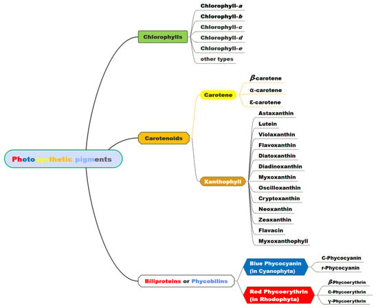
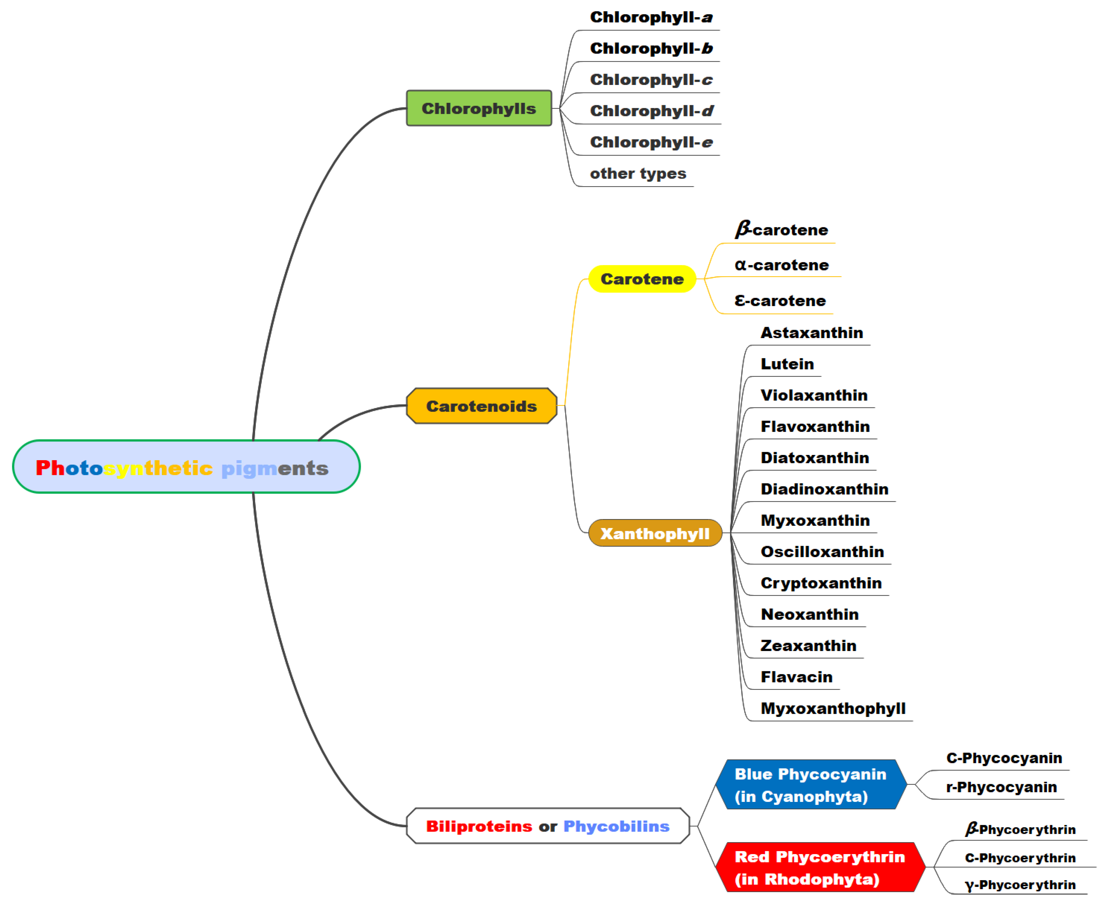
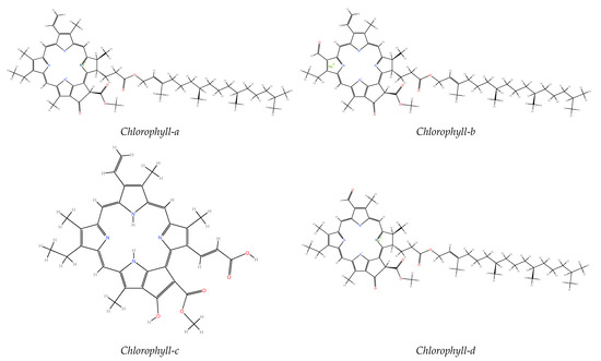
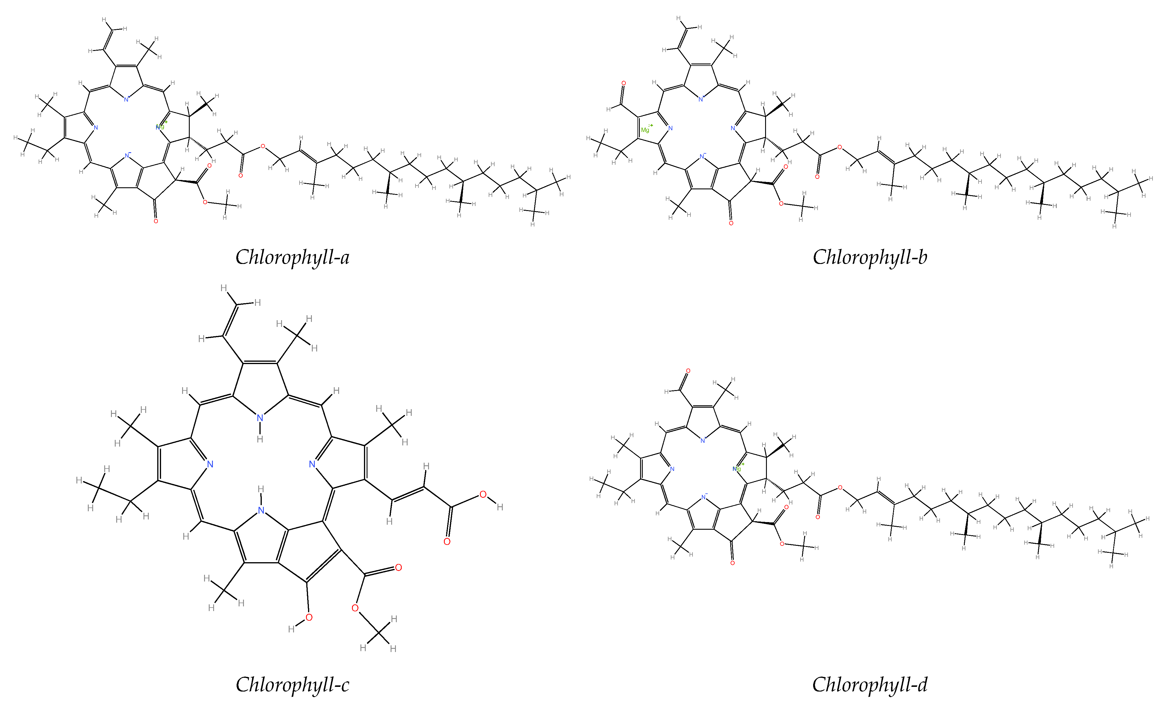
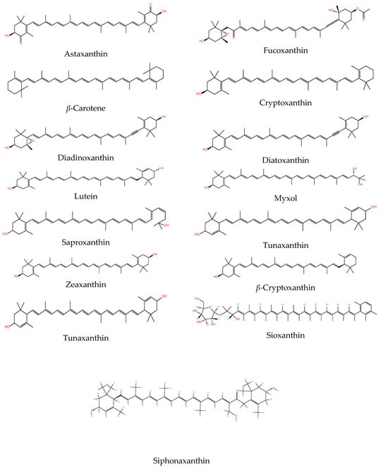
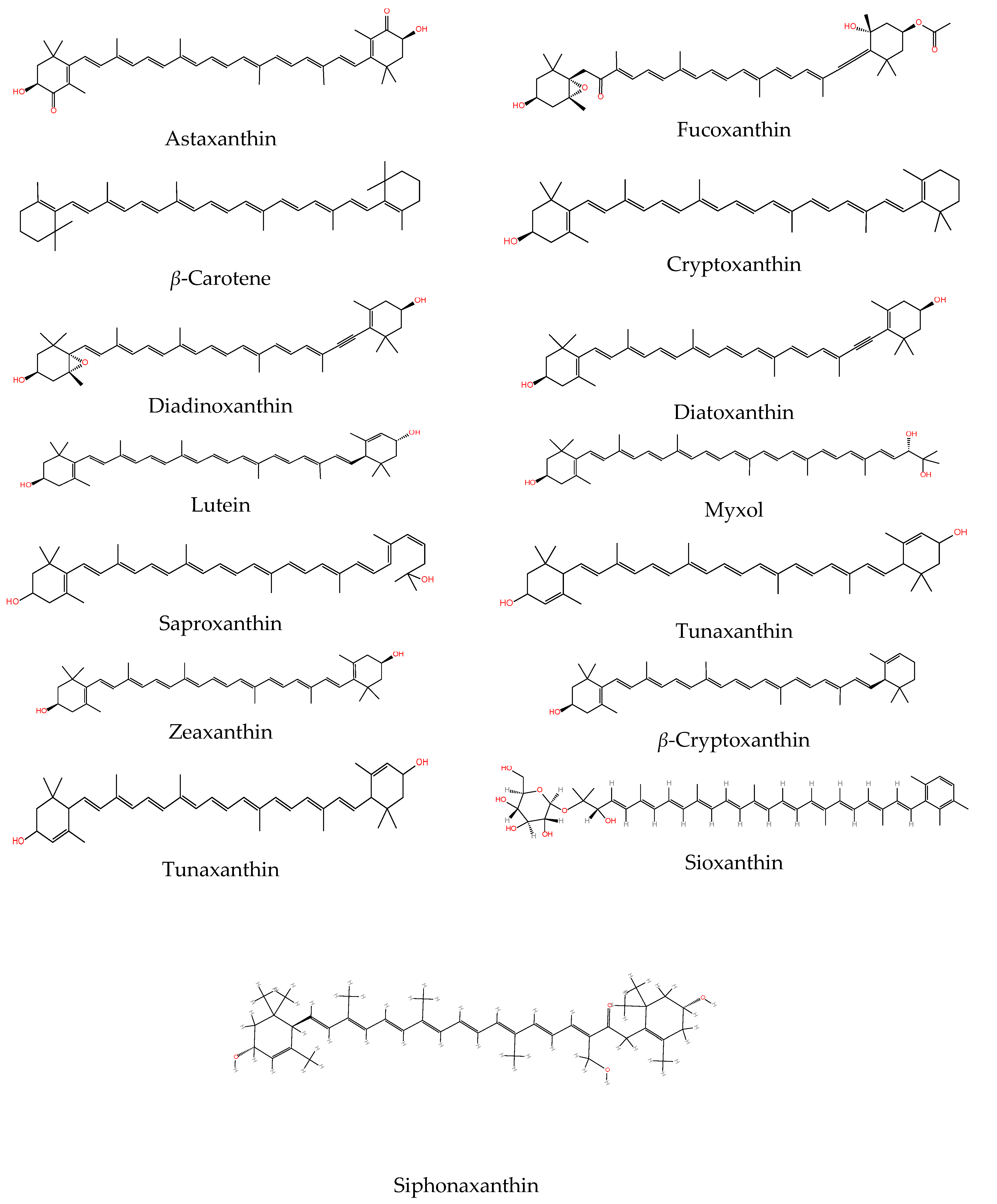
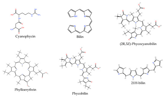
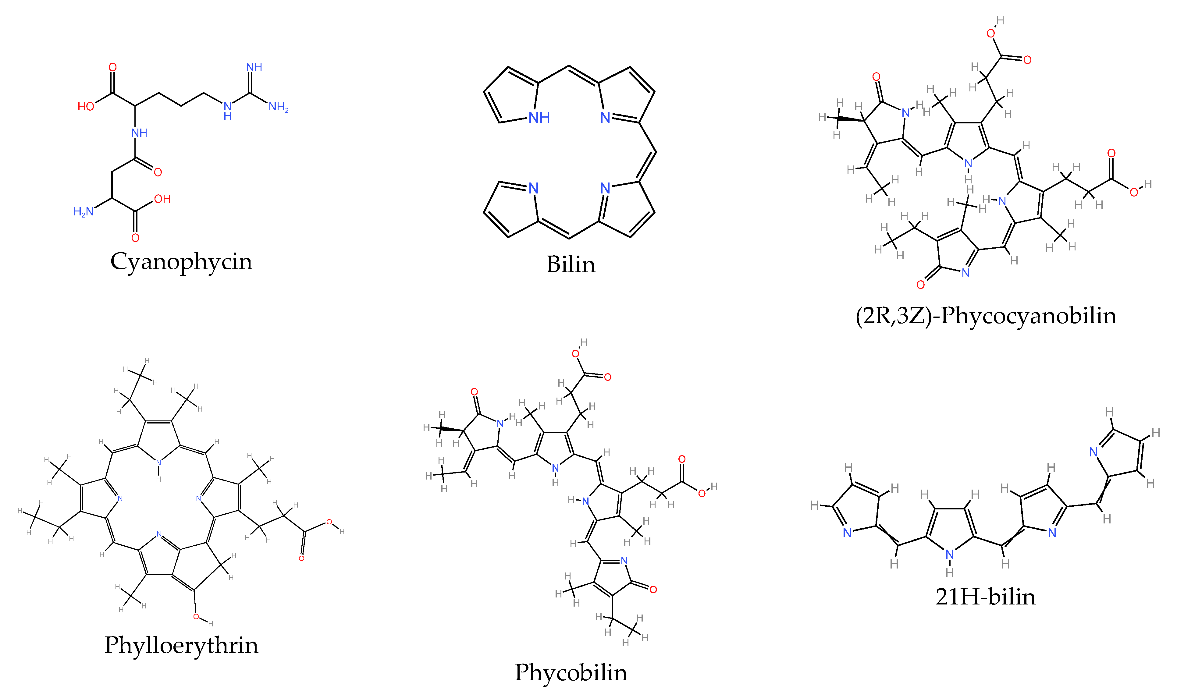
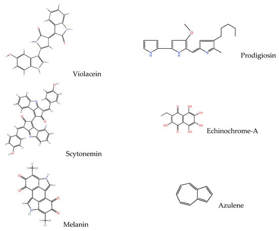
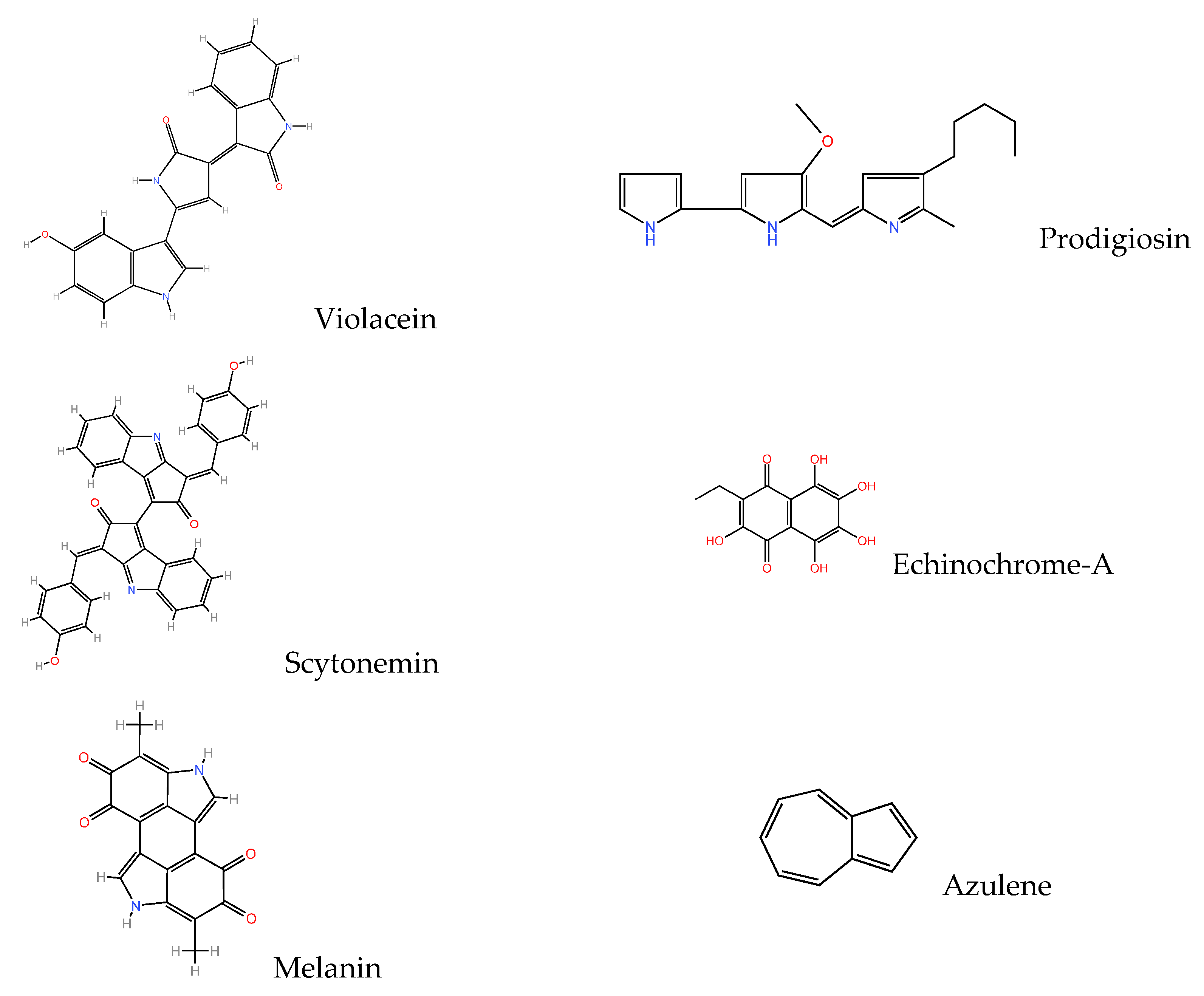
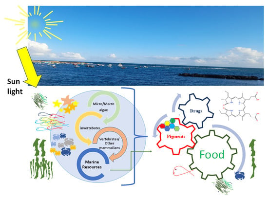
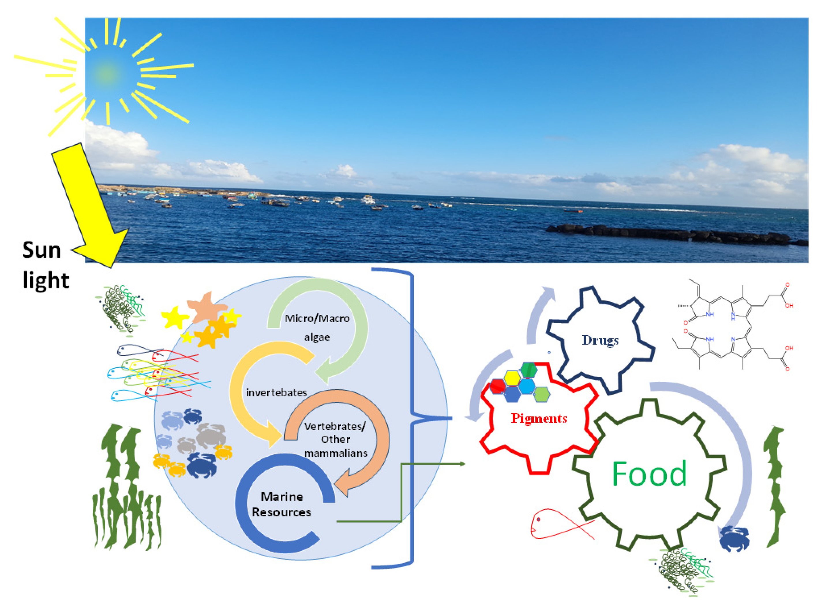

Disclaimer/Publisher’s Note: The statements, opinions and data contained in all publications are solely those of the individual author(s) and contributor(s) and not of MDPI and/or the editor(s). MDPI and/or the editor(s) disclaim responsibility for any injury to people or property resulting from any ideas, methods, instructions or products referred to in the content. |
© 2026 by the author. Licensee MDPI, Basel, Switzerland. This article is an open access article distributed under the terms and conditions of the Creative Commons Attribution (CC BY) license.