


Abstract
Lysine-specific demethylase 1 (LSD1) is a crucial epigenetic target closely correlated with the pathological progression of various solid tumors. In recent years, machine learning-driven virtual screening has been increasingly employed in novel drug discovery, providing promising paradigms for the rational development of LSD1 inhibitors. However, efficient screening of LSD1 inhibitors to prioritize candidates for experimental validation is still required. In this study, we propose a virtual screening framework that integrates a classification model, a drug–target affinity (DTA) prediction model (pharma-DTA), molecular docking, and binding free energy calculations. The optimal performance of the classification model is Random Forest with RDKit descriptors, which achieves a high precision of 0.930. The pharma-DTA model was established by integrating features of the ligand, pharmacophore, and the binding pocket of the protein. The model was fine-tuned on the dataset of LSD1 inhibitors, with the mean standard error of predicted pIC50 decreased to 0.5. These models were used to screen potential LSD1 inhibitors from the ZINC library, combined with molecular docking and MM-PBSA binding free energy calculations, and four top hits were identified. This virtual screening framework enables rapid identification of hit compounds for the discovery of potential LSD1 inhibitors.
1. Introduction
Lysine-specific demethylase 1 (LSD1), the first reported histone demethylase in 2004, is a prominent member of the extensively studied lysine demethylase family [1]. Its overexpression promotes aberrant cell proliferation, thereby driving rapid tumor growth and metastasis, and has been implicated in a wide range of cancers [2,3,4,5,6], including breast cancer [7], liver cancer [8], prostate cancer [9], stomach cancer [10], and acute myeloid leukemia [11]. Owing to its critical role in oncogenesis, LSD1 has rapidly emerged as a promising anti-cancer drug target [5]. Several potential LSD1 inhibitors have been discovered and are in clinical trials [12]. However, none have gained approval from the drug administration [13]. Conventional experimental methods for screening active compounds against a specific target from vast chemical libraries are often prohibitively costly and time-consuming [14,15]. Computer-aided drug design (CADD) technologies have accelerated pharmaceutical discovery, mitigating the need for blind experimentation and reducing early-stage research costs [16,17,18]. Meanwhile, artificial intelligence-aided drug discovery (AIDD) uses large datasets and machine learning models to identify targets and predict compound activity and potential adverse effects [19,20]. As a result, both CADD and AIDD have been successfully applied to the discovery of LSD1 inhibitors, with experimentally validated compounds confirming the effectiveness of this approach [21,22].
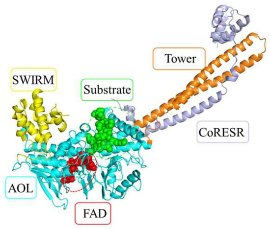
The structure of LSD1 comprises three major domains, as shown in Figure 1: the N-terminal SWIRM domain, the Tower domain, and the C-terminal amine oxidase-like (AOL) domain [23,24,25]. The AOL domain contains a flavin adenine dinucleotide (FAD) binding site and a substrate binding site, which together constitute the catalytic center responsible for the selective removal of mono- and dimethyl groups from lysine residues [26,27,28]. Notably, this active center constitutes the primary binding pocket for inhibitors [25,29]. Several key residues within this pocket have been identified as critical for ligand binding [22], including Trp695, Trp751, Tyr761, and Ala809.
Figure 1.
Three major domains of LSD1: SWIRM (yellow), Tower (orange), and AOL (cyan).
LSD1 inhibitors are classified as covalent or noncovalent according to their mechanism of inhibition [22]. Covalent inhibitors primarily function by forming adducts with the FAD cofactor. In recent decades, a variety of potent covalent inhibitors have been discovered. Notably, nine covalent inhibitors have entered clinical trials for the treatment of some solid tumors and blood tumors [4,13], including TCP, ORY-1001, ORY-2001, IMG-7289 [30], GSK-2879552 [31], INCB059872, TAK-418 [32], SYHA1807, and LH-1802 [13]. Despite offering prolonged therapeutic effects and enhanced efficacy, the irreversible binding of covalent inhibitors can lead to various side effects [33]. In comparison, noncovalent inhibitors have garnered growing attention owing to their potential advantages in safety and efficacy [34]. Numerous noncovalent LSD1 inhibitors with a broad range of chemical scaffolds have been reported [35,36], such as phenothiazine [37], pyridine [38], and thieno[3,2-b]pyrrole. To date, two noncovalent inhibitors have entered clinical trials, including CC-90011 [39] and SP-2577 [40]. Thus, continued discovery of noncovalent inhibitors constitutes a critical research priority [34].
CADD is a pivotal approach in contemporary preclinical drug discovery and has been successfully applied to the development of LSD1 inhibitors [18,22,41,42]. By integrating techniques such as molecular docking, molecular dynamics simulations, and binding energy calculations, CADD enables virtual screening of compound libraries to identify candidates [42,43,44,45,46,47]. For example, optimization of a hit from high-throughput screening produced a derivative with an IC50 of 0.162 µM, which later served as the basis for a more potent lead compound 49 with a lower IC50 of 6.7 nM [48]. Similarly, the Sharma group applied structure-based virtual screening and optimization to discover compound 12, which has a Ki of 31 nM [49]. These achievements show considerable potential of CADD in the discovery of new inhibitory agents [18].
AIDD has emerged as a powerful tool for identifying potential LSD1 inhibitors. Numerous machine learning and deep learning models were developed, trained on the extensive activity data available from databases such as ChEMBL [50] and ZINC. These models were used to identify active LSD1 inhibitors [51,52,53]. For example, virtual screening using a classification model based on existing compound libraries or drug banks identified numerous promising inhibitor skeletons [52]. The integration of machine learning with a regression model provided more profound insights into the binding mechanisms between inhibitors and LSD1 [53]. Compared with traditional drug screening methods, large-scale compound screening can be realized with modern drug discovery technologies. Most existing approaches have been optimized for DTA prediction by adopting diverse molecular features and network architectures, achieving high accuracy in regression tasks. Although this specialization enhances performance on dedicated tasks, it considerably restricts model generalization across different prediction scenarios. DTA is also widely applied in virtual screening (VS). Therefore, developing a robust predictive model for LSD1 inhibitor activity based on extensive datasets is crucial for addressing these challenges.
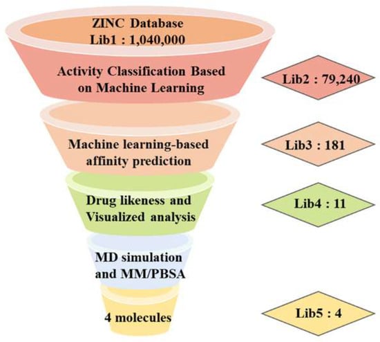
For the discovery of novel LSD1 inhibitors with structural diversity from an extensive compound library. CADD and AIDD can accelerate the process of discovering LSD1 inhibitors. In this study, we proposed pharma-DTA, a model that enhances DTA prediction by integrating features of the ligand and the protein binding pocket. Based on this model, we constructed a VS framework incorporating ML and CADD techniques and successfully identified active ligands. The DTA prediction model was fine-tuned to improve accuracy using a pre-trained topology-enhanced graph neural network [54]. Potential inhibitors were identified via virtual docking and further validated for stable binding to LSD1 through interaction analysis, molecular dynamics, and free energy calculations. The framework of virtual screening is depicted in Figure 2.
Figure 2.
The framework of virtual screening.
2. Materials and Methods
Dataset: First, an initial compound library (Lib1) was retrieved from the ZINC20 database, retaining molecules with molecular weight between 300 and 500 Da and logP between 1 and 4, yielding 1,040,000 compounds. Second, Lib1 was screened by the RF-RDK classification model; compounds with a predicted activity probability greater than 0.5 were retained as Lib2. Third, the fine-tuned pharma-DTA model ranked the candidates by predicted affinity, and the top 181 compounds with predicted pIC50 > 7.5 were designated as Lib3. Fourth, molecular docking with AutoDock Vina 1.2.0 was applied; all compounds in Lib3 showed docking scores below 8.00 kcal/mol, and 11 compounds were selected for Lib4 based on scaffold diversity and binding-pocket complementarity. Finally, MM-PBSA binding free energy calculations identified the compounds with binding affinities comparable to the reference inhibitor CC-90011, and four compounds (F1, F4, F8, and F11) were designated as Lib5 for subsequent analysis. Data curation for LSD1 noncovalent inhibitors: Data1 and Data2 were constructed by combining noncovalent inhibitor entries filtered from ChEMBL33 and additional noncovalent LSD1 inhibitor data collected from published literature [50]. Raw experimental inhibitory activity data were converted to standard pIC50 values (the negative base-10 logarithm of IC50 in M). All subsequent analysis, model training, and result visualization were performed on the unified pIC50 scale. After removing duplicates, the molecules were classified based on the statistical distribution of their IC50 values. Compounds with IC50 ≤ 1 µM were labeled as active, while those with IC50 ≥ 5 µM were defined as inactive [55]. The resulting dataset (denoted as Data1) comprised 800 active and 761 inactive compounds. For classification tasks, Data1 was randomly split into training (80%) and test (20%) sets. To rigorously evaluate the performance of our proposed model, we employed three widely recognized benchmark datasets: Davis, KIBA, and BindingDB in Table 1. To further fine-tune DTA prediction, a separate set of 1407 compounds with precise IC50 values was curated from ChEMBL33 as Data2. Before pre-training Pharma-DTA, all KDM1A/LSD1 entries were removed from BindingDB by matching the LSD1 protein sequence to eliminate cross-dataset overlap with our ChEMBL33 LSD1 fine-tuning dataset (Data2). This dataset was randomly split into training (70%), validation (15%), and test (15%) sets.
Table 1.
Sizes of the datasets.
Activity prediction with Model Generation and Evaluation: Multiple molecular fingerprint representations were computed as features for classification. For each compound, RDK (1024 dimensions), ECFP4 (1024 dimensions), and MACCS (166 dimensions) fingerprints [56] were derived from their Simplified Molecular Input Line Entry System (SMILES) strings using the RDKit 2022.09.1 toolkit in Python 3.11. Subsequently, to build a classification model for noncovalent LSD1 inhibitors, we used these fingerprint features as inputs to three machine learning algorithms, including Random Forest (RF) [57], Support Vector Machine (SVM) [58], and k-Nearest Neighbors (KNN). This framework produced nine distinct model configurations. Hyperparameters associated with the model are provided in Table S1. The evaluation of each model’s quality considered parameters such as true positives (TP), false positives (FP), true negatives (TN), and false negatives (FN). As shown in Table 2, a comprehensive evaluation was performed using standard metrics, including accuracy, precision, recall, F1-score, and Matthews correlation coefficient (MCC). Moreover, the area under the curve (AUC) is an essential index that quantifies a predictive model’s overall performance. The optimal model for each algorithm was obtained by evaluating combinations of fingerprint types and algorithms and selecting those with the best overall performance across multiple metrics.
Table 2.
Description of the evaluation metrics.
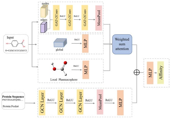
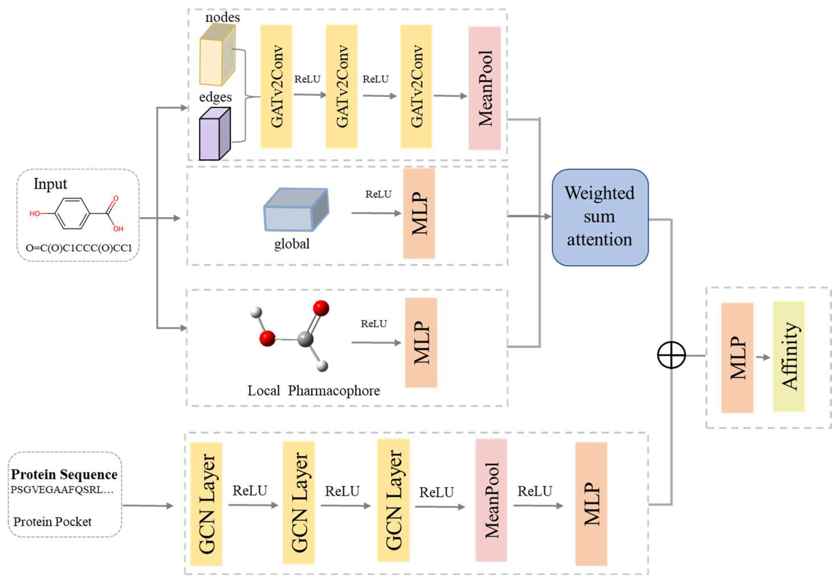
Architecture of the pharma-DTA model: As illustrated in Figure 3, we propose a multi-branch fused graph neural network framework, pharma-DTA, based on a previously reported pharmaGNN in our group [59,60]. The architecture incorporates three parallel sub-branches to extract features from the ligand and a graph encoder to characterize protein binding pockets. The hyperparameters used for model training are detailed in Table S2.
Figure 3.
The architecture of the pharma-DTA model.
For the ligands, the SMILES strings were converted into molecular graphs, and their 3D structures were optimized using MMFF94 in RDKit. From the molecular graph, node and edge features were extracted by three GATv2 layers, and global features were processed via a two-layer MLP. All these features were subsequently encoded into a unified embedding. Features of local pharmacophores are identified from the three-dimensional ligand structures, with a two-layer MLP implemented to learn a compact pharmacophore embedding. All these embeddings are fused in a weighted-summation attention block with adaptive weights to yield a unified ligand feature vector. For protein feature extraction, the protein binding pocket data were acquired from the CaviDB database. We utilize residue graphs to represent proteins with three GCN layers, followed by mean pooling and a readout MLP to generate a single embedding. The embeddings of the ligand and protein are concatenated to obtain a vector Z, which is then input into a four-layer MLP to predict the binding affinity. For detailed information, see Supplementary Material S2.
Fine-tuning the drug–target affinity prediction model: Although the pharma-DTA model performed well on the benchmark, its performance on the specific LSD1 dataset (Data2) was suboptimal. We employed a transfer learning strategy focused on selective layer optimization. After loading the pre-trained weights, the lower-level GNN layers were frozen to preserve the foundational molecular features. Concurrently, several layers were unfrozen and fine-tuned, including the final GNN layers, the readout module, and the fully connected prediction layers. This approach allowed the model to retain general knowledge from pre-training while efficiently adapting its final stages to the precise task of LSD1 affinity prediction. The training hyperparameters are as follows: the number of epochs (100), the batch size (256 to 32), and the learning rate (0.001 to 0.0001).
Molecular docking: The initial crystal structure of LSD1 was obtained from the RCSB Protein Data Bank [PDB ID: 2V1D] [61]. For docking simulations, a grid box centered at (47.053, 112.106, 70.261) with dimensions of 16 × 24 × 22 Å3 was used to include the active site [62]. The system underwent a two-stage energy minimization (5000 steps each), with a 500 kcal mol−1 Å−2 harmonic restraint applied to the solute during the first stage. Subsequently, the system was heated from 0 to 300 K over 50 ps with a restraint force of 25 kcal mol−1 Å−2. This was followed by a 1 ns density equilibration and a 4 ns constant-pressure equilibration. Finally, a 50 ns production simulation was performed at 300 K and 1 atm. Potential hit compounds were then identified via high-throughput molecular docking using AutoDock Vina 1.2.0. The empirical scoring function was used for binding mode prediction and affinity estimation, followed by elimination of compounds with low scores [63,64,65,66,67,68].
Molecular dynamics and MM/PBSA free energy calculations: Molecular dynamics (MD) simulations were performed using the Amber 24 package [69]. Ligand parameters were prepared by assigning AM1-BCC charges and the General Amber Force Field (GAFF) via antechamber, with missing parameters supplemented using “parmchk2” [70,71]. The protonation state of LSD1 was determined with PropKa, and the Amber ff19SB force field was applied to the protein [69]. All the missing hydrogen atoms were added using “tleap” [72]. Systems were neutralized with chloride counterions. Production MD runs of 50 ns each were performed using the GPU-accelerated PMEMD module [73]. K-means clustering was employed to cluster the molecular dynamics (MD) results, which were derived from four independent 50 ns MD simulations. Trajectory analysis, including the calculation of the root-mean-square deviation (RMSD) of protein heavy atoms relative to the initial structure, was carried out with the CPPTRAJ module [74]. The binding free energies were estimated using the Molecular Mechanics/Poisson–Boltzmann Surface Area (MM/PBSA) method, implemented via “MMPBSA.py” [75]. The non-equilibrated initial portion of the MD trajectory was excluded. System stabilization was verified by the converged RMSD of the protein–ligand complex, and the following 30 ns stable production trajectory was adopted for the MM-PBSA calculations.
3. Results
3.1. Machine Learning Model for Activity Screening
The performance of the activity classification models was evaluated on the test set using six metrics, including accuracy, precision, recall, F1-score, MCC, and AUC, as detailed in Table 3. The random forest (RF) model using RDK molecular fingerprints attained the optimal overall performance, with superior performance observed for all other evaluation metrics.
Table 3.
Performance evaluation of the 9 models based on test set prediction 1.
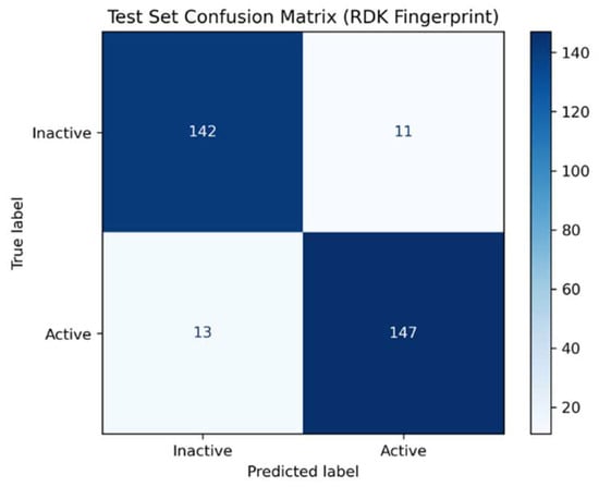
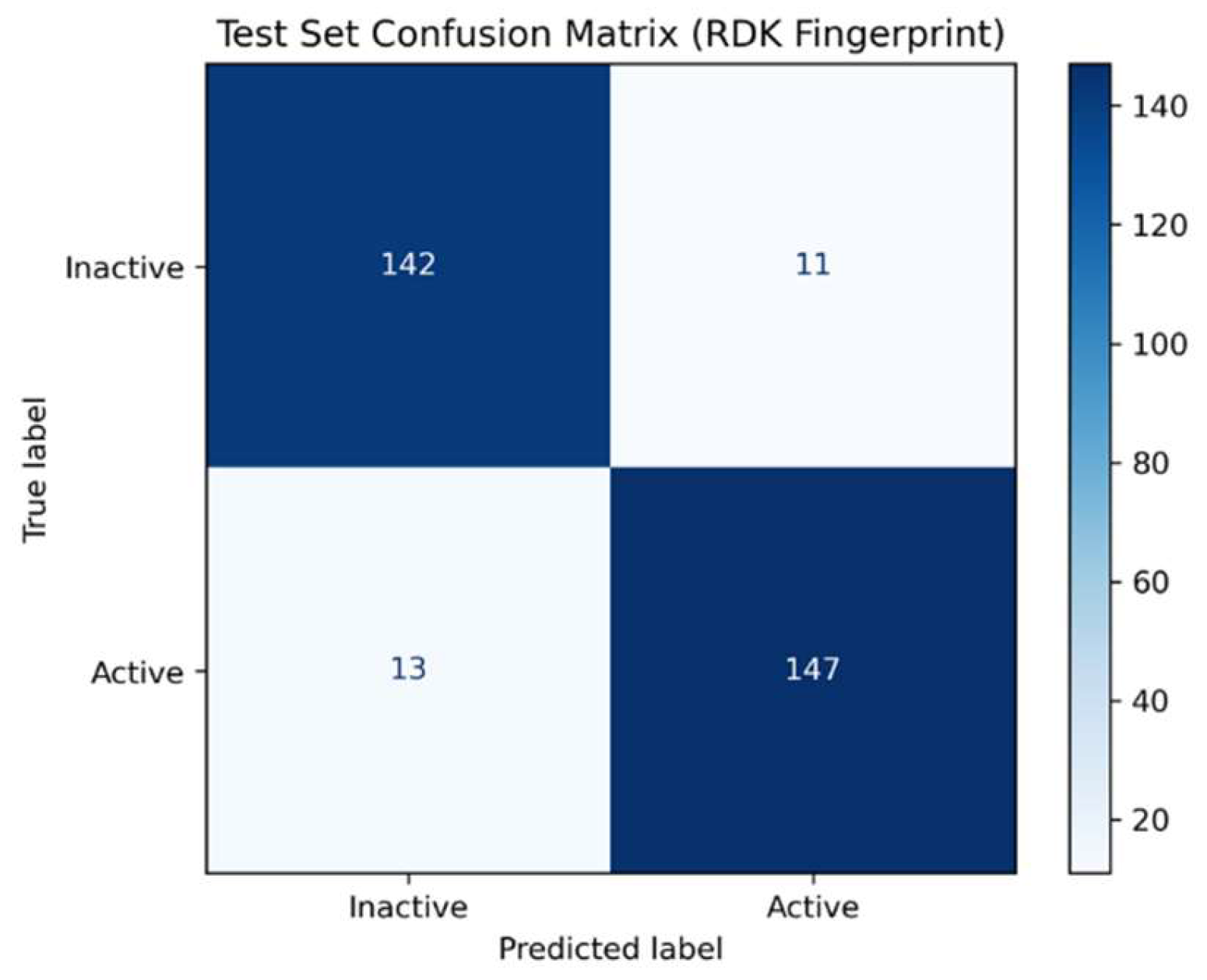
The confusion matrix of the RF-RDK model enabled visualization of the number of classified compounds in Figure 4. The test set contained 160 actives and 153 inactives. It revealed a TP of 147, an FN of 13, a TN of 142, and an FP of 11. This translates to a sensitivity of 92.5% (147/160) for identifying actives and a specificity of 92.8% (142/153) for identifying inactives, indicating the model has excellent, well-balanced classification capability. Subsequently, this high-performance model was applied to screen Lib1, retaining 79,240 compounds predicted with an activity probability greater than 0.5, designated as Lib2. Thus, this model was used in the following screening task.
Figure 4.
Confusion matrix of the RF-RDK model of predicted and true labels.
3.2. Performance of Pharma-DTA Model and Ablation Experiment
The hyperparameters of the proposed model are presented in Table S2. To evaluate the performance of the pharma-DTA model, we compared it with current state-of-the-art methods (including DeepDTA, AttentionDTA, GraphDTA, DGraphDTA, and DTA) on Davis, KIBA, and BindingDB datasets [76,77,78,79,80]. As shown in Table 4, the results show that the pharma-DTA model performs well on all three datasets. Compared with the best baseline (DTA), our model yielded relative MSE reductions of 3.8%, 0.8%, and 3.3% on the three datasets, showing considerable improvements on Davis and BindingDB and a modest gain on KIBA. In terms of the concordance index (CI), our model attained the highest values of 0.917 (Davis), 0.900 (KIBA), and 0.867 (BindingDB), outperforming DTA by 0.7%, 0.2%, and 3.0% on the three sets, respectively. These results show that our proposed representation achieves superior performance across diverse datasets.
Table 4.
The performance of the model on benchmark datasets 1.
To evaluate the generalization performance of individual modules on drugs in the model, ablation experiments on combinations of molecular feature modules were further performed on the BindingDB benchmark dataset. For the features of pharmacophore, edge, and global, each module was removed and compared with the full model. The full model achieves the lowest MSE of 0.532, outperforming the ablated models without the drug pharmacophore module (0.559), edge feature module (0.595), or global feature module (0.564). These results indicate that all feature components contribute to model performance, and the complete representation yields the strongest predictive ability. As shown in Table 5, ablation experiments indicate that removing any core module will cause a significant rise in MSE. This fully demonstrates that the local pharmacophore module, edge feature module, and global feature module all exert positive effects on improving the model’s prediction accuracy.
Table 5.
Comparison of ablation experiments on BindingDB datasets.
3.3. Validation of the Model and Virtual Screening of Compound Libraries
The model was fine-tuned on Data 2 for the specific task of predicting LSD1 inhibitor activity. The results of the fine-tuning process were assessed through two comparative analyses, as shown in Table 6. Model evaluation using three random seeds (25, 30, and 40) on Data2 showed that fine-tuning reduced the MSE from over 2.0 to 0.45–0.50. The results indicate that the fine-tuning process effectively improves the model’s accuracy for pIC50.
Table 6.
Comparison of MSE, predicted, and experimental pIC50 before and after fine-tuning.
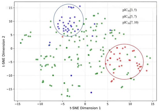
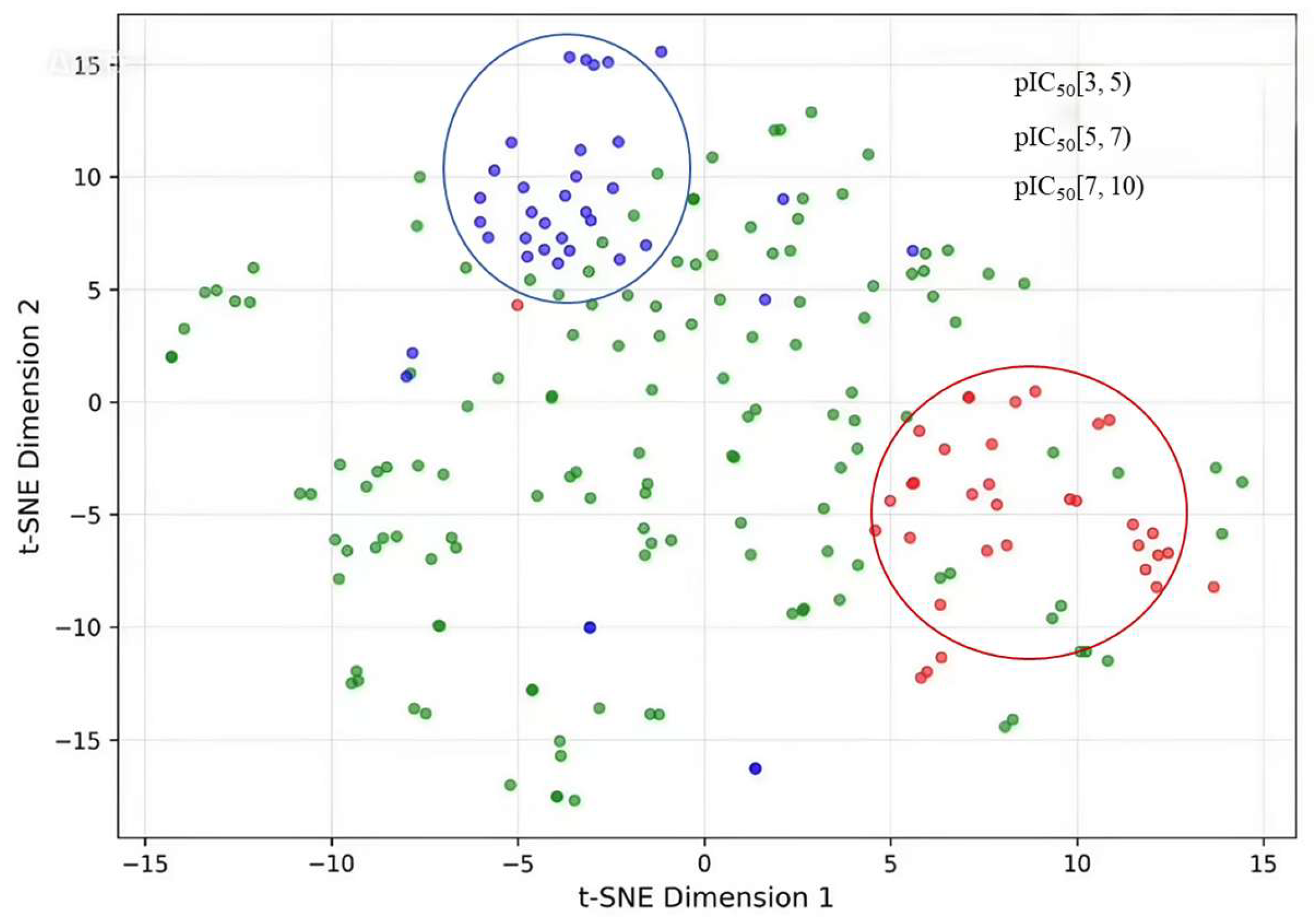
To elucidate the relationship between molecular descriptors and bioactivity (pIC50), we applied t-SNE to reduce the 256-dimensional feature vector Z from the penultimate layer of the fine-tuned pharma-DTA model to a two-dimensional latent space [81]. In Figure 5, compounds from the test set are visualized in this space, colored according to three pIC50 activity categories: low ([3, 5)), moderate ([5, 7)), and high ([7, 10)). The t-SNE visualization reveals that compounds segregate within the latent space according to their bioactivity. The highly active compounds are concentrated in the upper area, forming a well-defined cluster that is spatially segregated from other groups. In comparison, the low-activity compounds predominantly form a relatively compact cluster in the down-right region. This apparent separation in the t-SNE projection shows that the latent representation separates activity classes. The t-SNE visualization demonstrates that activity-related grouping emerges within the projected latent space, which may serve as a basis for subsequent feature-based activity analysis and molecular design.
Figure 5.
t-SNE visualization of the latent space colored by predicted pIC50 values. Compounds are categorized into low (red, [3, 5)), moderate (green, [5, 7)), and high (blue, [7, 10)) activity groups. The circles are added to highlight high activity (in blue) and low activity (in red) regions.
The screening process utilized our model to evaluate compounds in Lib2. The resulting candidates were ranked by predicted activity, and the top 181 compounds with pIC50 > 7.5 were retained as Lib3. Their chemical structures, molecular docking scores, and predicted binding affinities are provided in Figure S1 (Supplementary Material S3).
3.4. Molecular Docking
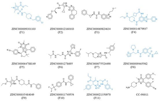
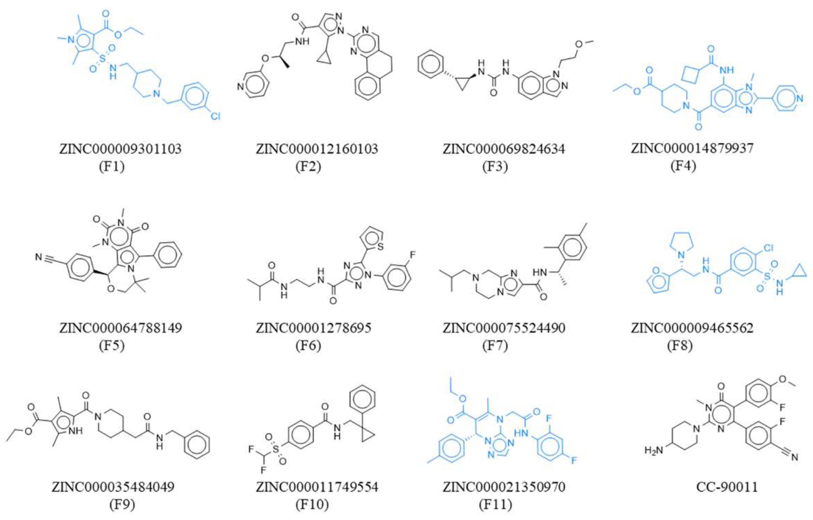
Molecular docking was performed to evaluate the binding affinity between LSD1 and compounds in Lib3. Notably, all docking scores were below −8.00 kcal/mol, with most reaching −9.00 kcal/mol or lower. The compounds prioritized by our classification and pIC50 prediction models achieved favorable docking scores and showed consistent promise across multiple virtual screening criteria. From this set, 11 compounds were selected for Lib4 based on scaffold diversity and binding-pocket complementarity. The chemical structures of the 11 selected compounds and the reference inhibitor CC-90011 are depicted in Figure 6.
Figure 6.
Chemical structures of the 11 selected compounds and CC-90011. Structures highlighted in blue have closest to or better activity than CC-90011.
3.5. MM-PBSA Binding Free Energy
The binding free energies of candidate ligands were computed using MM-PBSA (ΔGMM/PBSA) based on molecular dynamics (MD) simulations, with the decomposed energy components summarized in Table S6. As shown in Table 7, the results indicate that the ΔGMM/PBSA values of compounds F1, F4, F8, and F11 are comparable to those of CC-90011, and these four compounds were designated as Lib5. Notably, F1 showed the strongest binding affinity (−49.2 kcal/mol), with F4 also exhibiting a highly favorable energy (−48.3 kcal/mol). The ΔGMM/PBSA values for F8 and F11 were −41.3 kcal/mol and −39.7 kcal/mol, respectively, indicating strong binding. These results suggest that these four compounds can form stable complexes with LSD1. Consistent with the energy analysis, the MD trajectory analysis reveals that these ligands contribute to the structural stabilization of LSD1. Furthermore, the stability of all binding poses was confirmed by low root-mean-square deviation (RMSD) values throughout the simulations. Specifically, the average RMSD values for the F1-LSD1, F4-LSD1, F8-LSD1, and F11-LSD1 complexes were 2.1 Å, 2.1 Å, 2.0 Å, and 2.1 Å, respectively. Notably, all four ligand-bound complexes exhibited lower average RMSD values than the apo-LSD1 (2.3 Å), indicating that ligand binding stabilizes the overall protein structure and maintains stable binding within the pocket (Supplementary Material S4). Collectively, F1, F4, F8, and F11 emerge as promising candidates with binding affinities equal to or exceeding that of the reference compound. The physicochemical properties of the compounds predicted by Swiss-ADME [82] and all pharmacokinetic parameters were within satisfactory ranges, as detailed in Supplementary Material S5.
Table 7.
Results of docking score, predicted pIC50, and MMPBSA 1.
3.6. The Interactions Between 4 Screened Molecules and LSD1
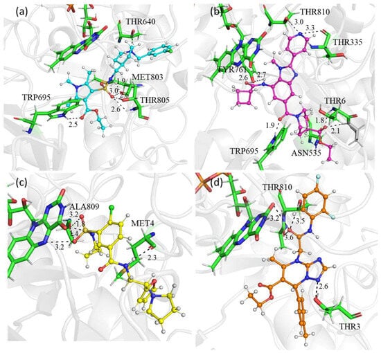
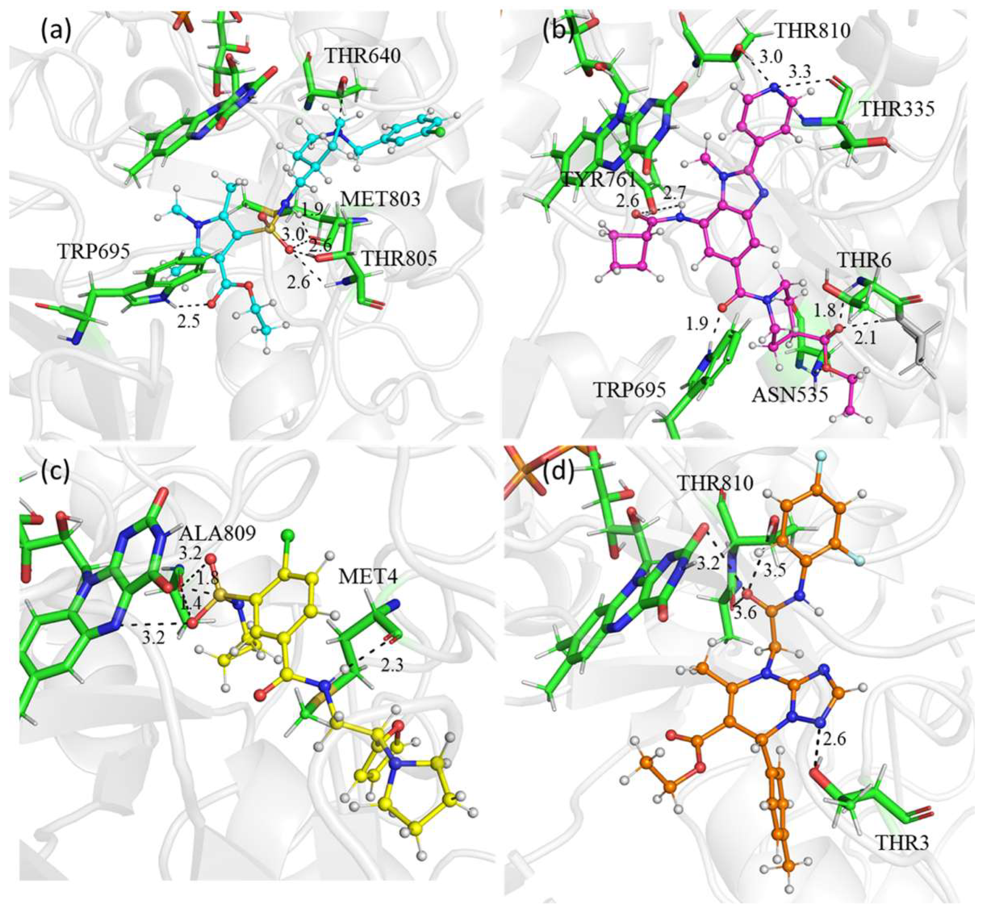
The representative binding modes were filtered based on the presence of H-bonds in the protein binding region. Figure 7 displays the H-bond interactions of representative conformations around the active site of the four complexes of lower affinity, namely F1, F4, F8, and F11. F1 binds to pockets of LSD1, forming relatively numerous and stable hydrogen bond interactions with amino acids such as THR640, TRP695, MET803, and THR805. Moreover, F4 establishes stable hydrogen bonds with amino acids ASN535, TRP695, TYR761, THR810, THR335, and THR6 in the pocket of LSD1. Additionally, F8 forms two hydrogen bonds with ALA809 and MET4 of LSD1. F11 also establishes stable hydrogen bonds with amino acids FAD, THR810, and THR3. Among these hydrogen-bonding-involved residues, TRP695, ASN535, TYR761, THR810, THR335, and ALA809 are well-documented favorable interaction residues for clinical-stage LSD1 inhibitors, including CC-90011 and TCP-derived ORY-1001. The observation of stable hydrogen bonds at these sites indicates that these four compounds can correctly occupy the LSD1 active-site pocket and form effective local intermolecular interactions, which provides a favorable structural basis for ligand–protein recognition.
Figure 7.
The hydrogen-bonding interactions between LSD1 and compounds: (a) F1, (b) F4, (c) F8, and (d) F11.
4. Conclusions
This study developed a hybrid virtual screening pipeline to identify novel LSD1 inhibitors, integrating machine learning, docking, and molecular dynamics simulations. We established a robust RF-RDK classification model, which achieved the highest precision of 0.930. An affinity prediction model (pharma-DTA) was established, and the fine-tuned model achieved high predictive accuracy, with predicted IC50 values very close to experimental values for LSD1 inhibitors. The t-SNE visualization was performed in the latent space to enable activity analysis of the molecular distribution. The compounds screened from the ZINC library by the initial classification and pIC50 prediction exhibited consistently low docking scores. The four top hits (F1, F4, F8, and F11) were further analyzed by MM-PBSA binding free energy calculations, indicating potentially favorable and stable predicted interactions with LSD1. This study not only identifies novel inhibitor candidates but also establishes an interpretable and efficient AI-augmented workflow that can be used to accelerate drug discovery for other targets.
Supplementary Materials
The following supporting information can be downloaded at https://www.mdpi.com/article/10.3390/chemistry8110162/s1, Figure S1: Structures and predicted activities of 181 compounds identified via virtual screening against LSD1. Predicted IC50 values (nM) and docking scores (kcal/mol) are shown in red and blue, respectively; Figure S2: RMSD plot of LSD1 with the identified compounds: (a) F1, (b) F4, (c) F8, and (d) F11; Figure S3: RMSD plot of apo-LSD1 (purple) and the F1, F4, F8, and F11-LSD1 complexes (black, red, blue, and green, respectively) during the 100 ns MD simulations; Table S1: Hyperparameters of the classification model; Table S2: Hyperparameters of pharma-DTA; Table S3: Node features in the representation of molecules; Table S4: Edge features in the representation of molecules; Table S5: Pharmacophore features; Table S6: MM/PB(GB)SA binding energies of the compounds and antiviral drugs (energies in kcal/mole); Table S7: In silico evaluation of pharmacokinetic properties of CC-90011, F1, F4, F8, and F11; Table S8. Comprehensive performance evaluation of the model on the Data2 test set.
Author Contributions
Conceptualization, J.S. and X.N.; methodology, X.N.; software, L.Z.; validation, Y.H.; resources, J.S.; writing—original draft preparation, L.Z.; writing—review and editing, J.S. and X.N.; funding acquisition, J.S. and L.Q. All authors have read and agreed to the published version of the manuscript.
Funding
This research was funded by the National Natural Science Foundation of China, grant number 22173083, and the Fundamental Research Funds of State Key Laboratory of Cotton Biobreeding and Integrated Utilization, grant number CBIUZ2025001.
Data Availability Statement
The original contributions presented in this study are included in the article/Supplementary Material. Further inquiries can be directed to the corresponding authors.
Conflicts of Interest
The authors declare no conflicts of interest.
Abbreviations
The following abbreviations are used in this manuscript:
| LSD1 | Lysine-specific demethylase 1 |
| ACC | Accuracy |
| AUC | Area under the receiver operating characteristic curve |
| MCC | Matthews correlation coefficient |
| CADD | Computer-aided drug design |
| AIDD | Artificial intelligence drug discovery |
| FN | False negative |
| FP | False positive |
| TN | True negative |
| TP | True positive |
| MD | Molecular dynamics |
| MMPBSA | Molecular mechanics Poisson–Boltzmann surface area |
| PDB | Protein Data Bank |
| RMSD | Root mean square deviation |
References
- Shi, Y.; Lan, F.; Matson, C.; Mulligan, P.; Whetstine, J.R.; Cole, P.A.; Casero, R.A.; Shi, Y. Histone demethylation mediated by the nuclear amine oxidase homolog LSD1. Cell 2004, 119, 941–953. [Google Scholar] [CrossRef] [Scilit]
- Hayami, S.; Kelly, J.D.; Cho, H.S.; Yoshimatsu, M.; Unoki, M.; Tsunoda, T.; Field, H.I.; Neal, D.E.; Yamaue, H.; Ponder, B.A.J.; et al. Overexpression of LSD1 contributes to human carcinogenesis through chromatin regulation in various cancers. Int. J. Cancer 2010, 128, 574–586. [Google Scholar] [CrossRef] [Scilit]
- Sheikh, K.A.; Iqubal, A.; Alam, M.M.; Akhter, M.; Khan, M.A.; Ehtaishamul Haque, S.; Parvez, S.; Jahangir, U.; Amir, M.; Khanna, S.; et al. A quinquennial review of potent LSD1 inhibitors explored for the treatment of different cancers, with special focus on SAR studies. Curr. Med. Chem. 2024, 31, 152–207. [Google Scholar] [CrossRef] [Scilit]
- Fu, X.; Zhang, P.; Yu, B. Advances toward LSD1 inhibitors for cancer therapy. Future Med. Chem. 2017, 9, 1227–1242. [Google Scholar] [CrossRef] [Scilit]
- Egolf, S.; Capell, B.C. LSD1: A viable therapeutic target in cutaneous squamous cell carcinoma? Expert Opin. Ther. Targets 2020, 24, 671–678. [Google Scholar] [CrossRef] [Scilit]
- Egolf, S.; Aubert, Y.; Doepner, M.; Anderson, A.; Maldonado-Lopez, A.; Pacella, G.; Lee, J.; Ko, E.K.; Zou, J.; Lan, Y.; et al. LSD1 inhibition promotes epithelial differentiation through derepression of fate-determining transcription factors. Cell Rep. 2019, 28, 1981–1992. [Google Scholar] [CrossRef] [Scilit]
- Lim, S.; Janzer, A.; Becker, A.; Zimmer, A.; Schüle, R.; Buettner, R.; Kirfel, J. Lysine-specific demethylase 1 (LSD1) is highly expressed in ER-negative breast cancers and a biomarker predicting aggressive biology. Carcinogenesis 2010, 31, 512–520. [Google Scholar] [CrossRef] [Scilit]
- Liu, C.; Liu, L.; Chen, X.; Cheng, J.; Zhang, H.; Zhang, C.; Shan, J.; Shen, J.; Qian, C. LSD1 stimulates cancer-associated fibroblasts to drive notch3-dependent self-renewal of liver cancer stem–like cells. Cancer Res. 2018, 78, 938–949. [Google Scholar] [CrossRef] [Scilit]
- Ketscher, A.; Jilg, C.A.; Willmann, D.; Hummel, B.; Imhof, A.; Rüsseler, V.; Hölz, S.; Metzger, E.; Müller, J.M.; Schüle, R. LSD1 controls metastasis of androgen-independent prostate cancer cells through PXN and LPAR6. Oncogenesis 2014, 3, e120. [Google Scholar] [CrossRef] [Scilit]
- Pan, H.-M.; Lang, W.-Y.; Yao, L.-J.; Wang, Y.; Li, X.-L. shRNA-interfering LSD1 inhibits proliferation and invasion of gastric cancer cells via VEGF-C/PI3K/AKT signaling pathway. World J. Gastrointest. Oncol. 2019, 11, 622–633. [Google Scholar] [CrossRef] [Scilit]
- Maiques-Diaz, A.; Spencer, G.J.; Lynch, J.T.; Ciceri, F.; Williams, E.L.; Amaral, F.M.R.; Wiseman, D.H.; Harris, W.J.; Li, Y.; Sahoo, S.; et al. Enhancer activation by pharmacologic displacement of LSD1 from GFI1 Induces differentiation in acute myeloid leukemia. Cell Rep. 2018, 22, 3641–3659. [Google Scholar] [CrossRef] [Scilit]
- Mould, D.P.; McGonagle, A.E.; Wiseman, D.H.; Williams, E.L.; Jordan, A.M. Reversible inhibitors of LSD1 as therapeutic agents in acute myeloid leukemia: Clinical significance and progress to date. Med. Res. Rev. 2014, 35, 586–618. [Google Scholar] [CrossRef] [Scilit]
- Fang, Y.; Liao, G.; Yu, B. LSD1/KDM1A inhibitors in clinical trials: Advances and prospects. J. Hematol. Oncol. 2019, 12, 129. [Google Scholar] [CrossRef] [Scilit]
- Paul, S.M.; Mytelka, D.S.; Dunwiddie, C.T.; Persinger, C.C.; Munos, B.H.; Lindborg, S.R.; Schacht, A.L. How to improve R&D productivity: The pharmaceutical industry’s grand challenge. Nat. Rev. Drug Discov. 2010, 9, 203–214. [Google Scholar] [CrossRef] [Scilit]
- DiMasi, J.A.; Grabowski, H.G.; Hansen, R.W. Innovation in the pharmaceutical industry: New estimates of R&D costs. J. Health Econ. 2016, 47, 20–33. [Google Scholar] [CrossRef] [Scilit]
- Xiang, M.; Cao, Y.; Fan, W.; Chen, L.; Mo, Y. Computer-aided drug design: Lead discovery and optimization. Comb. Chem. High Throughput Screen. 2012, 15, 328–337. [Google Scholar] [CrossRef] [Scilit]
- Baig, M.H.; Ahmad, K.; Rabbani, G.; Danishuddin, M.; Choi, I. Computer aided drug design and its application to the development of potential drugs for neurodegenerative disorders. Curr. Neuropharmacol. 2017, 16, 740–748. [Google Scholar] [CrossRef] [Scilit]
- Sabe, V.T.; Ntombela, T.; Jhamba, L.A.; Maguire, G.E.M.; Govender, T.; Naicker, T.; Kruger, H.G. Current trends in computer aided drug design and a highlight of drugs discovered via computational techniques: A review. Eur. J. Med. Chem. 2021, 224, 113705. [Google Scholar] [CrossRef] [Scilit]
- Zhang, O.; Lin, H.; Zhang, X.; Wang, X.; Wu, Z.; Ye, Q.; Zhao, W.; Wang, J.; Ying, K.; Kang, Y.; et al. Graph neural networks in modern AI-aided drug discovery. Chem. Rev. 2025, 125, 10001–10103. [Google Scholar] [CrossRef] [Scilit]
- Sun, Y.; Zhao, B.; Wang, Y.; Chen, Z.; Zhang, H.; Qu, L.; Zhao, Y.; Song, J. Optimization of potential non-covalent inhibitors for the SARS-CoV-2 main protease inspected by a descriptor of the subpocket occupancy. Phys. Chem. Chem. Phys. 2022, 24, 29940–29951. [Google Scholar] [CrossRef] [Scilit]
- Xu, W. Current status of computational approaches for small molecule drug discovery. J. Med. Chem. 2024, 67, 18633–18636. [Google Scholar] [CrossRef] [Scilit]
- Han, D.; Lu, J.; Fan, B.; Lu, W.; Xue, Y.; Wang, M.; Liu, T.; Cui, S.; Gao, Q.; Duan, Y.; et al. Lysine-specific demethylase 1 inhibitors: A comprehensive review utilizing computer-aided drug design technologies. Molecules 2024, 29, 550. [Google Scholar] [CrossRef] [Scilit]
- Stavropoulos, P.; Blobel, G.; Hoelz, A. Crystal structure and mechanism of human lysine-specific demethylase-1. Nat. Struct. Mol. Biol. 2006, 13, 626–632. [Google Scholar] [CrossRef] [Scilit]
- Kim, S.-A.; Zhu, J.; Yennawar, N.; Eek, P.; Tan, S. Crystal structure of the LSD1/CoREST histone demethylase bound to its nucleosome substrate. Mol. Cell 2020, 78, 903–914. [Google Scholar] [CrossRef] [Scilit]
- Hwang, S.; Schmitt, A.A.; Luteran, A.E.; Toone, E.J.; McCafferty, D.G. Thermodynamic Characterization of the Binding Interaction between the Histone Demethylase LSD1/KDM1 and CoREST. Biochemistry 2010, 50, 546–557. [Google Scholar] [CrossRef] [Scilit]
- Adamo, A.; Sesé, B.; Boue, S.; Castaño, J.; Paramonov, I.; Barrero, M.J.; Belmonte, J.C.I. LSD1 regulates the balance between self-renewal and differentiation in human embryonic stem cells. Nat. Cell Biol. 2011, 13, 652–659. [Google Scholar] [CrossRef] [Scilit]
- Lokken, A.A.; Zeleznik-Le, N.J. Breaking the LSD1/KDM1A addiction: Therapeutic targeting of the epigenetic modifier in AML. Cancer Cell 2012, 21, 451–453. [Google Scholar] [CrossRef] [Scilit]
- Klose, R.J.; Zhang, Y. Regulation of histone methylation by demethylimination and demethylation. Nat. Rev. Mol. Cell Biol. 2007, 8, 307–318. [Google Scholar] [CrossRef] [Scilit]
- Yang, K.; Xiong, J.; Shi, Y.; Yang, W.; Wang, W.; Song, Y.; Yu, B. Unveiling new binding sites and allosteric regulation mechanisms of LSD1 for novel therapeutics. J. Med. Chem. 2025, 68, 12125–12135. [Google Scholar] [CrossRef] [Scilit]
- Yacoub, A.; Rienhoff, H.Y., Jr. LSD1 Inhibition with IMG7289 (bomedemstat) rebalances megakaryopoiesis and erythropoiesis in a patient with MPN/MDS with refractory anemia with ringed sideroblast and marked thrombocytosis with JAK2 and SF3B1 mutations. Blood 2020, 136, 586–618. [Google Scholar] [CrossRef] [Scilit]
- Mohammad, H.; Smitheman, K.; van Aller, G.; Cusan, M.; Kamat, S.; Liu, Y.; Johnson, N.; Hann, C.; Armstrong, S.; Kruger, R. Novel anti-tumor activity of targeted LSD1 inhibition by GSK2879552. Eur. J. Cancer 2014, 50, 72. [Google Scholar] [CrossRef] [Scilit]
- Hattori, Y.; Matsumoto, S.; Morimoto, S.; Daini, M.; Toyofuku, M.; Matsuda, S.; Baba, R.; Murakami, K.; Iwatani, M.; Oki, H.; et al. Design, synthesis, and structure-activity relationship of TAK-418 and its derivatives as a novel series of LSD1 inhibitors with lowered risk of hematological side effects. Eur. J. Med. Chem. 2022, 239, 114522. [Google Scholar] [CrossRef] [Scilit]
- You, M.; Liu, H.; Li, C. The current toolbox for covalent inhibitors: From hit identification to drug discovery. J. Am. Chem. Soc. Au 2025, 5, 5866–5887. [Google Scholar] [CrossRef] [Scilit]
- Ota, Y.; Suzuki, T. Drug design concepts for LSD1-selective inhibitors. Chem. Rec. 2018, 18, 1782–1791. [Google Scholar] [CrossRef] [Scilit]
- Dai, X.J.; Liu, Y.; Xue, L.P.; Xiong, X.P.; Zhou, Y.; Zheng, Y.C.; Liu, H.M. Reversible lysine specific demethylase 1 (LSD1) inhibitors: A promising wrench to impair LSD1. J. Med. Chem. 2021, 64, 2466–2488. [Google Scholar] [CrossRef] [Scilit]
- Ma, L.-Y.; Zheng, Y.-C.; Wang, S.-Q.; Wang, B.; Wang, Z.-R.; Pang, L.-P.; Zhang, M.; Wang, J.-W.; Ding, L.; Li, J.; et al. Design, synthesis, and structure–activity relationship of novel LSD1 inhibitors based on pyrimidine–thiourea hybrids as potent, orally active antitumor agents. J. Med. Chem. 2015, 58, 1705–1716. [Google Scholar] [CrossRef] [Scilit]
- Dai, X.-J.; Zhao, L.-J.; Yang, L.-H.; Guo, T.; Xue, L.-P.; Ren, H.-M.; Yin, Z.-L.; Xiong, X.-P.; Zhou, Y.; Ji, S.-K.; et al. Phenothiazine-based LSD1 inhibitor promotes T-cell killing response of gastric cancer cells. J. Med. Chem. 2023, 66, 3896–3916. [Google Scholar] [CrossRef] [Scilit]
- Zheng, C.; Rej, R.K.; Wang, M.; Huang, L.; Fernandez-Salas, E.; Yang, C.-Y.; Wang, S. Discovery of Pyrrolo[2,3-c]pyridines as Potent and Reversible LSD1 Inhibitors. ACS Med. Chem. Lett. 2023, 14, 1389–1395. [Google Scholar] [CrossRef] [Scilit]
- Kanouni, T.; Severin, C.; Cho, R.W.; Yuen, N.Y.Y.; Xu, J.; Shi, L.; Lai, C.; Del Rosario, J.R.; Stansfield, R.K.; Lawton, L.N.; et al. Discovery of CC-90011: A potent and selective reversible inhibitor of lysine specific demethylase 1 (LSD1). J. Med. Chem. 2020, 63, 14522–14529. [Google Scholar] [CrossRef] [Scilit]
- Saladi, S.; Soldi, R.; Ghosh Halder, T.; Weston, A.; Thode, T.; Drenner, K.; Lewis, R.; Kaadige, M.R.; Srivastava, S.; Daniel Ampanattu, S.; et al. The novel reversible LSD1 inhibitor SP-2577 promotes anti-tumor immunity in SWItch/Sucrose-NonFermentable (SWI/SNF) complex mutated ovarian cancer. PLoS ONE 2020, 15, e0235705. [Google Scholar] [CrossRef] [Scilit]
- Pan, S.Y.; Zhou, S.F.; Gao, S.H.; Yu, Z.L.; Zhang, S.F.; Tang, M.K.; Sun, J.N.; Ma, D.L.; Han, Y.F.; Fong, W.F.; et al. New perspectives on how to discover drugs from herbal medicines: CAM’s outstanding contribution to modern therapeutics. Evid. Based Complement. Altern. Med. 2013, 2013, 627375. [Google Scholar] [CrossRef] [Scilit]
- Xu, Y.; He, Z.; Yang, M.; Gao, Y.; Jin, L.; Wang, M.; Zheng, Y.; Lu, X.; Zhang, S.; Wang, C.; et al. Investigating the binding mode of reversible LSD1 inhibitors derived from stilbene derivatives by 3D-QSAR, molecular docking, and molecular dynamics simulation. Molecules 2019, 24, 4479. [Google Scholar] [CrossRef] [Scilit]
- Yin, Z.; Liu, S.; Yang, X.; Chen, M.; Du, J.; Liu, H.; Yang, L. LSD1-based reversible inhibitors virtual screening and binding mechanism computational study. Molecules 2023, 28, 5315. [Google Scholar] [CrossRef] [Scilit]
- Thai, N.Q.; Nguyen, N.Q.; Nguyen, C.; Nguyen, T.Q.; Ho, K.; Nguyen, T.T.; Li, M.S. Screening potential inhibitors for cancer target LSD1 from natural products by steered molecular dynamics. Mol. Simul. 2017, 44, 335–342. [Google Scholar] [CrossRef] [Scilit]
- Pitsillou, E.; Liang, J.; Hung, A.; Karagiannis, T.C. Chromatin modification by olive phenolics: In silico molecular docking studies utilising the phenolic groups categorised in the OliveNet™ database against lysine specific demethylase enzymes. J. Mol. Graph. Model. 2020, 97, 107575. [Google Scholar] [CrossRef] [Scilit]
- Seraj, K.; Asadollahi-Baboli, M. In silico evaluation of 5-hydroxypyrazoles as LSD1 inhibitors based on molecular docking derived descriptors. J. Mol. Struct. 2019, 1179, 514–524. [Google Scholar] [CrossRef] [Scilit]
- Ding, L.; Wang, Z.-Z.; Sun, X.-D.; Yang, J.; Ma, C.-Y.; Li, W.; Liu, H.-M. 3D-QSAR (CoMFA, CoMSIA), molecular docking and molecular dynamics simulations study of 6-aryl-5-cyano-pyrimidine derivatives to explore the structure requirements of LSD1 inhibitors. Bioorg. Med. Chem. Lett. 2017, 27, 3521–3528. [Google Scholar] [CrossRef] [Scilit]
- Vianello, P.; Sartori, L.; Amigoni, F.; Cappa, A.; Fagá, G.; Fattori, R.; Legnaghi, E.; Ciossani, G.; Mattevi, A.; Meroni, G.; et al. Thieno[3,2-b]pyrrole-5-carboxamides as new reversible inhibitors of histone lysine demethylase KDM1A/LSD1. Part 2: Structure-based drug design and structure-activity relationship. J. Med. Chem. 2017, 60, 1693–1715. [Google Scholar] [CrossRef] [Scilit]
- Sorna, V.; Theisen, E.R.; Stephens, B.; Warner, S.L.; Bearss, D.J.; Vankayalapati, H.; Sharma, S. High-throughput virtual screening identifies novel N′-(1-Phenylethylidene)-benzohydrazides as potent, specific, and reversible LSD1 inhibitors. J. Med. Chem. 2013, 56, 9496–9508. [Google Scholar] [CrossRef] [Scilit]
- Gaulton, A.; Hersey, A.; Nowotka, M.; Bento, A.P.; Chambers, J.; Mendez, D.; Mutowo, P.; Atkinson, F.; Bellis, L.J.; Cibrián-Uhalte, E.; et al. The ChEMBL database in 2017. Nucleic Acids Res. 2017, 45, D945–D954. [Google Scholar] [CrossRef] [Scilit]
- Zhou, J.; Wu, S.; Lee, B.G.; Chen, T.; He, Z.; Lei, Y.; Tang, B.; Hirst, J.D. Machine-learning-enabled virtual screening for inhibitors of lysine-specific histone demethylase 1. Molecules 2021, 26, 7492. [Google Scholar] [CrossRef] [Scilit]
- Liu, S.; Yuan, F.; Chen, M.; Zhao, H.; Liu, H.; Nan, X.; Yang, L. Machine learning-based virtual screening of reversible inhibitors of LSD1 and binding mechanisms study. J. Mol. Struct. 2025, 1337, 142233. [Google Scholar] [CrossRef] [Scilit]
- Kuang, Z.-K.; Huang, Q.; Huang, L. Application of machine learning methods in predicting lysine-specific histone demethylase 1 (LSD1) inhibitors. Lett. Drug Des. Discov. 2024, 21, 4264–4277. [Google Scholar] [CrossRef] [Scilit]
- Vrbancic, G.; Podgorelec, V. Transfer learning with adaptive fine-tuning. IEEE Access 2020, 8, 196197–196211. [Google Scholar] [CrossRef] [Scilit]
- Di Stefano, M.; Galati, S.; Ortore, G.; Caligiuri, I.; Rizzolio, F.; Ceni, C.; Bertini, S.; Bononi, G.; Granchi, C.; Macchia, M.; et al. Machine learning-based virtual screening for the Identification of Cdk5 inhibitors. Int. J. Mol. Sci. 2022, 23, 10653. [Google Scholar] [CrossRef] [Scilit]
- Cereto-Massague, A.; Ojeda, M.J.; Valls, C.; Mulero, M.; Garcia-Vallve, S.; Pujadas, G. Molecular fingerprint similarity search in virtual screening. Methods 2015, 71, 58–63. [Google Scholar] [CrossRef] [Scilit]
- Breiman, L. Random Forests. Mach. Learn. 2001, 45, 5–32. [Google Scholar] [CrossRef] [Scilit]
- Noble, W.S. What is a support vector machine? Nat. Biotechnol. 2006, 24, 1565–1567. [Google Scholar] [CrossRef] [Scilit]
- Liu, Y.; Song, Q.; Shoaib, M.; Nan, X.; Ma, J.; Cui, K.; Shang, Y.; Song, J. PharmaGNN: A model for odor prediction based on graph neural networks. J. Sci. Food Agric. 2026, 106, 8114–8120. [Google Scholar] [CrossRef] [Scilit]
- Liu, Y.; Zheng, Y.; Tariq, A.; Nan, X.; Qu, L.; Song, J. PDCG: A Diffusion Model Guided by Pre-Training for Molecular Conformation Generation. Chemistry 2026, 8, 29. [Google Scholar] [CrossRef] [Scilit]
- Forneris, F.; Binda, C.; Adamo, A.; Battaglioli, E.; Mattevi, A. Structural basis of LSD1-CoREST selectivity in histone H3 recognition. J. Biol. Chem. 2007, 282, 20070–20074. [Google Scholar] [CrossRef] [Scilit]
- Wang, Q.Q.; Song, J.; Wei, D. Origin of chemoselectivity of halohydrin dehalogenase-catalyzed epoxide ring-opening reactions. J. Chem. Inf. Model. 2024, 64, 4530–4541. [Google Scholar] [CrossRef] [Scilit]
- Jorgensen, W.L.; Chandrasekhar, J.; Madura, J.D.; Impey, R.W.; Klein, M.L. Comparison of simple potential functions for simulating liquid water. J. Chem. Soc. 1983, 79, 926–935. [Google Scholar] [CrossRef] [Scilit]
- Jeon, W.; Kim, D. Autonomous molecule generation using reinforcement learning and docking to develop potential novel inhibitors. Sci. Rep. 2020, 10, 22104. [Google Scholar] [CrossRef] [Scilit]
- Araujo-Arcos, L.E.; Montaño, S.; Bello-Rios, C.; Garibay-Cerdenares, O.L.; Leyva-Vázquez, M.A.; Illades-Aguiar, B. Molecular Dynamics and Docking Reveal Enhanced Binding of HPV16 E6 Asian American Variants To non-phosphorylated DLG1. Cell Biochem. Biophys. 2026, 84, 2261–2277. [Google Scholar] [CrossRef] [Scilit]
- Tariq, A.; Shoaib, M.; Qu, L.; Nan, X.; Song, J. Functional group-dependent stabilization and dissociation mechanisms of EGFR allosteric inhibitors. J. Mol. Graph. Model. 2026, 149, 109586. [Google Scholar] [CrossRef] [Scilit]
- Tariq, A.; Shoaib, M.; Qu, L.; Nan, X.; Song, J. Elucidating the role of the T790M mutation in BLU-945 selectivity for mutant EGFR: Structural and energetic insights. J. Comput. Aided Mol. Des. 2026, 40, 66. [Google Scholar] [CrossRef] [Scilit]
- Shoaib, M.; Tariq, A.; Qu, L.; Shoukat, S.; Nan, X.; Song, J. Structure-Based Optimization and Evaluation of Gossypol Derivatives as Inhibitors of HPV16 E6 Oncoprotein: A Computational Study. ChemistrySelect 2026, 11, e00058. [Google Scholar] [CrossRef] [Scilit]
- Wang, J.; Wolf, R.M.; Caldwell, J.W.; Kollman, P.A.; Case, D.A. Development and testing of a general amber force field. J. Comput. Chem. 2004, 25, 1157–1174. [Google Scholar] [CrossRef] [Scilit]
- Tian, C.; Kasavajhala, K.; Belfon, K.A.A.; Raguette, L.; Huang, H.; Migues, A.N.; Bickel, J.; Wang, Y.; Pincay, J.; Wu, Q.; et al. Ff19SB: Amino-acid-specific protein backbone parameters trained against quantum mechanics energy surfaces in solution. J. Chem. Theory Comput. 2020, 16, 528–552. [Google Scholar] [CrossRef] [Scilit]
- Jakalian, A.; Bush, B.L.; Jack, D.B.; Bayly, C.I. Fast, efficient generation of high-quality atomic charges. AM1-BCC model: I. Method. J. Comput. Chem. 2000, 21, 132–146. [Google Scholar] [CrossRef] [Scilit]
- Gordon, J.C.; Myers, J.B.; Folta, T.; Shoja, V.; Heath, L.S.; Onufriev, A. H++: A server for estimating pKas and adding missing hydrogens to macromolecules. Nucleic Acids Res. 2005, 33, W368–371. [Google Scholar] [CrossRef] [Scilit]
- Gotz, A.W.; Williamson, M.J.; Xu, D.; Poole, D.; Le Grand, S.; Walker, R.C. Routine Microsecond Molecular Dynamics Simulations with AMBER on GPUs. 2. Explicit Solvent Particle Mesh Ewald. J. Chem. Theory Comput. 2012, 8, 1542–1555. [Google Scholar] [CrossRef] [Scilit]
- Roe, D.R.; Cheatham, T.E., 3rd. PTRAJ and CPPTRAJ: Software for Processing and Analysis of Molecular Dynamics Trajectory Data. J. Chem. Theory Comput. 2013, 9, 3084–3095. [Google Scholar] [CrossRef] [Scilit]
- Miller, B.R., 3rd; McGee, T.D., Jr.; Swails, J.M.; Homeyer, N.; Gohlke, H.; Roitberg, A.E. MMPBSA.py: An efficient program for end-state free energy calculations. J. Chem. Theory Comput. 2012, 8, 3314–3321. [Google Scholar] [CrossRef] [Scilit]
- Öztürk, H.; Özgür, A.; Ozkirimli, E. DeepDTA: Deep drug–target binding affinity prediction. Bioinformatics 2018, 34, i821–i829. [Google Scholar] [CrossRef] [Scilit]
- Zhao, Q.; Duan, G.; Yang, M.; Cheng, Z.; Li, Y.; Wang, J. AttentionDTA: Drug–Target Binding Affinity Prediction by Sequence-Based Deep Learning With Attention Mechanism. IEEE/ACM Trans. Comput. Biol. Bioinform. 2023, 20, 852–863. [Google Scholar] [CrossRef] [Scilit]
- Nguyen, T.; Le, H.; Quinn, T.P.; Nguyen, T.; Le, T.D.; Venkatesh, S.; Pier Luigi, M. GraphDTA: Predicting drug–target binding affinity with graph neural networks. Bioinformatics 2021, 37, 1140–1147. [Google Scholar] [CrossRef] [Scilit]
- Jiang, M.; Li, Z.; Zhang, S.; Wang, S.; Wang, X.; Yuan, Q.; Wei, Z. Drug–target affinity prediction using graph neural network and contact maps. RSC Adv. 2020, 10, 20701–20712. [Google Scholar] [CrossRef] [Scilit]
- Guo, H.; Zhang, C.; Shang, J.; Zhang, D.; Guo, Y.; Gao, K.; Yang, K.; Gao, X.; Yao, D.; Chen, W.; et al. Drug-Target Affinity Prediction Based on Topological Enhanced Graph Neural Networks. J. Chem. Inf. Model. 2025, 65, 3749–3760. [Google Scholar] [CrossRef] [Scilit]
- Kang, B.; García García, D.; Lijffijt, J.; Santos-Rodríguez, R.; De Bie, T. Conditional t-SNE: More informative t-SNE embeddings. Mach. Learn. 2020, 110, 2905–2940. [Google Scholar] [CrossRef] [Scilit]
- Daina, A.; Michielin, O.; Zoete, V. SwissADME: A free web tool to evaluate pharmacokinetics, drug-likeness and medicinal chemistry friendliness of small molecules. Sci. Rep. 2017, 7, 42717. [Google Scholar] [CrossRef] [Scilit]

Disclaimer/Publisher’s Note: The statements, opinions and data contained in all publications are solely those of the individual author(s) and contributor(s) and not of MDPI and/or the editor(s). MDPI and/or the editor(s) disclaim responsibility for any injury to people or property resulting from any ideas, methods, instructions or products referred to in the content. |
© 2026 by the authors. Licensee MDPI, Basel, Switzerland. This article is an open access article distributed under the terms and conditions of the Creative Commons Attribution (CC BY) license.