Next Issue
Volume 4, September
Previous Issue
Volume 4, March


Clocks & Sleep, Volume 4, Issue 2 (June 2022) – 8 articles

Cover Story (view full-size image): 
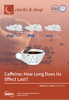
Caffeine is the most widely used stimulant on earth. It is generally assumed that consumption of a single dose of caffeine influences sleep only for the first 24 hours. This study aimed to record sleep and simultaneously perform electrophysiology in the lateral hypothalamus for at least 2 days after applying a single dose of caffeine. Recordings were performed in constant darkness as light and caffeine are known to interact in their effects on sleep and circadian rhythms. The data show that electroencephalogram theta (6–9 Hz) activity was increased for 48 hours, particularly in REM sleep. Neuronal activity in the lateral hypothalamus was increased in parallel. The data suggest that caffeine influences the brain longer than previously acknowledged, particularly under low light level conditions. View this paper
  • Issues are regarded as officially published after their release is announced to the table of contents alert mailing list.
  • You may sign up for e-mail alerts to receive table of contents of newly released issues.
  • PDF is the official format for papers published in both, html and pdf forms. To view the papers in pdf format, click on the "PDF Full-text" link, and use the free Adobe Reader to open them.
Order results
Result details
Section
Select all
Export citation of selected articles as:
21 pages, 277 KiB  
Conference Report
2021 Annual Meeting of the Swiss Society for Sleep Research, Sleep Medicine, and Chronobiology (SSSSC)
by Martin Hatzinger
Clocks & Sleep 2022, 4(2), 300-320; https://doi.org/10.3390/clockssleep4020026 - 07 Jun 2022
Viewed by 1954
Abstract
The 2021 meeting in Solothurn provided evidence-based education to advance the science and clinical practice of sleep medicine and sleep physiology, disseminates cutting-edge sleep and circadian research, promotes the translation of basic science into clinical practice, and fosters the future of the field [...] Read more.
The 2021 meeting in Solothurn provided evidence-based education to advance the science and clinical practice of sleep medicine and sleep physiology, disseminates cutting-edge sleep and circadian research, promotes the translation of basic science into clinical practice, and fosters the future of the field by allowing young clinicians and researchers to present their findings in talks and on posters [...] Full article
(This article belongs to the Section Human Basic Research & Neuroimaging)
13 pages, 1087 KiB  
Review
Neuroimaging in the Rare Sleep Disorder of Kleine–Levin Syndrome: A Systematic Review
by Juan Fernando Ortiz, Jennifer M. Argudo, Mario Yépez, Juan Andrés Moncayo, Hyder Tamton, Alex S. Aguirre, Ghanshyam Patel, Meghdeep Sen, Ayushi Mistry, Ray Yuen, Ahmed Eissa-Garces, Diego Ojeda and Samir Ruxmohan
Clocks & Sleep 2022, 4(2), 287-299; https://doi.org/10.3390/clockssleep4020025 - 31 May 2022
Cited by 3 | Viewed by 3383
Abstract
Kleine–Levin syndrome (KLS) is characterized by episodes of hypersomnia. Additionally, these patients can present with hyperphagia, hypersexuality, abnormal behavior, and cognitive dysfunction. Functional neuroimaging studies such as fMRI-BOLD, Positron Emission Tomography (PET) or SPECT help us understand the neuropathological bases of different disorders. [...] Read more.
Kleine–Levin syndrome (KLS) is characterized by episodes of hypersomnia. Additionally, these patients can present with hyperphagia, hypersexuality, abnormal behavior, and cognitive dysfunction. Functional neuroimaging studies such as fMRI-BOLD, Positron Emission Tomography (PET) or SPECT help us understand the neuropathological bases of different disorders. We conducted a systematic review to investigate the neuroimaging features of KLS patients and their clinical correlations. This systematic review was conducted by following the Meta-Analysis of Observational Studies in Epidemiology (MOOSE) and PRISMA protocol reporting guidelines. We aim to investigate the clinical correlation with neuroimaging among patients with KLS. We included only studies written in the English language in the last 20 years, conducted on humans; 10 studies were included. We excluded systematic reviews, metanalysis, and case reports. We found that there are changes in functional imaging studies during the symptomatic and asymptomatic periods as well as in between episodes in patients with K.L.S. The areas most reported as affected were the hypothalamic and thalamic regions, which showed hypoperfusion and, in a few cases, hyperperfusion; areas such as the frontal, parietal, occipital and the prefrontal cortex all showed alterations in cerebral perfusion. These changes in cerebral blood flow and regions vary according to the imaging (SPECT, PET SCAN, or fMRI) and the task performed while imaging was performed. We encountered conflicting data between studies. Hyper insomnia, the main feature of this disease during the symptomatic periods, was associated with decreased thalamic activity. Other features of K.L.S., such as apathy, hypersexuality, and depersonalization, were also correlated with functional imaging changes. There were also findings that correlated with working memory deficits seen in this stage during the asymptomatic periods. Hyperactivity of the thalamus and hypothalamus were the main features shown during the asymptomatic period. Additionally, functional imaging tends to improve with a longer course of the disease, which suggests that K.L.S. patients outgrow the disease. These findings should caution physicians when analyzing and correlating neuroimaging findings with the disease. Full article
(This article belongs to the Section Human Basic Research & Neuroimaging)
Show Figures

Figure 1

10 pages, 2230 KiB  
Article
Effect of Modified Yukmijihwang-Tang on Sleep Quality in the Rat
by SunYoung Lee, Hun-Soo Lee, Minsook Ye, Min-A Kim, Hwajung Kang, Sung Ja Rhie, Mi Young Lee, In Chul Jung, In-Cheol Kang and Insop Shim
Clocks & Sleep 2022, 4(2), 277-286; https://doi.org/10.3390/clockssleep4020024 - 27 May 2022
Viewed by 2220
Abstract
Many plants have been used in Korean medicine for treating insomnia. However, scientific evidence for their sedative activity has not been fully investigated. Thus, this study was carried out to investigate the sedative effects of the extracts of medicinal plants, including Yukmijihwang-tang and [...] Read more.
Many plants have been used in Korean medicine for treating insomnia. However, scientific evidence for their sedative activity has not been fully investigated. Thus, this study was carried out to investigate the sedative effects of the extracts of medicinal plants, including Yukmijihwang-tang and its various modified forms through the 5-HT2c receptor binding assay, and to further confirm its sleep-promoting effects and the underlying neural mechanism in rats utilizing electroencephalography (EEG) analysis. Enzyme-linked immunosorbent assay (ELISA) was used to measure serotonin (5-HT) in the brain. The water extracts of modified Yukmijihwang-tang (YmP) displayed binding affinity to the 5-HT2C receptor (IC50 value of 199.9 µg/mL). YmP (50 mg/kg) administration decreased wake time and increased REM and NREM sleep based on EEG data in rats. Additionally, treatment with YmP significantly increased the 5-HT level in the hypothalamus. In conclusion, the sedative effect of YmP can be attributed to the activation of the central serotonergic systems, as evidenced by the high affinity of binding of the 5-HT2C receptor and increased 5-HT levels in the brain of the rat. This study suggests that YmP can be a new material as a sleep inducer in natural products. Full article
(This article belongs to the Section Animal Basic Research)
Show Figures

Figure 1

17 pages, 2613 KiB  
Article
Long-Term Effect of a Single Dose of Caffeine on Sleep, the Sleep EEG and Neuronal Activity in the Peduncular Part of the Lateral Hypothalamus under Constant Dark Conditions
by Yumeng Wang and Tom Deboer
Clocks & Sleep 2022, 4(2), 260-276; https://doi.org/10.3390/clockssleep4020023 - 25 May 2022
Cited by 5 | Viewed by 3416
Abstract
Background: Caffeine is a central nervous system stimulant that influences both the sleep–wake cycle and the circadian clock and is known to influence neuronal activity in the lateral hypothalamus, an important area involved in sleep–wake regulation. Light is a strong zeitgeber and it [...] Read more.
Background: Caffeine is a central nervous system stimulant that influences both the sleep–wake cycle and the circadian clock and is known to influence neuronal activity in the lateral hypothalamus, an important area involved in sleep–wake regulation. Light is a strong zeitgeber and it is known to interact with the effect of caffeine on the sleep–wake cycle. We therefore wanted to investigate the long-term effects of a single dose of caffeine under constant dark conditions. Methods: We performed long-term (2 days) electroencephalogram (EEG)/electromyogram recordings combined with multi-unit neuronal activity recordings in the peduncular part of the lateral hypothalamus (PLH) under constant darkness in Brown Norway rats, and investigated the effect of a single caffeine treatment (15 mg/kg) or saline control given 1 h after the onset of the endogenous rest phase. Results: After a reduction in sleep and an increase in waking and activity in the first hours after administration, also on the second recording day after caffeine administration, rapid eye movement (REM) sleep was still reduced. Analysis of the EEG showed that power density in the theta range during waking and REM sleep was increased for at least two days. Neuronal activity in PLH was also increased for two days after the treatment, particularly during non-rapid eye movement sleep. Conclusion: Surprisingly, the data reveal long-term effects of a single dose of caffeine on vigilance states, EEG, and neuronal activity in the PLH. The absence of a light–dark cycle may have enabled the expression of these long-term changes. It therefore may be that caffeine, or its metabolites, have a stronger and longer lasting influence, particularly on the expression of REM sleep, than acknowledged until now. Full article
(This article belongs to the Section Animal Basic Research)
Show Figures

Figure 1

20 pages, 1027 KiB  
Review
Socioeconomic Position and Excessive Daytime Sleepiness: A Systematic Review of Social Epidemiological Studies
by Imene Bendaoud and Faustin Armel Etindele Sosso
Clocks & Sleep 2022, 4(2), 240-259; https://doi.org/10.3390/clockssleep4020022 - 16 May 2022
Cited by 7 | Viewed by 2877
Abstract
The objectives of this empirical study are to describe and discuss the current literature available on the relationship between excessive daytime sleepiness (EDS) and the socioeconomic position (SEP) as well as to provide recommendations for consideration of SEP in sleep medicine and biomedical [...] Read more.
The objectives of this empirical study are to describe and discuss the current literature available on the relationship between excessive daytime sleepiness (EDS) and the socioeconomic position (SEP) as well as to provide recommendations for consideration of SEP in sleep medicine and biomedical research. Databases Medline/PubMed, Web of Science, Google scholar and Scopus were screened from January 1990 to December 2020 using PRISMA guidelines and 20 articles were included in the final synthesis. Nineteen studies were cross-sectional and one study was longitudinal. Among these studies, 25.00% (n = 5) are focused on children and adolescent and the remaining 75.00% (n = 15) focused on adults and seniors. Ages ranged from 8 to 18 years old for children/adolescent and ranged from 18 to 102 years old for adults. Main SEP measures presented in these studies were education, income, perceived socioeconomic status and employment. The sample size in these studies varied from N = 90 participants to N = 33,865 participants. Overall, a lower educational level, a lower income and full-time employment were associated with EDS. Symptoms of EDS are prevalent in women, especially those with a low income or no job; and children and adolescents with difficult living conditions or working part time reported more sleep disturbances. SEP is already considered as an important determinant for many health outcomes, but if SEP is embedded in the experimental design in psychosomatic research, biomedical research and clinical practice as a constant variable regardless of outcome; it will move forward future investigations. Full article
(This article belongs to the Section Human Basic Research & Neuroimaging)
Show Figures

Figure 1

10 pages, 247 KiB  
Article
Combining Wake-Up-Back-to-Bed with Cognitive Induction Techniques: Does Earlier Sleep Interruption Reduce Lucid Dream Induction Rate?
by Daniel Erlacher, Vitus Furrer, Matthias Ineichen, John Braillard and Daniel Schmid
Clocks & Sleep 2022, 4(2), 230-239; https://doi.org/10.3390/clockssleep4020021 - 20 Apr 2022
Cited by 2 | Viewed by 4265
Abstract
Lucid dreaming offers the chance to investigate dreams from within a dream and by real-time dialogue between experimenters and dreamers during REM sleep. This state of consciousness opens a new experimental venue for dream research. However, laboratory study in this field is limited [...] Read more.
Lucid dreaming offers the chance to investigate dreams from within a dream and by real-time dialogue between experimenters and dreamers during REM sleep. This state of consciousness opens a new experimental venue for dream research. However, laboratory study in this field is limited due to the rarity of lucid dreamers. In a previous study, we were able to induce in 50% of the participants a lucid dream in a single sleep laboratory night by combining a wake-up-back-to-bed (WBTB) sleep routine and a mnemonic method (Mnemonic Induction of Lucid Dreams, MILD). In three experiments, we tried to replicate our earlier findings while we adapted our procedure in shortening (Exp1–3: 4.5 vs. 6 h of uninterrupted sleep in the first half of the night), simplifying (Exp2: time-based wakening vs. REM wakening in the second half of the night), and applying another induction technique (Exp3: reality testing vs. MILD). In the three conditions, four out of 15 (26%), zero out of 20 (0%), and three out of 15 (20%) participants reported a lucid dream. Compared to the original study, the earlier sleep interruption seems to reduce the lucid dream induction rate. Furthermore, without REM awakenings in the morning, lucid dream induction failed, whereas reality testing showed a lower success rate compared to MILD. Further systematic sleep laboratory studies are needed to develop reliable techniques for lucid dream research. Full article
(This article belongs to the Special Issue Dream and Sleep)
11 pages, 2461 KiB  
Systematic Review
Respiratory Muscle Training in Patients with Obstructive Sleep Apnoea: A Systematic Review and Meta-Analysis
by Rodrigo Torres-Castro, Lilian Solis-Navarro, Homero Puppo, Victoria Alcaraz-Serrano, Luis Vasconcello-Castillo, Jordi Vilaró and Roberto Vera-Uribe
Clocks & Sleep 2022, 4(2), 219-229; https://doi.org/10.3390/clockssleep4020020 - 11 Apr 2022
Cited by 4 | Viewed by 4244
Abstract
Background: Effective treatments for obstructive sleep apnoea (OSA) include positive pressure, weight loss, oral appliances, surgery, and exercise. Although the involvement of the respiratory muscles in OSA is evident, the effect of training them to improve clinical outcomes is not clear. We aimed [...] Read more.
Background: Effective treatments for obstructive sleep apnoea (OSA) include positive pressure, weight loss, oral appliances, surgery, and exercise. Although the involvement of the respiratory muscles in OSA is evident, the effect of training them to improve clinical outcomes is not clear. We aimed to determine the effects of respiratory muscle training in patients with OSA. Methods: A systematic review was conducted in seven databases. Studies that applied respiratory muscle training in OSA patients were reviewed. Two independent reviewers analysed the studies, extracted the data and assessed the quality of evidence. Results: Of the 405 reports returned by the initial search, eight articles reporting on 210 patients were included in the data synthesis. Seven included inspiratory muscle training (IMT), and one included expiratory muscle training (EMT). Regarding IMT, we found significant improvement in Epworth sleepiness scale in −4.45 points (95%CI −7.64 to −1.27 points, p = 0.006), in Pittsburgh sleep quality index of −2.79 points (95%CI −4.19 to −1.39 points, p < 0.0001), and maximum inspiratory pressure of −29.56 cmH2O (95%CI −53.14 to −5.98 cmH2O, p = 0.01). However, the apnoea/hypopnea index and physical capacity did not show changes. We did not perform a meta-analysis of EMT due to insufficient studies. Conclusion: IMT improves sleepiness, sleep quality and inspiratory strength in patients with OSA. Full article
(This article belongs to the Section Human Basic Research & Neuroimaging)
Show Figures

Figure 1

11 pages, 810 KiB  
Article
Direct Effects of Light on Sleep under Ultradian Light-Dark Cycles Depend on Circadian Time and Pulses Duration
by Fanny Fuchs, Ludivine Robin-Choteau, Laurence Hugueny, Dominique Ciocca and Patrice Bourgin
Clocks & Sleep 2022, 4(2), 208-218; https://doi.org/10.3390/clockssleep4020019 - 24 Mar 2022
Cited by 1 | Viewed by 2757
Abstract
Ultradian light–dark cycles in rodents are a precious tool to study the direct effects of repeated light exposures on sleep, in order to better understand the underlying mechanisms. This study aims to precisely evaluate the effects of light and dark exposures, according to [...] Read more.
Ultradian light–dark cycles in rodents are a precious tool to study the direct effects of repeated light exposures on sleep, in order to better understand the underlying mechanisms. This study aims to precisely evaluate the effects of light and dark exposures, according to circadian time, on sleep and waking distribution and quality, and to determine if these effects depend on the duration of light and dark pulses. To do this, mice were exposed to 24 h-long ultradian light–dark cycles with different durations of pulses: T2 cycle (1 h of light/1 h of dark) and T7 cycle (3.5 h of light/3.5 h of dark). Exposure to light not only promotes NREM and REM sleep and inhibits wake, but also drastically alters alertness and modifies sleep depth. These effects are modulated by circadian time, appearing especially during early subjective night, and their kinetics is highly dependent on the duration of pulses, suggesting that in the case of pulses of longer duration, the homeostatic process could overtake light direct influence for shaping sleep and waking distribution. Full article
(This article belongs to the Section Animal Basic Research)
Show Figures

Figure 1

Previous Issue
Next Issue
Back to TopTop