


Abstract
The mechanical interaction between the human body and foundation wear, which consists of normal load (pressure) and tangential load (shear), has a complicated influence on human physiological activities. In this study, five female subjects were asked to lay supine still, pressure loads of 20 mmHg and 40 mmHg and two levels of shear loads were exerted on the shank respectively, and the blood flow of skin undergoing loading was measured by using Advance Laser Blood Flowmeter. The results obtained were as follows: the low pressure could promote the skin blood flow; the shear load was effective in reducing the skin blood flow; little shear added to the pressure had important role on reducing the skin blood flow.
1. Introduction
As foundation wear helping to shape human figure for beauty becomes popular, the safety and comfort of such tight wear should be paid more attention in clothing design. The mechanical interaction between the human body and tight wear has a complicated influence on many human physiological activities. It has been reported that the mechanical force caused by tight garments can affect the heart beat, respiration, blood pressure, skin temperature, skin blood flow, and even autonomic nervous system both positively and negatively [1,2].
As the skin is the first layer under loads, the embedded microcirculation system must be affected. It is known that the mechanical force consists of normal load (pressure) and tangential load (shear). Much attention has been paid to that pressure could affect blood microcirculation in skin. It was reported that the capillaries could be squashed and skin blood flow (SBF) may decrease when the magnitude of pressure is higher than 35 mmHg [3]. The skin may ulcerate because of the SBF decrease and nutrient deficiency, if it is under pressure for a long time. Since the human body is soft and flexible, the shear exists and deforms the skin and soft tissue easily as women wearing foundation wear, and it also has great physiological effect on the skin. Early in 1958, Reichel proposed that shear is an important factor among other mechanical forces and its effect on soft tissue should not be ignored [4]. Zhang analyzed the mechanical mechanism of shear forces and found that the application of surface shear forces alters the internal stress distribution [5]. Some researchers investigated the etiological role of shear forces in decubitus ulcer. However, the effect of shear loads on skin microcirculation has seldom been reported, and the mechanism also needs to be discovered.
In this study, the change of skin blood flow was investigated when pressure, shear loads and the combined loads were applied on the lower limb respectively, and the mechanism of how the pressure and shear loads affect the skin blood flow is also discussed.
2. Methods
2.1. Subjects
Five healthy female undergraduates participated in this study. They were denoted as S1-S5 respectively. Their physical data (mean ± standard deviation) were as follows: age 20±1 yrs, height 163±3 cm, and weight 50±3 kg. The subjects had refrained from heavy exercise, salty food, alcohol and caffeine for 24 hours before measurement. To avoid the influence of climate change on SBF, the experiments were carried out in a climate chamber with constant temperature (20±2 °C), relative humidity (45±5%) and air velocity less than 0.1 m/s. The subjects were asked to rest for at least 30 minutes to reach a stable resting state before the tests began.
2.2. Loading Protocol
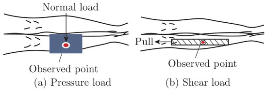
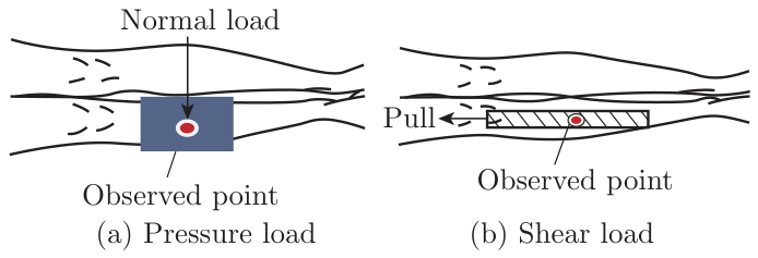
Two levels of pressure (20 mmHg and 40 mmHg) were exerted on the shank, with subjects lying still, by using the sphygmomanometer (Figure 1 (a)). The shear loads were produced by pulling upward the tape adhered to the front surface of the shank (Figure 1 (b)). The displacement of the tape top indicated the level of shear loads, and two levels, shear1 (6 mm) and shear2 (10 mm) were employed. Each level of load (pressure and shear) was exerted on and off for three times, and the interval was two minutes. The combined load pressure and shear was exerted: after a two-mins measurement of SBF under no load, the pressure was exerted on the shank for two minutes, and then the shear was added to the pressure load for the third two-mins. Pulling the tape under pressure was difficult, and the little shear (4 mm) was chosen, being noted as shear0. Shear0 was added to 20 mmHg and 40 mmHg respectively.
Figure 1.
Loading protocols.
2.3. Measurement of Skin Blood Flow
The skin blood flow under both no load and load was measured and it was observed by using Advance Laser Blood Flowmeter, ALF21R (Advance Co. Ltd., Japan). Three parameters of skin blood flow can be obtained simultaneously: “mass”, “velocity” and “flow”. “Mass” corresponds to the mass of blood in the measured area at a given time and can be obtained directly; “velocity” is the equivalent velocity of all the capillary flows within the measured skin region and can also be obtained directly; “flow” denotes the total blood volume of entering the skin soft tissue of 100 g per minute, which is expressed in terms of the product of the above two parameters. In this study, “mass” and “velocity” were analyzed, and a time constant of 0.1 s was selected for SBF signal sampling.
3. Results and Discussion
3.1. SBF Change under Pressure Loads
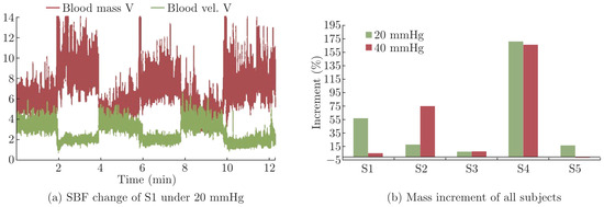
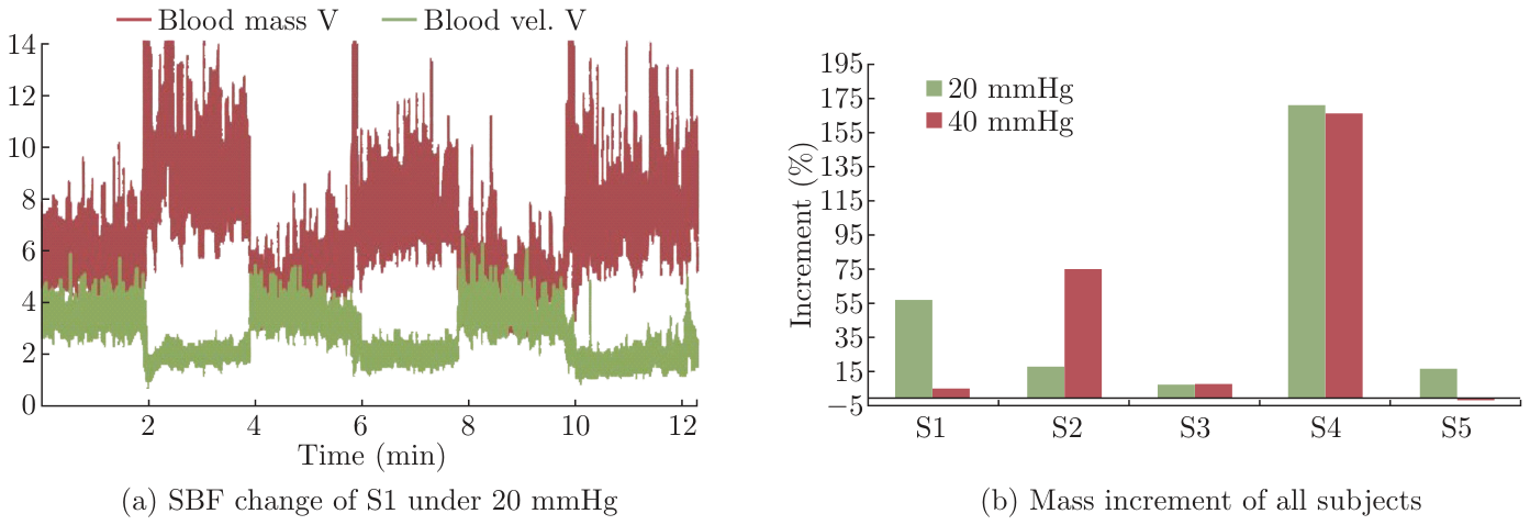
Figure 2 (a) shows that the SBF of S1 changed when 20 mmHg was exerted on and off: the blood mass increased and the velocity decreased. The SBF change was basically similar when the pressure was exerted for three times. The result was almost the same for all subjects under 20 mmHg and 40 mmHg.
Figure 2.
SBF change under pressures.
The mass increment under 20 mmHg and 40 mmHg was compared. The mean mass within each 2-mins interval was calculated, and those obtained from the first no load and pressure are used to calculate the relative mass increment. In Figure 2 (b), the blood mass increased under both pressures for all subjects. For S2, the mass increment climbed much when the pressure increased from 20 mmHg to 40 mmHg, while it decreased more or less for others. The similar results were found in the limbs about the skin blood flow: the SBF increased as lower pressure was exerted, and the increment rose or declined under 40 mmHg, if the pressure continues to increase, it could cause the blood mass to decrease [6]. Johansson et al. suggested that the skin blood flow in the hand decreased 50% when the pressure of 15∼33 kPa was exerted and it decreased continually as pressure increases [7]. It could be confirmed that the lower pressure could promote the skin blood flow from the above results.
Microcirculation system usually consists of arterioles, metarterioles, precapillary sphincters, capillaries, arteriovenous shunting, venula and other veins. The arterioles are wound by some smooth muscles and it regulates the blood flow depending on the contraction and relaxation of the smooth muscles. There are also some smooth muscles which are called precapillary sphincter winding through the metarterioles, and they play an important role on controlling the blood flow into the capillary. To study the mechanism of microcirculation regulation, Bai et al. put forward the concept of capillary unit and the model that the capillaries open or close in turn randomly [8]. A local capillary network is called a capillary unit (CU). It is assumed that the number of CU is “n” in some area. For a CU, its opentime and closetime change randomly, and the averages for the n-CUs are supposed to “µo” and “µc”. So the open-close frequency of capillaries (f = 1/(µo + µc)) and open rate (α = µo/(µo + µc)) could be gained.
The open and close of a CU is regarded to be regulated by the concentration of oxygen and metabolic waste products, respectively. When the shank wounded by the sphygmomanometer were compressed, the blood vessels embedded in the soft tissue were also squeezed and the resistance of blood flow increased. As the result shows, the blood velocity under pressure decreased, which hindered the transportation of the oxygen. In order to obtain the required oxygen concentration to maintain the body function, the “µo” needs to be prolonged, whereas the close time, “µc” mainly affected by the metabolism would not change. Thus it could be deduced that the open-close frequency “f” would decrease and the open rate “α” increase. That is to say, the total blood mass to capillaries at a time would also increase. This model exactly explains why “mass” increased with pressure. Brienza analyzed the skin blood flow signals at the sacrum under pressure by using wavelet analysis and found that the myogenic activity became active [9]. The same result was also found at a part of the sacral [10]. Through the frequency spectrum, it was found that the skin blood flow was influenced by these physiological activities: endothelial activity, neurogenic activity, myogenic activity, respiratory activity and heart activity. The frequency band of intrinsic myogenic activity is the interval of 0.052 Hz to 0.145 Hz, which just spans the frequency of the microvascular smooth muscle activity (0.067-0.167 Hz) [11].
3.2. SBF Change under Shear Loads
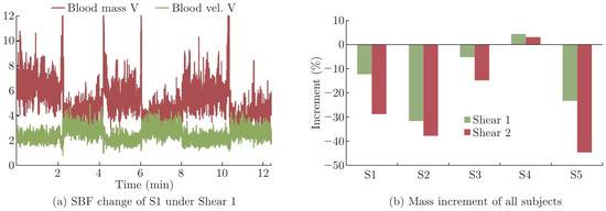
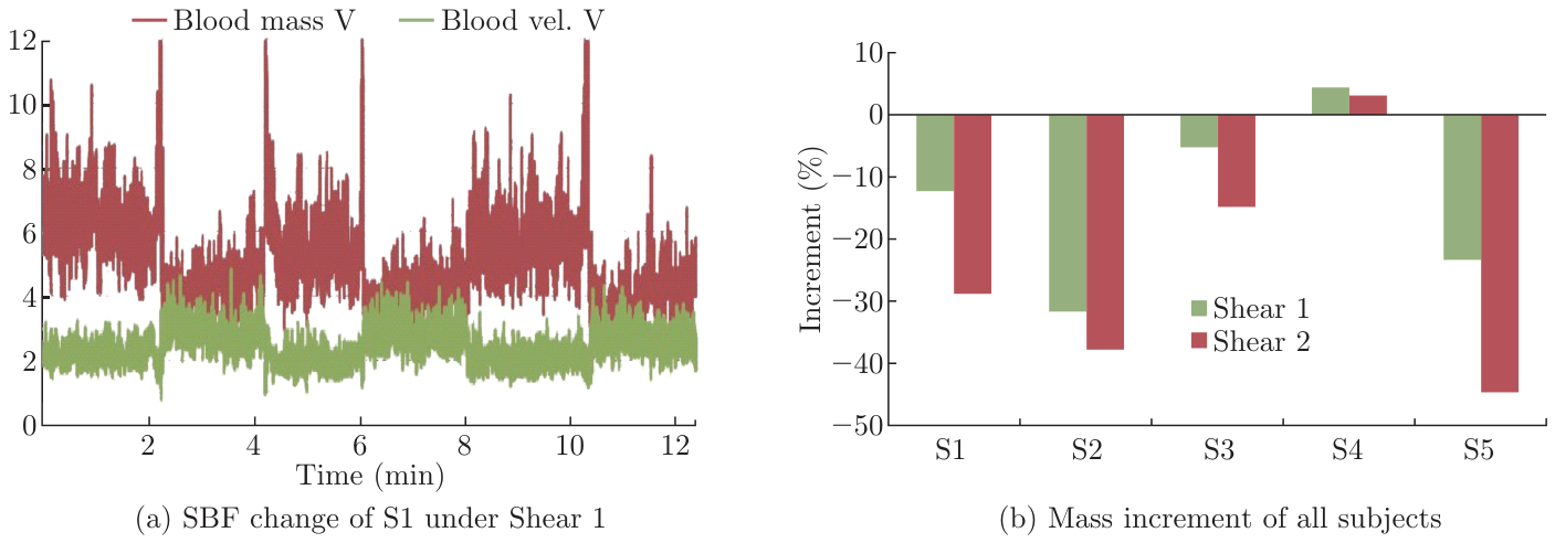
Figure 3 (a) shows that SBF changed when the shear load was exerted on and off: the blood mass decreased and the blood velocity increased. The SBF change was basically similar when the shear load was exerted for three times. The result was almost the same for all subjects under shear 1 2. The SBF changes under shears were different from those under pressures. The reason why the blood velocity increased under shear load is still unclear and remains to be further investigated. As shown in Figure 3 (b), for S4, the blood mass increased under both shears, it is consistent that the mass decreased more under shear2 than shear1 for others. It was confirmed that the shear load was effective in promoting the skin blood flow occlusion.
Figure 3.
SBF change under Shear loads.
Blood velocity increase means that the required blood perfusion and concentration of oxygen could be finished in short time. This also means that the “µo” would be cut down, whereas the close time “µc” is mainly affected by metabolism and would not change. Thus it could be deduced that the open-close frequency, “f” would increase and the open rate “α” decrease which means that the total blood mass to capillaries at a given time decreases. Therefore, this regulation explains the decrease of skin blood mass at a time under shear loads.
3.3. SBF Change under Combined Loads
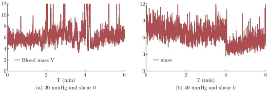
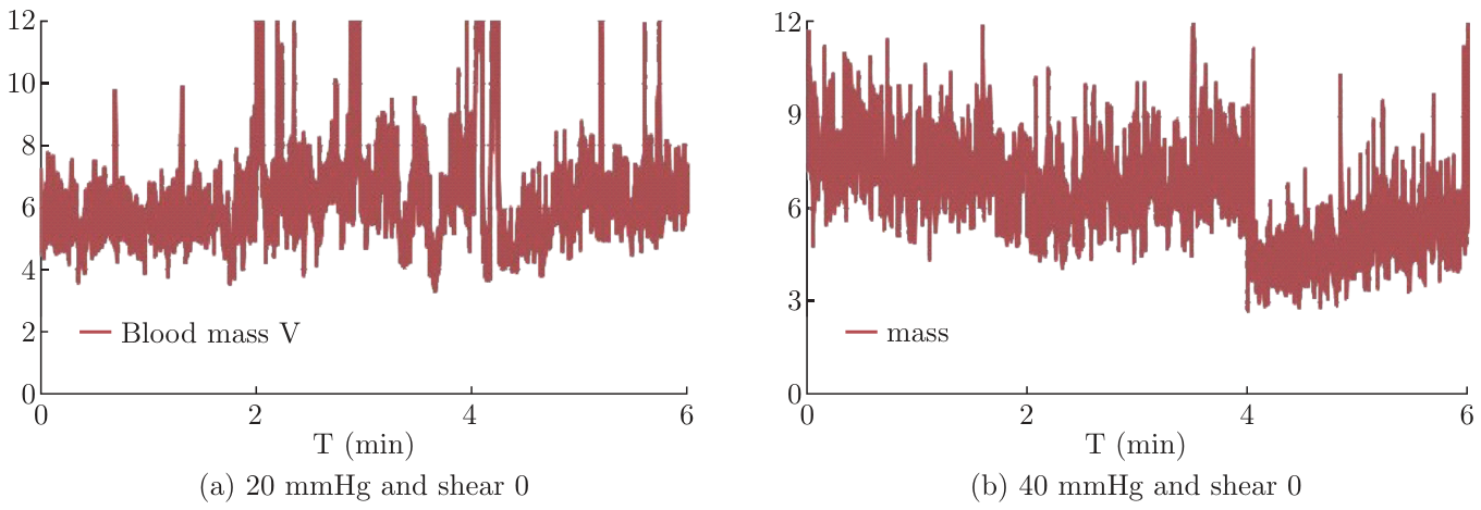
SBF changed when Shear0 was added to 20 mmHg and 40 mmHg respectively. As Figure 4 (a) shows, the blood mass of S1 increased a lot under 20 mmHg, and then the increment dropped when shear0 was added. In Figure 4 (b), the blood mass changed a little under 40 mmHg, and it decreased much when shear0 was added.
Figure 4.
Blood mass of S1 under combined loads.
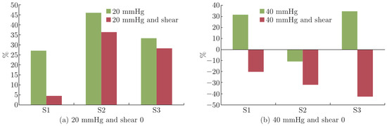
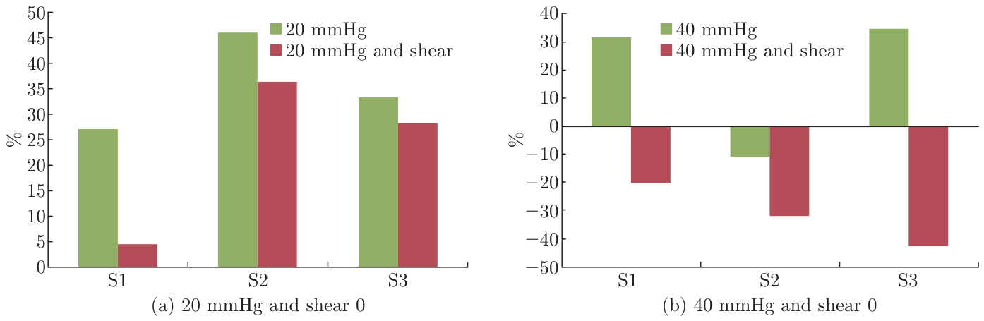
In Figure 5, the blood mass increments under pressure and combined loads were compared. Under 20 mmHg, the blood mass increased, while the increment dropped when the shear0 was added to the pressure (Figure 5 (a)). By comparison, under 40 mmHg, the blood mass increased around 30% for S1 and 2, it turned to decrease and the decrement was up to more than 30% as the shear load was added to the pressure (Figure 5 (b)). It was confirmed that the little shear load added to the pressure had a critical role on the decrement of blood mass. Katakura et al. applied both pressure and shear on the rabbits, and the skin blood flow decreased at a rate of 1.0 ml/min/100 g [12]. Goossen defined cut-off pressure as the level of external pressure exerted on skin when the skin oxygen tension is 1-3 kPa, at which the ischaemia of skin can be expected. They found that the cut-off pressure decreased much when the little shear was added [13]. The combined load is common as mechanical action and its effect on skin blood flow should be given much attention.
Figure 5.
Mass increments under combined loads.
4. Conclusion
In this study, the effects of pressure and shear on skin blood flow were investigated. And the mechanism was deduced based on the open-close model of capillaries. The findings were as follows: the blood mass increased under low pressure, and the velocity decreased; the blood mass decreased under shear loads, and the velocity increased; as little shear was added to the lower pressure, the blood mass increment dropped, however the combined loads of little shear and higher pressure caused much mass decrement; both pressure and shear loads changed the frequency of capillaries open-close, and thus the capillary opening rate, consequently changed the blood mass in the capillaries.
Acknowledgments
A project funded by the Priority Academic Program Development of Jiangsu higher education institutions (PAPD). This work was also partially funded through a research project entitled “Research and development of high performance foundation wear”, granted by the National Engineering Laboratory for Modern Silk, Soochow University. The authors are also grateful to all the volunteered subjects participated in this study.
References
- Sugita, A.; Okabe, K.; Kioka, E. Effect of girdles on comfort of aged women. J. Jpn. Res. Assoc. Text. End.-Uses 2002, 43, 365–376. [Google Scholar]
- Yokoi, R.; Yoshida, M.; Sasakawa, E.; Hirata, K. Effects of clothing pressure in wearing girdles on physiological functions. J. Jpn. Res. Assoc. Text. End.-Uses 2006, 47, 537–547. [Google Scholar]
- McGeown, J.G. Master Med. Physiol. (third edition); 2007; pp. 76–81. [Google Scholar]
- Reichel, S.M. Shearing forces as a factor in decubitus ulcers in paraplegics. J. Am. Med. Assoc. 1958, 166(7), 762–763. [Google Scholar] [CrossRef] [Scilit]
- Zhang, M.; Roberts, V.C. The effect of shear forces externally applied to skin surface on underlying tissues. J. Biomed. Eng. 1993, 15, 451–456. [Google Scholar] [CrossRef] [Scilit]
- Lu, Y.H.; Dai, X.Q.; Bai, L. Effect of garment pressure on peripheral skin blood flow. J. Donghua Univ. 2011, 28, 466–470. [Google Scholar]
- Johansson, L.; Hagg, G.M.; Fischer, T. Skin blood flow in the human hand in relation to applied pressure. Eur. J. Appl. Physiol. 2002, 86, 394–400. [Google Scholar] [CrossRef] [Scilit] [PubMed]
- Bai, L.; Bai, K. Mechanism of metabolic regulation in the microcirculation and its simulation. Front. Microcirc. Control Process. Clin. Appl. 2009, 22–23. [Google Scholar]
- Brienza, D.M.; Geyer, M.J.; Jan, Y.K. A comparison of changes in rhythms of sacral skin blood flow in response to heating and indentation. Arch. Phys. Med. Rehabil. 2005, 86, 1245–1251. [Google Scholar] [CrossRef] [Scilit]
- Jan, Y.K.; Blienza, D.M.; Geyer, M.J.; et al. Wavelet-Based spectrum analysis of sacral skin blood flow response to alternating pressure. Arch. Phys. Med. Rehabilifation 2008, 89(1), 137–145. [Google Scholar] [CrossRef] [Scilit]
- Golenhofen, K. Slow rhythms in smooth muscle. In Smooth Muscle; Buhlbnng, E., Brading, A.F., Jones, A.W., et al., Eds.; Edward Amold Ltd: London, 1970; pp. 316–342. [Google Scholar]
- Katakura, K.; Takeda, T.; Ishida Oyama, N. Jpn. J. Press. Ulcers ( L4144A) 2006, 8(4), 572–578.
- Goossens, R.H.M.; Zegers, R.; Hoek van Dijke, G.A.; Snijders, C.J. Influence of shear on skin oxygen tension. Physiology 1994, 14(1), 111–118. [Google Scholar] [CrossRef] [Scilit]
© 2012 Textile Bioengineering and Informatics Society. This article is an open access article distributed under the terms and conditions of the Creative Commons Attribution (CC BY) license (https://creativecommons.org/licenses/by/4.0/).