


Abstract
The human skin is the largest organ which protects the body from disease and physical damage. When the skin has been seriously damaged through disease or burns, the body cannot act fast enough to produce necessary replacement cells. Artificial skin grafts were developed as a way to prevent such consequences. This work is related to the design of advanced dermal scaffolds (non-woven fibrous mats) to provide multifunctional properties. Defined target properties are attained by distributing specified base materials at multiple length scales ranging from several nanometers to millimeters. Tissue scaffolds were developed using the electrospinning process, which creates a non-woven fibrous construction of high permeability and proper mechanical integrity similar to the scale of the extra-cellular matrix of cells.
1. Introduction
The problem of slow replacement cells production on a seriously damaged human skin, through diseases or high level burns, results death of the burnt victim, due to infection and loss of plasma. To overcome the problem, artificial dermal scaffolds (non-woven fibrous mats) are being developed, providing functional material concepts with enhanced properties [1].
The primary requirements for a scaffold are temporary mechanical support and tissue regeneration through cells and proteins [1]. The complex network of structural and functional proteins in the fibrous matrix of the ECM (extra-cellular matrix cells) is a major challenge in the dermal scaffold design.
Biocompatibility and biodegradability of the materials, chemo-mechanical properties and morphological characteristics at different length scales are the main demands for the cell-scaffold interface [2].
The process technique implemented nowadays for the production of the forth-mentioned fibrous mats is the electrospinning process. It is particularly appealing for fabricating large sheets of nanoscale fibrous scaffolds suitable for dermal implantation.
Recent studies have reported a multiscale 3D scaffold design via multimodal electrospinning (Soliman S. et all). The study concerns a modified electrospinning process (implementing two parallel syringes) used for the production of multimodal MIX scaffold (comprising nano and microfibers) that shows important improvements over the unimodal scaffolds in terms of mechanical and biological performance.
Another study has reported on the computer simulating approach of a cell growth inside the scaffolds. The cell activity during the tissue regeneration has been simulated by a lattice incorporated within a finite element model. Such developed mechano-biological model is reported to be a good prediction tool for the optimal design parameters for scaffolds. The investigations suggested experimental analysis as such that cells exposure is within optimal mechanical simulation and cell seeding optimal for vascular ingrowth’s [3,4].
An important issue investigated is the cells motility in a (e.g. damaged skin) during their growth (tissue regeneration). Multiscale models have reported on the cell and tissue dynamics. Single cell model simulates the deformations of the cell itself and the substrate. Mechanical principle incorporated in a single cell model is essential for a model simulation of cells interactions and tissues. With this model different traction patterns of the cells can be measured. Further numerical investigation is needed in order to represent a detailed cell-substrate dynamics [5].
When modeling biological systems, one should be able to produce different mathematical models at different scales and provide a link between each model to get a complete description of the system.
Molecular level is the fundamental scale for understanding or predicting the structure of a molecule. The stochastic changes of particle numbers in the volume elements representing intracellular space become possible to simulate through generalizations of Gillespie’s algorithm that include molecular diffusion events in addition to chemical reactions. The cell-cell contact, stochastic diffusion trajectories molecular particles and cellular membrane may be simulated. Models of cell state transitions and their consequences are formulated in terms of finite state automata, the cellular automata treat the single ‘cell’ as agents that can carry their states with them as they move on the grid space. There are some simulation examples of multicellular systems such as the adaptive immune system, populations and migrating cells, organ level phenomena. The aim of this paper is to examine the range of elastic and permeability properties using electrospinning.
.2. Initial work
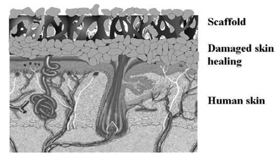
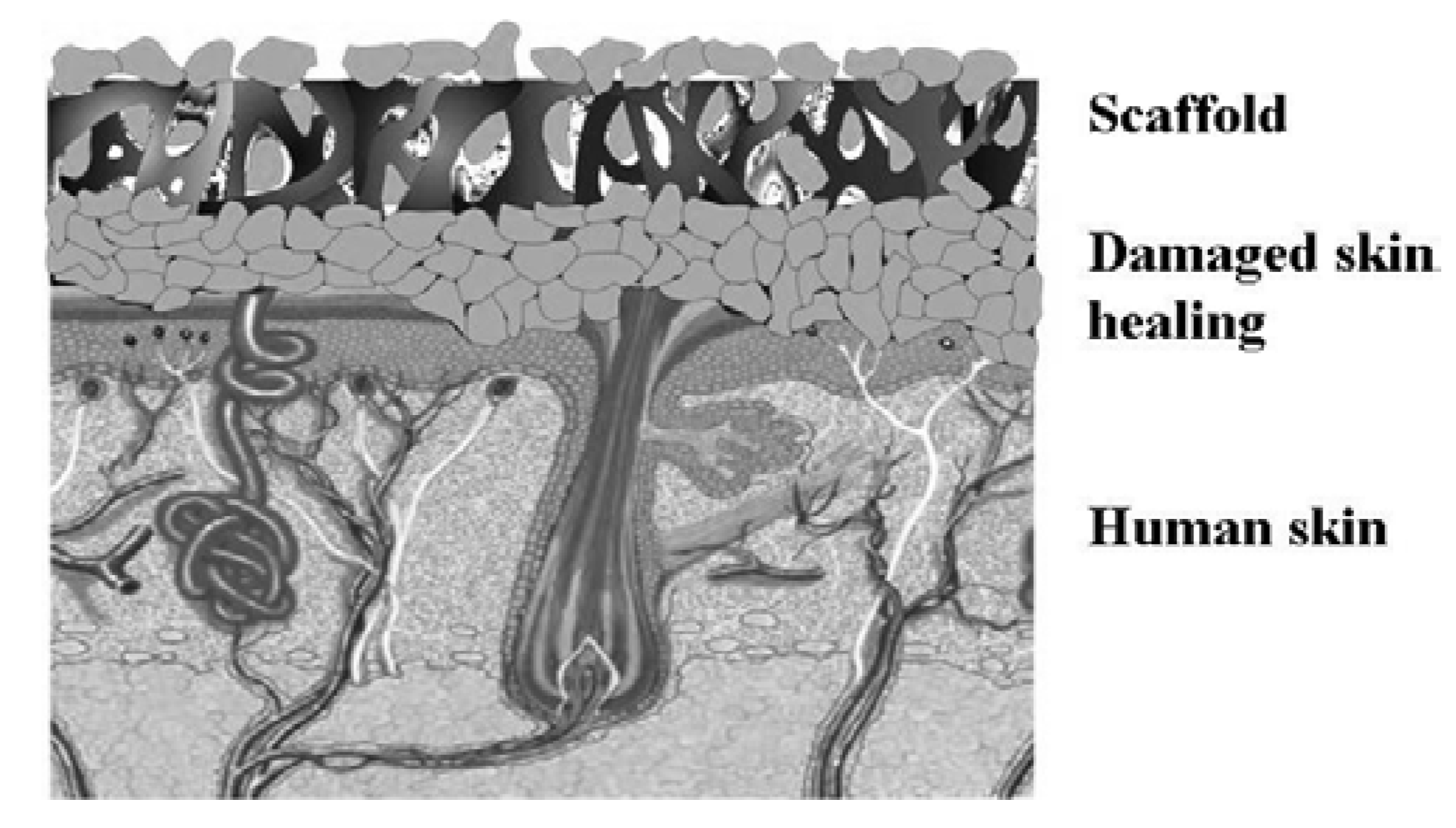
Tissue scaffolds were developed using the electrospinning process, which creates a non-woven fibrous construction of high permeability and proper mechanical integrity similar to the scale of the extracellular matrix of cells. Dramatically increased specific surface, excellent mechanical strength, and highly open porous structures are the unique properties of the electrospun fibers non-woven fabrics. Figure 1 illustrates dermal scaffold used for tissue regeneration of damaged human skin.
Figure 1.
Dermal scaffold for the acceleration of the cell growth on damaged skin.
The principle behind the electrospinning process is in the usage of electricity to move fluids (e.g. polymer solutions). A charged jet is ejected beyond a critical value of the electrical field intensity, at which the repulsive electrical forces overcome the surface tension of the polymer solution (or melt) droplet at the tip of the nozzle. Traveling towards the grounded collector the jet either cools down (in case of the melt) or the solvent evaporates (in case of the solution) to obtain ultrafine fibers in the form of a non-woven nanofabric. The non-woven fibrous structure resembles an intricate forest of overlaid fibers [2].
A number of nanofiber assemblies have been developed using electrospinning by incorporating functional agents to achieve antibacterial and magnetic, properties. The morphology of the electrospun fibers depends on a number of factors, such as solution properties (e.g., concentration, viscosity, conductivity, surface tension, etc.), processing conditions (e.g., electrical potential, collection distance, etc.), and ambient conditions (e.g., temperature, humidity, etc.).
2.1. Optimal scaffold design
Tissue engineering scaffolds must serve three primary purposes: they must define a space that will shape the regenerating tissue, they must provide temporary function in a defect while tissue regenerates and they must facilitate ingrowth of tissue and possibly allow inclusion of seeded cells, proteins and/or genes to accelerate tissue regeneration.
To describe how the load-bearing tissue grows within the scaffold pores, the remodeling rules established in nanofibre surface are considered applicable [7].
Therefore, cost function is often formulated in terms of the first invariants of permeability tensors. The constraints come as desired effective elasticity constants that are met within a specified tolerance and a porosity constraint.
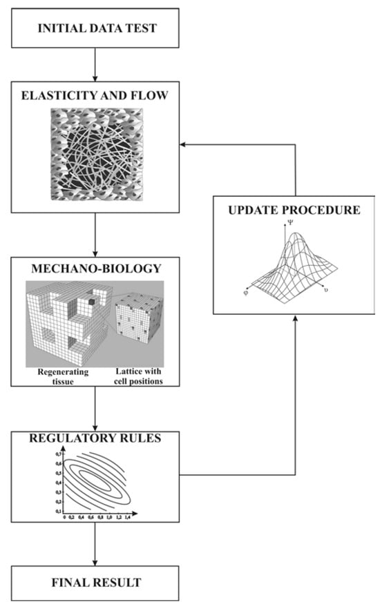
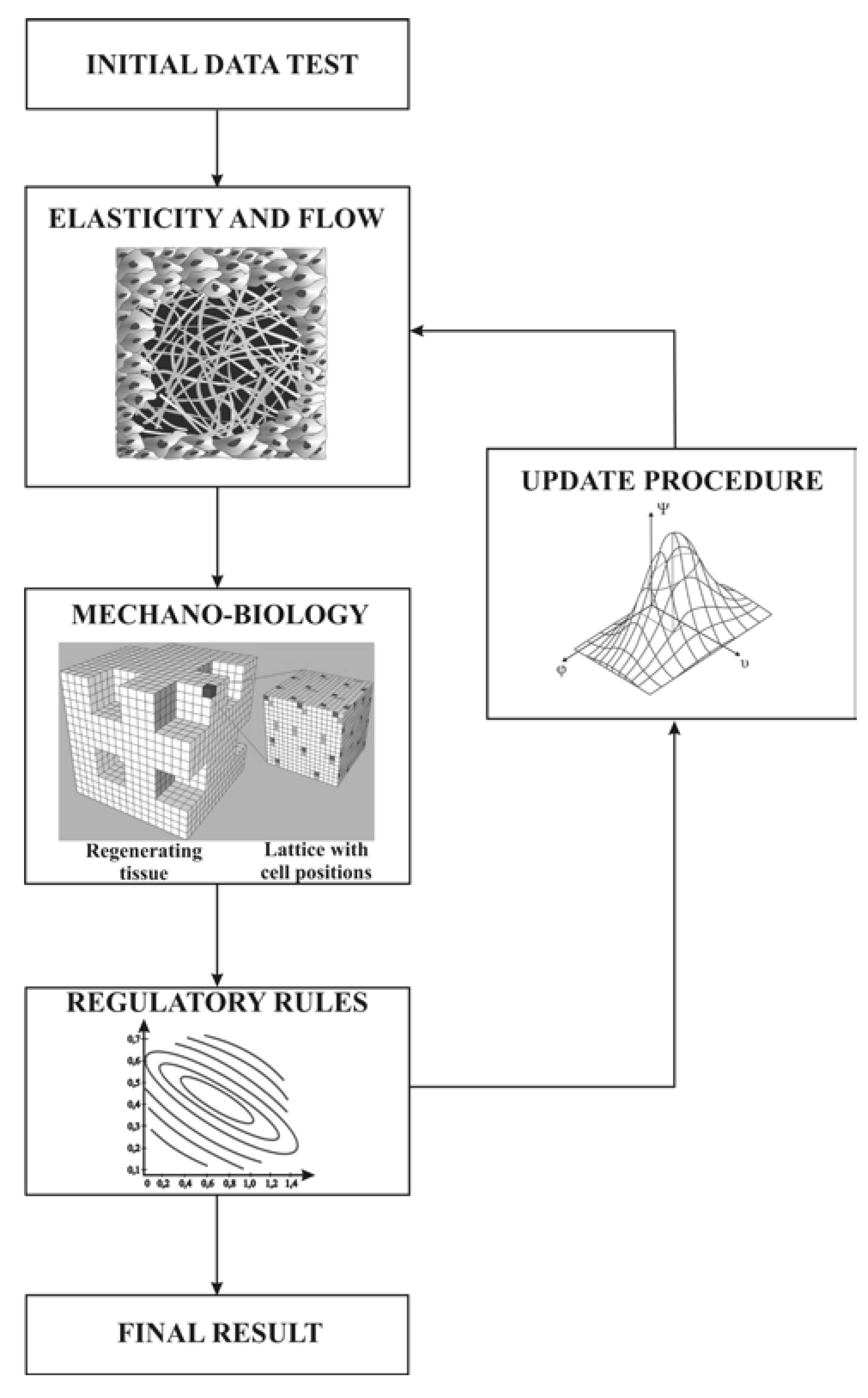
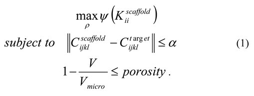 where Kiiscaffold denotes the first invariant term of the effective permeability tensor, Cijklscaffold is the effective elasticity tensor for the scaffold, Cijkltarget is the target value of the effective elasticity tensor, Vmicro denotes the volume of the unit cell architecture, V denotes the volume of the solid material, and α is a tolerance term that is gradually reduced during the course of iteration until convergence is achieved. Eq. 1 is a multiphysics optimization problem and is solved using the combination of a Method of Moving Asymptotes [8,9] to update elasticity, and an evolutionary structural optimization scheme to update the density distribution for permeability (Figure 2).
Figure 2.
Optimization scheme for the elasticity and density distribution to update permeability.
In this study, we focus on the biodegradable polymers whose degradation mechanism is assumed to be bulk erosion due to hydrolysis. The mechanical and transport properties such as stiffness and permeability depend on the molecular weight and crystallinity among others.
3. Materials and fibrous structure production
Poly (ε-caprolactone) (PCL) (Sigma–Aldrich) was spun into nanofiber mats from a polymer solution of 2 g of PCL and 14 ml of THF/DMF (1:1) solvents. Homogeneous solution is obtained by stirring the solution until the polymer is dissolved. Electrospun PCL fibers were produced using a 1 ml/h solution flow rate, a 21 kV potential and 18 cm distance between the needle tip and the collector. The ambient conditions were temperature T=16.8°C and a relative humidity of 27%. Heparin dissolved in water was added in one part of the PCL polymer solution and nanofibrous mats were electrospun as well. Later on the fiber distribution depending on heparin concentration was determined. The experimental data of the fiber diameter distribution concerning a fibrous structure depended on the processing parameters such as solution concentration c and applied electric field E and an additional microstructural parameter such as fiber density.
3.1. Results and discussion
With a multimodal fiber distribution principle a multiscale architecture can be constructed with a desired pore structure. Compounding nano- and microfibers in one, 3D multimodal scaffold should in principle allow different cell types to coexist, accelerating tissue growing phenomena.
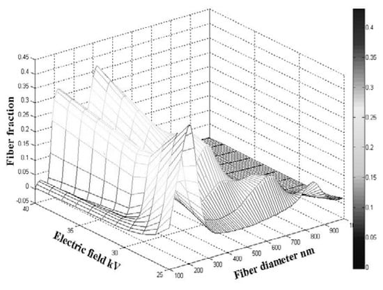
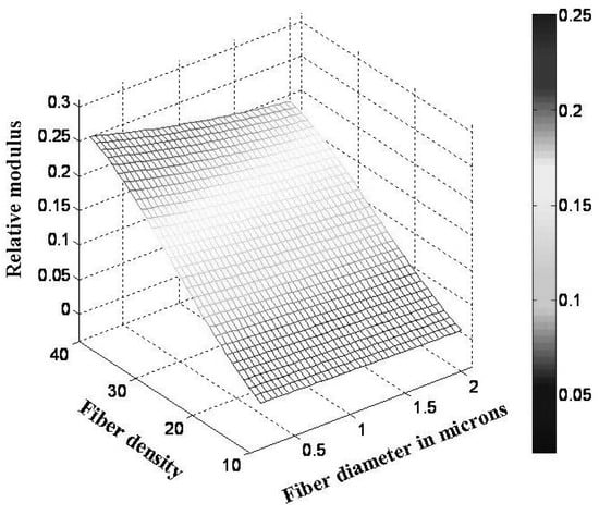
Figure 3 shows the fiber fraction depending on the fiber diameter and applied electric field. With the variation of the electric field voltage within different fiber diameters ranges the fiber fraction shows variation as well, with certain behavior repetitiveness.
Figure 3.
Fiber fraction dependence on the fiber diameter and the electric field.
At a certain fiber diameter, at the highest level of electric field voltage the fiber fraction gives its maximum, before and later, within fiber diameter range values, it increases and decreases respectively. The peaks of the fiber fraction within smaller diameters (and range) decreases with decreasing electric field until it reaches the value of around 33 kV, after which it increases to the same peak value at an electric field voltage of 30 kV.
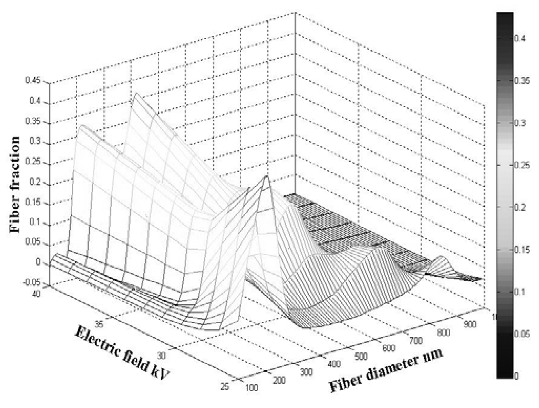
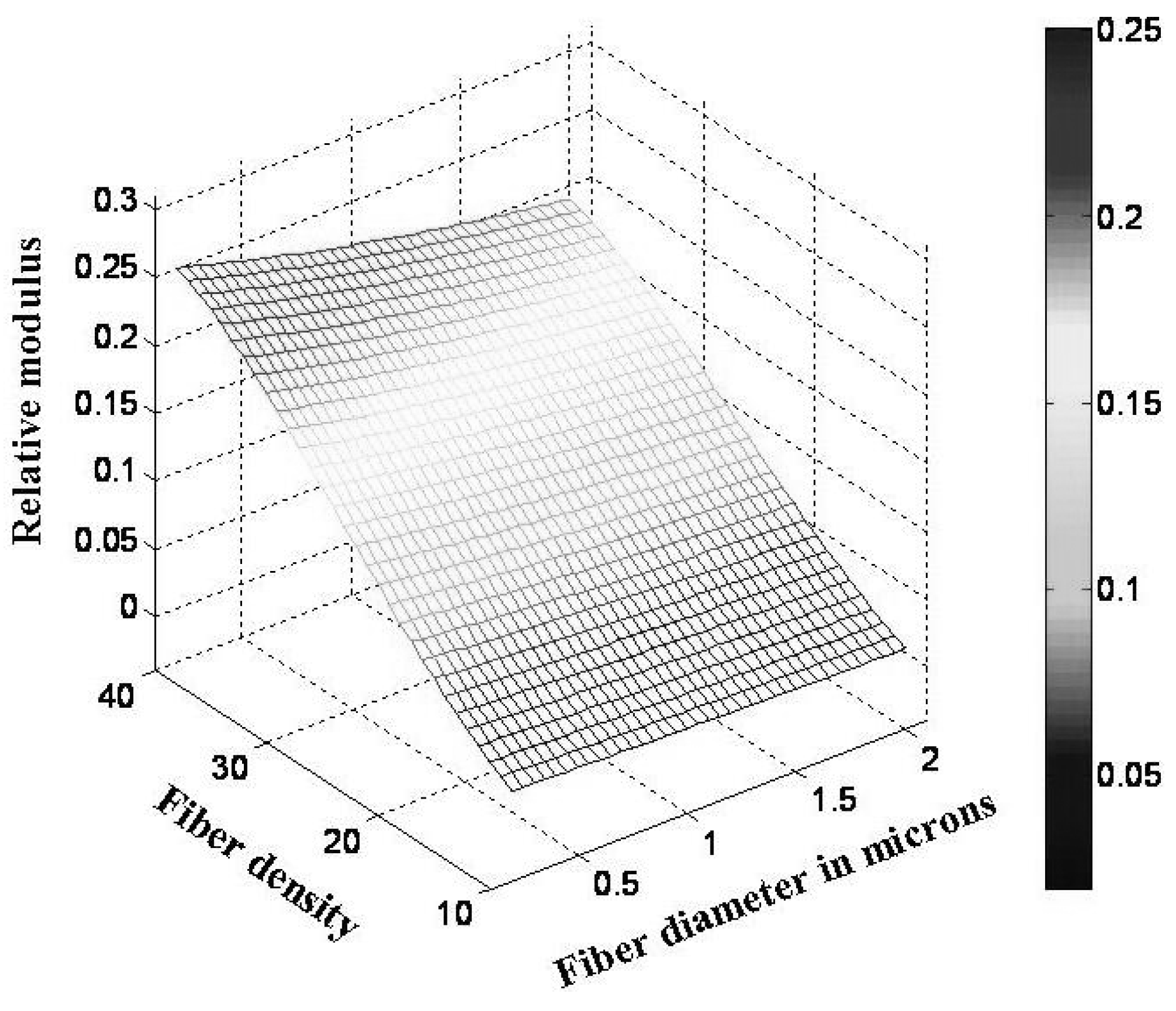
Within the second wider fiber diameter range or at higher fiber diameters the peak repetitiveness is bigger and shows decreasing maximal fiber fractions unlike the initial peak value at the highest electrical field voltage. The minimal value of the 3D surface is at the highest fiber diameter and applied electric field voltage. The modulus dependence on fiber diameter and density is given in Figure 4. The finest fibers have the highest densities and the highest modulus or small fiber diameters together with increasing fiber density give higher modulus. The plotted surface contours show the optimal parameter dependence.
Figure 4.
Relation between fiber density and diameter.
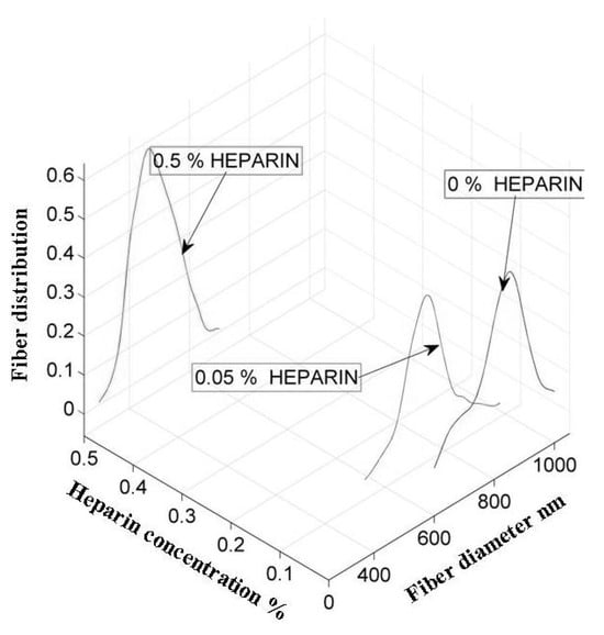
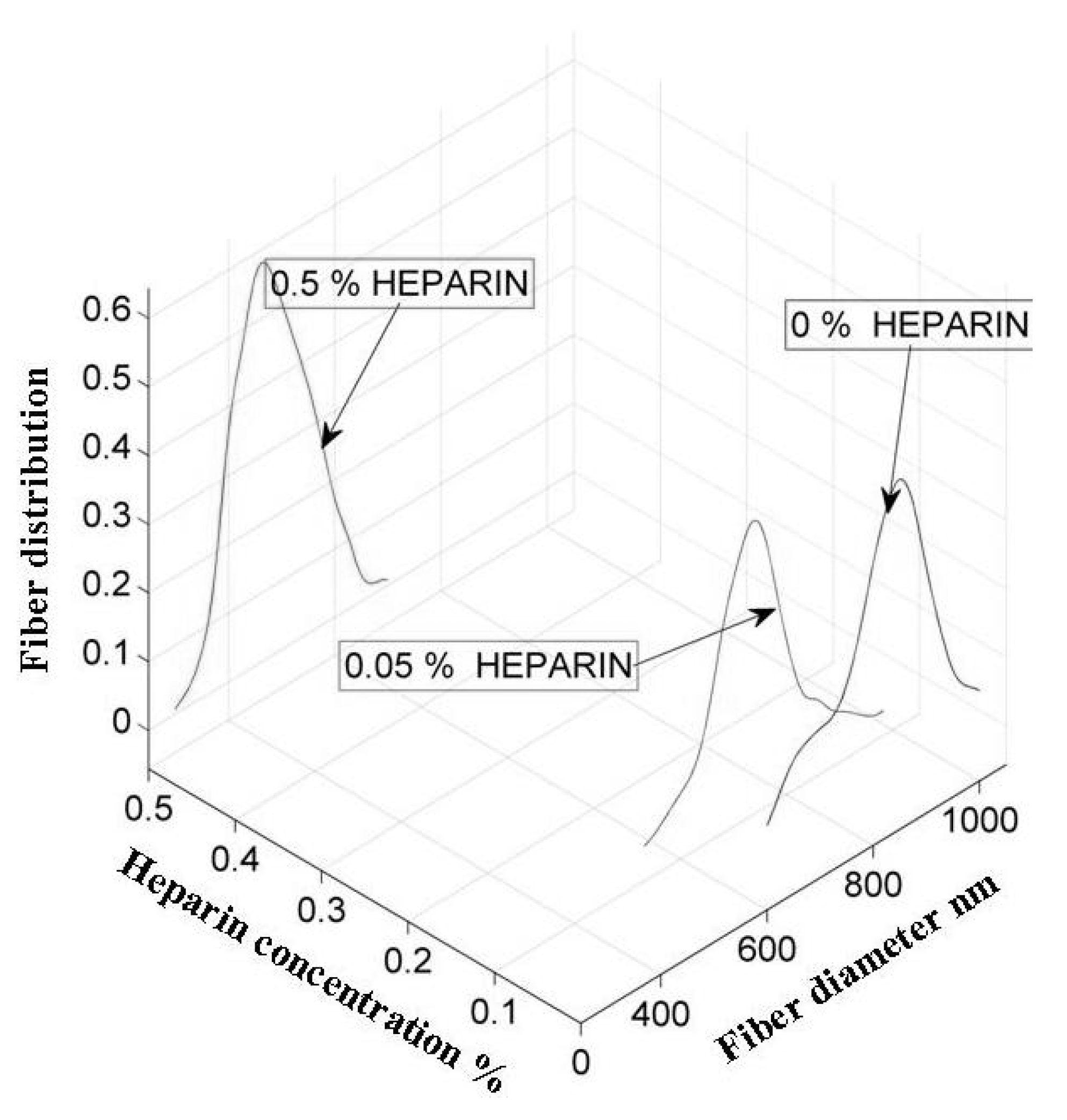
Figure 5 shows the fiber bimodal distribution dependence on the different heparin concentrations and the range of the fiber diameter. The behavior of the fiber distribution resulted in a bimodal normal distribution function with its mean values dependent on heparin concentration at a certain diameter. Before the mean value is achieved the fiber distribution increases with the fiber diameter at constant heparin concentration, as the fiber diameter continues to increase the fiber distribution decreases until it reaches the initial zero point. The standard deviation range is narrower at higher heparin concentrations but the fibers are finer. The overlapping of the fiber diameter type begins at the decreasing zone of the fiber distribution and it increases at lower heparin concentrations.
Figure 5.
Relation between fiber distribution and heparin concentration as well as fiber diameter.
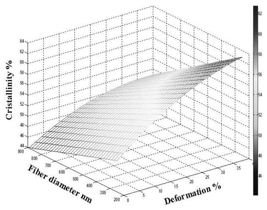
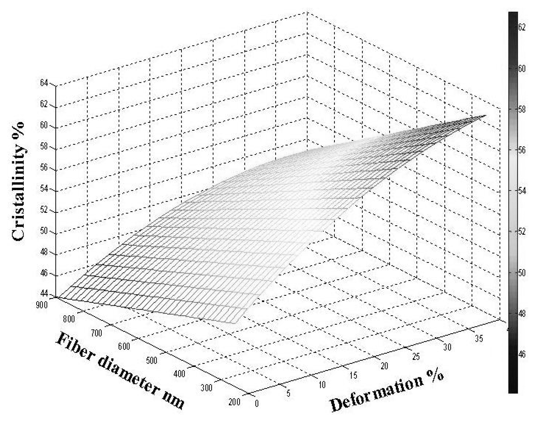
Relation between crystallinity and fiber diameter & deformation is plotted in Figure 6. The fiber diameter reduction and an increase of percentage deformation results in crystallinity increase in a continuous range.
Figure 6.
Relation between crystallinity and fiber diameter & deformation.
The minimal point of the plotted surface concerns the lowest level of cristallinity percentage at the highest fiber diameter with no deformation. The surface contour plots outline the surface interpolation of experimental data on parameters dependence (fiber diameter and deformation). The influence of the crystalline morphology and related molecular structure on properties can be studied in the future work. A PCL is a semicrystaline polymer, and its final properties, such as biodegradability and memory effect, depend to a great extent on the crystallinity, which depends on crystallization. The crystallization kinetics of PCL is of practical importance.
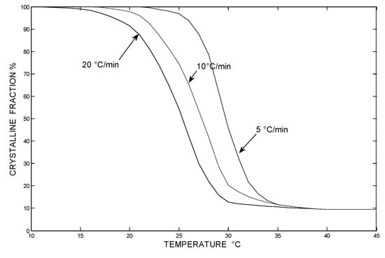
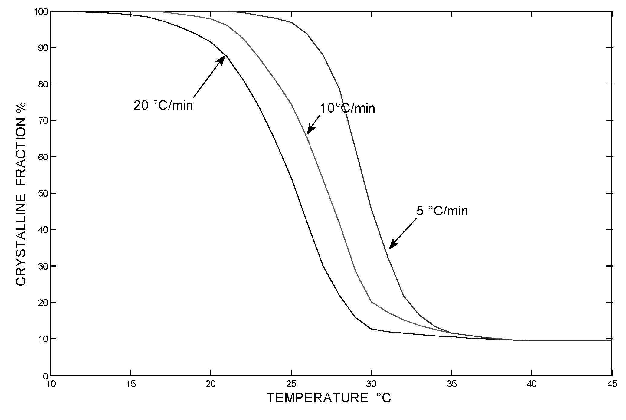
The relationship between crystalline fraction and temperature can be determined from DSC curves and cooling rates. Figure 7 shows that the total crystallization decreases with increasing cooling rate. As the cooling rate decreases, the temperature range of the crystallization becomes narrower.
Figure 7.
Relation between crystalline reactivity and temperature for different cooling rates.
Allowing larger over-cooling degree, same crystallinity can be reached at higher cooling rates [10].
4. Conclusion
The 3D multimodal scaffold comprising of micro and nanofibers will allow different cell types to coexist, which is important for the tissue regeneration enhancement. With the optimal electrospun fibrous scaffold production relationship between different parameters was revealed. The fiber fraction shows opposite behavior before and after its maximum value. It revealed increasing and decreasing character at the highest level of electrical field voltage within a certain diameter range.
An increasing modulus was the result of smaller fiber diameters with increasing fiber densities.
Bimodal normal fiber distribution resulted as a result of the different fiber and heparin concentrations. The fiber distribution increased with the fiber diameter at constant heparin concentration. Decreasing fiber distribution followed the mean value of the normal distribution function with the simultaneous increase of fiber diameter. The continuous rise of crystallinity resulted from the fiber diameter and percentage deformation decreased and increased, respectively.
The experimental results and related mathematical models reveal that the elastic and transport properties of fibrous meshes are sensitive to the microstructural architecture. The processing parameters influence process stability on one hand and microstructure parameter space on the other. Future work will focus on the use of scaffolds in functional tissue engineering scenarios, with the application of tissue growth phenomena.
References
- Metcalfe, A.D.; Ferguson, M.W.J. Bioengineering skin using mechanism of regeneration and repair. Biomaterials 2007, 28, 5100–5113. [Google Scholar] [CrossRef] [Scilit] [PubMed]
- Soliman, S.; Pagliari, S.; Rinaldi, A.; Forte, G.; Fiaccavento, R.; Pagliari, F.; Franzese, O.; Minieri, M.; Di Nardo, P.; Licoccia, S.; Travesa, E. Multiscale three-dimensional scaffolds for soft tissue engineering via multimodal electrospinning. Acta Biomater. 2010, 6, 1227–1237. [Google Scholar] [CrossRef] [Scilit] [PubMed]
- Byrne, D.P.; Lacroix, D.; Planell, J.A.; Kelly, D.J.; Prendergast, P.J. Simulation of tissue differentiation in a scaffold as a function of porosity, Young's modulus and dissolution rate: Application of mechanobiological models in tissue engineering. Biomaterials 2007, 28, 5544–5554. [Google Scholar] [CrossRef] [Scilit] [PubMed]
- Checa, S.; Patrick, J. Effect of cell seeding and mechanical loading on vascularization and tissue formation inside a scaffold: A mechano-biological model using lattice approach to simulate cell activity. J. Biomech. 2010, 43, 961968. [Google Scholar] [CrossRef] [Scilit] [PubMed]
- Stolarska, M.A.; Kim, Y.; Othmer, H.G. Multiscale models of cell and tissue dynamics. Philos. Trans. R. Soc. A Math. Phys. Eng. Sci. 2009, 367, 3525–3535. [Google Scholar] [CrossRef] [Scilit]
- Wong, S.C.; Baji, A.; Leng, S. Effect of fiber diameter on tensile properties of electrospun poly (εcaprolactone). Polymer 2008, 49, 4713–4722. [Google Scholar] [CrossRef] [Scilit]
- Hollister, S.J.; Maddox, R.D.; Taboas, J.M. Optimal design and fabrication of scaffolds to mimic tissue properties and satisfy biological constraints. Biomaterials 2002, 23, 4095–4103. [Google Scholar] [CrossRef] [Scilit] [PubMed]
- Zhang, W.H.; Fleury, C.; Duysinx, P.H.; Nguyen, V.H.; Laschet, I. A generalized method of moving asymptotes including equality constraints. Struct. Multidiscip. Optim. 1996, 12, 143–146. [Google Scholar] [CrossRef] [Scilit]
- Xie, Y.M.; Steven, G.P. Evolutionary structural optimization; Springer-Verlag, 1977. [Google Scholar]
- Zhu, G.; Xu, Q.; Qin, R.; Yan, H.; Liang, G. Effect of g-radiation on crystallizstion of polycarpolactone. Radiat. Phys. Chem. 2005, 74, 42–50. [Google Scholar]
© 2011 Textile Bioengineering and Informatics Society. This article is an open access article distributed under the terms and conditions of the Creative Commons Attribution (CC BY) license (https://creativecommons.org/licenses/by/4.0/).