


Abstract
Colorectal stents have been reported as an effective alternative to surgery for the palliation or as a “bridge to surgery” for patients with obstructing colorectal cancers. This review describes the need for colorectal stent placement, distinction from colostomy and resection with primary anastomosis, the requirements for placement, and the many efforts over the past century to accomplish this goal experimentally and clinically. This work briefly presents both commercially available stents and relevant newly developed stents with experimental bio-functional materials, including elastic polymers (polyurethane), the biodegradable and bioresorbable materials, and the naturally occurring materials, focusing on their potential applications in the development of future colorectal substitutes. This paper also examines the critical issues and scientific challenges that require further research and cooperation of multidisciplinary teams.
1. Introduction
Colorectal cancer (CRC) is one of the most common cancers in terms of both incidence and mortality in the world [1]. According to the World Health Organization, globally there are more than 800,000 newly diagnosed cases of colorectal cancer each year, with an overall annual mortality of more than 500,000 [2,3,4]. In particular, over 30,000 new cases are diagnosed in England and Wales each year, of which about half will have colorectal cancer registered as the underlying cause of death [5,6]; in the United States in 2009, 146,970 new cases of CRC were diagnosed with 49,920 dying of disease [7]; it comprises 10% of the over 500,000 annual cancer deaths, making it the third most common cancer in men and women; in the UK, it is about 25,000 new cases annually [3,8,9]. The same tendency is in China mainland and HK area and other countries and areas [10,11,12]. These data strongly suggest that more attention should be paid to the prevention and control of CRCs.
CRC is the principal cause of large bowel obstruction. Up to 75% of colorectal cancers occur in the left colon and a significant proportion (8-29%) of these will cause acute large bowel obstruction [12,13,14]. In 10-20% of all cases, partial colonic obstruction will develop, and complete obstruction occurs in an additional 5-20% [15,16].
Although surgery is considered the standard treatment for neoplastic large bowel obstruction, complications after surgery for large bowel obstruction are relatively frequent, with reported mortality rates ranging from 8.8% to 27% [17,18,19,20,21]. In addition, many of these patients couldn‟t undergo curative surgical treatment because they were unfit for general anesthesia, in poor general health, nor had concomitant disseminated neoplastic disease [22]. However, in case of without surgery, these patients have a median survival of 7 months [23]. Death occurs within 5 years from diagnosis in 80-90% of cases [24]. Moreover, these patients have complications that considerably worsen their quality of life [25], including intractable pain, occlusion, bleeding, perforation and septic diseases [26]. Therefore, only palliative treatment is a possible relief for these symptoms.
Resection can provide rapid palliative treatment, but this treatment has a morbidity rate of 4%-60% and a mortality rate of 3%-11% [8]. Colostomy to resolve bowel decompression is an alternative treatment, but one with high morbidity and mortality rates in patients with large-bowel obstruction caused by neoplasm [22]. Thus, resection and colostomy are also avoided; space is created for other types of complementary therapeutic symptom relief.
2. Stent placement
Recent systematic reviews found colonic stenting, when used as a “bridge to surgery”, can prevent acute colonic obstruction in patients who have impending obstruction detected either by colonoscopy or by diagnostic imaging (computed tomography canning, barium enema or water-soluble enema). This method is to be safe, with low mortality rates, and to have technical and clinical success in 92% and 88% of cases, respectively [27]. Earlier studies [28,29,30] have also demonstrated colonic stenting under combined endoscopic and fluoroscopic guidance in acute colonic obstruction is a minimally invasive, low-risk treatment for rapid relief of acute ileus. It allows relief of obstruction while avowing stoma formation in palliative cases, or it facilitates the completion of staging investigations, bowel decompression and preparation as a „„bridge to surgery‟‟ for those with resectable disease [31]. It allows optimal preoperative planning and gives the opportunity for the administration of any neoadjuvant chemo/radiotherapy if required [32]. In addition, it can provide time for systematic support and obviate the need for faecal diversion or on table lavage. The method rapidly resolve bowel obstruction and/or bleeding without the risk involved in surgical treatment [33].
2.1. Basic principles
Stents are composed of a variety of metal alloys or compound materials with varying shapes and sizes depending on the individual manufacturer and organ of placement, which will allow bowel motions to pass through an area of the bowel which has been blocked, either by scarring or by tumor [34]. During patient‟s colonoscopy, the blockage will be passed with the endoscope. This may require the blockage to be stretched (dilated), using a balloon passed through the endoscope. A thin wire will then be placed through the blockage and the endoscope removed. The stent will then be passed over the wire, through the blockage using X-ray control. At this point the stent is “released” which allows it to open up and hold open the blockage.
2.2. Materials
Since 1992, an increasing number of reports have described the use of stents for recanalization of malignant and benign bowel obstruction [35,36,37,38,39,40]. The common materials approved by FDA are listed in Table 1 and Table 2 as below [41,42,43,44,45,46,47,48]. As the development of molecular biology and biology engineering in recent years, some new bio-functional materials such as fibrin, collagen, PEG have been used for the treatment of colorectal diseases [25].
Table 1.
FDA-approved expandable metal esophageal stents.
Table 2.
FDA-approved self-expanding metal stents.
2.3. The types of stents
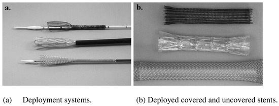
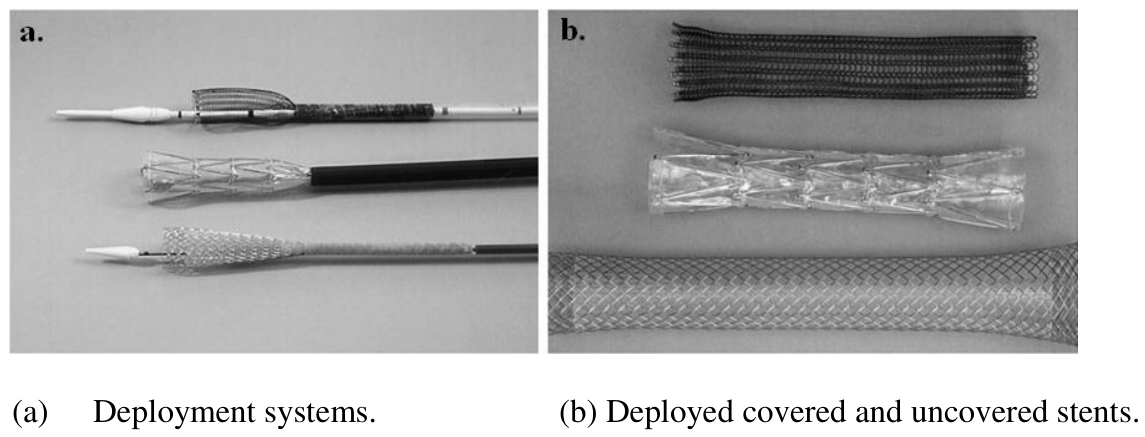
The first SEMS, the Z stent (Wilson Cook) was designed by Gianturco for use in vascular stenoses and described in use by Wright et al. [48] in 1985. They are available in a variety of diameters and lengths and are found in helical (eg, Esophacoil, Instent, Eden Prairie, MN), knitted (eg, Ultraflex, Boston Scientific, Oakland, NJ) and braided (Wallstent, Schneider, Zurich, Switzerland) varieties [31,49]. Different types of colorectal stents are as shown in Figure 1.
Figure 1.
Photographs of colorectal stents.
2.3.1. Uncovered stent
Kim et al. [50] explored metal stents that lack a covering membrane (uncovered stents), with good flexibility and stability and therefore easier to deploy [51,52,53,54,55,56,57,58,59,60], which have been successfully used for both preoperative decompression of acute neoplastic large bowel obstruction and long-term palliation of patients who are not candidates for surgery [49,50,61,62,63]. However, stent occlusion due to tumor ingrowth has been reported in 10% to 30% of cases in which uncovered stents were used [62,64].
2.3.2. Covered stent
Repici et al. [62], Tominaga et al. [65] reported the primary advantage of stents with covering membranes is the prevention of tissue ingrowth (tumor or mucosal hyperplasia and subsequent recurrent obstruction) through the mesh wall for patients who require stents for long-term colonic decompression [66,67]. This advantage is often outweighed by the stent‟s increased rigidity and tendency to migrate. Guan et al. [68] investigated the stent membranes using 5-FU and taxol particles; once deployed, the stent material becomes incorporated into both the tumor and surrounding tissue by pressure necrosis. This reaction allows both on curing colorectal tumors directly and anchoring of the stent and helps to prevent stent migration. With the use of covered stents, this integration does not always occur and a higher rate of stent migration is seen. Stent embedment accounts for some of the adverse effects seen when the pressure from expansion causes stent material to erode [64,69].
2.3.3. Self-expanding metallic stent (SEMS)
Self-expanding metal stents (SEMS) are currently used to treat variable sites of the gastrointestinal tract and biliary obstruction [70,71,72]. Three different self-expandable colonic stents are approved by the food and drug administration (FDA) in the United States for treatment of malignant obstruction [37, 73, and 74].
These are (a) the colonic Z-stent (Wilson–Cook Medical, Winston-Salem, NC) with diameters of 28 mm flanged ends and 25 mm mid-body; (b) the Enteral Wallstent (Mi-crovasive Corp., Natick, MA) with dimensions of 20 and 22 mm diameters; and (c) the Ultraflex Precision Colonic Stent (Microvasive, Boston Scientific Corp., Natick, MA) with a 30 mm diameter proximal flare and 25mm body [75].
The advantage of using the Enteral Wallstent over the other colonic stents is the much longer and smaller diameter (10 Fr) delivery system that allows passage of stents directly through the working channel of the endoscope. A theoretical advantage of the Wilson-Cook Z-stent and the Ultraflex Precision stent is the larger diameter of the lumen compared to the Enteral Wallstent. One further advantage of the Z-stent is that it does not shorten during deployment. [75]
2.3.4. Biodegradable stents
In 1996, Goldin et al. [76] firstly reported a kind of biodegradable stent. Sandha et al. [77] indicated this kind of novel stent is unnecessary to be recollected and the ideal colorectal stent. Guan et al. [68] indicated this biodegradable stent can be absorbed and catabolized in alternative term, which facilitates both local medicine release and temporary bracing; it can provide time for systematic support and obviate the need for faecal diversion or on-table lavage [78].
2.3.5. Other stents
Guan et al. [68] reported the radioactivity stent binding with 192Bo, Strontium and 125Iodine particles etc curing tumors directly; genophore-membrane stent was used for changing the genetics codes of the surrounding tissues by medical intervention.
2.4. Stent selection
Baron et al. [74] indicated the stent selection is subject to many factors, including the tumor sites, size, potential passages, fistula, degree of obstruction, length of stricture and personal experiences. Guan et al. [68] thought the uncovered and covered stents are often used for large bowl obstruction as a role of bridge to surgery and the long-term colonic decompression, respectively.
2.5. Description of procedure
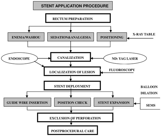
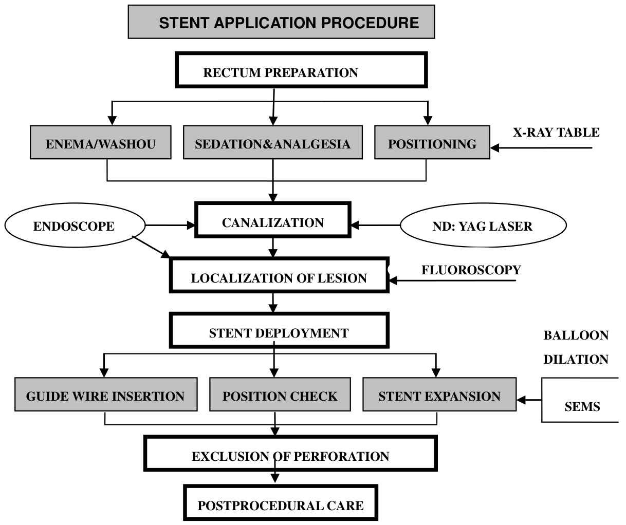
Stent placement can be divided into several distinct phases and is usually accomplished with a combination of endoscopic and fluoroscopic techniques [37,79,80,81,82]. The Schematic diagram of non-through the scope (TTS) stent placement is briefly presented in 5 steps: (a) A guide wire has been passed across the lesion under endoscopic and fluoroscopic guidance and exchanged for a super-stiff wire. (b) The endoscope is withdrawn leaving the guide wire in place across the stricture. (c) The predeployed stent is passed over the guide wire under fluoroscopic guidance. (d) The endoscope is reinserted alongside the predeployed stent to confirm proper positioning across the lesion before and during deployment. (e) Under endoscopic and fluoroscopic guidance, the stent is deployed. The stent application procedure is presented in Figure 2 [83,84,85,86,87].
Figure 2.
Stent application procsedure in patients with large bowel obstruction.
2.6. Indication
Many different endoluminal stents have been used by various investigators for different indications.
2.6.1. Malignant Fistulae
Patients with malignancy within the pelvis may suffer from fistulae to surrounding structures such as the vagina or bladder. Anastomotic leakage is the common and severe complication after colorectal surgeries. Intestinal canal being drawn repeatedly, over-cutting of the colorectal membrane all lead to the over tension at the stoma, which causes the phenomena of anastomotic leakage. So the key of surgery is to allow free drainage for the healing of leakage. Colorectal stents have been used to close such fistulae and allow nonsurgical palliation [34,88,89,90,91].
2.6.2. Obstruction
It has been estimated that between 8% and 29% of patients with colorectal cancer will have complete or partial intestinal obstruction [15, 92, and 93]. Colorectal stent placement may serve as a palliative modality of obstruction.
2.6.2.1. Benign stricture
Benign stoma stricture is the common complication, especially at the stoma of low rectum [94]. Guan et al. [95] indicated that patients with local pelvic tumors (ovarian carcinoma) or metastatic disease to the pelvis and colonic obstruction may achieve palliation of obstruction with colonic stenting. Forshaw et al. [96] reported SEMS was used for curing benign stoma stricture. However, this work has not given sufficient weight to the practical use in clinic [94,97]. Meisner et al. [98] thought SEMS placement didn‟t induce good result but with a high complication. There are a few materials at present mentioned the SEMS application status curing benign stoma stricture or obstruction.
2.6.2.2. Malignant obstruction
Gukovsky-Reicher et al. [99] concluded that it‟s difficult to find effective cure solutions to malignant diseases. Carter et al. [100] and Ren et al. [101] reported that stenting technology is successful both in curing primary colorectal cancers and large bowl obstruction caused by benign diseases. Shang et al. [102] illustrated that stent placement is a perspective solution for CRC, avoiding colostomy induced high morbidity and mortality.
Mauro et al. [103] reviewed the indications of stent placement as follows, (a) temporary decompression; it allows optimal preoperative planning and gives the opportunity for the administration of any neoadjuvant chemo/radiotherapy if required. (b) palliative treatment; it can provide palliation of obstruction in patients who are either unsuitable for laparotomy or incapable of surviving [2,104,105]. Evidence suggests that colo- rectal stents offer good palliation, and are safe and effective as a “bridge to surgery”. Stent usage can avoid the need for a stoma, and is associated with low rates of morbidity and mortality [101,104,105,106,107,108].
2.7. Complication
Stent placement may be resulting in varying rates of success and complications, some of them are fatal cases including stent reobstruction, stent migration, bleeding, perforation, fistula and so on [109,110,111,112,113,114,115,116,117,118]. Suzuki et al. [119] analyzed the complications (Table 3, Table 4) were defined as early and late (occurring≤30 days or>30 days after the procedure, respectively). As shown in Table 3 the complications of early 30 days are more than in late days. Khot and colleagues [27] were able to identify the complication in 598 instances. From Table 4, it can be analyzed that the migration is the most among all complications. All reports indicated stent placement is a feasible, effective and alternative method for acute colorectal obstruction [120].
Table 3.
Complications after stent insertion.
Table 4.
Complications of colorectal stent insertion [27].
Carter et al. [100] concluded that two important tips are helpful to avoid intra-procedural perforation. The first is limiting the amount of air insufflation during the exam, especially in patients with a dilated cecum. The second is avoiding present or post-stent dilation. Baron et al. [74] analyzed the improper stents cause reobstruction of large bowl. Keymling et al. [121] and Lagattolla et al. [122] explored the restenting is the most effective method, namely, “stent-graft”. Few report presents bleeding occurrence severely [123,124]. Spinelli et al. [125] and Mauro et al. [103] indicated perforation is a fatal complication and analyzed the reasons that caused perforation. If balloon dilatation was avoided, the perforation ratio would be below 5%.
2.8. Efficacy/Safety
Colorectal stents have been used to relieve acute obstruction of the lower bowel for the last ten years [6,126,127,128]. Evidence on which to based estimates of their efficacy and safety remains sparse. They have not been the object of any randomized controlled trials, though numerous case reports and small case series have established by their use obstruction can be successfully relieved.
In a literature search by Khot and colleagues [27] provided satisfactory evidence that stent placement is a relatively safe and effective alternative to colostomy, either as a “bridge to surgery” or for the palliative treatment of inoperable malignant disease.
Another reviewer Tack et al. [39], who included many of the same reports, reported the length of follow-up [129,130]. Diaz et al. [131] followed up 16 consecutive patients after palliative stent placement until death, or termination of the study at 44 months. The stents successfully resolved the clinical obstruction in all patients except one who required colostomy. No patients showed clinical symptoms of obstruction at the time of death or termination of the study. Camunez et al. [105] used stent placement as palliative treatment in 35 patients with a mean follow-up of 93 days. It was proved that it‟s safe and effective.
We may conclude from the above that for present purposes use of palliative stent may offer improved quality of life after placement, depending on the stage of disease, and is a satisfactory alternative to colostomy as well as a bridge to surgery.
2.9. New comprehensive stenting approach
Over the last 7 years, CRC death rates in men and women have decreased steadily (17% and 24%, respectively). These advances may be attributed to a better understanding of the disease genetics, improved surveillance, technical advances in the operations, increasing indications for the use neoadjuvant and adjuvant chemotherapy, radiation and better palliation [7].
2.9.1. Stenting integrated with laser technology
Nowadays, comprehensive stent treatment techniques are becoming more reliable. They are the combination with laser (Nd: YAG) photocoagulation, electrocoagulation, cryotherapy and the placement of metal stents in patients with a limited life expectancy. These methods rapidly resolve bowel obstruction and/or bleeding without the risk involved in surgical treatment [17,33].
The advantages of endoscopic placement of stents over interventional radiologic placement occurs because of greater accessibility to these sites and improved mechanical advantage of being able to pass some stents directly through the working channel of the endoscope.
2.9.2. Stenting integrated with chemotherapy and radiation
Many of these patients cannot undergo curative surgical treatment because who are unfit for general anesthesia, are in poor general health, or have concomitant disseminated neoplastic disease. Stent placement integrated with chemotherapy and radiation is the better solution for increasing survival by 3-6 months without increasing adverse effects on quality of life [13,16,117,118]. In the work of Camunez et al. [105] and Shang et al. [27], they presented that external radiotherapy used alone may eases pain in a high proportion of patients with locally advanced rectal cancer. In some patients, tumors have gone into complete remission or regressed sufficiently to permit curative surgery after prolonged fractionated radiotherapy of 45-50 Gy.
3. Future studies and directions
Despite the recent advances in stent technology in which it is still facing many problems, the search for the ideal enteral stent continues. The current studies mainly focus on the clinical therapies. Lan et al. [25] indicated the importance of using biomaterials for colorectal stents. However, few works are found to optimize the design and fabrication of new stents using bio-functional materials.
3.1. Biomaterials innovation
Biomedical materials and their production have been developed rapidly in the recent years. The global consumption of biomedical fibers and their products in 2006 reached above 318,000 tons; the current annual growth rate is about 3.5%. The growth rate of biomedical textiles has increased 25%-30% (data of 2008).
Synthetic polymers, used extensively, can be divided into permanent e.g., polyamide, polyester, polyethylene, polypropylene, PTFE and polyurethane and biodegradable which are mainly used in sutures and tissue engineering structures e.g., PHB polyhydroxybutyrate, PCL polycaprolactone, PLA poly(lactic acid), PMA polymerthylacrylate, PLGA copolymer of PLA and PGA poly(glycolic acid) and PLLA poly(L-lactic acid) [136,137,138,139,140,141,142,143,144,145,146,147,148]. Natural biological fibers include chitin (from the cells of crustacea) a polysaccharide renowned for its wound healing properties and incorporated into wound dressings; collagen (a fibrous protein found in connective tissue, tendons, etc.) used in cell engineering structures, for example artificial skin; and alginate fibers which can interact with the wound to form an absorbent gel, that acts as a protective barrier and still allows the wound to breathe [149,150,151,152,153,154,155,156,157,158,159,160,161,162,163,164,165,166,167]. However, the colorectal stents using the above biomaterials are sparse.
3.2. Design and fabrication innovations
On a worldwide basis, biomedical textile stents with braiding, knitting and woven technologies have been in routine clinical use for nearly five decades and have been implanted in hundreds of thousands of patients, due to their many advantages, such as good compatibility, softness, easy-bending, small quantity and a low-strength request on yarns [168,169,170,171,172]. Examples of these products are stents/scafford/graft/prosthesis of tracheobronchial [173,174,175,176,177,178,179,180,181]; esophageal [182,183,184,185,186], nerve [187,188], ligament [189,190,191] and vascular prosthesis [192,193,194,195,196] have been used in numerous patients. However, systematic investigations of colorectal stents made from braided and knitted structures using bio-functional materials are still limited. Until now, design and manufacture of the colorectal stents with good mechanical and biological performance remain a great challenge for material and medical specialists. It requires creativity and inspiration for the design and optimization of stent structures to simulate human colorectal functions. In addition, it is necessary to analyze and design stents by scientific models, including mathematical and animal models.
3.3. Modifications and biotechnology
The surface properties of materials in contact with biological systems play a key role in determining the outcome of biological-material interactions. Surface modification is a unique method to modify and optimize the surface properties of materials and components [197]. There are a multitude of surface modification methods available to engineer custom designed interfaces, including micro and nanostructuring of surfaces via various lithographic techniques, imprinting and laser micromaching; shot peening, laser ablation, plasma spraying, gas plasma treatments, ion bombardment, chemical etching, chemical and physical vapor deposition, as well as coatings of polymers, ceramics, metals, molecular self-assembled coatings, as well as biopolymeric coatings of proteins, sugars, lipids, polyelectrolyte coatings and more several surface modification methods [198,199]. The optimal surface will vary depending on the particular application, such as location of use for medical implants. More significantly, the optimal surface will vary with time, raising an interest in dynamic surfaces, as well as smart surfaces which react with a changing local biological environment. So, modifications and biotechnology may facilitate to produce better biocompatible colorectal stents.
3.4. Biomechanical properties
The mechanical properties play an important role in the colorectal stents practical application, but few studies have been conducted on this aspect about how to optimize its structure and process [200]. Dumoulin et al. [201], Francesco et al. [202] used FEM to set up 3-D models of tracheal stents, and attempted to simulate the stress distribution. The work by Kawaguchia et al. [203] also concentrated on the fabrication of mesh-type knitted structural tracheal Prostheses only, with PP for composite reinforcement.
However, the colorectal stents using the above approaches are scarce. In particular, what is missing is a precise mechanical analysis on the structures and process using mathematics models, offering no basis and effective methods for the application and optimization of colorectal stents.
4. Conclusion
As current operative and non-operative therapies have evolved and improved, the management of CRC is more complex and depends on a cooperative, multidisciplinary team approach, particularly in the face of an advanced disease. It is the multidisciplinary field involving biology, medicine, material engineering and mathematics that is likely to revolutionize the ways in which we improve the health and quality of life for millions of people worldwide by restoring, maintaining, or enhancing tissue and organ functions.
Acknowledgments
We would like to express our acknowledgment to Hong Kong Innovation and Technology Commission and Hong Kong Research Institute of Textile and Apparel for providing funding support through projects ITP-001-07-TP/ZR03 and ITP-031-08TP/ZR0M, and thank RGC and the Hong Kong Polytechnic University for supporting this study through project J-BB6Q and PolyU 5242/09E.
References
- Watson, A.J.M.; Shanmugam, V.; Mackay, I.; Chaturvedi, S.; Loudon, M.A.; Duddalwar, V.; Hussey, J.K. Outcomes after Placement of Colorectal Stents. Colorectal Dis. 2005, 7, 70–73. [Google Scholar] [CrossRef] [Scilit] [PubMed]
- Tilney, H.S.; Lovegrove, R.E.; Purkayastha, S. Comparison of colonic stenting and open surgery for malignant large bowel obstruction. Surg. Endosc. 2007, 21, 225–233. [Google Scholar] [CrossRef] [Scilit] [PubMed]
- Athreya, S.; Moss, J.; Urquhart, G. Colorectal stenting for colonic obstruction: the indications, complications, effectiveness and outcome 5 year review. Eur. J. Radiol. 2006, 60, 91–94. [Google Scholar] [CrossRef] [Scilit] [PubMed]
- Xinopoulos, D.; Dimitroulopoulos, D.; Theodos- opoulos, T. Stenting or stoma creation for patients with in operable malignant colonic obstruction? Results of a study and cost-effectiveness analysis. Surg. Endosc. 2004, 18, 421–426. [Google Scholar] [CrossRef] [Scilit] [PubMed]
- Lewis, R.; Flynn, A.; Dean, M.E.; Melville, A.; Eastwood, A.; Booth, A. Management of Colorectal Cancers. Qual. Saf. Health Care 2004, 13, 400–404. [Google Scholar] [CrossRef] [Scilit]
- National Institute for Clinical Excellence. Guidance on the Use of Irinotecan Oxaliplatin and Raltirexed for the Treatment of Advanced Colorectal Cancer, Tech. Appraisal Guidance No. 33. London: National Institute for Clinical Excellence 2002; http://www.nice.org.uk/ docref.asp?d=28735.
- Chokshi, R.J.; Abdel-Misih, S.; Bloomston, M. Surgical Management of Colorectal Cancer: a Review of the Literature. Indian J. Surg. 2009, 71, 350–355. [Google Scholar] [CrossRef] [Scilit] [PubMed]
- Deans, G.T.; Krukowski, Z.H.; Irwin, S.T. Malignant Obstruction of the Left Colon. Br. J. Surg. 1994, 81, 1270–1276. [Google Scholar] [CrossRef] [Scilit] [PubMed]
- Overgaard, M.; Overgaard, J.; Sell, A. Dose response relationship for radiation therapy of recurrent, residual and primarily inoperable colorectal cancer. Radiother. Oncol. 1984, 1, 217–225. [Google Scholar] [CrossRef] [Scilit] [PubMed]
- Lu, J.B.; Sun, X.B.; Dai, D.X.; Zhu, S.K.; Chang, Q.L.; Liu, S.Z.; Duan, W.J. Epidemiology of Gastroenterologic Cancer in Henan Province, China. World J. Gastroenterol. 2003, 9, 2400–2403. [Google Scholar] [CrossRef] [Scilit] [PubMed]
- Xu, A.G.; Jiang, B.; Yu, Z.J.; Zhong, X.H.; Gan, A.H.; Liu, J.H.; Luo, Q.Y.; Xiong, L.S. Epidemiology Investigation of Colorectal Cancer on Community Group in Guangdong Province. Chin. Med. J. 2007, 87, 1950–1953. [Google Scholar]
- Wei, Y.S.; Lu, J.C.; Wang, L.; Lan, P.; Zhao, H.J.; Pan, Z.Z.; Huang, J.; Wang, J.P. Risk Factors for Sporadic Colorectal Cancer in Southern Chinese. World J. Gastroenterol. 2009, 15, 2526–2530. [Google Scholar] [CrossRef] [Scilit] [PubMed]
- Baron, T.H.; Kozarek, R.A. Endoscopic stenting of colonic tumours. Best Pract. Res. Clin. Gastroenterol. 2004, 18, 209–229. [Google Scholar] [CrossRef] [Scilit] [PubMed]
- Kings Fund Forum. Cancer of the colon and rectum: consensus statement. Br. J. Surg. 1990, 70, 1063–1065. [Google Scholar] [CrossRef] [Scilit]
- Park, J.J.; Del Pino, A.; Orsay, C.P. Stoma complications: The Cook County Hospital experience. Dis. Colon Rectum 1999, 42, 1575–1580. [Google Scholar] [PubMed]
- Fan, Y.B.; Cheng, Y.S.; Chen, N.W.; Xu, H.M.; Yang, Z.; Wang, Y.; Huang, Y.Y.; Zheng, Q. Clinical application of self-expanding metallic stent in the management of acute left-sided colorectal malignant obstruction. World J. Gastroenterol. 2006, 12, 755–759. [Google Scholar] [CrossRef] [Scilit] [PubMed]
- Tekkis, P.P.; Kinsman, R.; Thompson, M.R.; Stamatakis, J.D. The Association of Coloproctology of Great Britain and Ireland study of large bowel obstruction caused by colorectal cancer. Ann. Surg. 2004, 240, 76–81. [Google Scholar] [CrossRef] [Scilit] [PubMed]
- Valerio, D.; Jones, P.F. Immediate resection in the treatment of large bowel emergencies. Br. J. Surg. 1978, 65, 712–716. [Google Scholar] [CrossRef] [Scilit] [PubMed]
- Serpell, J.W.; McDermott, F.T.; Katrivessis, H.; Hughes, E.S. Obstructing carcinomas of the colon. Br. J. Surg. 1989, 76, 965–969. [Google Scholar] [CrossRef] [Scilit] [PubMed]
- Phillips, R.K.; Hittinger, R.; Fry, J.S.; Fielding, L.P. Malignant large bowel obstruction. Br. J. Surg. 1985, 72, 296–302. [Google Scholar] [CrossRef] [Scilit] [PubMed]
- Buechter, K.J.; Boustany, C.; Caillouette, R.; Cohn, I. Surgical management of the acutely obstructed colon: a review of 127 cases. Am. J. Surg. 1988, 156, 163–168? [Google Scholar] [PubMed]
- Gregorio, M.A.D.; Mainar, A.; Tejero, E. Acute Colorectal Obstruction: Stent Placement for Palliative Treatment-Results of a Multicenter Study. Radiology 1998, 209, 117–120. [Google Scholar] [CrossRef] [Scilit] [PubMed]
- Gunderson, L.L.; Sosin, H. Areas of failure found at reoperation (second or symptomatic look) following curative surgery for adeno-carcinoma of the rectum: clinicopathologic correlation and implications for adjuvant therapy. Cancer 1974, 34, 1272–1292. [Google Scholar] [CrossRef] [Scilit] [PubMed]
- Turk, P.S.; Wanebo, H.J. Results of surgical treatment of non-hepatic recurrence of colorectal carcinoma. Cancer 1993, 71 (Suppl), 4267–4269. [Google Scholar] [CrossRef] [Scilit] [PubMed]
- Lan, P.; Peng, H. Devoting Much Attention to Heritage and Development of Colorectal and Anal Benign Disease. Chin. J. Bases Clin. General. Surg. 2010, 17, 105–108. [Google Scholar]
- Coco, C.; Cogliandolo, S.; Riccioni, M.E. Use of a self-expanding stent in the palliation of rectal cancer recurrences. Surg. Endosc. 2000, 14, 708–711. [Google Scholar] [CrossRef] [Scilit] [PubMed]
- Khot, U.P.; Lang, A.W.; Murali, K.; Parker, M.C. Systematic review of the efficacy and safety of colorectal stents. Br. J. Surg. 2002, 89, 1096–1102. [Google Scholar] [CrossRef] [Scilit] [PubMed]
- Binkert, C.A.; Ledermann, H.P.; Jost, R.; Saurenmann, P.; Decurtins, M.; Zollikofer, C.L. Acute colonic obstruction: Clinical aspects and cost effectiveness of preoperative and palliative treatment with self-expanding metallic stents. A preliminary report. Radiology 1998, 206, 199–204. [Google Scholar] [CrossRef] [Scilit] [PubMed]
- Zollikofer, C.L.; Jost, R.; Schoch, E.; Decurtins, M. Gastrointestinal stenting. Eur. Radiol. 2000, 10, 320–341. [Google Scholar] [CrossRef] [Scilit] [PubMed]
- Zollikofer, C.L.; Jost, R.; Schoch, E.; Decurtins, M. Gastroduodenal and colonic stents: Review article. Semin Interv. Radiol. 2001, 18, 265–280. [Google Scholar] [CrossRef] [Scilit]
- Harris, G.J.; Senagore, A.J.; Lavery, I.C.; Fazio, V.W. The management of neoplastic colorectal obstruction with colonic endo-lumenal stenting devices. Am. J. Surg. 2001, 181, 499–506. [Google Scholar] [CrossRef] [Scilit] [PubMed]
- Ell, C.; Hochberger, J.; May, A.; Fleig, W.E.; Hahn, E.G. Coated and uncoated self-expanding metal stents for malignant stenosis in the upper GI tract: preliminary clinical experiences with Wallstents. Am. J. Gastroenterol. 1994, 89, 1496–1500. [Google Scholar] [PubMed]
- Baraza, W.; Lee, F.; Brown, S.; Hurlstone, D.P. Combination endo-radiological colorectal stenting: a prospective 5-year clinical evaluation. Colorectal Dis. 2008, 10, 901–906. [Google Scholar] [CrossRef] [Scilit] [PubMed]
- Baron, T.H. Colorectal Stents. Tech. Gastrointest. Endosc. 2003, 5, 182–190. [Google Scholar] [CrossRef] [Scilit]
- Spinelli, P.; Dal Fante, M.; Mancini, A. Self expanding mesh stent for endoscopic palliation of rectal obstructing tumors: a preliminary report. Surg. Endosc. 1992, 6, 72–74. [Google Scholar] [CrossRef] [Scilit] [PubMed]
- Itabashi, M.; Hamano, K.; Kameoka, S.; Asahina, K. Self-expanding stainless steel stent application in rectosigmoid stricture. Dis. Colon Rectum 1993, 36, 508–511. [Google Scholar] [CrossRef] [Scilit] [PubMed]
- Baron, T.H.; Dean, P.A.; Yates, M.R.; Canon, C.; Koeheler, R.E. Expandable metal stents for the treatment of colonic obstruction: techniques and outcome. Gastrointest. Endosc. 1998, 47, 277–285. [Google Scholar] [CrossRef] [Scilit] [PubMed]
- Rey, J.F.; Romanczyk, T.; Greff, M. Metal stents for palliation of rectal carcinoma: a preliminary report on 12 patients. Endosc 1995, 27, 501–504. [Google Scholar] [CrossRef] [Scilit] [PubMed]
- Tack, J.; Gevers, A.M.; Rutgeerts, P. Self-expandable metallic stents in the palliation of rectosigmoid carcinoma: a follow-up study. Gastrointest. Endosc. 1998, 48, 267–271. [Google Scholar] [CrossRef] [Scilit] [PubMed]
- Mainar, A.; Teyero, E.; Mainar, M.; Ferral, H.; Castaneda, W. Colorectal obstruction: treatment with metallic stents. Radiology 1996, 198, 761–764. [Google Scholar] [CrossRef] [Scilit] [PubMed]
- Dohomoto, M.; Hunerbein, M.; Schlag, P.M. Application of rectal stents for palliation of obstructing rectosigmoid cancer. Surg. Endosc. 1997, 11, 758–761. [Google Scholar] [CrossRef] [Scilit] [PubMed]
- Dohomoto, M. Endoscopic implantation of rectal stents in palliative treatment of malignant stenosis [in Japanese]. Endosc. Dig. 1991, 3, 1507–1512. [Google Scholar]
- Arnell, T.; Stamos, M.J.; Takahashi, P.; Ojha, S.; Sze, G.; Eysselein, V. Colonic stents in colorectal obstruction. Ann. Surg. 1998, 64, 986–988. [Google Scholar]
- Wallis, F.; Campbell, K.L.; Eremin, O.; Hussey, J.K. Self-expanding metal stents in the management of colorectal carcinoma: a preliminary report. Clin. Radiol. 1998, 53, 251–254. [Google Scholar] [CrossRef] [Scilit] [PubMed]
- Mathus-Vliegen, E.M.; Tytgat, G.N. Laser photo coagulation in the palliation of colorectal malignancies. Cancer 1986, 57, 2212–2216. [Google Scholar] [PubMed]
- Saida, S.Y.; Sumiyama, Y.; Nagao, J.; Takase, M. Stent endoprosthesis for obstructing colorectal cancers. Dis. Colon Rectum 1996, 39, 552–555. [Google Scholar] [CrossRef] [Scilit] [PubMed]
- Paul, D.L.; Pinto, P.I.; Fernandez, L.R.; Montes, L.C. Palliative treatment of malignant colorectal strictures with metallic stent. Cardiovasc Interv. Radiol. 1999, 22, 29–36. [Google Scholar] [CrossRef] [Scilit] [PubMed]
- Wright, K.C.; Wallace, S.; Charnsangavej, C. Percutaneous endo-vascular stents: an experimental evaluation. Radiology 1985, 156, 69–72. [Google Scholar] [CrossRef] [Scilit] [PubMed]
- Park, K.B.; Do, Y.S.; Kang, W.K.; Choo, S.W.; Han, Y.H.; Suh, S.W.; Lee, S.J.; Park, K.S.; Choo, I.W. Malignant obstruction of gastric outlet and duodenum: palliation with flexible covered metallic stents. Radiol 2001, 219, 679–683. [Google Scholar] [CrossRef] [Scilit] [PubMed]
- Kim, J.H.; Lee, K.M.; Lee, K.J. Comparison of uncovered and covered through-the-scope (TTS) stent for malignant colorectal obstruction: long term results. Endosc 2003, 35 (Suppl), A55. [Google Scholar]
- Baere, T.; Harry, G.; Ducreux, M. Self-expanding metallic stents as palliative treatment of malignant gastroduodenal stenosis. Am. J. Roentgenol. 1997, 169, 1079–1083. [Google Scholar] [CrossRef] [Scilit] [PubMed]
- Yates, M.R.; Morgan, D.E.; Baron, T.H. Palliation of malignant and small intestinal strictures with self-expanding metal stents. Endosc 1998, 30, 266–272. [Google Scholar] [CrossRef] [Scilit] [PubMed]
- Binkert, C.A.; Jost, R.; Steiner, A. Benign and malignant stenoses of the stomach and duodenum: treatment with self-expanding metallic endoprostheses. Radiology 1996, 199, 335–338. [Google Scholar] [CrossRef] [Scilit] [PubMed]
- Feretis, C.; Benakis, P.; Dimopoulos, C. Palliation of malignant gastric outlet obstruction with self-expanding metal stents. Endosc 1996, 28, 225–228. [Google Scholar] [CrossRef] [Scilit] [PubMed]
- Freeman, M.L.; Cas, O.W. Interlocking expandable metal stents for simultaneous treatment of malignant biliary and duodenal obstruction. Gastroin Test. Endosc. 1996, 44, 98–99. [Google Scholar] [CrossRef] [Scilit] [PubMed]
- Keymling, M.; Wagner, J.H.; Vakil, N. Relief of malignant duodenal obstruction by percutaneous insertion of a metal stent. Gastroin Test. Endosc. 1993, 39, 439–441. [Google Scholar] [CrossRef] [Scilit] [PubMed]
- Kozarek, R.A.; Ball, T.J.; Patterson, D.J. Metallic self-expanding stent application in the upper gastrointestinal tract: caveats and concerns. Gastroin Test. Endosc. 1992, 38, 1–6. [Google Scholar] [CrossRef] [Scilit] [PubMed]
- Maetani, I.; Inoue, H.; Sato, M. Peroral insertion techniques of self-expanding metal stents for malignant gastric outlet and duodenal stenoses. Gastroin Test. Endosc. 1996, 44, 468–471. [Google Scholar] [CrossRef] [Scilit] [PubMed]
- Nevitt, A.W.; Vida, F.; Kozarek, R.A. Expandable metallic prostheses for malignant obstructions of gastric outlet and proximal small bowel. Gastroin Test. Endosc. 1998, 47, 271–276. [Google Scholar] [CrossRef] [Scilit] [PubMed]
- Pinto, I.T. Malignant gastric and duodenal stenosis: palliation by peroral implantation of a self-expanding metallic stent. Cardiovasc Interv. Radiol. 1997, 20, 431–434. [Google Scholar] [CrossRef] [Scilit] [PubMed]
- Lo, S. Expandable metal stents for colonic obstruction. Gastrointest. Endosc. Clin. N Am. 1999, 9, 459–446. [Google Scholar] [CrossRef] [Scilit]
- Repici, A.; Reggio, D.; Angelis, C.D. Covered metal stents for management of inoperable malignant colorectal strictures. Gastro Intest. Endosc. 2000, 52, 735–740. [Google Scholar] [CrossRef] [Scilit] [PubMed]
- Liu, X.S.; Ni, C.F.; Ding, Y. Metallic stent in the management of acute malignant colorectal obstruction. Foreign Med. Sci. Clin. Radiol. Fascicle 2000, 4, 196. [Google Scholar]
- Lee, H.P.; Gourley, L.; Duffy, S.W.; Estève, J.; Lee, J.; Day, N.E. Colorectal cancer and diet in an Asian population-a case-control study among Singapore Chinese. Int. J. Cancer 1989, 43, 1007–1016. [Google Scholar] [CrossRef] [Scilit] [PubMed]
- Tominaga, K.; Yoshida, M.; Maetani, I. Expandable metal stent placement in the treatment of a malignant anastomotic stricture of the transverse colon. Gastrointest. Endosc. 2001, 53, 524. [Google Scholar] [CrossRef] [Scilit] [PubMed]
- Song, H.Y.; Yang, D.H.; Kuh, J.H.; Choi, K.C. Obstructing cancer of the gastricantrum: palliative treatment with covered metallic stents. Radiology 1993, 187, 357–358. [Google Scholar] [CrossRef] [Scilit] [PubMed]
- Bethge, N.; Sommer, A.; Gross, U. Human tissue responses to metal stents implanted in vivo for the palliation of malignant stenoses. Gastrointest. Endosc. 1996, 43, 596–602. [Google Scholar] [CrossRef] [Scilit]
- Guan, Y.S.; Sun, L.; Li, X. Successful management of a benign anastomotic colonic stricture with self-expanding metallic stents: a case report. World J. Gastroenterol. 2004, 10, 3534. [Google Scholar] [CrossRef] [Scilit] [PubMed]
- Soetikno, R.M.; Lichtenstein, D.R.; Vander-voort, J. Palliation of malignant gastric outlet obstruction using an endo-scopically placed Wallstent. Gastroin Test. Endosc. 1998, 47, 267–270. [Google Scholar] [CrossRef] [Scilit] [PubMed]
- Mainar, A.; De Gregorio Ariza, M.A.; Tejero, E.; Tejero, Eloy; Tobío, R.; Alfonso, E.; Isabel Pinto, I.; Marcos Herrera, M.; Fernández, J.A. Acute colorectal obstruction: treatment with self-expandable metallic stents before scheduled surgery results of a multicenter study. Radiology 1999, 210, 65–69. [Google Scholar] [CrossRef] [Scilit] [PubMed]
- Baron, T.H. Expandable metal stents for the treatment of cancerous obstruction of the gastrointestinal tract. N Engl. J. Med. 2001, 344, 1681–1687. [Google Scholar] [CrossRef] [Scilit] [PubMed]
- Saida, Y.; Sumiyama, Y.; Nagao, J. Self-expandable metallic stent in the treatment of colorectal obstruction. Nippon Geka Gakkai Zasshi 2003, 104, 554. [Google Scholar] [PubMed]
- Whynes, D.K.; Neilson, A.R. Symptoms before and after surgery for colorectal cancer. Qual. Life Res. 1997, 3, 191. [Google Scholar] [CrossRef] [Scilit] [PubMed]
- Baron, T.H.; Rey, J.F.; Spinelli, P. Expandable metal stent placement for malignant colorectal obstruction. Endosc 2002, 34, 823–830. [Google Scholar] [CrossRef] [Scilit] [PubMed]
- Baron, T.H. Indications and Results of Endoscopic Rectal Stenting. Soc. Surg. Aliment. Trac 2004, 8, 266–269. [Google Scholar] [CrossRef] [Scilit] [PubMed]
- Goldin, E.; Fiorini, A.; Ratan, Y. A new biodegradable and self-expanding stent for benign esophageal strictures. Gastrointest. Endosc. 1996, 43, 294. [Google Scholar] [CrossRef] [Scilit]
- Sandha, G.S.; Marcon, N.E. Expandable metal stents for benign esophageal obstruction. Gastrointest. Endosc. Clin. N Am. 1999, 9, 437. [Google Scholar] [CrossRef] [Scilit]
- Tejero, E.; Mainar, A.; Fermandez, L.; Tobío, R.; De Gregorio, M.A. New procedure for the treatment of colorectal neoplastic obstructions. Dis. Colon Rectum 1994, 37, l158–1159. [Google Scholar] [CrossRef] [Scilit] [PubMed]
- Alcantara, M.; Serra, X.; Bombardó, J.; Falcó, J.; Perandreu, J.; Ayguavives, I.; Mora, L.; Hernando, R.; Navarro, S. Colorectal stenting as an effective therapy for preoperative and palliative treatment of large bowel obstruction: 9 years‟ experience. Tech Coloproctol. 2007, 11, 316–322. [Google Scholar] [CrossRef] [Scilit] [PubMed]
- Baigrie, R.J.; Berry, A.R. Management of advanced rectal cancer. Br. J. Surg. 1994, 81, 343–352. [Google Scholar] [CrossRef] [Scilit] [PubMed]
- Paul Diaz, L.; Pabon, I.P.; Lobato, R.F.; Lopez, C.M. Palliative treatment of malignant colorectal strictures with metallic stents. Cardiovasc Interv. Radiol. 1999, 22, 29–36. [Google Scholar] [CrossRef] [Scilit] [PubMed]
- Rupp, K.D.; Dohmoto, M.; Meffert, R. Cancer of the rectum-palliative endoscopic treatment. Eur. J. Surg. Oncol. 1995, 21, 644–647. [Google Scholar] [CrossRef] [Scilit] [PubMed]
- Lopera, J.E.; Ferral, H.; Wholey, M. Treatment of colonic obstructions with metallic stents: Indications, technique and complications. A J. R 1997, 169, 1285–1290. [Google Scholar] [CrossRef] [Scilit] [PubMed]
- Dohmoto, M.; Hunerbein, M.; Schlag, P.M. Application of rectal stents for palliation of obstructing recto sigmoid cancer. Surg. Endosc. 1997, 11, 758–761. [Google Scholar] [CrossRef] [Scilit] [PubMed]
- Canon, C.L.; Baron, T.H.; Morgan, D.E. Treatment of colonic obstruction with expandable metal stents: radio logic features. A J. R 1997, 168, 199–205. [Google Scholar] [CrossRef] [Scilit] [PubMed]
- Cwiekiel, W.; Andren-Sandberg, A. Malignant stricture with colovesical fistula: stent insertion in the colon. Radiology 1993, 186, 563–564. [Google Scholar] [CrossRef] [Scilit] [PubMed]
- Mainar, A.; Tejero, E.; Maynar, M. Colorectal obstruction: treatment with metallic stents. Radiol 1996, 198, 761–764. [Google Scholar] [CrossRef] [Scilit] [PubMed]
- Zeng, P. Metallic stent therapy for malignant rectal obstruction. J. Clin. Exp. Med. 2006, 5, 1780–1782. [Google Scholar]
- Umpleby, H.C.; Williamson, R.C.N.; Chin, M. Survival in acute obstructing colonectal carcinoma. Dis. Colon Rectum 1984, 27, 299–304. [Google Scholar] [CrossRef] [Scilit] [PubMed]
- Regland, J.J.; Londe, A.M.; Spratt, J.S. Correlation of the prognosis of obstructing colorectal carcinoma with clinical and pathologic variables. Am. J. Surg. 1971, 121, 552–556. [Google Scholar] [CrossRef] [Scilit] [PubMed]
- The SCOTIA Study Group. Single-stage treatment forma lignant left-sided colonic obstruction: a prospective randomized clinical trial comparing subtotal colectomy with segmental resection following intraoperative irrigation. Br. J. Surg. 1995, 82, 1622–1627. [Google Scholar] [CrossRef] [Scilit] [PubMed]
- Pérez-Díaz; Turégano-Fuentes; Calvo-Serrano; De Tomás-Palacios, J.; De Fuenmayor-Valera, M.L.; Del Valle Hernández, E.; Muñoz Jiménez, F.; Quintans-Rodrgíuez. Emergency subtotal colectomy as treatment of choicein obstructing carcinomas of the left colon. Colorectal Dis. 1999, 1, 15–18. [Google Scholar] [CrossRef] [Scilit] [PubMed]
- Lan, P.; Wang, J.P. Current therapy of short bowel syndrome. Chin. J. Pract. Surg. 1999, 19, 684. [Google Scholar]
- Yu, Z.Y.; Liu, L.; Zhang, C.Q.; Li, F. The analysis and control of 32 cases with benign colorectal anastomotic stricture. Sichuan Med. J. 2006, 27, 1043–1045. [Google Scholar]
- Guan, Y.S.; Sun, L. Clinical Application of Stents Deployment. Chin. J. Bases Clin. Gen. Surg. 2004, 11, 369. [Google Scholar] [CrossRef] [Scilit] [PubMed]
- Forshaw, M.J.; Sankararajah, D.; Stewart, M. Self-expanding metallic stents in the treatment of benign colorectal disease: indications and outcomes. Colorectal Dis. 2006, 8, 102. [Google Scholar] [CrossRef] [Scilit] [PubMed]
- Rees, J.R.; Carney, L.; Gill, T.S. Management of recurrent anastomotic stricture and iatrogenic stenosis by circular stapler. Dis. Colon Rectum 2004, 47, 944. [Google Scholar] [CrossRef] [Scilit] [PubMed]
- Meisner, S.; Hensler, M.; Knop, F.K. Self-expanding metal stents for colonic obstruction: experiences from 104 procedures in a single center. Dis. Colon Rectum 2004, 47, 444. [Google Scholar] [CrossRef] [Scilit] [PubMed]
- Gukovsky-Reicher, S.; Lin, R.M.; Sial, S. Self-expandable metal stents in palliation of malignant gastrointestinal obstruction: review of the current literature data and 5-year experience at Harbor-UCLA Medical Center. Med. Gen. Med. 2003, 5, 16. [Google Scholar]
- Carter, J.; Valmadre, S.; Dalrymple, C. Management of large bowel obstruction in advanced ovarian cancer with intraluminal stents. Gynecol. Oncol. 2002, 84, 176–179. [Google Scholar] [CrossRef] [Scilit] [PubMed]
- Ren, M.Y.; Huang, W. The application of colorectal stent on obstructive large bowel cancer. J. Coloproctological Surg. 2004, 10, 292. [Google Scholar]
- Shang, P.Z.; Liu, J.Z.; Lu, Y.Q.; Zhou, F.T.; Wang, T.S.; Zhang, Z.H. Shape Memory Alloy Stent for Intestinal Obstruction Due to Rectal Cancer. Chin. J. Bases Clin. General. Surg. 2002, 9, 34. [Google Scholar]
- Mauro, M.A.; Koehler, R.E.; Baron, T.H. Advances in gastrointestinal intervention: the treatment of gastroduodenal and colorectal obstructions with metallic stents. Radiology 2000, 215, 659. [Google Scholar] [CrossRef] [Scilit] [PubMed]
- Sebastian, S.; Johnston, S.; Geoghegan, T.; Torreggiani, W.; Buckley, M. Pooled analysis of the efficacy and safety of self-expanding metal stenting in malignant colorectal obstruction. Am. J. Gastroenterol. 2004, 99, 2051–2057. [Google Scholar] [CrossRef] [Scilit] [PubMed]
- Camunez, F.; Echenagusia, A.; Simo, G.; Turegano, F.; Vazquez, J.; Barreiro-Meiro, I. Malignant colorectal obstruction treated by means of self-expanding metallic stents: effectiveness before surgery and in palliation. Radiology 2000, 216, 492–497. [Google Scholar] [CrossRef] [Scilit] [PubMed]
- Ohman, U. Prognosis in patients with obstructing colorectal carcinoma. Am. J. Surg. 1982, 143, 742–747. [Google Scholar] [CrossRef] [Scilit] [PubMed]
- Yao, J.H.; Yue, B.; Hu, L.B. Obstruction of sigmoid and rectum cancer treated by metal stent in colon. Beijing Med. J. 2006, 28, 665. [Google Scholar]
- Fiori, E.; Lamazza, A.; De Cesare, A.; Bononi, M.; Volpino, P.; Schillaci, A.; Cavallaro, A.; Cangemi, V. Palliative management of malignant rectosigmoidal obstruction. Colostomy vs. endoscopic stenting. A randomized prospective trial. Anticancer Res. 2004, 24, 265. [Google Scholar] [PubMed]
- Makela, J.; Haukipuro, K.; Laitinen, S.; Kairaluoma, M.I. Surgical treatment of recurrent colorectal cancer. Arch. Surg. 1989, 124, 1029–1032. [Google Scholar] [CrossRef] [Scilit] [PubMed]
- Giovannucci, E. Metabolic syndrome, hyperinsulinemia, and colon cancer: a review. Am. J. Clin. Nutr. 2007, 86, 836S–842S. [Google Scholar] [CrossRef] [Scilit] [PubMed]
- Lan, P. Colorectal Cancer in Inflammatory Bowel Disease. Chin. J. Bases Clin. Gen. Surg. 2005, 12, 10–12. [Google Scholar]
- Ravo, B.; Ger, R. Temporary colostomy-an outmoded procedure? A report on the intracolonic bypass. Dis. Colon Rectum 1985, 28, 904–907. [Google Scholar] [PubMed]
- Wholey, M.H.; Levine, E.A.; Ferral, H.; Castaneda-Zuniga, W. Initial clinical experience with colonic stent placement. Am. J. Surg. 1998, 175, 194–197. [Google Scholar] [CrossRef] [Scilit] [PubMed]
- Raijman, I.; Siemens, M.; Marcon, N. Use of an expandable Ultraflex stent in the treatment of malignant rectal stricture. Endosc 1995, 27, 273–276. [Google Scholar] [CrossRef] [Scilit] [PubMed]
- Jost, R.S.; Jost, R.; Decurtins, M.; Schoch, E.; Brunner, B.; Zollikofer, C.L. Colorectal stenting: An effective and cost saving therapy in a cute left-sided obstruction [abstract]. Eur. Radiol. Suppl. 2004, 6, N39. [Google Scholar]
- Chen, W.Q.; Chen, C.Q.; Zhan, W.H.; Chen, Z.X.; Qin, J.Z.; Lan, P. Complication and indication of re-operation following pancreatoduodenectomy. Guangdong Med. J. 2000, 21, 129. [Google Scholar]
- Fernandez Lobato, R.; Pinto, I.; Paul, L.; Tejero, E.; Montes, C.; Fernandez, L. Self-expanding prostheses as a palliative method in treating advanced colorectal cancer. Int. Surg. 1999, 84, 159–162. [Google Scholar] [PubMed]
- Riedl, S.; Wiebelt, H.; Bergmann, U.; Hermanek, PJr. Post operative complications and fatalities in surgical therapy of colon carcinoma: Results of the German multicenter study by the Colorectal Carcinoma Study Group [in German]. Chirurgie 1995, 66, 597–606. [Google Scholar]
- Suzuki, N.; Saunders, B.P.; Thomas-Gibson, S. Colorectal Stenting for Malignant and Benign Disease, Outcomes in Colorectal Stenting. Dis. Colon Rectum 2004, 47, 1201–1207. [Google Scholar] [CrossRef] [Scilit] [PubMed]
- Kapadia, I, Ibrahim, M. “Arterial Graft”, WO Patent No. 89/00031, 1989.
- Keymling, M. Colorectal stenting. Endosc 2003, 35, 234. [Google Scholar] [CrossRef] [Scilit] [PubMed]
- Lagattollan, R.F.; Rowe, P.H.; Anderson, H.; Dunka, A. Restenting malignant oesophageal strictures. Br. J. Surg. 1998, 85, 261. [Google Scholar] [CrossRef] [Scilit] [PubMed]
- Lobato, R.F.; Pinto, I.; Paul, L.; Tejero, E.; Montes, C.; Fernández, L.; Moreno Azcoita, M.; Lozano, R. Self-expanding protheses as a palliative method in treating advanced colorectal cancer. Int. Surg. 1999, 84, 159–162. [Google Scholar]
- Han, Y.M.; Lee, J.M.; Lee, T.H. Delayed colon perforation after palliative t reatment for rectal carcinoma wit h bare rectal stent: a case report. Korean J. Radiol. 2000, 1, 169. [Google Scholar] [PubMed]
- Spinelli, P.; Mancini, A. Use of self-expanding metal stents for palliation of rectosigmoid cancer. Gastrointest. Endosc. 2001, 53, 203. [Google Scholar] [CrossRef] [Scilit] [PubMed]
- Benz S, Pfeffer F. “Suction stent” US Patent No. 2006/0095124 A1, 2006.
- Dohmoto, M. New method-endoscopic implantation of a rectal stent in palliative treatment of malignant stenosis. Endosc. Dig. 1991, 3, 1507–1512. [Google Scholar]
- Spinelli, P.; Dal Fante, M.; Mancini, A. Rectal metal stents for palliation of colorectal malignant stenosis. Bildgebung 1993, 60, 48–50. [Google Scholar] [PubMed]
- Egozi, L.; Sorrento, J.J.; Golub, R.; Schultz, E.H. Complication of the intracolonic bypass. Dis. Colon Rectum 1993, 36, 191–193. [Google Scholar] [CrossRef] [Scilit] [PubMed]
- Nugent, K.P.; Daniels, P.; Stewart, B.; Patankar, R.; Johnson, C.D. Quality of life in stoma patients. Dis. Colon Rectum 1999, 42, 1569–1574. [Google Scholar] [CrossRef] [Scilit] [PubMed]
- Diaz, L.P.; Pabón, I.P.; Lobato, R.F.; López, C.M. Palliative treatment of malignant colorectal strictures with metallic stents. Cardiovasc Interv. Radiol. 1999, 22, 29–36. [Google Scholar] [CrossRef] [Scilit] [PubMed]
- Gordon, N.L.; Dawson, A.A.; Bennett, B.; Innes, G.; Eremin, O.; Jones, P.F. Outcome in colorectal adenocarcinoma: two seven-year studies of a population. B M. J. 1993, 307, 707–710. [Google Scholar] [CrossRef] [Scilit] [PubMed]
- Colorectal Cancer Collaborative Group. Palliative chemotherapy for advanced colorectal cancer: systematic review and meta-analysis. Colorectal Cancer Collab. Group. B M. J. 2000, 321, 531–535. [Google Scholar] [CrossRef] [Scilit] [PubMed]
- Jonker, D.J.; Maroun, J.A.; Kocha, W. Survival benefit of chemotherapy in metastatic colorectal cancer: a meta-analysis of randomized controlled trials. Br. J. Cancer 2000, 82, 1789–1794. [Google Scholar] [CrossRef] [Scilit] [PubMed]
- Wang, J.P.; Ding, W.X.; Deng, Y.H.; Lan, P.; Pan, K.; Dong, G.H.; Deng, J.Z.; Wang, L.; Wu, X.J.; Guo, X.F.; Zheng, J. Preoperative chemoradiotherapy with FOLFOX in low rectal cancer: a multicenter study. Chin. J. Gastrointest. Surg. 2008, 11, 116–119. [Google Scholar]
- Bao, Q.B.; McCullen, G.M.; Higham, P.A.; Dumbleton, J.H.; Yuan, H.A. The artificial disc: theory, design and materials. Biomaterials 1996, 17, 1157–1167. [Google Scholar] [CrossRef] [Scilit] [PubMed]
- Bonfield, W.; Grynpas, M.D.; Tully, A.E.; Bowman, J.; Abram, J. Hydroxyapatite reinforced polyethylene-a mechanically compatible implant material for bone replacement. Biomaterials 1981, 2, 185–186. [Google Scholar] [CrossRef] [Scilit] [PubMed]
- Bostman, O.; Hirvensalo, E.; Makinen, J.; Rokkanen, P. Foreign-body reactions to fracture fixation implants of biodegradable synthetic polymers. J. Bone Jt. Surg. 1990, 72-B, 592–596. [Google Scholar] [CrossRef] [Scilit] [PubMed]
- Dauner, M.; Caramaro, L.; Missirlis, Y.; Panagiotopoulos, E. Resorbable continuous-fiber reinforced polymers for osteosynthesis. J. Mater. Sci. Mater. Med. 1998, 9, 173–179. [Google Scholar] [CrossRef] [Scilit] [PubMed]
- Davy, K.W.M.; Parker, S.; Braden, M.; Ward, I.M.; Ladizesky, H. Reinforcement of polymers of 2, 2 bis-4(2-hydroxy-3-methacryloyloxy propoxy) phenyl propane by ultra-high modulus polyethylene fibers. Biomaterials 1998, 13, 17–19. [Google Scholar]
- Goldner, J.L.; Urbaniak, J.R. The clinical experience with silicone-Dacron metacarpophalangeal and interphalangeal joint prostheses. J. BioMed Mater. Res. Symp. 1973, 4, 137–163. [Google Scholar] [CrossRef] [Scilit] [PubMed]
- Deb, S.; Wang, M.; Tanner, K.E.; Bonfield, W. Hydroxyapatite-polyethylene composites: effect of grafting and surface treatment of hydroxyapatite. J. Mater. Sci. Mater. Med. 1996, 7, 191–193. [Google Scholar] [CrossRef] [Scilit]
- Dehl, R.E. On the characterization of porosity in PTFE-carbon composite implant materials by mercury porosimetry. J. BioMed Mater. Res. 1982, 16, 715–719. [Google Scholar] [CrossRef] [Scilit] [PubMed]
- Deng, M.; Shalaby, S.W. Properties of self-reinforced ultra-high-molecular weight polyethylene composites. Biomaterials 1997, 18, 645–655. [Google Scholar] [CrossRef] [Scilit] [PubMed]
- Galletti, P.M.; Aebischer, P.; Sasken, H.F.; Goddard, M.B.; Chiu, T.H. Experience with fully bioresorbable aortic grafts in the dog. Surg 1988, 103, 231–241. [Google Scholar]
- Feofilova, E.P.; Nemtsev, D.V.; Tereshina, V.M.; Kozlov, V.P. Polyaminosaccharides of mycelial fungi: New biotechnological use and practical implications (review). Appl. Biochem Microbio 1996, 32, 437–445. [Google Scholar]
- Hake, U.; Gabbert, H.; Iversen, S.; Jakob, H.; Schmiedt, W.; Oelert, H. Evaluation of the healing of precoated vascular dacron prostheses. Langenbecks Arch. Chir. 1991, 376, 323–329. [Google Scholar] [CrossRef] [Scilit] [PubMed]
- Tokimitsu, H.; Ichikawa, H.; Saha, T.K.; Fukumori, Y.; Block, L.H. Design and preparation of gadolinium-loaded chitosan particles for cancer neutron capture therapy. S T P Pharma Sci. 2000, 10, 39–49. [Google Scholar]
- van der Lubben, I.M.; Verhoef, J.C.; can Aelst, A.C.; Borchard, G.; Junginger, H.E. Chitosan microparticles for oral vaccination: preparation, characterization and preliminary in vivo uptake studies in murine Peyer‟s patches. Biomaterials 2001, 22, 687–694. [Google Scholar] [CrossRef] [Scilit] [PubMed]
- Yusof, N.L.; Lim, L.Y.; Khor, E. Preparation and characterization of chitin beads as a wound dressing precursor. J. BioMed Matls Res. 2001, 541, 59–68. [Google Scholar] [CrossRef] [Scilit] [PubMed]
- Li, J.S.; Li, Y.; Li, L.; Mak, A.F.T.; Ko, F.; Qin, L. Fabrication of poly (L-laticacid) scaffolds with wool keratin for osteoblast cultivation. Adv. Mater. Res. 2008, 47, 845–848. [Google Scholar] [CrossRef] [Scilit]
- Gerhart, T.N.; Miller, R.L.; Kleshinski, S.J.; Hayes, W.C. In vitro characterization and biomechanical optimization of a biodegradable particulate composite bone cement. J. BioMed Mater. Res. 1988, 22, 1071–1082. [Google Scholar] [CrossRef] [Scilit] [PubMed]
- Gershon, B.; Cohn, D.; Marom, G. Utilization of composite laminate theory in the design of synthetic soft tissues for biomedical prostheses. Biomaterials 1990, 11, 548–552. [Google Scholar] [CrossRef] [Scilit] [PubMed]
- Gershon, B.; Cohn, D.; Marom, G. Compliance and ultimate strength of composite arterial prostheses. Biomaterials 1992, 13, 38–43. [Google Scholar] [CrossRef] [Scilit] [PubMed]
- Gilbert, J.L.; Ney, D.S.; Lautenschlager, E.P. Self-reinforced composite poly(methyl methacrylate): static and fatigue properties. Biomaterials 1995, 16, 1043–1055. [Google Scholar] [CrossRef] [Scilit] [PubMed]
- Li, J.S.; Li, Y.; Li, L.; Mak, A.F.T.; Ko, F.; Qin, L. Fabrication and degradation of poly (L-lacticacid) scaffolds with wool keratin. Comp. PartB 2009, 40, 664–667. [Google Scholar] [CrossRef] [Scilit]
- Li, L.; Li, Y.; Li, J.S. Antibacterial Properties of Nanosilver PLLA Fibrous Membranes. J. Nanomater 2009, 1–5. [Google Scholar]
- Li, Y.; Yuen, C.W.M.; Hu, J.Y.; Cheng, Y.F. Analysis of the Structural Characteristics of Nanoscale Silk Particles. J. Appl. Polym. Sci. 2006, 100, 268–274. [Google Scholar] [CrossRef] [Scilit]
- Sparrow, L.; Robinson, C.; Caine, J.; McMahon, D.; Strike, P. Type II intermediate-filament proteins from wool. The amino acid sequence of component 5 and comparison with component 7c. Biochem J. 1992, 282, 291–297. [Google Scholar] [CrossRef] [Scilit] [PubMed]
- Wilson, B.W.; Edwards, K.J.; Sleigh, M.J.; Byrne, C.R.; Ward, K.A. Complete sequence of a type-I microfibrillar wool keratin gene. Gene 1988, 3, 21–31. [Google Scholar] [CrossRef] [Scilit] [PubMed]
- Fuchs, E.; Weber, K. Intermediate filaments: structure, dynamics, function, and disease. Ann. Rev. Bio Chem. 1994, 63, 345–382. [Google Scholar] [CrossRef] [Scilit]
- Powell, B.C.; Beltrame, J.S. Characterisation of a hair (wool) keratin intermediate filament gene domain. J. Invest Dermatol. 1994, 102, 171–177. [Google Scholar] [CrossRef] [Scilit] [PubMed]
- Plowman, J.E.; Paton, L.N.; Bryson, W.G. The differential expression of proteins in the cortical cells of wool and hair fibres. Review article. Exp. Dermatol. 2007, 16, 707–714. [Google Scholar] [CrossRef] [Scilit] [PubMed]
- Plowman JE, Bryson WG. Protein composition of orthocortical and paracortical cells in New Zeal and merino wool fibres. Proceedings of the International Wool Textile Research Conference, Leeds, U.K.; 2005. CD publication.
- Ambrosio, L.; Carotenuto Nicolais, L. Composite materials. In Handbook of Biomaterial Properties; Black, J., Hastings, G., Eds.; Chapman and Hall: London, UK, 1998; pp. 214–269. [Google Scholar]
- Plowman, J.E. The proteomics of keratin proteins. J. Chromatogr. B 2007, 849, 181–189. [Google Scholar] [CrossRef] [Scilit] [PubMed]
- Louise N. Paton, Juliet A. Gerrard, Warren G. Two-dimensional gel electrophoresis of wool intermediate filament proteins. J proteomics 2008;71:439-447. [CrossRef] [Scilit] [PubMed]
- Summers, A.P.; Koob, T.J. The evolution of tendon-morphology and material properties. Comp. Biochem Physiol. A 2002, 133, 1159–1170. [Google Scholar] [CrossRef] [Scilit] [PubMed]
- Ramakrishna, S.; Mayer, J.; Wintermantel, E.; Leong, K.W. Biomedical Application of Polymer-composite materials: a Review. Compos. Sci. Technol. 2001, 61, 1189–1224. [Google Scholar] [CrossRef] [Scilit]
- Zhao HJ, Wang L, King MW. “Proceedings of Fourth International Conference on Healthcare and Medical Textiles”, Bolton University, UK 2007; July: 16-18.
- Huang, Z.M.; Ramakrishna, S. Development of knitted fabric reinforced composite material for prosthetic application. Adv. Compos. Lett. 1999, 86, 289–294. [Google Scholar] [CrossRef] [Scilit]
- Mei, Y.F.; Li, Y.L.; Chen, X.W. Research on Insertion the Continuous Weft by Rigid Rapier. Melliand China 2007, 2, 39–41. [Google Scholar]
- Li, Y.; Zhang, P.H.; Wang, W.Z. Fabrication of Knitted Composite Artificial Trachea (Stents). J. Donghua Univ. 2003, 29, 64–67. [Google Scholar]
- Wang, W.Z. Research and Development of Biological Composites Artificial Tracheal. Tech Tex. 2002, 20, 18–19. [Google Scholar]
- Korpela, A.; Aarnio, P.; Sariola, H.; Sariola, H.; Törmälä, P.; Harjula, A. Bioabsorbable self-reinforced poly-l-lactide, metallic, and silicone stents in the management of experimental tracheal stenosis. Chest 1999, 115, 490–495. [Google Scholar] [CrossRef] [Scilit] [PubMed]
- Dumon, J.R. A dedicated tracheobronchial stent. Chest 1990, 97, 328–332. [Google Scholar] [CrossRef] [Scilit] [PubMed]
- Suh, S.W.; Kim, J.; Baek, C.H.; Kim, H. Development of new tracheal Prostheses: autogenous mucosa-lined Prostheses made from polypropylene mesh. Int. J. Artif. Organs 2000, 23, 261–267. [Google Scholar] [CrossRef] [Scilit]
- Sakata, J.; Vacanti, C.A.; Schloo, B.; Healy, G.B.; Langer, R.; Vacanti, J.P. Tracheal composites tissue engineered from chondrocytes, tracheal epithelial cells, and synthetic degradable scaffolding. Transpl. Proc. 1994, 26, 3309–3310. [Google Scholar]
- Schultz, P.; Vautier, D.; Chluba, J.; Marcellin, L.; Debry, C. Survival analysis of rats implanted with porous titanium tracheal Prostheses. Ann. Thorac. Surg. 2002, 73, 1747–1751. [Google Scholar] [CrossRef] [Scilit] [PubMed]
- Teramachi, M.; Nakamura, T.; Yamamoto, Y.; Kiyotani, T.; Takimoto, Y.; Shimizu, Y. Porous-type tracheal Prostheses sealed with collagen sponge. Ann. Thorac. Surg. 1997, 64, 965–969. [Google Scholar] [CrossRef] [Scilit] [PubMed]
- Grillo, H.C. Tracheal Replacement: a Critical Review. Ann. Thorac. Surg. 2002, 73, 1995–2004. [Google Scholar] [CrossRef] [Scilit] [PubMed]
- Purushotham, A. D.; CarachiR; Gorham, S. D.; French, D.A.; Shivas, A.A. Use of a collagen coated vicryl tube in reconstruction of the porcine esophagus. Eur. J. Pediatr. Surg. 1991, 1, 80–84. [Google Scholar] [CrossRef] [Scilit] [PubMed]
- Takimoto, Y.; Okumura, N.; Nakamura, T.; Natsume, T.; Shimizu, Y. Long-term follow-up of the experimental replacement of the esophagus with a collagen-silicone composite tube. ASAIO J. 1993, 39, 736–739. [Google Scholar] [CrossRef] [Scilit]
- TakimotoY; NakamuraT; YamamotoY; Kiyotani, T.; Teramachi, M.; Shimizu, Y. The experimental replacement of a cervical esophageal segment with an artificial prosthesis with the use of collagen matrix and a silicone stent. J. Thorac. Cardiovasc Surg. 1998, 116, 98–106. [Google Scholar] [CrossRef] [Scilit] [PubMed]
- Saito, M.; Sakamoto, T.; Fujimaki, M.; Tsukada, K.; Honda, T.; Nozaki, M. Experimental study of an artificial esophagus using a collagen sponge, a latissimus dorsimuscle flap, and split-thickness skin. Surg. Today 2000, 30, 606–613. [Google Scholar] [CrossRef] [Scilit] [PubMed]
- Hayashi, K.; Ando, N.; Ozawa, S.; Kitagawa, Y.; Miki, H.; Sato, M.; Kitajima, M. A neo-esophagus reconstructed by cultured human esophageal epithelial cells, smoothmuscle cells, fibroblasts, and collagen. ASAIO J. 2004, 50, 261–266. [Google Scholar] [CrossRef] [Scilit] [PubMed]
- Chen, Y.S.; Hsieh, C.L.; Tsai, C.C.; Chen, T.H.; Cheng, W.C.; Hu, C.L.; Yao, C.H. Peripheral nerve regeneration using silicone rubber chambers filled with collagen, laminin and fibronectin. Biomaterials 2000, 21, 1541–1547. [Google Scholar] [CrossRef] [Scilit] [PubMed]
- Yoshii, S.; Oka, M. Collagen filaments as a scaffold for nerve regeneration. J. BioMed Mater. Res. 2001, 5, 401–405. [Google Scholar]
- Ambrosio, L.; De Santis, R.; Iannace, S.; Netti, P.A.; Nicolais, L. Viscoelastic Behavior of Composite Ligament Prosthesis. J. BioMed Mater. Res. 1998, 42, 6–12. [Google Scholar] [CrossRef] [Scilit]
- Berry, J.L.; Berg, W.S.; Stahurski, T.M.; Moran, J.M.; Morgan, E.M.; Greenwald, A.S. Evaluation of Dacron-covered and plain bovine xenografts as replacements for the anterior cruciate ligament. Clin. Orthop. 1988, 236, 270–278. [Google Scholar] [CrossRef] [Scilit]
- Iannace, S.; Sabatini, G.; Ambrosio, L.; Nicolais, L. „Mechanical behaviour of composite artificial tendons and ligaments. Biomaterials 1995, 16, 675. [Google Scholar] [CrossRef] [Scilit] [PubMed]
- King, M.W. Designing Fabrics for Blood Vessel Replacement. Can. Tex. J. 1991, 108, 25–28. [Google Scholar]
- Li, G.; Li, Y.L.; ChenXW; Ding, X.; Wang, L. Design and Manufacturing of Multi-layers Woven Artificial Vascular. J. Donghua Univ. (Natural Science Edition) 2009, 35, 264–269. [Google Scholar]
- Li, G.; Li, Y.L.; Chen, X.W. Research on the Manufacture of Bifurcated Artificial Vascular. Tech Tex. 2008, 26, 9–12. [Google Scholar]
- Blanchemain, N.; Haulon, S.; Boschin, F. Vascular Prostheses with Controlled Release of Antibiotics Part 1: Surface Modification with Cyclodextrins of PET Prostheses. Biomol. Eng. 2007, 24, 149–150. [Google Scholar] [PubMed]
- Li YL, Li G, Ding X, Wang L. Manufacture and Design of Multi-layer Bifurcated Weaving Vascular Prosthesis. Published Conference: 2008 international Symposium on Based Scaffolds for Tissue Engineering. ISTP.
- Gold, J. Surface modification of biomaterials from an academic research perspective. Eur. Cells Mater. 2005, 10, 2. [Google Scholar]
- Jiao, Y.P.; Cui, F.Z. Surface modification of polyester biomaterials for tissue engineering 2007. BioMed Mater. 2007, 2, 24–37. [Google Scholar] [CrossRef] [Scilit] [PubMed]
- Lan, P.; Yan, L.; Xiao, L. Induction of islet transplantation tolerance with anti-CD4, anti-CD8 immunotoxins and donor soluble antigen. Chin. Med. J. 1999, 112, 1109–1111. [Google Scholar] [PubMed]
- Hu, H.; Wan, S.; Zhou, R.X. Prediction of the thermo-elastic properties of knitted structural composites using FEM. J. Southeast Univ. 2003, 19, 280–282. [Google Scholar]
- Dumoulin, C.; Cochelin, B. Mechanical behavior modeling of balloon-expandable stents. J. Biomech. 2000, 4, 1461–1470. [Google Scholar] [CrossRef] [Scilit] [PubMed]
- Francesco, M.; Petrini, L.; Colombo, M.; Auricchio, F.; Pietrabissa, R.; Colombo, M. Mechanical behavior of coronary stents investigated through the finite element method. J. Biomech. 2002, 35, 803–811. [Google Scholar] [CrossRef] [Scilit] [PubMed]
- Kawaguchia, S.; Nakamurab, T.; Shimizub, Y. Mechanical properties of artificial tracheas composed of a mesh cylinder and a spiral stent. Biomaterials 2001, 22, 3085–3090. [Google Scholar] [CrossRef] [Scilit] [PubMed]

© 2011 Textile Bioengineering and Informatics Society. This article is an open access article distributed under the terms and conditions of the Creative Commons Attribution (CC BY) license (https://creativecommons.org/licenses/by/4.0/).