


Abstract
Silk fibroin (SF) / hyaluronic acid (HA) blend films were prepared using 1-ethyl-3- (3dimethylaminopropyl) carbodiimide hydrochloride (EDC) as crosslinking agent and N-hydroxysuccinimide (NHS), 2-morpholinoethanesulfonic acid (MES) as assistant agents. The physico-chemical properties of the blend films were examined and analyzed. The FTIR shows that HA interferes with SF to form crystal structures, whereas EDC induces SF to form Silk I. Compared to the uncross-linked blend films, the EDC cross-linked blend films show an obvious increase in elongation at break, and a decrease in tensile strength and Young’s modulus. The flexibility of the films improved significantly. L929 cells grew well on SF/HA blend films, demonstrating that the blend films cross-linked with EDC have no significant toxicity.
1. Introduction
In biological systems, besides protein there are large amounts of hyaluronic acid and other mucopolysaccharide in the extracellular matrix of the cartilage tissue and dermal connective tissue of vertebrates. Protein and polysaccharide, the two main components of extracellular matrix, have a few favorable effects on several cellular functions such as adhesion, migration, and proliferation. These components maintain normal structure and function of the organism. Ideal characteristics for the substrate to support tissue ingrowth include special and compositional properties that attract and guide the activity of the cell. Silk synthesized by Bombyx mori, consists of two kinds of proteins, sericin and fibroin (SF). SF is the structural fibrous protein and constitutes about 75% of the intact silk; sericin which surrounds and binds the fibroin fibers constitutes about 25% of the intact silk. The SF molecule consists of heavy chain, light chain and P25 glycoprotein of ~350 kDa, ~26 kDa and ~30 kDa, respectively, and the ratio is H:L:P25=6:6:1 [1]. SF has a good biocompatibility and silk has been commercially used as surgical sutures for decades. It is also an attractive natural fibrous protein for biomedical applications due to its permeability to oxygen and water, relatively low inflammatory response and protease susceptibility. Recently, there are many reports about the SF materials which have been widely applied in controlled drug delivery system [2], blood vessel engineering [3], peripheral nerve regeneration materials [4], femur defects [5], bone tissue engineering [6], artificial ligament [7], artificial tendon [8] etc.
Hyaluronic acid (HA) is a mucopolysaccharide found in various types of tissues. Its immunoneutrality makes it an excellent building block among biomaterials to be employed in tissue engineering [9]. HA has been widely applied in ophthalmology [10], osteoarthritis [11], wound healing [12], drug delivery [13] and tissue engineering [14] which demonstrates that HA has a good biocompatibility [15,16].
In this study, SF/HA blend films were prepared using 1-ethyl-3(3-dimethylaminopropyl) carbodiimide hydrochloride (EDC) as cross-linking agent and (NHS) N-hydroxysuccinimide, 2-morpholinoethansulfonic acid (MES) as assistant agents. The physico-chemical properties of SF/HA blend films as well as attachment and growth of fibroblast cells on films were investigated.
2. Experimental
2.1. Materials
Bombyx mori raw silk was purchased from Zhejiang province of China. Hyaluronic acid (sodium salt, Mw = 2.5×10⁶) was purchased from Shandong Freda Co., Ltd. (3-dimethylaminopropyl) carbodiimide hydrochloride (EDC) and N-hydroxysuccinimide (NHS) were purchased from Sigma Chemical Co., 2-morpholinoethanesulfonic acid (MES) was purchased from Fluka Chemical Co., L929 cells were provided by School of Medicine of Soochow University, DMEM (Dulbeccu’s Modified Eagle Medium, Gibico) was purchased from Shanghai Xi Basi Biological Technology Co., Ltd.
2.2. Preparation of SF/HA blend films
Raw silk fibers were degummed three times with 0.05% (w/w) Na2CO3 solution at 100 °C for 30 min and rinsed thoroughly and then dried in an oven. The extracted silk fibroin was dissolved in a ternary solvent system of CaCl2/CH3CH2OH/H2O (1:2:8 in molar ration) at 70±2 °C for 1 h. SF solution was obtained after dialysis and filtration. The HA aqueous solution was obtained by dissolving HA powder in deionized water.
The SF and HA solutions were mixed together at weight ratios of 100/0, 90/10, 80/20, 60/40, 40/60, 20/80, and 0/100, respectively. The EDC, NHS and MES were added to give the weight ratios of 20%, 10% and 20% against the total weight of SF and HA in solution, respectively. The blend films were prepared by casting the mixed solutions on the polyethylene dish and dried at 60 °C. The thickness of the blend films were about 50 μm.
2.3. Scanning Electron Microscopy (SEM)
Surface morphology of blend films was observed by HITACHI.S-570 scanning electron microscopy.
2.4. Fourier Transform Infrared (FT-IR)
The blend films were cut into micro-particles with radius less than 40 μm, and then samples were prepared in KBr pellets. FT-IR spectra were obtained with a Nicolet Avatar-IR360.
2.5. Tensile strength and elongation at break
Tensile strength and elongation at break were measured by an Instron 3365 Tester at 20 °C, 65% RH with a head speed of 20 mm/min. The specimen was 30 mm wide and 30 mm long between clamps.
2.6. Young’s Modulus
Initial tensile modulus was performed with an Y391 Yarn Elasticity Tester. The samples were cut into 120 mm×5 mm strips. Elongation value was obtained after a drawing load of 0.735 N was exerted on the samples for 5 secs. The experiment was performed under the following conditions: pretension of 0.2 N and gauge length of 50 mm. The initial tensile modulus was calculated according to the Equation (1):
where E is initial tensile modulus (MPa), P is drawing load (N), L0 is specimen gripping length at pretension (mm), S is specimen cross-section area (mm2) and ΔL is elongation value in 5 s (mm).
2.7. Cell culture
L929 fibroblasts were cultivated in 1% penicillin/streptomycin-10% FBS-Dulbecco’s modified Eagles medium at 37 °C in air with 5 (v/v)% CO2. The cells were trypsinized in the logarithmic growth state and evenly seeded (1×10⁴ cells/well) into 24 well plates to continue cultivation. The pictures of the L929 were obtained using inverted microscope (Olympus) after culturing for 5 days. The blend films were immersed in deionized water at 37 °C for 3 days and PBS for another 3 days, then were cut and placed carefully at the bottom of 24 well plates. Collagen film which was used as a negative control was obtained by casting collagen solution on 24 well plates directly, dried at 60 °C, then treated with 75% alcohol for 2 hrs and immersed in deionized water for 7 days.
3. Results and discussion
3.1. Morphology
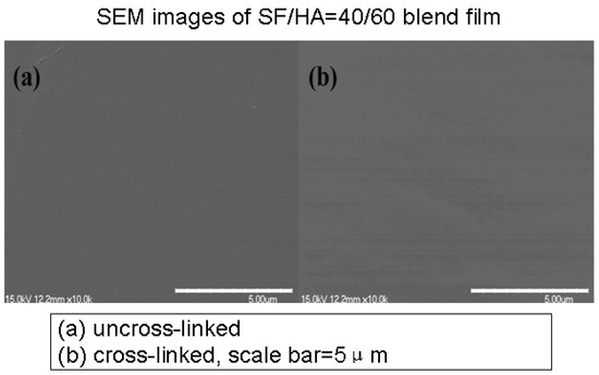
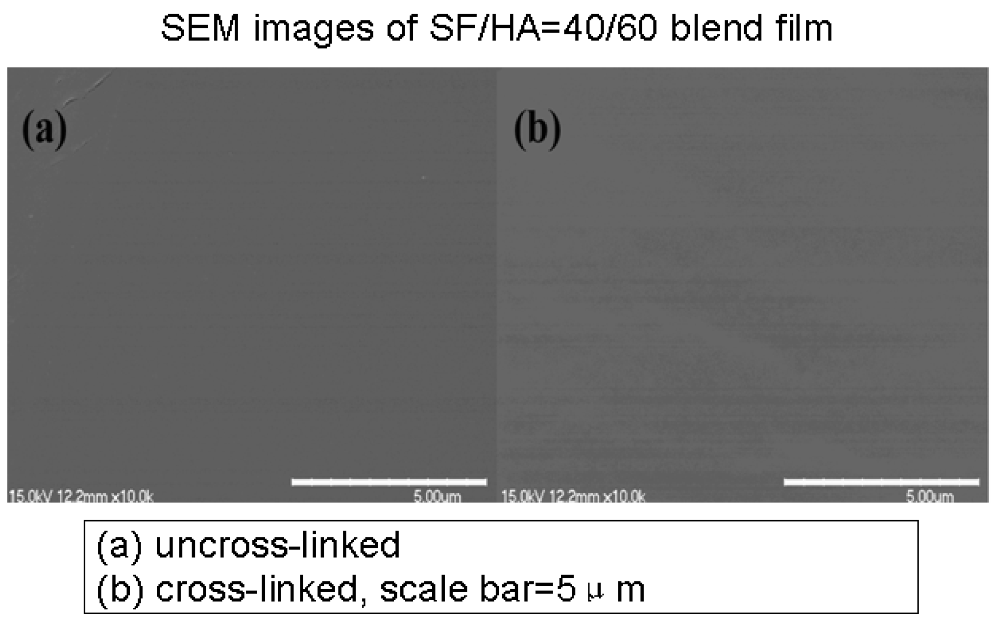
The surface of uncross-linked blend films as well as the cross-linked films exhibited uniform microstructure (Figure 1), and no phase separation was observed. SEM images of all other SF/HA blend films also show a similar uniform microstructure, indicating that HA and SF have good compatibility.
Figure 1.
SEM images of SF/HA=40/60 blend film.
3.2. FT-IR spectra
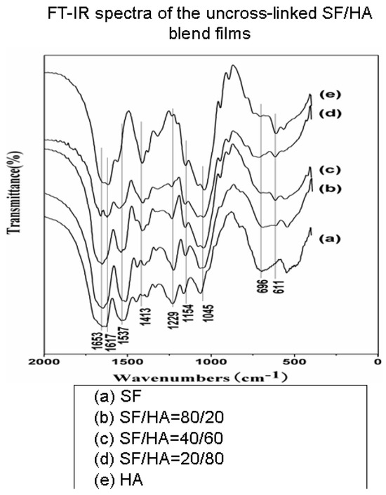
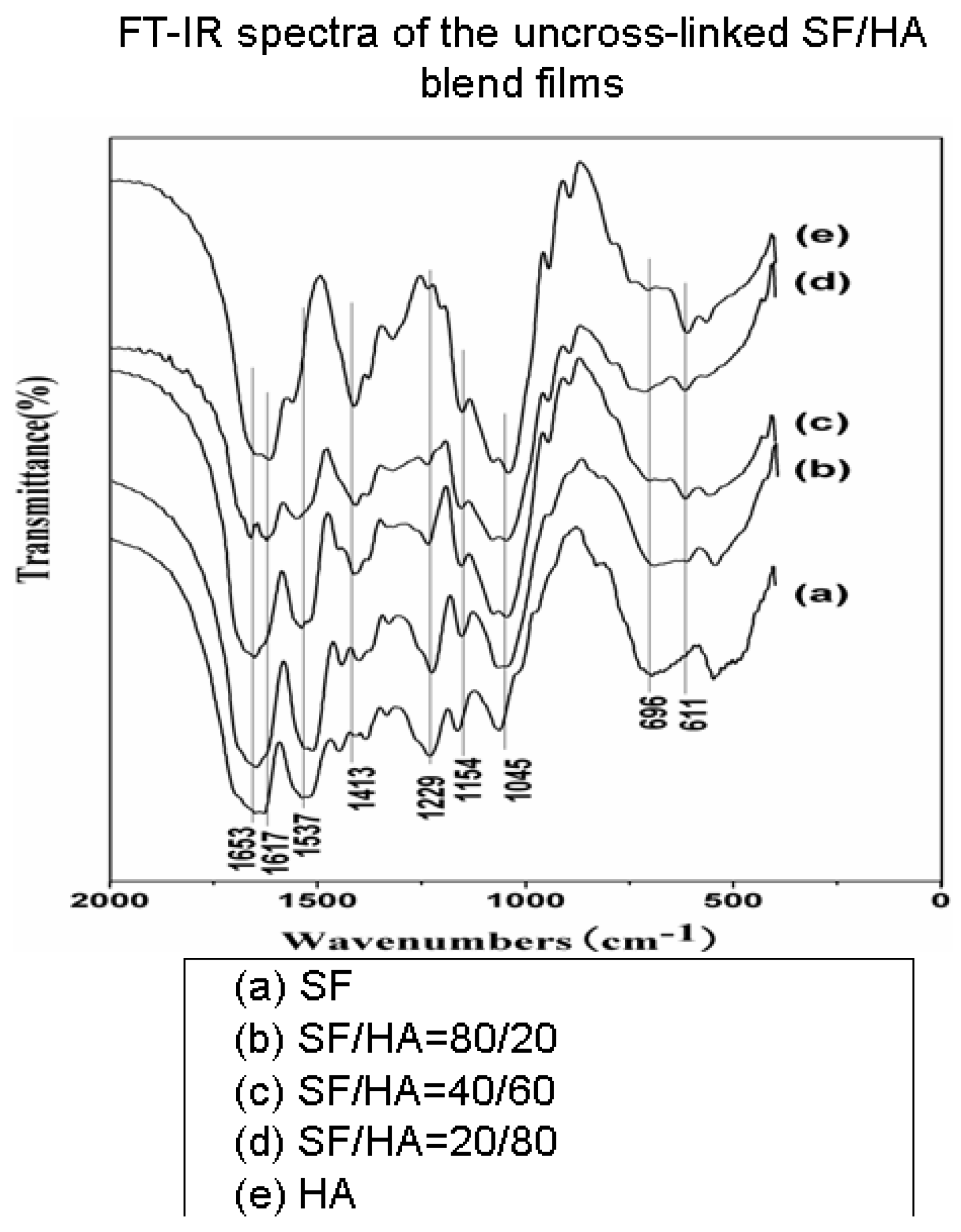
The SF usually shows the characteristic absorption bands at around 1650~1655 cm−1(amide I), 1525~1540 cm−1 (amide II), 1266 cm−1 (amide III), 669 cm−1 (amide V), which represent Silk I. 1620~1635 cm−1 (amide I), 1531 cm−1 (amide II), 1230~1235 cm−1 (amide III), 695 cm−1 (amide V), attribute to Silk II. 1655~1660 cm−1 (amide I), 1535~1545 cm−1 (amide II), 1235 cm−1 (amide III), 650 cm−1 (amide V) are assigned to random coil conformation [17,18,19,20].
The FT-IR spectra of uncross-linked SF/HA blend films are shown in Figure 2. The curve of SF film (Figure 2, curve (a)) shows strong absorption bands at 1651 cm−1, 1537 cm−1, 1229 cm−1 and 696 cm−1, indicating the presence of random coil conformation, Silk I and few Silk II. On the other hand, the curve of HA film (Figure 2, curve (e)) shows absorption bands at 1617 cm−1 and 1413 cm−1 attributed to anti-symmetric stretching and symmetric stretching of C=O, respectively. 611 cm−1 is assigned to the rocking vibration of N-H. 1154 cm−1 is assigned to anti-symmetric stretching of C-O-C, and 1045 cm−1 is assigned to the C-O stretching [21]. The amide I, amide II and amide III bands shifted to higher wave number with the increasing proportion of HA while the amide V shifted to lower wave number, indicating that the Silk I of SF in the blend films decreased with the increasing proportion of HA while the random coil conformation increased. In the SF/HA blend films, with the increasing proportion of HA, the chances of SF and HA combining with each other with the help of hydrogen bond and electrostatic interactions increased and the force to resist SF segmer combine with each other also increased. So it is more difficult for SF segmer to form a regular structure.
Figure 2.
FT-IR spectra of the uncross-linked SF/HA blend films.
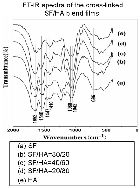
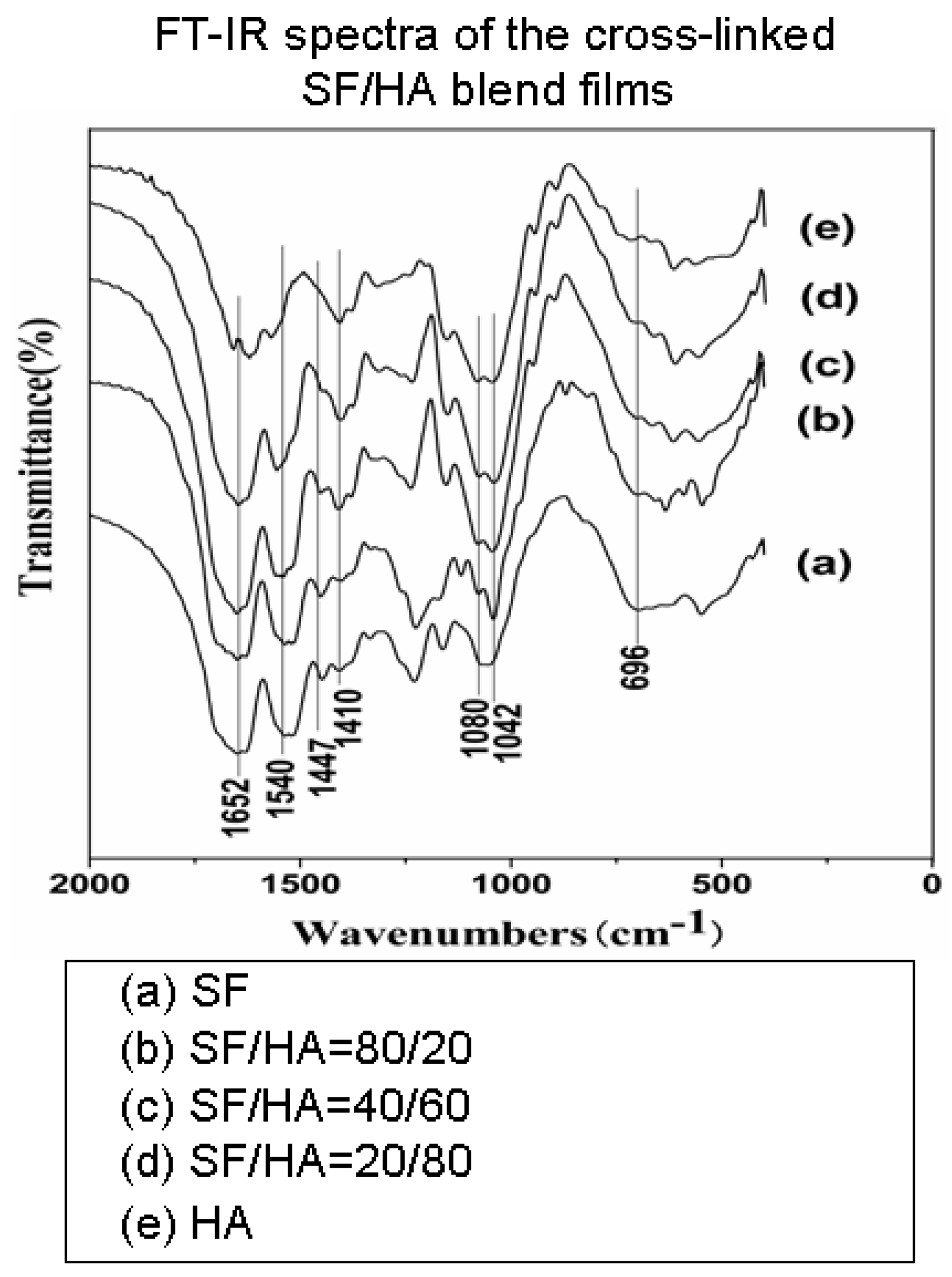
FT-IR spectra of cross-linked SF/HA blend films are shown in Figure 3. The amide bands around 1652 cm−1, 1540 cm−1 and 1447 cm−1 and C-O-C ester bands around 1080 cm−1 and 1042 cm−1 are slightly increased. Carboxyl characteristic absorption band around 1410 cm−1 decreased, indicating that amide or ester linkages are formed in all the blend films after being cross-linked by EDC. EDC reacts with the carboxyl of SF or HA and forms an intermediate product O-urea, which may readily react with the hydroxyl group of HA or amino group of SF, resulting in the formation of ester linkage and amide linkage respectively. The amide I, amide II and amide III bands slightly shift to a higher wave number with the increasing proportion of HA while the amide V shifts to lower wave number, indicating that the random coil conformation increases with the increasing proportion of HA.
Figure 3.
FT-IR spectra of the cross-linked SF/HA blend films.
3.3. Tensile strength and elongation at break
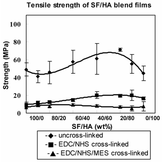
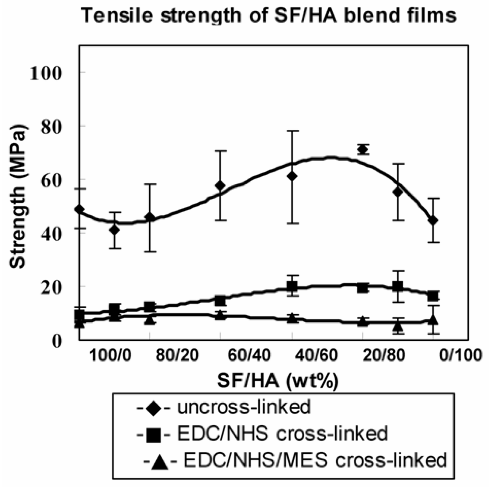
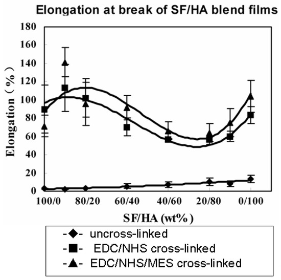
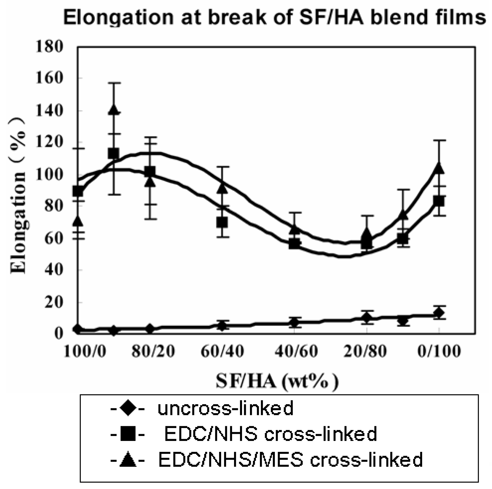
As shown in Figure 4 and Figure 5, the tensile strength of uncross-linked SF/HA blend films is much larger at various proportions of HA, whereas elongation at break is small, displaying the characteristic of brittle materials. After being cross-linked by EDC, the elongation at break increased obviously while tensile strength decreased, showing the characteristic of flexible materials.
Figure 4.
Tensile strength of SF/HA blend films.
Figure 5.
Elongation at break of SF/HA blend films.
It can be considered that the original hydrogen bond between the SF molecule in uncross-linked blend films is broken when the HA content is lower, such as SF/HA=90/10, hence the tensile strength decreased. Though the tensile strength has a slight increase with the increase of HA content, this may be due to the fact that HA contains many hydroxyl groups, carboxyl groups and more hydrogen bands formed between SF and HA, makes itstill lower than the SF film. When the HA content exceeds 70%, the tensile strength decreased again due to which HA mainly exists with random coil in aqueous solution. The elongation of the blend films increased with the increase HA content. A decrease of the tensile strength with a simultaneous increase of elongation is also a characteristic property of other polysaccharide or protein films plasticized with different hydrophilic compounds [22,23].
As previously reported [24], the EDC mainly reacted with the carboxyl of SF or HA, the amino group of SF and hydroxyl group of HA. Thus, after being cross-linked with EDC, the hydrogen bond and electrostatic interactions between SF or HA macromolecular segments in the blend films decreased remarkably. On the other hand, some small molecular compounds which can dissolve in water are formed. The interspaces of the macromolecular bonds increased and hindered the formation of hydrogen bonds between the macromolecular segmers. And the displacement ability of the segmers enhanced. Therefore, the tensile strength of the cross-linked blend films is smaller than that of uncross-linked films, and the elongation at break increased remarkably. Nevertheless, the tensile strength is still in the moderate range and this characteristic may be favorable for its application in biomedical field.
It can be considered that the covalent linkage formed between EDC and SF or EDC and HA molecule chain consequently resulted in a network structure being formed. The density of secondary bond such as hydrogen bond and electrostatic interactions between SF or HA segmer decreased remarkably, and the displacement ability of the segmer enhanced.
Therefore, the elongation at break increased obviously. However, because of the three-dimensional network structure of SF and HA chains, when an external force at certain direction is applied on the blend films, the different domain segmer of SF or HA molecule suffered the force from different directions, which makes it difficult for the chain to parallel along the direction of external force and to endure the force at the same time, leading to the decrease of tensile strength.
3.4. Young’s modulus
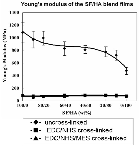
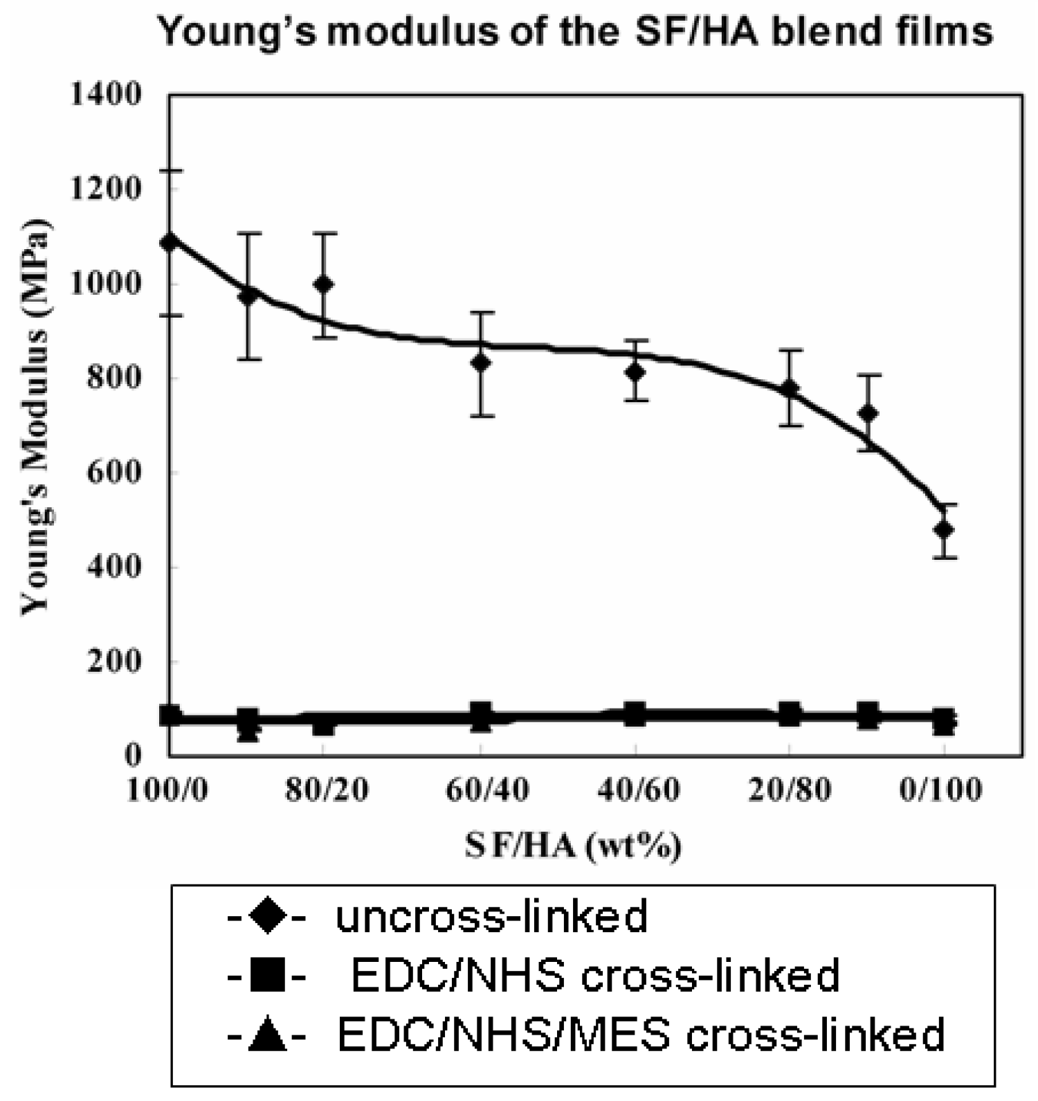
The smaller the Young’s modulus, the easier the materials deform. Therefore, Young’s modulus is an important indicator to measure the flexibility of materials. As shown in Figure 6, the Young’s modulus of blend films decreased gradually with increasing proportion of HA, indicating that the flexibility of the films improved gradually. This change can be considered as a result of the flexibility of HA macromolecules chain.
Figure 6.
Young’s modulus of the SF/HA blend films.
It is worth mentioning that compared to the uncross-linked blend films, the Young’s modulus of EDC cross-linked films decreased obviously and flexibility of blend films improved remarkably, indicating that the displacement ability enhanced obviously between SF and HA segmer after crosslinking.
3.5. Cell culture
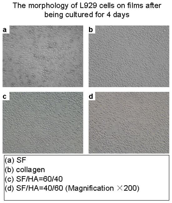
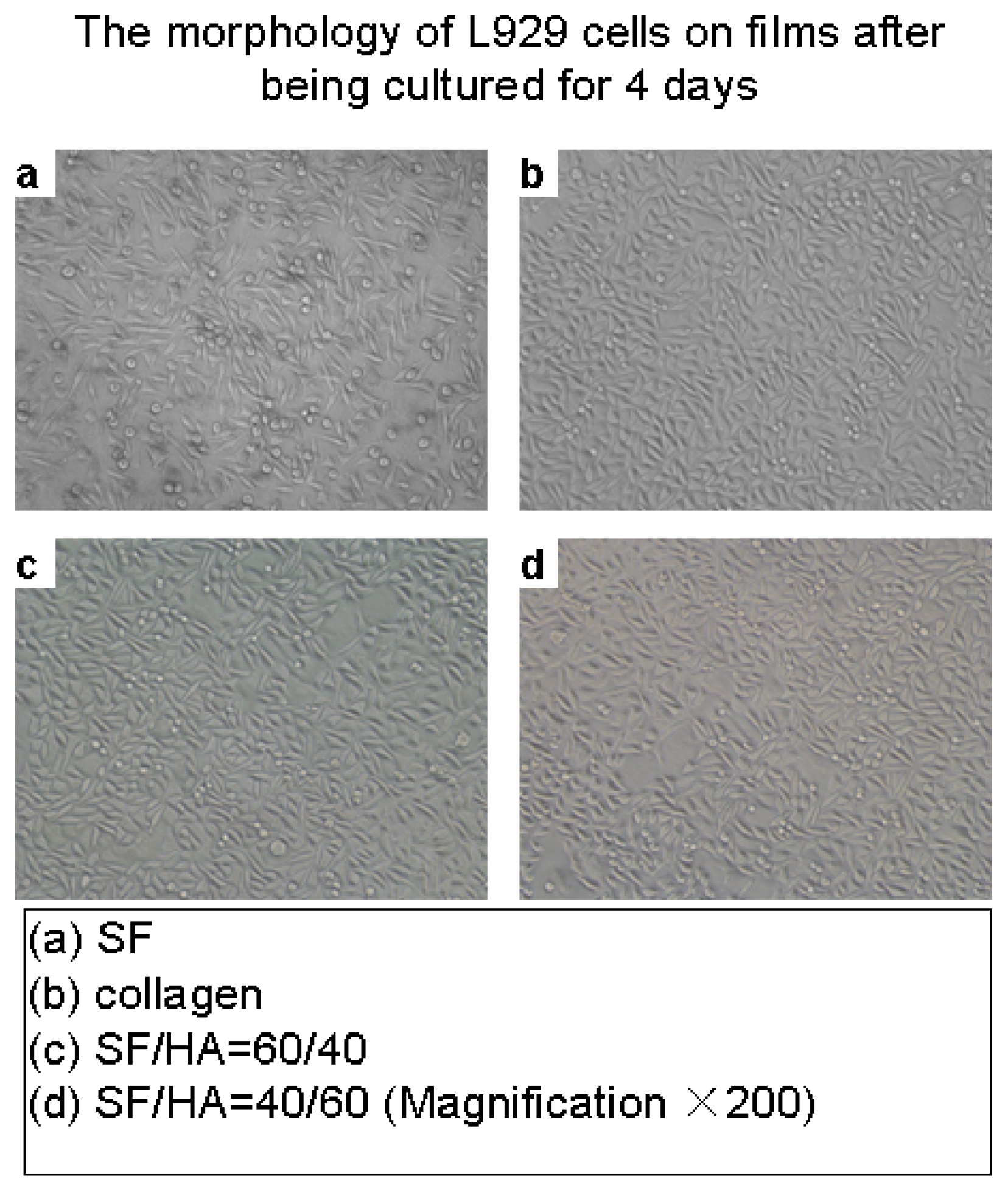
The proliferations of cultivated L929 cells on SF/HA blend films and SF films is very similar to those on collagen films (Figure 7). Cells present fusiform and eumorphism, and they do not exhibit any morphological changes, indicating that the blend films can support the cell adhesion and proliferation well; the blend films have a good biocompatibility.
Figure 7.
The morphology of L929 cells on films after being cultured for 4 days.
4. Conclusions
The SF/HA blend films cross-linked with EDC were prepared by flow casting method. Effective crosslinking reactions took place between EDC and SF or HA macromolecules. Compared to the SF film, the broken elongation of blend films increased obviously when the blending ratio of SF/HA was 90/10 and then decreased with the increase of HA content. HA interferes with SF to form crystalline structures, whereas EDC can induce SF to form Silk I.
Acknowledgments
This work was supported by the Key Basic Research Program of China (973 program, 2005CB623902), National Natural Science Foundation of China (30970714), College Natural Science Research Project of Jiangsu Province (07KJA43010) and The Program of Science and Technology of Jiangsu Province (BA2007055).
References
- Inoue, S.; Tanaka, K.; Arisaka, F.; Kimura, S.; Ohtomo, K.; Mizuno, S. Silk Fibroin of Bombyx mori Is Secreted, Assembling a High Molecular Mass Elementary Unit Consisting of H-chain, L-chain, and P25, with a 6:6:1 Molar Ratio. J. Biol. Chem. 2000, 275, 40517–40528. [Google Scholar] [CrossRef] [Scilit] [PubMed]
- Hofmann, S.; Wong, P.F.C.T.; Rossetti, F.; Textor, M.; Vunjak-Novakovic, G.; Kaplan, D.L.; Merkle, H.P.; Meinel, L. Silk fibroin as an organic polymer for controlled drug delivery. J. Control Release 2006, 111, 219–227. [Google Scholar] [CrossRef] [Scilit] [PubMed]
- Lovett, M.; Cannizzaro, C.; Daheron, L.; Messmer, B.; Vunjak-Novakovic, G.; Kaplan, D.L. Silk fibroin microtubes for blood vessel engineering. Biomaterials 2007, 28, 5271–5279. [Google Scholar] [CrossRef] [Scilit] [PubMed]
- Yang, Y.; Ding, F.; Wu, J.; Hu, W.; Liu, W.; Liu, J.; Gu, X. Development and evaluation of silk fibroin-based nerve grafts used for peripheral nerve regeneration. Biomaterials 2007, 28, 5526–5535. [Google Scholar] [CrossRef] [Scilit] [PubMed]
- Meinel, L.; Betz, O.; Fajardo, R.; Hofmann, S.; Nazarian, A.; Cory, E.; Hilbe, M.; McCool, J.; Langer, R.; Vunjak-Novakovic, G.; Merkle, H.P.; Rechenberg, B.; Kaplan, D.L.; Kirker-Head, C. Silk based biomaterials to heal critical sized femur defects. Bone 2006, 39, 922–931. [Google Scholar] [CrossRef] [Scilit] [PubMed]
- Li, C.; Vepari, C.; Jin, H.J.; Kim, H.J.; Kaplan, D.L. Electrospun silk-BMP-2 scaffolds for bone tissue engineering. Biomaterials 2006, 27, 3115–3124. [Google Scholar] [CrossRef] [Scilit] [PubMed]
- Altman, G.H.; Horan, R.L.; Lu, H.H.; Moreau, J.; Martin, I.; Richmond, J.C.; Kaplan, D.L. Silk matrix for tissue engineered anterior cruciate ligaments. Biomaterials 2002, 23, 4131–4141. [Google Scholar] [CrossRef] [Scilit] [PubMed]
- Vepari, C.; Kaplan, D.L. Silk as a biomaterial. Prog. Polym. Sci. 2007, 32, 991–1007. [Google Scholar] [CrossRef] [Scilit] [PubMed]
- Rapport, M.M.; Weissman, B.; Linker, A. Isolation of a crystalline disaccharide, hyalobiuronic acid from hyaluronic acid. Nature 1951, 168, 996–997. [Google Scholar] [CrossRef] [Scilit] [PubMed]
- Kazuaki, K.; Mitsuhiro, K.; Shin-ichi, Y. Hyaluronic acid: separation and biological implications. J. Chromatogr. B 2003, 797, 347–355. [Google Scholar] [CrossRef] [Scilit] [PubMed]
- Campbell, J.; Bellamy, N.; Gee, T. Differences between systematic reviews on hyaluronan/hyaluronic acid/hylan. Osteoarthr. Cartil. 2007, 15, 1424–1436. [Google Scholar]
- Goa, K.L.; Benfield, P. A review of its pharmacology and use as a surgical aid in ophthalmology and its therapeutic potential in joint disease and wound healing. Drugs 1994, 47, 536–566. [Google Scholar] [PubMed]
- Mayol, L.; Quaglia, F.; Borzacchiello, A.; Ambrosio, L.; Rotonda, M.I.L. A novel poloxamers/hyaluronic acid in situ forming hydrogel for drug delivery: Rheological, mucoadhesive and in vitro release properties. Eur. J. Pharm. Biopharm. 2008, 70, 199206. [Google Scholar] [CrossRef] [Scilit] [PubMed]
- Tan, H.; Chu, C.R.; Payne, K.A.; Marra, K.G. Injectable in situ forming biodegradable chitosan-hyaluronic acid based hydrogels for cartilage tissue engineering. Biomaterials 2009, 30, 2499–2506. [Google Scholar] [CrossRef] [Scilit] [PubMed]
- Benedetti, L.; Cortivo, R.; Berti, T.; Berti, A.; Pea, F. Biocompatibility and biodegradation of different hyaluronan derivatives (HYAFF) implanted in rats. Biomaterials 1993, 14, 1154–1160. [Google Scholar] [CrossRef] [Scilit] [PubMed]
- Larsen, N.E.; Pollak, C.T.; Reiner, K.; Leshchiner, E.; Balazs, E.A. Hylan gel biomaterial: dermal and immunologic compatibility. J. BioMed Mater. Res. 1993, 27, 1129–1134. [Google Scholar] [CrossRef] [Scilit] [PubMed]
- LiuY; Qian, J.; Liu, H.; Zhang, X.; Deng, J.; Yu, T. Blend membrane of regenerated silk fibroin, poly(vinyl alcohol), and peroxidase and its application to a ferrocene-mediating hydrogen peroxide sensor. J. Appl. Polym. Sci. 1996, 61, 641647. [Google Scholar] [CrossRef] [Scilit]
- Asakura, T.; Kuzuhara, A.; Tabeta, R.; Saito, H. Conformation characterization of bombyx-mori silk fibroin in the solid-state by high-frequency C13 cross polarization magic angle spinning nmr, X-ray-diffraction, and infrared-spectroscopy. Macromolecules 1985, 18, 1841–1845. [Google Scholar]
- Venyaminov, S.Y.; Kalnin, N.N. Quantitative IR spectrophotometry of peptide compounds in water (H2O) solution. I. Spectral parameters of amino acid residue absorption bands. Biopolymers 1990, 30, 1243–1257. [Google Scholar] [CrossRef] [Scilit] [PubMed]
- Freddi, G.; Tsukada, M.; Beretta, S. Structure and physical properties of silk fibroin/polyacrylamide blend films. J. Appl. Polym. Sci. 1999, 71, 15631571. [Google Scholar] [CrossRef] [Scilit]
- Gilli, R.; Kacuráková, M.; Mathlouthi, M.; Navarini, L.; Paoletti, S. FTIR studies of sodium hyaluronate and its oligomers in the amorphous solid phase and in aqueous solution. Carbohydr. Res. 1994, 263, 315–326. [Google Scholar] [CrossRef] [Scilit] [PubMed]
- Arvanitoyannis, I.S.; Biliaderis, C.G. Physical properties of polyol-plasticized edible blends made of methyl cellulose and soluble starch. Carbohydr. Polym. 1999, 38, 47–58. [Google Scholar] [CrossRef] [Scilit]
- Arvanitoyannis, I.S.; Nakayama, A.; Aiba, S. Chitosan andgelatin based edibfilms: state e diagrams, mechanical and permeation properties. Carbohydr. Polym. 1998b, 37, 371–382. [Google Scholar]
- Choi, Y.S.; Hong, S.R.; Lee, Y.M.; Song, K.W.; Park, M.H.; Nam, Y.S. Studies on gelatin-containing artificial skin: II. Preparation and characterization of crosslinked gelatin-hyaluronate sponge. J. BioMed Mater. Res. 1999, 48, 631–639. [Google Scholar] [CrossRef] [Scilit]
© 2010 Textile Bioengineering and Informatics Society. This article is an open access article distributed under the terms and conditions of the Creative Commons Attribution (CC BY) license (https://creativecommons.org/licenses/by/4.0/).